Review

The Emerging Role of Autophagy as a Target of Environmental Pollutants: An Update on Mechanisms

by
Md. Ataur Rahman
1,2,*,
Md Saidur Rahman
3,
Md. Anowar Khasru Parvez
4 and
Bonglee Kim
1,2,*
1
Department of Pathology, College of Korean Medicine, Kyung Hee University, 1-5 Hoegidong Dongdaemun-gu, Seoul 02447, Republic of Korea
2
Korean Medicine-Based Drug Repositioning Cancer Research Center, College of Korean Medicine, Kyung Hee University, Seoul 02447, Republic of Korea
3
Department of Animal Science & Technology and BET Research Institute, Chung-Ang University, Anseong 17546, Republic of Korea
4
Department of Microbiology, Jahangirnagar University, Savar, Dhaka 1342, Bangladesh
*
Authors to whom correspondence should be addressed.
Toxics 2023, 11(2), 135; https://doi.org/10.3390/toxics11020135
Submission received: 15 November 2022 / Revised: 9 January 2023 / Accepted: 28 January 2023 / Published: 30 January 2023
(This article belongs to the Section Exposome Analysis and Risk Assessment)

Abstract

:
Autophagy is an evolutionarily conserved cellular system crucial for cellular homeostasis that protects cells from a broad range of internal and extracellular stresses. Autophagy decreases metabolic load and toxicity by removing damaged cellular components. Environmental contaminants, particularly industrial substances, can influence autophagic flux by enhancing it as a protective response, preventing it, or converting its protective function into a pro-cell death mechanism. Environmental toxic materials are also notorious for their tendency to bioaccumulate and induce pathophysiological vulnerability. Many environmental pollutants have been found to influence stress which increases autophagy. Increasing autophagy was recently shown to improve stress resistance and reduce genetic damage. Moreover, suppressing autophagy or depleting its resources either increases or decreases toxicity, depending on the circumstances. The essential process of selective autophagy is utilized by mammalian cells in order to eliminate particulate matter, nanoparticles, toxic metals, and smoke exposure without inflicting damage on cytosolic components. Moreover, cigarette smoke and aging are the chief causes of chronic obstructive pulmonary disease (COPD)-emphysema; however, the disease’s molecular mechanism is poorly known. Therefore, understanding the impacts of environmental exposure via autophagy offers new approaches for risk assessment, protection, and preventative actions which will counter the harmful effects of environmental contaminants on human and animal health.

1. Introduction

It has recently been shown that the cellular autophagy process, which involves lysosomes fusing with undesired or accumulated defective cellular components, is crucial for maintaining cellular function and homeostasis [1]. Autophagy is an active component of cell defense and helps cancer cells maintain their cytostatic link during the growth process. [2]. Phagophore assembly sites (PAS), which are structures that come before autophagosomes, are initiated by autophagy process. [3]. The endoplasmic reticulum (ER)-associated phosphatidylinositol 3-kinase (PI3K) is crucial for beginning the production of PAS [4]. AMP-activated protein kinase (AMPK), the mammalian target of rapamycin (mTOR), and unc-51-like autophagy activating kinase-1 (ULK1) facilitate phagophore formation during autophagy induction [5]. Phagophore recruiters include PI3K catalytic subunit type 3 (PIK3C3/Vps34), PI3K regulatory subunit 4 (PIK3R4/Vps15/p150), and beclin-1 (BECN1). Then, the membrane is expanded and sealed to lengthen it in preparation for autophagosome production. When autophagosomes reach maturity, they bind to lysosomes to create autolysosomes [6]. After acid hydrolases break down autolysosomes and their inner cargos, creating nutrients, additional metabolite recycling maintains cellular equilibrium [7] (Figure 1). The mTOR-independent autophagy mechanism has several therapeutic targets for neurodegenerative disorders [8]. mTOR-independent network regulating mammalian autophagy, encompassing cAMP-Epac-PLCϵ-IP3 and Ca2+-calpain-Gαs pathways, provides multiple therapeutic targets for neurological disorders. Enhancing autophagy through this mTOR-independent route is protective in different models [9].
Currently, global pollution threatens human health. Modern cultures worry about air pollution, including particulate particles and heavy metals. Due to their toxicity, endurance, and bioaccumulation, heavy metals, including cadmium, lead, and arsenic, are widespread contaminants [10]. Autophagy can be disrupted by various environmental pollutants, including pesticides, particulate particles, and heavy metals [11,12]. A single drug can have varying effects on the autophagy process depending on factors such as cell type, exposure length, and dosage. Therefore, understanding the effects of exposure to chemicals on autophagy has gained importance [13]. This understanding provides new avenues for environmental pollutant risk assessment, protection, and preventative measures that can be taken to protect against their adverse effects on human health [14].
Autophagy is a preprogrammed system that cells use to manage their internal homeostasis and reduce or eliminate the effects of foreign toxic chemicals that enter them (Figure 2) [15,16]. Therefore, autophagy induction may be a new restorative strategy in the toxicity field [17]. This review focuses on the autophagic pathways induced by various environmental metal pollutants to fill the knowledge gap on autophagy’s function in eliminating environmental toxicities.

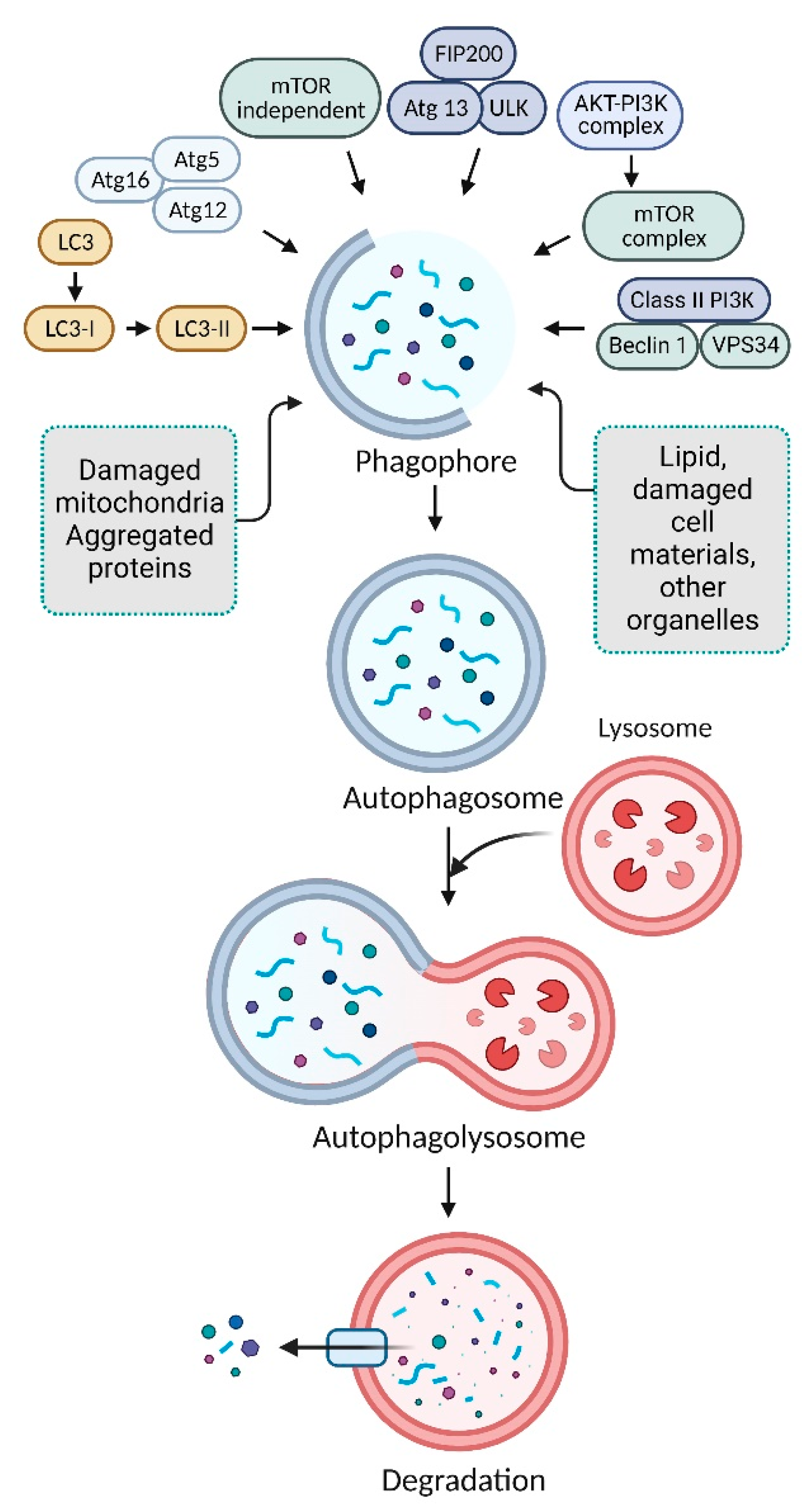
2. Mechanism of Autophagy Pathways

Numerous studies have been developed to reflect the different targeted subcellular component types that could be destroyed by autophagy [18]. Several autophagy receptors play a role in the autophagy pathways that are presented in Figure 3, including aggrephagy, mitophagy, nanoparticulophagy, reticulophagy, xenophagy, zymophagy, lipophagy, ribophagy, and pexophagy [19,20]. However, autophagy and its participation in the responses to various common environmental exposures such as metals, airborne particulate matter, nanoparticles, and cigarette smoke as well as some common single environmental toxins might be removed from the environment.
Autophagy selectively degrades lipids, called lipophagy, and sequestosome 1 (SQSTM1) autophagy receptors connect with lipid droplets through autophagy [21]. Mitophagy is the autophagic process that selectively degrades mitochondria [22]. During mitophagy, autophagy adaptors and receptors, such as the neighbor of BRCA1 gene 1 (NBR1), optineurin (OPTN), the nuclear dot protein 52 kDa (NDP52), and SQSTM1, recognize mitochondria [22]. During reticulophagy, the ER recognizes autophagy adaptors autophagy-related 40 (ATG40) and the ER-anchored autophagy receptor (reticulophagy regulator 1) [23]. In ribophagy, autophagy selectively degrades ribosomes, which bind to nuclear fragile X mental retardation-interacting protein 1 (NUFIP1), an autophagy receptor-like protein [24]. Midbody degradation is the selective destruction of midbody rings produced during cytokinesis via autophagy [25]. Midbody rings recognize autophagy receptors such as SQSTM1 and NBR1 during selective autophagy [26]. Pexophagy is the selective destruction of peroxisomes via autophagy; peroxisomes recognize autophagy adaptors autophagy-related 36 (ATG36), ATG40, and peroxisomal biogenesis factor 3 (PEX3), and autophagy receptors SQSTM1 and NBR1 [27]. Zymophagy selectively degrades damaged or surplus zymogen granules via autophagy; zymogen particles bind to autophagy receptors, including SQSTM1 [28]. Several proteins participate in selective autophagy, such as TAR DNA-binding protein 43 (TDP43), glucocerebrosidase (GBA), presenilin 1 (PSEN1), ATPase cation transporter 13A2 (ATP13A2), and superoxide dismutase-1 (SOD1) [29]. Selective autophagy processes contain proteins and specialized autophagic receptors which identify the cargo, generally mediated via cargo ubiquitination [30]. Through its interaction with a scaffold protein, the receptor either binds to cargoes or may be an integral component of these cargoes, connecting them to the autophagy machinery in the cell [31]. Protein aggregates are associated with Alzheimer’s, Parkinson’s, and Huntington’s diseases [2,32]. In yeasts, flies, and mammalians, cells mediate amyloid beta peptide, tau, poly-Q, alpha-synuclein, and mutant huntingtin protein aggregates which might be removed by aggrephagy, a selective disposal of protein aggregates [2,33]. Therefore, understanding the impacts of environmental pollutants on autophagy offers new ways for risk assessment, protection, and preventive actions to offset the harmful effects of environmental contaminants on human health.

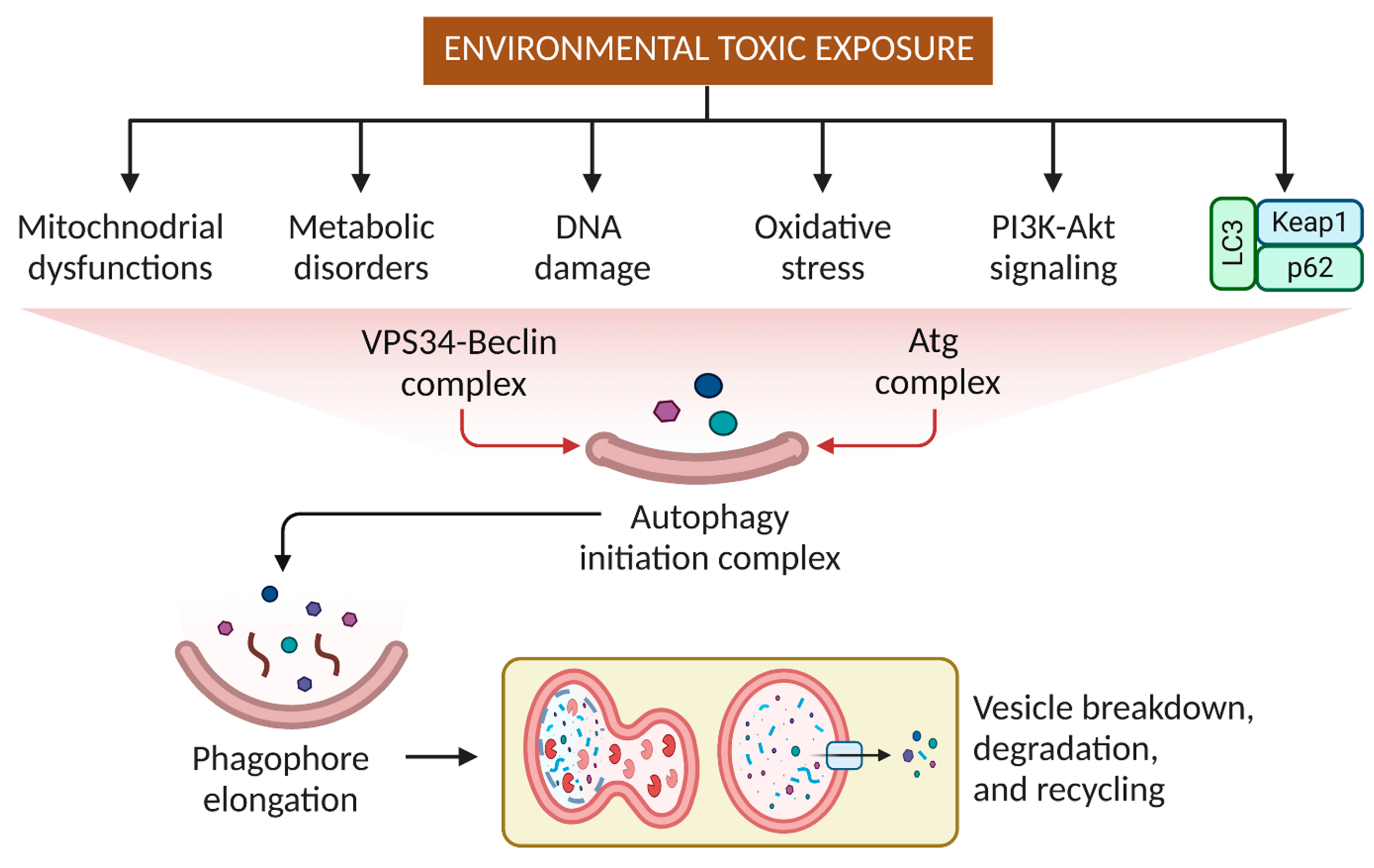
3. Effects of Pesticides and Other Small Molecular Weight Environmental Toxins on Autophagy

Understanding the impacts of pesticides, small molecular weight, and chemical exposure on autophagy offers new ways for risk assessment, protection, and preventative actions to offset the harmful effects of environmental contaminants on human health [13]. Cellular components and protein kinases can activate several signaling pathways that result in autophagy, apoptosis, and necrosis [34]. Nanoparticles and metals act as strong autophagy activators in cell and animal systems [35]. When cells respond to metals/metalloids and nanoparticles, AMPK, mitogen-activated protein kinase (MAPK), AKT serine/threonine kinase 1 (AKT1/AKT), PI3K, death protein kinases, and mTOR are the main factors inducing or inhibiting autophagy [36]. Several receptors (e.g., NBR1, p62, Tax1 binding protein 1 [TAX1BP1], and OPTN) that recognize the autophagy adaptor (lipidated microtubule-associated proteins 1A/1B light chain 3A [MAP1LC3A/LC3; LC3II]) attract tagged mitochondria and facilitate autophagy vacuole engulfment [37]. Autophagy prevents chlorpyrifos (CPF)-induced reactive oxygen species (ROS)-mediated toxicity. CPF increases mitochondria-mediated apoptosis-related ROS production and autophagy in human neuroblastoma cells [38]. ROS connect environmental (pesticides, herbicides, heavy metals) and endogenous and genetic PD risk factors. Environmental toxins and medicines, such as 1-methyl-4-phenylpyridinium (MPP+), rotenone, paraquat (PQ), and metamphetamine, have been linked to autophagy dysregulation in neurotoxin-induced dopaminergic cell death models [39]. Epidemiological studies relate rural life, farming, well water, and agrichemicals to an increased risk of PD. Several agrichemicals harm dopaminergic neurons, suggesting an environmental foundation for sporadic PD [40]. Dopaminergic neurons are uniquely sensitive to the herbicide paraquat, with other populations of neurons unaffected, associated with diminished motor activity and dose-dependent striatal dopaminergic nerve fiber losses. Paraquat-treated animals showed upregulation and aggregation of -synuclein [-Syn] in the substantia nigra [41]. Anti-apoptotic proteins interact with BECN1 and BCL2-associated X apoptosis regulator (BAX) or BCL2 antagonist/killer 1 (BAK1/BAK) [42,43]. Interestingly, BECN1 is a PI3K component and autophagy activator that normally interacts with anti-apoptotic proteins (e.g., B-cell lymphoma 2 [BCL2]) to suppress autophagy [44]. Stress interrupts the connection, promoting autophagy. Stress affects the connection between BCL2 and BAX/BAK, increasing apoptosis [45]. During continued stress, BECN1 is cleaved by caspase and translocates to the mitochondria, increasing apoptosis [46]. Depending on its length, BECN1 can induce autophagy or apoptosis [43]. In stressed cells, calpain or caspases also degrade autophagy-related 5 (ATG5) and autophagy and beclin-1 regulator 1 (AMBRA1), shifting autophagy towards apoptosis [47]. Moreover, arsenic inhibits p62-mediated selective autophagy, stabilizing FTO protein. FTO overexpression can prevent autophagy, maintaining FTO accumulation in a positive feedback loop [34]. Physical, chemical, and biological processes that occur in plants, animals, and humans exposed to environmental toxins that result in autophagy are presented in Figure 4.
Autophagy and membrane trafficking degrade and recycle macromolecules in lysosomes. It has also been found that lysosomal membranes and ATP-dependent proton pumps keep the lumen acidic for enzyme activity [47]. However, lumen pH or lysosomal membrane permeability variations cause lysosome dysfunction, disrupting the autophagosome–lysosome fusion [48]. Changes in membrane permeability can produce acidification and necrosis. Perfluorooctanoic acid, arsenic, and cadmium inhibit lysosomal functioning [49]. Moreover, rotenone increases ROS production, inhibiting the mitochondrial electron chain and causing autophagy in neuroblastoma cells [50]. Longer exposure causes autophagosome accumulation and lysosomal pH disruption. Malathion causes autophagosome accumulation in SH-SY5Y cells [51]. It inhibits acetylcholinesterase, causing neurotoxicity, and destabilizes lysosomal membranes, impairing autophagosome–lysosome fusion and causing autophagosome buildup.
Agriculture uses the neurotoxic pesticide fipronil [52]. Pre-treating SH-SY5Y cells with rapamycin enhanced cell viability after fipronil treatments and reduced apoptosis [13]. N-acetylcysteine, a ROS scavenger, reduced fipronil-induced autophagy and apoptosis, showing that oxidative stress is required for toxicity and autophagy [13]. Polybrominated diphenyl ethers BDE-153 and BDE-100 induced autophagy in human liver HepG2 cells through oxidative stress and mitochondrial dysfunction (mitophagy) [53]. Recent research suggests that direct oxidation of catalytic thiol-groups on autophagy-related 3 (ATG3) and 7 (ATG7) might block LC3’s conjugation with phosphatidylethanolamine, which is essential for effective autophagy [54]. ER stress promotes ROS production and can affect redox equilibrium in the cell. Particularly, cadmium, perfluorooctanoic acid (PFOA), paraquat (PQ), cigarette smoke, and chloropicrin trigger ER stress and autophagy. ER stress is caused by inadequate protein folding or diminished ER folding capacity [55]. In particular, PQ is a non-selective herbicide that induces ER stress and autophagy in SH-SY5Y cells [56]. However, inhibiting autophagy increased apoptosis, suggesting it protects against PQ-induced toxicity. PFOA disrupts lipid metabolism, increases ROS levels, and causes ER stress. It induced autophagic vacuole accumulation and disrupted autophagosome–lysosome fusion in the mouse liver in vivo and in a human hepatocyte culture in vitro [57].

4. Targeting Autophagy Modulation to Eliminate Environmental Substances

Mammalian cells use selective autophagy, a critical process, to destroy environmental toxins and damaged organelles without damaging cytosolic elements. Depending on the autophagy receptors and cargo targeted, selective autophagy can be categorized as either inducing or inhibiting.

4.1. Elimination of Particulate Matter by Autophagy

Air pollution has emerged as a significant problem in the environment, particularly due to the presence of extremely minute pollutant particles and pathogenic microbes, which can cause significant harm to the human body. Filtration of the air is one method of cleaning the air that has proven to be both popular and successful [58]. Particulate matter (PM) comprises tiny particles or liquid droplets that are so tiny that they can be inhaled and cause significant harm to an individual’s health [59]. PM, metals, black carbon, nitrate, organic aerosols, polycyclic aromatic hydrocarbons, automotive exhaust, and sulfates comprise microscopic particles and liquid droplets floating in the air [60]. PM has been associated with various health conditions, especially respiratory illnesses. Several studies have associated PM exposure with autophagy and airway dysfunction [61,62]. PM activated the nuclear factor kappa-light-chain-enhancer of the activated B cells (NF-κβ) pathway, airway inflammation, and mucus hyper-production in human bronchial epithelial (HBE) cells [63]. Fine PM triggered cytotoxicity and enhanced autophagy, oxidative stress, and the tumor necrosis factor (TNF/TNFα) pathway in human lung epithelial cells [64]. It has been reported to activate autophagy and inflammation in HBE cells in vitro and in vivo [65]. An autophagy inhibitor (3-methyladenine) suppressed PM-activated pro-inflammatory cytokine expression in vitro and in vivo in PM-treated mice [66]. Additionally, diesel exhaust particle exposure triggered autophagy and citrullination in normal HBE (NHBE) cells. Both Euro 4 and Euro 5 carbon particles could severely alter cell viability, inducing autophagy, apoptosis, and necrosis and stimulating pro-inflammatory cytokine interleukin (IL)-18 production, protein citrullination, and protein arginine deiminase activity in NHBE cells [67]. Therefore, PM has been associated with several health problems in humans, notably respiratory conditions, which can be reduced via the autophagy pathway.

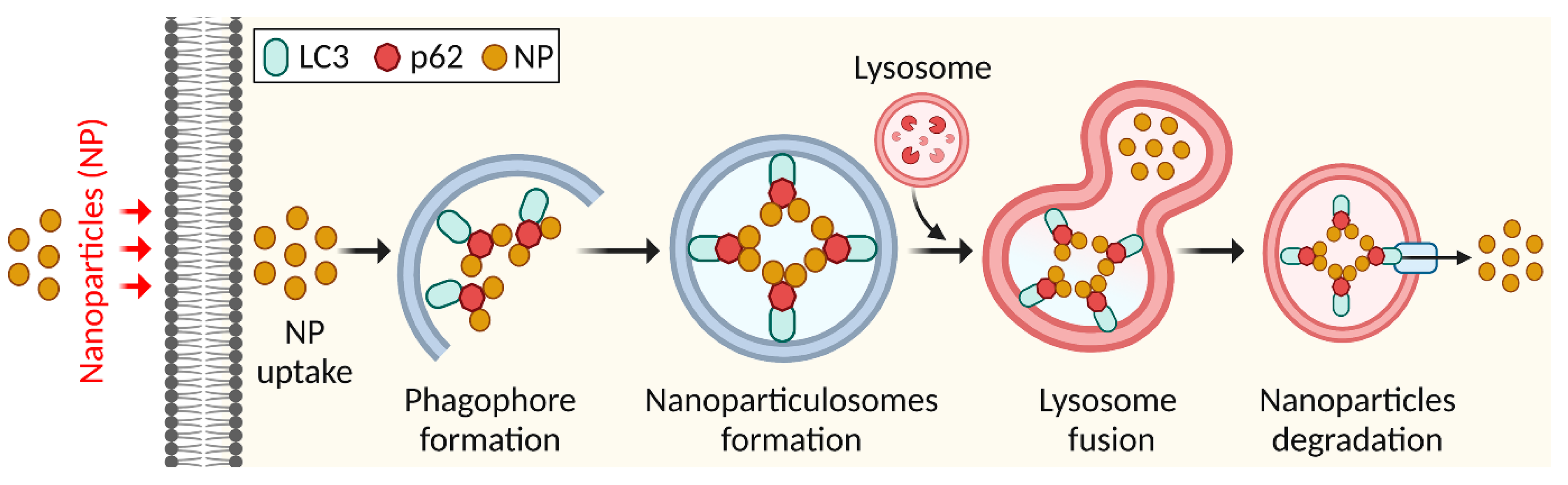
4.2. Elimination of Nanoparticles by Autophagy

A nanoparticle, a type of ultrafine particle, is often described as a particle of matter with a diameter of 1-100 nm. Nanoparticles exist naturally and are studied in chemistry, physics, geology, and biology [68]. One of the mechanisms of intrinsic toxicity that are exhibited by NPs is the disruption of autophagy. The disruption of autophagy that NPs cause must be understood in order to ensure the safety of nanotechnology [10]. Autophagy induced by nanoparticles via endocytosis or other routes may have therapeutic effects, indicating biological applications, although their processing and destruction via selective autophagy remains unknown. Nanoparticles are foreign entities that are destroyed by cells [69]. Nanoparticles enter cells via endocytosis or other uptake routes. Nanoparticles colocalize with autophagy receptors or markers to produce nanoparticle-containing autophagosomes called nanoparticulosomes [70]. These ubiquitinated nanoparticles engage with autophagy receptor proteins, SQSTM1 bound to LC3, forming an autophagosome (Figure 5) [70]. Nanoparticles affect autophagy by increasing autophagosome production and flux or causing autophagic malfunction [71]. Nanoparticles enhance LC3 levels in various categories [35]. In autophagy malfunction, SQSTM1 levels increase because it is no longer degraded. Carbon nanotubes, poly(amidoamine) dendrimers, iron oxide nanoparticles, and graphene oxide caused autophagosome accumulation by blocking autophagic flow [72]. Silver and iron oxide nanoparticles induce autophagy by producing ROS [73]. Alumina, fullerenes, cationic dendrimers, carbon nanotubes, quantum dots, gold, zinc oxides, and silica were found to activate autophagy by blocking mTOR or promoting the expression and phosphorylation of autophagy-related proteins [74]. Lanthanum oxide, cerium dioxide, europium oxide, and manganese also triggered autophagy [75]. Caveolin 1 (CAV1) is an important membrane protein for cell membrane trafficking and autophagy [76]. Additionally, biodegradable ferric phosphate nanosheets are coated with doxorubicin for targeted tumor eradication via an autophagy inhibition-enhanced apoptosis/ferroptosis pathway [77]. Therefore, nanoparticulophagy shows intracellular trafficking mechanisms other than degradation routes for digesting nanoparticles and nanodrugs with therapeutic and pathophysiological consequences.

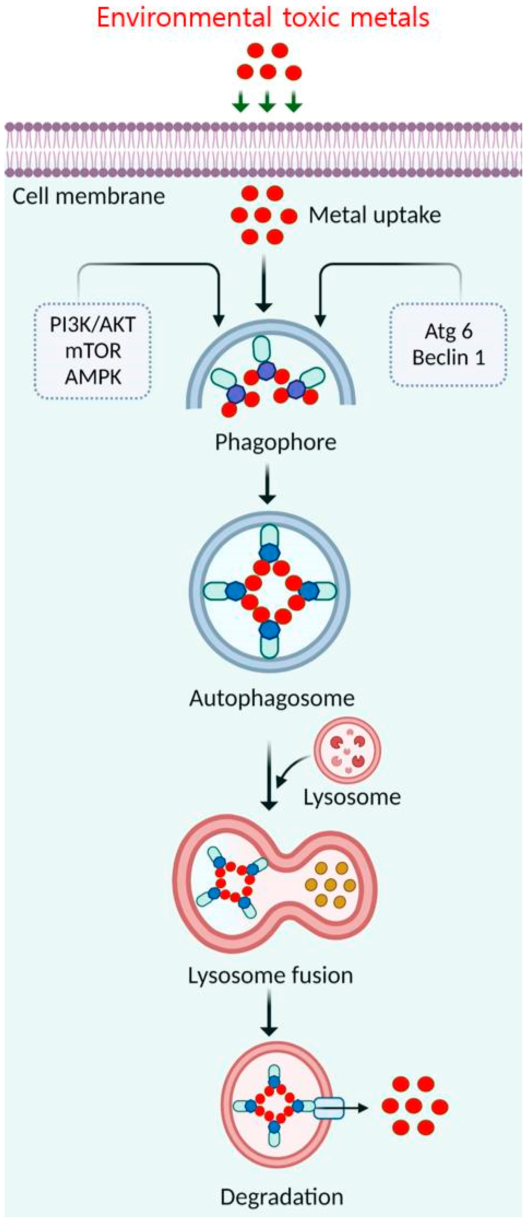
4.3. Elimination of Toxic Metals by Autophagy

Metals and metalloids are human and environmental toxicants. Recently, autophagy has been studied for eliminating physicochemical metal factors that exacerbate toxicity [78]. Autophagy begins when the flat membrane wraps around cytosol or organelles, forming the double-membrane autophagosome vesicle. During autophagosome formation, membranes expand and form a cup-like phagophore [79]. Generally, phagophores are formed by isolating the original membrane and assimilating lipids or repurposing existing compartments. Vesicles sequester cytosolic material and transport it to the lysosomal lumen, forming single membrane autophagolysosomes that digest their contents [80]. The lysosomal membrane-associated protein (LAMP) maintains cellular homeostasis through ubiquitination during maturation. Light chain 3B (LC3B) is ubiquitinated to form an integral membrane protein complex in a nascent autophagosome [8]. Autophagy’s molecular basis could be a technique for removing toxic, hazardous metals from the environment (Figure 6). Arsenic causes DNA damage, apoptosis, and oxidative stress [15]. Arsenic’s short-term activation of autophagy protects against apoptosis [81]. Prolonged exposure to environmental dosages impairs autophagy. Cadmium promotes DNA strand breaks, ER stress, ROS production, and calcium homeostasis [82]. Prolonged contact with these metals reduces the rate at which the p62 protein is degraded by autophagy, resulting in its accumulation [83]. BCL2 interacting protein 3 (BNIP3) is essential for arsenic trioxide (As2O3)-induced autophagy in malignant glioma cells [84]. As2O3-induced autophagic cell death involves LC3 and mitochondrial membrane rupture but not caspase activation [85]. As2O3 is a powerful autophagy inducer that appears to need MAPK kinase (MEK)/extracellular signal-regulated kinase (ERK) pathway activation but not MAPK8/JNK or AKT/mTOR [86]. However, arsenic induces autophagy, modifies autolysosomal gene expression, and inhibits cellular growth in human lymphoblastoid cell lines [87]. Additionally, the Ca2+-mitochondrial-caspase and Ca2+-ERK-LC3 signaling pathways increase cytosolic cadmium levels to promote autophagy and cell death in MES-13 cells [88]. Cadmium accumulated in rat kidney proximal convoluted tubule lysosomes, stimulating cell growth and autophagy [89]. However, further investigations on the roles of arsenic and cadmium in triggering autophagic cell death are needed.
In addition, mercury (Hg) toxicity causes DNA damage, suppresses DNA and RNA synthesis, and induces protein structural changes in vivo and in vitro [90]. Hg poisoning triggers autophagy in rat hepatocytes by modulating the ATG5-autophagy-related 12 (ATG12)-LC3B covalent-conjugation pathway via ubiquitination [11]. In response to Hg, autophagy monitors cell fate by recruiting caspase-8 (CASP8) to autophagosomes via its Fas-associated death domain [91]. High chromium (Cr[III]) damaged DNA [92]. Hexavalent Cr can trigger autophagy in stem/progenitor cells. Stem/progenitor cells subjected to subtoxic and toxic Cr concentrations had preserved tissue regeneration potential [93]. Autophagy indicates Cr toxicity in cord blood hematopoietic stem cells [94]. However, the hematopoietic lineage responds to Cr(VI)-mediated toxic stress via apoptosis and autophagy. Molecular switching between these two pathways may be mediated by stem/progenitor cell differentiation [95]. Iron (Fe) excess causes brain necrosis and apoptosis. DNA damage and oxidative stress exacerbate Fe2+-mediated toxicity [96]. Fe2+-mediated cell death is not necessarily via apoptosis. Recent studies showed that upregulating the ferritin stress protein complex is a quick adaptation mechanism, with ferritin autophagy influencing cellular susceptibility to the oxidative stress response [97]. Nuclear receptor coactivator 4 (NCOA4) acts with GABA type A receptor-associated protein-like 2 (GABARAPL2/ATG8) to recruit a specific cargo-receptor complex into autophagosomes, called ferritinophagy, which is crucial for Fe homeostasis [98]. Therefore, it has been suggested that mammalian cells use autophagy as a cytoprotective defense against several types of metabolic toxicity or organelle damage.

4.4. Elimination of Smoke by Autophagy

In the indoor environment, one of the most significant contributors of particulate matter and chemical pollutants is the act of smoking. A combination of the main stream of smoke that is expelled from the lungs of smokers and the side stream of smoke that is generated straight from the burning cigarette, pipe, or cigar is what makes up second-hand smoke [99]. Epoxide hydrolase 2 (Ephx2)-deficient animals were found to have less lung inflammation and autophagy due to cigarette smoke exposure than normal mice [100]. The autophagy signaling pathway is enhanced by nicotine exposure, causing the heart to adopt an ischemic-sensitive phenotype. It offers an autophagy suppression therapeutic approach that may be innovative for treating ischemic heart disease [101]. The immunological response is commonly associated with autophagy activation caused by cigarette smoke. In cigarette-exposed mice, the rise in pulmonary p62 is highly linked with increased expression of bicaudal D1 (BICD1), an adapter protein that binds to the dynein motor apparatus linking microtubule transport to lysosomes [102]. Recently, cigarette smoke was found to induce autophagy and trigger immune- and oxidation-related responses that damage the airway and alveolar epithelium [103]. The development of smoke aggregates, generally cleared by autophagy, is under the control of the multifunctional protein p62 [104]. However, in vitro exposure of bronchial epithelial cell line BEAS-2B to cigarette smoke extract causes ubiquitinated protein aggregates that colocalize with LC3B and p62 [105]. Carbamazepine reduces these aggregates. In mice exposed to cigarette smoke, aggresomes, LC3B, and p62 increase in peripheral lung tissue, correlating with cellular senescence [106]. Recently, cigarette smoke was shown to be responsible for the accretion of an additional autophagy-related protein called the transcription factor EB (TFEB) in the mouse lung in vivio and HBE cells in vitro [107]. In airway epithelial cells, mTOR was found to regulate cigarette smoke-activated apoptosis, autophagy, inflammation, and necroptosis [108,109]. In stable chronic obstructive pulmonary disease (COPD), the majority of studies have demonstrated an impairment in autophagy, with reduced autophagic flux and accumulation of abnormal mitochondria (defective mitophagy), and are linked to cellular senescence [110]. Acute exposure to cigarette smoke may activate autophagy, resulting in ciliary dysfunction and death of airway epithelial cells [111]. It is challenging to target autophagy therapeutically since the level of autophagy might vary from cell type to cell type and from one environment to another inside a cell [112]. However, these medications are not specific, and researchers are currently working on developing drugs that are more selective. These drugs have the potential to be beneficial as innovative agents in the treatment of asthma and COPD in the future.

5. Conclusions

Several recent studies have found that distinct signaling pathways ultimately resulting in autophagy are activated in cells by cellular proteins or kinases as a protective response to environmental toxins and are associated with increased cell survival. Many studies indicate that autophagy plays a key role in cellular reactions to environmental toxins. However, its significance in environmental toxicant exposures remains unknown. Therefore, environmental chemical exposures must be further studied to determine autophagy’s molecular pathways. However, unknown relationships exist between autophagy, immunological responses, and other cellular activities induced by chemical stress. Additionally, novel technologies and animal models will be needed to identify the complicated autophagic routes in metal-induced cytotoxicity. Therefore, further studies are urgently needed to explore how chemicals affect autophagy. These findings will be important for undertaking risk assessments, protective measures, and prevention activities for environmental contaminants with health impacts. Therefore, determining how to apply autophagy in environmental areas related to human health and establishing associations between autophagy and environmental exposure are appealing subjects for further research.

Author Contributions

Conceptualization, M.A.R.; writing—original draft, M.A.R.; data curation, M.A.R.; editing and modifying draft preparation, M.A.R.; review, M.A.K.P.; supervision, B.K.; funding, B.K. Figures were prepared by M.S.R. All authors have read and agreed to the published version of the manuscript.

Funding

This research was supported by the Basic Science Research Program through the National Research Foundation of Korea (NRF) funded by the Ministry of Education (NRF-2020R1I1A2066868), and the National Research Foundation of Korea (NRF) grant funded by the Korean Government (MSIT) (No. 2020R1A5A2019413).

Institutional Review Board Statement

Not applicable.

Informed Consent Statement

Not applicable.

Data Availability Statement

Request upon corresponding author.

Conflicts of Interest

The authors declare no conflict of interest.

References

  1. Guo, H.; Ouyang, Y.; Yin, H.; Cui, H.; Deng, H.; Liu, H.; Jian, Z.; Fang, J.; Zuo, Z.; Wang, X.; et al. Induction of autophagy via the ROS-dependent AMPK-mTOR pathway protects copper-induced spermatogenesis disorder. Redox Biol. 2022, 49, 102227. [Google Scholar] [CrossRef] [PubMed]
  2. Rahman, M.A.; Rahman, H.; Mamun-Or-Rashid, A.N.M.; Hwang, H.; Chung, S.; Kim, B.; Rhim, H. Autophagy Modulation in Aggresome Formation: Emerging Implications and Treatments of Alzheimer’s Disease. Biomedicines 2022, 10, 1027. [Google Scholar] [CrossRef] [PubMed]
  3. Ma, X.; Li, P.; Ge, L. Targeting of biomolecular condensates to the autophagy pathway. Trends Cell Biol. 2022; online ahead of print. [Google Scholar] [CrossRef] [PubMed]
  4. Vitto, V.A.M.; Bianchin, S.; Zolondick, A.A.; Pellielo, G.; Rimessi, A.; Chianese, D.; Yang, H.; Carbone, M.; Pinton, P.; Giorgi, C.; et al. Molecular Mechanisms of Autophagy in Cancer Development, Progression, and Therapy. Biomedicines 2022, 10, 1596. [Google Scholar] [CrossRef]
  5. Rahman, M.A.; Cho, Y.; Nam, G.; Rhim, H. Antioxidant compound, oxyresveratrol, inhibits APP production through the AMPK/ULK1/mTOR-mediated autophagy pathway in mouse cortical astrocytes. Antioxidants 2021, 10, 408. [Google Scholar] [CrossRef]
  6. Rodgers, S.J.; Jones, E.I.; Arumugam, S.; Hamila, S.A.; Danne, J.; Gurung, R.; Eramo, M.J.; Nanayakkara, R.; Ramm, G.; McGrath, M.J.; et al. Endosome maturation links PI3Kα signaling to lysosome repopulation during basal autophagy. EMBO J. 2022, 41, e110398. [Google Scholar] [CrossRef]
  7. Ravichandran, R.; PriyaDharshini, L.C.; Sakthivel, K.M.; Rasmi, R.R. Role and regulation of autophagy in cancer. Biochim. Biophys. Acta (BBA)—Mol. Basis Dis. 2022, 1868, 166400. [Google Scholar]
  8. Uddin, M.S.; Rahman, A.; Kabir, T.; Behl, T.; Mathew, B.; Perveen, A.; Barreto, G.E.; Bin-Jumah, M.N.; Abdel-Daim, M.M.; Ashraf, G.M. Multifarious roles of mTOR signaling in cognitive aging and cerebrovascular dysfunction of Alzheimer’s disease. Iubmb Life 2020, 72, 1843–1855. [Google Scholar] [CrossRef]
  9. Sarkar, S. Chemical screening platforms for autophagy drug discovery to identify therapeutic candidates for Huntington’s disease and other neurodegenerative disorders. Drug Discov. Today Technol. 2013, 10, e137–e144. [Google Scholar] [CrossRef]
  10. Zhou, X.; Jin, W.; Sun, H.; Li, C.; Jia, J. Perturbation of autophagy: An intrinsic toxicity mechanism of nanoparticles. Sci. Total Environ. 2022, 823, 153629. [Google Scholar] [CrossRef]
  11. Chatterjee, S.; Sarkar, S.; Bhattacharya, S. Toxic metals and autophagy. Chem. Res. Toxicol. 2014, 27, 1887–1900. [Google Scholar] [CrossRef] [PubMed]
  12. He, B.; Wang, X.; Yang, C.; Zhu, J.; Jin, Y.; Fu, Z. The regulation of autophagy in the pesticide-induced toxicity: Angel or demon? Chemosphere 2020, 242, 125138. [Google Scholar] [CrossRef] [PubMed]
  13. Pesonen, M.; Vähäkangas, K. Autophagy in exposure to environmental chemicals. Toxicol. Lett. 2019, 305, 1–9. [Google Scholar] [CrossRef] [PubMed]
  14. Ventriglio, A.; Bellomo, A.; di Gioia, I.; Di Sabatino, D.; Favale, D.; De Berardis, D.; Cianconi, P. Environmental pollution and mental health: A narrative review of literature. CNS Spectr. 2021, 26, 51–61. [Google Scholar] [CrossRef] [PubMed]
  15. Rahman, M.A.; Rahman, M.S.; Uddin, M.J.; Mamum-Or-Rashid, A.N.M.; Pang, M.-G.; Rhim, H. Emerging risk of environmental factors: Insight mechanisms of Alzheimer’s diseases. Environ. Sci. Pollut. Res. 2020, 27, 44659–44672. [Google Scholar] [CrossRef] [PubMed]
  16. Rahman, M.A.; Rhim, H. Therapeutic implication of autophagy in neurodegenerative diseases. BMB Rep. 2017, 50, 345–354. [Google Scholar] [CrossRef] [Green Version]
  17. Martínez-García, G.G.; Mariño, G. Autophagy role in environmental pollutants exposure. Prog. Mol. Biol. Transl. Sci. 2020, 172, 257–291. [Google Scholar]
  18. Tian, L.; Ji, H.; Wang, W.; Han, X.; Zhang, X.; Li, X.; Guo, L.; Huang, L.; Gao, W. Mitochondria-targeted pentacyclic triterpenoid carbon dots for selective cancer cell destruction via inducing autophagy, apoptosis, as well as ferroptosis. Bioorganic Chem. 2023, 130, 106259. [Google Scholar] [CrossRef]
  19. Lin, P.-H. Highlights in Autophagy—From Basic Mechanisms to Human Disorder Treatments. Cell 2023, 12, 188. [Google Scholar] [CrossRef]
  20. Guan, X.; Iyaswamy, A.; Sreenivasmurthy, S.G.; Su, C.; Zhu, Z.; Liu, J.; Kan, Y.; Cheung, K.-H.; Lu, J.; Tan, J.; et al. Mechanistic Insights into Selective Autophagy Subtypes in Alzheimer’s Disease. Int. J. Mol. Sci. 2022, 23, 3609. [Google Scholar] [CrossRef]
  21. Berardi, D.E.; Bock-Hughes, A.; Terry, A.R.; Drake, L.E.; Bozek, G.; Macleod, K.F. Lipid droplet turnover at the lysosome inhibits growth of hepatocellular carcinoma in a BNIP3-dependent manner. Sci. Adv. 2022, 8, eabo2510. [Google Scholar] [CrossRef] [PubMed]
  22. Zellner, S. Exploring Selective Autophagy Cargo and Machinery Using Proximity Proteomics. 2022. Available online: edoc.ub.uni-muenchen.de (accessed on 27 January 2023).
  23. Chino, H.; Mizushima, N. ER-Phagy: Quality and Quantity Control of the Endoplasmic Reticulum by Autophagy. Cold Spring Harb. Perspect. Biol. 2022, 15, a041256. [Google Scholar] [CrossRef] [PubMed]
  24. Trendel, J.; Aleksić, M.; Bertolini, M.; Jochem, M.; Kramer, G.; Pfeffer, S.; Bukau, B.; Krijgsveld, J. Translational Activity Controls Ribophagic Flux and Turnover of Distinct Ribosome Pools. bioRxiv 2022. [Google Scholar] [CrossRef]
  25. Halcrow, E.F.J.; Mazza, R.; Diversi, A.; Enright, A.; D’Avino, P.P. Midbody Proteins Display Distinct Dynamics during Cytokinesis. Cells 2022, 11, 3337. [Google Scholar] [CrossRef] [PubMed]
  26. Almacellas, E.; Mauvezin, C. Emerging roles of mitotic autophagy. J. Cell Sci. 2022, 135, jcs255802. [Google Scholar] [CrossRef]
  27. Wu, P.; Choo, C.Y.L.; Lu, H.; Wei, X.; Chen, Y.; Yago, J.I.; Chung, K. Pexophagy is critical for fungal development, stress response, and virulence in Alternaria alternata. Mol. Plant Pathol. 2022, 23, 1538–1554. [Google Scholar] [CrossRef]
  28. Wang, Q.; Yu, J.; Gao, W.; Sun, Y.; Liu, X.; Lv, Z.; Li, L.; Xue, D. The lncRNA TCONS_00021785/miR-21-5p/Trim33 axis regulates VMP1-mediated zymophagy, reduces the activation of trypsinogen, and promotes acinar cell recovery. Cell Death Discov. 2022, 8, 65. [Google Scholar] [CrossRef]
  29. Boland, B.; Yu, W.H.; Corti, O.; Mollereau, B.; Henriques, A.; Bezard, E.; Pastores, G.M.; Rubinsztein, D.C.; Nixon, R.A.; Duchen, M.; et al. Promoting the clearance of neurotoxic proteins in neurodegenerative disorders of ageing. Nat. Rev. Drug Discov. 2018, 17, 660–688. [Google Scholar] [CrossRef]
  30. Tedesco, B.; Vendredy, L.; Timmerman, V.; Poletti, A. The chaperone-assisted selective autophagy complex dynamics and dysfunctions. Autophagy 2023, 1–23. [Google Scholar] [CrossRef]
  31. Yu, G.; Klionsky, D.J. Life and Death Decisions—The Many Faces of Autophagy in Cell Survival and Cell Death. Biomolecules 2022, 12, 866. [Google Scholar] [CrossRef]
  32. Rahman, M.A.; Rahman, M.R.; Zaman, T.; Uddin, M.S.; Islam, R.; Abdel-Daim, M.M.; Rhim, H. Emerging Potential of Naturally Occurring Autophagy Modulators Against Neurodegeneration. Curr. Pharm. Des. 2020, 26, 772–779. [Google Scholar] [CrossRef] [PubMed]
  33. Simonsen, A.; Wollert, T. Don’t forget to be picky–selective autophagy of protein aggregates in neurodegenerative diseases. Curr. Opin. Cell Biol. 2022, 75, 102064. [Google Scholar] [CrossRef] [PubMed]
  34. Cui, Y.-H.; Yang, S.; Wei, J.; Shea, C.R.; Zhong, W.; Wang, F.; Shah, P.; Kibriya, M.G.; Cui, X.; Ahsan, H.; et al. Autophagy of the m6A mRNA demethylase FTO is impaired by low-level arsenic exposure to promote tumorigenesis. Nat. Commun. 2021, 12, 2183. [Google Scholar] [CrossRef]
  35. Paskeh, M.D.A.; Entezari, M.; Clark, C.; Zabolian, A.; Ranjbar, E.; Farahani, M.V.; Saleki, H.; Sharifzadeh, S.O.; Far, F.B.; Ashrafizadeh, M.; et al. Targeted regulation of autophagy using nanoparticles: New insight into cancer therapy. Biochim. Biophys. Acta (BBA)—Mol. Basis Dis. 2022, 1868, 166326. [Google Scholar] [CrossRef]
  36. Patwa, J.; Flora, S.J.S. Heavy metal-induced cerebral small vessel disease: Insights into molecular mechanisms and possible reversal strategies. Int. J. Mol. Sci. 2020, 21, 3862. [Google Scholar] [CrossRef] [PubMed]
  37. Li, W.; He, P.; Huang, Y.; Li, Y.-F.; Lu, J.; Li, M.; Kurihara, H.; Luo, Z.; Meng, T.; Onishi, M.; et al. Selective autophagy of intracellular organelles: Recent research advances. Theranostics 2021, 11, 222. [Google Scholar] [CrossRef] [PubMed]
  38. Chen, R.; Cui, Y.; Zhang, X.; Zhang, Y.; Chen, M.; Zhou, T.; Lan, X.; Dong, W.; Pan, C. Chlorpyrifos induction of testicular-cell apoptosis through generation of reactive oxygen species and phosphorylation of AMPK. J. Agric. Food Chem. 2018, 66, 12455–12470. [Google Scholar] [CrossRef]
  39. Janda, E.; Isidoro, C.; Carresi, C.; Mollace, V. Defective autophagy in Parkinson’s disease: Role of oxidative stress. Mol. Neurobiol. 2012, 46, 639–661. [Google Scholar] [CrossRef]
  40. Dagda, R.K.; Das Banerjee, T.; Janda, E. How Parkinsonian toxins dysregulate the autophagy machinery. Int. J. Mol. Sci. 2013, 14, 22163–22189. [Google Scholar] [CrossRef]
  41. Wills, J.; Credle, J.; Oaks, A.W.; Duka, V.; Lee, J.-H.; Jones, J.; Sidhu, A. Paraquat, but not maneb, induces synucleinopathy and tauopathy in striata of mice through inhibition of proteasomal and autophagic pathways. PLoS ONE 2012, 7, e30745. [Google Scholar] [CrossRef]
  42. Rahman, M.A.; Bishayee, K.; Habib, K.; Sadra, A.; Huh, S.-O. 18alpha-Glycyrrhetinic acid lethality for neuroblastoma cells via de-regulating the Beclin-1/Bcl-2 complex and inducing apoptosis. Biochem. Pharm. 2016, 117, 97–112. [Google Scholar] [CrossRef] [PubMed]
  43. Rahman, M.A.; Bishayee, K.; Sadra, A.; Huh, S.-O. Oxyresveratrol activates parallel apoptotic and autophagic cell death pathways in neuroblastoma cells. Biochim. Biophys. Acta Gen. Subj. 2017, 1861, 23–36. [Google Scholar] [CrossRef]
  44. Rotimi, D.E.; Singh, S.K. Interaction between apoptosis and autophagy in testicular function. Andrologia 2022, 54, e14602. [Google Scholar] [CrossRef] [PubMed]
  45. Wang, H.; Wang, A.; Wang, X.; Zeng, X.; Xing, H. AMPK/PPAR-γ/NF-κB axis participates in ROS-mediated apoptosis and autophagy caused by cadmium in pig liver. Environ. Pollut. 2022, 294, 118659. [Google Scholar] [CrossRef] [PubMed]
  46. Rahman, M.A.; Akter, S.; Dorotea, D.; Mazumder, A.; Uddin, M.N.; Hannan, M.A.; Hossen, M.J.; Ahmed, M.S.; Kim, W.; Kim, B.; et al. Renoprotective potentials of small molecule natural products targeting mitochondrial dysfunction. Front. Pharmacol. 2022, 13, 925993. [Google Scholar] [CrossRef]
  47. Seyrek, K.; Wohlfromm, F.; Espe, J.; Lavrik, I.N. The cross-talk of autophagy and apoptosis in breast carcinoma: Implications for novel therapies? Biochem. J. 2022, 479, 1581–1608. [Google Scholar] [CrossRef]
  48. Riederer, E.; Cang, C.; Ren, D. Lysosomal Ion Channels: What Are They Good For and Are They Druggable Targets? Annu. Rev. Pharmacol. Toxicol. 2022, 63, 19–41. [Google Scholar] [CrossRef]
  49. Wu, D.-L.; Cheng, L.; Rao, Q.-X.; Wang, X.-L.; Zhang, Q.-C.; Yao, C.-X.; Chen, S.-S.; Liu, X.; Song, W.; Zhou, J.-X. Toxic Effects and Transcriptional Responses in Zebrafish Liver Cells Following Perfluorooctanoic Acid Exposure. Aquat. Toxicol. 2022, 253, 106328. [Google Scholar] [CrossRef]
  50. Ramalingam, M.; Jeong, H.-S.; Hwang, J.; Cho, H.-H.; Kim, B.C.; Kim, E.; Jang, S. Autophagy Signaling by Neural-Induced Human Adipose Tissue-Derived Stem Cell-Conditioned Medium during Rotenone-Induced Toxicity in SH-SY5Y Cells. Int. J. Mol. Sci. 2022, 23, 4193. [Google Scholar] [CrossRef]
  51. Venkatesan, R.; Park, Y.U.; Ji, E.; Yeo, E.-J.; Kim, S.Y. Malathion increases apoptotic cell death by inducing lysosomal membrane permeabilization in N2a neuroblastoma cells: A model for neurodegeneration in Alzheimer’s disease. Cell Death Discov. 2017, 3, 17007. [Google Scholar] [CrossRef]
  52. Elmorsy, E.; Al-Ghafari, A.; Al Doghaither, H.; Salama, M.; Carter, W.G. An Investigation of the Neurotoxic Effects of Malathion, Chlorpyrifos, and Paraquat to Different Brain Regions. Brain Sci. 2022, 12, 975. [Google Scholar] [CrossRef] [PubMed]
  53. Pereira, L.C.; Duarte, F.V.; Varela, A.T.I.F.; Rolo, A.P.; Palmeira, C.M.M.; Dorta, D.J. Exposure to BDE-153 induces autophagy in HepG2 cells. Toxicol. Vitr. 2017, 42, 61–68. [Google Scholar] [CrossRef]
  54. Zavadskiy, S.; Sologova, S.; Moldogazieva, N. Oxidative distress in aging and age-related diseases: Spatiotemporal dysregulation of protein oxidation and degradation. Biochimie 2022, 195, 114–134. [Google Scholar] [CrossRef] [PubMed]
  55. Zhang, L.; Xia, Q.; Zhou, Y.; Li, J. Endoplasmic reticulum stress and autophagy contribute to cadmium-induced cytotoxicity in retinal pigment epithelial cells. Toxicol. Lett. 2019, 311, 105–113. [Google Scholar] [CrossRef] [PubMed]
  56. Rakowski, M.; Porębski, S.; Grzelak, A. Nutraceuticals as Modulators of Autophagy: Relevance in Parkinson’s Disease. Int. J. Mol. Sci. 2022, 23, 3625. [Google Scholar] [CrossRef]
  57. Du, Y.; Cai, Z.; Zhou, G.; Liang, W.; Man, Q.; Wang, W. Perfluorooctanoic acid exposure increases both proliferation and apoptosis of human placental trophoblast cells mediated by ER stress-induced ROS or UPR pathways. Ecotoxicol. Environ. Saf. 2022, 236, 113508. [Google Scholar] [CrossRef]
  58. Su, Q.; Huang, Y.; Wei, Z.; Zhu, C.; Zeng, W.; Wang, S.; Long, S.; Zhang, G.; Yang, J.; Wang, X. A novel multi-gradient PASS nanofibrous membranes with outstanding particulate matter removal efficiency and excellent antimicrobial property. Sep. Purif. Technol. 2023, 307, 122652. [Google Scholar] [CrossRef]
  59. Onaiwu, G.E.; Okuo, J.M. Quantification of Fine Particulate Matter (PM2. 5) and Its Correlation with Meteorological Parameters Within the Ambient Air of Automobile Workshops in Benin City. Aerosol. Sci. Eng. 2022, 1–10. [Google Scholar] [CrossRef]
  60. Park, S.; Ku, J.; Lee, S.-M.; Hwang, H.; Lee, N.; Kim, H.; Yoon, K.-J.; Kim, Y.; Choi, S.Q. Potential toxicity of inorganic ions in particulate matter: Ion permeation in lung and disruption of cell metabolism. Sci. Total Environ. 2022, 824, 153818. [Google Scholar] [CrossRef]
  61. Liu, Y.; He, X.; Liu, J.; Zhang, L.; Xiong, A.; Wang, J.; Liu, S.; Jiang, M.; Luo, L.; Li, G. Transcriptome analysis identifies IL24 as an autophagy modulator in PM2. 5 caused lung dysfunction. Ecotoxicol. Environ. Saf. 2022, 244, 114039. [Google Scholar] [CrossRef]
  62. Kaur, M.; Chandel, J.; Malik, J.; Naura, A.S. Particulate matter in COPD pathogenesis: An overview. Inflamm. Res. 2022, 71, 797–815. [Google Scholar] [CrossRef] [PubMed]
  63. Mwase, C.; Phung, T.-K.N.; O’Sullivan, M.J.; Mitchel, J.A.; De Marzio, M.; Kılıç, A.; Weiss, S.T.; Fredberg, J.J.; Park, J.-A. Mechanical Compression of Human Airway Epithelial Cells Induces Release of Extracellular Vesicles Containing Tenascin C. Cells 2022, 11, 256. [Google Scholar] [CrossRef] [PubMed]
  64. Costa-Beber, L.C.; Guma, F.T.C.R. The macrophage senescence hypothesis: The role of poor heat shock response in pulmonary inflammation and endothelial dysfunction following chronic exposure to air pollution. Inflamm. Res. 2022, 71, 1433–1448. [Google Scholar] [CrossRef] [PubMed]
  65. Zeng, Z.; Huang, H.; Zhang, J.; Liu, Y.; Zhong, W.; Chen, W.; Lu, Y.; Qiao, Y.; Zhao, H.; Meng, X.; et al. HDM induce airway epithelial cell ferroptosis and promote inflammation by activating ferritinophagy in asthma. FASEB J. 2022, 36, e22359. [Google Scholar] [CrossRef] [PubMed]
  66. Das, D.N. Elucidating Mechanisms of Benzo [a] Pyrene Mediated Apoptotic and Autophagic Cell Death and Its Prevention with Phytotherapeutics. Ph.D. Thesis, National Institute of Technology, Rourkela, India, 2015. [Google Scholar]
  67. Colasanti, T.; Fiorito, S.; Alessandri, C.; Serafino, A.; Andreola, F.; Barbati, C.; Morello, F.; Alfè, M.; Di Blasio, G.; Gargiulo, V.; et al. Diesel exhaust particles induce autophagy and citrullination in Normal Human Bronchial Epithelial cells. Cell Death Dis. 2018, 9, 1–15. [Google Scholar] [CrossRef] [Green Version]
  68. Bhardwaj, L.K.; Rath, P.; Choudhury, M. A Comprehensive Review on the Classification, Uses, Sources of Nanoparticles (NPs) and Their Toxicity on Health. Aerosol Sci. Eng. 2022, 1–18. [Google Scholar] [CrossRef]
  69. Zaher, S.; Soliman, M.E.; Elsabahy, M.; Hathout, R.M. Protein nanoparticles as natural drugs carriers for cancer therapy. Adv. Tradit. Med. 2022, 1–30. [Google Scholar] [CrossRef]
  70. Raj, E.N.; Lin, Y.; Chen, C.; Liu, K.; Chao, J. Selective autophagy pathway of nanoparticles and nanodrugs: Drug delivery and pathophysiological effects. Adv. Ther. 2020, 3, 2000085. [Google Scholar] [CrossRef]
  71. Shang, M.; Niu, S.; Chang, X.; Li, J.; Zhang, W.; Guo, M.; Wu, T.; Zhang, T.; Tang, M.; Xue, Y. Silver nanoparticle-induced impaired autophagic flux and lysosomal dysfunction contribute to the microglia inflammation polarization. Food Chem. Toxicol. 2022, 170, 113469. [Google Scholar] [CrossRef]
  72. Negi, S.; Chaudhuri, A.; Kumar, D.N.; Dehari, D.; Singh, S.; Agrawal, A.K. Nanotherapeutics in autophagy: A paradigm shift in cancer treatment. Drug Deliv. Transl. Res. 2022, 12, 2589–2612. [Google Scholar] [CrossRef]
  73. Alavi, M.; Kamarasu, P.; McClements, D.J.; Moore, M.D. Metal and metal oxide-based antiviral nanoparticles: Properties, mechanisms of action, and applications. Adv. Colloid Interface Sci. 2022, 306, 102726. [Google Scholar] [CrossRef] [PubMed]
  74. Zhang, Y. Tuning Autophagy-Inducing Activity and Toxicity for Lanthanide Nanocrystals; Springer Nature: Berlin/Heidelberg, Germany, 2022. [Google Scholar]
  75. Rehman, Y.; Qutaish, H.; Kim, J.H.; Huang, X.-F.; Alvi, S.; Konstantinov, K. Microenvironmental Behaviour of Nanotheranostic Systems for Controlled Oxidative Stress and Cancer Treatment. Nanomaterials 2022, 12, 2462. [Google Scholar] [CrossRef] [PubMed]
  76. Jiang, Y.; Krantz, S.; Qin, X.; Li, S.; Gunasekara, H.; Kim, Y.-M.; Zimnicka, A.; Bae, M.; Ma, K.; Toth, P.T.; et al. Caveolin-1 controls mitochondrial damage and ROS production by regulating fission-fusion dynamics and mitophagy. Redox Biol. 2022, 52, 102304. [Google Scholar] [CrossRef] [PubMed]
  77. Yang, Q.; Zhang, W.; Lu, S.-Y.; Cai, X.; Chen, C.; Zhang, Q.; Duan, Y.; Xie, D.; Zhang, Q.; Ran, H.; et al. Biodegradable doxorubicin-loaded ferric phosphate nanosheets for specific tumor elimination through autophagy inhibition-enhanced apoptosis/ferroptosis pathway. Chem. Eng. J. 2023, 454, 140455. [Google Scholar] [CrossRef]
  78. Xiong, P.; Huang, X.; Ye, N.; Lu, Q.; Zhang, G.; Peng, S.; Wang, H.; Liu, Y. Cytotoxicity of Metal—Based Nanoparticles: From Mechanisms and Methods of Evaluation to Pathological Manifestations. Adv. Sci. 2022, 9, 2106049. [Google Scholar] [CrossRef]
  79. Rahman, M.A.; Rahman, M.S.; Uddin, M.J.; Mamun-Or-Rashid, A.N.M. Proteostasis and Neurodegeneration: Perspectives in the Pathogenesis of Molecular and Cellular Mechanisms. In Quality Control of Cellular Protein in Neurodegenerative Disorders; IGI Global: Hershey, PA, USA, 2020; pp. 154–178. [Google Scholar]
  80. Uddin, M.S.; Mamun, A.A.; Rahman, M.A.; Behl, T.; Perveen, A.; Hafeez, A.; Bin-Jumah, M.N.; Abdel-Daim, M.M.; Ashraf, G.D. Emerging proof of protein misfolding and interactions in multifactorial Alzheimer’s disease. Curr. Top. Med. Chem. 2020, 20, 2380–2390. [Google Scholar] [CrossRef]
  81. Kumari, B.; Bharti, V.K. Environmental toxicology of arsenic: Current understanding of toxicity, detection, and remedial strategies. Res. Sq. 2022; preprint. [Google Scholar]
  82. Esmaeili, Y.; Yarjanli, Z.; Pakniya, F.; Bidram, E.; Łos, M.J.; Eshraghi, M.; Klionsky, D.J.; Ghavami, S.; Zarrabi, A. Targeting autophagy, oxidative stress, and ER stress for neurodegenerative diseases treatment. J. Control. Release 2022, 345, 147–175. [Google Scholar] [CrossRef]
  83. Zhao, X.; Shi, X.; Yao, Y.; Li, X.; Xu, S. Autophagy flux inhibition mediated by lysosomal dysfunction participates in the cadmium exposure-induced cardiotoxicity in swine. BioFactors 2022, 48, 946–958. [Google Scholar] [CrossRef]
  84. Lee, H.-Y.; Oh, S.-H. Arsenite-induced cytotoxicity is regulated by p38-SQSTM1/p62 and JNK-BNIP3L/Nix signaling in lung cancer cells. Biochem. Biophys. Res. Commun. 2022, 587, 16–23. [Google Scholar] [CrossRef]
  85. Tang, X.; Wang, Z.; Hu, S.; Zhou, B. Assessing Drug-Induced Mitochondrial Toxicity in Cardiomyocytes: Implications for Preclinical Cardiac Safety Evaluation. Pharmaceutics 2022, 14, 1313. [Google Scholar] [CrossRef] [PubMed]
  86. Djavaheri-Mergny, M.; Giuriato, S.; Tschan, M.P.; Humbert, M. Therapeutic Modulation of Autophagy in Leukaemia and Lymphoma. Cells 2019, 8, 103. [Google Scholar] [CrossRef] [Green Version]
  87. Oršolić, N.; Jembrek, M.J. Molecular and Cellular Mechanisms of Propolis and Its Polyphenolic Compounds against Cancer. Int. J. Mol. Sci. 2022, 23, 10479. [Google Scholar] [CrossRef]
  88. Wang, S.H.; Shih, Y.L.; Ko, W.C.; Wei, Y.H.; Shih, C.M. Cadmium-induced autophagy and apoptosis are mediated by a calcium signaling pathway. Cell. Mol. Life Sci. 2008, 65, 3640–3652. [Google Scholar] [CrossRef] [PubMed]
  89. Zhao, C. Evaluation of Protective Effect of Naringenin on Cadmium-Induced Kidney Injury in Rats. Pak. J. Zoöl. 2022. [Google Scholar] [CrossRef]
  90. Balali-Mood, M.; Naseri, K.; Tahergorabi, Z.; Khazdair, M.R.; Sadeghi, M. Toxic mechanisms of five heavy metals: Mercury, lead, chromium, cadmium, and arsenic. Front. Pharmacol. 2021, 12, 643972. [Google Scholar] [CrossRef]
  91. Kolettis, N. Interplay of Apoptosis and Autophagy in Acute Lymphoblastic Leukemic Cells; California State University: Northridge, CA, USA, 2014. [Google Scholar]
  92. Lankford, E.; Thomas, C.S.; Marchi, S.; Brown, S.; Woski, S.A.; Vincent, J.B. Examining the Potential Formation of Ternary Chromium-Histidine-DNA Complexes and Implications for Their Carcinogenicity. Biol. Trace Elem. Res. 2022, 200, 1473–1481. [Google Scholar] [CrossRef]
  93. Ye, L.; Li, X.; Chen, X.; Lian, Q.; Ge, R.-S. Environmental toxicants on Leydig cell function. In Spermatogenesis; CRC Press: Boca Raton, FL, USA, 2018; pp. 245–267. [Google Scholar]
  94. Di Gioacchino, M.; Petrarca, C.; Perrone, A.; Farina, M.; Sabbioni, E.; Hartung, T.; Martino, S.; Esposito, D.L.; Lotti, L.V.; Mariani-Costantini, R. Autophagy as an ultrastructural marker of heavy metal toxicity in human cord blood hematopoietic stem cells. Sci. Total Environ. 2008, 392, 50–58. [Google Scholar] [CrossRef]
  95. Scharf, P.; Broering, M.F.; Da Rocha, G.H.O.; Farsky, S.H.P. Cellular and molecular mechanisms of environmental pollutants on hematopoiesis. Int. J. Mol. Sci. 2020, 21, 6996. [Google Scholar] [CrossRef]
  96. Ranjbary, A.G.; Saleh, G.K.; Azimi, M.; Karimian, F.; Mehrzad, J.; Zohdi, J. Superparamagnetic Iron Oxide Nanoparticles Induce Apoptosis in HT-29 Cells by Stimulating Oxidative Stress and Damaging DNA. Biol. Trace Elem. Res. 2022. online ahead of print. [Google Scholar] [CrossRef]
  97. Tam, E.; Reno, C.; Nguyen, K.; Cho, S.; Sweeney, G. Importance of Autophagy in Mediating Cellular Responses to Iron Overload in Cardiomyocytes. Rev. Cardiovasc. Med. 2022, 23, 167. [Google Scholar] [CrossRef]
  98. Gubas, A.; Dikic, I. A guide to the regulation of selective autophagy receptors. FEBS J. 2022, 289, 75–89. [Google Scholar] [CrossRef] [PubMed]
  99. Yeh, K.; Li, L.; Wania, F.; Abbatt, J.P. Thirdhand smoke from tobacco, e-cigarettes, cannabis, methamphetamine and cocaine: Partitioning, reactive fate, and human exposure in indoor environments. Environ. Int. 2022, 160, 107063. [Google Scholar] [CrossRef] [PubMed]
  100. Li, Y.; Yu, G.; Yuan, S.; Tan, C.; Lian, P.; Fu, L.; Hou, Q.; Xu, B.; Wang, H. Cigarette smoke-induced pulmonary inflammation and autophagy are attenuated in Ephx2-deficient mice. Inflammation 2017, 40, 497–510. [Google Scholar] [CrossRef] [Green Version]
  101. Zhang, P.; Li, Y.; Fu, Y.; Huang, L.; Liu, B.; Zhang, L.; Shao, X.M.; Xiao, D. Inhibition of autophagy signaling via 3-methyladenine rescued nicotine-mediated cardiac pathological effects and heart dysfunctions. Int. J. Biol. Sci. 2020, 16, 1349. [Google Scholar] [CrossRef] [Green Version]
  102. Mercado, N.; Colley, T.; Baker, J.R.; Vuppussetty, C.; Kono, Y.; Clarke, C.; Tooze, S.; Johansen, T.; Barnes, P.J. Bicaudal D1 impairs autophagosome maturation in chronic obstructive pulmonary disease. FASEB BioAdv. 2019, 1, 688–705. [Google Scholar] [CrossRef]
  103. Zhang, J.; Chen, Z.; Shan, D.; Wu, Y.; Zhao, Y.; Li, C.; Shu, Y.; Linghu, X.; Wang, B. Adverse effects of exposure to fine particles and ultrafine particles in the environment on different organs of organisms. J. Environ. Sci. 2022; in press. [Google Scholar] [CrossRef]
  104. Koskela, A.; Manai, F.; Basagni, F.; Liukkonen, M.; Rosini, M.; Govoni, S.; Monte, M.D.; Smedowski, A.; Kaarniranta, K.; Amadio, M. Nature-Inspired Hybrids (NIH) Improve Proteostasis by Activating Nrf2-Mediated Protective Pathways in Retinal Pigment Epithelial Cells. Antioxidants 2022, 11, 1385. [Google Scholar] [CrossRef]
  105. Tran, I.; Ji, C.; Ni, I.; Min, T.; Tang, D.; Vij, N. Role of cigarette smoke–induced aggresome formation in chronic obstructive pulmonary disease–emphysema pathogenesis. Am. J. Respir. Cell Mol. Biol. 2015, 53, 159–173. [Google Scholar] [CrossRef] [Green Version]
  106. Vij, N.; Chandramani-Shivalingappa, P.; Van Westphal, C.; Hole, R.; Bodas, M. Cigarette smoke-induced autophagy impairment accelerates lung aging, COPD-emphysema exacerbations and pathogenesis. Am. J. Physiol.-Cell Physiol. 2018, 314, C73–C87. [Google Scholar] [CrossRef] [Green Version]
  107. Duszka, K.; Gregor, A.; Guillou, H.; König, J.; Wahli, W. Peroxisome Proliferator-Activated Receptors and Caloric Restriction—Common Pathways Affecting Metabolism, Health, and Longevity. Cells 2020, 9, 1708. [Google Scholar] [CrossRef] [PubMed]
  108. Ismail, S. The Regulation of Inflammatory Responses of Airway Epithelial Cells and Fibroblasts to Rhinoviral Infection. Ph.D. Thesis, University of Sheffield, Sheffield, UK, 2015. [Google Scholar]
  109. Zhang, M.; Fang, L.; Zhou, L.; Molino, A.; Valentino, M.R.; Yang, S.; Zhang, J.; Li, Y.; Roth, M. MAPK15-ULK1 signaling regulates mitophagy of airway epithelial cell in chronic obstructive pulmonary disease. Free. Radic. Biol. Med. 2021, 172, 541–549. [Google Scholar] [CrossRef] [PubMed]
  110. Barnes, P.J.; Baker, J.; Donnelly, L.E. Autophagy in asthma and chronic obstructive pulmonary disease. Clin. Sci. 2022, 136, 733–746. [Google Scholar] [CrossRef] [PubMed]
  111. Dias-Teixeira, K.L.; Gh, M.S.; Romano, J.; Norouzi, F.; Laurie, G.W. Autophagy in the normal and diseased Cornea. Exp. Eye Res. 2022, 225, 109274. [Google Scholar] [CrossRef]
  112. Carinci, M.; Palumbo, L.; Pellielo, G.; Agyapong, E.D.; Morciano, G.; Patergnani, S.; Giorgi, C.; Pinton, P.; Rimessi, A. The Multifaceted Roles of Autophagy in Infectious, Obstructive, and Malignant Airway Diseases. Biomedicines 2022, 10, 1944. [Google Scholar] [CrossRef]
Figure 1. The autophagic pathway’s molecular mechanism. The development of a pre-autophagosomal structure triggers autophagy. The pre-autophagosomal structure is partly formed by PI3K-AMPK, and mTOR. The BECN1 complex, ULK1, Vps34, and phagophore production are all stimulated. Phagophore nucleation is extended, followed by autophagosome binding. Autolysosomes are created when a mature autophagosome binds to a lysosome. Acid hydrolases finally destroy autolysosomes, creating nutrients and recycling metabolites.
Toxics 11 00135 g001
Figure 2. Interaction between the many distinct cell death types and survival induction by environmental pollutants.
Toxics 11 00135 g002
Figure 3. Many selective autophagy processes contribute to environmental toxin removal.
Toxics 11 00135 g003
Figure 4. Physiological and biochemical reactions in plants, animals, and humans in response to environmental toxin exposure.
Toxics 11 00135 g004
Figure 5. Schematic diagram of nanoparticles eliminated by autophagy.
Toxics 11 00135 g005
Figure 6. Schematic diagram and molecular mechanism of autophagy as a method for removing hazardous toxic metals from the environment.
Toxics 11 00135 g006
Disclaimer/Publisher’s Note: The statements, opinions and data contained in all publications are solely those of the individual author(s) and contributor(s) and not of MDPI and/or the editor(s). MDPI and/or the editor(s) disclaim responsibility for any injury to people or property resulting from any ideas, methods, instructions or products referred to in the content.

