Abstract
Increased environmental pollution, urbanization, and a wide variety of anthropogenic activities have led to the release of toxic pollutants into the environment, including heavy metals (HMs). It has been found that increasing concentrations of HMs lead to toxicity, mineral imbalances, and serious diseases, which are occurring more and more frequently. Therefore, testing has become imperative to detect these deficiencies in a timely manner. The detection of traces of HMs, especially toxic ones, in human tissues, various biological fluids, or hair is a complex, high-precision analysis that enables early diagnosis, addressing people under constant stress or exposed to a toxic environment; the test also targets people who have died in suspicious circumstances. Tissue mineral analysis (TMA) determines the concentration of toxic minerals/metals at the intracellular level and can therefore determine correlations between measured concentrations and imbalances in the body. Framing the already-published information on the topic, this review aimed to explore the toxicity of HMs to human health, the harmful effects of their accumulation, the advantages vs. the disadvantages of choosing different biological fluids/tissues/organs necessary for the quantitative measurement of HM in the human body, as well as the choice of the optimal method, correlated with the purpose of the analysis.
1. Introduction
Any substance with the potential to produce a negative biological effect can be considered a toxic agent. There are several distinct and intricate classification systems for toxic substances, each based on different standards depending on the desired application. They are classified as pesticides, solvents, food additives, metals, and war gases based on their target purpose. Furthermore, classification based on the chemical structure of toxicants includes metals, non-metals, acids and bases, and organic toxicants, while analytical characterization divides them into volatile toxicants, extractives, metals, and metalloids. Therefore, metal toxicity is regularly encountered in a variety of forms, making the analytical assessment of heavy metals (HMs) highly important in the context of public health [1].
HMs are found naturally in the environment, in volcanic eruptions or soil, but they are also the result of various human activities, such as mining, automobile emissions, and industrial waste, etc. The term toxic metals is often synonymous with the term HMs, but there are also light metals which, through accumulation, or in certain forms or doses, become toxic [2]. They adversely affect living organisms, the environment and are non-biodegradable. The criteria for assigning the term HMs are as follows: atomic weight, atomic number, position in the periodic table, and specific gravity >5 g/cm3 (i.e., five times that of water) [3,4,5]. HMs are substances with high electrical conductivity, luster, malleability, and the property to split electrons, forming cations.
Concern over HMs contaminating the ecosystem has grown steadily, addressing the issues of environmental and global public health. As a consequence of their use increasing exponentially in a variety of manufacturing, agricultural, residential, and technical applications, human exposure has also increased significantly [6]. Based on the contamination of the air and water, or the accumulation of HMs in plants and meat, the population can be exposed to HMs directly by ingestion, inhalation, or direct skin contact. Furthermore, exposure to HM toxicity is more likely to occur in certain occupations [7].
Critical contaminants with the possibility of harming both human health and the ecological balance are non-essential metals. The toxicity manifested by non-essential HMs is different for each chemical element. Effects increase from a low level, as in the case of Barium (Ba), Lithium (Li), Aluminum (Al), and Tin (Sn) to elements with a high degree of toxicity, including Lead (Pb), Arsenic (As), Cadmium (Cd), and Mercury (Hg), responsible for numerous negative health consequences [8]. A classification system of HMs according to their role in the body is depicted in Figure 1.
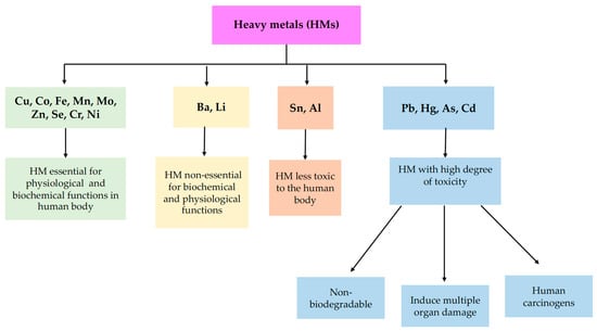
Figure 1.
Classification of heavy metals based on their role in the human body.
HMs act as cofactors of the enzyme system, in biochemical or metabolic processes. Metal balance in the body is a widely acknowledged fact. When there is an imbalance, lowering or raising specific elements causes a decrease or increase in others. Such a disturbance of the balance can be the cause of many diseases/deficiencies [9].
Framing the already-published information in the literature on the topic (depicted in the bibliometric analysis), this paper aims to provide updated knowledge and a documented and validated database on the recognized toxicity of HMs, the harmful effects of their accumulation on human health, the benefits vs. disadvantages of choosing different biological fluids/tissues/organs for the identification and quantitative measurement of HMs in the human body, as well as on the choice of the optimal method of HMs analysis, depending on the purpose.
2. Criteria for Literature Selection
This paper reviews and centralizes publications on the toxicological impact of heavy metals on human health with an emphasis on modern analytical methods of detection and quantification, outlining the current state of knowledge in the field. In order to achieve this objective, advanced searches have been performed based on predefined algorithms using Boolean operators in large and scientifically validated databases (i.e., Web of Science, PubMed, Scopus, Nature, and ScienceDirect). Moreover, due to its export, filtering, analysis and interpretation facilities, Web of Science has provided the support for a bibliometric analysis of the data starting from a simple search. In total, 146 resources corresponded to the selection and evaluation processes and constituted a defining framework supporting the information in this analysis. The stages of bibliographic selection have been represented in Figure 2, which was realized according to Page et al.’s guidelines [10].
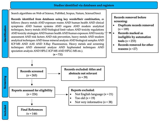
Figure 2.
PRISMA 2020 flow diagram describing literature selection.
3. Bibliometric Analysis of the Topic in the Literature
3.1. Data Collection
The Web of Science (WOS) database is one of the largest abstract and citation databases, and it was used to conduct the literature search. Using the following search parameters (TS = (heavy metal) AND TS = (human health)), 13,198 documents were identified. These documents are classified into the categories presented in Figure 3 (one document can fall into multiple categories).
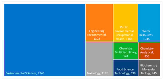
Figure 3.
Treemap of WOS categories with over 400 documents. Under the category name, the number of articles is presented.
Out of the 13,198 documents, only the documents written in English were included in this research (12,994). The leading countries in publishing papers, the relationship between them, the top-ten journals (according to the number of published articles), the network map of co-authorship, the top-ten most-cited articles, and the bubble map of the words with the highest occurrence and the average citations of these words are presented in the following section of the article.
3.2. Literature Results Obtained and Discussion
Pier, S. published the first document that fit the search parameters in 1975. The title of this paper is: “Role of heavy-metals in human health”; it was published in Texas reports on biology and medicine, and it has 21 citations. From 1975 to 2000, 159 articles were published. Then, from 2001 until 2022, the subject started to gain traction, and the number of articles published yearly started to increase steadily.
Figure 3 presents the number of articles published each year. Due to the low number of published articles before 1991, Figure 4 presents only the data from 1991 to 2021. The first year in which there were over 100 articles published was 2006.
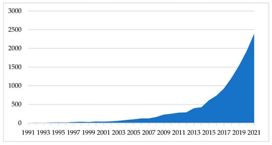
Figure 4.
Number of documents published/year.
The data was downloaded from the WoS export menu, using the following settings: “Full Record and Cited References.” Then, the bibliometric analysis was conducted using VOSviewer (CWTS, Leiden University, Leiden, The Netherlands).
3.2.1. From 1975 to 2010
In this period, 1401 documents were published. Out of these documents, 1083 (77.30%) were articles, 280 (19.99%) were proceedings papers, and 164 were review articles (11.70%). The rest of the documents were classified into other categories. The most prolific countries were the United States, China, India, Spain, and England.
Figure 5 (21 items) shows the keywords co-occurrence map. The bubble size is directly proportional to the number of occurrences, the bubble color corresponds with the average citation/document that includes the keywords: if two bubbles are closer to each other, they have a higher co-occurrence level. Only words with a minimum occurrence of 50 are presented. Words with a high occurrence are presented in Table 1.
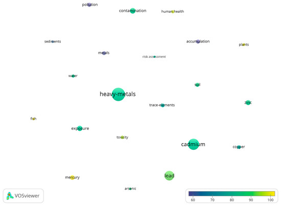
Figure 5.
Bubble map of keywords co-occurrence from 1975 to 2000.

Table 1.
The top-10 keywords for each period based on the number of occurrences.
3.2.2. From 2011 to Present
A total of 11,593 documents were published in this period. Out of these documents, 9749 (85.01%) were articles, 1461 (12.60%) were review papers, and 469 (4.04%) were proceedings papers. The rest were classified into other categories. China was the most productive country, followed by the United States, India, Pakistan, and Iran. Figure 6 (52 items) shows the keywords co-occurrence map for this period. The minimum word occurrence was set to 300.
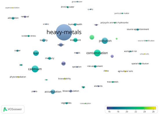
Figure 6.
Bubble map of keywords co-occurrence, from 2011 to present.
Table 1 compares the most-occurring keywords for both periods. Keywords, such as “heavy-metals” and “cadmium” had the highest occurrence for both periods. Words that represent HMs and were in the top-10 words based on occurrence, for the first period, were “cadmium,” “lead,” “zinc,” and mercury. For the second period, only “cadmium” and “lead” made it into the top-10 keywords list.
3.2.3. Leading Countries in Publishing Papers Regarding Heavy Metal Detection
Authors from 167 countries published the 12,994 articles. Table 2 presents the top-10 countries according to the number of published articles, the number of citations, the average citation/document, and the total link strength (TLS—a score representing the strength of a country’s co-authorship with other countries). China had the highest number of published documents, with 3944 (30.35%), followed by the United States with 1346 (10.36%) publications. Countries that stood out because of the high average citations/document were the United States (39.33), Australia (34.49), Spain (31.27), and Pakistan (31.02)
Table 2.
Top-10 countries according to the number of articles published.
VOS viewer was used to create the network map of country co-authorship (Figure 7). Countries that collaborated in writing articles were grouped in clusters. The bubble size is directly proportional to the number of published documents, and the thickness of the line is directly proportional to the link strength (a measure of co-authorship between two countries). The bubble color indicates the clusters.
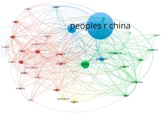
Figure 7.
Bubble map of co-authorship by country (made with VOSviewer).
Countries with at least 100 published documents are presented. A total of 34 out of 167 countries met this threshold. The four clusters included the following countries:
- The red cluster includes Belgium, Brazil, Czech Republic, England, France, Germany, Greece, Iran, Italy, Mexico, Poland, Portugal, Romania, Russia, Spain, Sweden, Switzerland, and Turkey;
- The green cluster includes Australia, Bangladesh, Egypt, India, Japan, Malaysia, Saudi Arabia, South Korea, Taiwan, and Thailand;
- The blue cluster includes Canada, Pakistan, China, and the United States;
- The yellow cluster includes Nigeria and South Africa.
The two countries that collaborated the most were the United States and China, China and Pakistan, and China with Australia. Other countries that showed a strong collaboration were Saudi Arabia and Egypt.
Furthermore, a bubble map representing the top-20 countries was created based on the number of documents to compare the two periods (Figure 8a,b). The bubble color indicates the average publication year of a country, and the bubble size is directly proportional to the number of published documents. Two bubbles are in closer proximity to each other if they collaborate often. For the first period, the United States was the major contributor in this field, but China overtook the United States in the second period regarding the number of published articles.
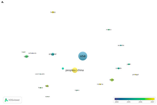
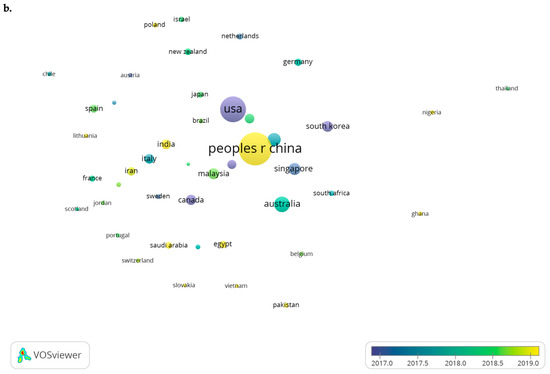
Figure 8.
Most prolific countries: (a). In the period 1975–2010; and (b). In the period 2011 to present.
3.2.4. Most Cited Articles and Leading Journals in Publishing Papers Regarding Heavy Metal Detection
The top-five most-cited articles are presented in Table 3. The most-cited article fitting the search parameters was “Hazards of HM contamination,” published in the British Medical Bulletin (IF 4.291) by Jarup L. in 2003, followed by the article published by Kampa M. in 2008 in Environmental Pollution (IF 8.071). The top-10 journals ranked by the number of published documents are presented in Table 4. The most prolific journal was Environmental Science and Pollution Research, with 750 documents published and an IF of 4.223. The journal Science of the Total Environment had the highest average citation/article, and the Journal of Hazardous Material had the highest IF of the presented journals (10.588).
Table 3.
Top-five most-cited documents.
Table 4.
Top-10 journals according to the number of articles published.
VOSviewer was used to create a bubble map of the sources that published the most documents in each period (Figure 9). The bubble size is directly proportional to the number of published documents. The bubble color represents the average publication year. For the 1975–2010 period, the most prolific journal was Science of the Total Environment, with an average publication year of 2004.82, followed by Chemosphere with an average publication year of 2006.62, and Environmental Monitoring and Assessment with an average publication year of 2006.78. The most prolific journal in the second period was Environmental Science and Pollution Research with an average publication year of 2018.78.
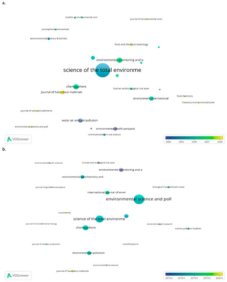
Figure 9.
Most prolific journals: (a). In the period 1975–2010; and (b). In the period 2011 to present.
4. Exposure Routes to HMs and Toxicity Regulations
Among the most toxic HMs (affecting human health), the following can be mentioned: As, Al, Hg, Pb, Ni, and Cd. Although they are extremely toxic, the population’s exposure to them is constantly increasing [15]. The HMs pathways into the human body are:
- ingestion (via food or water, reaching the bloodstream and various organs, such as the pancreas, and liver, etc., through the absorption process), as is the case for As, Pb, Hg, and Cd;
- inhalation (through the inhalation of air, vapors or aerosols, toxic metals enter the respiratory tract, reach the lungs and then the bloodstream), as is the case for Pb, Hg, and Al;
- and by dermal absorption for As, as is depicted in Figure 10 [16].
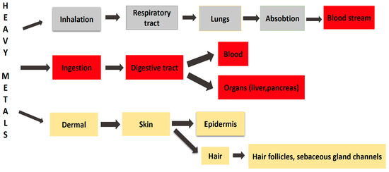 Figure 10. Exposure route to heavy metals.
It is increasingly recognized that HMs have an increased affinity for certain target organs (i.e., liver, brain, kidneys, and the bones). The accumulation and toxic effect of HMs depends on several factors, such as concentration, chemical form, sex, age, and the time of exposure. Most of them, once ingested, are distributed through the bloodstream to various organs and tissues, where they cause damage in different systems and organs. HMs’ harmful action derives from stimulating the formation of free radicals and reactive oxygen derivatives, which cause lipid peroxidation and oxidative stress in the body [17,18]. Furthermore, it must be considered the excess of HMs in the human body which may lead to neurological disorders and adverse emotional changes [19,20,21]. Table 5 summarizes the toxic effects on some organs and systems.
Table 5.
The toxic effects of heavy metals on different systems.
Several institutions manage substance safety concerns and have developed legislation regulating HM-related issues. As a result of the harmful effects of HMs on human health, the Environmental Protection Agency (EPA) made several decisions, including decreasing the Acceptable Daily Intake (ADI) and establishing a Reference Dose (RfD), with an acceptable safety level on health, for developmental and non-carcinogenic effects [38].
The official estimations from Institutions, such as the EPA, Joint Food and Agriculture Organization and World Health Organization (FAO/WHO) Expert Committee on Food Additives (JECFA), Food and Drug Administration (FDA), and Agency for Toxic Substances and Disease Registry (ATSDR) that provide the minimum risk limits for HMs, are used to determine the daily maximum safe exposure levels. All the minimal risk values originate from dose-response measurements correlating chronic exposures with the impacts seen in individuals or animal models. The minimum risk values are all based on the prevention of adverse health consequences. Moreover, the chronic oral minimum risk values regulated by the agencies for adults are 2.14 μg/kg/day for inorganic As (JECFA), 1 μg/kg/day for Cd (EPA), 0.16 μg/kg/day for Pb (FDA), 0.3 μg/kg/day for methylHg (ATSDR), 1500 μg/kg/day for Cr (III), and 3 μg/kg/day for Cr (VI), both results being provided by the EPA [39]. The risk posed by HM pollution to human health, can be assessed by using different indices, such as the daily intake of metals (DIM), the transfer factor (TF), and the health risk index (HRI), etc. [40].
Due to the difficulty in quantifying the effects of HMs and due to the low (trace) concentrations in different biological matrices, biological monitoring (BM) of human exposure to HMs has become a real challenge. Therefore, HMs toxicity, related to their concentration and accumulation in the human body, over a long period, are triggering factors of various negative effects on human health [41].
The scientific literature includes experimental studies on the acute and subacute toxicity of HMs. The bacterial bioluminescent test of toxicity (BBTT) is utilized to assess the acute toxicity of several HMs. The toxicity assays involved the evaluation of the effective concentration levels over different periods and the relative molar toxicity estimates. An experimental study found that Hg was the most harmful of the group of studied HMs (Hg, Pb, Zn, Cu, Cd, As, Cr, Co, and Ni) to the bioindicator Photobacterium phosphoreum, while Ni has been found to be the least toxic [42]. Another study assessed the acute toxicity of Cu, Pb, and Hg on juvenile Cyprinus carpio, determining the concentration that is lethal to 50% of the species (LC50) after 96 h of exposure (0.30 ppm for Cu, 0.44 ppm for Pb, and 0.16 ppm for Hg) [43]. To study the impacts of acute toxicity on the survival rate and bioaccumulation of HMs in tissues, Oreochromis sp. was examined with increasing HM concentrations and times of exposure. The 96 h LC50 of Cu, Zn, and Cd were found to be 0.45, 2.1, and 0.7 mg . Inductively coupled plasma optical emission spectrometry (ICP-OES) was used to examine fish tissues. A higher concentration of hazardous metals was associated with considerably higher tilapia fish mortality. The results showed that, in these experimental conditions, Cu was more toxic than Zn or Cd [44]. Furthermore, when HMs are present in the environment (water, soil, and food crops) at levels higher than the reference values, there is a significant risk to human health. The well water standard regulation’s reference values are 0.07 µg for Cd, 2.52 µg for Cu, 0.39 µg for Pb, and 1.36 µg for As. The mean maximum allowable levels (MALs) for the presence of HMs in soils are 13.7 mg for As, 28.5 mg for Pb, 0.18 mg for Cd, and 32. 8 mg for Cu. Moreover, the MALs for the presence of As, Pb, Cd, and Cu in vegetables and cereals are 0.1 mg , 0.2 mg , 0.125 mg , and 10 mg [45], respectively.
The most commonly used methods for exposure assessment include sample selection and preparation steps, followed by sample digestion prior to qualitative and quantitative analysis. The properties of HMs make atomic absorption spectroscopy (AAS) a frequently used method due to its high sensitivity and specificity. The evaluation of the relevant certified reference materials is used to verify the validity of the analytical processes [46].
The basic standard necessary to protect individuals from risks to their health and safety inflicted or anticipated to be caused by the presence of HMs at work or as a result of any occupational activity using chemical agents is regulated by law. Furthermore, it is mandatory to respect the biological limit values tolerable by employees, with the concentrations of chemical agents kept as low as possible. Table 6 presents the binding biological limit values (BBLVs), including HMs, established by the Scientific Committee for Occupational Exposure Limits (SCOEL), Chemical Agents Directive (CAD) 98/24/EC establishing the biological limit value at the European Union level and national regulations [47].
Table 6.
Binding biological limit values concerning occupational exposure.
According to Omrane et al., three strategies have been applied for the assessment of occupational exposure to HMs in order to correlate with the models’ predictions, including mathematical algorithms to estimate analytes’ concentrations, the determination of indoor HM concentrations, and biological testing of HMs in the urine of consenting adults. Moreover, in this experimental study, inhalation was the principal exposure route based on the selection criteria. The concentration of HMs is predicted by using a variety of methods (i.e., the Eddy Diffusion Turbulent model, the Well Mixed Box, and the Near Field and Far Field) [48].
The measurement of a chemical agent or its metabolites in a biological sample is one method of determining exposure through BM (usually blood, urine or breath). The benefit of BM is that it incorporates all exposure pathways and sources [49,50,51,52]. BM can be separated into exposure assessment and effect observation, for which internal dose and effect markers are being used. In the context of environmental health, biomarkers mostly refer to measurements used for risk evaluation or diagnosis. The phrase is now being used to describe BM measurements in occupational medicine or industrial health, too. The monitoring of exposure and effects correlate with the indicators of exposure and effects [53].
In the domain of environmental health, a number of biological methods and biomarkers are helpful for determining the risk of lead exposure. The most popular biomarker for lead exposure is blood lead. While the significant effects of lead on bone marrow can be considered an effect biomarker, this indicator assesses body burden, soft tissue lead, and absorbed dosages of lead. The interaction of lead with certain enzymes involved in heme production is the primary cause of lead’s effects in the bone marrow [53].
The suppression of delta-aminolevulinic acid dehydratase (ALAD) and changes in the quantities of certain metabolites, such as delta-aminolevulinic acid in urine (ALA-U), zinc protoporphyrin (ZP) in blood, delta-aminolevulinic acid in plasma (ALA-P), delta-aminolevulinic acid in blood (ALA-B), and coproporphyrin in urine (CP), are the key biomarkers of impact. The aforementioned markers do not, however, all accurately reflect the dose and the internal dose/effect correlation [54].
The heme pathway’s rate-limiting enzyme, ALA synthetase (ALAS), creates ALA in the mitochondria from succinyl-CoA and glycine. Lead exposure causes a reduced ALAD activity, and indirect ALAS induction caused by negative feedback control, resulting in an increase in ALA in different tissues and plasma and an increase in ALA excretion in urine. Although ALA in urine (ALA-U) has been suggested as a biomarker of lead toxicity, ALA in plasma or blood indicates the impact of lead on bone marrow more specifically. In a wide spectrum of Pb-B values, an exponential link between plasma and urine ALA was noticed [55].
The proportion of biological anomalies may be indicated by the free erythrocyte protoporphyrin (FEP) values. Microcytic anemia is linked to noticeably high BLLs. The importance of FEP measurement may be muddled by iron deficiency, which is also linked to anemia [56].
The levels of zinc protoporphyrin (ZPP) are frequently applied as indicators for lead toxicity. The other essential enzyme that facilitates the inclusion of iron into protoporphyrin IX is ferrochelatase. However, this enzyme is suppressed, and the route is blocked in Pb toxicity conditions. Additionally, if there is not enough iron available, Zn is used in place of Fe, which raises the concentrations of ZPP. These increases do not manifest in the blood until Pb levels approach 35 μg/dL, which limits this modification as a significant diagnostic characteristic [57].
5. The Importance of Tissue Mineral Analysis
In the United States in the 1970s, the first mineralograms were made by mass spectrometry, a technique used at the time by NASA, to analyze the moon rocks, as well as to analyze the metals from the spacecraft’s shell. Starting from here, this technique was also used in medicine, as an analysis for the determination of the metal content in animal fur, as well as in the human hair [58].The idea of mineral analysis prompted several American researchers, for example: Dr. George Watson (Nobel Prize), Dr. Clivet, Dr. David Watts, Dr. Wilson, and Dr. Paul Eck, to study numerous cases for more than 40 years, and to conclude that the mineral levels from the hair, correlated well with the mineral level from the tissues. In 1980, the EPA recognized the tissue mineral analysis (TMA), as a scientific method of determining toxic substances, minerals, metals from the human body.
Research on TMA has shown that it is a very advanced and modern method in medicine, which shows the relationship between human health and the concentration of various HMs/nutrients. We can find information about the time of exposure, absorption, and the distribution of all toxic substances in human tissues/organs. After absorption, HMs pass into the bloodstream and are stored into the target organs/tissues, which shows that the blood does not reflect an older exposure, this fact being found in the studies on children previously exposed to Pb. In conclusion, we can say TMA is probably the most important barometer regarding the evaluation of HMs on human health [19]. With this method, in a single analysis, the most important 36 minerals from the body can be determined, of which 30 are essential and 6 are toxic minerals (HMs), in extremely low quantities—parts per million (ppm) or even per billion.
The human body’s organic structure is made up of 80% water and 19% minerals. Minerals play a very important role in the normal functioning of the body, by controlling certain biochemical reactions, stabilizing components of proteins and enzymes, functioning as mineral cofactors for many enzymes and antioxidant molecules [59]. The enzyme with a mineral cofactor (a bivalent ion of Fe, Zn, Mn, and Co) forms an active configuration. HMs, such as Pb, Hg, Cr, As, and Al, can interfere with enzymatic reactions and disrupt their enzymatic activity because of their affinity for thiol groups (-SH) from the enzyme [60].
As was mentioned before, using TMA, the concentrations of HMs and minerals are determined, whose values provide information at the intracellular level, enabling us to identify the tendency towards functional imbalances, chronic intoxications, and concentrations, which cannot be determined by blood or urine analysis (this analysis provides information only at the extracellular level). In other words, we can say that this analysis shows the stress response phase, the mineral requirement of each human organism, the HM amount, carbohydrate metabolism, and the tissue hormone efficiency, etc. Thus it can be stated that TMA with an individualized clinical examination, allows an accurate diagnosis, the establishment of a nutritional strategy, the amelioration of various mineral and hormonal imbalances, and can even identify possible HM intoxication [61]. In the last years, TMA has greatly improved through Hair Analysis (HMS—Hair Metabolic System), which is a more representative method in terms of prevention for preventive diagnosis. Therefore by using HMS, the concentrations of different minerals/HMs can be determined, for up to 3 months or even more, and can be used for individual personalized treatments (in HM intoxication: the chelation therapy or homoeopath treatment; in the case of mineral deficiencies by taking dietary supplements or vitamins) [62]. The continuous development of TMA has led to the discovery of many correlations between disease and emotions. It is well known, that most diseases have an emotional character, and emotions are enchained with the biochemical changes in the body. Emotional and mental factors can lead to increased excretion and absorption of minerals/HMs by the human body. From this, we deduce that strong emotional reactions will produce nutritional, hormonal, neurological, and metabolic changes, which may be quantified and TMA can be that barometer/instrument for assessing the psycho–somatic and somatic–psychic relationship [19].
6. Human Biomarkers—An Assessing Health Status Factor
One of the assessment methods used in the determination of HM toxicity to human health, is human biomarkers. The low concentration of HM in biological matrices, the quantification of the contributions of sources of exposure, environmental and genetic factors, have led to the difficulty of monitoring human exposure. However, the most widely used human biomarkers in toxicological study remain: blood, urine, nails, and hair [63]. Being a noninvasive matrix, the hair and nails are most-often used as a representative human biomarker in long-term exposures to HMs [64,65]. On the other hand, biomarkers, such as the blood and organ tissues, can decrease the rate of participation in the population, requiring invasive sampling. The majority of HMs have a relativity short lifetime, which makes their blood and urine dosing a standard only for short-term exposures, for example, 2–3 h or 3–4 days (acute exposure) [66]. Some of them, the most toxic HMs, such as Pb or Cd, remain in the blood and tissues for a long time. A quantitative assessment of them can only be made if the bioaccumulation method is well known.
6.1. Hair, Nails, Urine, Blood, or Vitreous Humor Analysis
The utility of studying and measuring the concentrations of HMs in hair, nails, various biological fluids (blood, urine, and vitreous humor), and some organs or tissues, has become increasingly vital, because all these biomarkers are a perfect barometer for the disturbances that occur in the human body. The determination of HMs/trace elements from the human blood or urine, is a traditional/classic procedure, which shows the concentration of HMs/trace elements at the present time, but the action of external factors and bio-kinetics, lead to fluctuations in the HMs/trace element concentrations in the urine and blood. The hair and nails are considered to be final metabolic products, which, compared to other human body compartments, provide clear and precise information about any trace elements, because we presume that they can incorporate very easily any trace elements (toxic or not) during the growth process, due to a complex structure called amino acids, made from sulfur and fibrous proteins [63]. In addition to the hair and nails, in some cases, hard tissues can be used as a biomarker, depending on the HM which must be determined [67]. All this HM accumulation in human body can induce certain diseases, intoxications or imbalances in the human body [68].
6.1.1. Human Hair and the Trace Elements Stored in It
Human hair is made of approximately 80% protein, 10–15% water, 5–10% pigments, minerals, lipids, and the mineral content is 0.25–0.95% [69]. The scalp is usually made up of about 100,000 hairs, of which about 10% is at rest. The growth rate of the hair is generally 0.3 mm/day, which means about 1 cm/month. The hair follicle is strongly vascularized, due to the blood, which bathes it and is a very good transport medium for toxic and trace elements. As the hair grows, it suffers a keratinization process (hardening) and during this process, it incorporates all the traces of toxic and essential elements into the protein structure [68]. In 1929, the hair scalp was used for the first time, to assess the human systemic level of elements [70]. Following this analysis, the researchers established different correlations between the essential/toxic elements and different diseases, nutritional status, and metabolic disorders [71,72].
Even nowadays, the hair remains the most important biological matrix for health assessment, and for identifying possible intoxications of various etiologies (HMs, drugs, and pesticides), as well as mineral imbalances in the human body. Compared to other tissues/body fluids, such as the blood or urine, it was found that the concentration of HMs from the hair is in direct correlation with various diseases. HMS provides information about the history of long-term exposure, compared to other traditional analysis, which shows the toxic elements in the initial phase. The hair analysis is a non-invasive method, which does not require a large amount of biological material, is simple and straightforward. Hair has long been used, both in animal and human studies, to highlight the human body’s exposure to HMs. In this way, analyses can be performed on samples that are hundreds of years old [73].
Difficulties arise due the lack of well-defined reference areas of concentration. This is a consequence of the huge difference in the element’s levels of concentrations, depending on the region, sex, age, hair color, and lifestyle. Other external factors/sources (hair dyeing, use of cosmetics to treat hair, hair washing, and bleaching), may also influence the correct interpretation of the results [74]. The concentration of toxic elements in human hair, may also be affected by seasonal variations or the effect of synergism and antagonism [75].
6.1.2. Finger- and Toenails
The most attractive and interesting samples sources are the nails and human hair, because they are very easy to collect, store and transport, unlike other biological fluids, such as the blood, urine, and vitreous humor, which can be easily contaminated if the collection and storage procedures are not followed correctly. These metabolic products have fairly consistent content of HMs due to the growth process [22].
The human nail is a thin, horny plate, which grows on the upper part of the phalanx of the fingernail and toes, having an important sensory and protective function. It is made of a strong protein material, called keratin, that incorporates all the essential trace elements or toxic elements. Like any other part of the human body, they are exposed to various environmental and occupational factors or to imbalances in the human body. Toenails and fingernails, are considered to be very good biomarkers for tracking accumulation of various toxic elements [76]. The trace elements and HMs are collected and stored for a very long period, which allows nail analysis, along with hair analysis, to bring valuable information about possible intoxications or diseases that exist in the body or favor the appearance of various diseases [77]. On average, the fingernail growth rate is about 3–5 mm/month, and the toenails increase by about 1.6 mm/month. This rate of nail growth depends on sex, age, health conditions, and metabolic rate, etc. However, toenails are more protected than fingernails from any possible external factors. Because nails incorporate any trace elements very easily in high quantity, they are considered to be an important barometer for long-term exposures, for all kinds of toxic elements [61].
Nails are made of a rich layer of keratin, which easily incorporates any trace elements, which makes by them an important source of vital information to the researcher. Along with hair, they are the most suitable sample for a long-term assessment of trace elements, which can cause intoxications or imbalances in the body. The taken sample does not require special conditions for collection, storage, and transport. In the case of some trace elements/toxins, it does not always provide all the necessary information. There may be differences in toenail analysis compared to the analysis of the fingernails, because the toenails are more protected from the action of various toxic factors and are washed less often than the hands. The growth of the toenails is slower than that of the fingernails, so the accumulation of HM is slower, on average about 2–12 months after exposure. Nails can be very easily contaminated due to the use of nail polish or some medications. Sometimes the amount of nails collected is quite small, which makes the limit of detection lower [76].
6.1.3. Vitreous Humor and Epithelial Tissues (Human Eyes)
The toxicity of HMs is explained by the interaction between the HM ion and the specific target protein. This action changes the function and structure of the protein. The cells involved in the transport of HMs are very susceptible to their toxic effects [78,79]. The retinal epithelium is a chelating metal tissue, with a high affinity for HMs, due to melanin (the retinal tissue pigment), which forms bonds with HMs. Results have shown that Pb may accumulate in the retina and choroid [80].
6.1.4. Blood and Urine
There are other biological matrices that can provide important information about the accumulation of HMs, because some of the trace elements are not found in the blood (note: it is preferable to determine the inorganic forms of Hg and As from blood). Some HMs can be stored in various tissues, such as the kidney, liver, brain, bones, and lungs, which are considered target organs, where HMs can cause a lot of injuries without increasing the values in the blood. The HMs remain only for a short time in the blood (blood has the role of transporting nutrients from one tissue to another), where they are fixed in the tissues or organs, so their values from blood are relevant for a short period of time. As a biological matrix for the determination of Pb, the blood is preferred, because it shows a better correlation with some diseases. In addition, the determination of MeHg salts is recommended from the blood. Blood and urine show us the amount of HMs at the present time, or better said, at the time of exposure ±2 days and not the cumulative degree of exposure.
The HM determination from the blood, provides information about recent exposure (hours or days). Blood determination is an invasive method, and the blood levels are transient, independent of those stored in human tissues (the toxic minerals are purified quickly) and vary from one component to other (e.g. the serum, and plasma, etc.) In the case of urine, it can be seen, that HMs are excreted at the time of sampling and provide information about relatively recent exposure (days or weeks), which makes this determination suitable for measuring acute exposure.
6.2. Different Tissues and Organs
6.2.1. Teeth and Bones
Along with other biological fluids and tissues, the teeth and bones can be considered an important compartment in terms of exposure to HMs on a large scale [81]. It is well known that the teeth and bones are calcified tissues, which have as their main component hydroxyapatite. Although calcified (hard) tissues are chemically stable, it is presumed that some HMs/trace elements can accumulate in the skeletal structure, by displacing Ca2+ from hydroxyapatite, which makes the teeth and bones a bioindicator/biomarker for monitoring the accumulation of HMs in the human body [82]. From a morphological point of view, there are two types of bone tissues: spongy and compact. Spongy bone can provide information about a relative recent exposure to HMs, while within the compact bone, HMs can accumulate for a much longer period. During the formation of tooth tissues, there is no release of mineral substances, thus the teeth can be considered a permanent and stable recording in terms of exposure to HMs over a longer period [81,83]. In the case of bone tissues, in pathological and physiological conditions, a release and fluctuation of HMs/minerals from the bone tissues in the blood was found. Deciduous teeth cannot be considered a bioindicator of HM exposure, because they have a short lifespan, and later they are replaced by permanent teeth. Permanent teeth, especially the ‘dentin’ (which does not suffer any turnover mineral phase), can provide information about HM accumulation, but the tooth enamel, which is constantly in direct contact with various factors within the oral cavity (drugs, caries, smoke, food, and medications), can modify the mineral composition of the surface layers of the tooth, which may affect their credibility as a bioindicator [82]. Teeth are considered to be easy-to-extract biological tissues, with a low rate of pollutant removal [84]. As mentioned earlier, HMs, once ingested and absorbed, are distributed in target organs and those in large quantities, with a long half-life, are stored in the human skeleton, which is an ideal long-term storage place [85]. With aging, senescent cells begin to accumulate in organs and tissues. They can cause inflammatory reactions, and decreased vitality of cells and tissues. Thus, the bones may be affected by the distribution of HMs in the aging process. Human bone, being a hard, calcified tissue during growth, incorporates HMs, similar to the nails and hair. Compared to the blood, the skeletal system incorporates and retains HMs for a very long time, and with aging, the storage capacity can change.
Many articles have shown that there are no significant differences in HM concentrations between different bones (e.g., the femur, and tibia) [72]. However, a potential factor of influence is the place of sampling. Another disadvantage of bone sampling is the amount of the sample; the human bone is not always accessible for measurement or sampling [71].
6.2.2. Tissues and Organs
It is well known that HMs have an increased affinity for certain organs (target organs) and systems. Both organic and inorganic HMs derivatives are toxic, but inorganic derivatives are the most toxic (creating irreversible injuries); they penetrate the fat-rich tissues, such as the brain, and liver, due to their increased fat solubility (i.e., Pb penetrates the bones and teeth 80–95%; the liver, lung and brain as soft tissues [22]; Hg (depending on the organic form) targets the lung, kidney (40–60%), heart, liver, brain, and tissues [86]; As targets the blood, lung, heart, kidney, and neurological systems; Al targets mainly the brain and bones, lung, muscle, heart, and spleen).
As previously stated, the determination of HMs from various organs and tissues, is extremely important, but too inaccessible (organs or tissues sampling may be performed only in the case of surgery or forensic cases/necropsy). As can be expected, any determination of HMs from various biological fluids, organs, or tissues, has advantages and disadvantages. In the following, the advantages and disadvantages of the biological matrix that contributes to the determination of HMs, are described and summarized in Table 7.
Table 7.
Advantages and disadvantages of different biological matrices used to identify heavy metals (HMs).
7. Modern Analytical Methods of Determination
At the present time, the chemistry of HMs or HM traces, present or accumulated in the human body over time, can be determined through the continuous improvement of various specific, modern, selective, and highly sensitive analyses of several types of samples [100]. One of the most widespread analysis techniques, both in the field of elemental and molecular analysis, is spectrometry. When precise determinations are sought, it is imperative to apply spectrometric techniques, which cover an extremely wide field, and have precision and accuracy in determinations, which make the use of these techniques more than necessary [101]. Some of the most commonly used techniques are: Lymphoblastic Transformation Test (LTT)-HMs test, X-ray Fluorescence, AAS, Inductively Coupled Plasma Mass Spectrometry (ICP-MS) or Inductively Coupled Plasma-Atomic Emission Spectrometry (ICP-AES), and the reason behind these choices is mentioned later. The last three spectrometric techniques are the most often used to determine traces of heavy metals in various biological matrices (Figure 11) [102,103].
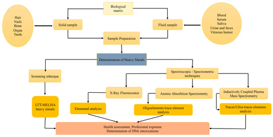
Figure 11.
Biological matrix, Sample Preparation and Modern Analytical techniques in HM determination.
7.1. Lymphoblastic Transformation Test—HMs
One of the routine tests when it comes to human body sensitivity to HMs, is the Patch Test, or rather the Epicutaneous Patch Test. However, this test has some disadvantages: it can be false-negative or false-positive, it is subjectively assessed, it has low reproducibility, it can cause exacerbated irritation, and it is generally used to determine dermal allergens, the most common ones being Ni and Co [104,105].
The in vivo lymphocyte transformation test (LTT) is an alternative to the epicutaneous test (Patch test), which in turn is optimized by the LTT-MELISA® (Memory Lymphocyte Immune Stimulation Assay) test-method accredited in Germany since 2001. LTT-MELISA® is a test with high specificity, reproducibility, sensitivity, and reliability for the detection of sensitivity in patients with metal sensitivity [106].
The LTT is a test that shows the degree of proliferation (reactivity) of T lymphocytes, after exposure to various allergens (HM in this case). LTT-HM is the validated test for various allergens: respiratory, medicinal, and food; it does not produce sensitization, has an increased reproducibility, and provides objective results. It is indicated in a case of monitoring symptomatic persons, who are periodically exposed to the action of HMs. Thus, it was found that in the case of monitoring a patient exposed to the action of HMs, after avoiding exposure of the patient for a long time, the lymphocyte reactivity had normalized (LTT-negative) [107]. The test is performed through venous blood sampling, and it allows us to detect concentrations of HMs in saline solutions, such as Al, Gold (Au), Silver (Ag), Beryllium (Be), Cd, Co, Cr, Cu, Gallium (Ga), Indium (In), Iridium (Ir), Mo, Ni, Palladium (Pd), Pb, Platinum (Pt), Ruthenium (Ru), Sn, Titanium (Ti) and oxides of titanium (TiO2), Vanadium (V), as well as Hg compounds (i.e., ethyl Hg, MeHg, PhenylHg (PhHg), and Hg chloride (HgCl2) [108].
The MELISA®LTT-test is important in clinical relevance in the case of exposed patients, for those who are about to undergo surgery (i.e., prosthesis or dental work), or for those who suffer from chronic infections, fatigue or various autoimmune diseases, whose aim is to detect and monitor the action of HMs [108]. Research on the reliability of MELISA testing is inconsistent [109]. Some claim the MELISA test is reliable and clinically helpful [107,108,109,110]. However, other studies claim that MELISA testing is non-specific, therefore it has little clinical use [111,112].
7.2. X-ray Fluorescence
X-ray Fluorescence Spectrometry (X-ray Fluorescence) is a primary analytical technique, one of the oldest nuclear techniques that relies on the interaction of X-rays with a material, in order to determine its elemental composition [113]. X-ray fluorescence allows the determination of each individual element with high selectivity. This technique is suitable for flat, homogeneous, and very thin samples, as it has a low energy, which is easily absorbed by the sample matrix [60]. It is also suitable for the determination of heavy metals with Z > 18, from various biological materials. It is possible that with this method, a low concentration of elements can be found, which should be analyzed by other analytical techniques [114]. Compared to other analytical methods, this non-invasive technique does not require sample preparation. Due to the extremely penetrating nature of these X-rays, since their discovery, they have been used for non-invasive” observation, for the diagnostic purposes of various biological samples (i.e., synovial fluid, blood, plasma, and bones), environmental samples (i.e., soil, water, and oil), as well as food samples (i.e., milk, wine, and beer) [87]. It is a fast, simple, relatively low-cost method that can be used to determine several HMs in various solid samples, but only in small samples [115]. Thus, it can be said that X-ray fluorescence spectrometry is an ideal technique, especially in environmental samples [116], forensic toxicology, for rapid, simultaneous, quantitative mapping of metalloids and metals/trace elements in very low concentrations (even traces) in various human tissues (hard tissues, bones, nails, and teeth) with reasonable specificity, sensitivity, and resolution (Figure 11) [117]. The XRF technique has the ability to simultaneously detect trace elements in very low concentrations, the preparation of the sample for analysis is simple, and regarding the origin of the sample (human nature), the size is limited [115]. However, this method has some drawbacks, such as: the technique/equipment requires the use of a radiation source, LOD depending on sample matrix and specific elements, and for a blood sample it is necessary to identify the parameters that could affect the analysis (depositing the blood sample on the reflector) [118,119].
7.3. Atomic Absorption Spectrometry
AAS is a modern analytical method widely used for high sensitivity determinations in various fields, such as: the pharmaceutical industry, water and environmental quality control, clinical and forensic toxicology when involving suspicions of HM poisoning. This method makes it possible to determine concentrations of HMs by the order of ppm or even ppb (using a graphite furnace AAS) [86]. As opposed to classical photometric methods, this method is much more accurate; the analysis is performed in aqueous or organic solutions and does not require a high consumption of reagents. It is a recognized method for qualitative and quantitative determinations of HMs, with a high resolution and a relatively simple way of working [120,121,122]. AAS cannot simultaneously analyze numerous elements, as the light source bulb must be changed while determining the elements and fails to identify elements with resonance lines in the ultraviolet (UV) vacuum area. Also, complex sample matrices might cause interference issues that are difficult to resolve. All of these are shortcomings [123].
7.4. Inductively Coupled Plasma Mass Spectrometry
ICP-MS is a type of analytical technique, which uses an inductively coupled plasma to ionize the sample. By atomizing the sample, it creates small polyatomic and atomic ions, which are then detected. It is used and known for its ability to detect metals and non-metals in liquid samples at very low concentrations [124]. ICP-MS is currently the most recent and widely used method for the detection of HMs and metalloids in traces/ultra-traces [125] with a lot of applications in the chemical industry, food processing, environmental analysis, and medical analysis [126]. Emerging since the 1980s and used as a method for the analysis of various multi-element clinical samples, ICP-MS has been increasingly developed for the detection and quantification of metals and metalloids, such as: As, Se, and Pb, etc. [127]. It is considered the gold analytical technique used for the determination of trace elements in biological specimens [128]. This is a superior HM detection method, preferable in comparison to other methods (AAS), because of its high detection speed, accuracy, flexibility, simplicity, and reproducibility. It allows the detection of several elements of the periodic table with an atomic mass ranging from 7 to 250, i.e., from Li to Uranium(U); allowing detection in parts per trillion (ppt) or nanogram/liter concentrations, and can detect and measure variations in isotopic compositions in environmental and geological samples [129,130]. ICP-MS is generally unable to directly identify minor metal ions in samples from the environment. The interaction of the samples’ matrix and decreased concentration metal ions causes ICP-MS’s direct determination to be extremely challenging. Preconcentration and separation are important to increase the sensitivity of trace metal detection [131,132].
ICP-MS is frequently used in the laboratories of pharmacology, clinical and forensic toxicology for quantitative analysis [118,133], and biochemistry, aiming to monitor occupationally exposed people, those with different metal allergies or to identify different HMs or metalloids that may cause intoxication [134]. It also allows the simultaneous analysis of approximatively 30 elements in a single detection, from multiple biological matrices (blood, urine, plasma, hair, nails, tissues, and organs), resulting in an optimal gain in sensitivity [127]. Performing the analysis involves a high cost as the samples need to be pre-dissolved into solutions. Due to low salt tolerance, the limits of detection can really be up to 50 times worse. Several light elements cause significant interference in ICP-MS, such as Ca, S, K, Fe, and Se [121].
Table 8 presents the most modern analytical techniques used for HM determination in biological samples.
Table 8.
Modern analytical methods used for HM determination in biological samples.
7.5. Hyphenated Techniques and Speciation Analytics
Hyphenated analytical techniques include approaches that combine chromatography with spectroscopic or spectrometric methods to analyze a variety of biochemical or toxicological compounds. Furthermore, these techniques are generally preferred, especially in trace analysis, since they greatly limit the danger of cross-contamination due to sample manipulation. Additionally, the fraction of interest cannot typically be transferred quantitatively using off-line procedures [135,136].
Over time, the beneficial effects of combining spectroscopic or spectrometric techniques with separation methods for the quantitative and qualitative assessment of unidentified chemicals in complex fractions and extracts has been established. Spectroscopic or spectrometric detection techniques, such as mass spectrometry (MS), Fourier transform infrared (FTIR), ultraviolet-visible (UV-Vis) absorbance or fluorescence emission, nuclear magnetic resonance spectroscopy (NMR), and photodiode array (PDA), are linked to liquid chromatography (LC), typically high-performance liquid chromatography (HPLC), capillary electrophoresis (CE), or gas chromatography (GC), to obtain detailed information on the structure, leading to the detection of different compounds, including HMs, present in various biological samples [137].
Analytical techniques for detecting and speciating metals and metalloids have drawn considerable scientific interest because of their significant environmental and public health impacts [138].
Speciation analytics refers to the assessment of different chemical and physical forms of a specific element. The concept of speciation can be used to identify the presence of several configurations of a certain analyte in the sample under observation, such as the attachment to various ligands or the presence of various oxidation states. Moreover, selectivity in order to ensure the compounds of interest are identified and sensitivity in order to accurately assess the concentration of the analyte in the sample are specific parameters required for an appropriate speciation analysis [139].
The distinction between physical and chemical speciation is also feasible. Chemical speciation can be divided into distribution speciation involving the exploring or evaluating of particular chemical structures and identifying particular chemical forms in particular elements of the samples and screening speciation that involves seeking and ascertaining selected chemical structures [140,141]. According to the outcomes of toxicological testing, a specific entity’s proportion in a mixture often has a more significant impact on living organisms than its overall concentration [139].
The inorganic ions of As, Se, Hg, Pb, and Sn, whose toxicological characteristics are varied, are those for which speciation analyses are most frequently utilized. As speciation is a topic that is constantly evolving due to advancements in analytical methods and growing toxicological knowledge. As speciation is not frequently provided as an examination in clinical laboratories, despite the fact that speciation procedures have been widely developed. Furthermore, HPLC-ICP-MS is typically used to perform As speciation on urine and blood samples. Micro-liquid chromatography coupled to an ICP-MS spectrometer (μLC-ICP-MS) is an experimental approach established as a novel and quick analytical method for determining the different forms of As [139,142].
Another experimental study assessed the concentration of Se in human serum by also applying hyphenated techniques. After an enzymatic hydrolysis, the resulting compound (i.e., selenomethionine) was evaluated using reversed phase-HPLC (RP-HPLC), combined with collision/reaction cell inductively coupled plasma-quadrupole mass spectrometry (CRC ICP-QMS) [143].
The detection of Hg, Pb, and Sn species in urine samples has been conducted by applying a GC/MS-MS method. Capillary GC combined with an ion-trap mass spectrometer with electron impact ionization in the tandem-MS mode (MS-MS) was used to separate and identify the toxic metallic ions. Furthermore, for increased accuracy, a very efficient method of sample preparation was established, focusing on chemical modifications with sodium tetraethylborate and subsequent headspace solid phase microextraction [144].
The hyphenated techniques broaden the novel and extend the prospects. Their primary benefits are extremely low detection and measurement thresholds, a negligible impact of external interference on the analytical assay, and extremely high accuracy and repeatability of outputs [139]. The disadvantages and expensive price of the equipment are undoubtedly the limits of the hyphenated approaches. Since they are difficult to acquire, laboratories do not frequently use them. Additionally, the use of hyphenated procedures needs a thorough understanding of the analytical techniques and equipment. Rather than being used for the usual analyses, these devices are exceedingly expensive and are only used in research. However, the hyphenated strategies have been evolving and becoming more and more significant, which is supported by the growing body of publications on the topic and by new methods/techniques [139,145].
8. Conclusions and Future Perspectives
The potential of HMs to damage DNA and membranes, as well as to interfere with the activity of proteins and enzymes has demonstrated that they represent a significant risk to human health.
At the present moment, AAS is the dominant analytical technique for the detection of trace elements (metals, and oligo-elements) in the human body, and we are witnessing a more and more evident shift from flame-electro thermal atomic spectrometry to ICP-MS, a more modern and effective method. However, the choice of the analytical method used for the detection of the different metals will remain at the discretion of the toxicologist/chemist who considers the metals to be detected, their number, the analyzed matrix (biological samples), the concentration of the analyte in the matrix, and the intended purpose (toxicological/nutritional screening, therapeutic purpose, and prevention, etc.).
TMA is of great importance for monitoring the health of the population, not only for studying the effects of occupational exposure, but also for health in general, by determining both nontoxic minerals in the human body and toxic minerals (HMs). Although some authors claim that TMA is an alternative method of investigation or even a fraud (some considered TMA to be “unscientific, economically wasteful and probably illegal”), its importance cannot be denied [146]. In 2011, a publication emerged in the scientific literature that clarified to some extent the status of TMA. This diagnostic method cannot accurately, clearly, and realistically determine what causes an abnormal concentration of minerals in the hair. However, it can be stated that the results of the TMA, in conjunction with laboratory biochemical analyses (i.e., determination of δ-aminolevulinic acid (ALA), coproporphyria/Pb poisoning, or determination of As acute hemolytic anemia, the dark color of urine, detection of plasma Hg, coproporphyria or DMPS challenge test etc.) can lead to an accurate diagnosis in terms of the disorders that the patient suffers from, possible intoxication or possible procedures that the patient undergoes [22].
In the case of an HM detection in forensic practice, it is extremely important to collect as many biological fluids, tissues, and organs as possible, to process them as quickly as possible and to analyze them with modern high-performance equipment. All of these factors, together with a histopathological examination and an investigation into possible diseases and habitual behaviors, can lead to the most accurate determination of the cause of death.
Hyphenated techniques, which combine multimodal detectors with chromatographic separative methods, are becoming effective substitutes with potential use in the evaluation of HMs.
Given the fact that HMs are toxic elements for both human health and the environment, and due to the high degree of bioaccumulation in the human body, it would be advisable to monitor them continuously. Nowadays, due to the modernization of laboratories, the development of biological methods of identification and quantification, using biomarkers it is possible to establish certain criteria and standards that would help protect human health and the environment from HM contamination. TMA, combined with high-performance laboratory analysis, can be considered a vital barometer of the population’s health, as it creates a true mapping of all the biochemical reactions in the body, which through their disturbances can trigger various diseases or even intoxications.
Funding
The APC was funded by the University of Oradea through an internal project.
Institutional Review Board Statement
Not applicable.
Informed Consent Statement
Not applicable.
Data Availability Statement
Not applicable.
Acknowledgments
The authors would like to express their gratitude to the University of Oradea for supporting the APC.
Conflicts of Interest
The authors declare no conflict of interest.
References
- Singhal, H.R.; Mani, N.K.; Kodgi, A.; Mehendale, N.; Sharma, S.; Garlapati, V.K. Chapter 10-Miniaturized microfluidic heuristics for the detection of polluting molecules in the environment. In Handbook on Miniaturization in Analytical Chemistry; Hussain, C.M., Ed.; Elsevier: Amsterdam, The Netherlands, 2020; pp. 221–235. [Google Scholar]
- Griffin, R.M. WebMD, Heavy Metal Poisoning. Available online: https://www.webmd.com/a-to-z-guides/what-is-heavy-metal-poisoning (accessed on 17 April 2022).
- Huamain, C.; Chunrong, Z.; Cong, T.; Yongguan, Z. Heavy metal pollution in soils in China: Status and countermeasures. Ambio 1999, 28, 130–134. [Google Scholar]
- Järup, L. Hazards of heavy metal contamination. Br. Med. Bull. 2003, 68, 167–182. [Google Scholar] [CrossRef] [PubMed]
- Chen, T.-C.; Priambodo, R.; Huang, R.-L.; Huang, Y.-H. The Effective Electrolytic Recovery of Dilute Copper from Industrial Wastewater. J. Waste Manag. 2013, 2013, 164780. [Google Scholar] [CrossRef]
- Witkowska, D.; Słowik, J.; Chilicka, K. Heavy Metals and Human Health: Possible Exposure Pathways and the Competition for Protein Binding Sites. Molecules 2021, 26, 6060. [Google Scholar] [CrossRef]
- Bolan, S.; Kunhikrishnan, A.; Seshadri, B.; Choppala, G.; Naidu, R.; Bolan, N.S.; Ok, Y.S.; Zhang, M.; Li, C.-G.; Li, F.; et al. Sources, distribution, bioavailability, toxicity, and risk assessment of heavy metal(loid)s in complementary medicines. Environ. Int. 2017, 108, 103–118. [Google Scholar] [CrossRef]
- Nabulo, G.; Black, C.; Young, S. Trace metal uptake by tropical vegetables grown on soil amended with urban sewage sludge. Environ. Pollut. 2011, 159, 368–376. [Google Scholar] [CrossRef]
- Savic Gajic, I.; Savic, I.; Gajic, D. The role and health risk of heavy metals in human organism. In Heavy Metals and Health; Castillo, L., Ed.; Nova Science Publisher: Hauppauge, NY, USA, 2016; pp. 47–89. [Google Scholar]
- Page, M.J.; McKenzie, J.E.; Bossuyt, P.M.; Boutron, I.; Hoffmann, T.C.; Mulrow, C.D.; Shamseer, L.; Tetzlaff, J.M.; Akl, E.A.; Brennan, S.E.; et al. The PRISMA 2020 statement: An updated guideline for reporting systematic reviews. BMJ 2021, 372, n71. [Google Scholar] [CrossRef]
- Kampa, M.; Castanas, E. Human health effects of air pollution. Environ. Pollut. 2008, 151, 362–367. [Google Scholar] [CrossRef]
- Ali, H.; Khan, E.; Sajad, M.A. Phytoremediation of heavy metals—Concepts and applications. Chemosphere 2013, 91, 869–881. [Google Scholar] [CrossRef]
- Salt, D.E.; Smith, R.D.; Raskin, I. PHYTOREMEDIATION. Annu. Rev. Plant Physiol. Plant Mol. Biol. 1998, 49, 643–668. [Google Scholar] [CrossRef]
- Li, Z.; Ma, Z.; van der Kuijp, T.J.; Yuan, Z.; Huang, L. A review of soil heavy metal pollution from mines in China: Pollution and health risk assessment. Sci. Total Environ. 2014, 468, 843–853. [Google Scholar] [CrossRef] [PubMed]
- Engwa, G.; Okeke, P.; Nwalo, N.; Unachukwu, M. Mechanism and Health Effects of Heavy Metal Toxicity in Humans. In Poisoning in the Modern World—New Tricks for an Old Dog; Intechopen: London, UK, 2019; pp. 1–23. [Google Scholar]
- Briffa, J.; Sinagra, E.; Blundell, R. Heavy metal pollution in the environment and their toxicological effects on humans. Heliyon 2020, 6, e04691. [Google Scholar] [CrossRef]
- Flora, S.J. Arsenic-induced oxidative stress and its reversibility. Free Radic. Biol. Med. 2011, 51, 257–281. [Google Scholar] [CrossRef]
- Jaishankar, M.; Tseten, T.; Anbalagan, N.; Mathew, B.B.; Beeregowda, K.N. Toxicity, mechanism and health effects of some heavy metals. Interdiscip. Toxicol. 2014, 7, 60. [Google Scholar] [CrossRef] [PubMed]
- Watts, D.L. Trace elements and neuropsychological problems as reflected in tissue mineral analysis (TMA) patterns. J. Orthomol. Med. 1990, 5, 159–166. [Google Scholar]
- Behl, T.; Kaur, G.; Sehgal, A.; Bhardwaj, S.; Singh, S.; Buhas, C.; Judea-Pusta, C.; Uivarosan, D.; Munteanu, M.A.; Bungau, S. Multifaceted Role of Matrix Metalloproteinases in Neurodegenerative Diseases: Pathophysiological and Therapeutic Perspectives. Int. J. Mol. Sci. 2021, 22, 1413. [Google Scholar] [CrossRef]
- Kabir, M.T.; Uddin, M.S.; Zaman, S.; Begum, Y.; Ashraf, G.M.; Bin-Jumah, M.N.; Bungau, S.G.; Mousa, S.A.; Abdel-Daim, M.M. Molecular Mechanisms of Metal Toxicity in the Pathogenesis of Alzheimer’s Disease. Mol. Neurobiol. 2021, 58, 1–20. [Google Scholar] [CrossRef]
- Flora, G.; Gupta, D.; Tiwari, A. Toxicity of lead: A review with recent updates. Interdiscip. Toxicol. 2012, 5, 47. [Google Scholar] [CrossRef]
- Karagas, M.R.; Choi, A.L.; Oken, E.; Horvat, M.; Schoeny, R.; Kamai, E.; Cowell, W.; Grandjean, P.; Korrick, S. Evidence on the human health effects of low-level methylmercury exposure. Environ. Health Perspect. 2012, 120, 799–806. [Google Scholar] [CrossRef]
- Hong, Y.-S.; Kim, Y.-M.; Lee, K.-E. Methylmercury exposure and health effects. J. Prev. Med. Public Health 2012, 45, 353. [Google Scholar] [CrossRef]
- Charkiewicz, A.; Backstrand, J. Lead Toxicity and Pollution in Poland. Int. J. Environ. Res. Public Health 2020, 17, 4385. [Google Scholar] [CrossRef] [PubMed]
- Bhattacharjee, S.; Chen, C.; Ahn, W.-S. Chromium terephthalate metal–organic framework MIL-101: Synthesis, functionalization, and applications for adsorption and catalysis. RSC Adv. 2014, 4, 52500–52525. [Google Scholar] [CrossRef]
- Andrade, V.; Aschner, M.; Marreilha Dos Santos, A. Neurotoxicity of metal mixtures. Neurotox. Met. 2017, 18, 227–265. [Google Scholar]
- Ratnaike, R.N. Acute and chronic arsenic toxicity. Postgrad. Med. J. 2003, 79, 391–396. [Google Scholar] [CrossRef]
- Bjørklund, G.; Oliinyk, P.; Lysiuk, R.; Rahaman, M.; Antonyak, H.; Lozynska, I.; Lenchyk, L.; Peana, M. Arsenic intoxication: General aspects and chelating agents. Arch. Toxicol. 2020, 94, 1879–1897. [Google Scholar] [CrossRef]
- Ikechukwu, U.R.; Okpashi, V.E.; Oluomachi, U.N.; Paulinus, N.C.; Obiageli, N.F.; Precious, O. Evaluation of heavy metals in selected fruits in Umuahia market, Nigeria: Associating toxicity to effect for improved metal risk assessment. J. Appl. Biol. Biotechnol. 2019, 7, 3–5. [Google Scholar]
- Syversen, T.; Kaur, P. The toxicology of mercury and its compounds. J. Trace Elem. Med. Biol. 2012, 26, 215–226. [Google Scholar] [CrossRef]
- Jiao, J.; Wang, M.; Wang, Y.; Sun, N.; Li, C. Lead exposure increases blood pressure by increasing angiotensinogen expression. J. Environ. Sci. Health A Tox. Hazard. Subst. Environ. Eng. 2016, 51, 434–439. [Google Scholar] [CrossRef]
- Gambelunghe, A.; Sallsten, G.; Borné, Y.; Forsgard, N.; Hedblad, B.; Nilsson, P.; Fagerberg, B.; Engström, G.; Barregard, L. Low-level exposure to lead, blood pressure, and hypertension in a population-based cohort. Environ. Res. 2016, 149, 157–163. [Google Scholar] [CrossRef]
- Sharma, B.; Singh, S.; Siddiqi, N. Biomedical Implications of Heavy Metals Induced Imbalances in Redox Systems. BioMed Res. Int. 2014, 2014, 640754. [Google Scholar] [CrossRef]
- Dongre, N.N.; Suryakar, A.N.; Patil, A.J.; Hundekari, I.A.; Devarnavadagi, B.B. Biochemical effects of lead exposure on battery manufacture workers with reference to blood pressure, calcium metabolism and bone mineral density. Indian J. Clin. Biochem. 2013, 28, 65–70. [Google Scholar] [CrossRef] [PubMed]
- Kall, J.; Just, A.; Aschner, M. What is the risk? Dental amalgam, mercury exposure, and human health risks throughout the life span. In Epigenetics, the Environment, and Children’s Health Across Lifespans; Springer: Berlin/Heidelberg, Germany, 2016; pp. 159–206. [Google Scholar]
- Agency for Toxic Substances and Disease Registry, Toxicological Profile for Mercury. Available online: https://wwwn.cdc.gov/TSP/ToxProfiles/ToxProfiles.aspx?id=115&tid=24 (accessed on 10 December 2021).
- Tchounwou, P.B.; Yedjou, C.G.; Patlolla, A.K.; Sutton, D.J. Heavy Metal Toxicity and the Environment. In Molecular, Clinical and Environmental Toxicology: Volume 3: Environmental Toxicology; Luch, A., Ed.; Springer: Basel, Switzerland, 2012; pp. 133–164. [Google Scholar]
- Wong, C.; Roberts, S.M.; Saab, I.N. Review of regulatory reference values and background levels for heavy metals in the human diet. Regul. Toxicol. Pharmacol. 2022, 130, 105122. [Google Scholar] [CrossRef] [PubMed]
- Khan, S.; Cao, Q.; Zheng, Y.M.; Huang, Y.Z.; Zhu, Y.G. Health risks of heavy metals in contaminated soils and food crops irrigated with wastewater in Beijing, China. Environ. Pollut. 2008, 152, 686–692. [Google Scholar] [CrossRef]
- Naz, A.; Chowdhury, A.; Mishra, B.K.; Karthikeyan, K. Distribution of heavy metals and associated human health risk in mine, agricultural and roadside soils at the largest chromite mine of India. Environ. Geochem. Health 2018, 40, 2155–2175. [Google Scholar] [CrossRef] [PubMed]
- Kafka, Z.; Punčochárová, J.; Švadlenka, J.; Kuraš, M. Determination of acute toxicity of heavy metals. Toxicol. Environ. Chem. 1997, 63, 119–124. [Google Scholar] [CrossRef]
- Alam, M.K.; Maughan, O.E. Acute toxicity of heavy metals to common carp (Cyprinus carpio). J. Environ. Sci. Health Part A Environ. Sci. Eng. Toxicol. 1995, 30, 1807–1816. [Google Scholar] [CrossRef]
- Aldoghachi, M.; Rahman, M.; Yusoff, I.; Azirun, M. Acute toxicity and bioaccumulation of heavy metals in red tilapia fish. JAPS J. Anim. Plant Sci. 2016, 26, 507–513. [Google Scholar]
- Cai, L.-M.; Xu, Z.-C.; Qi, J.-Y.; Feng, Z.-Z.; Xiang, T.-S. Assessment of exposure to heavy metals and health risks among residents near Tonglushan mine in Hubei, China. Chemosphere 2015, 127, 127–135. [Google Scholar] [CrossRef]
- Tang, J.; Huang, Z.; Pan, X.-D. Exposure assessment of heavy metals (Cd, Hg, and Pb) by the intake of local foods from Zhejiang, China. Environ. Geochem. Health 2014, 36, 765–771. [Google Scholar] [CrossRef]
- Government Decision no. 1216 of 2006 on the Establishment of Minimum Occupational Safety and Health Requirements to Ensure the Protection of Workers against Risks Related to the Presence of Chemical Agents. Available online: https://legislatie.just.ro/Public/DetaliiDocument/75978 (accessed on 26 September 2022).
- Omrane, F.; Gargouri, I.; Khadhraoui, M.; Elleuch, B.; Zmirou-Navier, D. Risk assessment of occupational exposure to heavy metal mixtures: A study protocol. BMC Public Health 2018, 18, 314. [Google Scholar] [CrossRef]
- Schenk, L. and Johanson, G. Will worker DNELs derived under the European REACH regulation extend the landscape of occupational exposure guidance values? Arch Toxicol 2019, 93, 1187–1200. [Google Scholar] [CrossRef] [PubMed]
- European Commission; Directorate-General for Employment; Social Affairs and Inclusion. Methodology for Derivation of Occupational Exposure Limits of Chemical Agents: The General Decision-Making Framework of the Scientific Committee on Occupational Exposure Limits (SCOEL) 2017; Publications Office of the European Union: Luxembourg, 2018. [Google Scholar]
- European Agency for Safety and Health at Work. Information about the European Opinion Polls on Safety and Health at Work; 2016. Available online: https://osha.europa.eu/en (accessed on 10 September 2022).
- MAK- und BAT-Werte-Liste 2020. Available online: https://series.publisso.de/sites/default/files/documents/series/mak/lmbv/Vol2020/Iss1/Doc001/mbwl_2020_deu.pdf (accessed on 10 November 2022).
- Sakai, T. Biomarkers of lead exposure. Ind. Health 2000, 38, 127–142. [Google Scholar] [CrossRef] [PubMed]
- Asadauskaite, R.; Naginiene, R.; Abdrachmanovas, O. δ-Aminolevulinic Acid Dehydratase in Blood as a Biomarker for Low-Level Lead Exposure. Cent. Eur. J. Public Health 2007, 15. Available online: https://hdl.handle.net/20.500.12512/83745 (accessed on 20 November 2022).
- World Health Organization. Recommended Health-Based Limits in Occupational Exposure to Heavy Metals: Report of a WHO Study Group [Meeting Held in Geneva from 5 to 11 June 1979]; World Health Organization: Geneva, Switzerland, 1980. [Google Scholar]
- Lead Toxicity Workup. Available online: https://emedicine.medscape.com/article/1174752-workup (accessed on 11 October 2022).
- Flora, S. Metals. In Biomarkers in Toxicology; Gupta, R.C., Ed.; Academic Press: San Diego, CA, USA, 2014; Volume 1, pp. 485–519. [Google Scholar]
- Daphnelab. Available online: https://www.daphnelab.com/ro/home-ro/ (accessed on 19 January 2022).
- Dubey, P.; Thakur, V.; Chattopadhyay, M. Role of minerals and trace elements in diabetes and insulin resistance. Nutrients 2020, 12, 1864. [Google Scholar] [CrossRef]
- Shahid, M.; Pourrut, B.; Dumat, C.; Nadeem, M.; Aslam, M.; Pinelli, E. Heavy-metal-induced reactive oxygen species: Phytotoxicity and physicochemical changes in plants. Rev. Environ. Contam. Toxicol. 2014, 232, 1–44. [Google Scholar]
- Soetan, K.; Olaiya, C.O.; Oyewole, O.E. The importance of mineral elements for humans, domestic animals and plants—Review. Afr. J. Food Sci. 2010, 4, 200–222. [Google Scholar]
- Ghorbani, A.; Darmani kuhi, H.; Mohit, A. A review Hair tissue analysis: An analytical method for determining essential elements, toxic elements, hormones and drug use and abuse. Int. Res. J. Appl. Basic Sci. 2013, 4, 3675–3688. [Google Scholar]
- Mohmand, J.; Eqani, S.A.M.A.S.; Fasola, M.; Alamdar, A.; Mustafa, I.; Ali, N.; Liu, L.; Peng, S.; Shen, H. Human exposure to toxic metals via contaminated dust: Bio-accumulation trends and their potential risk estimation. Chemosphere 2015, 132, 142–151. [Google Scholar] [CrossRef]
- Tamás, M.; Sharma, S.; Ibstedt, S.; Jacobson, T.; Christen, P. Heavy Metals and Metalloids As a Cause for Protein Misfolding and Aggregation. Biomolecules 2014, 4, 252–267. [Google Scholar] [CrossRef]
- Ab Razak, N.H.; Praveena, S.M.; Hashim, Z. Toenail as a biomarker of heavy metal exposure via drinking water: A systematic review. Rev. Environ. Health 2015, 30, 1–7. [Google Scholar] [CrossRef]
- Cabral, M.; Toure, A.; Garçon, G.; Diop, C.; Bouhsina, S.; Dewaele, D.; Cazier, F.; Courcot, D.; Tall-Dia, A.; Shirali, P. Effects of environmental cadmium and lead exposure on adults neighboring a discharge: Evidences of adverse health effects. Environ. Pollut. 2015, 206, 247–255. [Google Scholar] [CrossRef] [PubMed]
- He, K. Trace elements in nails as biomarkers in clinical research. Eur. J. Clin. Investig. 2011, 41, 98–102. [Google Scholar] [CrossRef]
- Katz, S.A. On the use of hair analysis for assessing arsenic intoxication. Int. J. Environ. Res. Public Health 2019, 16, 977. [Google Scholar] [CrossRef] [PubMed]
- Chatt, A.; Katz, S.A. Hair Analysis: Applications in the Biomedical and Environmental Sciences; VCH: Weinheim, Germany, 1989. [Google Scholar]
- Quig, D. Trace element analysis in hair: Factors determining accuracy, precision, and reliability. Altern. Med. Rev. 2001, 6, 472–481. [Google Scholar]
- Park, S.B.; Choi, S.W.; Nam, A.Y. Hair tissue mineral analysis and metabolic syndrome. Biol. Trace Elem. Res. 2009, 130, 218–228. [Google Scholar] [CrossRef] [PubMed]
- Yasuda, H.; Yoshida, K.; Segawa, M.; Tokuda, R.; Tsutsui, T.; Yasuda, Y.; Magara, S. Metallomics study using hair mineral analysis and multiple logistic regression analysis: Relationship between cancer and minerals. Environ. Health Prev. Med. 2009, 14, 261–266. [Google Scholar] [CrossRef] [PubMed]
- Kintz, P.; Ginet, M.; Marques, N.; Cirimele, V. Arsenic speciation of two specimens of Napoleon’s hair. Forensic Sci. Int. 2007, 170, 204–206. [Google Scholar] [CrossRef]
- Morton, J.; Carolan, V.A.; Gardiner, P.H. Removal of exogenously bound elements from human hair by various washing procedures and determination by inductively coupled plasma mass spectrometry. Anal. Chim. Acta 2002, 455, 23–34. [Google Scholar] [CrossRef]
- Chojnacka, K.; Górecka, H.; Chojnacki, A.; Górecki, H. Inter-element interactions in human hair. Environ. Toxicol. Pharmacol. 2005, 20, 368–374. [Google Scholar] [CrossRef]
- Kaur, N. Photochemical mediated reactions in five-membered O-heterocycles synthesis. Synth. Commun. 2018, 48, 2119–2149. [Google Scholar] [CrossRef]
- Rashed, M.N.; Hossam, F. Heavy Metals in Fingernails and Scalp Hair of Children, Adults and Workers from Environmentally Exposed Areas at Aswan, Egypt. Environ. Bioindic. 2007, 2, 131–145. [Google Scholar] [CrossRef]
- Erie, J.C.; Butz, J.A.; Good, J.A.; Erie, E.A.; Burritt, M.F.; Cameron, J.D. Heavy Metal Concentrations in Human Eyes. Am. J. Ophthalmol. 2005, 139, 888–893. [Google Scholar] [CrossRef] [PubMed]
- Tamahkar, E.; Denizli, A. Metal ion coordination interactions for biomolecule recognition: A review. Hittite J. Sci. Eng. 2014, 1, 21–26. [Google Scholar] [CrossRef]
- Eichenbaum, J.W.; Zheng, W. Distribution of lead and transthyretin in human eyes. J. Toxicol. Clin. Toxicol. 2000, 38, 377–381. [Google Scholar] [CrossRef]
- Alomary, A.; Al-Momani, I.; Massadeh, A. Lead and cadmium in human teeth from Jordan by atomic absorption spectrometry: Some factors influencing their concentrations. Sci. Total Environ. 2006, 369, 69–75. [Google Scholar] [CrossRef]
- Bryła, E.; Dobrzyński, M.; Konkol, D.; Kuropka, P.; Styczyńska, M.; Korczyński, M. Toxic Metals Content in Impacted Third Molars and Adjacent Bone Tissue in Different Groups of Patients. Materials 2021, 14, 793. [Google Scholar] [CrossRef]
- Sharma, S.; Kaur, G.; Kaur, C. Trend in the analysis of heavy metals in human teeth dentine: A review. J. Indo-Pac. Acad. Forensic Odontol. 2020, 9, 93. [Google Scholar]
- Asaduzzaman, K.; Khandaker, M.U.; Baharudin, N.A.B.; Amin, Y.B.M.; Farook, M.S.; Bradley, D.; Mahmoud, O. Heavy metals in human teeth dentine: A bio-indicator of metals exposure and environmental pollution. Chemosphere 2017, 176, 221–230. [Google Scholar] [CrossRef]
- Chang, L.; Shen, S.; Zhang, Z.; Song, X.; Zhang, P. Study on the relationship between age and the concentrations of heavy metal elements in human bone. Ann. Transl. Med. 2018, 6, 320. [Google Scholar] [CrossRef]
- Jan, A.T.; Azam, M.; Siddiqui, K.; Ali, A.; Choi, I.; Haq, Q.M. Heavy metals and human health: Mechanistic insight into toxicity and counter defense system of antioxidants. Int. J. Mol. Sci. 2015, 16, 29592–29630. [Google Scholar] [CrossRef]
- Klotz, K.; Göen, T. Human biomonitoring of lead exposure. Met. Ions Life Sci. 2017, 17, 99–121. [Google Scholar]
- Mikulewicz, M.; Chojnacka, K.; Wołowiec, P. Release of metal ions from fixed orthodontic appliance: An in vitro study in continuous flow system. Angle Orthod. 2014, 84, 140–148. [Google Scholar] [CrossRef] [PubMed]
- Waseem, A.; Arshad, J. A review of Human Biomonitoring studies of trace elements in Pakistan. Chemosphere 2016, 163, 153–176. [Google Scholar] [CrossRef] [PubMed]
- Dos Santos, M.; Soares, M.C.F.; Baisch, P.R.M.; Baisch, A.L.M.; da Silva Júnior, F.M.R. Biomonitoring of trace elements in urine samples of children from a coal-mining region. Chemosphere 2018, 197, 622–626. [Google Scholar] [CrossRef]
- Park, S.J.; Kwon, K.-E.; Choi, N.-K.; Park, K.; Woo, S.J. Prevalence and Incidence of Exudative Age-Related Macular Degeneration in South Korea: A Nationwide Population-Based Study. Ophthalmology 2015, 122, 2063–2070. [Google Scholar] [CrossRef]
- Schmeling, M.; Davidson, J.; Eng, P.; Stubbs, J.; Jurewicz, A.; Burnett, D. Grazing Incidence X-ray Fluorescence Measurements of Genesis Sample 30580 for Determination of Manganese and Nickel Fluences. In Proceedings of the Lunar and Planetary Science Conference, Woodlands, Texas, 16–20 March 2015; p. 2238. [Google Scholar]
- Kempson, I.M.; Lombi, E. Hair analysis as a biomonitor for toxicology, disease and health status. Chem. Soc. Rev. 2011, 40, 3915–3940. [Google Scholar] [CrossRef]
- Grassin-Delyle, S.; Martin, M.; Hamzaoui, O.; Lamy, E.; Jayle, C.; Sage, E.; Etting, I.; Devillier, P.; Alvarez, J.-C. A high-resolution ICP-MS method for the determination of 38 inorganic elements in human whole blood, urine, hair and tissues after microwave digestion. Talanta 2019, 199, 228–237. [Google Scholar] [CrossRef]
- Kaur, I.; Jakhar, D. Intramatricial platelet-rich plasma therapy: A novel treatment modality in refractory nail disorders. Dermatol. Ther. 2019, 32, e12831. [Google Scholar] [CrossRef]
- Ab Razak, N.H.; Praveena, S.M.; Aris, A.Z.; Hashim, Z. Drinking water studies: A review on heavy metal, application of biomarker and health risk assessment (a special focus in Malaysia). J. Epidemiol. Glob. Health 2015, 5, 297–310. [Google Scholar] [CrossRef]
- Malara, P.; Fischer, A.; Malara, B. Selected toxic and essential heavy metals in impacted teeth and the surrounding mandibular bones of people exposed to heavy metals in the environment. J. Occup. Med. Toxicol. 2016, 11, 56. [Google Scholar] [CrossRef]
- Tvinnereim, H.M.; Eide, R.; Riise, T. Heavy metals in human primary teeth: Some factors influencing the metal concentrations. Sci. Total Environ. 2000, 255, 21–27. [Google Scholar] [CrossRef] [PubMed]
- Kershaw, J.L.; Hall, A.J. Mercury in cetaceans: Exposure, bioaccumulation and toxicity. Sci. Total Environ. 2019, 694, 133683. [Google Scholar] [CrossRef] [PubMed]
- Morais, S.; Costa, F.; Pereira, M. Heavy Metals and Human Health. In Environmental Health—Emerging Issues and Practice; Oosthuizen, J., Ed.; IntechOpen: London, UK, 2012; pp. 227–246. [Google Scholar]
- Davidson, C.M. Methods for the Determination of Heavy Metals and Metalloids in Soils. In Heavy Metals in Soils: Trace Metals and Metalloids in Soils and Their Bioavailability; Alloway, B.J., Ed.; Springer: Dordrecht, The Netherlands, 2013; pp. 97–140. [Google Scholar]
- Batista, B.L.; Rodrigues, J.L.; de Oliveira Souza, V.C.; Barbosa, F. A fast ultrasound-assisted extraction procedure for trace elements determination in hair samples by ICP-MS for forensic analysis. Forensic Sci. Int. 2009, 192, 88–93. [Google Scholar] [CrossRef] [PubMed]
- Bolann, B.J.; Rahil-Khazen, R.; Henriksen, H.; Isrenn, R.; Ulvik, R.J. Evaluation of methods for trace-element determination with emphasis on their usability in the clinical routine laboratory. Scand. J. Clin. Lab. Investig. 2007, 67, 353–366. [Google Scholar] [CrossRef] [PubMed]
- Schalock, P.C.; Crawford, G.; Nedorost, S.; Scheinman, P.L.; Atwater, A.R.; Mowad, C.; Brod, B.; Ehrlich, A.; Watsky, K.L.; Sasseville, D.; et al. Patch Testing for Evaluation of Hypersensitivity to Implanted Metal Devices: A Perspective From the American Contact Dermatitis Society. Dermatitis 2016, 27, 241–247. [Google Scholar] [CrossRef] [PubMed]
- Granchi, D.; Cenni, E.; Giunti, A.; Baldini, N. Metal hypersensitivity testing in patients undergoing joint replacement: A systematic review. J. Bone Jt. Surg. Br. 2012, 94, 1126–1134. [Google Scholar] [CrossRef]
- Valentine-Thon, E.; Schiwara, H.-W. Validity of MELISA for metal sensitivity testing. Neuro Endocrinol. Lett. 2003, 24, 57–64. [Google Scholar] [PubMed]
- Cerghizan, D.; Iulia, N.A.; Maris, M.; Laura, B.D.; Condurache, G.; Gabriela, T.; Matei, M. Interrelation between Prosthetic and Orthodontic Treatment. Rom. J. Oral Rehabil. 2020, 12, 145–156. [Google Scholar]
- Valentine-Thon, E.; Müller, K.; Guzzi, G.; Kreisel, S.; Ohnsorge, P.; Sandkamp, M. LTT-MELISA is clinically relevant for detecting and monitoring metal sensitivity. Neuro Endocrinol. Lett. 2006, 27 (Suppl. S1), 17–24. [Google Scholar]
- Jones, S.L.; Campbell, B.; Hart, T. Laboratory tests commonly used in complementary and alternative medicine: A review of the evidence. Ann. Clin. Biochem. 2019, 56, 310–325. [Google Scholar] [CrossRef] [PubMed]
- Vrbova, R.; Podzimek, S.; Himmlova, L.; Roubickova, A.; Janovska, M.; Janatova, T.; Bartos, M.; Vinsu, A. Titanium and Other Metal Hypersensitivity Diagnosed by MELISA® Test: Follow-Up Study. BioMed Res. Int. 2021, 2021, 5512091. [Google Scholar] [CrossRef] [PubMed]
- Koene, R. The’memory lymphocyte immunostimulation assay’(MELISA) is useless for the detection of metal allergy. Ned. Tijdschr. Voor Geneeskd. 2005, 149, 2090–2092. [Google Scholar]
- Vadalà, M.; Laurino, C.; Palmieri, B. The memory lymphocyte immunostimulation assay in immune system disorders: Is useful or useless? J. Lab. Physicians 2017, 9, 223–226. [Google Scholar] [CrossRef] [PubMed]
- Pushie, M.J.; Pickering, I.J.; Korbas, M.; Hackett, M.J.; George, G.N. Elemental and Chemically Specific X-ray Fluorescence Imaging of Biological Systems. Chem. Rev. 2014, 114, 8499–8541. [Google Scholar] [CrossRef]
- Mikušová, P.P.V.; Oľga, S.; Sýkorová, M.; Havránek, E. Determination of heavy metals in samples of different states by radionuclide x-ray fluorescence. Acta Fac. Pharm. Univ. Comen. 2006, 53, 268–274. [Google Scholar]
- Roux, C.; Lennard, C. X-ray Fluorescence in Forensic Science. In Encyclopedia of Analytical Chemistry; Meyers, R.A., Brettell, T.C., Eds.; John Wiley & Sons: Oxford, UK, 2006; pp. 1–9. [Google Scholar]
- Ene, A.; Bosneaga Sion, A.; Georgescu, L. Determination of heavy metals in soils using XRF technique. Rom. J. Phys. 2010, 55, 815–820. [Google Scholar]
- Zhang, R.; Li, L.; Sultanbawa, Y.; Xu, Z.P. X-ray fluorescence imaging of metals and metalloids in biological systems. Am. J. Nucl. Med. Mol. Imaging 2018, 8, 169–188. [Google Scholar]
- Taylor, A.; Day, M.; Hill, S.; Marshall, J.; Patriarca, M.; White, M. Atomic spectrometry update: Review of advances in the analysis of clinical and biological materials, foods and beverages. J. Anal. At. Spectrom. 2014, 29, 386. [Google Scholar] [CrossRef]
- Tiwari, M.K.; Singh, A.K.; Sawhney, K.J.S. Sample Preparation for Evaluation of Detection Limits in X-ray Fluorescence Spectrometry. Anal. Sci. 2005, 21, 143–147. [Google Scholar] [CrossRef][Green Version]
- Goullé, J.P.; Mahieu, L.; Castermant, J.; Neveu, N.; Bonneau, L.; Lainé, G.; Bouige, D.; Lacroix, C. Metal and metalloid multi-elementary ICP-MS validation in whole blood, plasma, urine and hair. Reference values. Forensic Sci. Int. 2005, 153, 39–44. [Google Scholar] [CrossRef]
- Parsons, P.J.; Barbosa, F. Atomic spectrometry and trends in clinical laboratory medicine. Spectrochim. Acta 2007, 62, 992–1003. [Google Scholar] [CrossRef]
- Freedman, J.C. Biophysical Chemistry of Physiological Solutions. In Cell Physiology Source Book, 4th ed.; Chapter 1; Sperelakis, N., Ed.; Academic Press: San Diego, CA, USA, 2012; pp. 3–17. [Google Scholar]
- Jin, M.; Yuan, H.; Liu, B.; Peng, J.; Xu, L.; Yang, D. Review of the distribution and detection methods of heavy metals in the environment. Anal. Methods 2020, 12, 5747–5766. [Google Scholar] [CrossRef] [PubMed]
- Mittal, M.; Kumar, K.; Anghore, D.; Rawal, K.R. ICP-MS: Analytical Method for Identification and Detection of Elemental Impurities. Curr. Drug Discov. Technol. 2017, 14, 106–120. [Google Scholar] [CrossRef] [PubMed]
- Becker, J.; Jakubowski, N. The synergy of elemental and biomolecular mass spectrometry: New analytical strategies in life sciences. Chem. Soc. Rev. 2009, 38, 1969–1983. [Google Scholar] [CrossRef] [PubMed]
- Cavey, T.; Ropert, M.; Loréal, O.; Bendavid, C.; Peoc’h, K. Métaux: Applications cliniques courantes en spectrométrie de masse à plasma à couplage inductif. In Proceedings of the Annales de Biologie Clinique, Paris, France, 1 October 2019; pp. 495–504. [Google Scholar]
- Goullé, J.-P.; Saussereau, E.; Mahieu, L.; Guerbet, M. Current role of ICP–MS in clinical toxicology and forensic toxicology: A metallic profile. Bioanalysis 2014, 6, 2245–2259. [Google Scholar] [CrossRef] [PubMed]
- Yang, H.S.; LaFrance, D.R.; Hao, Y. Elemental Testing Using Inductively Coupled Plasma Mass Spectrometry in Clinical Laboratories: An ACLPS Critical Review. Am. J. Clin. Pathol. 2021, 156, 167–175. [Google Scholar] [CrossRef]
- Laur, N.; Kinscherf, R.; Pomytkin, K.; Kaiser, L.; Knes, O.; Deigner, H.-P. ICP-MS trace element analysis in serum and whole blood. PLoS ONE 2020, 15, e0233357. [Google Scholar] [CrossRef]
- Krachler, M.; Irgolic, K. The potential of inductively coupled plasma mass spectrometry (ICP-MS) for the simultaneous determination of trace elements in whole blood, plasma and serum. J. Trace Elem. Med. Biol. 1999, 13, 157–169. [Google Scholar] [CrossRef]
- Sigel, A.; Sigel, H.; Sigel, R. Cadmium: From Toxicity to Essentiality; Springer: New York, NY, USA; Dordrecht, The Netherlands, 2013; Volume 1. [Google Scholar]
- Suh, J.H.; Lee, Y.Y.; Lee, H.J.; Kang, M.; Hur, Y.; Lee, S.N.; Yang, D.-H.; Han, S.B. Dispersive liquid–liquid microextraction based on solidification of floating organic droplets followed by high performance liquid chromatography for the determination of duloxetine in human plasma. J. Pharm. Biomed. Anal. 2013, 75, 214–219. [Google Scholar] [CrossRef]
- Abduljabbar, T.N.; Sharp, B.L.; Reid, H.J.; Barzegar-Befroeid, N.; Peto, T.; Lengyel, I. Determination of Zn, Cu and Fe in human patients’ serum using micro-sampling ICP-MS and sample dilution. Talanta 2019, 204, 663–669. [Google Scholar] [CrossRef]
- González-Domínguez, R.; García-Barrera, T.; Gómez-Ariza, J.L. Characterization of metal profiles in serum during the progression of Alzheimer’s disease. Metallomics 2014, 6, 292–300. [Google Scholar] [CrossRef] [PubMed]
- Patel, K.N.; Patel, J.K.; Patel, M.P.; Rajput, G.C.; Patel, H.A. Introduction to hyphenated techniques and their applications in pharmacy. Pharm Methods 2010, 1, 2–13. [Google Scholar] [CrossRef] [PubMed]
- Sarker, S.D.; Nahar, L. Hyphenated Techniques. In Natural Products Isolation; Sarker, S.D., Latif, Z., Gray, A.I., Eds.; Humana Press: Totowa, NJ, USA, 2005; pp. 233–267. [Google Scholar]
- Meermann, B.; Sperling, M. Hyphenated techniques as tools for speciation analysis of metal-based pharmaceuticals: Developments and applications. Anal. Bioanal. Chem. 2012, 403, 1501–1522. [Google Scholar] [CrossRef]
- Das, A.K.; de la Guardia, M.; Cervera, M.L. Literature survey of on-line elemental speciation in aqueous solutions. Talanta 2001, 55, 1–28. [Google Scholar] [CrossRef] [PubMed]
- Michalski, R.; Szopa, S.; Jabłońska, M.; Łyko, A. Application of hyphenated techniques in speciation analysis of arsenic, antimony, and thallium. Sci. World J. 2012, 2012, 902464. [Google Scholar] [CrossRef]
- Alonso, E.; Santos, A.; Callejón, M.; Jiménez, J.C. Speciation as a screening tool for the determination of heavy metal surface water pollution in the Guadiamar river basin. Chemosphere 2004, 56, 561–570. [Google Scholar] [CrossRef]
- Van Landeghem, G.F.; De Broe, M.E.; D’Haese, P.C. Al and Si: Their speciation, distribution, and toxicity. Clin. Biochem. 1998, 31, 385–397. [Google Scholar] [CrossRef]
- Morton, J.; Leese, E. Arsenic speciation in clinical samples: Urine analysis using fast micro-liquid chromatography ICP-MS. Anal. Bioanal. Chem. 2011, 399, 1781–1788. [Google Scholar] [CrossRef]
- Jitaru, P.; Goenaga-Infante, H.; Vaslin-Reimann, S.; Fisicaro, P. A systematic approach to the accurate quantification of selenium in serum selenoalbumin by HPLC–ICP-MS. Anal. Chim. Acta 2010, 657, 100–107. [Google Scholar] [CrossRef]
- Dunemann, L.; Hajimiragha, H.; Begerow, J. Simultaneous determination of Hg(II) and alkylated Hg, Pb, and Sn species in human body fluids using SPME-GC/MS-MS. Fresenius J. Anal. Chem. 1999, 363, 466–468. [Google Scholar] [CrossRef]
- Seger, C.; Sturm, S. NMR-Based Chromatography Readouts: Indispensable Tools to “Translate” Analytical Features into Molecular Structures. Cells 2022, 11, 3526. [Google Scholar] [CrossRef] [PubMed]
- Barrett, S. Commercial hair analysis. Science or scam? JAMA 1985, 254, 1041–1045. [Google Scholar] [CrossRef] [PubMed]


Publisher’s Note: MDPI stays neutral with regard to jurisdictional claims in published maps and institutional affiliations. |
© 2022 by the authors. Licensee MDPI, Basel, Switzerland. This article is an open access article distributed under the terms and conditions of the Creative Commons Attribution (CC BY) license (https://creativecommons.org/licenses/by/4.0/).