Review

Unveiling the Value of Amomum tsaoko Crevost & Lem.: A Review from Bioactive Compounds to Health Benefits and Industrial Applications


1
College of Pharmacy, Dali University, Dali 671000, China
2
Yunnan International Joint Laboratory of Characteristic Medicinal and Edible Resources, Dali 671000, China
*
Authors to whom correspondence should be addressed.
Foods 2026, 15(14), 2513; https://doi.org/10.3390/foods15142513
Submission received: 12 June 2026 / Revised: 8 July 2026 / Accepted: 14 July 2026 / Published: 16 July 2026
(This article belongs to the Section Nutraceuticals, Functional Foods, and Novel Foods)

Abstract

Amomum tsaoko Crevost & Lem. (AT) is a representative edible, medicinal spice widely used in Southeast Asia for food seasoning and flavor enhancement. Growing evidence suggests that it is a rich source of bioactive phytochemicals with diverse health-promoting properties, necessitating a systematic synthesis of its functional attributes and underlying mechanisms to better guide future applications. AT has been reported to contain flavonoids, diarylheptanoids, phenolic acids, terpenoids, steroids, and volatile oils. These constituents are associated with a broad spectrum of biological activities, including antimicrobial, antidiabetic, antioxidant, anti-inflammatory, anticancer, neuroprotective, anti-obesity, gastrointestinal protective, and immunomodulatory effects. Due to its functional properties and economic value, AT shows considerable potential for application in functional foods, pharmaceuticals, cosmetics, and agriculture. However, despite its extensive utilization, integrated reviews that systematically link bioactivities, toxicological evidence, and industrial applications remain scarce. This review comprehensively summarizes recent advances in the bioactive compounds, health functions, toxicological evaluation, and industrial applications of AT. Current research progress, key limitations, and future perspectives are critically discussed. Additionally, by providing a comprehensive overview of its multifaceted benefits and applications, this review fills an important gap and offers insights to support further research and multi-sectoral exploitation of AT.

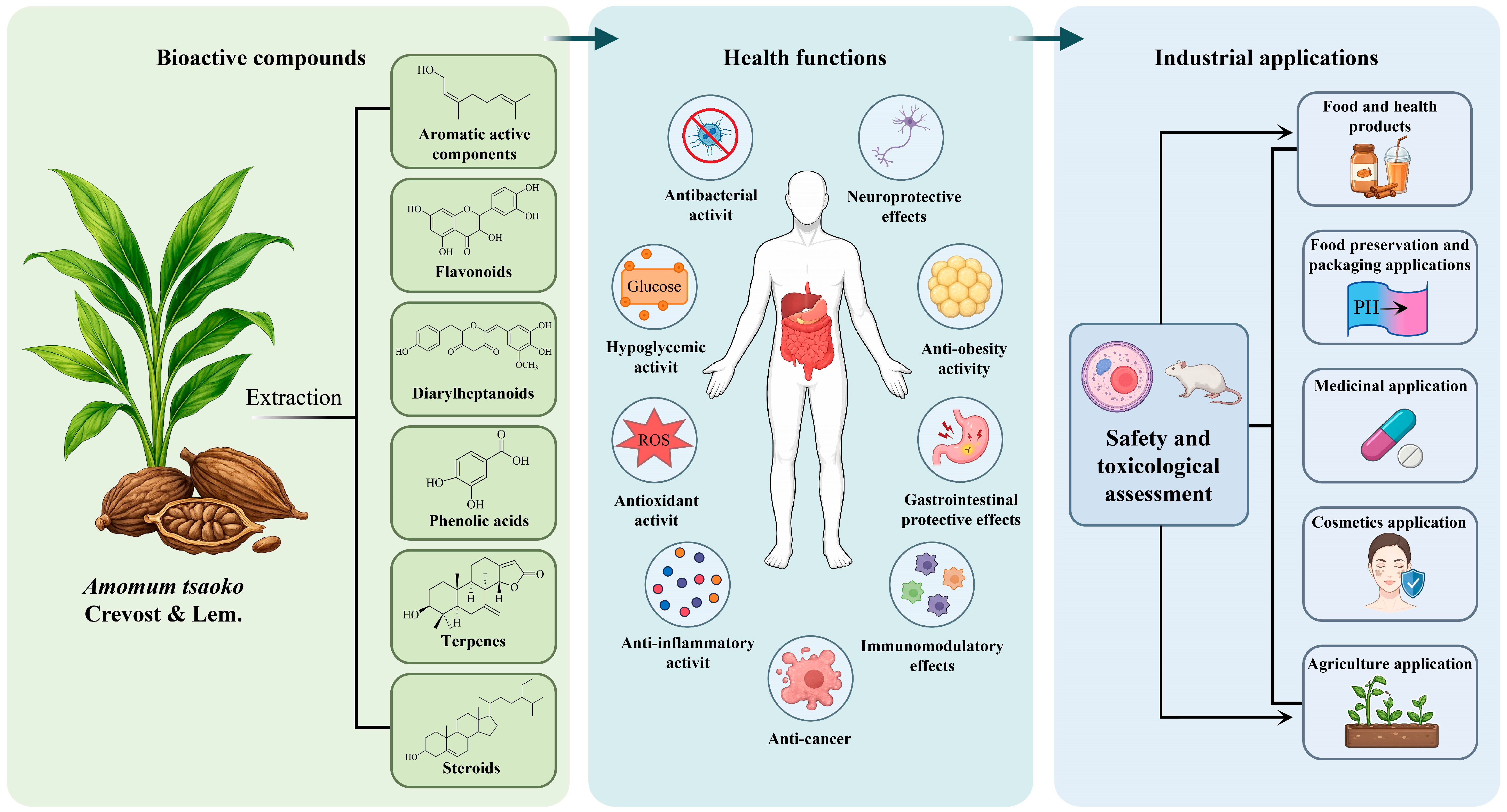
Graphical Abstract

1. Introduction

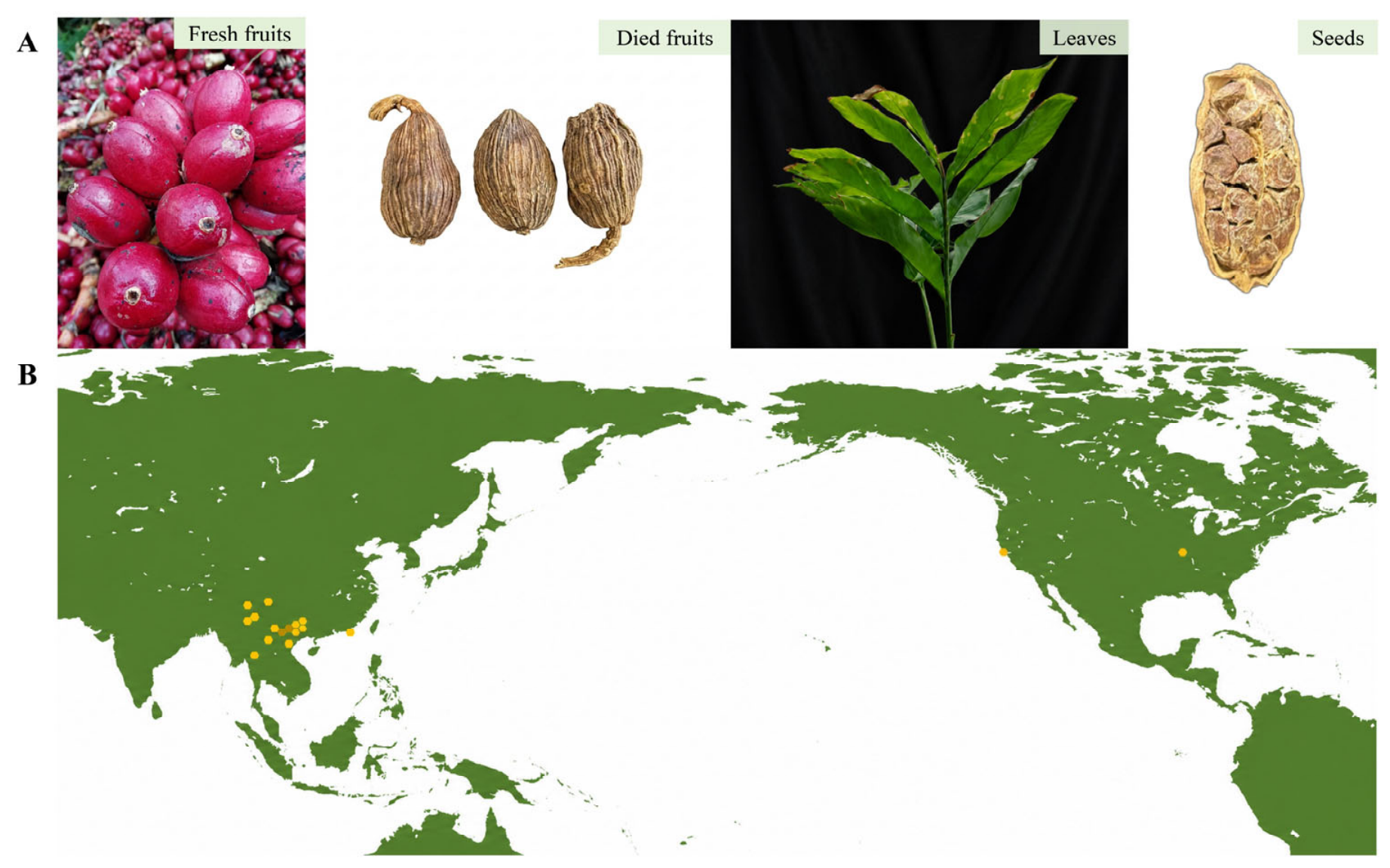
Amomum tsaoko Crevost & Lemarié (AT), a perennial species belonging to the genus Amomum (Zingiberaceae), is predominantly distributed across Southwest China, northern Vietnam, and other parts of Asia [1] (Figure 1). China is the primary production region for AT, with Yunnan Province accounting for more than 95% of the national cultivation area [2]. The dried ripe fruits of AT have long been valued for their dual roles as both a culinary spice and a medicinal resource. In Chinese cuisine, the fruits are commonly added to meat dishes, hotpots, and soups to enhance flavor and mask undesirable odors, an effect primarily attributed to their rich volatile constituents [3]. In traditional Chinese medicine, AT has been extensively used to treat disorders associated with cold–dampness affecting the spleen and stomach, as well as infectious diseases such as malaria [4]. Its medicinal use can be traced back to the Song Dynasty, where it was recorded in the classical medical text Taiping Huimin Heji Jufang [5]. Furthermore, AT is officially listed in the Pharmacopoeia of the People’s Republic of China 2025, which records various prescriptions containing this species [6].
With advances in natural product chemistry and functional food research, increasing attention has been directed toward the phytochemical composition and biological activities of AT. To date, numerous bioactive constituents have been isolated and characterized from AT, including phenolics, flavonoids, diarylheptanoids, bicyclic nonanes, and steroids. These compounds exert diverse pharmacological and health-promoting effects through multiple molecular targets and complex mechanisms, including the regulation of gastrointestinal function, antimicrobial and anti-inflammatory activities, anticancer effects, and neuroprotection. These characteristics are closely linked to the health benefits of AT [7]. Nevertheless, current research has largely focused on the isolation and bioactivity assessment of constituents derived from the fruits and essential oils, whereas comprehensive investigations of other plant tissues and non-volatile constituents remain scarce. In parallel, growing interest has been directed toward the utilization of AT in the food industry, particularly as a natural preservative, flavoring agent, and functional ingredient. Despite these promising applications, challenges related to stability, standardization, and industrial-scale implementation remain unresolved. Furthermore, the expanding use of AT has highlighted the importance of comprehensive safety evaluation. Although AT has long been regarded as safe in traditional use, there remains a lack of comprehensive evaluation of its safety and toxicological profile.
Therefore, this review aims to provide a systematic summary of the bioactive compounds, health functions, application potential, and safety profile of AT. Based on this summary, we discuss the limitations of current research and outline future directions, with the goal of providing guidance for its further development in food and health-related fields.

2. Methodology

This review comprehensively summarizes the current knowledge regarding the phytochemical constituents, pharmacological activities, structure–activity relationships, industrial applications, and safety of AT based on a systematic literature search. Relevant literature was primarily retrieved from the following major international databases: Web of Science, PubMed, ScienceDirect, Google Scholar, Scopus, and CNKI. The search covered publications from database inception to March 2026, with particular emphasis on studies published after 2020. Boolean operators (AND/OR) were combined with targeted keywords, including: “Amomum tsaoko”, “Chemical composition”, “essential oil”, “volatile compounds”, “bioactive compounds”, “pharmacological activity”, “aromatic components”, “diarylheptanoids”, “flavonoids”, “structure-function relationship”, “application”, “toxicity”, and “safety”. Priority was given to original research articles published in peer-reviewed journals, while authoritative review articles were consulted to provide background information and identify additional relevant studies. Patent literature was additionally consulted to support the discussion of industrial applications and technological developments. Master’s and doctoral theses were considered for inclusion only when they reported original experimental findings unavailable in the peer-reviewed literature and provided essential evidence relevant to specific topics discussed in this review. Studies lacking sufficient experimental evidence, non-peer-reviewed conference abstracts, and non-English publications with limited accessibility were excluded. The titles and abstracts of the retrieved records were initially screened for relevance. Subsequently, the full texts of potentially eligible publications were assessed according to the predefined inclusion and exclusion criteria, and the selected studies were categorized according to the major topics of this review, including phytochemistry, pharmacological activities, structure–activity relationships, industrial applications, and safety.

3. Chemicals

3.1. Nutrients

As a popular spice in food processing, AT also possesses certain nutritional value. FAT has been reported to contain eight saturated fatty acids, with palmitic acid (125–181 mg/kg) being the most abundant. Furthermore, five unsaturated fatty acids have also been identified in FAT, with oleic acid (120–181 mg/kg) being the most abundant, followed by linoleic acid (54.3–113 mg/kg) and linolenic acid (18.7–45.3 mg/kg) [8]. Notably, as drying progresses, the content of most fatty acids initially decreases before subsequently increasing, reaching a relatively high concentration after 24 h of oven drying at 60 °C. This trend is likely attributable to the accumulation of fatty acids caused by progressive moisture loss during drying [8]. In addition to lipid components, AT also contains polysaccharides. Crude polysaccharides (ATP) were isolated from AT via hot water extraction and ethanol precipitation, with a yield of 5.22% (dry weight basis). Subsequent purification resulted in the isolation of ATP-4, a novel acidic heteropolysaccharide with immunomodulatory activity. Its monosaccharide composition includes rhamnose (Rha), galacturonic acid (GalA), glucose (Glc), galactose, xylose (Xyl), and arabinose, with a molar ratio of 9.03:52.37:7.89:12.34:6.47:11.89 and a molecular weight of 4.23 × 10⁴ Da. Structural characterization revealed that the backbone of ATP-4 is predominantly composed of 1,4-linked GalpA (47.87%), displaying characteristic homogalacturonan regions interspersed with rhamnogalacturonan domains (1,2-linked-Rhap and 1,2,4-linked-Rhap). The presence of 1,3-linked-Glcp, T-Glcp, and 1,4-linked-Xylp linkages further differentiates ATP-4 from conventional pectic polysaccharide structures. Furthermore, the ATP-4 degradation product AO-2 and the modified polysaccharide ATP-4e have demonstrated enhanced immunomodulatory activity [9].

3.2. Bioactive Compounds

Recent advances in analytical methodologies and structural characterization techniques have greatly facilitated the systematic investigation of secondary metabolites in AT, thereby broadening their potential applications. To date, at least 493 structurally diverse compounds have been identified from AT [7], including flavonoids, diphthalides, phenolic acids, terpenes, steroids, and aroma components. Detailed information on these compounds is provided in Table S1.

3.2.1. Aromatic Active Components

AT is characterized by its distinctive aromatic and pungent properties, which are primarily attributed to its volatile constituents (essential oils). A large number of aromatic components include monoterpenes, oxygenated monoterpenes, sesquiterpenes, oxygenated sesquiterpenes, esters, aldehydes, and alcohols. Among these, aldehydes represent the most abundant class of aromatic components (2607 to 3848 mg/kg) and significantly contribute to the overall aromatic profile [8]. Numerous studies have demonstrated that various factors, including storage duration, harvest season, extraction method, geographical distribution, and drying method, influence the composition and abundance of aromatic compounds in AT. For instance, during natural drying, while the number of aromatic compounds remains constant, their concentrations undergo dynamic changes [10]. In contrast, oven drying at 50 °C significantly reduces drying time while preventing mold growth and fruit cracking, effectively retaining high levels of essential oils and volatile compounds in AT [11]. In terms of controllability and overall aroma acceptance, oven drying offers distinct advantages and is therefore considered more suitable for industrial processing [12]. In addition, the length of AT fruits and seeds is positively correlated with both aroma intensity and biological activity [13]. It has been reported that the differences in aroma profiles between FAT and DAT are mainly attributed to variations in the concentrations of aldehydes and terpenes. Specifically, drying leads to a significant reduction in aldehyde levels, while terpene levels increase. Consequently, DAT generally exhibits a stronger fatty and spicy aroma, making it a popular spice [14]. In terms of extraction techniques, Cui et al. employed a green and efficient modified solvent-free microwave extraction (M-SFME) method for the extraction of AEO. Under optimal conditions, the yield reached 1.13%, significantly surpassing that achieved by conventional steam distillation (0.84%) and conventional SFME (0.91%). Furthermore, M-SFME offers advantages such as a short extraction time, low energy consumption, and minimal environmental impact, making it a promising alternative technology for the extraction of essential oils from AT and other aromatic plants [15]. Another study utilized two methods, hydrodistillation (HD) and solvent extraction combined with solvent-assisted flavor evaporation (SE-SAFE), to isolate the volatile components of AT cobs to evaluate the influence of extraction techniques on key odor-active compounds. The results indicated that most aromatic active compounds showed higher flavor dilution factors in the SE-SAFE method [16].

3.2.2. Flavonoids

ATF is a rich source of flavonoids, and its methanolic extract has been reported to contain substantial amounts of flavonoids (1.21 mg QE per g DW) [4]. The major flavonoids identified in AT include quercetin (73), catechin (69), and epicatechin (68), all of which exhibit diverse biological activities. For instance, quercetin (73) has been reported to exhibit potent neuroprotective and antioxidant effects, while epicatechin demonstrates excellent anti-inflammatory activity [17]. Further analysis revealed that flavonoids are the primary phenolic compounds present in the ethyl acetate fraction of the AT ethanol extract [18]. In recent years, a series of structurally novel flavanol hybrids have also been discovered in AT, including flavanol–menthane conjugates, flavanol–fatty alcohol hybrids, and flavanol–monoterpenoid hybrids, which are believed to have potential applications in diabetes treatment [19,20,21]. Recent research indicates that the flavanol–fatty alcohol hybrids Tsaokoflavanols A1–J1 (129–138) exhibit inhibitory activity against HPL and may provide therapeutic benefits for obesity and related metabolic disorders [22]. Furthermore, the ultrasonic-assisted extraction and HPD300 macroporous resin purification process, optimized through one-factor experiments combined with response surface methodology, significantly improved the extraction efficiency of flavonoids from AT, achieving a yield of 3.33%, which is markedly higher than that reported in previous methods. During the purification process, flavonoids eluted with 20% and 30% ethanol exhibited high flavonoid purity (>90%) and remarkable antioxidant activity, surpassing that of vitamin C (Vc). Further metabolomic analysis revealed that the major components of this flavonoid-enriched fraction include epicatechin, isoquercitrin, astragalin, kaempferol-3-O-rutinoside, and procyanidin B2. These constituents were collectively responsible for the excellent in vitro antioxidant and α-glucosidase inhibitory activities of the ATEE [23]. To maximize the retention of flavonoids, it is essential to select appropriate extraction and purification methods and to optimize the process conditions accordingly.

3.2.3. Diarylheptanoids

Diarylheptanoids are a class of natural products characterized by a distinctive structural motif, consisting of a seven-carbon aliphatic chain connecting two aromatic rings at the 1- and 7-positions. These substances are considered possible components for nutritional supplements and pharmaceuticals due to their varied biological activities, which include anti-inflammatory, antitumor, antioxidant, and neuroprotective properties [24]. Clinical studies have demonstrated that this class of compounds (curcumin) exhibits a favorable safety profile at daily doses of up to 12 g, although their systemic bioavailability remains relatively limited [25]. Notably, diarylheptanoids are considered characteristic constituents of AT [26]. In AT, diarylheptanoids are predominantly found in the fruits and leaves, among which linear diarylheptanoids represent the predominant structural type. Representative compounds include tsaokoarylone (144) and hannokinol (174) [26]. Linear diarylheptanoids accumulate predominantly in mature fruits, with concentrations peaking in fruits harvested in October and November, suggesting that their biosynthesis and accumulation are closely associated with fruit developmental stages. Furthermore, through co-expression network analysis and phylogenetic analysis, several candidate genes potentially involved in diarylheptanoid accumulation were identified, among which two key genes, AmT044854 and AmT007727, were verified to participate in the biosynthesis of linear diarylheptanoids. These findings suggest that the accumulation of diarylheptanoids in AT is developmentally regulated, providing molecular evidence for their potential as quality markers and functional components [26]. Nevertheless, further studies are required to ascertain the exact concentrations of these compounds and their biological effects.

3.2.4. Phenolic Acids

As shown in Table S1, phenolic acids are predominantly distributed in ATF, but they have also been detected in the seeds, including vanillic acid (209), 3-O-methylgallic acid (204), 3,4-dihydroxybenzoic acid (206), and 3,4-dimethoxybenzoic acid (203). These phenolic acids are well known for their antioxidant and anti-inflammatory activities [27]. Emerging evidence suggests that the phenolic acids identified in AT exhibit potential antidiabetic, anti-obesity, and anti-Alzheimer’s effects [18,28,29], highlighting their potential applications in the prevention and management of chronic metabolic and neurodegenerative disorders. Furthermore, these compounds can serve as precursors for various bioactive substances, holding significant development value in the food industry, pharmaceuticals, cosmetics, and related engineering fields [30]. Regarding extraction methods, since phenolic acids in plant tissues often form relatively stable complexes with proteins or polysaccharides, the extraction efficiency of single-solvent systems is typically limited. In contrast, mixed systems of organic solvents and water have been demonstrated to improve the extraction efficiency of phenolic acids from AT, thereby facilitating subsequent isolation, characterization, and bioactivity evaluation [31].

3.2.5. Terpenes and Steroids

Terpenoids identified in AT mainly comprise monoterpenes, sesquiterpenes, diterpenes, and bicyclononane derivatives, most of which have been isolated from its fruits. Among these, monoterpenes and sesquiterpenes are considered key components of AEO and fragrance. Notably, certain oxygen-containing monoterpenes, namely 4-indanecarbaldehyde (271), 5-indanecarbaldehyde (270), and trans- and cis-2,3,3a,7a-tetrahydro-1H-indene-4-carbaldehyde (256), have thus far been reported exclusively in AT samples originating from China and Vietnam and have not been identified in other species. These compounds may therefore serve as valuable chemotaxonomic markers for AT, independent of the region of origin [32]. Wang et al. were the first to identify isospongian diterpenoids in the leaves of AT; these compounds exhibit a variety of health benefits, including anticancer, anti-inflammatory, antiviral, antifungal, and antihypertensive effects [33]. Furthermore, bicyclononane is a unique chemical compound found in AT; this class of compounds forms a bicyclic core utilizing hexane and pentane rings as their basic skeletons [7]. The bicyclononane compounds isolated from ATF are also important bioactive substances.
Phytosterols are a class of bioactive compounds widely found in plants and are recognized for their beneficial effects on lipid metabolism and cardiovascular health [34]. Sitosterol (274) and daucosterol (275), isolated from ATF, are representative phytosterols. Among them, sitosterol has been extensively studied for its diverse health-promoting properties, including cardioprotective, antitumor, and glucose-regulating activities [35]. Additionally, sitosterol serves as an important precursor for the in vitro synthesis of vitamin D analogs, such as D2, D4, and D5 [36]. Likewise, accumulating evidence has demonstrated the antitumor, neuroprotective, and hypoglycemic effects of daucosterol [37]. Collectively, these compounds contribute to the health-promoting potential of AT and support its application in functional foods, nutraceuticals, and pharmaceutical products.

3.2.6. Others

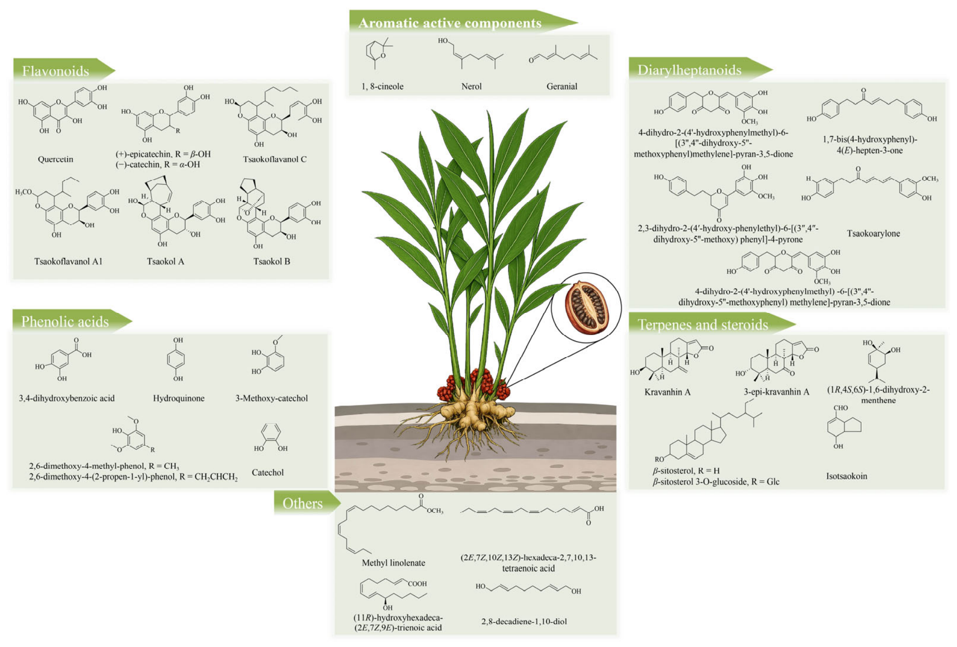
Beyond the bioactive constituents discussed above, other types of compounds have been isolated from AT. These compounds encompass fatty acids, fatty aldehydes, fatty alcohols, aliphatic esters, pyrroles, and phenylethanoid glycosides. Among these constituents, the fatty acids (2E,7Z,10Z,13Z)-hexadeca-2,7,10,13-tetraenoic acid (288) and (2E,7Z)-tetradeca-2,7-dienoic acid (289), isolated from ATF, have demonstrated significant antibacterial, anti-obesity, and antioxidant activities [38]. Furthermore, the fatty acid (11R)-hydroxyhexadeca-(2E,7Z,9E)-trienoic acid (290), obtained from the ethanol extract of AT seeds, has been shown to inhibit sphingosine phosphatase SPHK1 or SPHK2 [28], suggesting its potential involvement in regulating lipid metabolism and associated signaling pathways. The structures of the representative compounds are shown in Figure 2.

4. Health Functions

Numerous studies have demonstrated that AT is rich in bioactive compounds that offer a broad spectrum of health benefits, including antibacterial, hypoglycemic, antioxidant, anti-inflammatory, anticancer, neuroprotective, anti-obesity, gastroprotective, and immunomodulatory effects, which will be briefly discussed below. The potential health benefits and bioactivities of AT are presented in Table 1.

4.1. Antibacterial Activity

In vitro studies have demonstrated that AT exhibits broad-spectrum antibacterial and antifungal activities. Its antibacterial activity is thought to be primarily attributed to AEO, which successfully inhibits both Gram-positive and Gram-negative bacteria as well as various fungi [39]. Among the tested microorganisms, AEO exhibits potent activity against Staphylococcus aureus, with a MIC of 0.20 mg/mL and an MBC ranging from 0.3–0.78 mg/mL [40], significantly outperforming other essential oils such as Cinnamomum cassia essential oil (2.5 mg/mL) [41] and Zanthoxylum schinifolium essential oil (10 mg/mL) [42]. Furthermore, 1,8-cineole, (E)-dec-2-enal, citral, α-pinene, and α-terpineol are considered the major contributors to the antimicrobial activity of AEO [40]. Mechanistic studies further demonstrated that AEO exerts rapid antibacterial effects against the foodborne bacterium Escherichia coli by disrupting membrane integrity and permeability, resulting in the leakage of intracellular nucleic acids and proteins [43]. In a mouse infection model, AEO protected animals from infections caused by Staphylococcus aureus or Escherichia coli in vivo, highlighting its potential for the management of bacterial infections, particularly those caused by multidrug-resistant pathogens [44]. It is worth noting that antimicrobial efficacy varies considerably for AEO obtained through different extraction methods. Compared to HD and SMFE extracts, the essential oil obtained via M-SFME exhibits stronger antimicrobial and antioxidant activities, primarily attributed to its higher proportion of oxygenated monoterpenes (55.8%) [15]. Furthermore, in vitro studies have also demonstrated that AEO exhibits pronounced antifungal activity against such fungi as Botrytis cinerea, Aspergillus oryzae, Mucor, and Penicillium [45].
In addition to AEO, ATE also exhibits notable antibacterial activity in vitro. Studies have shown that ATE inhibits bacterial growth by suppressing cellular respiration, mainly through disruption of the Krebs cycle during glucose metabolism [46]. Furthermore, by damaging bacterial cell wall integrity and morphology, it effectively inhibits Listeria monocytogenes and Bacillus subtilis, with MIC and MBC values of 1.25 mg/mL for both [47]. Beyond its direct antibacterial effects, ATE markedly suppresses biofilm formation, virulence factor production, and motility in various foodborne pathogens (such as Staphylococcus aureus, Salmonella typhimurium, and Pseudomonas aeruginosa) by interfering with bacterial quorum-sensing systems [48]. This demonstrates its potential value as a natural quorum-sensing inhibitor and anti-biofilm agent in controlling food spoilage and preventing bacterial infections. Interestingly, ATE exhibited remarkable stability under diverse environmental conditions. Its antibacterial activity showed high tolerance to changes in salt concentration, temperature, ultraviolet light, and pH, and exposure to salt, strong acids, and strong bases moderately enhanced its antibacterial activity. However, its antibacterial activity is relatively sensitive to high-sugar environments, where high-sugar environments reduce its antibacterial efficacy [49]. Antibacterial activity also varies considerably among extracts obtained using different solvents, with ethyl acetate extracts exhibiting the strongest antibacterial activity. For example, 95% ethanol and ethyl acetate extracts of ATF demonstrated excellent inhibitory activity against Klebsiella pneumoniae [38]. In addition, bicyclic nonane isotsaokoin (262), isolated from an ATFME, exhibited antifungal activity against Trichophyton mentagrophytes [50]. Nevertheless, current research primarily focuses on in vitro antimicrobial evaluations and preliminary investigations into mechanisms of action. Future studies should focus on identifying the key active components and clarifying their stability and safety in complex food systems or physiological environments to advance their practical application in food preservation and health-related fields.

4.2. Hypoglycemic Activity

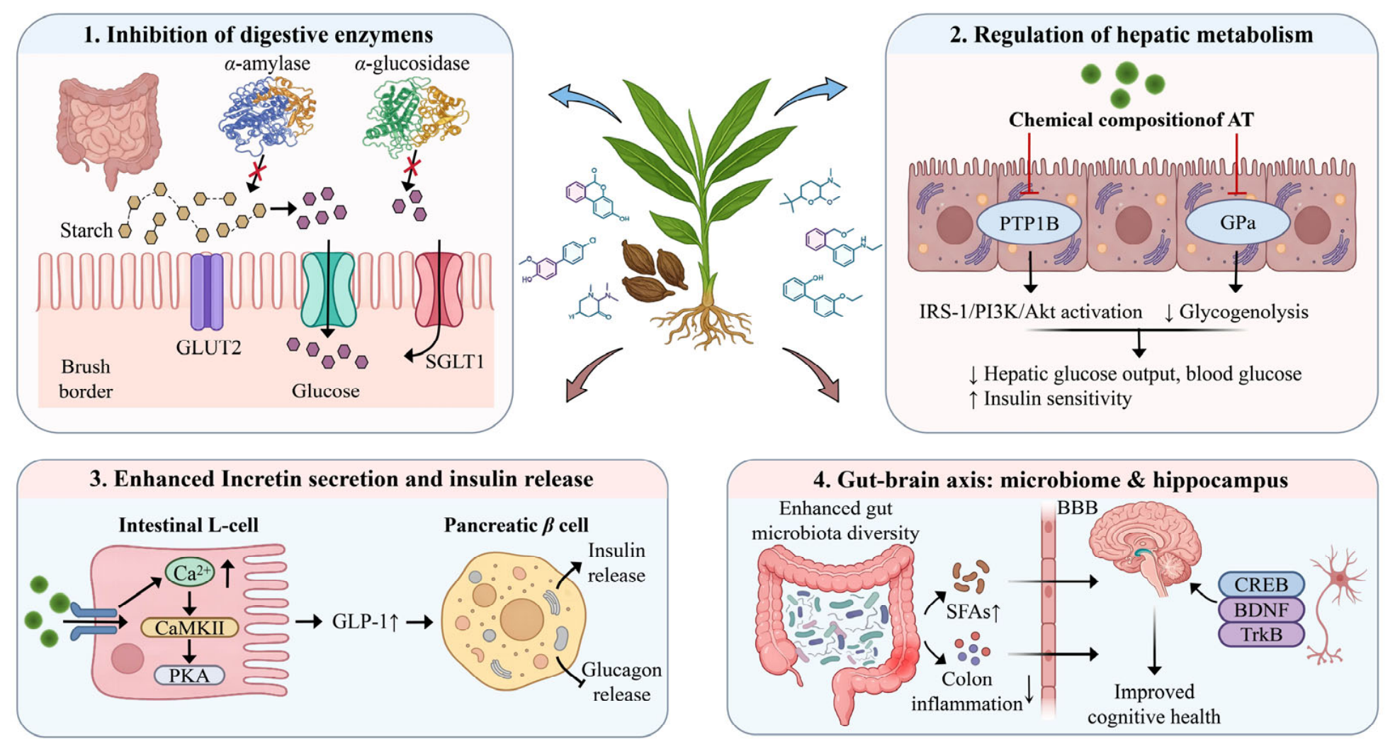
Diabetes is a major metabolic disorder associated with severe complications [51]. Many dietary natural products play an important role in the prevention and management of diabetes. As shown in Figure 3, AT has shown considerable potential for glycemic control and glucose metabolism regulation. In vitro studies showed that the ATME exhibits potent α-glucosidase inhibitory activity, with an IC50 value of 0.145 mg/mL, exceeding that of the clinical α-glucosidase inhibitor acarbose (IC50, 0.273 mg/mL) [52]. Similarly, both the AT water extract (seeds and pericarp) and the 50% ethanol–water extract exhibited significant inhibitory effects on α-glucosidase [53], while the water extract (seeds and pericarp) also effectively inhibited α-amylase activity [54]. Additionally, the extraction process using solvents of varying polarities markedly affects the antidiabetic activity of AT extracts. Fan et al. found that EF and BF exhibited superior α-glucosidase inhibitory activity compared to other fractions, with IC50 values of 20.14 ± 0.78 and 18.30 ± 0.42 μg/mL, respectively [18]. Evidence from in vivo studies further supports the antidiabetic potential of AT. In high-fat diet-induced diabetic mouse models, both the ATEE and ATME exhibited significant antidiabetic effects [4,18,52]. Mechanistic studies revealed that ATEE significantly reversed glucose metabolism disorders and cognitive deficits in mice with T2DM. These effects were associated with modulation of the gut microbiota, increased SCFA production, reduced inflammation in the hippocampus and colon, and activation of the CREB/BDNF/TrkB pathway [55]. These effects may be attributed to the phenolic compounds in AT, which may exert independent or synergistic actions on the aforementioned activities [54].
Bioactivity-guided fractionation and in vitro screening have identified numerous bioactive constituents responsible for the antidiabetic activity of AT. Several diarylheptanoids, including Tsaokopyranols A–M (184–196), exhibited potent α-glucosidase inhibitory activity, with IC50 values ranging from 59.4 to 116.5 μM, all showing stronger activity than acarbose (IC50 = 219.0 μM) [53]. Among these, Amomutsaokols H (191) and J (193) act as non-competitive α-glucosidase inhibitors, with Ki values of 18.5 and 213.0 μM, respectively [56]. Notably, 2-hydroxymusaitinerin A (169), a diarylheptanoid isolated from AT leaves, was identified as a novel inhibitor of GPa, PTP1B, and α-glucosidase, while platyphyllone (179) exhibited mixed-mode inhibition on α-glucosidase through both non-competitive and anti-competitive mechanisms [57]. Moreover, several flavonoids have also demonstrated promising antidiabetic activity in vitro. Among them, Tsaokols A (140) and B (139) showed notable α-glucosidase inhibitory activity, which was significantly stronger than acarbose [21]. Amomutsaokins A (101) and F (106) were identified as mixed-type α-glucosidase inhibitors, with Ki values of 36.2 and 24.4 μM, respectively [19]. Tsaokoflavanols A (110), B (111), F (115), and K (120) displayed dual inhibitory activity against α-glucosidase and PTP1B [20]. In addition, diterpenoids have also shown antidiabetic potential. Kravanhin A (259) and 3-epi-kravanhin A (258), isolated from AT leaves, significantly enhanced GLP-1 secretion in STC-1 cells via the Ca2+/CaMKII and PKA pathways [33]. Although AT extracts lower blood glucose in animal models, most isolated compounds have only been tested in enzyme- and cell-based assays. Future studies should validate their in vivo efficacy and evaluate pharmacokinetics and bioavailability.

4.3. Antioxidant Activity

Oxidative stress is associated with various pathological conditions, including cardiovascular disease, neurodegenerative diseases, cancer, and aging [58]. Therefore, antioxidant activity is essential for maintaining physiological homeostasis and can be enhanced by dietary antioxidants [59]. In in vitro experiments, the ATFME exhibited strong radical-scavenging activity against DPPH. (IC50: 0.044 mg/mL) and ABTS+ (IC50: 0.040 mg/mL), indicating potent antioxidant activity [52]. Further studies suggest that the antioxidant constituents in AT are mainly concentrated in the polar fractions, particularly the methanol extract [60,61]. Different polar fractions of AT also exhibit antioxidant activity in vitro, although their relative effectiveness depends on the assay employed. Specifically, the EF showed the strongest DPPH. scavenging activity, while the BF exhibited the greatest superoxide radical-scavenging and ferric-reducing capacities [62]. Similarly, the ethyl acetate fraction exhibited potent antioxidant activity with IC50 values of 0.17 and 0.07 mg/mL for DPPH. and ABTS+, respectively, and a FRAP value of 546.10 mg VCE/g DW. This activity may be associated with the presence of phenolic hydroxyl groups or the synergistic effects of phenolic and flavonoid compounds [18]. Evidence from animal studies further supports the antioxidant potential of AT. Administration of ATFME significantly elevated SOD, GSH, and GSH-Px levels while reducing MDA and 8-ISO-PGF2α [52].
Chemical free radical-scavenging assays were used to further identify a number of antioxidant components in AT. Studies have demonstrated that AT polyphenols exhibit strong DPPH. (IC50 = 42.46 μg/mL) and ABTS+ (IC50 = 85.47 μg/mL) scavenging activities, with antioxidant activity positively correlated with polyphenol content [63]. Further studies showed that the flavonoid quercetin (73) (inhibition rate > 80%), the phenolic acid 3,4-dihydroxybenzoic acid (206) (inhibition rate > 90%), and the diarylheptanoid CG-B (142) (inhibition rate > 79%) also exhibited strong DPPH scavenging activity at a concentration of 100 μg/mL, comparable to that of Vc at the same concentration [17,38,64]. Current antioxidant evidence relies largely on chemical radical-scavenging assays, which do not fully predict biological antioxidant efficacy. Future studies should assess whether these properties translate into physiological benefits in vivo.

4.4. Anti-Inflammatory Activity

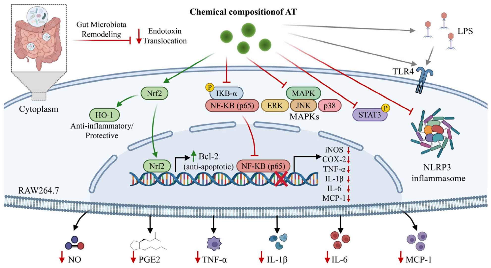
Increasing evidence has highlighted the anti-inflammatory potential of AT extracts and their bioactive constituents. As shown in Figure 4, AT and its active components have demonstrated significant anti-inflammatory effects in both in vitro and in vivo studies. At the cellular level, the ATEE suppresses inflammatory responses in LPS-activated macrophages via Nrf2-dependent HO-1 expression and inhibition of NF-κB signaling [65]. Likewise, ATFME markedly inhibits NO production in LPS-stimulated BV2 microglia [66]. Mechanistic studies further showed that the ATFME reduces inflammation by inhibiting NO generation through the stimulation of the ROS/MAPK/Nrf2-mediated HO-1 signaling pathway [67]. In addition, AEO exhibited potent anti-inflammatory activity in LPS-stimulated macrophages. At non-cytotoxic concentrations (0–20 μg/mL), AEO reduced NO production and decreased the expression of TNF-α, IL-1β, IL-6, and MCP-1 in a dose-dependent manner. Concurrently, AEO effectively suppressed the overexpression of iNOS and COX-2 while reducing the levels of NF-κBp-p65 and MAPKp-ERK, suggesting that its anti-inflammatory activity is mediated by the inhibition of the NF-κB and MAPK signaling pathways [68]. Notably, evidence from animal studies further supports the anti-inflammatory potential of AT. In an LPS-induced mouse model of neuroinflammation, AEO improved cognitive performance and preserved neuronal integrity, upregulated the expression of the anti-apoptotic protein Bcl-2, and effectively alleviated the neuroinflammatory response. Multi-omics analyses further suggested involvement in the regulation of the MAPK signaling pathway, as well as the coordinated regulation of multiple metabolic pathways, including histidine metabolism, pantothenic acid and coenzyme A biosynthesis, and phenylalanine metabolism [69]. Recent studies suggest that AT alleviates ulcerative colitis through inhibiting STAT3 phosphorylation and suppressing necrotic apoptosis [70]. Additionally, AT flavonoids may ameliorate colitis by reshaping the gut microbiota, reducing endotoxin translocation, and thereby inhibiting the downstream TLR4/NF-κB/NLRP3 inflammatory signaling pathways [71].
Several anti-inflammatory compounds have now been identified in AT, with most of the evidence to date coming from cell-based experiments. In LPS-stimulated RAW 264.7 macrophages, the fatty acid methyl linolenate (283) inhibited NO production (IC50 = 61.2 μM) [72]. Two diarylheptanoids isolated from ATF, CG-A (141) and CG-B (142), also showed dose-dependent anti-inflammatory effects in vitro [73]. Further studies showed that the fatty alcohol compound 2,8-decadiene-1,10-diol (DDO) (281) suppresses NO and prostaglandin E2 production by downregulating the expression of iNOS and COX-2 and reduces the production of pro-inflammatory cytokines such as IL-6 and TNF-α. These effects are associated with the inactivation of MAPK signaling pathways, including ERK, JNK, and p38 MAPK, together with suppression of NF-κB signaling (including IκB-α degradation and NF-κB nuclear localization) [74]. Similarly, the flavonoids (+)-epicatechin (68) and (−)-catechin (69), as well as the terpenoid compound (1R,4S,6S)-1,6-dihydroxy-2-menthene (254), effectively reduced NO production in LPS-stimulated RAW 264.7 cells through inhibition of iNOS expression and inflammatory cytokine production. Among these, (+)-epicatechin (68) and (−)-catechin (69) exert anti-inflammatory effects mainly through inhibition of NF-κB nuclear translocation [75,76]. In summary, various extracts, essential oils, and isolated compounds from AT have demonstrated significant anti-inflammatory activity across multiple inflammatory models, primarily through modulation of oxidative stress and suppression of inflammatory signaling pathways.

4.5. Anticancer Activity

Cancer remains a major global health challenge. Natural products have attracted considerable attention as sources of anticancer agents because of their accessibility, suitability, and low cytotoxicity, and have been widely explored for cancer prevention and treatment [77]. Current evidence suggests that AT possesses promising antitumor activity in both in vitro and in vivo models. The AEO exhibits significant cytotoxicity against several human cancer cell lines, including HepG2, HeLa, and Bel-7402, in vitro. The strongest effect was observed against HepG2 cells (IC50 = 31.80 µg/mL), while its toxicity to normal cells, such as HUVEC and HL-7702, was low, suggesting selective anticancer activity. Mechanistically, AEO exerts its antitumor effects through apoptosis induction [78]. In addition to AEO, the ATEE also exhibits broad-spectrum antitumor activity. Studies have shown that the 95% ethanol extract and its ethyl acetate fraction exhibit potent cytotoxicity toward cervical cancer cells (HeLa), liver cancer cells (HepG-2 and SMMC-7721), and lung cancer cells (A549) [64]. Both cell-based and animal studies have further demonstrated that the ATEE suppresses ovarian tumor growth and reduces tumor-associated angiogenesis. Mechanistic analysis indicates that the p-STAT3/NF-κB signaling pathway forms a positive feedback circuit that promotes IL-6 and VEGF expression, while IL-6 and VEGF further activate this pathway, promoting tumor progression. The ATEE disrupts this cascade amplification effect by inducing endoplasmic reticulum stress, thereby suppressing angiogenesis and tumor progression [79].
In AT, several small-molecule constituents have shown cytotoxic and antiproliferative activities. Multiple fatty acids and diarylheptanoids isolated from ATF exhibited antiproliferative activity in the murine neuroblastoma cell line N2a. Among these compounds, tsaokoarylone (144) showed the strongest activity [80]. Additionally, hannokinol (174) and the flavonoid CG-B (142), isolated from the EF, showed significant cytotoxic action against HepG-2, SMMC-7721, HeLa, and A549 cell lines. CG-B was particularly active against SMMC-7721 cells (IC50 = 44.66 µg/mL); this activity even surpassed that of the positive control 5-fluorouracil [64]. At the same time, the flavanol hybrid tsaokoflavanol C (112) induced apoptosis and inhibited the proliferation of HepG2 cells [22]. However, current research is primarily conducted on various tumour cell lines in vitro. Future investigations should evaluate whether these cytotoxic effects can be reproduced in animal tumor models while minimizing toxicity toward normal tissues.

4.6. Neuroprotective Effects

Numerous studies have demonstrated that AT exhibits broad neuroprotective effects across multiple neurological disease models through multi-target mechanisms. Compared to other spices such as cinnamon, cardamom, black pepper, and white pepper, AT exhibited stronger anti-Alzheimer’s disease (AD) activity. Specifically, AEO demonstrated significant acetylcholinesterase inhibitory activity and improved behavioral deficits in an AlCl3-induced zebrafish dementia model, supporting its neuroprotective potential [81]. In a mouse model of pentylenetetrazol (PTZ)-induced seizures, the ethanol extract of ATF exerted antiepileptic and neuroprotective effects through antioxidant, anti-inflammatory, neurotransmitter-regulating, and energy metabolism-modulating activities [82]. In a mouse model of diabetes-induced depression, ATEE improved metabolic disorders and depressive-like behaviors, increased neuroendocrine hormone secretion, modulated microglial activation, and promoted neurogenesis. It also inhibited the expression of inflammation-related proteins such as HMGB1, TLR4, and NF-κB, thereby alleviating diabetes-associated depression, suggesting that the HMGB1/TLR4/NF-κB signaling axis may be a key mechanism underlying its effects [83]. Furthermore, in a mouse model of Parkinson’s disease induced by low-dose rotenone, AT flavonoids alleviated motor dysfunction and constipation, protected dopaminergic neurons, reduced inflammatory responses, and enhanced intestinal barrier function. Notably, their anti-Parkinson’s mechanism may also be associated with the regulation of gut microbiota [84].
Cell-based studies have indicated that the representative flavonoid quercetin showed significant neuroprotective effects against H2O2-induced injury in PC12 cells, significantly improving cell viability at a concentration of 50 μg/mL [17]. Furthermore, diarylheptanoids CG-A (141) and CG-B (142) significantly attenuated H2O2-induced cytotoxicity, with CG-A (141) exhibiting greater neuroprotective activity than CG-B (142), showing activity comparable to that of vitamin C at the tested concentration [73]. Another study reported that compounds isolated from ATF, including tsaokoic acid (264), tsaokoin (263), vanillin (214), and tsaokoarylone (144), have shown potential cognitive-enhancing and anti-Alzheimer’s disease activities [29]. Collectively, available evidence suggests that AT has promising neuroprotective potential, primarily by modulating oxidative stress and neuroinflammatory pathways. Future studies should confirm whether these effects can be replicated in more clinically relevant models and establish the long-term safety of AT supplementation.

4.7. Anti-Obesity Activity

ATE has significant anti-obesity effects. In diet-induced obese mice, the ATEE alleviated obesity-associated dyslipidemia as evidenced by reduced body weight gain, visceral adiposity, and dyslipidemia-related biomarkers [85]. Similarly, studies in ovariectomized mice demonstrated that the ATEE dose-dependently suppressed estrogen deficiency-induced weight gain and significantly reduced abnormal lipid accumulation in adipose tissue, the liver, and bone marrow, suggesting a beneficial role in hormone-related metabolic disorders [86]. Moreover, based on the results of network pharmacology analysis, AT contains several active compounds that are effective for treating obesity, with concentrations being relatively higher in AT harvested in September and October [87]. In addition, in HFD-induced NAFLD mice, AT effectively reduced lipid accumulation, oxidative stress, and inflammation while improving gut microbiota composition and lipid metabolism. Its protective effects are closely associated with the regulation of the gut–liver axis, suggesting potential applications in NAFLD prevention and management [88].
Among the isolated compounds, the flavanol–fatty alcohol hybrids Tsaokoflavanols A1 (129), B1 (130), E1 (133), and F1 (134) isolated from AT exhibited potent HPL inhibitory activity, with IC50 values ranging from 0.017 to 0.193 mM. Notably, Tsaokoflavanol A1 (129) exhibited stronger HPL inhibition than the positive control orlistat (IC50 = 0.067 mM) [22]. Similarly, (2E,7Z)-tetradeca-2,7-dienoic acid (289) and (E)-tetradec-2-enoic acid (287) also showed stronger lipase inhibitory activity than orlistat at a concentration of 50 μg/mL [38]. Furthermore, methyl linolenate (283) and catechol (233), identified from the ATEE via bioactivity-guided fractionation, displayed significant anti-adipogenic activity in the 3T3-L1 preadipocyte model. Both compounds reduced lipid accumulation in a dose-dependent manner and were more effective than other co-isolated constituents [89].
Mechanistically, current evidence suggests that the anti-obesity effects of AT are mediated through multiple complementary mechanisms, including inhibition of dietary lipid digestion via pancreatic lipase, suppression of adipocyte differentiation, attenuation of ectopic lipid accumulation, and modulation of the gut–liver axis. Among these, pancreatic lipase is the only molecular target that has been directly identified for AT extracts. Although methyl linolenate (283) and catechol (233) exhibit anti-adipogenic activity in 3T3-L1 preadipocytes, their effects on key adipogenic regulators, such as peroxisome proliferator-activated receptor γ (PPARγ) and CCAAT/enhancer-binding protein α (C/EBPα), remain unknown. Likewise, no studies have investigated whether AT promotes white adipose tissue browning or enhances lipolysis by regulating thermogenic markers (e.g., UCP1, PRDM16, and PGC-1α) or lipolytic enzymes (e.g., ATGL and HSL). Overall, current evidence supports the anti-obesity effects of AT mainly through reduced lipid digestion, suppression of adipogenesis, and improved lipid metabolism. Future studies should determine whether AT also modulates thermogenesis and lipolysis and identify the underlying molecular targets.

4.8. Gastrointestinal Protective Effects

Traditionally, AT has been used to alleviate gastrointestinal discomfort. Recent studies have provided growing evidence supporting its gastrointestinal benefits. Relevant studies have confirmed that its active components include essential oils, water extracts, and flavonoids. Diets containing AT significantly modulated the intestinal microbiota of zebrafish and tilapia, promoting beneficial microorganisms, suppressing potentially harmful bacteria, and improving gut morphology. These findings suggest that AT can improve gut health by regulating the gut microbiome and intestinal morphology, supporting its application as a natural feed additive in aquaculture [90]. Additionally, AEO dose-dependently alleviated loperamide-induced gastrointestinal hypomotility in rats. Non-targeted metabolomics indicated that AEO normalizes several dysregulated metabolite levels by modulating pathways such as primary bile acid biosynthesis. Microbiome-metabolite correlation analysis further revealed that AEO improved intestinal homeostasis and motility by regulating the gut microbiota structure and metabolic function [91].
Beyond AEO, AT water extracts and their flavonoid compounds have also shown promising laxative activity. In a mouse model of loperamide-induced constipation, medium- and high-dose AT aqueous extract (ATAE) (750 and 1000 mg/kg/day) significantly improved gastrointestinal transit and defecation parameters, confirming its laxative effect [92]. Recent studies have further confirmed that ATAE effectively alleviates loperamide-induced slow-transit constipation, with the bioactivity primarily attributed to the ethanol-soluble fraction of AT (ATES) rather than the ethanol precipitate of ATAE. Flavonoids are the primary active components of ATES, and the flavonoid fraction (ATTF) purified from ATES also exhibits pronounced laxative activity. Mechanistic investigations indicate that ATTF may alleviate constipation by modulating gut microbiota composition (e.g., Lactobacillus and Bacillus), regulating metabolites associated with serotonergic signaling pathways (such as PGJ2 and TXB2), and improving serotonergic synaptic pathway-related factors (such as 5-HT), thereby promoting intestinal motility and maintaining gut homeostasis [93].
Although AT supplementation beneficially modulates gut microbiota composition and improves intestinal homeostasis, its effect on specific pathobionts such as Fusobacterium nucleatum remains unexplored. F. nucleatum is a key intestinal pathobiont linked to gut barrier dysfunction, chronic inflammation, inflammatory bowel disease, and colorectal cancer [94]. AT supplementation has been shown to increase beneficial bacteria, suppress potentially harmful microorganisms, improve gut morphology, and regulate microbial metabolites linked to intestinal function. In addition, AT and its bioactive constituents exhibit broad-spectrum antimicrobial, anti-inflammatory, and barrier-protective activities, which may indirectly create an intestinal microenvironment unfavorable for the colonization or expansion of F. nucleatum. Future studies should assess whether AT alters intestinal F. nucleatum abundance by combining microbiome sequencing with targeted bacterial quantification.

4.9. Immunomodulatory Effects

Both in vitro and in vivo studies have demonstrated that the AT polysaccharide ATP-4 can enhance immune responses through direct activation of immune cells and indirect modulation of the gut microbiota. Subsequent studies revealed that the polysaccharide ATP-4e, generated through structural modification of ATP-4 while preserving the active domain AO-2, exhibits enhanced immunomodulatory activity relative to native ATP-4 [9]. In addition to polysaccharides, the ATSEE and its low-molecular-weight components also exhibit immunomodulatory effects. In vitro studies have shown that the ATSEE can effectively inhibit the activity of sphingosine phosphatase (SPHK1/2). Tsaokol A (140), further isolated from the extract, exhibited strong inhibitory effects on both SPHK1 and SPHK2, with inhibition rates of 59.75% and 25.40%, respectively. Furthermore, 8-hydroxy-2,6-dimethyl-1,6-octadien-3-one (246) exhibited significant inhibitory activity against SPHK2 (inhibition rate of 22.75%), outperforming the positive control [28]. These results suggest that the ATSEE and its bioactive small molecules may participate in immune regulation by modulating sphingosine metabolism-related pathways. Additionally, various phenolic compounds isolated from AT have demonstrated notable anti-complement activity in vitro. Related studies have shown that these compounds primarily exert their effects by inhibiting the classical and alternative pathways of the complement system. Among them, 1,7-bis(4-hydroxyphenyl)-4(E)-hepten-3-one (183) and hydroquinone (232) can interact with multiple key complement components, including C1q, C2, and C3, thereby regulating the complement cascade [95]. In summary, the immunomodulatory effects of AT involve immune cell activation, gut microbiota regulation, modulation of sphingosine metabolism, and inhibition of the complement system. These findings support the potential use of AT in immune-related functional foods or health products. However, its specific molecular mechanisms and in vivo actions require further study.

4.10. Others

Beyond the pharmacological activities described above, AT has also been reported to possess anti-atherosclerotic, antiviral, nephroprotective, anti-Trichomonas vaginalis, cholesterol-lowering, and anti-osteoporotic effects.
AT may exert anti-atherosclerotic effects through the regulation of lipid metabolism, attenuation of inflammation, and modulation of gut microbiota composition [96]. The available evidence indicates that the cardiovascular benefits of AT are currently supported primarily through indirect mechanisms. By improving hyperglycemia, obesity, dyslipidemia, oxidative stress, chronic inflammation, and gut microbiota dysbiosis, AT may reduce multiple cardiometabolic risk factors that contribute to the development of atherosclerosis. However, direct evidence demonstrating cardiovascular protection, such as vascular endothelial protection, improvement of vascular function, or attenuation of myocardial injury, remains limited. Therefore, future studies are warranted to determine whether AT exerts direct cardiovascular effects independent of its metabolic regulatory activities. Furthermore, AEO can interact with the SARS-CoV-2 spike protein RBD in aerosols, thereby preventing RBD–hACE2 binding and potentially interrupting viral transmission, thereby demonstrating significant potential for preventing aerosol transmission of the virus [97]. In a gentamicin-induced rat model, administration of AEO reduced serum urea and creatinine levels in the rats and improved pathological changes and oxidative stress in renal tissue. Concurrently, AEO alleviated inflammatory responses and apoptosis in the rats by regulating the MAPK pathway, indicating a nephroprotective effect of AEO [98]. Additionally, both AEO and geraniol (38) exhibited anti-T. vaginalis activity, which may result from damage to cell membranes and organelles [99]. In another study, AEO and polyphenol extracts reduced cholesterol levels by upregulating hepatic CYP7A1 expression, promoting bile acid excretion, and modulating gut microbiota involved in cholesterol metabolism, such as Ruminococcaceae [100]. Regarding anti-osteoporosis effects, Shim et al. demonstrated that ATEE significantly improved bone microarchitecture, bone mineral density, and bone volume fraction in an ovariectomized mouse model. This effect was primarily attributed to the inhibition of osteoclast differentiation. In vitro experiments confirmed that ATEE effectively inhibits osteoclast formation by downregulating the RANKL-induced NF-κB/Fos/NFATc1 signaling pathway, thereby suppressing bone resorption [86]. Despite the promising pharmacological activities reported for AT, current evidence is mainly based on cell and animal studies. Clinical validation remains limited, and future randomized controlled trials are needed to confirm its efficacy and safety in humans.
Table 1. Health functions and related mechanisms of Amomum tsaoko Crevost & Lem. (“↓”, decrease; “↑”, increase).
Bioactivities/ComponentsAssaysTesting SubjectsEffects/MechanismsReferences
Antibacterial activity
AEOIn vitroAgar disc diffusion assay, MICStaphylococcus aureus CCTCC AB91118 (MIC and MBC = 0.20 g/L)[39]
AEOIn vitroAgar disc diffusion assay, MIC, MBCStaphylococcus aureus (MIC = 0.20 mg/mL, MBC = 0.39–0.78 mg/mL)[40]
AEOIn vitroAgar disk diffusion assay, MIC, MBCEscherichia coli (MIC = 3.13, MBC = 6.25 mg/mL)[43]
AEOIn vitroMICGram-positive and Gram-negative bacteria (MIC = 22.49 to 1438.91 μg/mL)[44]
AEOIn vivoMiceInhibiting Escherichia coli, Staphylococcus aureus[44]
AEOIn vitroMIC, MBCGram-positive and Gram-negative bacteria (MIC = 2.94–5.86 mg/mL)[15]
ATEIn vitroMIC, MBCBacillus subtilis and Listeria monocytogenes (MIC and MBC = 1.25 mg/mL)[47]
ATFMEIn vitroDisk diffusion assay, MICStaphylococcus aureus (MIC = 1 mg/mL); Salmonella typhimurium (MIC = 2 mg/mL); Pseudomonas aeruginosa (MIC = 2 mg/mL)[48]
95% ethanol and ethyl acetate fractionIn vitroKlebsiella pneumoniaeInhibiting Klebsiella pneumoniae[38]
(2E,7Z,10Z,13Z)-hexadeca-2,7,10,13-tetraenoic acidIn vitroKlebsiella pneumoniaeInhibiting Klebsiella pneumoniae (inhibition rate > 99%)[38]
(2E,7Z)-tetradeca-2,7-dienoic acidIn vitroKlebsiella pneumoniaeInhibiting Klebsiella pneumoniae (inhibition rate > 99%)[38]
(E)-tetradec-2-enoic acidIn vitroKlebsiella pneumoniaeInhibiting Klebsiella pneumoniae (inhibition rate > 99%)[38]
(E)-dodec-2-enoic acidIn vitroKlebsiella pneumoniaeInhibiting Klebsiella pneumoniae (inhibition rate > 99%)[38]
CoronadieneIn vitroKlebsiella pneumoniaeInhibiting Klebsiella pneumoniae (inhibition rate > 99%)[38]
Vanillic acidIn vitroKlebsiella pneumoniaeInhibiting Klebsiella pneumoniae (inhibition rate > 99%)[38]
3,4-dihydroxybenzoic acidIn vitroKlebsiella pneumoniaeInhibiting Klebsiella pneumoniae (inhibition rate > 99%)[38]
IsotsaokoinIn vitroAgar disc diffusion assayInhibiting Trichophyton mentagrophytes[50]
AEOIn vitroEscherichia coli, Staphylococcus albus, Bacillus subtilis, Penicillium, Mucor, and Aspergillus oryzaeInhibiting Bacillus subtilis, Staphylococcus albus, and Escherichia coli; inhibiting Aspergillus oryzae, Mucor, and Penicillium[45]
ATEIn vitroStaphylococcus aureusInhibiting bacterial growth, altering cell morphology and cell membrane structure, inhibiting cellular respiration and metabolism[46]
ATEIn vitroMICEscherichia coli and Candida albicans (MIC = 1.5 mg/mL); Bacillus subtilis and Proteus vulgaris (MIC = 3.0 mg/mL); Aspergillus niger (MIC = 6.0 mg/mL).[49]
Hypoglycemic activity
ATMEIn vitroα-glucosidase, α-amylaseInhibiting α-glucosidase and α-amylase[61]
EFIn vitroα-glucosidaseInhibiting α-glucosidase (IC50 = 20.14 ± 0.78 μg/mL)[18]
BFIn vitroα-glucosidaseInhibiting α-glucosidase (IC50 = 18.30 ± 0.42 μg/mL)[18]
EFIn vivoHFD-induced diabetes miceFasting blood glucose ↓, glucose tolerance ↑[18]
ATMEIn vitroα-glucosidaseInhibiting α-glucosidase (IC50 = 0.154 mg/mL)[52]
ATMEIn vivoSTZ-induced diabetic miceFasting blood glucose, area under the curve of the oral glucose tolerance test, HOMA-IR ↓, HOMA-β ↑[52]
50% ethanol–water extractIn vitroα-glucosidaseInhibiting α-glucosidase (IC50 = 38.6 μg/mL)[53]
Tsaokopyranol EIn vitroα-glucosidaseInhibiting α-glucosidase (IC50 < 100 μM)[53]
Tsaokopyranol HIn vitroα-glucosidaseInhibiting α-glucosidase (IC50 < 100 μM)[53]
Tsaokopyranol IIn vitroα-glucosidaseInhibiting α-glucosidase (IC50 < 100 μM)[53]
Tsaokopyranol JIn vitroα-glucosidaseInhibiting α-glucosidase (IC50 < 100 μM)[53]
Tsaokopyranol KIn vitroα-glucosidaseInhibiting α-glucosidase (IC50 < 100 μM)[53]
phaeoheptanoxideIn vitroα-glucosidaseInhibiting α-glucosidase (IC50 < 100 μM)[53]
AT aqueous extractsIn vitroα-glucosidase, α-amylaseInhibiting α-amylase and α-glucosidase[54]
ATEIn vivoHFD and STZ-induced T2DM miceGlucose tolerance, SOD ↑, FBG, insulin, MDA ↓, maintaining pancreatic structure and function[4]
ATEIn vitroα-glucosidase, α-amylaseInhibiting α-glucosidase (IC50 = 1.76 mg/mL) and α-amylase (IC50 = 14.23 mg/mL)[4]
ATEEIn vivoHFD and STZ-induced T2DM miceThe expression of proteins in the CREB/BDNF/TrkB pathway, SCFA ↑, inflammatory responses in the hippocampus, the loss of colonic tight junction proteins, levels of colonic inflammatory factors ↓, reshaping the gut microbiota[55]
2-Hydroxymusaitinerin AIn vitroEnzyme inhibition assayInhibiting GPa (99.0%), PTP1B (59.4%), and α-glucosidase (55.9%)[55]
PlatyphylloneIn vitroα-glucosidaseInhibiting α-glucosidase via non-competitive and competitive mechanisms (IC50 = 25.8 μM)[55]
Tsaokol AIn vitroα-glucosidaseInhibiting α-glucosidase (IC50 = 18.8 μmol/L)[21]
Tsaokol BIn vitroα-glucosidaseInhibiting α-glucosidase (IC50 = 38.6 μmol/L)[21]
proanthocyanidin A-2In vitroEnzyme inhibition assayInhibiting PTP1B (IC50 = 201.45 μM); Inhibiting α-glucosidase (IC50 = 3.73 μM)[19]
Amomutsaokin BIn vitroEnzyme inhibition assayInhibiting PTP1B (IC50 = 314.00 μM); Inhibiting α-glucosidase (IC50 = 73.05 ± 4.60 μM)[19]
Amomutsaokin CIn vitroEnzyme inhibition assayInhibiting PTP1B (IC50 = 266.31 μM); Inhibiting α-glucosidase (IC50 = 76.23 ± 0.57 μM)[19]
Amomutsaokin FIn vitroEnzyme inhibition assayInhibiting PTP1B (IC50 = 317.51 μM); Inhibiting α-glucosidase (IC50 = 29.50 ± 9.05 μM)[19]
flavanocoumarinIn vitroEnzyme inhibition assayInhibiting PTP1B (IC50 = 285.62 μM)[19]
Amomutsaokin AIn vitroEnzyme inhibition assayInhibiting α-glucosidase (IC50 = 61.45 ± 12.80 μM)[19]
Amomutsaokin EIn vitroEnzyme inhibition assayInhibiting α-glucosidase (IC50 = 48.45 ± 0.07 μM)[19]
Amomutsaokin GIn vitroEnzyme inhibition assayInhibiting α-glucosidase (IC50 = 34.94 ± 3.08 μM)[19]
Amomutsaokin HIn vitroEnzyme inhibition assayInhibiting α-glucosidase (IC50 = 30.70 ± 1.13 μM)[19]
(+)-afzelechinIn vitroEnzyme inhibition assayInhibiting α-glucosidase (IC50 = 44.10 ± 2.55 μM)[19]
sappanone BIn vitroEnzyme inhibition assayInhibiting α-glucosidase (IC50 = 64.95 ± 7.71)[19]
BrazilinIn vitroEnzyme inhibition assayInhibiting α-glucosidase (IC50 = 34.40 ± 1.41 μM)[19]
Tsaokoflavanols A, B, F, K, RIn vitroEnzyme inhibition assayInhibiting α-glucosidase (IC50 = 5.2–9.0 μM)[20]
Tsaokoflavanols F, J, K, L, SIn vitroEnzyme inhibition assaySelectively inhibiting PTP1B/TCPTP (IC50 = 56.4–80.4 μM)[20]
ATEIn vivodb/db miceThe random blood glucose and fasting blood glucose ↓[20]
Kravanhin CIn vitroEnzyme inhibition assayInflammation, oxidative stress, and insulin resistance ↓[33]
3-epi-kravanhin A, kravanhin AIn vitroSTC-1 cellsGLP-1 secretion in STC-1 cells ↑[33]
Antioxidant activity
ATFMEIn vitroIn vitro antioxidant assaysScavenging DPPH. and ABTS+.[52]
ATFMEIn vivoD-galactose plus HFD induced oxidative damage mouseSOD, GSH, GSH-Px ↑, MDA and 8-ISO-PGF2α ↓[52]
ATMEIn vitroIn vitro antioxidant assaysScavenging DPPH.[61]
ATMEIn vivoMicePlasma TBARS ↓[61]
EFIn vitroIn vitro antioxidant assaysScavenging DPPH. (IC50 = 0.17 ± 0.01 mg/mL), ABTS+. (IC50 = 0.07 ± 0.01 mg/mL); FRAP (546.10 ± 6.61 mg VCE/g DW)[18]
QuercetinIn vitroIn vitro antioxidant assaysScavenging DPPH. (inhibition rate > 80% at a concentration of 100 mg/mL)[17]
(2E,7Z,10Z,13Z)-hexadeca-2,7,10,13-tetraenoic acidIn vitroIn vitro antioxidant assaysScavenging DPPH. (inhibition rate = 77.08% at a concentration of 100 mg/mL)[38]
3,4-dihydroxybenzoic acidIn vitroIn vitro antioxidant assaysScavenging DPPH. (inhibition rate > 90% at a concentration of 100 mg/mL)[38]
95% ethanol extract and ethyl acetate fractionIn vitroIn vitro antioxidant assaysScavenging DPPH. (inhibition rate > 90% at a concentration of 200 mg/mL)[64]
4-dihydro-2-(4′-hydroxy-phenylmethyl)-6- [(3″,4″-dihydroxy-5″-methoxyphenyl) methylene]-pyran-3,5- dioneIn vitroIn vitro antioxidant assaysScavenging DPPH. (inhibition rate > 60% at a concentration of 80 g/mL); scavenging DPPH. (inhibition rate = 79.04%)[64]
2,3-dihydro-2-(4′-hydroxy-phenylethyl)-6- [(3″,4″-dihydroxy-5″- methoxy) phenyl]-4-pyroneIn vitroIn vitro antioxidant assaysScavenging DPPH. (inhibition rate = 58.55% at a concentration of 100 g/mL)[64]
AEOIn vitroIn vitro antioxidant assaysScavenging DPPH. (IC50 = 5.27 mg/mL); the β-carotene/linoleic acid bleaching assay (IC50 = 0.63 mg/mL)[15]
3,4-dihydroxybenzoic acidIn vitroIn vitro antioxidant assaysScavenging DPPH. (inhibition rate > 90% at a concentration of 100 μg/mL)[38]
ATEIn vitroIn vitro antioxidant assaysScavenging ABTS+. (IC50 = 3.49 mg mL−1); ORAC = 34,276.57 μM TE/100 g DW; FRAP = 207.42 μM Fe2+ per g DW[4]
EFIn vitroIn vitro antioxidant assaysScavenging DPPH. (99.82%)[62]
BFIn vitroIn vitro antioxidant assaysScavenging DPPH. (91.79%); scavenging ABTS+. (74.65%); reducing Fe3+ to Fe2+[62]
AT polyphenolsIn vitroIn vitro antioxidant assaysScavenging DPPH. (IC50 = 42.46 μg/mL); scavenging ABTS+. (IC50 = 85.47 μg/mL)[63]
Anti-inflammatory activity
ATEEIn vitroRAW 264.7 macrophagesRelease of pro-inflammatory mediators, phosphorylation and degradation of IκB-α, the nuclear translocation of NF-κB p65 ↓[65]
ATFMEIn vitroBV2 microgliaNO ↓[66]
ATMEIn vitroRAW 264.7 murine macrophage cell lineiNOS expression ↓, HO-1 expression, Nrf2 expression, nuclear accumulation and the binding of Nrf2 to ARE ↑[67]
ATMEIn vivoLPS-induced murine model of sepsisSerum NO levels, hepatic iNOS expression ↓, HO-1 expression and survival rate ↑[67]
AEOIn vitroRAW 264.7 macrophagesNO, the expression levels of TNF-α, IL-1β, IL-6, MCP-1, iNOS, COX-2, NF-κB p-p65, and p-ERK ↓[68]
AEOIn vivoLPS-induced neuroinflammation miceBcl-2 expression ↑, the neuroinflammatory response ↓, improving cognitive function in LPS-induced neuroinflammatory mice, maintaining neuronal structural integrity[69]
ATIn vitroHT-29 cell lineCell morphology and viability ↑, the phosphorylation of RIPK1, RIPK3, and MLKL ↓[70]
ATIn vivoDSS-induced ulcerative colitis injury miceExpression of tight junction proteins ↑, weight loss, disease activity indices, distribution and expression of phosphorylated RIPK3 and MLKL ↓, improving intestinal histopathology[70]
AT flavonoidsIn vivoUlcerative coliti miceSerum LPS, activation of the colonic TLR4/NF-κB/NLRP3 signaling pathway, Escherichia, Shigella, Colidextribacter, and Oscillibacter ↓, mRNA expression of tight junction proteins, Akkermansia, Bifidobacterium, unclassified_f__Atopobiaceae ↑, Improving body weight, disease activity index scores, and colon length in ulcerative coliti mice, alleviating colonic tissue damage[71]
Amotsaokonal BIn vitroRAW 264.7 macrophagesInhibiting NO production (IC50 = 94.8 μM)[72]
Methyl linolenateIn vitroRAW 264.7 macrophagesInhibiting NO production (IC50 = 61.2 μM)[72]
CG-AIn vitroRAW 264.7 macrophagesInhibiting NO production (60.46 ± 0.23%)[73]
CG-BIn vitroRAW 264.7 macrophagesInhibiting NO production (48.62 ± 0.38%)[73]
2,8-decadiene-1,10-diolIn vitroRAW 264.7 macrophagesExpression of inducible nitric oxide synthase and cyclooxygenase-2, NO and prostaglandin E2, pro-inflammatory cytokines such as IL-6 and TNF-α ↓[74]
(+)-epicatechinIn vitroRAW 264.7 cellsiNOS expression, inflammatory cytokines such as TNF-α, IL-1β, and IL-10, NO, nuclear localization of NF-κB ↓[75]
(−)-catechinIn vitroRAW 264.7 cellsiNOS expression, inflammatory cytokines such as TNF-α, IL-1β, and IL-10, NO, nuclear localization of NF-κB ↓[75]
(1R,4S,6S)-1,6-dihydroxy-2-mentheneIn vitroRAW 264.7 cellsiNOS expression, NO ↓[76]
Anticancer activity
AEOIn vitroHepG2, Bel-7402, Hela, A549, SGC -7901, PC-3 cellsInhibiting growth of HepG2 (IC50 = 31.80 ± 1.18 μg/mL), HeLa, Bel-7402, A549, SGC-7901 and PC-3 cells[78]
95% ethanol extract and ethyl acetate fractionIn vitroHepG-2, SMMC-7721, HeLa, A549 cellsInhibiting growth of HepG-2, SMMC-7721 (71.4%), HeLa, A549 cells[64]
95% ethanol extract and petroleum ether fractionIn vitroHepG-2, SMMC-7721, HeLa, A549 cellsInhibiting growth of HepG-2, Hela, A549 cells[64]
IsotsaokoinIn vitroHepG-2, SMMC-7721, HeLa, A549 cellsInhibiting growth of Hela (inhibition rate > 50%), HepG-2, SMMC-7721, A549 cells[64]
HannokinolIn vitroHepG-2, SMMC-7721, HeLa, A549 cellsInhibiting growth of A549 (65.9%) and HepG-2 (66.7%)[64]
2,3-dihydro-2-(4′-hydroxy-phenylethyl)-6-[(3′′,4′’-dihydroxy-5′′-methoxy) phenyl]-4-pyroneIn vitroHepG-2, SMMC-7721, HeLa, A549 cellsInhibiting growth of A549 (70.03%)[64]
4-dihydro-2-(4′-hydroxyphenylmethyl)-6-[(3′′,4′’-dihydroxy-5′′-methoxyphenyl) methylene]-pyran-3,5-dioneIn vitroHepG-2, SMMC-7721, HeLa, A549 cellsInhibiting growth of SMMC-7721 (73.4%) and HepG-2 (68.3%)[64]
ATEEIn vivoBALB/c nude miceInhibiting tumor growth and angiogenesis[79]
ATEEIn vitroSKOV3, HUVEC cellsThe p-STAT3/NF-κB positive feedback loop, IL-6, VEGF, migration, invasion, and tubulogenesis of vascular endothelial cells ↓[79]
(2E,6E)-8-hydroxy-2,6-dimethyl-2,6-octadienalIn vitroMouse neuroblastoma cell line N2aInhibiting tumor proliferative (IC50 = 82 ± 2 μM)[80]
TsaokoaryloneIn vitroMouse neuroblastoma cell line N2aInhibiting tumor proliferative (IC50 = 46 ± 7 μM)[80]
(2E,8E)-10-hydroxy-decadienalIn vitroMouse neuroblastoma cell line N2aInhibiting tumor proliferative (IC50 = 52 ± 2 μM)[80]
Tsaokoflavanol CIn vitroHepG2 cellsInducing HepG2 cells apoptosis (CC50 = 14.96 ± 0.62 Mm)[22]
Neuroprotective effects
AEOIn vivoAlCl3-induced dementia in zebrafishImproving the behavioral deficits in AlCl3-induced dementia zebrafish[81]
AEOIn vitroAChEInhibiting AChE (IC50 = 62.3 ± 11.0 μg/mL)[81]
3-careneIn vitroAChEInhibiting AChE (IC50 = 1.73 μg/mL)[81]
α-pineneIn vitroAChEInhibiting AChE (IC50 = 2.66 μg/mL)[81]
β-pineneIn vitroAChEInhibiting AChE (IC50 = 14.75 μg/mL)[81]
AT fruit ethanol extractIn vivoPTZ-induced seizure in miceGABA, glutamate, and dopamine levels, Ca2+-ATPase and Na+-K+-ATPase activity ↑, frequency and duration of seizures, NF-κB, IL-1β, TLR-4, TNF-α, and COX-2 mRNA expression ↓[82]
ATEEIn vivodiabetic depression miceNeurotransmitter levels, HMGB1, TLR4, and NF-κB proteins expression ↓, the secretion of neuroendocrine hormones ↑, improving glucose and lipid metabolism, the activation of microglia and the intensity of neurogenic immunofluorescence, alleviating depression-like behavior[83]
AT flavonoidsIn vivoRotenone-induced PD mouseDopaminergic neuron loss and inflammatory gene expression (TNF-α, IL-1β, IL-6, COX-2, and MCP-1) ↓, intestinal barrier-related gene expression (Muc-2, ZO-1, Occludin, Claudin-3, and Claudin-4) ↑, improving motor dysfunction and constipation symptoms, reversing rotenone-induced gut dysbiosis.[84]
QuercetinIn vitroPC-12 cellsProtecting PC-12 cells (survival rate = 78.9%)[17]
EFIn vitroPC-12 cellsProtecting PC-12 cells[17]
DaucosterolIn vitroPC-12 cellsProtecting PC-12 cells (survival rate = 75.6%)[17]
EpicatechinIn vitroPC-12 cellsProtecting PC-12 cells (survival rate = 70.4%)[17]
Quercetin-7-O-β-glucosideIn vitroPC-12 cellsProtecting PC-12 cells (survival rate = 68.1%)[17]
Quercetin-3-O-β-glucosideIn vitroPC-12 cellsProtecting PC-12 cells (survival rate = 68.1%)[17]
Meso-hannokinolIn vitroPC-12 cellsProtecting PC-12 cells (survival rate = 63.8%)[17]
CG-AIn vitroPC-12 cellsProtecting PC-12 cells (survival rate = 80.34 ± 1.78%)[73]
CG-BIn vitroPC-12 cellsProtecting PC-12 cells (survival rate = 69.82 ± 1.57%)[73]
Tsaokoic acidIn vitroAChEInhibiting AChE (IC50 = 32.78 μM)[29]
TsaokoinIn vitroAChEInhibiting AChE (IC50 = 41.70 μM)[29]
VanillinIn vitroAChEInhibiting AChE (IC50 = 39.25 μM)[29]
TsaokoaryloneIn vitroAChEInhibiting AChE (IC50 = 31.13 μM)[29]
Anti-obesity activity
ATEEIn vivoC57BL/6 mice fed HCDBody weight gain, visceral fat accumulation, subcutaneous fat accumulation, adipocyte size, plasmaTC and TG, LDL cholesterol, atherogenic index, cardiac risk factor, hepatic TC and TG content, hepatic lipid droplet accumulation ↓, HDL cholesterol ↑[85]
ATEEIn vivoovariectomy miceWeight gain, fat accumulation, osteoclast differentiation ↓, preventing ovariectomy-induced deterioration of bone density and trabecular bone microstructure[86]
AT aqueous extractIn vivoHFD-induced NAFLD miceWeight gain, blood glucose, TG and cholesterol levels in serum and hepatic, pro-inflammatory cytokines (TNF-α, IL-6, and IL-1β) expression, hepatic lipogenesis genes (such as Ppar-γ and Fatp) expression ↓, hepatic antioxidant capacity, beneficial bacterial phyla abundance ↑, reversing HFD-induced dysbiosis, restoring microbial community diversity[88]
Tsaokoflavanol A1In vitroHPLInhibiting HPL (IC50 = 0.017 mM,)[22]
Tsaokoflavanol B1, D1, E1, F1, G1, I1In vitroHPLInhibiting HPL (IC50 = 0.091–0.483 mM)[22]
(2E,7Z)-tetradeca-2,7-dienoic acidIn vitrolipaseInhibiting lipase (61.56%)[38]
(E)- tetradec-2-enoic acidIn vitrolipaseInhibiting lipase (59.37%)[38]
Methyl linolenateIn vitro3T3-L1 cellsLipid accumulation in 3T3-L1 adipocytes ↓[89]
CatecholIn vitro3T3-L1 cellslipid accumulation in 3T3-L1 adipocytes ↓[89]
Gastrointestinal protective effects
AT PowderIn vivoZebrafish, tilapiaBeneficial bacteria, length of intestinal villi ↑, harmful bacteria ↓[90]
AT Total FlavonoidsIn vivoLoperamide-induced constipation miceDominant commensals (such as Lachnospiraceae) ↓, Lactobacillus and Bacillus, 5-HT, the mRNA expression of 5-HT2A, PLA2, and COX2, TRPA1 and MLC3 expression ↑[93]
AEOIn vivoLoperamide hydrochloride-induced gastrointestinal motility inhibition ratsRestoring the abundance of the Firmicutes and Verrucomicrobia phyla, regulating metabolic pathways such as primary bile acid biosynthesis[91]
AT aqueous extractIn vivoLoperamide-induced constipation miceFirst black faeces time ↓, the fecal water content, fecal weight and the number of defecations in 6 h, the intestinal transit ratio ↑[92]
Immunomodulatory effects
ATP-4In vivoCTX-induced bone marrow suppression mouseActivating immune cells and regulating the gut microbiota[9]
ATP-4In vitroRAW264.7 CellsActivating immune cells and regulating the gut microbiota[9]
Tsaokol AIn vitroSPHK1/2Inhibiting SPHK1 (40%), Inhibiting SPHK2 (70%)[28]
8-hydroxy-2,6-dimethyl-1,6-octadien-3-oneIn vitroSPHK1/2Inhibiting SPHK2 (70%)[28]
1,7-bis(4-hydroxyphenyl)-4(E)-hepten-3-oneIn vitroAntibodies specific to complement componentsInhibiting the complement system components C1q, C2, C3, C4, C5, and C9[95]
hydroquinoneIn vitroAntibodies specific to complement componentsInhibiting the complement system components C1q, C2, C3, C4, C5, and C9[95]
Anti-atherosclerotic
ATEIn vivoatherosclerosis miceTC, LDL, inflammatory responses in the aorta and liver ↓, regulating the gut microbiota, alleviating oxidative stress[96]
Nephroprotective
AEOIn vivogentamicin-induced acute kidney injury ratsSerum urea and creatinine levels, inflammatory responses, apoptosis ↓, improving renal histopathological changes and oxidative stress[98]
Anti-Trichomonas vaginalis
AEOIn vitroTrichomonas vaginalis (Tv1, Tv2)Damaging the cell membrane and organelles, T. vaginalis isolate Tv1 (MLC = 44.97 mg/mL, IC50 = 22.49 mg/mL), T. vaginalis isolate Tv2 (MLC = 89.93 mg/mL, IC50 = 44.97 mg/mL)[99]
GeraniolIn vitroTrichomonas vaginalis (Tv1, Tv2)Damaging the cell membrane and organelles, T. vaginalis isolate Tv1 (MLC = 342.96 mg/mL, IC50 = 171.48 mg/mL); T. vaginalis isolate Tv2 (MLC = 342.96 mg/mL, IC50 = 171.48 mg/mL)[99]
Cholesterol-lowering
AT polyphenol extractIn vivoMale Golden Syrian hamstersExcretion of total acidic sterols, proliferation of the genus Ruminococcus_2 ↑, plasma TC, the growth of Allobaculum and Desulfovibrio ↓[100]
AEOIn vivoMale Golden Syrian hamstersExcretion of total acidic sterols, proliferation of the genus Ruminococcus_2 ↑, plasma TC, the growth of Allobaculum and Desulfovibrio ↓[100]
Anti-osteoporotic
ATEEIn vivoOvariectomized miceWeight gain, fat accumulation, osteoclast differentiation ↓, preventing deterioration of bone density and trabecular microstructure[86]

5. Relationships Between Structure and Function

5.1. Structure–Function Relationship of Diarylheptanoids

Most bioactive diarylheptanoids identified in AT possess a linear skeleton [26]. Studies have shown that the introduction of fluorine and/or methoxy groups onto one or both aromatic rings of the linear di-aryl-heptane backbone markedly reduces cytotoxicity while largely preserving topoisomerase inhibitory activity, thereby improving the safety profile of these compounds [101]. Regarding antioxidant activity, the introduction of a catechol structure significantly enhances the antioxidant capacity of linear diarylheptanoids. At the same time, antioxidant potency is further affected by steric hindrance around the substitution site and the degree of unsaturation of the heptane chain [102]. Furthermore, the α-glucosidase inhibition is strongly influenced by the skeletal structure and the distribution of functional groups. Compounds featuring a linear 1,7-diarylheptane skeleton, a carbonyl group, and multiple free phenolic hydroxyl groups, such as platyphyllone (179), exhibit significantly enhanced inhibitory activity, as their hydroxyl and carbonyl groups facilitate stable hydrogen–bond interactions with the enzyme active site. In contrast, cyclized structures or highly substituted diarylheptanoid derivatives generally show weaker α-glucosidase inhibition due to conformational constraints or the shielding of key functional groups [57]. Further systematic analysis of Tsaokopyranols A–M (184–196) and Amomutsaokols A–K (146–156) indicates that hydroxylation modifications on the tetrahydrofuran ring are favorable for biological activity, with the configuration of the C-5 hydroxyl group on the C7 chain being a key factor influencing this activity. Additionally, the presence of two adjacent hydroxyl groups on the benzene ring is associated with enhanced activity, whereas acylation of the benzene ring and an increase in the number of methoxy groups both tend to diminish activity. Notably, the presence of a conjugated double bond may adversely affect activity, and the introduction of adjacent hydroxyl groups at the C-7 and C-8 positions appears to provide little or no benefit to activity [53,56].
Pharmacokinetic information on diarylheptanoids from AT is currently unavailable. However, the representative diarylheptanoid curcumin has been reported to exhibit poor oral bioavailability because of limited absorption, rapid metabolism, and rapid systemic elimination, highlighting pharmacokinetic limitations that may hinder the further development of this class of compounds [24,103]. Therefore, systematic pharmacokinetic evaluation of AT diarylheptanoids is warranted to support their future application as functional food ingredients and therapeutic candidates.

5.2. Structure–Function Relationship of Flavanol Hybrids

Existing studies indicate that the enzyme-inhibitory activity of flavanols and their hybrid derivatives is strongly influenced by their molecular aggregation form, substitution position, side-chain structure, and spatial conformation. Specifically, flavanol dimers generally display stronger α-glucosidase inhibitory activity than their monomeric counterparts. For flavanol–menthane conjugates, derivatives with menthane attached at the C-8 position show slightly stronger PTP1B inhibitory activity compared to compounds with menthane attached at the C-6 position, suggesting that the linkage position of the menthane moiety affects activity [19]. For flavanol–fatty alcohol hybrids, side-chain architecture plays a critical role in target enzyme selectivity. The study found that longer side chains enhanced PTP1B inhibition, while having minimal impact on α-glucosidase. Molecular docking further indicated that the hemiacetal hydroxyl group, the 3,4-dihydroxyphenyl group, and the fatty chain are essential for inhibitory activity against both α-glucosidase and PTP1B. These functional groups stabilize enzyme–ligand complexes through hydrogen-bonding and hydrophobic interactions [20]. Additionally, stereochemistry and substitution patterns further influence biological activity. Yang et al. demonstrated that the substitution and configuration of the hydroxyl group at the C-11 position of the flavanol–fatty alcohol hybrids, as well as the length of the aliphatic chain at the C-13 position, are critical determinants of cytotoxicity toward HepG2 cells. Meanwhile, hybrids with a β-configured 3,4-dihydroxyphenyl group exhibited stronger HPL inhibitory activity compared to those with an α-configured group [22]. In contrast, another type of flavanol–monoterpene hybrid compound, characterized by a higher content of ring structures, tends to adopt cage-like conformations. This conformation hinders effective interactions with the amino acids within the active site of HPL. The resulting steric hindrance limits productive binding in these flavanol–monoterpene hybrids, thereby reducing or abolishing HPL inhibition [22].
Current studies on flavanol hybrids have primarily focused on their biological activities and underlying structure–activity relationships, whereas their pharmacokinetic properties remain largely unexplored. A recent systematic review of 49 human intervention studies reported a mean bioavailability of 31 ± 23% for flavan-3-ols and identified up to 180 circulating metabolites, highlighting the extensive phase II conjugation and gut microbiota-mediated biotransformation of this class of compounds in vivo [104]. Considering that flavanol hybrids retain the flavanol scaffold while possessing distinct structural modifications, future studies should further elucidate how these structural characteristics affect their pharmacokinetic behavior and biological efficacy.

6. Safety and Toxicological Assessment

The safety of ATE has been evaluated in both animal and cellular studies. Park et al. assessed the oral toxicity of ATEE in Balb/c mice over 3 weeks, administering doses of 250, 500, 1000, and 2000 mg/kg/day. The results indicated that no treatment-related adverse effects were observed with respect to mortality, clinical signs, organ function, or body weight. Additionally, hematological and serum biochemical parameters remained within normal ranges and were comparable to those of the control group. These findings indicate that ATEE did not induce observable toxicity in Balb/c mice after 3 weeks of continuous administration of ATEE, with a no-observed-adverse-effect level (NOAEL) greater than 2000 mg/kg/day [105]. Another study conducted an acute oral toxicological assessment of ATE. The results indicated that the maximum tolerated dose exceeded 10 g/kg in both female and male mice, meeting the criteria for classification as a ‘substantially non-toxic substance’ [106]. These results support the safety profile of AT and its potential application in functional foods, cosmetics, and pharmaceuticals.
Furthermore, the cytotoxic properties of AEO and its active components have also attracted attention. Studies have shown that AEO exhibits low toxicity toward normal human cells while exhibiting selective cytotoxicity toward cancer cells [78]. Another study showed that AEO and its major components, eucalyptol and limonene, exhibited notable contact and fumigant toxicity against Tribolium castaneum and Lasioderma serricorne, suggesting their potential application in the control of grain storage pests [107]. In LPS-induced RAW 264.7 cell experiments, even at concentrations exceeding 300 μM, DDO (281) had virtually no effect on cell morphology and viability, supporting its favorable safety profile in cellular models [74]. Additionally, geraniol (38), a relatively abundant constituent of AEO, exhibited no obvious local irritation or toxic side effects on the mouse vagina at doses of 0.11, 0.22, and 0.44 g/kg, suggesting a favorable safety profile for the treatment of vulvovaginal candidiasis [108]. However, geraniol (38) has also been reported to induce allergic contact dermatitis, indicating that its potential sensitization risk warrants attention [109]. On the other hand, citral, a major constituent of AEO, has been reported to induce oxidative stress and exhibit hepatotoxicity at high doses [110]. Another representative constituent of AEO, 1,8-cineole (50), has shown subchronic toxicity in animal studies, with a no-observed-adverse-effect level (NOAEL) of 64.15 mg/kg and a lowest-observed-adverse-effect level (LOAEL) of 192.45 mg/kg. Notably, 1,8-cineole (50) affected body weight, hematological parameters, and serum biochemical indices in rats to varying degrees, and induced infectious injury as well as hepatic and renal damage in mice; however, these toxic effects were reversible [111]. Despite these findings, clinical evidence regarding the safety and efficacy of AT in humans remains unavailable. Therefore, well-designed toxicological and clinical studies are needed to establish its safety profile and therapeutic potential.

7. Industrial Applications

AT exhibits broad application prospects in the food, pharmaceutical, cosmetics, and agriculture sectors (Figure 5). Its diverse range of applications highlights the adaptability and functional potential of this plant resource across various industrial systems, providing insights for valuable research and development aimed at the sustainable use of natural resources.

7.1. Food and Health Products

The dried ripe fruits of AT are widely recognized as a medicinal and edible resource in Southeast Asia and have long been used as spices and flavoring agents [13]. Previous studies have shown that NaNO2 may, to some extent, suppress nitrosamine formation by inhibiting the reaction between nitrite and amine compounds, suggesting potential applications in food safety enhancement [112]. In recent years, the development of AT-based products in fermented alcoholic beverages, functional alcoholic drinks, and related foods has largely been reflected in Chinese patent publications. Patented technologies have explored the incorporation of AT into the baijiu daqu system to enhance characteristic aroma profiles, as well as its application in the preparation of specific aromatic baijiu or functional alcoholic beverages (Chinese Patent CN120209950A). Additional developments include AT-based liquid beverages (CN118923783A) and AT-flavored foods (CN116114868A), aimed at broadening its utilization in functional beverages and snack foods while preserving its characteristic flavor. AT has also been proposed for use alone or in combination with other medicinal and edible plants to promote probiotic proliferation, enrich beneficial microbiota, and suppress potentially harmful microorganisms (CN121081578A).

7.2. Food Preservation and Packaging Applications

Beyond direct food and health-related products, increasing attention has been directed toward the use of AEO in food preservation and functional packaging systems [113]. Studies have shown that AEO, owing to its antibacterial, anti-biofilm, and antioxidant properties, can function as a natural preservative in diverse food systems, particularly for refrigerated foods, including meat, seafood, fruits, and vegetables. In food preservation applications, AEO has been successfully incorporated into packaging materials through various carrier systems. Microencapsulation and nanoemulsion technologies improve the stability of essential oils and enable controlled release, thereby maintaining sustained antimicrobial activity under low-temperature storage conditions. For example, active films prepared by loading microencapsulated AEO onto a PVA/mixed starch matrix can significantly delay quality deterioration during the storage of seafood and extend shelf life [114]. Similarly, chitosan-based films containing AEO nanoemulsions demonstrate superior overall preservation effects compared to the base material alone in the preservation of fresh meat [115]. In addition to its antibacterial effects, AEO exhibits significant anti-biofilm activity, which is particularly relevant to food safety applications. Relevant studies have demonstrated that AEO can effectively inhibit and disrupt biofilms formed by various foodborne pathogens. The underlying mechanisms involve inhibiting initial adhesion, reducing the secretion of extracellular polymers (EPS), and regulating the expression of biofilm-related genes [116,117]. These properties confer unique advantages on AEO in the preservation of meat products and the control of microorganisms on the surfaces of food contact materials. With advancements in materials science and food engineering, AEO has also been integrated into high-performance active and smart packaging systems. For instance, the stable loading and controlled release of essential oils via nanocarriers can improve the preservation performance of packaging films in the refrigerated preservation of fruits and vegetables [118]. Furthermore, the incorporation of AEO as a natural antimicrobial agent into pH-responsive smart packaging films, in combination with purple potato anthocyanins, enables visual monitoring of the spoilage process in seafood products while simultaneously preserving freshness. This approach broadens the application potential of AEO in the domain of smart food packaging [119].

7.3. Medicinal Application

Bioactive compounds in AT have demonstrated significant potential in treating various diseases, including diabetes, cardiovascular diseases, neurological disorders, gastrointestinal diseases, cancer, and inflammatory conditions. These findings support the development of AT-based functional products and natural health formulations.
Traditionally, AT has been widely used as a principal component in herbal formulations. Representative formulations are primarily used for two purposes: the management of gastrointestinal disorders associated with the traditional concept of spleen–stomach cold-damp syndrome and the treatment of infectious diseases, including malaria [120]. Modern research indicates that AT has been shown to improve gastrointestinal function in rats with functional dyspepsia and is clinically used to alleviate symptoms such as abdominal distension and pain [121]. Recent patent evidence has further highlighted the potential application of AEO in oral healthcare products. The results indicate that this essential oil can inhibit the proliferation, biofilm formation, and acid-producing capacity of pathogenic bacteria associated with dental caries, periodontitis, and halitosis, including Streptococcus mutans, Porphyromonas gingivalis, and Fusobacterium nucleatum, supporting the development of AEO-based oral care products in the development of oral care products (CN118909696A).

7.4. Cosmetics and Agriculture Applications

Beyond the applications discussed above, AT also shows considerable potential in the cosmetics and agricultural sectors. FAT is primarily characterized by its fatty notes, accompanied by citrus and green nuances, resulting in a well-balanced and intense aroma profile that contributes to its broad use in food and cosmetic products [122]. In the cosmetic field, ATE has been reported to exhibit strong UV-absorbing properties, highlighting its potential application in sunscreen formulations [123]. Patented formulations have further proposed the incorporation of ATE as an active ingredient in emulsion-based products. In addition, ATE has been shown to inhibit tyrosinase activity and reduce melanin production in zebrafish embryos, supporting its application in skin-whitening and skincare products (CN119950392A). In agriculture, AT has been reported to improve the gut microbiota of aquaculture animals, potentially reducing the dependence on conventional antibiotics and highlighting its promise as a sustainable feed additive [90]. Moreover, AEO and its major constituents, eucalyptol and limonene, exhibit insecticidal and fumigant activities against grain storage pests, supporting their potential use as natural insecticides and grain storage protectants [107].

8. Conclusions and Future Perspectives

In recent years, AT has attracted significant attention in academic circles because of its dual role as a food and medicinal resource. This review summarizes the bioactive compounds, health functions, industrial application potential, and safety profile of AT. AT is abundant in various bioactive compounds, including flavonoids, diarylheptanoids, phenolic acids, terpenes, steroids, and essential oils. These constituents contribute to its antibacterial, hypoglycemic, antioxidant, anti-inflammatory, anticancer, neuroprotective, anti-obesity, gastroprotective, and immunomodulatory effects. These characteristics highlight its potential for modern therapeutic and industrial applications.
Despite the considerable progress achieved to date, several critical challenges and research gaps remain to be addressed: (a) Current studies on the chemical composition of AT have largely focused on volatile constituents, while comparatively little attention has been devoted to the systematic identification of non-volatile components and their functional contributions. It is necessary to combine metabolomics with functional evaluation methods to systematically analyze and attribute functions to non-volatile components. (b) Existing studies have reported that AT possesses various bioactivities, but the relationship between these activities and their specific medical applications remains poorly understood. Future research should integrate molecular and cellular studies with in vivo experiments to identify key bioactive components and their mechanisms of action, with a particular focus on bioavailability, to deepen our understanding of their potential therapeutic value. (c) Essential oils serve as the primary basis for AT’s distinctive aroma and antimicrobial activity; however, their poor stability, low solubility, and intense flavor may adversely affect food texture and bioactivity. Therefore, it is necessary to develop more efficient essential oil delivery and controlled-release systems to enhance their stability and antimicrobial efficacy. (d) The sensory quality, nutritional composition, and functional properties of AT are strongly influenced by multiple factors, including variety, origin, drying methods, processing techniques, and storage conditions. Future research should focus on establishing standardized processing procedures and a comprehensive quality control system across the entire industrial chain to ensure the quality of AT and promote the healthy development of the industry. (e) Systematic evaluations of AT’s toxicity and mechanisms of action remain relatively insufficient. Therefore, in-depth research in these areas is essential to mitigate the risks associated with product development and clinical applications. (f) Large quantities of stems, leaves, and seeds are often discarded as waste, resulting in resource wastage and environmental pollution. These AT by-products also require commercially viable bioprocessing solutions.
In conclusion, the growing body of evidence highlights AT as a valuable medicinal and edible resource with diverse bioactive constituents, broad health-promoting properties, and promising industrial applications. Addressing the current limitations in phytochemical characterization, mechanistic studies, bioavailability, safety assessment, quality control, and resource utilization will be essential for unlocking its full potential. These efforts will provide a solid scientific foundation for the sustainable development and commercialization of AT in functional foods, nutraceuticals, pharmaceuticals, and other health-related industries.

Supplementary Materials

The following supporting information can be downloaded at: https://www.mdpi.com/article/10.3390/foods15142513/s1, Table S1: Bioactive chemicals isolated from Amomum tsaoko Crevost & Lem. References [124,125,126,127,128,129,130] are cited in the Supplementary Materials.

Author Contributions

Methodology, Y.P. and J.W.; validation, J.W.; investigation, Y.P.; data curation, C.L., Z.X., K.L., H.Z., Y.Y. and C.X.; writing—original draft preparation, Y.P.; writing—review and editing, Y.P., Y.Y. and C.X.; supervision, Y.P., Y.Y. and C.X.; funding acquisition, Y.Y. and C.X. All authors have read and agreed to the published version of the manuscript.

Funding

This work was supported by the Yunnan expert workstation (202505AF350055); Yunnan International Joint Laboratory of Characteristic Medicinal and Edible Resources (202503AP140006); National Natural Science Foundation of China (82560839); Yunnan Characteristic Plant Extraction Laboratory Co., Ltd., Yunnan 650106, China (YKKF2024001); Dali Science and Technology Program (20242903B030017); Dali University Doctoral Start-up Funding (KY2396121440); Yunnan Provincial Science and Technology Special Envoy (202504BL090065).

Institutional Review Board Statement

Not applicable.

Informed Consent Statement

Not applicable.

Data Availability Statement

No data was used for the research described in the article.

Conflicts of Interest

The authors declare that they have no known competing financial interests or personal relationships that could have appeared to influence the work reported in this paper.

Glossary

5-HT: 5-hydroxytryptamineIL-1β: interleukin-1β
5-HT2A: 5-hydroxytryptamine receptor 2AIL-6: interleukin-6
8-ISO-PGF2α: 8-iso-prostaglandin F2αiNOS: inducible nitric oxide synthase
ABTS: 2,2’-azinobis-(3-ethylbenzthiazoline-6-sulphonate)IκB-α: inhibitor of κB alpha
AChE: acetylcholinesteraseJNK: c-Jun n-terminal kinase
AEO: Amomum tsaoko essentialLDL: low-density lipoprotein
AKI: acute kidney injuryLPS: lipopolysaccharide
ARE: antioxidant response elementMAPK: mitogen-activated protein kinases
AT: Amomum tsaoko Crevost & LemariéMBC: minimum bactericidal concentration
ATE: Amomum tsaoko extractMCP-1: monocyte chemotactic protein-1
ATEE: Amomum tsaoko ethanol extractMDA: malondialdehyde
ATF: Amomum tsaoko fruitsMIC: minimum inhibitory concentration
ATFME: Amomum tsaoko fruit methanol extractMLC: minimum lethal concentration
ATME: Amomum tsaoko methanol extractMLC3: myosin light chain 3
ATSEE: Amomum tsaoko seeds ethanol extract MLKL: mixed lineage kinase domain-like protein
Bcl-2: B-cell lymphoma 2NFATc1: nuclear factor of activated T cells, cytoplasmic 1
BDNF: brain-derived neurotrophic factorNF-κB: nuclear factor-κB
BF: n-butanol fractionNLRP3: NOD-like receptor protein 3
Ca2+/CaMKII: calcium/calmodulin-dependent protein kinase IINO: nitric oxide
CC50: half maximal cytotoxic concentrationNrf2: nuclear factor erythroid 2-related factor 2
CG-A: 2,3-dihydro-2-(4′-hydroxyphenylethyl)-6-[(3″,4″-dihydroxy-5″-methoxy) phenyl]-4-pyronep-ERK: phosphorylated extracellular signal-regulated kinase
CG-B: 4-dihydro-2-(4′-hydroxyphenylmethyl)-6-[(3″,4″-dihydroxy-5″-methoxyphenyl) methylene]-pyran-3,5-dionePKA: protein kinase A
COX-2: cyclooxygenase-2PLA2: phospholipase A2
CREB: cAMP-response element binding proteinp-p65: phosphorylated-p65
CYP7A1: cholesterol 7α-hydroxylasep-STAT3: phosphorylated signal transducer and activator of transcription 3
DAT: died Amomum tsaokoPTP1B: protein tyrosine phosphatase 1B
DPPH: 2,2-diphenyl-1-pyridyl hydrazine radicalRAW 264.7: mouse monocyte macrophage leukemia cell line
EF: ethyl acetate fractionRBD: receptor-binding domain
ERK: extracellular signal-regulated kinaseRIPK1: receptor-interacting protein kinase 1
FAT: Fresh Amomum tsaokoRIPK3: receptor-interacting protein kinase 3
Fos: FBJ murine osteosarcoma viral oncogene homologROS: oxygen species
FRAP: ferric reducing antioxidant powerSARS-CoV-2: severe acute respiratory syndrome coronavirus 2
GABA: gamma-aminobutyric acidSCFAs: short-chain fatty acids
GLP-1: glucagon-like peptide-1SOD: superoxide dismutase
GPa: glycophorin ASPHK1: sphingosine kinase 1
GSH: glutathioneSPHK2: sphingosine kinase 2
GSH-Px: glutathione peroxidaseT2DM: type 2 diabetes
H2O2: hydrogen peroxideTBARS: thiobarbituric acid reactive substances
hACE2: human angiotensin-converting enzyme 2TC: total cholesterol
HMGB1: high mobility group box 1TLR4: toll-like receptor 4
HO-1: heme oxygenase-1TNF-α: tumor necrosis factor-α
HPL: human pancreatic lipaseTrkB: tropomyosin receptor kinase B
IC50: half maximal inhibitory concentrationTRPA1: transient receptor potential ankyrin 1
IL-10: interleukin-10VEGF: vascular endothelial growth factor

References

  1. Yan, S.; Liu, Y.; Zhao, W.; Zhao, H.; Xue, X. Chemical markers of a rare honey from the traditional spice plant Amomum tsao-ko Crevost et Lemarie, via integrated GC-MS and LC-MS approaches. Food Res. Int. 2023, 172, 113234. [Google Scholar] [CrossRef] [Scilit] [PubMed]
  2. Ma, M.; Wang, T.; Lu, B. Assessment of genetic diversity in Amomum tsao-ko Crevost & Lemarié, an important medicine food homologous crop from Southwest China using SRAP and ISSR markers. Genet. Resour. Crop Evol. 2021, 68, 2655–2667. [Google Scholar] [CrossRef] [Scilit] [PubMed]
  3. Wang, J.; Li, Y.; Lu, Q.; Hu, Q.; Liu, P.; Yang, Y.; Li, G.; Xie, H.; Tang, H. Drying temperature affects essential oil yield and composition of black cardamom (Amomum tsao-ko). Ind. Crops Prod. 2021, 168, 113580. [Google Scholar] [CrossRef] [Scilit]
  4. Zhang, X.F.; Tang, Y.J.; Guan, X.X.; Lu, X.; Li, J.; Chen, X.L.; Deng, J.L.; Fan, J.M. Flavonoid constituents of Amomum tsao-ko Crevost et Lemarie and their antioxidant and antidiabetic effects in diabetic rats—in vitro and in vivo studies. Food Funct. 2022, 13, 437–450. [Google Scholar] [CrossRef] [Scilit] [PubMed]
  5. Qin, H.W.; Wang, Y.Z.; Yang, M.Q.; Yang, S.B.; Zhang, J.Y. Medicinal textual research of tsaoko fructus. Chin. J. Exp. Tradit. Med. Formulae 2021, 27, 139–148. [Google Scholar] [CrossRef]
  6. Chinese Pharmacopoeia Commission. Pharmacopoeia of the People’s Republic of China; China Medical Science and Technology: Beijing, China, 2025.
  7. He, G.; Yang, S.B.; Wang, Y.Z. The potential of Amomum tsao-ko as a traditional Chinese medicine: Traditional clinical applications, phytochemistry and pharmacological properties. Arab. J. Chem. 2023, 16, 104936. [Google Scholar] [CrossRef] [Scilit]
  8. Liang, M.; Xin, R.; Guan, W.; Liu, Y. Effect of moisture loss on aroma profile, key odorants, and fatty acids of Amomum tsaoko during vacuum oven drying. Food Chem. 2025, 468, 142421. [Google Scholar] [CrossRef] [Scilit] [PubMed]
  9. Shao, J.; Li, T.; Wang, X.; Wang, Z.; Liu, L.; Liu, H.; Li, H.; Yao, X.; Yu, Y. Structural elucidation and optimization of an immunomodulatory acidic polysaccharide from Amomum tsao-ko: Activity evaluation and degradation-analysis-modification paradigm. Carbohydr. Polym. 2025, 369, 124271. [Google Scholar] [CrossRef] [Scilit] [PubMed]
  10. Wu, Y.; Xin, R.; Liang, M.; Zhang, Z.; Guan, W.; Wang, R.; Liu, Y. Investigation on the changes of odor-active compounds of Amomum tsao-ko during natural drying. J. Food Compos. Anal. 2023, 122, 105432. [Google Scholar] [CrossRef] [Scilit]
  11. Qin, H.; Yang, S.; Yang, M.; Wang, Y.; Yang, T.; Zhang, J. Effects on volatile oil and volatile compounds of Amomum tsao-ko with different pre-drying and drying methods. Ind. Crops Prod. 2021, 174, 114168. [Google Scholar] [CrossRef] [Scilit]
  12. Yang, H.; Ge, X.; Gu, L.; Li, S.; Ge, C.; Xu, Z.; Xiao, Z. Characterization of key flavor compounds of different dried Amomum tsao-ko using a sensomics approach. J. Food Compos. Anal. 2025, 139, 107101. [Google Scholar] [CrossRef] [Scilit]
  13. Li, G.; Lu, Q.; Wang, J.; Hu, Q.; Liu, P.; Yang, Y.; Li, Y.; Tang, H.; Xie, H. Correlation analysis of compounds in essential oil of Amomum tsaoko seed and fruit morphological characteristics, geographical conditions, locality of growth. Agronomy 2021, 11, 744. [Google Scholar] [CrossRef] [Scilit]
  14. Liang, M.; Wu, Y.; Wang, R.; Zhang, Z.; Xin, R.; Liu, Y. Insights into the key odorants in fresh and dried Amomum tsaoko using the sensomics approach. Food Chem. X 2024, 22, 101344. [Google Scholar] [CrossRef] [Scilit] [PubMed]
  15. Cui, Q.; Wang, L.T.; Liu, J.Z.; Wang, H.M.; Guo, N.; Gu, C.B.; Fu, Y.J. Rapid extraction of Amomum tsao-ko essential oil and determination of its chemical composition, antioxidant and antimicrobial activities. J. Chromatogr. B 2017, 1061, 364–371. [Google Scholar] [CrossRef] [Scilit] [PubMed]
  16. Wu, Y.; Xin, R.; Liang, M.; Guan, W.; Wang, R.; Miao, Z.; Liu, Y. Characterization of the key odorants in Amomum tsao-ko cobs by sensory directed flavor analysis. Microchem. J. 2024, 204, 111034. [Google Scholar] [CrossRef] [Scilit]
  17. Zhang, T.T.; Lu, C.L.; Jiang, J.G. Bioactivity evaluation of ingredients identified from the fruits of Amomum tsaoko Crevost et Lemaire, a Chinese spice. Food Funct. 2014, 5, 1747–1754. [Google Scholar] [CrossRef] [Scilit] [PubMed]
  18. Fan, H.; Chen, M.; Dai, T.; Deng, L.; Liu, C.; Zhou, W.; Chen, J. Phenolic compounds profile of Amomum tsaoko Crevost et Lemaire and their antioxidant and hypoglycemic potential. Food Biosci. 2023, 52, 102508. [Google Scholar] [CrossRef] [Scilit]
  19. He, X.-F.; Chen, J.-J.; Huang, X.-Y.; Hu, J.; Zhang, X.-K.; Guo, Y.-Q.; Zhang, X.-M.; Geng, C.-A. The antidiabetic potency of Amomum tsao-ko and its active flavanols, as PTP1B selective and α-glucosidase dual inhibitors. Ind. Crops Prod. 2021, 160, 112908. [Google Scholar] [CrossRef] [Scilit]
  20. He, X.F.; Chen, J.J.; Li, T.Z.; Zhang, X.K.; Guo, Y.Q.; Zhang, X.M.; Hu, J.; Geng, C.A. Nineteen new flavanol-fatty alcohol hybrids with alpha-glucosidase and PTP1B dual inhibition: One unusual type of antidiabetic constituent from Amomum tsao-ko. J. Agric. Food Chem. 2020, 68, 11434–11448. [Google Scholar] [CrossRef] [Scilit] [PubMed]
  21. He, X.-F.; Chen, J.-J.; Li, T.-Z.; Hu, J.; Zhang, X.-K.; Guo, Y.-Q.; Zhang, X.-M.; Geng, C.-A. Tsaokols A and B, unusual flavanol-monoterpenoid hybrids as α-glucosidase inhibitors from Amomum tsao-ko. Chin. Chem. Lett. 2021, 32, 1202–1205. [Google Scholar] [CrossRef] [Scilit]
  22. Yang, T.R.; Huang, S.C.; Wang, Y.F.; Lou, Z.X.; Dai, S.X.; Su, L.H.; Xu, M. Tsaokoflavanols A1-J1: Flavanol-fatty alcohol hybrids with HPL inhibitory activity from Amomum tsao-ko. Phytochemistry 2024, 219, 113982. [Google Scholar] [CrossRef] [Scilit] [PubMed]
  23. Huang, Z.; Zhao, Y.; Yang, W.; Lang, L.; Sheng, J.; Tian, Y.; Gao, X. Preparation of flavonoids from Amomum tsaoko and evaluation of their antioxidant and alpha-glucosidase inhibitory activities. Food Chem. X 2025, 25, 102177. [Google Scholar] [CrossRef] [Scilit] [PubMed]
  24. Sun, D.J.; Zhu, L.J.; Zhao, Y.Q.; Zhen, Y.Q.; Zhang, L.; Lin, C.C.; Chen, L.X. Diarylheptanoid: A privileged structure in drug discovery. Fitoterapia 2020, 142, 104490. [Google Scholar] [CrossRef] [Scilit] [PubMed]
  25. Patel, S.S.; Acharya, A.; Ray, R.S.; Agrawal, R.; Raghuwanshi, R.; Jain, P. Cellular and molecular mechanisms of curcumin in prevention and treatment of disease. Crit. Rev. Food Sci. Nutr. 2020, 60, 887–939. [Google Scholar] [CrossRef] [Scilit] [PubMed]
  26. Li, P.; Long, J.; Bai, G.; Zhang, J.; Cha, Y.; Gao, W.; Luan, X.; Wu, L.; Mu, M.; Kennelly, E.J.; et al. Metabolomics and transcriptomics reveal that diarylheptanoids vary in Amomum tsao-ko fruit development. J. Agric. Food Chem. 2023, 71, 7020–7031. [Google Scholar] [CrossRef] [Scilit] [PubMed]
  27. Cordeiro, M.L.D.S.; Martins, V.G.D.Q.A.; Silva, A.P.D.; Rocha, H.A.O.; Rachetti, V.D.P.S.; Scortecci, K.C. Phenolic acids as antidepressant agents. Nutrients 2022, 14, 4309. [Google Scholar] [CrossRef] [Scilit] [PubMed]
  28. Lee, S.; Lee, J.C.; Subedi, L.; Cho, K.H.; Kim, S.Y.; Park, H.J.; Kim, K.H. Bioactive compounds from the seeds of Amomum tsaoko Crevost et Lemaire, a Chinese spice as inhibitors of sphingosine kinases, SPHK1/2. RSC Adv. 2019, 9, 33957–33968. [Google Scholar] [CrossRef] [Scilit] [PubMed]
  29. Kim, H.; Lee, H.; Jung, H.J.; Noh, S.G.; Youn, I.; Kwak, H.; Lee, Y.; Nam, S.J.; Kang, S.; Chung, H.Y.; et al. Tsaokoic acid: A new bicyclic nonene from the fruits of Amomum tsao-ko with acetylcholinesterase inhibitory activity. Molecules 2023, 28, 2602. [Google Scholar] [CrossRef] [Scilit] [PubMed]
  30. Stuper-Szablewska, K.; Perkowski, J. Phenolic acids in cereal grain: Occurrence, biosynthesis, metabolism and role in living organisms. Crit. Rev. Food Sci. Nutr. 2019, 59, 664–675. [Google Scholar] [CrossRef] [Scilit] [PubMed]
  31. Shen, H.; Guan, H.; Zhong, Y.; Tang, Y.; Xu, X.; Dai, M.; Pu, Z. Optimization of the extraction technology of phenolic acid from Amomum tsaoko. China Pharm. 2021, 32, 1698–1702. [Google Scholar]
  32. Sim, S.; Tan, S.K.; Kohlenberg, B.; Braun, N.A. Amomum tsao-ko—Chinese black cardamom: Detailed oil composition and comparison with two other cardamom species. Nat. Prod. Commun. 2019, 14, 1934578X1985767. [Google Scholar] [CrossRef] [Scilit]
  33. Wang, Y.; Wu, S.L.; Li, X.Y.; Gongpan, P.; Fu, H.; Liao, X.M.; Yang, Y.; Huang, M.; Huang, X.Y.; Ma, Y.B.; et al. Isospongian diterpenoids from the leaves of Amomum tsao-ko promote GLP-1 secretion via Ca(2+)/CaMKII and PKA pathways and inhibit DPP-4 enzyme. Chem. Biodivers. 2024, 21, e202401407. [Google Scholar] [CrossRef] [Scilit] [PubMed]
  34. Zhang, R.; Han, Y.; McClements, D.J.; Xu, D.; Chen, S. Production, characterization, delivery, and cholesterol-lowering mechanism of phytosterols: A review. J. Agric. Food Chem. 2022, 70, 2483–2494. [Google Scholar] [CrossRef] [Scilit] [PubMed]
  35. Adhimoolam, K.; Sureshbabu, A.; Smirnova, E.; Muthuramalingam, P.; Do Thi, C.T.; Senthil, K.; Min, T. β-Sitosterol-Dietary sources and role in cancer and diabetes management. Food Sci. Nutr. 2024, 12, 8870–8886. [Google Scholar] [CrossRef] [Scilit] [PubMed]
  36. Komba, S.; Kotake-Nara, E.; Tsuzuki, W. Simultaneous synthesis of vitamins D2, D4, D5, D6, and D7 from commercially available phytosterol, β-sitosterol, and identification of each vitamin D by HSQC NMR. Metabolites 2019, 9, 107. [Google Scholar] [CrossRef] [Scilit] [PubMed]
  37. El Omari, N.; Jaouadi, I.; Lahyaoui, M.; Benali, T.; Taha, D.; Bakrim, S.; El Menyiy, N.; El Kamari, F.; Zengin, G.; Bangar, S.P.; et al. Natural sources, pharmacological properties, and health benefits of daucosterol: Versatility of actions. Appl. Sci. 2022, 12, 5779. [Google Scholar] [CrossRef] [Scilit]
  38. Liu, H.; Yan, Q.; Zou, D.; Bu, X.; Zhang, B.; Ma, X.; Leng, A.; Zhang, H.; Li, D.; Wang, C. Identification and bioactivity evaluation of ingredients from the fruits of Amomum tsaoko Crevost et Lemaire. Phytochem. Lett. 2018, 28, 111–115. [Google Scholar] [CrossRef] [Scilit]
  39. Yang, Y.; Yan, R.W.; Cai, X.Q.; Zheng, Z.L.; Zou, G.L. Chemical composition and antimicrobial activity of the essential oil of Amomum tsao-ko. J. Sci. Food Agric. 2008, 88, 2111–2116. [Google Scholar] [CrossRef] [Scilit]
  40. Li, W.; Li, J.; Qin, Z.; Wang, Y.; Zhao, P.; Gao, H. Insights into the composition and antibacterial activity of Amomum tsao-ko essential oils from different regions based on GC-MS and GC-IMS. Foods 2022, 11, 1402. [Google Scholar] [CrossRef] [Scilit] [PubMed]
  41. Huang, D.F.; Xu, J.G.; Liu, J.X.; Zhang, H.; Hu, Q.P. Chemical constituents, antibacterial activity and mechanism of action of the essential oil from Cinnamomum cassia bark against four food-related bacteria. Microbiology 2014, 83, 357–365. [Google Scholar] [CrossRef] [Scilit]
  42. Diao, W.R.; Hu, Q.P.; Feng, S.S.; Li, W.Q.; Xu, J.G. Chemical composition and antibacterial activity of the essential oil from green huajiao (Zanthoxylum schinifolium) against selected foodborne pathogens. J. Agric. Food Chem. 2013, 61, 6044–6049. [Google Scholar] [CrossRef] [Scilit] [PubMed]
  43. Guo, N.; Zang, Y.P.; Cui, Q.; Gai, Q.Y.; Jiao, J.; Wang, W.; Zu, Y.G.; Fu, Y.J. The preservative potential of Amomum tsaoko essential oil against E. coil, its antibacterial property and mode of action. Food Control 2017, 75, 236–245. [Google Scholar] [CrossRef] [Scilit]
  44. Min, D.; Cheng, P.; Fenghui, S. Anti-infectious efficacy of essential oil from Caoguo (Fructus Tsaoko). J. Tradit. Chin. Med. 2016, 36, 799–804. [Google Scholar] [CrossRef] [Scilit] [PubMed]
  45. Meng, D.W.; Li, W.; Wang, P.J.; Shigematsu, M.; Ren, H.F.; Lu, Z.G. Identification of compositions of Amomum tsao-ko essential oil using GC-MS and its antibacterial activity. J. Food Sci. Technol. 2013, 31, 24–30. [Google Scholar]
  46. Huang, C.L.; Peng, M.F.; Chen, W.X. Bacteriostatic Action and Mechanism of Extract from Amomum tsao-ko on Staphylococcus aureus. Food Ind. 2015, 36, 185–188. [Google Scholar]
  47. Tang, Z.; Zhao, M.; Han, Y.; Gao, L.; Chen, J.; Chen, W.; Li, Y. Study on antibacterial activity of Amomum tsao-ko extracts against Bacillus subtilis and Listeria monocytogenes and preliminary investigation of its antibacterial mchanism. IOP Conf. Ser. Earth Environ. Sci. 2020, 512, 012080. [Google Scholar] [CrossRef] [Scilit]
  48. Rahman, M.R.T.; Lou, Z.; Yu, F.; Wang, P.; Wang, H. Anti-quorum sensing and anti-biofilm activity of Amomum tsaoko (Amommum tsao-ko Crevost et Lemarie) on foodborne pathogens. Saudi J. Biol. Sci. 2017, 24, 324–330. [Google Scholar] [CrossRef] [Scilit] [PubMed]
  49. Luo, S.S.; Liu, X.L.; Qiu, H.Y. Study on antibacterial activity and stability of Amomum tsao-ko extract. China Condiment 2013, 38, 27–31. [Google Scholar]
  50. Moon, S.S.; Lee, J.Y.; Cho, S.C. Isotsaokoin, an antifungal agent from Amomum tsao-ko. J. Nat. Prod. 2004, 67, 889–891. [Google Scholar] [CrossRef] [Scilit] [PubMed]
  51. Wang, P.C.; Zhao, S.; Yang, B.Y.; Wang, Q.H.; Kuang, H.X. Anti-diabetic polysaccharides from natural sources: A review. Carbohydr. Polym. 2016, 148, 86–97. [Google Scholar] [CrossRef] [Scilit] [PubMed]
  52. Xie, L.; Yu, D.; Li, Y.; Ju, H.; Chen, J.; Hu, L.; Yu, L. Characterization, hypoglycemic activity, and antioxidant activity of methanol extracts from Amomum tsao-ko: In vitro and in vivo studies. Front. Nutr. 2022, 9, 869749. [Google Scholar] [CrossRef] [Scilit] [PubMed]
  53. He, X.F.; Zhang, X.K.; Geng, C.A.; Hu, J.; Zhang, X.M.; Guo, Y.Q.; Chen, J.J. Tsaokopyranols A-M, 2,6-epoxydiarylheptanoids from Amomum tsao-ko and their alpha-glucosidase inhibitory activity. Bioorg. Chem. 2020, 96, 103638. [Google Scholar] [CrossRef] [Scilit] [PubMed]
  54. Hussain, S.A.; Hameed, A.; Fu, J.; Xiao, H.; Liu, Q.; Song, Y. Comparative in vitro analysis of anti-diabetic activity of Indo-Pak black cardamom (Amomum subulatum Roxb.) and Chinese black cardamom (Amomum tsao-ko Crevost et Lemaire). Prog. Nutr. 2018, 20, 403–414. [Google Scholar] [CrossRef] [Scilit]
  55. Wang, C.; Ren, H.; Wang, Z.; Min, R.; Chen, X.; Ma, Y.; Zhang, X. Amomum tsao-ko attenuates hyperglycemia and cognitive impairment via regulating gut microbiota, SCFAs, and CREB/BDNF/TrkB signaling pathway in T2DM mice. Neurogastroenterol. Motil. 2025, 37, e70065. [Google Scholar] [CrossRef] [Scilit] [PubMed]
  56. He, X.F.; Wang, H.M.; Geng, C.A.; Hu, J.; Zhang, X.M.; Guo, Y.Q.; Chen, J.J. Amomutsaokols A-K, diarylheptanoids from Amomum tsao-ko and their alpha-glucosidase inhibitory activity. Phytochemistry 2020, 177, 112418. [Google Scholar] [CrossRef] [Scilit] [PubMed]
  57. Wang, Y.; Li, X.Y.; Wu, S.L.; Gongpan, P.; Yang, Y.; Huang, M.; Li, D.H.; Geng, C.A. Antidiabetic diarylheptanoids from the leaves of Amomum tsao-ko and their inhibition mechanism against alpha-glucosidase. Fitoterapia 2025, 183, 106566. [Google Scholar] [CrossRef] [Scilit] [PubMed]
  58. López-Alarcón, C.; Denicola, A. Evaluating the antioxidant capacity of natural products: A review on chemical and cellular-based assays. Anal. Chim. Acta 2013, 763, 1–10. [Google Scholar] [CrossRef] [Scilit] [PubMed]
  59. Kontoghiorghes, G.J. Development of iron-chelating/antioxidant nutraceuticals and natural products as pharmaceuticals for clinical use in diseases with free radical pathologies. Nutrients 2025, 17, 3270. [Google Scholar] [CrossRef] [Scilit] [PubMed]
  60. Yu, L.; Shirai, N.; Suzuki, H.; Hosono, T.; Nakajima, Y.; Kajiwara, M.; Takatori, K. Effect of lipid extracted from tsao-ko (Amomum tsao-ko Crevost et Lemaire) on digestive enzyme activity, antioxidant activity, plasma and liver lipids, and blood glucose levels of mice. J. Nutr. Sci. Vitaminol. 2008, 54, 378–383. [Google Scholar] [CrossRef] [Scilit] [PubMed]
  61. Yu, L.; Shirai, N.; Suzuki, H.; Sugane, N.; Hosono, T.; Nakajima, Y.; Kajiwara, M.; Takatori, K. The effect of methanol extracts of tsao-ko (Amomum tsao-ko Crevost et Lemaire) on digestive enzyme and antioxidant activity in vitro, and plasma lipids and glucose and liver lipids in mice. J. Nutr. Sci. Vitaminol. 2010, 56, 171–176. [Google Scholar] [CrossRef] [Scilit] [PubMed]
  62. Chen, S.; Huang, B.; Huang, S. Study on in vitro Antioxidant Activities of Different Polar Parts of Ethanol Extract from Amomum tsao-ko. China Pharm. 2020, 31, 953–956. [Google Scholar]
  63. Li, Z.J.; Wan, H.Y.; Gu, L.L.; Han, Y.; Zheng, Y.L.; Zhang, M.X.; Liu, W.T. Extraction and LC-MS/MS analysis of the polyphenols from Amomum tsaoko. Food Ind. Technol. 2017, 38, 294–299. [Google Scholar] [CrossRef]
  64. Zhang, T.T.; Lu, C.L.; Jiang, J.G. Antioxidant and anti-tumour evaluation of compounds identified from fruit of Amomum tsaoko Crevost et Lemaire. J. Funct. Foods 2015, 18, 423–431. [Google Scholar] [CrossRef] [Scilit]
  65. Li, B.; Choi, H.J.; Lee, D.S.; Oh, H.; Kim, Y.C.; Moon, J.Y.; Park, W.H.; Park, S.D.; Kim, J.E. Amomum tsao-ko suppresses lipopolysaccharide-induced inflammatory responses in RAW264.7 macrophages via Nrf2-dependent heme oxygenase-1 expression. Am. J. Chin. Med. 2014, 42, 1229–1244. [Google Scholar] [CrossRef] [Scilit] [PubMed]
  66. Lee, K.Y.; Kim, S.H.; Sung, S.H.; Kim, Y.C. Inhibitory constituents of lipopolysaccharide-induced nitric oxide production in BV2 microglia isolated from Amomum tsao-ko. Planta Med. 2008, 74, 867–869. [Google Scholar] [CrossRef] [Scilit] [PubMed]
  67. Shin, J.S.; Ryu, S.; Jang, D.S.; Cho, Y.W.; Chung, E.K.; Lee, K.T. Amomum tsao-ko fruit extract suppresses lipopolysaccharide-induced inducible nitric oxide synthase by inducing heme oxygenase-1 in macrophages and in septic mice. Int. J. Exp. Pathol. 2015, 96, 395–405. [Google Scholar] [CrossRef] [Scilit] [PubMed]
  68. Liao, L.; Yang, S.; Li, R.; Zhou, W.; Xiao, Y.; Yuan, Y.; Cha, Y.; He, G.; Li, J. Anti-inflammatory effect of essential oil from Amomum Tsaoko Crevost et Lemarie. J. Funct. Foods 2022, 93, 105087. [Google Scholar] [CrossRef] [Scilit]
  69. Li, H.; Li, J.; Kou, Y.; Luo, S.; Wang, S.; Zhang, L. Essential oil from seven homologous Zingiberaceae alleviates lipopolysaccharide-induced neuroinflammation: Integrated assessment of molecular mechanisms, gene regulation, and metabolite control. Ind. Crops Prod. 2025, 232, 121257. [Google Scholar] [CrossRef] [Scilit]
  70. Wang, Y.; Lu, K.; Gong, Y.; Shao, Y.; Liu, S.; Fang, Y.; Shi, Y.; Xu, E.; Yang, Y.; Yuan, S.; et al. Amomum tsaoko extract from Nujiang alleviates DSS-induced colitis through inhibiting necroptosis. Front. Cell Dev. Biol. 2026, 14, 1768630. [Google Scholar] [CrossRef] [Scilit] [PubMed]
  71. Huang, Z.; Zhao, Y.; Yang, W.; Duan, C.; Sheng, J.; Tian, Y.; Peng, L.; Gao, X. Amomum tsaoko flavonoids attenuate ulcerative colitis by inhibiting TLR4/NF-kappaB/NLRP3 signaling pathway and modulating gut microbiota in mice. Front. Microbiol. 2025, 16, 1557778. [Google Scholar] [CrossRef] [Scilit] [PubMed]
  72. Hong, S.S.; Lee, J.H.; Choi, Y.H.; Jeong, W.; Ahn, E.K.; Lym, S.H.; Oh, J.S. Amotsaokonal A–C, benzaldehyde and cycloterpenal from Amomum tsao-ko. Tetrahedron Lett. 2015, 56, 6681–6684. [Google Scholar] [CrossRef] [Scilit]
  73. Zhang, T.T.; Lu, C.L.; Jiang, J.G. Neuroprotective and anti-inflammatory effects of diphenylheptanes from the fruits of Amomum tsaoko, a Chinese spice. Plant Foods Hum. Nutr. 2016, 71, 450–453. [Google Scholar] [CrossRef] [Scilit] [PubMed]
  74. Kim, M.S.; Ahn, E.K.; Hong, S.S.; Oh, J.S. 2,8-Decadiene-1,10-Diol inhibits lipopolysaccharide-induced inflammatory responses through inactivation of mitogen-activated protein kinase and nuclear factor-kappaB signaling pathway. Inflammation 2016, 39, 583–591. [Google Scholar] [CrossRef] [Scilit] [PubMed]
  75. Choi, C.W.; Shin, J.Y.; Seo, C.; Su Hong, S.; Ahn, E.-K.; Jung, Y.H.; Oh, J.S. In Vitro Anti-Inflammatory Activity of the Components of Amomum tsao-ko in murine macrophage raw 264.7 Cells. Afr. J. Tradit. Complement. Altern. Med. 2018, 15, 26–34. [Google Scholar] [CrossRef] [Scilit]
  76. Hong, S.S.; Lee, J.E.; Jung, Y.W.; Park, J.H.; Lee, J.A.; Jeong, W.; Ahn, E.K.; Choi, C.W.; Oh, J.S. Monoterpenoids from the fruits of Amomum tsao-ko have inhibitory effects on nitric oxide production. Plants 2021, 10, 257. [Google Scholar] [CrossRef] [Scilit] [PubMed]
  77. Hashem, S.; Ali, T.A.; Akhtar, S.; Nisar, S.; Sageena, G.; Ali, S.; Al-Mannai, S.; Therachiyil, L.; Mir, R.; Elfaki, I.; et al. Targeting cancer signaling pathways by natural products: Exploring promising anti-cancer agents. Biomed. Pharmacother. 2022, 150, 113054. [Google Scholar] [CrossRef] [Scilit] [PubMed]
  78. Yang, Y.; Yue, Y.; Runwei, Y.; Guolin, Z. Cytotoxic, apoptotic and antioxidant activity of the essential oil of Amomum tsao-ko. Bioresour. Technol. 2010, 101, 4205–4211. [Google Scholar] [CrossRef] [Scilit] [PubMed]
  79. Chen, C.; You, F.; Wu, F.; Luo, Y.; Zheng, G.; Xu, H.; Liu, Y. Antiangiogenesis efficacy of ethanol extract from Amomum tsaoko in ovarian cancer through inducing ER stress to suppress p-STAT3/NF-kB/IL-6 and VEGF Loop. Evid.-Based Complement. Altern. Med. 2020, 2020, 2390125. [Google Scholar] [CrossRef] [Scilit] [PubMed]
  80. Yang, X.; Küenzi, P.; Plitzko, I.; Potterat, O.; Hamburger, M. Bicyclononane aldehydes and antiproliferative constituents from Amomum tsao-ko. Planta Med. 2009, 75, 543–546. [Google Scholar] [CrossRef] [Scilit] [PubMed]
  81. Chen, S.X.; Xiang, J.Y.; Han, J.X.; Yang, F.; Li, H.Z.; Chen, H.; Xu, M. Essential oils from spices inhibit cholinesterase activity and improve behavioral disorder in AlCl(3) induced dementia. Chem. Biodivers. 2022, 19, e202100443. [Google Scholar] [CrossRef] [Scilit] [PubMed]
  82. Wang, K.; Liu, Y.; Shi, Y.; Yan, M.; Rengarajan, T.; Feng, X. Amomum tsaoko fruit extract exerts anticonvulsant effects through suppression of oxidative stress and neuroinflammation in a pentylenetetrazol kindling model of epilepsy in mice. Saudi J. Biol. Sci. 2021, 28, 4247–4254. [Google Scholar] [CrossRef] [Scilit] [PubMed]
  83. Wang, Z.; Ren, H.; Wang, C.; Min, R.; Ma, Y.; Ma, Y.; Zhang, X. Amomum tsao-ko crevost et lemaire ameliorates depression-like behaviors and hippocampal inflammation by inhibiting microglia activation and HMGB1/TLR4/NF-kappaB pathway in diabetic mice with depression. Metab. Brain Dis. 2025, 40, 164. [Google Scholar] [CrossRef] [Scilit] [PubMed]
  84. Liu, L.; Zhao, Y.; Yang, W.; Han, L.; Mo, X.; Sheng, J.; Tian, Y.; Gao, X. Correlations between amelioration of Rotenone-induced parkinson’s symptoms by Amomum tsaoko flavonoids and gut microbiota in mice. Int. J. Mol. Sci. 2025, 26, 1676. [Google Scholar] [CrossRef] [Scilit] [PubMed]
  85. Park, J.H.; Ahn, E.K.; Hwang, M.H.; Park, Y.J.; Cho, Y.R.; Ko, H.J.; Jeong, W.; Yang, S.H.; Seo, D.W.; Oh, J.S. Improvement of obesity and dyslipidemic activity of Amomum tsao-ko in C57BL/6 mice fed a high-carbohydrate diet. Molecules 2021, 26, 1638. [Google Scholar] [CrossRef] [Scilit] [PubMed]
  86. Shim, K.S.; Hwang, Y.H.; Jang, S.A.; Kim, T.; Ha, H. Ethanol extract of Amomum tsao-ko ameliorates Ovariectomy-induced trabecular loss and fat accumulation. Molecules 2021, 26, 784. [Google Scholar] [CrossRef] [Scilit] [PubMed]
  87. He, G.; Yang, S.B.; Wang, Y.Z. Analysis of chemical changes during maturation of Amomum tsao-ko based on GC-MS, FT-NIR, and FT-MIR. ACS Omega 2024, 9, 29857–29869. [Google Scholar] [CrossRef] [Scilit] [PubMed]
  88. Du, Z.; Yu, L.; Dong, J.; Shan, L.; He, J.; Shen, Y.; Li, Y.; Lu, X.; Duan, S.; Fu, J.; et al. Modulation of Amomum tsao-ko on lipid metabolism and gut microbiota in high-fat diet-fed mice with NAFLD. J. Agric. Food Res. 2025, 24, 102379. [Google Scholar] [CrossRef] [Scilit]
  89. Hong, S.S.; Choi, C.W.; Lee, J.E.; Jung, Y.W.; Lee, J.A.; Jeong, W.; Choi, Y.-H.; Cha, H.; Ahn, E.-K.; Oh, J.S. Bioassay-guided isolation and identification of anti-obesity phytochemicals from fruits of Amomum tsao-ko. Appl. Biol. Chem. 2021, 64, 2. [Google Scholar] [CrossRef] [Scilit]
  90. Peng, W.; Liao, G.; Wu, L.; Zhang, J.; He, J.; Gao, P.; Cha, Y.; You, S.; Huang, M.; Zhou, J.; et al. Antibacterial and gut health effects of Amomum tsao-ko in aquatic feed: A sustainable alternative to chemical antibiotics. J. Fish Biol. 2024, 105, 649–662. [Google Scholar] [CrossRef] [Scilit] [PubMed]
  91. Hu, M.M.; Hong, Y.B.; Zhao, R.; Wu, Y.; Lei, S.N.; Xu, C.P.; Che, Y.Y. Optimization of extraction technology of essential oil from tsaoko fructus and its mechanism of promoting gastrointestinal peristalsis. Nat. Prod. Res. Dev. 2026, 38, 107–120. [Google Scholar] [CrossRef]
  92. Yang, W.Q.; Tian, Y.; Zhang, A.J.; Chang, S.L.; Sun, N.; Gao, X.Y.; Fan, Y.H. Effect of aqueous extract from Amomum tsao-ko on constipation symptoms induced by ameliorates loperamide in mice. Southwest China J. Agric. Sci. 2020, 33, 2209–2214. [Google Scholar] [CrossRef]
  93. Hu, Y.; Gao, X.; Zhao, Y.; Liu, S.; Luo, K.; Fu, X.; Li, J.; Sheng, J.; Tian, Y.; Fan, Y. Flavonoids in Amomum tsaoko Crevost et Lemarie ameliorate Loperamide-induced constipation in mice by regulating gut microbiota and related metabolites. J. Mol. Sci. 2023, 24, 7191. [Google Scholar] [CrossRef] [Scilit] [PubMed]
  94. Gao, X.; Cao, F.; Li, Y.; Huang, J.; Hu, X.; Li, Z. Fusobacterium nucleatum: A transboundary pathogen in host-microbiota networks. Gut Pathog. 2025, 17, 92. [Google Scholar] [CrossRef] [Scilit] [PubMed]
  95. Jin, J.; Cheng, Z.; Chen, D. Two new compounds and anti-complementary constituents from Amomum tsao-ko. Nat. Prod. Commun. 2013, 8, 1934578X1300801214. [Google Scholar] [CrossRef] [Scilit]
  96. Wang, Q.; Niu, Y.; Huang, J.; Huang, J.; Zhang, J.; Zhang, B.; Guo, Z.; Feng, S. Amomum tsao-ko Crevost et Lemarie extract targets the gut-liver axis to combat atherosclerosis in ApoE(-/-) mice. Front. Microbiol. 2025, 16, 1641035. [Google Scholar] [CrossRef] [Scilit] [PubMed]
  97. Liu, J.Z.; Lyu, H.C.; Fu, Y.J.; Cui, Q. Amomum tsao-ko essential oil, a novel anti-COVID-19 Omicron spike protein natural products: A computational study. Arab. J. Chem. 2022, 15, 103916. [Google Scholar] [CrossRef] [Scilit]
  98. Xu, X.J.; Zhang, M.L.; Hou, Y.M.; Zhang, K.; Yao, D.H.; Li, G.Y.; Kou, W.B.; Wang, H.Y.; Wang, J.H. The Amomum tsao-ko essential oils inhibited inflammation and apoptosis through p38/JNK MAPK signaling pathway and alleviated gentamicin-induced acute kidney injury. Molecules 2022, 27, 7121. [Google Scholar] [CrossRef] [Scilit] [PubMed]
  99. Dai, M.; Peng, C.; Peng, F.; Xie, C.; Wang, P.; Sun, F. Anti-Trichomonas vaginalis properties of the oil of Amomum tsao-ko and its major component, geraniol. Pharm. Biol. 2016, 54, 445–450. [Google Scholar] [CrossRef] [Scilit] [PubMed]
  100. Liu, L.; Zhao, Y.; Ming, J.; Chen, J.; Zhao, G.; Chen, Z.Y.; Wang, Y.; Lei, L. Polyphenol extract and essential oil of Amomum tsao-ko equally alleviate hypercholesterolemia and modulate gut microbiota. Food Funct. 2021, 12, 12008–12021. [Google Scholar] [CrossRef] [Scilit] [PubMed]
  101. Motiur Rahman, A.F.M.; Lu, Y.; Lee, H.-J.; Jo, H.; Yin, W.; Alam, M.S.; Cha, H.; Kadi, A.A.; Kwon, Y.; Jahng, Y. Linear diarylheptanoids as potential anticancer therapeutics: Synthesis, biological evaluation, and structure-activity relationship studies. Arch. Pharmacal Res. 2018, 41, 1131–1148. [Google Scholar] [CrossRef] [Scilit] [PubMed]
  102. Ma, A.N.; Yao, T.; Guo, Y.J.; Zhao, C.Y.; Wang, B.J.; Li, G.; Qiu, F. Research progress on the antioxidant activity of natural diarylheptanoids: Mechanisms and structure-activity relationships. Curr. Med. Chem. 2025, 32, 2494–2523. [Google Scholar] [CrossRef] [Scilit] [PubMed]
  103. Anand, P.; Kunnumakkara, A.B.; Newman, R.A.; Aggarwal, B.B. Bioavailability of curcumin: Problems and promises. Mol. Pharm. 2007, 4, 807–818. [Google Scholar] [CrossRef] [Scilit] [PubMed]
  104. Di Pede, G.; Mena, P.; Bresciani, L.; Achour, M.; Lamuela-Raventós, R.M.; Estruch, R.; Landberg, R.; Kulling, S.E.; Wishart, D.; Rodriguez-Mateos, A.; et al. Revisiting the bioavailability of flavan-3-ols in humans: A systematic review and comprehensive data analysis. Mol. Asp. Med. 2022, 89, 101146. [Google Scholar] [CrossRef] [Scilit] [PubMed]
  105. Park, J.H.; Cho, Y.R.; Ko, H.J.; Jeong, W.; Ahn, E.K.; Oh, J.; Oh, J.S. Evaluation of 3-week repeated dose oral toxicity on Amomum tsao-ko extract in Balb/c mice. J. Appl. Biol. Chem. 2015, 58, 139–143. [Google Scholar] [CrossRef] [Scilit]
  106. Liu, X. Studies on Extraction, Application and Safety Evaluation of Antibacterial Substances from Amomum tsao-ko. Master’s Thesis, Hainan University, Haikou, China, 2011. [Google Scholar]
  107. Wang, Y.; You, C.X.; Wang, C.F.; Yang, K.; Chen, R.; Zhang, W.J.; Du, S.S.; Geng, Z.F.; Deng, Z.W. Chemical constituents and insecticidal activities of the essential oil from Amomum tsaoko against two stored-product insects. J. Oleo Sci. 2014, 63, 1019–1026. [Google Scholar] [CrossRef] [Scilit] [PubMed]
  108. Dai, M.; Peng, C. Effect of geraniol on mice with candidal vaginitis. Chin. Tradit. Pat. Med. 2013, 35, 1831–1836. [Google Scholar] [CrossRef] [Scilit]
  109. Tamagawa-Mineoka, R.; Katoh, N.; Kishimoto, S. Allergic contact cheilitis due to geraniol in food. Contact Dermat. 2007, 56, 242–243. [Google Scholar] [CrossRef] [Scilit]
  110. Tang, H.; Long, N.; Dai, M.; Lin, L.; Li, J.; Sun, F.; Guo, L. Effect of citral on mouse hepatic cytochrome P450 enzymes. Pharm. Biol. 2018, 56, 337–343. [Google Scholar] [CrossRef] [Scilit] [PubMed]
  111. Du, Y.H.; Wang, C.; Zhou, L.J.; Zhang, P.; Wei, Q.; Yin, Z.Q.; Jiang, Q.J.; Jiang, J.H. Effects of sub-chronic intoxication of 1, 8-cineole on blood biochemical indexes of mice. J. Northwest A&F Univ. Nat. Sci. Ed. 2014, 42, 29–34. [Google Scholar] [CrossRef]
  112. Lu, Z.G.; Meng, D.W.; Li, W.; Wang, P.J.; Ren, H.F. Chemical components and NaNO2 scavenging ability of Amomum tsao-ko essential oil extacted by clevenger method. Nat. Prod. Res. Dev. 2013, 25, 207–211. [Google Scholar] [CrossRef]
  113. Xue, F.; Gu, Y.; Wang, Y.; Li, C.; Adhikari, B. Encapsulation of essential oil in emulsion based edible films prepared by soy protein isolate-gum acacia conjugates. Food Hydrocoll. 2019, 96, 178–189. [Google Scholar] [CrossRef] [Scilit]
  114. Li, Y.; Huang, X.; Zhao, X.; Zou, Y.; Wang, X.; He, J.; Mu, M.; Chu, Y.; Gao, Y.; Liao, L.; et al. The refrigerated preservation effect of Amomum tsao-ko essential oil and its microcapsules loaded on polyvinyl alcohol/starch composite film based on broad-spectrum antibacterial properties on golden pompanos. LWT 2025, 222, 117595. [Google Scholar] [CrossRef] [Scilit]
  115. Zhang, X.; Niu, Y.; Shu, J.; Fan, Y.; Zheng, T.; Song, S.; Fan, J. Formulation optimization, antimicrobial mechanisms, and application of Amomum tsao-ko essential oil nanoemulsion. Food Control 2025, 176, 111393. [Google Scholar] [CrossRef] [Scilit]
  116. Yan, Z.; Guo, J.; Chen, Q.; Wan, S.; Qin, Z.; Gao, H. Antibiofilm activity of Amomum tsaoko essential oil on Staphylococcus aureus and Its application in pork preservation. Foods 2025, 14, 662. [Google Scholar] [CrossRef] [Scilit] [PubMed]
  117. Yang, L.; Wan, S.; Guo, J.; Chen, Q.; Dong, L.; Zhang, D.; Qin, Z.; Gao, H. Effect of Amomum tsaoko essential oil on the inhibition and removal of Cronobacter sakazakii biofilm on food contact materials. Food Biosci. 2025, 71, 107258. [Google Scholar] [CrossRef] [Scilit]
  118. Qin, Y.; Yu, H.; Chen, H.; Zhuang, Y.; Liu, Q.; Cui, R.; Zhang, C.; Brennan, C.; Huang, Y. Preparation and characterization of Ag@MOF-Amomum tsaoko essential oil/waterborne polyurethane composite films for strawberry preservation. Food Control 2025, 178, 111533. [Google Scholar] [CrossRef] [Scilit]
  119. He, Y.; Yuan, Y.; Gao, Y.; Chen, M.; Li, Y.; Zou, Y.; Liao, L.; Li, X.; Wang, Z.; Li, J.; et al. Enhancement of colorimetric pH-sensitive film incorporating Amomum tsao-ko essential oil as antibacterial for mantis shrimp spoilage tracking and fresh-keeping. Foods 2024, 13, 1638. [Google Scholar] [CrossRef] [Scilit] [PubMed]
  120. Yang, S.; Xue, Y.; Chen, D.; Wang, Z. Amomum tsao-ko Crevost & Lemarié: A comprehensive review on traditional uses, botany, phytochemistry, and pharmacology. Phytochem. Rev. 2022, 21, 1487–1521. [Google Scholar] [CrossRef] [Scilit] [PubMed]
  121. Li, Z.; Yang, T.; Wang, L.; Liu, X.; Qu, Y.; Xu, Z.; Zhang, J. Comparison of the effects of Amomum tsaoko and its adulterants on functional dyspepsia rats based on metabolomics analysis. J. Pharm. Biomed. Anal. 2024, 246, 116208. [Google Scholar] [CrossRef] [Scilit] [PubMed]
  122. Liang, M.; Zhang, Z.; Wu, Y.; Wang, R.; Liu, Y. Comparison of Amomum tsaoko crevost et Lemaire from four regions via headspace solid-phase microextraction: Variable optimization and volatile characterization. Ind. Crops Prod. 2023, 191, 115924. [Google Scholar] [CrossRef] [Scilit]
  123. Chen, Q.S.; Meng, X.; Gong, S.Z.; Kong, S.Z.; Sun, Y. Application of composite UV absorbent in sun-screening cosmetics. China Surfactant Deterg. Cosmet. 2014, 44, 273–277. [Google Scholar] [CrossRef]
  124. Kim, J.G.; Le, T.P.L.; Hong, H.R.; Han, J.S.; Ko, J.H.; Lee, S.H.; Lee, M.K.; Hwang, B.Y. Nitric Oxide Inhibitory Constituents from the Fruits of Amomum tsao-ko. Nat. Prod. Sci. 2019, 25, 76–80. [Google Scholar] [CrossRef] [Scilit]
  125. Wang, W.; Wang, C.; Zhang, Y. Phenolic Constituents from the Fruits of Amomum tsao-ko (Zingiberaceae). Plant Divers. 2009, 31, 284–288. [Google Scholar] [CrossRef] [Scilit]
  126. Kim, J.G.; Jang, H.; Le, T.P.L.; Hong, H.R.; Lee, M.K.; Hong, J.T.; Lee, D.; Hwang, B.Y. Pyranoflavanones and Pyranochalcones from the Fruits of Amomum tsao-ko. J. Nat. Prod. 2019, 82, 1886–1892. [Google Scholar] [CrossRef] [Scilit] [PubMed]
  127. Moon, S.S.; Cho, S.C.; Lee, J.Y. Tsaokoarylone, a cytotoxic diarylheptanoid from Amomum tsao-ko fruits. Bull. Korean Chem. Soc. 2005, 26, 447–450. [Google Scholar] [CrossRef] [Scilit]
  128. Martin, T.S.; Kikuzaki, H.; Hisamoto, M.; Nakatani, N. Constituents of Amomum tsao-ko and their radical scavenging and antioxidant activities. J. Am. Oil Chem. Soc. 2000, 77, 667–673. [Google Scholar] [CrossRef] [Scilit]
  129. Liu, H.; Wang, M.; Xu, Y.; Zhang, H.X.; Ge, W.H. Chemical constituents from Amomum tsao-ko. Chin. Tradit. Pat. Med. 2020, 42, 2648–2651. [Google Scholar]
  130. Starkenmann, C.; Mayenzet, F.; Brauchli, R.; Wunsche, L.; Vial, C. Structure elucidation of a pungent compound in black cardamom Amomum tsao-ko Crevost et Lemarié (Zingiberaceae). J. Agric. Food Chem. 2007, 55, 10902–10907. [Google Scholar] [CrossRef] [Scilit] [PubMed]
Figure 1. The fresh fruits, dried fruits, leaves and seeds of Amomum tsaoko Crevost & Lem. (A). The global distribution map of Amomum tsaoko Crevost & Lem. (adapted from the Global Biodiversity Information Facility database, https://www.gbif.org/, accessed on 11 June 2026) (B).
Foods 15 02513 g001
Figure 2. Structures of representative compounds in Amomum tsaoko Crevost & Lem.
Foods 15 02513 g002
Figure 3. Hypoglycemic activity and its mechanisms of active compounds in Amomum tsaoko Crevost & Lem.
Foods 15 02513 g003
Figure 4. Anti-inflammatory activity and its mechanisms of active compounds in Amomum tsaoko Crevost & Lem.
Foods 15 02513 g004
Figure 5. Industrial applications of Amomum tsaoko Crevost & Lem. and its by-products.
Foods 15 02513 g005
Disclaimer/Publisher’s Note: The statements, opinions and data contained in all publications are solely those of the individual author(s) and contributor(s) and not of MDPI and/or the editor(s). MDPI and/or the editor(s) disclaim responsibility for any injury to people or property resulting from any ideas, methods, instructions or products referred to in the content.