Article

Disrupting Pathogenicity in Foodborne Staphylococcus aureus: Biofilm Inhibition and Attenuation of Resistance and Virulence by Tunisian Aromatic Plant Essential Oils


1
Laboratory of Antimicrobial Resistance LR99ES09, Faculty of Medicine of Tunis, University of Tunis El Manar, Tunis 1007, Tunisia
2
Department of Clinical Biology B, Faculty of Pharmacy of Monastir Tunisia, University of Monastir, Monastir 5000, Tunisia
3
Laboratory of Pharmaceutical, Chemical and Pharmacological Drug Development LR12ES09, Faculty of Pharmacy, University of Monastir, Monastir 5000, Tunisia
4
Process Engineering, Materials and Environment Laboratory, Faculty of Technology, Djillali Liabes University of Sidi Bel Abbes, Sidi Bel Abbès 22000, Algeria
5
Department of Botany and Microbiology, College of Science, King Saud University, Riyadh 11451, Saudi Arabia
6
Department of Genetic, Faculty of Medicine of Tunis, Tunis El Manar University, Tunis 1007, Tunisia
*
Author to whom correspondence should be addressed.
Foods 2026, 15(13), 2361; https://doi.org/10.3390/foods15132361
Submission received: 6 June 2026 / Revised: 27 June 2026 / Accepted: 30 June 2026 / Published: 2 July 2026

Abstract

The proliferation of methicillin-resistant Staphylococcus aureus (MRSA) in food processing is an escalating public health issue. This circumstance has intensified the quest for ecological alternatives to impede pathogen proliferation and avert food degradation. This study firstly investigated the chemical compositions of three essential oils (EOs) sourced from Eucalyptus, Rosemary and Lavender plants using GC-MS. Subsequently, the antibacterial and antibiofilm activities of the tested EOs were assessed against MRSA strains. The effects of these EOs on the expression of antibiotic resistance-related (mecA), virulence regulatory (agrA and sarA), and enterotoxin (sea) genes in MRSA strains were also evaluated by real-time PCR. Concerning the composition analyses performed on the EOs, our results revealed a total of 82 compounds, which accounted for 99.20, 98.10 and 92.78% of Eucalyptus, Rosemary and Lavender EOs, respectively. The anti-staphylococcal activity showed that Eucalyptus EO had the greatest effect, with diameter of inhibition exceeding 41 mm. Moreover, the association between Rosemary EO and the antibiotic (cefoxitin) highlighted the enhancement of the antibacterial effect against the MRSA reference strain. Additionally, Eucalyptus EO showed the highest inhibitory effect against both strains, with MIC values ranging from 0.781 to 1.563 mg/mL, followed by the Rosemary and Lavender EOs. All the tested EOs displayed a bactericidal effect against the tested MRSA strains. Regarding the antibiofilm activity, Rosemary and Lavender EOs had varying impacts on the pre-formed biofilms, with percentage reduction values ranging from 36% to 73% and 37% to 68%, respectively. Finally, the mRNA expression of the MRSA gene A mecA and virulence genes agrA, sarA and sea declined following EO treatment compared with the control. The findings of this study highlighted the efficacy of locally tested EOs in reducing MRSA biofilm formation and the expression of virulence factors and suggested their potential use in food safety and culinary applications.

1. Introduction

Methicillin-resistant Staphylococcus aureus (MRSA) infections have become significantly more common in community settings in recent years [1]. Recently, MRSA has been a significant and serious threat among pathogenic bacteria, and it has been accountable for almost 100,000 deaths [2]. With a case fatality rate between 15% and 30%, MRSA is the most common bacterial cause of global mortality [3]. Skin and subcutaneous tissue infections, pneumonia, bacteremia and food poisoning are only a few of the ailments caused by this opportunistic pathogen [2]. MRSA usually overcomes the effects of beta-lactams by producing penicillinase and altering the binding pocket for cell wall synthesis [3]. Furthermore, anti-glycopeptide medication resistance has started to emerge in the more current form of MRSA, which has made treating the infection difficult [4].
Alongside conventional resistance mechanisms, a distinctive characteristic of S. aureus pathogenesis is its capacity to endure on both living and non-living surfaces in a biofilm condition [5]. The creation of biofilm protects this bacterium against host immune system attacks, antibiotics, and external threats [6]. Additionally, a significant aspect in the pathogenic success of S. aureus is its array of virulence determinants, including secreted toxins, exoenzymes, and cofactors that activate host zymogens [7]. Taken together, all of these factors result in host tissue damage and the manipulation of its immune responses.
The investigation of innovative alternative antimicrobials, including natural essential oils (EOs), is receiving more attention due to the drawbacks of traditional antibiotics. EOs are being increasingly studied as natural, biodegradable antimicrobials to address multidrug-resistant bacteria and substitute synthetic agents [8]. Their abundance in chemicals such as 1.8 cineol, menthol, thymol, eugenol, carvacrol and cinnamaldehyde eradicates microorganisms by compromising cell membranes and subsequently restricting the development of bacterial resistance [9]. However, it was previously highlighted that the chemical compositions of EOs vary markedly according to various internal and external factors such as environmental and climatic conditions, as plants modify their secondary metabolism to adapt to stress [10,11]. Subsequently the variations in these factors affect the concentrations of terpenes and terpenoids in EOs, which, in turn, can impact or change their biological properties, notably their antimicrobial effects.
Although the antibacterial activities of EOs are well described, their modes of action in bacterial cells, particularly in pathogenic factor gene expression, need further investigation to better enable their valorization. Thus, our study aimed firstly to analyze the chemical compositions of local Lamiaceae and Myrtaceae species’ EOs, then to determine their antibacterial and antibiofilm activities against MRSA strains and finally to assess their effects in MRSA pathogenic factor gene expression.

2. Materials and Methods

2.1. Essential Oils and Chemical Composition

Three essential oils from two plant families, Lamiaceae (Rosmarinus officinalis; Lavandula officinalis) and Myrtaceae (Eucalyptus globulus), were purchased from a local producer (Herbéos, Khniss, Tunisia). Gas chromatography–mass spectrometry (GC–MS) and a gas chromatography–flame ionization detector (GC–FID) were used to investigate the essential oils’ chemical compositions [12].

2.2. Bacterial Strains

One biofilm forming the MRSA strain Sa12 [13] and one biofilm forming the MRSA reference strain NCTC12493 were used in this study for antibacterial and antibiofilm activities and for the assessment of the EOs’ effects in pathogenic factor gene expression.

2.3. Disk Diffusion Test

The antagonistic effects of R. officinalis, L. officinalis and E. globulus EOs were assessed against the two strains using the disk diffusion approach, as previously described [14]. The results were determined as the inhibitory zones (mm) surrounding EO-impregnated disks. As positive controls, cefoxitin (Fox 30 µg) disks (Oxoid, Thermo Fisher, Basingstoke, UK) were used.

2.4. Combined Disk Diffusion Test

For all of the MRSA strains, a combined disk diffusion assay was used to investigate potential interactions between the three EOs and cefoxitin. A Petri plate filled with Mueller–Hinton (MH) agar (Biolife, Milan, Italy) was covered with a bacterial inoculum suspension of each MRSA strain prepared in saline solution, achieving a turbidity of 0.5 McFarland. Then, cefoxitin (Oxoid, Dardilly, France) disks were impregnated with 10 μL of each EO and placed on the surfaces of the MH agar plates. After incubation for 24 h at 37 °C, the inhibition zones were determined, as mentioned in Section 2.2.

2.5. Microdilution Assay

The minimal inhibition concentration (MIC) and the minimal bactericidal concentration (MBC) of R. officinalis, L. officinalis and E. globulus EOs were assessed against the two MRSA strains, as previously described [15]. A range of concentrations of the studied substances, varying from 50 to 0.098 mg/mL, was prepared by performing serial twofold dilutions with dimethyl sulfoxide (DMSO), followed by dilution in MH broth.

2.6. Antibiofilm Activities

2.6.1. Biofilm Inhibition

The biofilm-inhibiting properties of the selected EOs were assessed as previously described [16]. Sub-inhibitory concentrations (1/8 to 1 × MIC) of the investigated substances were applied to each bacterial strain formerly cultured in BHI (with 2% glucose). Non-adherent cells were eliminated following a 24 h incubation at 37 °C, and biofilm cells dyed with Crystal Violet (1%) were measured at 570 nm. Wells containing bacterial inoculum without essential oils were used as the positive control, whereas wells with BHI broth only were reserved for the negative control.

2.6.2. Biofilm Eradication

R. officinalis, L. officinalis and E. globulus EOs’ capacity to eradicate biofilms was evaluated in accordance with earlier reports [16]. The selected EOs were added to pre-established biofilms (48 h) at different concentrations, ranging from MIC to 4 × MIC, and then incubated for an additional 24 h. CV (1%) was used to stain the treated biofilm biomass, and its absorbance at 570 nm was used to measure the results. The following formula was used to estimate the percentage of biofilm eradication:
[(OD growth control − OD sample)/OD growth control] × 100

2.7. Evaluation of Virulence Gene Expression by Quantitative Real-Time PCR

2.7.1. Bacterial RNA Extraction

After the overnight treatment of clinical and MRSA strains with MIC of R. officinalis, L. officinalis and E. globulus EOs, the total RNA from each bacterial strain was isolated using the FavorPrep™ Tissue Total RNA MicroElute Kit (Favorgen, Tainan, China), following the manufacturer’s protocol adapted for bacterial samples. The extracted RNA was treated with DNase I to remove residual genomic DNA. RNA quality and concentration were assessed using a NanoDrop 2000 spectrophotometer (Thermo Scientific, Waltham, MA, USA).

2.7.2. Quantitative RT-PCR Assay

Reverse transcription was performed at a final volume of 20 μL containing 100 ng of RNA, 1 mM of dNTP mix, 100 ng of random hexamers, 4 μL of 5 × SCRIPT RT buffer, and 200 U of SCRIPT Reverse Transcriptase (Jena Bioscience, Thuringia, Germany; Cat. No. PCR-505L). The reaction was incubated for 20 min at 42 °C, followed by 30 min at 50 °C, and terminated by heating at 70 °C for 10 min. The resulting cDNA was subjected to quantitative real-time PCR using the Rotor-Gene Q MDx system (Qiagen, Hilden, Germany). Each sample was analyzed in triplicate in a 20 μL reaction containing 10 μL of qPCR GreenMaster (Jena Bioscience; Cat. No. PCR-372L), 2 μL of cDNA, 1 μL of each primer (10 μM), and 6 μL of PCR-grade water. The amplification conditions were 95 °C for 2 min, followed by 40 cycles of 95 °C for 20 s and 60 °C for 60 s. Cycle threshold (Ct) values were determined using the Rotor-Gene Q Series Software v2.0. PCR specificity was confirmed by high-resolution melting (HRM) analysis by increasing the temperature from 50 °C to 95 °C. The relative expression of virulence factor genes was calculated according to the Pfaffl method [17]. Primer sequences are listed in Table 1, and normalization was performed using the 16S rRNA gene.

2.8. Statistical Analysis

All experiments were performed in triplicate, and the results are shown as the mean values ± standard deviations. The average values were calculated using the SPSS 25.0 statistical package for Windows. A significance test was conducted on the treatments using a two-way ANOVA. Differences in means were calculated using Duncan’s multiple range tests for means with 95% confidence intervals (p ≤ 0.05).

3. Results

3.1. Chemical Composition of the Essential Oils

GC-MS-based chemical composition analyses of the studied EOs revealed a total of 82 compounds, as reported in Table 2. They accounted for 99.20, 98.10 and 92.78% of Eucalyptus, Rosemary and Lavender EOs. 1.8-cineol (83.4%) and α-pinene (8.2%) were the major components of Eucalyptus essential oil. Regarding Rosemary EO, the main components were 1.8-cineol (46.9%), L-camphor (13.1%) and α-pinene (12.36%). For Lavender EO, Linalool (35.7%) and Linalyl acetate (33.4%) were found to be the main components.

3.2. Disk Diffusion Susceptibility Test

The antibacterial effects of the selected EOs were firstly assessed by the disk diffusion assay against the clinical and reference strains of MRSA, both alone and combined with the antibiotic cefoxitin. Our results revealed that the Eucalyptus EO exhibited the highest effect, with a diameter of inhibition exceeding 41 mm. Moreover, the association between the Rosemary EO and the antibiotic showed the enhancement of the antibacterial effect against the reference strain (18.50 mm) compared to when the EO was tested alone (16.67). A slight amelioration of this activity was also registered with the Lavender and Eucalyptus EOs against the same tested strain (Table 3); however, no positive effect of this association (EO/ATB) was noted against the clinical strain Sa12.

3.3. MIC and MBC Determination

The results for the antibacterial effects of the selected EOs against the MRSA strains are summarized in Table 4. The Eucalyptus EO showed the highest inhibitory effect against both strains, with MIC values ranging from 0.781 to 1.563 mg/mL, followed by the Rosemary EO and the Lavender EO. Additionally, the obtained results for the MBCs of the investigated oils range from 1.563 to 6.250 mg/mL. Taken together, all the tested EOs exhibited a bactericidal effect against MRSA bacteria, with MBC/MIC values ≤ 4.

3.4. Anti-Adhesion Effect

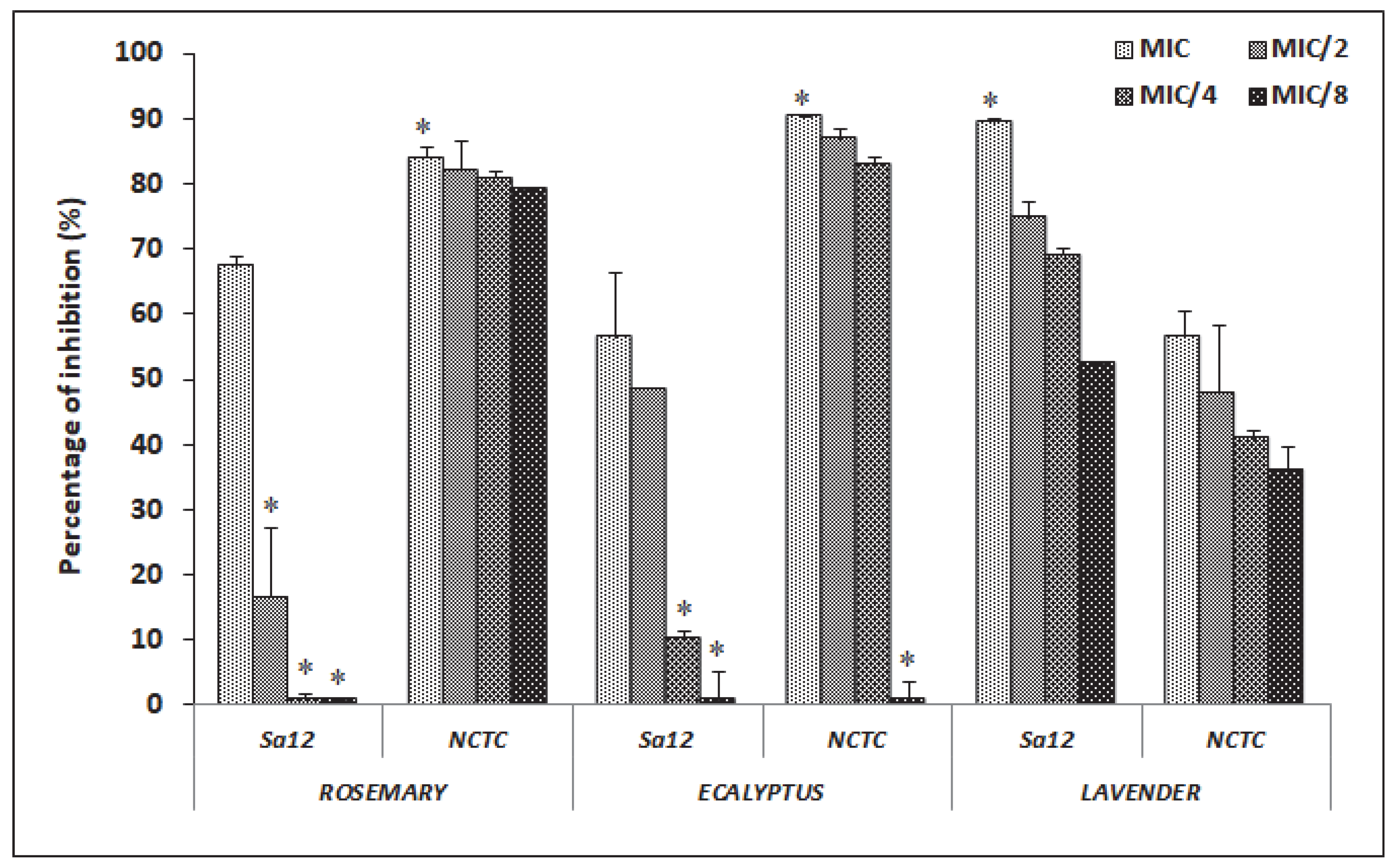
Following bacterial incubation for 24 h with sub-inhibitory concentrations of the investigated agents, the pre-formed biofilm was stained with Crystal Violet, and the percentage of anti-adhesion activity was determined after comparison with untreated bacteria. The Rosemary and Eucalyptus EOs were more effective against the reference strain, with the percentage of biofilm inhibition ranging from 71% to 93%. However, the Lavender EO showed a potent anti-attachment effect against the clinical MRSA strain, with the percentage of inhibition exceeding 52% at a low dose (1/8 MIC) (Figure 1).

3.5. Biofilm Eradication Effect

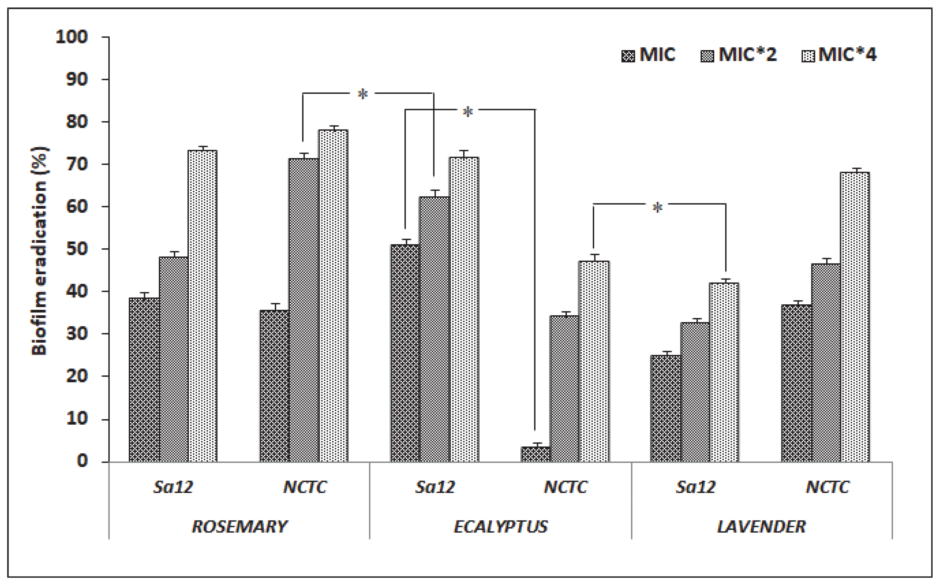
MRSA strains’ biofilms were treated with the selected EOs at different concentrations ranging from MIC to 4 × MIC. The results of this test are presented in Figure 2. The Rosemary and Lavender EOs had varying impacts on pre-formed biofilms, with percentage reduction values ranging from 36% to 73% and 37% to 68%, respectively (Figure 2). In contrast, the Eucalyptus EO was more efficient against the clinical MRSA biofilm, with the percentage of eradication exceeding 50%, even at low concentration (1 × MIC).

3.6. Pathogenic Factor Gene Expression

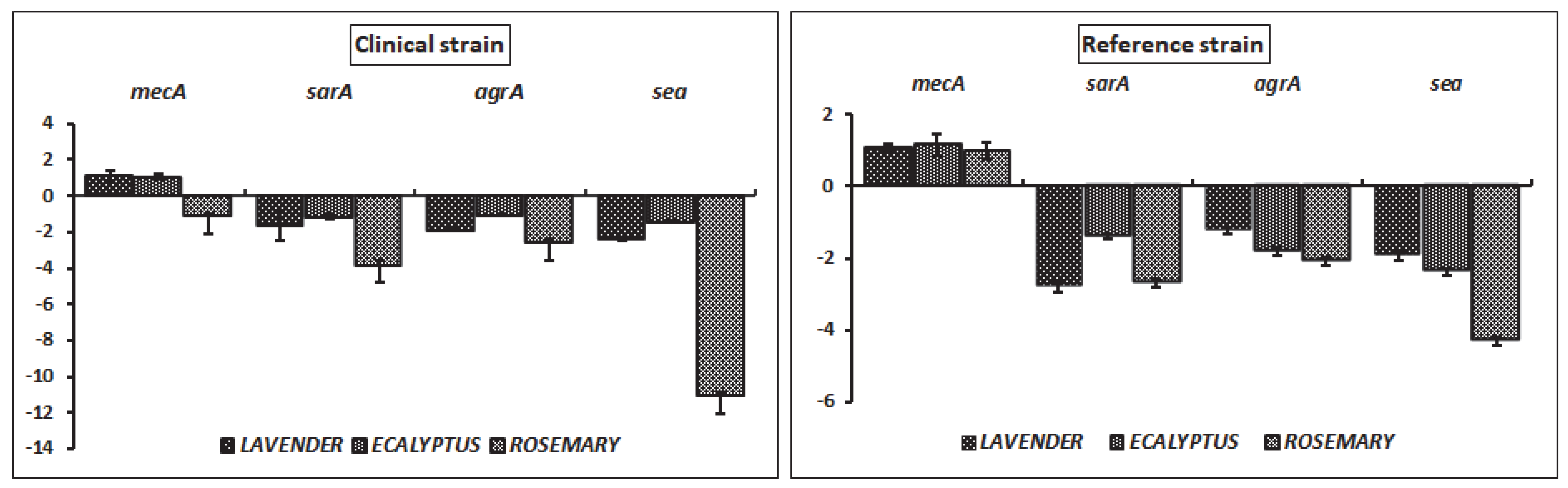
By analyzing the mRNA expression of the mecA, agrA, sarA, and sea genes in MRSA strains, we evaluated the impacts of the EOs on virulence-related gene expression and antibiotic resistance. Treated strains were subjected to MICs of each EO, and the data presented in Figure 3 demonstrate the suppression of gene expression for all target genes in comparison to untreated bacterial cells.
Quantitative real-time PCR analysis revealed variable gene expression since mecA expression in the reference MRSA strain remained unaffected or slightly upregulated, with fold changes of +1.07, +1.14 and 1.00 for Lavender, Eucalyptus, and Rosemary EO treatments, respectively (Figure 3). In contrast, consistent down-regulation was observed for the regulatory genes sarA and agrA, with the strongest inhibition induced by Rosemary oil (−2.72 and −2.08, respectively), followed by Lavender (−2.81 and −1.22) and Eucalyptus (−1.40 and −1.82) oils. The sea gene exhibited the most pronounced repression, particularly under Rosemary treatment (−4.33), compared to Eucalyptus (−2.38) and Lavender (−1.95). Regarding the Sa12 strain, a different expression pattern was observed, characterized by a greater sensitivity to essential oil exposure. Notably, consistent down-regulation of sarA and agrA was detected, with fold changes of −3.79 and −2.54, respectively, indicating a particularly strong inhibitory effect from Rosemary oil. Remarkably, the sea gene exhibited a dramatic reduction in expression in this clinical MRSA strain in the presence of Rosemary oil (−11.02) compared to other treatments.

4. Discussion

Essential oils are being extensively studied due to their abundant and varied bioactive constituents, which provide a natural, safe and effective option to address drug-resistant microorganisms. In the first part of our study, the chemical composition analyses of three local EOs (Eucalyptus, Rosemary and Lavender) performed by GC-MS revealed a total of 82 compounds pertaining to Monoterpene oxide (1.8-cineol), Monoterpene hydrocarbon (α-pinene), Monoterpene keton (L-camphor), Monoterpene alcool (Linalool), and Monoterpene ester (Linalyl acetate). Previous studies performing GC-MS analysis of the Lamiaceae (Rosemary and Lavender) and Myrtaceae (Eucalyptus) species’ EOs, studying species with different geographical origins (Morocco, France, Italy, Greece and Turkey), revealed the presence of the same main constituents but with difference percentage contents [19,20,21]. The variations in these compositions arise from the confluence of biological and genetic variables, environmental conditions and even the difference between the methodologies used for extraction and storage [22,23]. Additionally, differences in minor compounds of EOs have been described [24,25], and the profile compound fluctuations were registered even within the same plant organs [26,27]. All these fluctuations in EOs’ compositions certainly affected their biological activities, making the analysis of their constituents important to better understand their modes of action in pathogenic bacterial cells.
The antibacterial effects of the selected EOs were firstly assessed using a disk diffusion assay against clinical and reference strains of MRSA. Our results revealed that the Eucalyptus EO exhibited the greatest effect, with diameters of inhibition ranging between 38 and 41.33 mm. This important activity may be due to the high percentage of 1.8-cineol (83.4%) present in this analyzed EO. More specifically, it was previously reported that Eucalyptus (1.8-cineol) induces reactive oxygen species (ROS)-mediated oxidative stress and compromises the integrity of the cell membrane in MRSA strains [28]. In fact, the surge in ROS generation influenced antioxidant enzyme function and compromised macromolecules, resulting in bacterial damage and subsequent cell death [29]. Moreover, the association between the tested EOs and the antibiotic (Fox) showed the enhancement of the antibacterial effect against the reference strain; however, no positive effect of this association was noted against the clinical MRSA strain. Our results are in agreement with other reports showing a synergistic effect between EOs and antibiotics [30,31]. The observed differences may be related to the types of interactions of the minor and major compounds of EOs with the antibiotic and the susceptibility of the tested bacterial strain. The antibacterial effects of the selected EOs against the MRSA strains evaluated by the determination of MICs were found to be in agreement with those for the disk diffusion assay, since the Eucalyptus EO showed the greatest inhibitory effect against both strains, with MIC values ranging from 0.781 to 1.563 mg/mL, followed by the Rosemary EO and the Lavender EO. All tested EOs exhibited bactericidal effects against MRSA bacteria with MBC/MIC values ≤ 4 [32].
The roles of bacterial biofilms in antibiotic resistance, along with the different control strategies used, were studied in [33,34]. Our study investigated a biological biofilm control approach based on plant-extracted EOs. The percentages of anti-adhesion activity, determined through comparison with untreated bacteria strains, revealed that Rosemary and Eucalyptus EOs were more effective against the MRSA reference strains, with the percentage of biofilm inhibition reaching 93%. Regarding the Lavender EO, a potent anti-attachment effect was noted against the clinical MRSA strain at a low dose (1/8 MIC). Since initial adhesion is a crucial step in biofilm formation [35], it is of interest to impede this phase and subsequently to prevent biofilm development and maturation. In agreement with our study, it was revealed that EOs are effective natural agents for initial biofilm suppression [36,37]. In addition to biofilm attachment inhibition, we investigated the biofilm eradication potency of the selected EOs. Our results showed that the Eucalyptol EO was more efficient against the clinical MRSA biofilm, with the percentage of eradication exceeding 50% even at low concentrations (1 × MIC). Regarding the Rosemary and Lavender EOs, varying effects on pre-formed biofilms formation were observed, with percentage reduction values ranging from 36% to 73%. Accordingly, plant-derived bioactive chemicals, exemplified by those in Eucalyptus, Rosemary and Lavender, may proficiently destroy biofilm architectures by processes including quorum-sensing (QS) inhibition [15,38,39]. In fact, the QS mechanism acts as an intercellular signaling system through chemical signals, enabling bacterial communication and control of their biofilm formation [40]. Therefore, EOs function as natural anti-quorum-sensing agents against MRSA by interrupting bacterial communication, which consequently prevents biofilm formation and virulence factor generation [41,42].
The last part of our study was conducted to assess the effects of local EOs on methicillin resistance and virulence-related gene expression in both MRSA strains by quantitative PCR. This comparative analysis highlights a more pronounced transcriptional response in the clinical Sa12 strain, particularly for sea and sarA genes, suggesting increased susceptibility to EO-mediated regulation. Among the tested oils, Rosemary consistently exerted the strongest inhibitory effect on pathogenicity-related gene expression in both strains, followed by Eucalyptus and Lavender. Remarkably, upregulated expression of the antibiotic resistance gene mecA was noted following treatment with the majority of EOs. Our findings were in agreement with a recent report revealing that the mRNA expression of pivotal genes implicated in MRSA resistance and pathogenicity declined in a concentration-dependent manner following Chrysanthemum zawadskii EO treatment compared to untreated bacteria [18]. Overall, these findings indicate that essential oils, especially Rosemary, significantly repress key virulence determinants in MRSA in a strain-dependent manner, potentially through interference with global regulatory systems such as sarA and agrA.

5. Conclusions

The present study highlighted the variety and abundance of chemical components present in local essential oils from Lamiaceae and Myrtaceae species. This diversity in bioactive constituents justifies the observed and notable antibacterial, anti-adhesion and antibiofilm properties of these EOs against MRSA pathogenic strains. Additionally, our study revealed that the tested EOs inhibited the expression of methicillin resistance, staphylococcal enterotoxin A, staphylococcal accessory regulator A and accessory gene regulator A, which are considered major genes linked to the resistance and pathogenicity of MRSA. Accordingly, this molecular targeting approach requires further investigation for potential use in food applications aiming at lowering MRSA’s virulence and infectivity.

Author Contributions

Conceptualization, A.M. (Abderrahmen Merghni) and L.H.; methodology, A.M. (Amal Makhlouf), H.E., S.M., A.E. and A.M. (Abderrahmen Merghni); validation, H.E., L.H. and A.M. (Abderrahmen Merghni); writing—original draft preparation, A.M. (Amal Makhlouf), H.E., S.M., K.M.A. and A.M. (Merghni Abderrahmen); writing—review and editing, A.M. (Amal Makhlouf), A.R.B., K.M.A., L.H. and A.M. (Abderrahmen Merghni); supervision, A.M. (Abderrahmen Merghni) and L.H. All authors have read and agreed to the published version of the manuscript.

Funding

This work was supported by the Ministry of Higher Education and Scientific Research (MHESR) of Tunisia.

Institutional Review Board Statement

Not applicable.

Informed Consent Statement

Not applicable.

Data Availability Statement

The original contributions presented in this study are included in the article. Further inquiries can be directed to the corresponding author.

Conflicts of Interest

The authors declare no conflicts of interest.

Correction Statement

This article has been republished with a minor correction to resolve spelling and grammatical errors. This change does not affect the scientific content of the article.

References

  1. Algammal, A.M.; Hetta, H.F.; Elkelish, A.; Alkhalifah, D.H.H.; Hozzein, W.N.; Batiha, G.E.; El Nahhas, N.; Mabrok, M.A. Methicillin-Resistant Staphylococcus aureus (MRSA): One Health Perspective Approach to the Bacterium Epidemiology, Virulence Factors, Antibiotic-Resistance, and Zoonotic Impact. Infect. Drug Resist. 2020, 13, 3255–3265. [Google Scholar]
  2. Ali Alghamdi, B.; Al-Johani, I.; Al-Shamrani, J.M.; Musamed Alshamrani, H.; Al-Otaibi, B.G.; Almazmomi, K.; Yusnoraini Yusof, N. Antimicrobial resistance in methicillin-resistant Staphylococcus aureus. Saudi J. Biol. Sci. 2023, 30, 103604. [Google Scholar] [CrossRef] [PubMed]
  3. Tong, S.Y.C.; Fowler, V.G., Jr.; Skalla, L.; Holland, T.L. Management of Staphylococcus aureus Bacteremia: A Review. JAMA 2025, 334, 798–808. [Google Scholar] [PubMed]
  4. Tobin, E.H.; Jogu, P.; Koirala, J. Methicillin-Resistant Staphylococcus aureus. In StatPearls; StatPearls Publishing: Treasure Island, FL, USA, 2025. [Google Scholar]
  5. Cameron, D.R.; Jiang, J.H.; Kostoulias, X.; Foxwell, D.J.; Peleg, A.Y. Vancomycin susceptibility in methicillin-resistant Staphylococcus aureus is mediated by YycHI activation of the WalRK essential two-component regulatory system. Sci. Rep. 2016, 6, 30823. [Google Scholar] [PubMed]
  6. Craft, K.M.; Nguyen, J.M.; Berg, L.J.; Townsend, S.D. Methicillin-resistant Staphylococcus aureus (MRSA): Antibiotic-resistance and the biofilm phenotype. Medchemcomm 2019, 10, 1231–1241. [Google Scholar] [PubMed]
  7. Peng, Q.; Tang, X.; Dong, W.; Sun, N.; Yuan, W. A Review of Biofilm Formation of Staphylococcus aureus and Its Regulation Mechanism. Antibiotics 2022, 12, 12. [Google Scholar] [CrossRef] [PubMed]
  8. Tam, K.; Torres, V.J. Staphylococcus aureus Secreted Toxins and Extracellular Enzymes. Microbiol. Spectr. 2019, 7. [Google Scholar] [CrossRef] [PubMed]
  9. Mabrouk, S.; Esraa, A.A.; Mohamed, A.H.; Tariq, A.; Al-Asmari, F.; Khulood, F.A.; Haiying, C.; Lin, L. Essential oils as antibacterials against multidrug-resistant foodborne pathogens: Mechanisms, recent advances, and legal considerations. Food Biosci. 2025, 64, 105937. [Google Scholar] [CrossRef]
  10. Khwaza, V.; Aderibigbe, B.A. Antibacterial Activity of Selected Essential Oil Components and Their Derivatives: A Review. Antibiotics 2025, 14, 68. [Google Scholar] [CrossRef] [PubMed]
  11. Abdelmohsen, U.R.; Elmaidomy, A.H. Exploring the therapeutic potential of essential oils: A review of composition and influencing factors. Front. Nat. Prod. 2025, 4, 1490511. [Google Scholar] [CrossRef]
  12. Yu, J. Chemical Composition of Essential Oils and Their Potential Applications in Postharvest Storage of Cereal Grains. Molecules 2025, 30, 683. [Google Scholar] [CrossRef] [PubMed]
  13. Moumni, S.; Elaissi, A.; Trabelsi, A.; Merghni, A.; Chraief, I.; Jelassi, B.; Chemli, R.; Ferchichi, S. Correlation between chemical composition and antibacterial activity of some Lamiaceae species essential oils from Tunisia. BMC Complement. Med. Ther. 2020, 20, 103. [Google Scholar] [CrossRef] [PubMed]
  14. Merghni, A.; Noumi, E.; Hadded, O.; Dridi, N.; Panwar, H.; Ceylan, O.; Mastouri, M.; Snoussi, M. Assessment of the antibiofilm and antiquorum sensing activities of Eucalyptus globulus essential oil and its main component 1,8-cineole against methicillin-resistant Staphylococcus aureus strains. Microb. Pathog. 2018, 118, 74–80. [Google Scholar] [CrossRef] [PubMed]
  15. Perez, L.R.; Freitas, A.L.; Barth, A.L.; Dias, C.A. Direct disk diffusion susceptibility testing from respiratory tract specimens: Focus on Pseudomonas aeruginosa. Int. J. Infect. Dis. 2014, 26, 47–48. [Google Scholar] [CrossRef] [PubMed]
  16. Magina, M.D.; Dalmarco, E.M.; Wisniewski, A., Jr.; Simionatto, E.L.; Dalmarco, J.B.; Pizzolatti, M.G.; Brighente, I.M.C. Chemical composition and antibacterial activity of essen-tial oils of Eugenia species. J. Nat. Med. 2009, 63, 345–350. [Google Scholar] [PubMed]
  17. Merghni, A.; Marzouki, H.; Hentati, H.; Aouni, M.; Mastouri, M. Antibacterial and antibiofilm activities of Laurus nobilis L. essential oil against Staphylococcus aureus strains associated with oral infections. Pathol. Biol. 2015. Epub ahead of printing. [Google Scholar]
  18. Pfaffl, M.W.; Horgan, G.W.; Dempfle, L. Relative expression software tool (REST) for group-wise comparison and statistical analysis of relative expression results in real-time PCR. Nucleic Acids Res. 2002, 30, e36. [Google Scholar] [PubMed]
  19. Kim, J.-H.; Park, B.-I.; Kim, Y.-H.; Yoon, J.-S.; Choi, N.-Y.; Kim, K.-J. Chrysanthemum zawadskii var. latilobum Flower Essential Oil Reduces MRSA Pathogenicity by Inhibiting Virulence Gene Expression. Molecules 2025, 30, 553. [Google Scholar] [CrossRef] [PubMed]
  20. Satyal, P.; Jones, T.H.; Lopez, E.M.; McFeeters, R.L.; Ali, N.A.; Mansi, I.; Al-Kaf, A.G.; Setzer, W.N. Chemotypic Characterization and Biological Activity of Rosmarinus officinalis. Foods 2017, 6, 20. [Google Scholar] [CrossRef] [PubMed]
  21. Čmiková, N.; Galovičová, L.; Schwarzová, M.; Vukic, M.D.; Vukovic, N.L.; Kowalczewski, P.Ł.; Bakay, L.; Kluz, M.I.; Puchalski, C.; Kačániová, M. Chemical Composition and Biological Activities of Eucalyptus globulus Essential Oil. Plants 2023, 12, 1076. [Google Scholar] [CrossRef] [PubMed]
  22. Aguerd, O.; Elhrech, H.; El Omari, N.; Benali, T.; Akhazzane, M.; Mostakim, M.; Ouma, S.; Khattabi, L.; Amanullah, M.; Menyiy, N.E.; et al. Chemical composition and biological effects of Lavandula angustifolia Mill., essential oils. AMB Express 2025, 15, 164. [Google Scholar] [CrossRef] [PubMed]
  23. Khan, M.H.; Dar, N.A.; Alie, B.A.; Dar, S.A.; Lone, A.A.; Mir, G.H.; Fayaz, U.; Ali, S.; Tyagi, A.; El-Sheikh, M.A.; et al. Unraveling the Variability of Essential Oil Composition in Different Accessions of Bunium persicum Collected from Different Temperate Micro-Climates. Molecules 2023, 28, 2404. [Google Scholar] [CrossRef] [PubMed]
  24. Guelifet, K.; Kherraz, K.; Messaoudi, M.; Ferhat, M.A.; Khattabi, L.; Bendrihem, K.A.; Zahnit, W.; Addad, D.; Benmohamed, M.; Azoudj, Y.; et al. Seasonal and Extraction-Dependent Variation in the Composition and Bioactivity of Essential Oils from Wild Rosmarinus officinalis L. Molecules 2025, 30, 4258. [Google Scholar] [CrossRef] [PubMed]
  25. Guedri Mkaddem, M.; Zrig, A.; Ben Abdallah, M.; Romdhane, M.; Okla, M.K.; Al-Hashimi, A.; Alwase, Y.A.; Hegab, M.Y.; Madany, M.M.Y.; Hassan, A.H.A.; et al. Variation of the Chemical Composition of Essential Oils and Total Phenols Content in Natural Populations of Marrubium vulgare L. Plants 2022, 11, 612. [Google Scholar] [CrossRef] [PubMed]
  26. Cheng, Y.; Fu, Y.; Gu, D.; Huang, Y.; Lu, Y.; Liu, Y.; Li, X.; Yao, X.; Zhang, X.; Jian, W.; et al. Seasonal Variation in Chemical Composition and Antioxidant and Antibacterial Activity of Essential Oil from Cinnamomum cassia Leaves. Plants 2025, 14, 81. [Google Scholar]
  27. Marzouki, H.; Piras, A.; Salah, K.B.; Medini, H.; Pivetta, T.; Bouzid, S.; Marongiu, B.; Falconieri, D. Essential oil composition and variability of Laurus nobilis L. growing in Tunisia, comparison and chemometric investigation of different plant organs. Nat. Prod. Res. 2009, 23, 343–354. [Google Scholar] [CrossRef] [PubMed]
  28. Pluhár, Z.; Kun, R.; Cservenka, J.; Neumayer, É.; Tavaszi-Sárosi, S.; Radácsi, P.; Gosztola, B. Variations in Essential Oil Composition and Chemotype Patterns of Wild Thyme (Thymus) Species in the Natural Habitats of Hungary. Horticulturae 2024, 10, 150. [Google Scholar] [CrossRef]
  29. Merghni, A.; Belmamoun, A.R.; Urcan, A.C.; Bobiş, O.; Lassoued, M.A. 1,8-Cineol (Eucalyptol) Disrupts Membrane Integrity and Induces Oxidative Stress in Methicillin-Resistant Staphylococcus aureus. Antioxidants 2023, 12, 1388. [Google Scholar] [CrossRef] [PubMed]
  30. Vaishampayan, A.; Grohmann, E. Antimicrobials Functioning through ROS-Mediated Mechanisms: Current Insights. Microorganisms 2021, 10, 61. [Google Scholar] [CrossRef] [PubMed]
  31. Langeveld, W.T.; Veldhuizen, E.J.; Burt, S.A. Synergy between essential oil components and antibiotics: A review. Crit. Rev. Microbiol. 2014, 40, 76–94. [Google Scholar] [PubMed]
  32. Shahin, H.H.; Baroudi, M.; Dabboussi, F.; Ismail, B.; Salma, R.; Osman, M.; El Omari, K. Synergistic Antibacterial Effects of Plant Extracts and Essential Oils Against Drug-Resistant Bacteria of Clinical Interest. Pathogens 2025, 14, 348. [Google Scholar] [CrossRef] [PubMed]
  33. Makade, C.S.; Shenoi, P.R.; Bhongade, B.A.; Shingane, S.A.; Ambulkar, P.C.; Shewale, A.M. Estimation of MBC: MIC Ratio of Herbal Extracts against Common Endodontic Pathogens. J. Pharm. Bioallied Sci. 2024, 16, S1414–S1416. [Google Scholar] [CrossRef] [PubMed]
  34. Abebe, G.M. The Role of Bacterial Biofilm in Antibiotic Resistance and Food Contamination. Int. J. Microbiol. 2020, 2020, 1705814. [Google Scholar] [CrossRef] [PubMed]
  35. Rather, M.A.; Gupta, K.; Mandal, M. Microbial biofilm: Formation, architecture, antibiotic resistance, and control strategies. Braz. J. Microbiol. 2021, 52, 1701–1718. [Google Scholar] [CrossRef] [PubMed]
  36. Zhao, A.; Sun, J.; Liu, Y. Understanding bacterial biofilms: From definition to treatment strategies. Front. Cell Infect. Microbiol. 2023, 13, 1137947. [Google Scholar] [CrossRef] [PubMed]
  37. Khashei, S.; Fazeli, H.; Rahimi, F.; Karbasizadeh, V. Antibiotic-potentiating efficacy of Rosmarinus officinalis L. to combat planktonic cells, biofilms, and efflux pump activities of extensively drug-resistant Acinetobacter baumannii clinical strains. Front. Pharmacol. 2025, 16, 1558611. [Google Scholar] [PubMed]
  38. Luță, E.-A.A.; Biță, A.; Moroșan, A.; Mihaiescu, D.E.; Mihai, D.P.; Popescu, L.; Bejenaru, L.E.; Bejenaru, C.; Popovici, V.; Olaru, O.T. Implications of Rosemary and Thyme (Lamiaceae) Cultivation in Plant Communities for the Development of Antioxidant Therapies. Int. J. Mol. Sci. 2023, 24, 11670. [Google Scholar] [PubMed]
  39. Ali, M.K.; Galut, H.S.; Matar, E.E.T.M.; Mamand, S.F.; Sabrie, L.S.; Mala, S.F.; Jghef, M.M. Exploring the antibacterial and anti-biofilm properties of Rosmarinus officinalis extracts: A natural strategy against methicillin-resistant Staphylococcus aureus. Open Vet. J. 2025, 15, 5549–5561. [Google Scholar] [PubMed]
  40. Abraham, W.R. Going beyond the Control of Quorum-Sensing to Combat Biofilm Infections. Antibiotics 2016, 5, 3. [Google Scholar] [CrossRef] [PubMed]
  41. Cáceres, M.; Hidalgo, W.; Stashenko, E.; Torres, R.; Ortiz, C. Essential oils of aromatic plants with antibacterial, anti-biofilm and anti-quorum sensing activities against pathogenic bacteria. Antibiotics 2020, 9, 147. [Google Scholar] [PubMed]
  42. Barak, T.H.; Karaca, B.; Servi, H.; Kara Ertekin, S.; Şentürk, T.B.; Dinc, M.; Ustuner, H.; Eryilmaz, M. Antimicrobial: Antibiofilm, Anti-Quorum Sensing and Cytotoxic Activities of Dorystoechas hastata Boiss & Heldr. ex Bentham Essential Oil. Antibiotics 2025, 14, 1019. [Google Scholar] [CrossRef] [PubMed]
Figure 1. The effects of the Eucalyptus, Rosemary and Lavender essential oils on the adhesion of the clinical (Sa12) and reference (NCTC12493) MRSA strains treated with sub-inhibitory concentrations (1/8 to 1 × MIC), expressed as a percentage of inhibition (%) and evaluated by the Crystal Violet staining assay. Values are the averages of at least three independent determinations. Error bars represent standard deviations. (*) Differences were considered significant at p < 0.05.
Foods 15 02361 g001
Figure 2. The effects of the Eucalyptus, Rosemary and Lavender essential oils on pre-formed biofilms of the clinical (Sa12) and reference (NCTC12493) MRSA strains treated with various concentrations (MIC to MIC × 4), expressed as a percentage of biofilm eradication (%) and evaluated by the Crystal Violet staining assay. Values are the averages of at least three independent determinations. Error bars represent standard deviations. (*) Differences were considered significant at p < 0.05.
Foods 15 02361 g002
Figure 3. The relative expression levels (fold change) of antibiotic resistance (mecA) and virulence (sarA, agrA, and sea) genes in clinical (Sa12) and reference (NCTC12493) MRSA strains following exposure to Lavender, Eucalyptus, and Rosemary essential oils. Gene expression was quantified by real-time quantitative PCR (RT-qPCR) and normalized to the untreated control condition. Fold change values represent relative transcription levels calculated using the 2−ΔΔCt method. Negative values indicate down-regulation, whereas positive values indicate the upregulation of gene expression compared to the control. Among the tested treatments, the Rosemary essential oil induced the strongest down-regulation of virulence-associated genes, particularly sea and sarA, with a more pronounced effect observed in the clinical strain compared to the reference strain.
Foods 15 02361 g003
Table 1. The primers used in the RT-PCR experiments [18].
GeneSequences (5′ → 3′)Tm (°C)Base Count (nt)GC Ratio (%)
agrAForwardTGATAATCCTTATGAGGTGCTT53.72236.36
ReverseTGATAATCCTTATGAGGTGCTT55.62142.86
mecAForwardGTTAGATTGGGATCATAGCGTCATT58.12540
ReverseTGCCTACTCATGTGTTCCTGTAT592737.04
sarAForwardTGTTATCAATGGTCACTTATGCTG56.32437.5
ReverseTCTTTGTTTTCGCTGATGTATGTC57.12437.5
seaForwardATGGTGCTTATTAGGTGTATC50.12133.33
ReverseCGTTTCCAAAGGTAGTGTTATT52.92138.1
16s rRNAForwardACTGGGATAACTTCGGGAAA55.22045
ReverseCGTTGCCTTGGTAAGCC54.91758.82
Table 2. The chemical compositions of the tested EOs.
TRIRCompoundsRosemaryEucalyptusLavender
13.561852Cis-3-hexenol0.01*Tr
24.556910Zeta-fenchene0.02**
34.537917Alpha-fenchene*0.18*
44.946925Tricyclène0.11*0.01
55.043928Alpha-Thujène0.14tr0.03
65.227936Alpha-pinene12.368.200.77
75.579949Camphéne2.920.060.15
85.718954Verbenene0.05*0.01
96.230974Sabinene0.21*0.12
106.330978Beta-Pinène5.830.450.71
116.53479863-octanone**0.05
126.717993Myrcène1.530.150.37
136.8252997Butyl butyrate**0.03
147.1321006Alpha-Phellandrene0.220.110.05
157.3291012Delta-3-Carene0.19*0.07
167.37661013Hexyl acetate**0.02
177.5291017Alpha-Terpinene0.130.020.02
187.7871024P-Cymène3.522.290.58
198.08710331,8-cinéole46.8983.405.55
207.92701028Limonène**0.55
218.2461037Trans-beta-Ocimene0.09*0.24
228.34511040N-Butyl isovalerate**0.02
238.5991047Cis-beta-Ocimene0.06*0.11
248.9691057Gamma-Terpinene0.550.060.14
259.4541070Cis-Sabinene hydrate0.05**
269.26141065Trans-sabinene hydrate**0.10
279.3831068Cis-Linalool oxide*0.17*
289.45661070Trans-Linalool oxide**0.16
2910.0201086P-alpha-dimethylstyrene*0.42*
3010.0651087Alpha-Terpinolene0.220.270.20
3110.3531095Terpinolene*0.08*
3210.5101100Linalool0.520.0335.68
3310.6621103Isoamyl isovalerate0.020.07*
3411.04621112Cis-rose oxide**0.16
3511.3491119Fenchol0.05**
3611.4991123Campholenal0.050.02*
3711.50931123Chrysanthenone**0.18
3811.9641134Trans-Pinocarveol*2.36*
3912.2541141L-camphor13.120.074.46
4012.4211145Exo-methyl-camphenilol0.07**
4112.51841147Hexyl isobutyrate**0.11
4212.78361153Isoborneol**0.60
4312.9821158Pinocarvone0.030.05*
4413.1601162Borneol L.2.270.123.53
4513.3771167Isopinocamphone*0.13*
4613.6421173Terpinene-4-ol0.480.122.21
4713.9031180P-cymen-8-ol*0.06*
4814.0261182Is-p-Mentha-1(7),8-dien-2-ol*0.12*
4914.2201187Alpha-Terpineol1.34 0.52
5014.5301194Myrtenol0.120.110.04
5114.6871198Methyl chavicol0.03**
5214.98911205Verbenone*0.030.04
5315.22141210Octyl acetate**0.02
5415.4461215Trans-(+)-carveol0.040.040.02
5515.8021223Bornyl formate0.03**
5615.80281223Isobornyl formate**0.03
5715.93181226Nerol**0.08
5816.52921240Hexyl isovalerate**0.13
5917.35151258Linalyl acetate**33.40
6018.3681281Bornyle acetate0.730.020.03
6118.70641289Lavandulyl acetate**1.44
6218.9541294Thymol0.07**
6319.0981297Carvacrol0.020.02*
6421.2141345Alpha-cubebene0.06**
6521.4841351Eugenol0.09**
6622.1361366Alpha-yalangene0.10**
6722.3321370Alpha-Copaene0.20**
6822.9991385Beta-Elemene0.05**
6923.5761398Methyl eugenol0.08**
7024.1691412Beta-Caryophyllene1.95**
7124.5681422Alpha cedrene0.07**
7224.9901431Aromadendrene0.07*0.04
7325.6031446Alpha-Humulène0.26**
7425.8781452(E)-beta-farnesene0.05**
7526.6191470Alloaromadendrene0.17**
7626.7661473Germacrene D0.02**
7727.3741487Ar-Curcumene0.08**
7827.6241493Beta-selinene 0.09**
7928.0101502Beta-bisabolene0.06**
8028.1591506Alpha-amorphene0.11**
8128.5681516Delta-cadinene0.22**
8230.8781574Caryophyllene oxide0.26**
Total (%) 98.1099.2092.78
*: Trace (<0.1%).
Table 3. The disk diffusion assay.
EOMRSAD.I (mm)ATB (Fox)EO + ATB
RosemarySa1219.67 ± 0.586.0019.33 ± 0.58
NCTC1249316.67 ± 1.156.0018.50 ± 1.15
EucalyptusSa1241.33 ± 1.15 *6.0034.67 ± 1.15 **
NCTC1249338.00 *6.0038.67 ± 0.58 **
LavenderSa1218.006.0018.00
NCTC1249319.67 ± 0.586.0020.50 ± 0.58
D.I: Diameter of inhibition; ATB: antibiotic; FOX: cefoxitin; * indicates a statistically significant difference between the activity of the EOs tested without the antibiotic; ** indicates a statistically significant difference between the activity of the EOs tested in association with the antibiotic.
Table 4. The determination of the MICs and MBCs of the EOs.
EOSa12NCTC12493
MICMBCMBC/MICMICMBCMBC/MIC
Rosemary1.5633.12523.1256.2502
Eucalyptus0.7811.56321.5631.5631
Lavender3.1256.25026.2506.2501
Sa12: clinical MRSA strain; NCTC12493: reference MRSA strain; MIC: minimal inhibition concentration; MBC: minimal bactericidal concentration. MIC and MBC values are expressed as mg/mL.
Disclaimer/Publisher’s Note: The statements, opinions and data contained in all publications are solely those of the individual author(s) and contributor(s) and not of MDPI and/or the editor(s). MDPI and/or the editor(s) disclaim responsibility for any injury to people or property resulting from any ideas, methods, instructions or products referred to in the content.