Article

Effect of Selenium Fortification on Growth Performance and Nutritional Compounds of Kale (Brassica oleracea L. Var. acephala DC.)

1
Ganzhou General Inspection and Testing Institute, China National Quality and Inspection Center for Se-Rich and Camellia Oleifera Products (Jiangxi), Ganzhou 341000, China
2
School of Public Health and Health Management, Gannan Medical University, Ganzhou 341000, China
3
Key Laboratory of Development and Utilization of Gannan Characteristic Food Function Component of Ganzhou, Gannan Medical University, Ganzhou 341000, China
*
Authors to whom correspondence should be addressed.
Foods 2025, 14(18), 3283; https://doi.org/10.3390/foods14183283
Submission received: 1 August 2025 / Revised: 17 September 2025 / Accepted: 19 September 2025 / Published: 22 September 2025

Abstract

This study aims to investigate the effects of selenium (Se) fortification on growth performance and the Se content in kale using Se fertilizer, and it determines the influences of Se fortification on the metabolic profile of kale using quasi-targeted metabolomics. The results showed that Se fortification increased the plant height and leaf weight of kale, up-regulated the total Se content and decreased the chlorophyll and total phenolic contents in kale leaf. Se fortification elevated selenate (Se(IV)), selenite (Se(VI)), selenocystine (SeCys2), Se-methylselenocysteine (Se-MeSeCys) and selenomethionine (SeMet) contents, as well as total contents of Se in different forms in kale leaf. Se fortification also changed the metabolic profile of kale leaf, via six particular types of compounds (amino acid and its derivatives; organic acid and its derivatives; carbohydrates and its derivatives; lipids; flavonoids; organoheterocyclic compounds) and eight metabolic pathways (alanine, aspartate and glutamate metabolism; amino sugar and nucleotide sugar metabolism; sulfur metabolism; starch and sucrose metabolism; taurine and hypotaurine metabolism; glycolysis/gluconeogenesis; fructose and mannose metabolism; nitrogen metabolism). Moreover, 24 metabolic biomarkers were screened for kale leaf affected by Se fortification. Furthermore, correlations were observed between metabolic biomarkers and Se contents as well as speciation. These results indicate that Se fortification has a significant influence on the growth performance and nutritional compounds of kale, providing references for the future study on the production and bioactivity of Se-enriched kale.

Graphical Abstract

1. Introduction

Kale (Brassica oleracea L. Var. acephala DC.), an important vegetable belonging to the Brassica family, is commonly consumed in Europe, Asia and America. In recent years, it has gained great popularity as a “superfood” and is listed in many “lists of the healthiest vegetables” [1]. This vegetable is rich in nutrients and bioactive compounds, such as amino acids, vitamins, minerals, dietary fiber and phytochemicals [2]. Kale extracts have been reported to possess numerous preventive and therapeutic properties, such as antimicrobial, anti-carcinogenic, antioxidant and hypoglycemic activities [3]. In view of this, kale is increasingly considered a source of nutraceuticals and undergoes processing to become value-added products (bread, juice, beverages, etc.) [4].
Kale plant is an annual crop, whose yield, size and nutritional variation greatly depend on its growing conditions. The effects of bio-slurry and inorganic N fertilizer [5], sulfur treatment [2], temperature stress [6,7], nitrogen and magnesium nutrients [8], iodoquinoline biofortification [9] and selenium (Se) biofortification [10,11] on the growth performance, nutrients and/or bioactive compounds of kale are some of the areas that have been investigated to date.
It is well known that Se is an essential trace element that plays a crucial role in maintaining animal and human health. Se deficiency is closely related to several diseases such as Kashan, Kashin–Beck and hypothyroidism [12], while its overdose causes intoxication events [13]. Approximately one billion people suffer from Se deficiency globally, as the soil of many countries lacks this element [14]. In fact, the organic compositions of Se species, especially selenoproteins and selenoamino acids, are responsible for the function of Se in human health. Since plants can generally convert inorganic Se into organic Se, the aforementioned Se biofortification has been widely applied to produce Se-enriched foods [12], thereby increasing Se intake in human diets and satisfying the recommended dietary allowance [14]. Like with other Brassica plants (turnip, cabbage, broccoli, nozawana, komatsuna, etc.), kale can also significantly absorb, accumulate and transform inorganic Se into organic Se [12]. Thus, kale has great potential to produce as Se-enriched diet.
Se biofortification has demonstrated the ability to enrich the Se content in kale. In a study by Tavan et al. [14], kale microgreens successfully accumulated up to 893.3 and 24 µg Se/kg of dry matter under a Se-rich soilless medium and with foliar application, at a 20 µM concentration. Leamsamrong et al. [10] found that the total Se contents of the air-dried matter in Se-enriched Chinese kale seedlings (433 ± 22 mg·Se/kg) were significantly higher than those of the regular Chinese kale seedlings. Maneetong et al. [12] discovered that total Se concentrations of kale with all Se-supplemented treatments were higher than those with the control treatment. Furthermore, the investigation conducted by Zagrodzki et al. [11] indicated that Se fortification stimulated the production of phenolic acids (sinapic, chlorogenic, isochlorogenic and caffeic acids) in the kale sprouts. Paśko et al. [15] observed that Se fortification affected the synthesis of sulfur and phenolic compounds in the kale sprouts. However, the influences of Se biofortification on comprehensive nutritional compounds in kale are still necessary to examine. Therefore, a comprehensive profiling method is required to elucidate the underlying metabolic changes.
Quasi-targeted metabolomics, based on LC-MS/MS technology, is a new type of metabolomics detection technique that combines the high-throughput advantages of untargeted metabolomics with the high accuracy and sensitivity advantages of targeted metabolomics. This tool has been widely used to identify chemical constituents and metabolic changes in plants [16,17,18,19]. For instance, Ren et al. [20] examined an integrated metabolome to reveal the differential metabolites in a vegetable cucumber fruit (Cucumis sativus L.) after grafting. Moreover, other researchers have used an untargeted metabolomics strategy to study the nutrient profiles of Chinese kale [21]. In this light, quasi-targeted metabolomics is suitable for understanding the effect of Se biofortification on nutritional compounds in kale.
Hence, in this study, the effects of Se fortification on growth performance and the Se content in kale were detected. Moreover, the influences of Se fortification on the metabolic profile of kale were systematically determined and analyzed using quasi-targeted metabolomics. Furthermore, correlations between the metabolic biomarkers, Se contents and speciation in kale were analyzed. This study is beneficial for people to better understand the impact of selenium fortification on the nutritional compounds in kale, and it can guide future research on the potential biological activities of Se-biofortified kale.

2. Materials and Methods

2.1. Materials and Chemicals

Kale seeds were purchased from Beijing Dongsheng Seed Industry Co., Ltd. (Beijing, China). Amino acid water-soluble fertilizer (containing Se ≥ 1000 mg/L) was bought from Suzhou Setek Co., Ltd. (Suzhou, China). A mixed standard solution of 28 metal elements was obtained from the National Center of Analysis and Testing for Nonferrous Metals and Electronic Materials, and standard solutions of Se(IV), Se(VI), SeCys2, Se-MeSeCys and SeMet were gained from the National Institute of Metrology, China. Gallic acid was purchased from LEMEITIAN MEDICINE (Chengdu, China), and Folin–Ciocalteu reagent was bought from Shanghai Macklin Biochemical Co., Ltd. (Shanghai, China). LC-MS-grade water was obtained from Merck KGaA (Darmstadt, Germany), and LC-MS-grade methanol, formic acid and acetonitrile were gained from Thermo Fisher Scientific (Waltham, MA, USA). All other chemicals that were used were of analytical grade and bought from Sinopharm Chemical Reagent Co., Ltd. (Shanghai, China).

2.2. Experimental Site, Growing Conditions and Cultivation

The experiment was conducted at the test base of the Ganzhou General Inspection and Testing Institute (25.8351° E, 114.8957° N) in Ganzhou City, Jiangxi province, China. Kale seeds were purchased, soaked, germinated and then sown in a tray using a seedling substrate to grow seedlings. One month later, kale seedlings with thick main stems and well-developed root systems, having 3–4 true leaves and showing consistent growth, were selected and transplanted into 100 L vegetable boxes (3 plants in each box). The Se contents of soil in all vegetable boxes were ≤0.4 mg/kg. Three months later, kale plants were divided into six groups (6 plants per group): the control group (treated with 0 mg/L Se); the 1 mg/L Se group; the 2 mg/L Se group; the 5 mg/L Se group; the 10 mg/L Se group; and the 20 mg/L Se group. The Se solutions with different concentrations that were used in this study were prepared using an amino acid water-soluble fertilizer (containing Se ≥ 1000 mg/L). The leaf surface of each plant and the corresponding soil were sprayed with 500 mL of prepared Se solutions for two months. After the experimental period, biometric measurements of kale were taken, and the 4th to 10th kale leaves were collected from top to bottom for the following determinations.

2.3. Determination of Biometric Measurements of Kale

The plant height of kale was determined by measuring the distance from the bottom to the top of the stem using a vernier caliper. In addition, all leaves of each kale plant were picked and then weighed using an electronic scale (ME104E, Mettler Toledo, Shanghai, China), and the weight was recorded as leaf weight.

2.4. Determination of Chlorophyll Contents in Kale Leaf

The chlorophyll content in kale leaf was detected according to the method recommended by the Standardization Administration of the People’s Republic of China (NY/T 3082-2017) [22]. To achieve this, 0.5 g of kale leaf was placed in a conical flask, and it was mixed with 100 mL of an absolute ethyl alcohol–acetone solution (v/v = 1:1). The conical flask was sealed with a sealing film, and then the mixture was kept standing in the dark at room temperature for 5 h. After filtration, the absorbances of the filtrates were measured at 645 nm and 663 nm, respectively. Finally, the chlorophyll content was calculated using the Arnon Equation (1):
w = (8.05 × A1 + 20.29 × A2) × v/(1000 × m)
where w is the chlorophyll content; A1 and A2 are the absorbance of the sample at 645 nm and 663 nm, respectively; v is the sample volume; and m is the sample weight.

2.5. Determination of Total Phenolic Contents in Kale Leaf

The total phenolic content in kale leaf was colorimetrically determined using the Folin–Ciocalteu method, referring to previous study [23] with slight modifications. In the process, 0.2 g of kale leaves was weighed and adequately homogenized with 50% methanol (v/v). The homogenates were centrifuged to collect the supernatants, and 1 mL of the supernatant was mixed with 1 mL of the Folin–Ciocalteu reagent and 2 mL of a Na2CO3 solution (20 g/L). Then, the mixture was reacted in the dark at room temperature for 2 h. Finally, the absorbance was measured at 765 nm. In this determination, gallic acid was used as the standard.

2.6. Determination of Total Se Contents in Kale Leaf

The total Se content in kale leaf was measured according to the method recommended by the Standardization Administration of the People’s Republic of China (GB 5009.93-2017) [24]. To determine the content, 0.5 g of kale leaves was acid-digested using 10 mL of HNO3 solution and 2 mL of H2O2 solution in a microwave digestion system (MARS6, CEM Corp., Matthews, NC, USA). Then, 5 mL of HCl solution (6 mol/L) was added and heated until it was clear and colorless, with white smoke emerging. After being cooled, the mixture was transferred to a 10 mL volumetric flask, where 2.5 mL of potassium ferricyanide solution was added and the remaining volume was made up of water. Finally, the solution was detected on an inductively coupled plasma mass spectrometer (iCAP RQ, Thermo Fisher Scientific, Waltham, MA, USA) using a mixed standard solution of 28 metal elements (including Se).

2.7. Determination of Speciation of Se Compounds in Kale Leaf

The speciation of Se compounds in kale leaf was detected using the method recommended by the Standardization Administration of the People’s Republic of China (NY/T 3556-2020) [25]. First, 1.0 g of kale leaves was placed in a 50 mL centrifuge tube, 10 mL of Tris-HCl buffer solution (30 mmol/L, pH 7.5) was added and the mixture underwent ultrasonication for 30 min. After the addition of 25 mg of protease (≥3.5 U/mg), the mixture was enzymatically hydrolyzed on a thermostatic oscillator (37 °C, 300 r/min) for 20 h. Then, the reaction mixture was taken out and centrifuged (5000 r/min, 10 min) to collect the supernatant. Subsequently, 2 mL of the supernatant was transferred to a 10 mL volumetric flask, and the remaining volume was made up of the mobile phase (15 mmol/L ammonium acetate, 0.2 mmol/L tetrabutyl ammonium hydroxide and 5% methanol; pH 5.5). After mixing, the samples were filtered using a 0.22 μm filter membrane and subjected to Se speciation determination on a high-performance liquid chromatography-inductively coupled plasma mass spectrometer (LC300-NexlON5000G, PerkinElmer, Waltham, MA, USA), using standard solutions of Se(IV), Se(VI), SeCys2, Se-MeSeCys and SeMet.

2.8. Quasi-Targeted Metabolomics Analysis of Kale Leaf

Kale leaves from the control group and the 10 mg/L Se group (labeled as the selenate group) were collected for quasi-targeted metabolomics analysis (each group had at least four replicates), which was performed according to the methods outlined in the study by Wu et al. [26], under the support of Novogene Co., Ltd. (Beijing, China). The procedures are provided in more detail below.

2.8.1. Sample Preparation

Lyophilized kale leaves (100 mg) were placed in Eppendorf tubes, and 500 μL of a pre-cooled 80% methanol aqueous solution was added and extensively vortexed. The samples were kept standing in an ice bath for 5 min and then centrifuged (15,000× g, 20 min) at 4 °C to obtain the supernatant. Subsequently, some of the supernatant was diluted with LC-MS-grade water to a final concentration containing 53% methanol. The diluents underwent centrifugation (15,000× g, 20 min, 4 °C) to acquire supernatants and were then subjected to HPLC-MS/MS analysis.

2.8.2. HPLC-MS/MS Analysis

HPLC-MS/MS analysis was performed using an ExionLCTM AD system (SCIEX) coupled with a QTRAP@ 6500+ mass spectrometer (SCIEX) from Novogene Co., Ltd. (Beijing, China). Samples were eluted on an Xselect HSS T3 (2.1 × 150 mm, 2.5 μm) column (Waters, Milford, CT, USA) using eluent A (0.1% formic acid–water) and eluent B (0.1% formic acid–acetonitrile) at a flow rate of 0.4 mL/min, under the following gradient elution procedures: 0 min, 98% A and 2% B; 2 min, 98% A and 2% B; 15 min, 0% A and 100% B; 17 min, 0% A and 100% B; 17.1 min, 98% A and 2% B; and 20 min, 98% A and 2% B. Furthermore, the QTRAP@ 6500+ mass spectrometer was operated in a positive/negative polarity mode under the following conditions: curtain gas, 35 psi; collision gas, medium; ionspray voltage, 5500 V/−4500 V; temperature, 550 °C; ion source gas, 1:60; and ion source gas 2:60.

2.8.3. Metabolite Identification and Quantification

The samples were detected using multiple reaction monitoring based on the Novogene database. Q1, Q3, RT (retention time), DP (declustering potential) and CE (collision energy) variables were used for the identification of metabolites, while Q3 alone was used for the quantification of metabolites. The data files acquired via HPLC-MS/MS analysis were opened using SCIEX OSV1.4 software, and then the integration and correction of the peaks were performed by this software. The main parameters for peak screening were as follows: minimum peak height, 500; signal/noise ratio, 5; and Gaussian smooth width, 1. The area of each peak represents the relative content of the corresponding metabolite.

2.8.4. Data Analysis

The identified metabolites were annotated using the Kyoto Encyclopedia of Genes and Genomes (KEGG, https://www.genome.jp/kegg/pathway.html; accessed on 5 May 2024), Human Metabolome Database (HMDB, https://hmdb.ca/metabolites; accessed on 5 May 2024) and Lipidmaps (http://www.lipidmaps.org/; accessed on 5 May 2024) databases. Multivariate statistical analysis was conducted using principal component analysis (PCA) and partial least squares discriminant analysis (PLS-DA) on metaX 1.4.16 software, and univariate analysis was performed using Student’s t-test. The metabolites with variable importance in the projection (VIP) > 1, p-value < 0.05 and fold change ≥ 2 or fold change ≤ 0.5 were considered to be differential metabolites. Meanwhile, volcano plots drawn by ggplot2 in the R project were used to filter metabolites of interest, which were based on the VIP, Log2 (fold change) and −log10 (p-value) values. Moreover, KEGG (http://www.kegg.jp/; accessed on 5 May 2024) pathway analysis and KEGG enrichment analysis were carried out based on previously screened differential metabolites.

2.9. Correlation Analyses Between Metabolic Biomarkers, Se Contents and Se Speciation

Correlation analyses between metabolic biomarkers, Se contents and Se speciation were carried out by Pearson analysis in Origin 2022 software (OriginLab Corporation, Northampton, MA, USA), using the “Correlation Plot” package. The corresponding correlation heatmaps were processed and exported.

2.10. Statistical Analysis

All data are expressed as mean ± standard error. Differences between all experimental groups were examined by a one-way analysis of variance (ANOVA) combined with Tukey’s analysis, using SPSS 23.0 software (Chicago, IL, USA). In addition, a statistical analysis of the data from quasi-targeted metabolomics was performed using Student’s t-test. All results with p-values < 0.05 were considered statistically significant.

3. Results and Discussion

3.1. Effect of Se Fortification on the Growth Performance of Kale

Se fortification has been demonstrated to affect the growth performance of kale microgreens [14] and kale seedlings [12]. In Figure 1A, the plant height of kale in the control group was 20.39 ± 1.98 cm, while that in the Se treatment groups ranged from 31.50 ± 3.82 cm to 46.50 ± 2.29 cm. Compared with the control group, the plant height of kale was significantly increased in the Se treatment groups. No remarkable difference was seen in the plant height of kale between the 1, 2 and 5 mg/L Se groups, between the 1 and 10 mg/L Se groups and between the 10 and 20 mg/L Se groups. However, the plant height of kale in the 20 mg/L Se group was clearly higher than that in the 1, 2 and 5 mg/L Se groups. Meanwhile, in comparison with the 2 and 5 mg/L Se groups, the plant height of kale in the 10 mg/L Se group was dramatically higher. Similarly, the combined application of bio-slurry and inorganic nitrogen has been reported to markedly increase the plant height of kale [5]. As shown in Figure 1B, only the 20 mg/L Se treatment notably enhanced the leaf weight of kale (0.30 ± 0.05 g) compared to the control group (0.10 ± 0.03 g). However, compared with the control group, 1, 2, 5 and 10 mg/L Se treatments did not significantly influence the leaf weight of kale. Similarly, Se [12] and sulfur [2] supplementations elevated the weights of kale leaf and kale seedlings, respectively.

3.2. Effect of Se Fortification on Chlorophyll, Total Phenolic and Total Se Contents in Kale Leaf

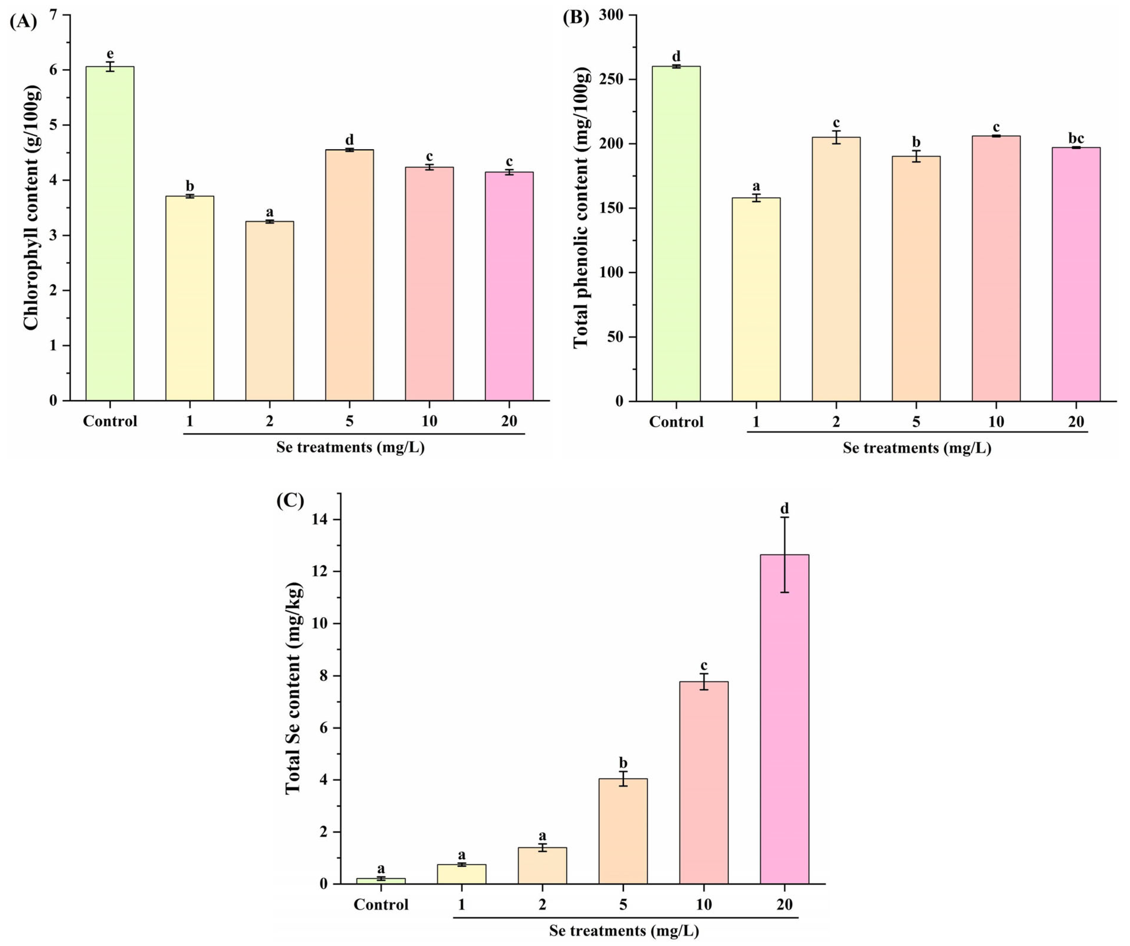
The development and expansion of leaf were associated with an increase in chlorophyll content [27]. In Figure 2A, Se treatments (1, 2, 5, 10 and 20 mg/L) significantly decreased the chlorophyll content in kale leaf compared to the control group. Meanwhile, notable differences were observed among the 1, 2, 5, 10 and/or 20 mg/L Se groups, except between the 10 and 20 mg/L Se groups. In previous studies, no significant influence of Se on the accumulation of chlorophyll in kale microgreens was observed [28], whereas Se fortification notably added to the chlorophyll content in cauliflower [29]. Our observation revealed that Se treatments at 1, 2, 5, 10 and 20 mg/L could inhibit the biosynthesis of chlorophyll in kale leaf. This might be explained by the different species. Moreover, the results indicated that Se fortification might down-regulate the nutritional value of chlorophyll in kale leaf, which might in turn affect the metabolic profiles of other nutrients.
The accumulation of polyphenols in plants is usually associated with stress conditions during the growth cycle [30]. Moreover, phenolic compounds are important specialized metabolites that contribute to the health-promoting properties of kale [31]. In Figure 2B, Se treatments (1, 2, 5, 10 and 20 mg/L) also significantly decreased the total phenolic content in kale leaf, as compared with the control group. Similarly, Ortega-Hernández et al. [32] found that Se treatments markedly decreased the phenolic accumulation in kale sprouts. It might be speculated that Se treatments caused stress alterations to kale, which should be further confirmed as there were different findings in previous studies concerning other cultivars of kale or other plants. For example, in an investigation conducted by Viltres-Portales et al. [28], the total polyphenolic compound content of kale microgreens is not affected by Se treatment. And Se fortification has been demonstrated to significantly increase the concentration of some phenolic acids, especially chlorogenic and protocatechuic acids in kale sprouts [15]. At the same time, Se fortification notably elevated the total polyphenolic content in cauliflower [29]. Furthermore, compared with the 1 mg/L Se group, the total phenolic contents in kale leaf were observably increased in other Se treatment groups. Similarly, Paweł et al. [11] found that Se fortification stimulated the production of phenolic acids in kale sprouts, depending on the Se dose. Based on the above findings, it is necessary to determine the comprehensive changes in nutritional compounds of kale using metabolomics analysis.
One of the simplest and most robust techniques to increase the Se content in plants is by growing plants in high-Se soil and applying Se fertilizers [33]. As illustrated in Figure 2C, as compared to the control group, 5, 10 and 20 mg/L Se treatments notably up-regulated the total Se contents in kale leaf. Moreover, Se treatments significantly increased the total Se contents of kale leaf in a dose-dependent manner, in the Se concentration range of 2–20 mg/L. This was consistent with the previous findings that Se biofortification was effective in enriching the Se content in kale microgreens [14], seedlings [10,12] and sprouts [11,15].

3.3. Effect of Se Fortification on Speciation of Se Compounds in Kale Leaf

The speciation of Se in plant tissue is important for understanding the efficiency of Se absorption for animals and humans [34]. Both inorganic Se compounds (i.e., Se(IV) and Se(VI)) and organic Se compounds (i.e., SeCys2, Se-MeSeCys and SeMet) are frequently found in plants [35]. As shown in Figure 3A,E, the Se(IV) and SeMet contents in kale leaf were significantly increased in the 10 and 20 mg/L Se groups compared to those in the control group. In Figure 3C,F, Se fortifications (1–20 mg/L) notably elevated the SeCys2 content and total contents of Se in different forms in kale leaf.
In Figure 3B, compared with the control group, the Se(VI) content in kale leaf was clearly enhanced in the 2, 5, 10 and 20 mg/L Se groups. In Figure 3D, the Se-MeSeCys content in kale leaf was dramatically increased in the 5, 10 and 20 mg/L Se groups compared to that in the control group. These results indicate that Se fortification added inorganic and organic Se compounds to kale leaf. To the best of our knowledge, the median lethal doses of inorganic Se compounds (Se(IV) and Se(VI)) were 7 mg/kg·bw and ~13 mg/kg·bw, respectively. However, organic Se species can have greater bioactivities with less toxicity compared to inorganics species [33]. As a result, the bioactivities of kale leaf tend to be enriched by Se fortification. Furthermore, Figure 3 displays that the contents of organic Se compounds in Se-fortified kale are higher than those of inorganic Se compounds. From this perspective, Se fortification is desirable for kale as long as it is consumed within a safe dosage.

3.4. Effect of Se Fortification on Bioactive Compounds in Kale Leaf

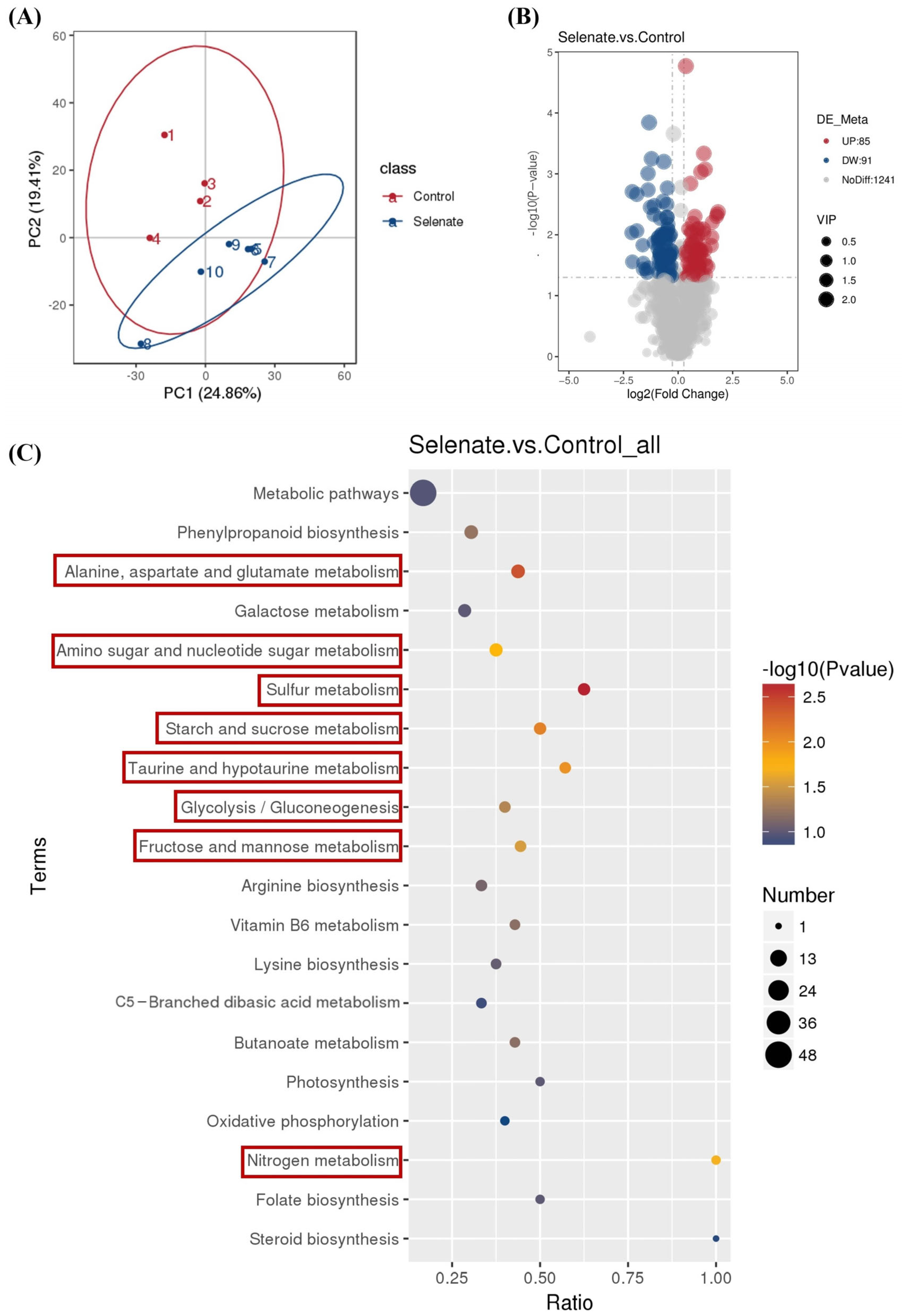
There are few reports on the effect of selenium supplementation on the concentration of bioactive compounds present in kale plants [36]. Thus, quasi-targeted metabolomics analysis was performed to ascertain the effect of Se fortification on bioactive compounds in kale leaf. The representative total ion chromatograms of kale samples from the control and selenate groups in positive ion (ESI+) and negative ion (ESI−) modes can be seen in the Supplementary Materials (Figures S1 and S2). In the PCA score plot (Figure 4A), samples of kale leaf were clearly separated between the control group and the selenate group, suggesting that Se fortification changed the metabolic profile of kale leaf. As shown in Figure 4B, a total of 176 differential metabolites (85 up-regulated and 91 down-regulated) were identified. Among them, 113 and 63 differential metabolites were detected in positive ion (ESI+) and negative ion (ESI−) modes, respectively. The differential metabolites can be mainly classified into six types of compounds, including amino acid and its derivatives (Table 1); organic acid and its derivatives (Table 2); carbohydrates and its derivatives (Table 3); lipids (Table 4); flavonoids (Table 5); and organoheterocyclics (Table 6).
Using KEGG analysis based on these differential metabolites, 46 terms were enriched. The top 20 enriched KEGG pathways are displayed in Figure 4C. There were eight significantly enriched KEGG pathways, including alanine, aspartate and glutamate metabolism; amino sugar and nucleotide sugar metabolism; sulfur metabolism; starch and sucrose metabolism; taurine and hypotaurine metabolism; glycolysis/gluconeogenesis; fructose and mannose metabolism; and nitrogen metabolism. These metabolic pathways have been reported to be clearly affected by Se fortification or other treatments in kale or other plants. For example, high-voltage electrostatic field treatment has been demonstrated to significantly regulate alanine, aspartate and glutamate metabolism of kale [37]. Se fortification has been indicated to significantly regulate amino sugar and nucleotide sugar metabolism of alfalfa sprouts [38]. Plants absorb inorganic selenium and convert it into various organic selenides via the sulfur metabolism pathway [39]. Because the absorption and assimilation of sulfur by plants are coordinated with the absorption and assimilation of nitrogen, by influencing sulfur metabolism, selenium also affects nitrogen metabolism [40]. Se fortification has been demonstrated to facilitate the production of sulfur compounds in kale sprouts [15,41]. Se fortification has been found to markedly modulate starch and sucrose metabolism and glycolysis/gluconeogenesis metabolic pathways of alfalfa leaves [42]. Selenium nanoparticle treatment has been observed to clearly regulate taurine and hypotaurine metabolism in soybean seedlings [43]. Se treatment has been reported to dramatically regulate fructose and mannose metabolism in rice [44].

3.4.1. Effect of Se Fortification on Amino Acid and Its Derivatives in Kale Leaf

After further analysis of the differential metabolites, 32 compounds (19 detected in ESI+ mode and 13 detected in ESI− mode) belonging to amino acid and its derivatives were identified, as shown in Table 1. Among them, 15 (i.e., trimethyllysine, 2-amino-2-deoxy-D-gluconate, homocysteine, L-asparagine, aspartate, ala-Gln, etc.) and 17 (D-threonine, L-glutamic acid, N alpha-acetyl-L-Arginine, ureidosuccinic acid, O-acetylserine, L-homoserine, etc.) compounds were significantly up-regulated and down-regulated, respectively. In particular, the quantitative values of trimethyllysine and ala-Gln in kale leaf were significantly increased to more than 2-fold in the selenate group compared to those in the control group. A previous study has reported that trimethyllysine is an important post-translationally modified amino acid with functions in the biosynthesis of carnitine, which is importantly involved in the transport of long-chain fatty acids from the cytosol to the mitochondria in both eukaryotes and some prokaryotes [45]. Moreover, Ala-Gln supplementation has been proven to possess an intestinal protection function [46,47]. On the other hand, the quantitative values of kynurenine, santacruzamate A and 3-(2-Naphthyl)-L-alanine clearly declined approximately 0.23-, 0.27- and 0.27-fold in the selenate group compared to those in the control group. Among them, 3-(2-Naphthyl)-L-alanine in dark tea has been proven to show significant correlations with digestive enzyme inhibition [48]. These observations indicated that Se fortification had significant influences on the contents of amino acid and its derivatives in kale leaf, which might enhance some biological activities like intestinal protection function.

3.4.2. Effect of Se Fortification on Organic Acid and Its Derivatives in Kale Leaf

After further analysis of the differential metabolites, 19 compounds (9 identified in ESI+ mode and 10 detected in ESI− mode) belonging to organic acid and its derivatives were determined, as illustrated in Table 2. Of those, 9 compounds including N-Acetyl-L-carnosine, maleamic acid, 4-hydroxybenzoate, p-aminobenzoate, alpha-ketoglutaric acid, L-cysteinesulfinic acid, hydroxycitric acid and 2-aminoethanesulfonic acid were notably up-regulated, whereas 10 compounds including 2-aminoethylphosphonate, dodecanedioic acid, trans-aconitic acid, phosphoric acid, succinic acid, methylmalonate, glutaconic acid, 3-hydroxyanthranilic acid, vanillic acid and isovanillic acid were evidently down-regulated. In particular, the quantitative values of p-aminobenzoate and L-cysteinesulfinic acid were markedly enhanced to approximately 3.45- and 2.57-fold in the selenate group as compared to those in the control group. Of those, p-aminobenzoate has been reported to possess numerous biological activities, such as antioxidant, antibacterial, antimutagenic, anticoagulant, fibrinolytic and immunomodulating activities, protection against UV irradiation, as well as chemical induction associated with thermotolerance and pathogen resistance in plants [49]. L-Cysteinesulfinic acid has been demonstrated to have anti-methanogenic and antimicrobial activities [50]. These results suggested that Se fortification had significant effects on the contents of organic acid and its derivatives in kale leaf, which might elevate some biological activities like antibacterial action.

3.4.3. Effect of Se Fortification on Carbohydrates and Their Derivatives in Kale Leaf

After further analysis of the differential metabolites, 25 compounds (7 detected in ESI+ mode and 18 detected in ESI− mode) belonging to carbohydrates and their derivatives were identified, as displayed in Table 3. Of those, 22 (i.e., isomaltose, DIMBOA glucoside, icariside B2, sorbitol-6-phosphate, D-glucose 1-phosphate, D-fructose 6-phosphate, etc.) and 3 (D-threose, pteroside A and sedoheptulose anhydride) compounds were dramatically up-regulated and down-regulated, respectively. In particular, the quantitative value of DIMBOA glucoside in kale leaf was significantly increased to be approximately 2.89-fold in the selenate group compared to that in the control group. It is worth noting that DIMBOA glucoside has been reported to enhance the resistance of wheat and maize to aphids [51]. Moreover, low-molecular-weight carbohydrates are compounds directly related to the kale flavor and nutritional quality [52]. According to Table 3, the quantitative values of D-glucopyranose, D-galactaric acid, L-sorbose, D-glucuronic acid, D-galactose, alpha-D-glucose, D-ribose, D-glucose and D-xylose were notably up-regulated. Among them, glucose is one of the major soluble sugars found in kale [3,4]. These findings implied that Se fortification had notable impacts on the amounts of carbohydrates and their derivatives in kale leaf, which might alter the kale flavor and nutritional quality.

3.4.4. Effect of Se Fortification on Lipids in Kale Leaf

Plant lipids have become increasingly popular as functional components in the creation of functional foods among all bioactive phytochemicals [3]. After further analysis of the differential metabolites, 17 compounds (12 identified in ESI+ mode and 5 identified in ESI− mode) belonging to lipids were determined, as revealed in Table 4. Among them, 4 (methyl oleate, 1-hexadecanol, (E)-3-(4-Hydroxyphenyl)propenoic acid (2S)-3-(beta-D-glucopyranosyloxy)-2-hydroxypropyl ester and hexadecanedioic acid) and 13 (i.e., palmitoleic acid, LPC (1-acyl 16:2), trans-2-hexenal, LysoPC 18:3 (2n isomer), 9,10-EODE, 3-methyladipic acid, etc.) compounds were significantly up-regulated and down-regulated, respectively. In particular, the quantitative value of 1-hexadecanol in kale leaf was observably increased to be more than 2-fold in the selenate group compared to that in the control group. In previous studies, 1-hexadecanol has been found to exhibit antimicrobial [53] and anticancer [54] activities. These results hinted that Se fortification produced clear influences on the contents of lipids in kale leaf, which might enhance some bioactivities.

3.4.5. Effect of Se Fortification on Flavonoids in Kale Leaf

Flavonoids, being prevalent in vegetables, are essential to the diverse stages of their growth, development and storage [55], and they have been shown to have multiple pharmacological activities, such as antioxidant, anti-inflammatory, antibacterial, antiviral, anticancer, antidiabetic and immune function modulation [56]. Kale is a good source of flavonoids compared to other commonly consumed vegetables [4]. Organic and bio-organic fertilizers have been demonstrated to enhance the flavonoids of broccoli (Brassica leracea, Var. Italica) [57]. After further analysis of the differential metabolites, 14 compounds (10 detected in ESI+ mode and 4 detected in ESI− mode) belonging to flavonoids were identified, as listed in Table 5. Of those, seven compounds (8-C-hexosyl-apigenin O-hexosyl-O-hexoside, methylChrysoeriol-5-O-hexoside, irisolidone-7-O-beta-d-glucoside, quercetin-O-glucoside, C-pentosyl-chrysoeriol 7-O-feruloylhexoside, chrysin O-malonylhexoside and sakuranetin) were notably up-regulated, and seven compounds (methylLuteolin-C-hexoside, daidzin, chrysin O-hexoside, 4′-O-glucosylvitexin, genistin, heptamethoxyflavone and azaleatin) were notably down-regulated. In particular, the quantitative value of C-pentosyl-chrysoeriol 7-O-feruloylhexoside in kale leaf was significantly increased to approximately 2.86-fold in the selenate group compared to that in the control group. Similarly, the C-pentosyl-chrysoeriol 7-O-feruloylhexoside content of goji fruit was significantly increased with phosphorus fertilizer levels [58]. These observations indicated that Se fortification generated significant effects on the contents of flavonoids in kale leaf, which might elevate some pharmacological activities.

3.4.6. Effect of Se Fortification on Organoheterocyclic Compounds in Kale Leaf

Organoheterocyclic compounds have been reported for the treatment of cancer and kidney diseases [59]. Moreover, the accumulation of glucosinolates induced by thermal stress has been found to significantly affect organoheterocyclic compounds of kale [60]. After further analysis of the differential metabolites, 14 compounds (12 identified in ESI+ mode and 2 identified in ESI− mode) belonging to organoheterocyclic compounds were determined, as shown in Table 6. Among them, 4 compounds (deoxynojirimycin, 3-indoleacetonitrile, NNK and indole-2-carboxylic acid) were significantly up-regulated, and 10 compounds (i.e., hypoxanthine, zarzissine, N-methylnicotinamide, (S)-2-phenyloxirane, 4-pyridoxate, brazilin, etc.) were significantly down-regulated, respectively. In particular, the quantitative value of indole-2-carboxylic acid was markedly up-regulated to approximately 2.19-fold in the selenate group as compared to that in the control group. In a previous report [61], indole-2-carboxylic acids have been demonstrated to be MCL-1 inhibitors, thereby showed anticancer activity. These findings suggested that Se fortification had significant impacts on the amounts of organoheterocyclic compounds in kale leaf, which might alter some bioactivities.

3.4.7. Effect of Se Fortification on Other Differential Metabolites in Kale Leaf

There were other differential metabolites screened between the selenate group and the control group, including 10 phenylpropanoids and polyketides, 12 nucleotides and their derivates, 6 terpenoids, 4 amines, 5 vitamins, 4 phenols and their derivatives, 6 phenolic acids, 2 phytohormones, 2 alkaloids and their derivatives, 1 polyamine, 1 organooxygen compound, 1 benzene and substituted derivatives and 1 alcohol and polyol, as shown in Table 7. In a previous study [62], terpenoids and alkaloids were speculated to be bioactive constituents for the antibacterial activity of kale leaf extracts. Moreover, Se fortification has been widely reported to affect the synthesis of phenolic compounds in kale sprouts, which is related to cytotoxic, antioxidant and anti-inflammatory activities [11,15,32,41]. Furthermore, phytohormones are known to participate in plant Se accumulation [63], and polyamines are involved in the regulation of the cellular growth, apoptosis, rooting, flower development and stress responses of plants [64]. In Table 7, the quantitative value of caffeic acid in kale leaf is significantly increased to more than 2-fold in the selenate group compared to that in the control group. It has been reported that caffeic acid is one of the most abundant phenolic acids in kale leaves, and this compound is highly correlated with antioxidant and antibacterial activities [65]. These results implied that Se fortification produced significant effects on the contents of other differential metabolites like phenylpropanoids and polyketides in kale leaf, which might enhance some bioactivities.

3.5. Metabolic Biomarkers in Kale Leaf Affected by Se Fortification

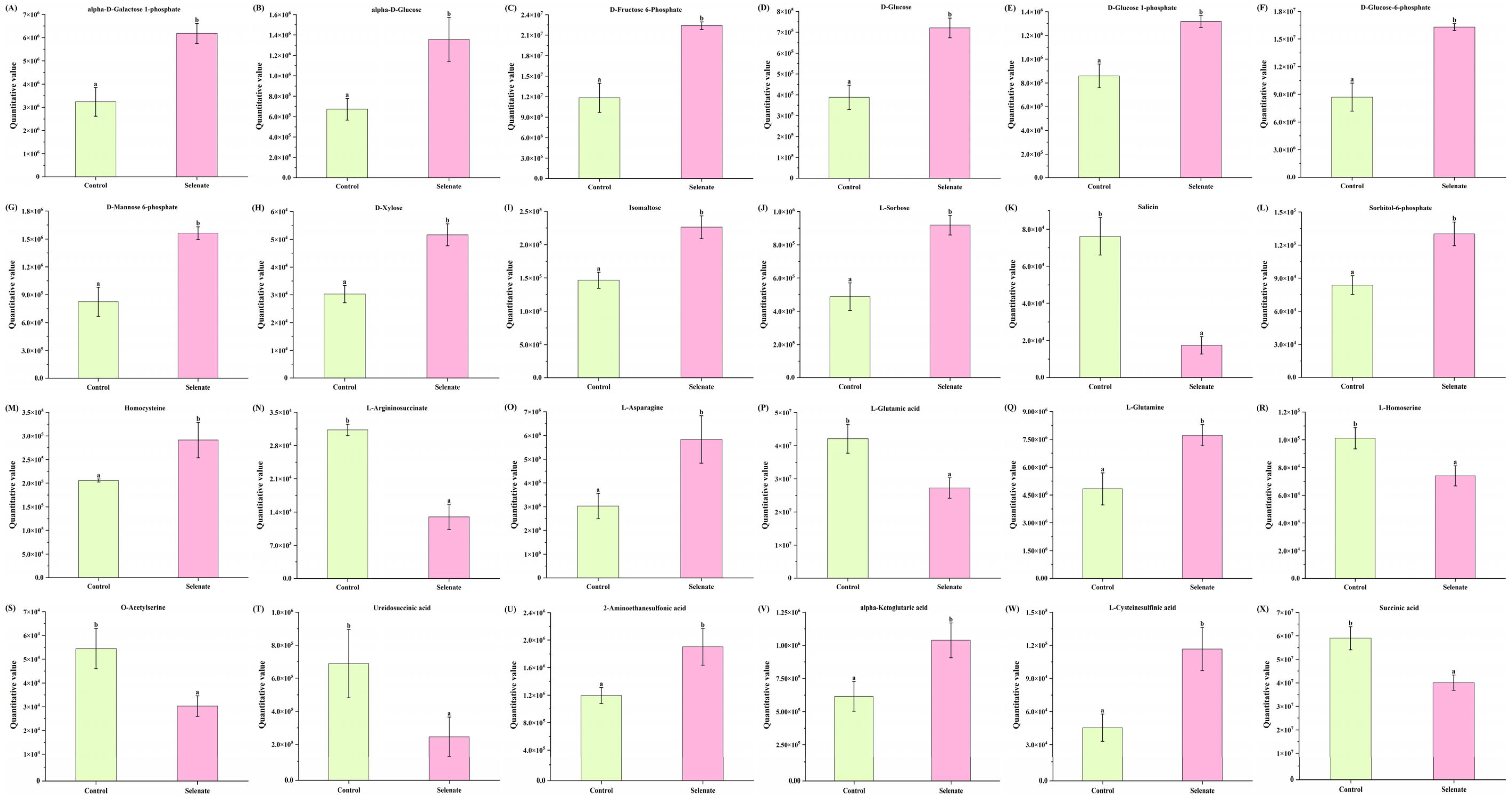
As mentioned in Figure 4, eight different metabolic pathways of kale leaf that were significantly regulated by Se fortification were determined. When combined, there were 24 differential metabolites (12 carbohydrates and their derivatives, 8 amino acids and their derivatives and 4 organic acid and their derivatives) enriched in the eight metabolic pathways, as shown in Figure 5. These 24 metabolites could be considered as biomarkers indicating the changes in nutrients of kale caused by selenium fortification. The interaction of sugars and amino acids could affect plant quality and aroma [66]. Moreover, the contents of amino acids could influence nitrogen accumulation in plants [67], and amino acids’ or compounds’ synthesis play important roles in the adaptions of plants to stress and adversity [68]. In Figure 5, the quantitative values of 17 (i.e., alpha-D-galactose 1-phosphate, alpha-D-glucose, D-fructose 6-phosphate, D-glucose, homocysteine and L-asparagine, etc.) and 7 (salicin, L-argininosuccinate, L-glutamic acid, L-homoserine, O-acetylserine, ureidosuccinic acid and succinic acid) compounds ae notably up-regulated and down-regulated, respectively. Similarly, nano-Se foliar application increased the glucose content in Siraitia grosvenorii [66]. The D-glucose-6-phosphate content was significantly increased, whilst that of L-glutamic acid was dramatically decreased in alfalfa affected by Se fortification [67]. L-Asparagine and L-glutamine in red pitaya fruit have been demonstrated to be greatly increased by nano-selenium biofortification [68]. Of note, some of the above-metabolic biomarkers have been demonstrated to possess health benefits. For instance, 2-aminoethanesulfonic acid (also known as taurine) has been reported to play important roles in the treatment of diseases of the muscle, the central nervous system and the cardiovascular system [69]. Alpha-ketoglutaric acid is involved in both energy metabolism and carbon and nitrogen metabolism, exhibiting a variety of functions, and therefore it may act as a regulator in the progression of a variety of diseases [70]. L-Cysteinesulfinic acid has been proven to modulate cardiovascular function in the periaqueductal gray area of rats [71]. Therefore, the changes in metabolic biomarkers induced by Se fortification might be related to the growth, quality and nutritive value of kale. This speculation should be confirmed in the future.

3.6. Correlations Between Metabolic Biomarkers and Se Contents as Well as Speciation

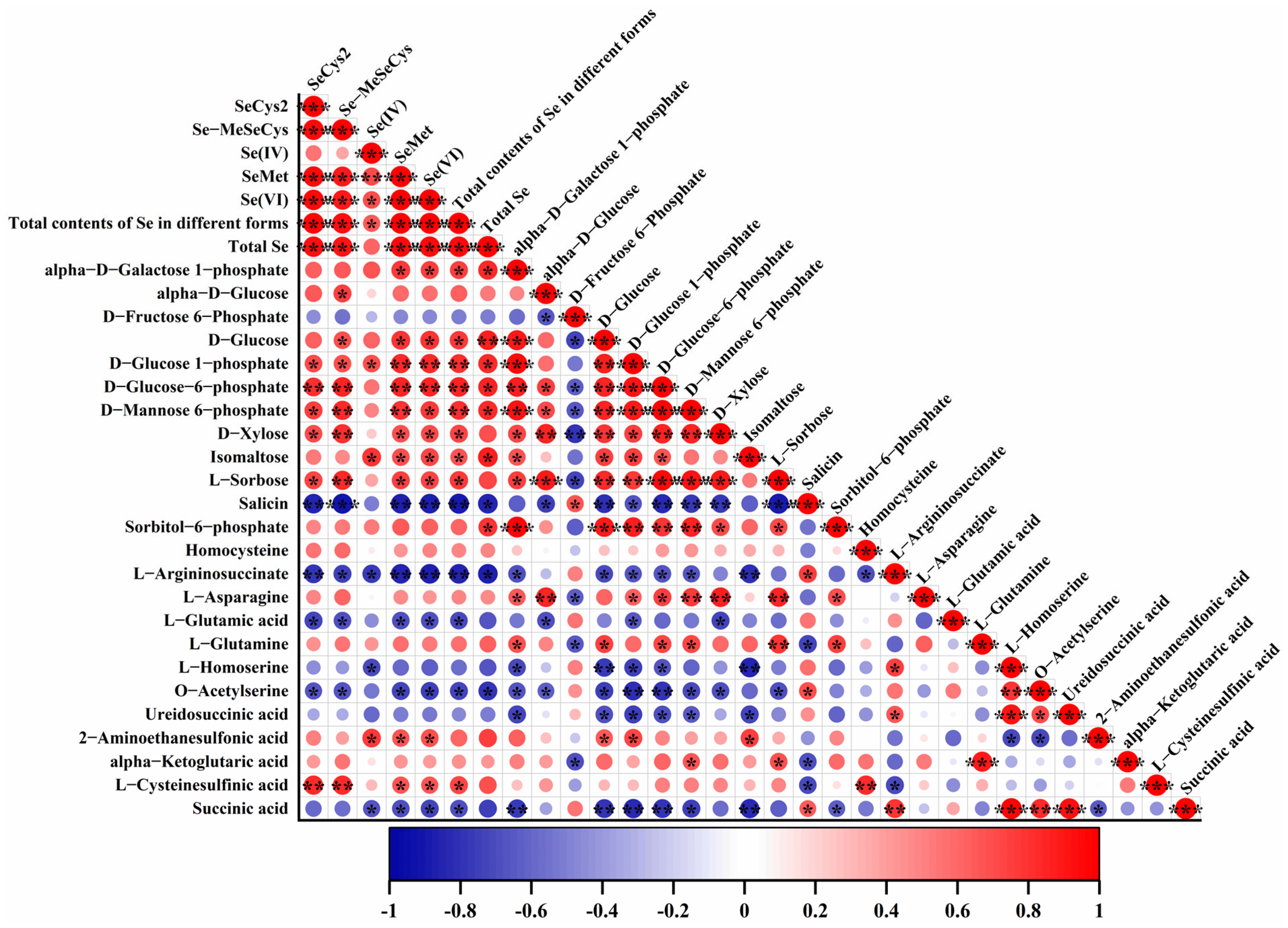
Pearson correlation analyses were performed to explore whether the contents of metabolic biomarkers in kale leaf were related to Se contents and speciation, as illustrated in Figure 6. Many significant positive correlations were observed between carbohydrates and their derivatives and Se contents as well as speciation: SeCys2 with D-glucose 1-phosphate, D-glucose-6-phosphate, D-mannose 6-phosphate, D-xylose and L-sorbose; Se-MeSeCys with alpha-D-glucose, D-glucose, D-glucose 1-phosphate, D-glucose-6-phosphate, D-mannose 6-phosphate, D-xylose and L-sorbose; Se(IV) with D-glucose 1-phosphate and isomaltose; SeMet and Se(VI) with alpha-D-galactose 1-phosphate, D-glucose, D-glucose 1-phosphate, D-glucose-6-phosphate, D-mannose 6-phosphate, D-xylose, isomaltose, L-sorbose; total contents of Se in different forms with alpha-D-galactose 1-phosphate, D-glucose, D-glucose 1-phosphate, D-glucose-6-phosphate, D-mannose 6-phosphate, D-xylose, isomaltose, L-sorbose; total Se with alpha-D-galactose 1-phosphate, D-glucose, D-glucose 1-phosphate, D-glucose-6-phosphate, D-mannose 6-phosphate, isomaltose and sorbitol-6-phosphate. Meanwhile, some clear positive correlations were seen between organic acid and its derivatives and Se contents as well as speciation: SeCys2 with L-cysteinesulfinic acid; Se-MeSeCys with L-cysteinesulfinic acid; Se(IV) with 2-aminoethanesulfonic acid; SeMet and Se(VI) with 2-aminoethanesulfonic acid and L-cysteinesulfinic acid; total contents of Se in different forms with L-cysteinesulfinic acid.
On the other hand, some remarkable negative correlations were seen between amino acid and its derivatives and Se contents as well as speciation: SeCys2 and Se-MeSeCys with L-argininosuccinate, L-glutamic acid and O-acetylserine; Se(IV) with L-argininosuccinate and L-homoserine; SeMet, Se(VI) and total contents of Se in different forms with L-argininosuccinate, L-glutamic acid and O-acetylserine; total Se with L-argininosuccinate and O-acetylserine. Moreover, SeCys2, Se-MeSeCys, Se(IV), SeMet, Se(VI), total contents of Se in different forms and total Se showed significant negative correlations with one (salicin) of the carbohydrates and its derivatives. Meanwhile, Se(IV), SeMet, Se(VI) and total contents of Se in different forms were clearly negatively correlated with one (succinic acid) organic acid and its derivatives.
Furthermore, prominent positive and negative correlations were found in themselves of metabolic biomarkers or Se contents as well as speciation. Similarly, clear correlations between differential metabolites and Se along with differential metabolites and differential metabolites have been discovered in purple rice grains exposed to different selenium concentrations [72]. In the study by Zagrodzki et al. [11], Se was proven to be correlated only with caffeic acid in Se fortification of kale. Paśko et al. [15] found that Se speciation in kale sprouts fortified with novel organic Se compounds was strongly correlated with one another.
In general, Se fortification produced positive effects on the accumulation of carbohydrates and their derivatives along with organic acid and its derivatives, whereas this generated negative influences on the production of amino acid and its derivatives.

4. Conclusions

Se deficiency is an urgent problem that needs to be addressed, and Se fortification is commonly applied to produce Se-enriched foods. In the present study, Se fortification using Se fertilizer significantly increased the contents of total Se and different Se speciation in kale leaf. Meanwhile, Se fortification clearly increased the plant height and leaf weight of kale, and it notably decreased the chlorophyll and total phenolic contents in kale leaf. Moreover, Se fortification markedly changed the metabolic profile of kale leaf, especially involved in six types of compounds and eight metabolic pathways. Twenty-four metabolites were screened as biomarkers indicating the changes in nutrients of kale caused by selenium fortification. Furthermore, correlation analysis reflected that Se fortification produced positive effects on the accumulation of carbohydrates and their derivatives as well as organic acid and its derivatives, while it generated negative influences on the production of amino acid and its derivatives, in kale leaf. These results indicate that Se fortification has a significant influence on growth performance and nutritional compounds of kale. In contrast with previous studies, our findings are beneficial for comprehensively understanding the impact of selenium fortification on the nutritional compounds in kale, and they can guide future research on the potential biological activities of Se-biofortified kale. This is of great significance for the development and future research of the kale industry. However, several limitations of this study should be addressed in future research: the nutritive value and bioactivity of Se-enriched kale remain to be thoroughly investigated; the mechanisms through which selenium fortification influences the growth performance and nutritional compounds of kale require further elucidation; the effects of other fortification methods on kale growth and nutrients of kale also merit comparative study. In the next work, we will focus on resolving these issues.

Supplementary Materials

The following supporting information can be downloaded at https://www.mdpi.com/article/10.3390/foods14183283/s1, Figure S1. Representative total ion chromatograms of kale samples from control and selenate groups in positive ion mode. Figure S2. Representative total ion chromatograms of kale samples from control and selenate groups in negative ion mode.

Author Contributions

Conceptualization, X.-Y.Z., M.W., Q.Z. and X.-Y.W.; methodology, X.-Y.Z., H.L., Q.Z. and X.-Y.W.; investigation, X.-Y.Z., H.L., L.-C.S. and Q.Z.; formal analysis, X.-Y.Z., L.-C.S. and T.-T.L.; writing—original draft preparation, X.-Y.Z.; writing—review and editing, Q.Z. and X.-Y.W.; supervision, M.W. and X.-Y.W.; project administration, H.L., L.-C.S. and T.-T.L.; funding acquisition, X.-Y.Z., Q.Z. and X.-Y.W. All authors have read and agreed to the published version of the manuscript.

Funding

This work was financially supported by the Science and Technology Innovation Talent Project in Ganzhou [2022CXRC9540] and the University-Level Scientific Research Projects of Gannan Medical University [QD201913, QD202128, TD202313 and TD202313-1].

Institutional Review Board Statement

Not applicable.

Informed Consent Statement

Not applicable.

Data Availability Statement

The original contributions presented in the study are included in the article/Supplementary Materials. Further inquiries can be directed to the corresponding authors.

Conflicts of Interest

The authors declare no conflicts of interest.

References

  1. Šamec, D.; Urlić, B.; Salopek-Sondi, B. Kale (Brassica oleracea var. acephala) as a superfood: Review of the scientific evidence behind the statement. Crit. Rev. Food Sci. 2019, 59, 2411–2422. [Google Scholar] [CrossRef]
  2. Park, Y.J.; Lee, H.M.; Shin, M.J.; Arasu, M.V.; Chung, D.Y.; Al-Dhabi, N.A.; Kim, S.J. Effect of different proportion of sulphur treatments on the contents of glucosinolate in kale (Brassica oleracea var. acephala) commonly consumed in Republic of Korea. Saudi J. Biol. Sci. 2018, 25, 349–353. [Google Scholar] [CrossRef]
  3. Khalid, W.; Fareed, A.; Abdul, R.M.; Asma, A.R.; Hadiqa, F.u.R.; Sajid, A.M.; Saadia, A.; Muhammad, Z.; Saira, S.; Ammar, A.F.; et al. Industrial applications of kale (Brassica oleracea var. sabellica) as a functional ingredient: A review. Int. J. Food Prop. 2023, 26, 489–501. [Google Scholar] [CrossRef]
  4. Satheesh, N.; Workneh Fanta, S. Kale: Review on nutritional composition, bio-active compounds, anti-nutritional factors, health beneficial properties and value-added products. Cogent Food Agric. 2020, 6, 1811048. [Google Scholar] [CrossRef]
  5. Haile, A.; Ayalew, T. Comparative study on the effect of bio-slurry and inorganic N-fertilizer on growth and yield of kale (Brassica oleracea L.). Afr. J. Plant Sci. 2018, 12, 81–87. [Google Scholar]
  6. Rodríguez, V.M.; Soengas, P.; Alonso-Villaverde, V.; Sotelo, T.; Cartea, M.E.; Velasco, P. Effect of temperature stress on the early vegetative development of Brassica oleracea L. BMC Plant Biol. 2015, 15, 145. [Google Scholar] [CrossRef]
  7. Šamec, D.; Ljubej, V.; Redovniković, I.R.; Fistanić, S.; Salopek-Sondi, B. Low temperatures affect the physiological status and phytochemical content of flat leaf kale (Brassica oleracea var. acephala) Sprouts. Foods 2022, 11, 264. [Google Scholar] [CrossRef] [PubMed]
  8. Mao, Y.X.; Chai, X.R.; Zhong, M.; Zhang, L.; Zhao, P.Y.; Kang, Y.Y.; Guo, J.X.; Yang, X. Effects of nitrogen and magnesium nutrient on the plant growth, quality, photosynthetic characteristics, antioxidant metabolism, and endogenous hormone of Chinese kale (Brassica albograbra Bailey). Sci. Hortic. 2022, 303, 111243. [Google Scholar] [CrossRef]
  9. Krawczyk, K.K.; Smoleń, S.; Wisła-Świder, A.; Kowalska, I.; Kiełbasa, D.; Pitala, J.; Krzemińska, J.; Waśniowska, J.; Koronowicz, A. Kale (Brassica oleracea L. var. sabellica) biofortified with iodoquinolines: Effectiveness of enriching with iodine and influence on chemical composition. Sci. Hortic. 2024, 323, 112519. [Google Scholar] [CrossRef]
  10. Leamsamrong, K.; Tongjaroenbuangam, W.; Maneetong, S.; Chantiratikul, A.; Chinrasri, O.; Chantiratikul, P. Physicochemical contents, antioxidant activities, and acute toxicity assessment of selenium-enriched Chinese Kale (Brassica oleracea var. alboglabra L.) seedlings. J. Chem. 2019, 2019, 7983038. [Google Scholar] [CrossRef]
  11. Zagrodzki, P.; Paśko, P.; Galanty, A.; Tyszka-Czochara, M.; Wietecha-Posłuszny, R.; Rubió, P.S.; Bartoń, H.; Prochownik, E.; Muszyńska, B.; Sułkowska-Ziaja, K.; et al. Does selenium fortification of kale and kohlrabi sprouts change significantly their biochemical and cytotoxic properties? J. Trace Elem. Med. Biol. 2020, 59, 126466. [Google Scholar] [CrossRef] [PubMed]
  12. Maneetong, S.; Chookhampaeng, S.; Chantiratikul, A.; Chinrasri, O.; Thosaikham, W.; Sittipout, R.; Chantiratikul, P. Hydroponic cultivation of selenium-enriched kale (Brassica oleracea var. alboglabra L.) seedling and speciation of selenium with HPLC-ICP-MS. Microchem. J. 2013, 108, 87–91. [Google Scholar] [CrossRef]
  13. Li, J.; Liang, D.L.; Qin, S.Y.; Feng, P.Y.; Wu, X.P. Effects of selenite and selenate application on growth and shoot selenium accumulation of pak choi (Brassica chinensis L) during successive planting conditions. Environ. Sci. Pollut. R 2015, 22, 11076–11086. [Google Scholar] [CrossRef]
  14. Tavan, M.; Wee, B.; Fuentes, S.; Pang, A.; Brodie, G.; Viejo, C.G.; Gupta, D. Biofortification of kale microgreens with selenate-selenium using two delivery methods: Selenium-rich soilless medium and foliar application. Sci. Hortic. 2024, 323, 112522. [Google Scholar] [CrossRef]
  15. Paśko, P.; Galanty, A.; Zagrodzki, P.; Żmudzki, P.; Bieniek, U.; Prochownik, E.; Domínguez-Álvarez, E.; Bierła, K.; Łobiński, R.; Szpunar, J.; et al. Varied effect of fortification of kale sprouts with novel organic selenium compounds on the synthesis of sulphur and phenolic compounds in relation to cytotoxic, antioxidant and anti-inflammatory activity. Microchem. J. 2022, 179, 107509. [Google Scholar] [CrossRef]
  16. Niu, Y.Q.; Zheng, M.X.; Tian, D.G.; Zhu, Y.M.; Chen, H.; Zhu, Y.J.; Su, H.L. Quasi-targeted metabonomics reveals metabolites associated with antioxidant activity of Mesona chinensis Benth Cultivar Xiaoye. Plants 2025, 14, 1585. [Google Scholar] [CrossRef]
  17. Fang, X.; Xue, R.; Xiao, J.Y.; Pu, Q.; Wang, Y.F.; Yuan, Y.; Liu, B.; Sui, M.Y.; Jiang, G.X.; Niaz, R.; et al. Effects of different fermentation modes on tea leaves: Revealing the metabolites modification by quasi-targeted metabolomics. Food Biosci. 2024, 62, 105223. [Google Scholar] [CrossRef]
  18. Ma, X.Y.; Jin, W.; Wang, L.F.; Chen, W.W.; Wang, Y.F.; Wen, H.B.; Cao, X.J. Metabolic response of Sinosolenaia oleivora to heat and drought stress using a quasi-targeted metabolomics approach. Comp. Biochem. Phys. D 2025, 55, 101499. [Google Scholar] [CrossRef]
  19. Zhang, X.Y.; Li, M.C.; Zhen, L.; Wang, Y.D.; Wang, Y.F.; Qin, Y.Y.; Zhang, Z.H.; Zhao, T.R.; Cao, J.X.; Liu, Y.P.; et al. Ultra-high hydrostatic pressure pretreatment on White Que Zui Tea: Chemical constituents, antioxidant, cytoprotective, and anti-Inflammatory activities. Foods 2023, 12, 628. [Google Scholar] [CrossRef]
  20. Ren, J.; Yang, L.; Cao, R.F.; Wang, Y.D.; Zhang, C.; Yu, X.J.; Meng, W.D.; Ye, X.L. Integrated metabolome and transcriptome analysis provides new insights into the glossy graft cucumber fruit (Cucumis sativus L.). Int. J. Mol. Sci. 2023, 24, 12147. [Google Scholar] [CrossRef] [PubMed]
  21. Wang, Y.Q.; Hu, L.P.; Liu, G.M.; Zhang, D.S.; He, H.J. Evaluation of the nutritional quality of Chinese Kale (Brassica alboglabra Bailey) using UHPLC-Quadrupole-Orbitrap MS/MS-based metabolomics. Molecules 2017, 22, 1262. [Google Scholar] [CrossRef]
  22. NY/T 3082-2017; Determination of Chlorophyll Content in Fruits, Vegetables and Derived Products-Spectrophotometry Method. Ministry of Agriculture of the People’s Republic of China: Beijing, China, 2017.
  23. Liao, Q.; Liang, P.X.; Xing, Y.; Yao, Z.F.; Chen, J.P.; Pan, L.P.; Deng, Y.Q.; Liu, Y.X.; Huang, D. Optimizing selenium application for enhanced quality and nutritional value of spring tea (Camellia sinensis). Horticulturae 2025, 11, 423. [Google Scholar] [CrossRef]
  24. GB 5009.93-2017; National Food Safety Standard Determination of Selenium in Food. China Standard Publishing House: Beijing, China, 2017.
  25. NY/T 3556-2020; Determination of Selenocysteine and Selenomethionine in Cereals– Liquid Chromatography–Inductively Coupled Plasma Mass Spectrometry. Ministry of Agriculture of the People’s Republic of China: Beijing, China, 2020.
  26. Wu, Y.; Xiao, H.W.; Zhang, H.; Pan, A.L.; Shen, J.; Sun, J.; Liang, Z.H.; Pi, J.S. Quasi-targeted metabolomics approach reveal the metabolite differences of three poultry eggs. Foods 2023, 12, 2765. [Google Scholar] [CrossRef] [PubMed]
  27. Lefsrud, M.; Kopsell, D.; Wenzel, A.; Sheehan, J. Changes in kale (Brassica oleracea L. var. acephala) carotenoid and chlorophyll pigment concentrations during leaf ontogeny. Sci. Hortic. 2007, 112, 136–141. [Google Scholar] [CrossRef]
  28. Viltres-Portales, M.; Sánchez-Martín, M.J.; Llugany, M.; Boada, R.; Valiente, M. Selenium biofortification of microgreens: Influence on phytochemicals, pigments and nutrients. Plant Physiol. Biochem. 2024, 206, 108283. [Google Scholar] [CrossRef]
  29. Saeedi, M.; Soltani, F.; Babalar, M.; Izadpanah, F.; Wiesner-Reinhold, M.; Baldermann, S. Selenium fortification alters the growth, antioxidant characteristics and secondary metabolite profiles of cauliflower (Brassica oleracea var. botrytis) cultivars in hydroponic culture. Plants 2021, 10, 1537. [Google Scholar] [CrossRef]
  30. Gupta, M.; Gupta, S. An overview of selenium uptake, metabolism, and toxicity in plants. Front. Plant Sci. 2017, 7, 2074. [Google Scholar] [CrossRef] [PubMed]
  31. Bianchi, G.; Picchi, V.; Tava, A.; Doria, F.; Walley, P.G.; Dever, L.; di Bella, M.C.; Arena, D.; Ben Ammar, H.; Lo Scalzo, R.; et al. Insights into the phytochemical composition of selected genotypes of organic kale (Brassica oleracea L. var. acephala). J. Food Compos. Anal. 2024, 125, 105721. [Google Scholar] [CrossRef]
  32. Ortega-Hernández, E.; Antunes-Ricardo, M.; Cisneros-Zevallos, L.; Jacobo-Velázquez, D.A. Selenium, sulfur, and methyl jasmonate treatments improve the accumulation of lutein and glucosinolates in kale Sprouts. Plants 2022, 11, 1271. [Google Scholar] [CrossRef]
  33. Tangjaidee, P.; Swedlund, P.; Xiang, J.Q.; Yin, H.Q.; Quek, S.Y. Selenium-enriched plant foods: Selenium accumulation, speciation, and health functionality. Front. Nutr. 2023, 9, 962312. [Google Scholar] [CrossRef]
  34. Silva, M.A.; de Sousa, G.F.; Bañuelos, G.; Amaral, D.; Brown, P.H.; Guilherme, L.R.G. Selenium speciation in Se-enriched soybean grains from biofortified plants grown under different methods of selenium application. Foods 2023, 12, 1214. [Google Scholar] [CrossRef]
  35. Bañuelos, G.S.; Freeman, J.L.; Arroyo, I.S. Selenium content and speciation differences in selenium enriched soups made from selenium biofortified plants. J. Food Compos. Anal. 2022, 105, 104255. [Google Scholar] [CrossRef]
  36. Ortega-Hernández, E.; Antunes-Ricardo, M.; Jacobo-Velázquez, D.A. Improving the health-benefits of kales (Brassica oleracea L. var. acephala DC) through the application of controlled abiotic stresses: A review. Plants 2021, 10, 2629. [Google Scholar] [CrossRef] [PubMed]
  37. Huang, Y.X.; Liu, C.; Shen, C.X.; Ran, B.H.; Yang, Z.P.; Zhou, L.; Xu, H.H.; Tang, Y. The impact of high voltage electrostatic field on the storage quality and metabolism of Chinese kale. J. Food Meas. Charact. 2024, 18, 3405–3424. [Google Scholar] [CrossRef]
  38. Ren, Y.R.; Zhang, Q.; Li, X.; Zhang, T.Y.; Tian, D.C.; Liu, L.; Dong, X.Y.; Wang, Z.Y.; Chai, M.F. Effects of selenium content on growth, antioxidant activity, and key selenium-enriched gene expression in alfalfa sprouts. Foods 2024, 13, 2261. [Google Scholar] [CrossRef]
  39. Chao, W.; Rao, S.; Chen, Q.W.; Zhang, W.W.; Liao, Y.L.; Ye, J.B.; Cheng, S.Y.; Yang, X.Y.; Xu, F. Advances in research on the involvement of selenium in regulating plant ecosystems. Plants 2022, 11, 2712. [Google Scholar] [CrossRef]
  40. Skrypnik, L.; Feduraev, P.; Golovin, A.; Maslennikov, P.; Styran, T.; Antipina, M.; Riabova, A.; Katserov, D. The integral boosting effect of selenium on the secondary metabolism of higher plants. Plants 2022, 11, 3432. [Google Scholar] [CrossRef]
  41. Zagrodzki, P.; Wiesner, A.; Marcinkowska, M.; Jamrozik, M.; Domínguez-Álvarez, E.; Bierła, K.; Łobiński, R.; Szpunar, J.; Handzlik, J.; Galanty, A.; et al. Relationships between molecular characteristics of novel organic selenium compounds and the formation of sulfur compounds in selenium biofortified kale sprouts. Molecules 2023, 28, 2062. [Google Scholar] [CrossRef]
  42. Hu, H.F.; Hu, J.K.; Wang, Q.D.; Xiang, M.L.; Zhang, Y.R. Transcriptome analysis revealed accumulation-assimilation of selenium and physio-biochemical changes in alfalfa (Medicago sativa L.) leaves. J. Sci. Food Agric. 2022, 102, 4577–4588. [Google Scholar] [CrossRef]
  43. Xiong, Y.Z.; Xiang, X.M.; Xiao, C.M.; Zhang, N.; Cheng, H.; Rao, S.; Cheng, S.Y.; Li, L. Illumina RNA and SMRT sequencing reveals the mechanism of uptake and transformation of selenium nanoparticles in soybean seedlings. Plants 2023, 12, 789. [Google Scholar] [CrossRef]
  44. Liu, N.X.; Yu, Q.T.; Chen, B.C.; Li, C.X.; Bu, F.S.; Li, J.R.; Peng, X.L.; Lu, Y.C. Selenium alleviates low-temperature stress in rice by regulating metabolic networks and functional genes. Agriculture 2025, 15, 1489. [Google Scholar] [CrossRef]
  45. Maas, M.N.; Hintzen, J.C.J.; Porzberg, M.R.B.; Mecinović, J. Trimethyllysine: From carnitine biosynthesis to epigenetics. Int. J. Mol. Sci. 2020, 21, 9451. [Google Scholar] [CrossRef]
  46. Tian, J.; He, G.; Mai, K.S.; Liu, C.D.; Zhou, H.H.; Wen, H. Dietary ala-Gln ameliorated growth suppression and intestinal injury induced by soya saponin in zebrafish. Aquaculture 2020, 529, 735748. [Google Scholar] [CrossRef]
  47. Liu, J.; Zong, C.G.; Yu, X.; Ding, Y.; Chang, B.; Wang, R.Y.; Sang, L.X. Alanyl-glutamine (Ala-Gln) ameliorates dextran sulfate sodium (DSS)-induced acute colitis by regulating the gut microbiota, PI3K-Akt/NF-κB/STAT3 signaling, and associated pulmonary injury. ACS Infect. Dis. 2023, 9, 979–992. [Google Scholar] [CrossRef] [PubMed]
  48. Yu, J.; Wu, Y.J.; Zeng, C.X.; Wu, H.R.; Jia, G.F.; Ye, J.H.; Qin, S.; Liu, Z.H.; Shi, M. Phytochemicals from fractioned dark tea water extract enhance the digestive enzyme inhibition, antioxidant capacities and glucose-lipid balance. Food Res. Int. 2025, 204, 115957. [Google Scholar] [CrossRef]
  49. Sumalan, R.L.; Croitor, L.; Petric, M.; Radulov, I.; Bourosh, P.; Sumalan, R.M.; Crisan, M. p-Aminobenzoate organic salts as potential plant growth regulators for tomatoes. Molecules 2020, 25, 1635. [Google Scholar] [CrossRef] [PubMed]
  50. Levent, G.; Božić, A.; Petrujkić, B.T.; Callaway, T.R.; Poole, T.L.; Crippen, T.L.; Harvey, R.B.; Ochoa-García, P.; Corral-Luna, A.; Yeater, K.M.; et al. Assessment of potential anti-methanogenic and antimicrobial activity of ethyl nitroacetate, α-lipoic acid, taurine and L-cysteinesulfinic acid in vitro. Microorganisms 2024, 12, 34. [Google Scholar] [CrossRef]
  51. Li, B.; Förster, C.; Robert, C.A.M.; Züst, T.; Hu, L.; Machado, R.A.R.; Berset, J.D.; Handrick, V.; Knauer, T.; Hensel, G.; et al. Convergent evolution of a metabolic switch between aphid and caterpillar resistance in cereals. Sci. Adv. 2018, 4, eaat6797. [Google Scholar] [CrossRef]
  52. Megías-Pérez, R.; Hahn, C.; Ruiz-Matute, A.I.; Behrends, B.; Albach, D.C.; Kuhnert, N. Changes in low molecular weight carbohydrates in kale during development and acclimation to cold temperatures determined by chromatographic techniques coupled to mass spectrometry. Food Res. Int. 2020, 127, 108727. [Google Scholar] [CrossRef]
  53. Ganesh, M.; Mohankumar, M. Extraction and identification of bioactive components in Sida cordata (Burm. f.) using gas chromatography-mass spectrometry. J. Food Sci. Tech. 2017, 54, 3082–3091. [Google Scholar] [CrossRef]
  54. Cui, W.W.; Liu, D.; Gu, W.; Chu, B. Peroxisome-driven ether-linked phospholipids biosynthesis is essential for ferroptosis. Cell Death Differ. 2021, 28, 2536–2551. [Google Scholar] [CrossRef]
  55. Zheng, X.Y.; Zhang, X.J.; Zeng, F.K. Biological functions and health benefits of flavonoids in fruits and vegetables: A contemporary review. Foods 2025, 14, 155. [Google Scholar] [CrossRef]
  56. Peng, P.; Zou, J.R.; Zhong, B.; Zhang, G.X.; Zou, X.F.; Xie, T.P. Protective effects and mechanisms of flavonoids in renal ischemia-reperfusion injury. Pharmacology 2023, 108, 27–36. [Google Scholar] [CrossRef]
  57. Naguib, A.E.M.M.; El-Baz, F.K.; Salama, Z.A.; Hanaa, H.A.E.B.; Ali, H.F.; Gaafar, A.A. Enhancement of phenolics, flavonoids and glucosinolates of Broccoli (Brassica olaracea, var. Italica) as antioxidants in response to organic and bio-organic fertilizers. J. Saudi Soc. Agric. Sci. 2012, 11, 135–142. [Google Scholar] [CrossRef]
  58. Wei, F.; Shi, Z.G.; Wan, R.; Li, Y.X.; Wang, Y.J.; An, W.; Qin, K.; Cao, Y.L.; Chen, X.Y.; Wang, X.Y.; et al. Impact of phosphorus fertilizer level on the yield and metabolome of goji fruit. Sci. Rep. 2020, 10, 14656. [Google Scholar] [CrossRef]
  59. Wang, T.; Tang, C.Y.; Chen, J.B.; Liang, J.; Li, Y.L.; Li, X.Z. Accumulation characteristics of natural Ophiocordyceps sinensis metabolites driven by environmental factors. Metabolites 2024, 14, 414. [Google Scholar] [CrossRef]
  60. Díaz-Urbano, M.; Velasco, P.; Cartea, M.E.; Rodríguez, V.M. Metabolism reorganization in kale (Brassica oleracea L. var acephala) populations with divergent glucosinolate content under thermal stresses. Agronomy 2022, 12, 2652. [Google Scholar] [CrossRef]
  61. Leverson, J.D.; Zhang, H.; Chen, J.; Tahir, S.K.; Phillips, D.C.; Xue, J.; Nimmer, P.; Jin, S.; Smith, M.; Xiao, Y.; et al. Potent and selective small-molecule MCL-1 inhibitors demonstrate on-target cancer cell killing activity as single agents and in combination with ABT-263 (navitoclax). Cell Death Dis. 2015, 6, e1590. [Google Scholar] [CrossRef] [PubMed]
  62. Murugesan, K.; Mulugeta, K.; Hailu, E.; Tamene, W.; Alagar Yadav, S. Insights for integrative medicinal potentials of Ethiopian Kale (Brassica carinata): Investigation of antibacterial, antioxidant potential and phytocompounds composition of its leaves. Chin. Herb. Med. 2021, 13, 250–254. [Google Scholar] [CrossRef] [PubMed]
  63. Golubkina, N.; Moldovan, A.; Kekina, H.; Kharchenko, V.; Sekara, A.; Vasileva, V.; Skrypnik, L.; Tallarita, A.; Caruso, G. Joint biofortification of plants with selenium and iodine: New field of discoveries. Plants 2021, 10, 1352. [Google Scholar] [CrossRef]
  64. Kiełkowska, A.; Adamus, A. Exogenously applied polyamines reduce reactive oxygen species, enhancing cell division and the shoot regeneration from Brassica oleracea L. var. capitata protoplasts. Agronomy 2021, 11, 735. [Google Scholar] [CrossRef]
  65. Ayaz, F.A.; Hayırlıoglu-Ayaz, S.; Alpay-Karaoglu, S.; Grúz, J.; Valentová, K.; Ulrichová, J.; Strnad, M. Phenolic acid contents of kale (Brassica oleraceae L. var. acephala DC.) extracts and their antioxidant and antibacterial activities. Food Chem. 2008, 107, 19–25. [Google Scholar] [CrossRef]
  66. Zhou, C.R.; Zhang, J.B.; Wu, Y.L.; Cheng, H.Y.; Pang, Q.L.; Xiao, Y.H.; Li, D.; Pan, C.P. Metabolomic analysis on the mechanism of nanoselenium biofortification improving the Siraitia grosvenorii nutritional and health value. Foods 2022, 11, 3019. [Google Scholar] [CrossRef]
  67. Sun, P.B.; Ge, G.T.; Sun, L.; Bao, J.; Zhao, M.Q.E.; Hao, J.F.; Zhang, Y.H.; Liu, G.S.; Wang, Z.J.; Jia, Y.S. Metabolomics combined with physiology and transcriptomics reveal the regulation of key nitrogen metabolic pathways in alfalfa by foliar spraying with nano-selenium. J. Nanobiotechnol. 2025, 23, 7. [Google Scholar] [CrossRef] [PubMed]
  68. Yu, H.; Miao, P.J.; Li, D.; Wu, Y.L.; Zhou, C.R.; Pan, C.P. Improving red pitaya fruit quality by nano-selenium biofortification to enhance phenylpropanoid and betalain biosynthesis. Ecotoxicol. Environ. Saf. 2023, 267, 115653. [Google Scholar] [CrossRef]
  69. Schaffer, S.; Kim, H.W. Effects and mechanisms of taurine as a therapeutic agent. Biomol. Ther. 2018, 26, 225–241. [Google Scholar] [CrossRef] [PubMed]
  70. Meng, X.Q.; Liu, H.Q.; Peng, L.X.; He, W.G.; Li, S.Y. Potential clinical applications of alpha-ketoglutaric acid in diseases (Review). Mol. Med. Rep. 2022, 25, 151. [Google Scholar] [CrossRef] [PubMed]
  71. Maione, S.; Leyva, J.; Palazzo, E.; Stella, L.; Rossi, F. L-Cysteinesulfinic acid modulates cardiovascular function in the periaqueductal gray area of rat. J. Cardiovasc. Pharmacol. 1998, 32, 650–653. [Google Scholar] [CrossRef]
  72. Xiong, Q.Q.; Wu, H.; Peng, Y.H.; Du, M.Y.; Luo, H.H. Metabolomics revealed the enrichment of nutritional components in purple rice grains exposed to different selenium concentrations. J. Cereal Sci. 2025, 125, 104249. [Google Scholar] [CrossRef]
Figure 1. Effect of Se fortification on plant height (A) and leaf weight (B) of kale (n = 3). Different letters represent significant differences (p < 0.05) from each other.
Foods 14 03283 g001
Figure 2. Effect of Se fortification on chlorophyll (A), total phenolic (B) and total Se contents (C) of kale leaf (n = 3). Different letters represent significant differences (p < 0.05) from each other.
Foods 14 03283 g002
Figure 3. Effect of Se fortification on Se speciation in kale leaf (n = 3). (A) Se(IV) content; (B) Se(VI) content; (C) SeCys2 content; (D) Se-MeSeCys content; (E) SeMet content; (F) total contents of Se in different forms. Different letters represent significant differences (p < 0.05) from each other.
Foods 14 03283 g003
Figure 4. Effect of Se fortification on metabolic profile of kale leaf. (A) PCA score plot; (B) volcano plots of differential metabolites; (C) top 20 enriched KEGG pathways of differential metabolites.
Foods 14 03283 g004
Figure 5. The levels of the 24 metabolic biomarkers (enriched in 8 metabolic pathways of alanine, aspartate and glutamate metabolism, amino sugar and nucleotide sugar metabolism, sulfur metabolism, starch and sucrose metabolism, taurine and hypotaurine metabolism, glycolysis/gluconeogenesis, fructose and mannose metabolism and nitrogen metabolism) in kale leaf of selenate group vs. control group. Different letters (a and b) represent significant differences (p < 0.05) from each other.
Foods 14 03283 g005
Figure 6. Pearson correlation analysis between the 24 metabolic biomarkers and Se contents as well as speciation in kale. *, ** and *** indicate a significant difference value of 0.05, 0.01 and 0.001, respectively.
Foods 14 03283 g006
Table 1. Significantly differential metabolites belonging to amino acid and its derivatives in selenate group vs. control group.
NumberRT (min)NameFormulaMolecular Weight (Da)Fold Changep-ValueChange
ESI+ mode
10.760TrimethyllysineC9H21N2O2189.2752.964140.006252Up
20.8402-Amino-2-deoxy-D-gluconateC6H13NO6195.1711.490110.016303Up
30.840HomocysteineC4H9NO2S135.1851.416060.033335Up
40.860D-ThreonineC4H9NO3119.1190.693600.048622Down
50.880N-Hydroxyl-tryptamineC10H12N2O176.2151.644350.045438Up
60.886L-Glutamic acidC5H9NO4147.1300.647000.025971Down
70.960N-Acetyl-L-valineC7H13NO3159.1831.584980.006555Up
81.000D-Proline betaineC7H13NO2143.1801.303470.022577Up
91.008StachydrineC7H13NO2143.1801.828990.008002Up
101.040S-Methyl-L-cysteineC4H9NO2S135.1851.280901.70E-05Up
111.040N alpha-Acetyl-L-arginineC8H16N4O3216.2380.694160.024535Down
121.830Homocysteic acidC4H9NO5S183.1800.798280.035047Down
131.840L-Methionine sulfoneC5H11NO4S181.2100.772000.010351Down
141.840N-Acetyl-Dl-glutamic acidC7H11NO5189.1661.395980.019346Up
151.860D-HomocysteineC4H9NO2S135.1850.738080.011723Down
162.270DL-m-TyrosineC9H11NO3181.1900.697500.005192Down
173.780KynurenineC10H12N2O3208.2140.233970.027744Down
188.680Santacruzamate AC15H22N2O3278.3500.269580.002170Down
1918.290N-AcetyltryptamineC12H14N2O202.2521.248390.049374Up
ESI− mode
10.843L-AsparagineC4H8N2O3132.1201.926490.030460Up
20.850AspartateC4H7NO4133.1031.463120.014558Up
30.850Ala-GlnC8H15N3O4217.2202.420310.044904Up
40.868L-GlutamineC5H10N2O3146.1401.598560.045253Up
51.031Ureidosuccinic acidC5H8N2O5176.1300.356660.037044Down
61.056O-AcetylserineC5H9NO4147.1300.555730.020191Down
71.110AllysineC6H11NO3145.0741.654560.017013Up
81.900L-HomoserineC4H9NO3119.1190.732230.035539Down
92.150N-AcetylalanineC5H9NO3131.1300.608780.029064Down
104.090PhenprobamateC10H13NO2179.2160.741040.025886Down
115.410L-ArgininosuccinateC10H18N4O6290.2730.414860.003546Down
126.0703-(2-Naphthyl)-L-alanineC13H13NO2215.0950.269350.008577Down
136.2203-(2-Naphthyl)-D-alanineC13H13NO2215.2480.761050.021510Down
Table 2. Significantly differential metabolites belonging to organic acid and its derivatives in selenate group vs. control group.
NumberRT (min)NameFormulaMolecular Weight (Da)Fold Changep-ValueChange
ESI+ mode
10.980N-Acetyl-L-carnosineC11H16N4O4268.2701.391970.008014Up
21.490Maleamic acidC4H5NO3115.0902.003400.047445Up
34.1204-HydroxybenzoateC7H6O3138.1211.273110.018775Up
44.450p-AminobenzoateC7H7NO2137.1363.453230.004585Up
56.0704-Hydroxy-3,5-diisopropylbenzaldehydeC13H18O2206.2001.695620.029888Up
66.4702-AminoethylphosphonateC2H8NO3P125.0640.565160.021502Down
79.452Dodecanedioic acidC12H22O4230.3000.329320.036416Down
810.950trans-Aconitic acidC6H6O6174.0160.691210.009376Down
911.010Phosphoric acidH3O4P97.9950.794500.018272Down
ESI− mode
10.870alpha-Ketoglutaric acidC5H6O5146.0981.687130.041975Up
20.880L-Cysteinesulfinic acidC3H7NO4S153.1602.571920.033105Up
30.900Hydroxycitric acidC6H8O8208.1241.448180.037908Up
41.0402-Aminoethanesulfonic acidC2H7NO3S125.0151.592220.045750Up
51.936Succinic acidC4H6O4118.0900.679410.011762Down
61.938MethylmalonateC4H6O4118.0900.666650.013200Down
73.420Glutaconic acidC5H6O4130.0990.649170.009588Down
84.4503-Hydroxyanthranilic acidC7H7NO3153.1350.588870.031454Down
95.901Vanillic acidC8H8O4168.1500.530960.011337Down
106.100Isovanillic acidC8H8O4168.1500.557370.012052Down
Table 3. Significantly differential metabolites belonging to carbohydrates and their derivatives in selenate group vs. control group.
NumberRT (min)NameFormulaMolecular Weight (Da)Fold Changep-ValueChange
ESI+ mode
10.900IsomaltoseC12H22O11342.2971.543500.008773Up
21.660D-ThreoseC4H8O4120.0420.665520.024747Down
34.260DIMBOA glucosideC15H19NO10374.1082.889630.010864Up
45.730Pteroside AC21H30O8410.4640.744400.045354Down
56.050Icariside B2C19H30O8386.1941.951320.007890Up
67.4707-O-Methylaloeresin AC29H30O11554.5501.967900.022436Up
710.150Sedoheptulose anhydrideC7H12O6192.1670.790700.009938Down
ESI− mode
10.860Sorbitol-6-phosphateC6H15O9P262.0451.554250.013097Up
20.860D-Glucose 1-phosphateC6H13O9P260.1361.533880.022855Up
30.860D-Fructose 6-phosphateC6H13O9P260.1361.890670.033529Up
40.860D-Glucose-6-phosphateC6H13O9P260.0301.871760.036772Up
50.870D-GlucopyranoseC6H12O6180.1561.734180.019685Up
60.870D-Galactaric acidC6H10O8210.0381.446180.021811Up
70.870alpha-D-Galactose 1-phosphateC6H13O9P260.1361.912700.032810Up
80.870D-Mannose 6-phosphateC6H13O9P260.1361.896300.044741Up
90.890L-SorboseC6H12O6180.1601.880910.033118Up
100.894D-Glucuronic acidC6H10O7194.1401.880420.046651Up
110.900D-GalactoseC6H12O6180.1601.825450.033758Up
120.9106-Phosphogluconic acidC6H13O10P276.1351.645610.039733Up
130.912alpha-D-GlucoseC6H12O6180.1602.014940.027591Up
140.920D-RiboseC5H10O5150.1301.482510.001443Up
150.940D-GlucoseC6H12O6180.1561.857060.031938Up
160.984N-AcetylglucosamineC8H15NO6221.2101.544610.009389Up
171.010D-XyloseC5H10O5150.1311.705020.005010Up
184.989SalicinC13H18O7286.2800.228900.009265Down
Table 4. Significantly differential metabolites belonging to lipids in selenate group vs. control group.
NumberRT (min)NameFormulaMolecular Weight (Da)Fold Changep-ValueChange
ESI+ mode
11.060Methyl oleateC19H36O2296.4881.815020.039804Up
25.2501-HexadecanolC16H34O242.4413.558890.004228Up
310.900Palmitoleic acidC16H30O2254.4080.792560.032195Down
410.990LPC (1-acyl 16:2)C24H46NO7P491.6000.558500.019798Down
511.005trans-2-HexenalC6H10O98.1400.767490.022241Down
611.330LysoPC 18:3 (2n isomer)C26H48NO7P517.2000.690230.026496Down
711.360LysoPC 14:0 (2n isomer)C22H46NO7P467.3000.384790.000983Down
811.370Lysopc 14:0C22H46NO7P467.5770.385280.001830Down
911.450LysoPC 15:1C23H46NO7P479.1000.595580.026791Down
1011.540LPC(1-acyl 16:1)C24H48NO7P493.6100.620690.002815Down
1111.570LysoPC 16:1C24H48NO7P493.3000.644770.009996Down
1211.940LysoPC 15:0C23H48NO7P481.3000.784970.044473Down
ESI− mode
11.6909,10-EODEC18H32O3296.0000.619870.040417Down
21.860(E)-3-(4-Hydroxyphenyl)Propenoic Acid (2S)-3-(beta-D-Glucopyranosyloxy)-2-Hydroxypropyl EsterC18H24O10400.3772.322770.025892Up
35.7473-Methyladipic acidC7H12O4160.1700.744420.034135Down
411.270Lysope 14:0C19H40NO7P425.5000.429450.000565Down
511.908Hexadecanedioic acidC16H30O4286.4101.440600.040142Up
Table 5. Significantly differential metabolites belonging to flavonoids in selenate group vs. control group.
NumberRT (min)NameFormulaMolecular Weight (Da)Fold Changep-ValueChange
ESI+ mode
10.710methylLuteolin-C-hexosideC22H22O11448.4000.392360.034781Down
20.9008-C-hexosyl-apigenin O-hexosyl-O-hexosideC33H40O20756.6691.975830.017199Up
30.970methylChrysoeriol-5-O-hexosideC23H24O11476.4302.066760.000935Up
40.970Irisolidone-7-O-beta-d-glucosideC23H24O11476.4301.593130.013537Up
56.180DaidzinC21H20O9416.1110.528850.016847Down
66.190Chrysin O-hexosideC21H20O9416.2000.468920.003323Down
76.2804′-O-GlucosylvitexinC27H30O15594.5180.664800.042288Down
86.540Quercetin-O-glucosideC21H20O12464.3762.106790.018089Up
97.520C-pentosyl-chrysoeriol 7-O-feruloylhexosideC37H38O18770.2102.857140.014988Up
107.590Chrysin O-malonylhexosideC24H22O12502.4312.107250.019679Up
ESI− mode
11.010SakuranetinC16H14O5286.2791.720060.020090Up
26.780GenistinC21H20O10432.3800.535600.010864Down
37.080HeptamethoxyflavoneC22H24O9432.4200.658850.011248Down
47.300AzaleatinC16H12O7316.2650.374520.041797Down
Table 6. Significantly differential metabolites belonging to organoheterocyclic compounds in selenate group vs. control group.
NumberRT (min)NameFormulaMolecular Weight (Da)Fold Changep-ValueChange
ESI+ mode
10.730DeoxynojirimycinC6H13NO4163.1731.632250.044442Up
21.0903-IndoleacetonitrileC10H8N2156.1801.745730.028890Up
31.509HypoxanthineC5H4N4O136.1100.589540.009671Down
41.830ZarzissineC5H5N5135.0540.719980.008105Down
51.840N-MethylnicotinamideC7H8N2O136.1510.726670.006758Down
62.010(S)-2-PhenyloxiraneC8H8O120.0580.746140.032044Down
72.9104-PyridoxateC8H9NO4183.1610.233290.001957Down
83.9504-Methyl-5-thiazoleethanolC6H9NOS143.1000.543100.030762Down
94.480N-gamma-Acetyl-N-2-Formyl-5-MethoxykynurenamineC13H16N2O4264.2770.541890.013823Down
105.898NNKC10H13N3O2207.2301.870940.028485Up
117.142Indole-3-carboxylic acidC9H7NO2161.1600.463360.004700Down
1212.565AlantolactoneC15H20O2232.3200.636200.042875Down
ESI− mode
10.927Indole-2-carboxylic acidC9H7NO2161.1602.191700.045829Up
25.905BrazilinC16H14O5286.2790.537420.025127Down
Table 7. Other differential metabolites in selenate group vs. control group.
NumberRT (min)NameFormulaMolecular Weight (Da)ClassFold Changep-ValueChange
ESI+ mode
10.8904-HydroxycinnamaldehydeC9H8O2148.159Phenylpropanoids and polyketides0.685760.034948Down
21.840Caffeic aldehydeC9H8O3164.100Phenylpropanoids and polyketides0.709680.005165Down
35.7204-Methylumbelliferyl phenylphosphonateC16H13O5P316.240Phenylpropanoids and polyketides1.832700.048113Up
46.424FangchinolineC37H40N2O6608.720Phenylpropanoids and polyketides0.398100.000144Down
57.158DihydrocoumarinC9H8O2148.159Phenylpropanoids and polyketides0.819870.018214Down
69.2386-MethylcoumarinC10H8O2160.170Phenylpropanoids and polyketides1.846640.010285Up
79.290Ethyl coumarin-3-carboxylateC12H10O4218.205Phenylpropanoids and polyketides0.582990.023577Down
810.992Methyl eugenolC11H14O2178.230Phenylpropanoids and polyketides0.635960.000633Down
90.9602′-deoxy-5-(hydroxymethyl)cytidineC10H15N3O5257.240Nucleotide and its derivates1.344180.048231Up
101.029CytidineC9H13N3O5243.220Nucleotide and its derivates1.742570.034440Up
111.030NADPC21H28N7O17P3743.400Nucleotide and its derivates0.324750.014813Down
123.928XanthosineC10H12N4O6284.225Nucleotide and its derivates0.466490.013369Down
134.0401-MethylguanosineC11H15N5O5297.267Nucleotide and its derivates0.647680.023146Down
144.1402′-O-methyladenosineC11H15N5O4281.268Nucleotide and its derivates1.960540.017207Up
154.540N6-Succinyl adenosineC14H17N5O8383.313Nucleotide and its derivates0.711370.001703Down
165.1303′-Aenylic acidC10H14N5O7P347.221Nucleotide and its derivates0.590980.032129Down
171.060BorneolC10H18O154.249Terpenoids1.554830.044955Up
186.040Citroside AC19H30O8386.194Terpenoids2.002100.018877Up
199.751LongifoleneC15H24204.350Terpenoids0.757070.048815Down
209.970Beta-IononeC13H20O192.297Terpenoids0.828620.048116Down
2110.140ElemolC15H26O222.198Terpenoids0.756250.023813Down
2210.795Tanshinone II BC19H18O4310.347Terpenoids0.762230.021306Down
230.880N1-AcetylspermineC12H28N4O244.377Amines0.726960.008203Down
244.430N,N-Dimethyl-1,4-phenylenediamineC8H12N2136.194Amines2.433450.031564Up
256.3501-Decanoyl-2-hydroxy-sn-glycero-3-phosphocholineC18H38NO7P411.500Amines1.398880.045250Up
269.5404-D-HydroxysphinganineC18H39NO3317.507Amines0.699540.003165Down
270.800PyridoxamineC8H12N2O2168.090Vitamins1.590190.034441Up
280.970Pyridoxine di-O-hexosideC20H31NO13493.465Vitamins1.755540.034493Up
291.050Vitamin D2C28H44O396.648Vitamins1.351180.029204Up
301.530PyridoxineC8H11NO3169.178Vitamins1.755540.018088Down
315.100N-Feruloyl putrescineC14H20N2O3264.100Phenols and its derivatives2.334330.017474Up
326.490SinapaldehydeC11H12O4208.211Phenols and its derivatives0.530380.012306Down
337.532FerulaldehydeC10H10O3178.185Phenols and its derivatives0.828190.020825Down
349.209Bornyl acetateC12H20O2196.290Phenols and its derivatives0.568640.005409Down
354.910N-p-CoumaroylputrescineC13H18N2O2234.290Phenolic acids2.260590.000463Up
365.540HydroxycinnamateC9H8O3164.158Phenolic acids0.618480.045476Down
3711.6305-O-Caffeoylshikimic acidC16H16O8336.293Phenolic acids1.728150.038816Up
381.000iP9GC16H23N5O5365.384Phytohormones2.281960.009327Up
394.620trans-zeatin N-glucosideC16H23N5O6381.200Phytohormones1.678910.024310Up
401.540LupinineC10H19NO169.267Alkaloids and derivatives0.770600.043363Down
412.834HordenineC10H15NO165.230Alkaloids and derivatives0.332020.028973Down
420.680SpermidineC7H19N3145.246Polyamine0.630590.035179Down
434.2855-HydroxymethylfurfuralC6H6O3126.110Organooxygen compounds0.602150.020847Down
446.120SattabacinC13H18O2206.280Benzene and substituted derivatives1.859130.011933Up
ESI− mode
10.690Thymidine 5′-diphosphateC10H16N2O11P2402.190Nucleotide and its derivates0.598200.009880Down
20.730dATPC10H16N5O12P3491.001Nucleotide and its derivates0.715960.019214Down
30.9802-deoxyglucose-6-phosphateC6H13O8P244.136Nucleotide and its derivates2.354860.000842Up
44.289ThymidineC10H14N2O5242.230Nucleotide and its derivates0.528710.041315Down
51.038Shikimic acidC7H10O5174.150Phenolic acids2.227610.008091Up
65.801Caffeic acidC9H8O4180.160Phenolic acids3.391260.004990Up
76.0402,6-Di-tert-butylphenolC14H22O206.320Phenolic acids1.984450.033550Up
81.040P-Coumaryl alcoholC9H10O2150.174Phenylpropanoids and polyketides1.366020.019139Up
96.970OxyresveratrolC14H12O4244.240Phenylpropanoids and polyketides0.655300.029290Down
100.950Bisulfurous acidC11H8O2172.180Vitamins1.868490.017363Up
115.9203-O-p-coumaroyl shikimic acid O-hexosideC22H26O12482.100Alcohols and polyols1.747800.012347Up
Disclaimer/Publisher’s Note: The statements, opinions and data contained in all publications are solely those of the individual author(s) and contributor(s) and not of MDPI and/or the editor(s). MDPI and/or the editor(s) disclaim responsibility for any injury to people or property resulting from any ideas, methods, instructions or products referred to in the content.