Article

Loop-Mediated Isothermal Amplification for Detecting Four Major Foodborne Pathogens in Meat and Meat Products

1
School of Pharmaceutical Sciences, Zhengzhou University, Zhengzhou 450001, China
2
Key Laboratory of Food Safety Quick Testing and Smart Supervision Technology for State Market Regulation, Henan Province Food Inspection Research Institute, Zhengzhou 450003, China
3
State Key Laboratory of Cotton Bio-Breeding and Integrated Utilization, Zhengzhou University, Zhengzhou 450001, China
4
Zhengzhou Zhongdao Biotechnology Co., Ltd., Zhengzhou 450007, China
5
Institute of Chemistry, Henan Academy of Sciences, Zhengzhou 450002, China
*
Authors to whom correspondence should be addressed.
Foods 2025, 14(13), 2321; https://doi.org/10.3390/foods14132321
Submission received: 16 May 2025 / Revised: 20 June 2025 / Accepted: 25 June 2025 / Published: 30 June 2025
(This article belongs to the Section Food Microbiology)

Abstract

Listeria monocytogenes, Staphylococcus aureus, Salmonella enterica, and Escherichia coli O157:H7 are four major foodborne pathogenic bacteria found in meat and meat products, which pose significant threats to human health. In this study, we developed specific loop-mediated isothermal amplification (LAMP) primers targeting these four pathogenic bacteria. Following the optimization of system components and reaction parameters, four rapid and simplified LAMP-based detection assays were established, which enabled the visual detection of these four pathogenic bacteria within 40–50 min. The three established LAMP assays targeting L. monocytogenes, S. aureus, and E. coli O157:H7 achieved species-level discrimination, whereas the LAMP method for Salmonella exhibited genus-level specificity. The detection limits of the LAMP assays were determined as follows: 1.8 × 101 colony forming units (CFU)/mL for L. monocytogenes, 5.1 × 101 CFU/mL for S. aureus, 1.2 × 101 CFU/mL for S. enterica, and 3.3 × 10³ CFU/mL for E. coli O157:H7, with sensitivity improved by 10–1000-fold compared to conventional PCR. The developed LAMP assays were used to analyze 52 meat and meat product samples, and 7 samples were positive, which was consistent with the results of the conventional PCR and culture-based methods, demonstrating an accuracy rate of 100% for the LAMP methods. In conclusion, the established LAMP assays exhibit high specificity, enhanced sensitivity, and result visualization, making them suitable for on-site rapid detection in food safety monitoring.

1. Introduction

Meat and meat products provide high-quality protein, vitamins, and other nutrients in human diets [1]. The high nutrient content and moisture levels in meat and meat products create ideal conditions for the rapid proliferation of microorganisms and foodborne pathogens, leading to severe economic losses and health hazards [2]. Common foodborne pathogenic bacteria mainly include Listeria monocytogenes, Staphylococcus aureus, Salmonella, and Escherichia coli, and food products contaminated by these pathogens pose many disease risks to millions of consumers annually [3].
L. monocytogenes can survive under a wide range of temperatures, broad pH levels, and high salt concentrations, as well as other adverse conditions. It can adapt to and even grow in diverse food production environments [4]. Listeriosis is a rare, preventable, and treatable foodborne disease, but has high hospitalization and high case fatality rates of 20–50%, presenting as gastroenteritis, septicemia, central nervous system (CNS) infections, and maternal–fetal infections [5]. S. aureus lives in different niches, including the skin, nares, and mucosal surfaces of more than one-third of the human population [6]. S. aureus commonly causes staphylococcal food poisoning and other clinical syndromes through the ingestion of heat-stable staphylococcal enterotoxins preformed in food [7]. Salmonella predominantly resides in the intestinal tract of humans and animals [8]. Salmonella transmission to humans happens along the farm-to-fork continuum via contaminated animal- and plant-derived foods [9]. Infections caused by Salmonella can lead to various illnesses, including gastroenteritis, bacteremia, and septicemia [8]. E. coli O157:H7 is the most common serotype of E. coli and specifically colonizes the large intestine [10], causing acute gastroenteritis, hemorrhagic colitis, and hemolytic uremic syndrome in humans [11]. These four foodborne pathogenic bacteria can cause rapid infection, resulting in significant risks to public health and a substantial economic burden worldwide [12]. Therefore, the development of rapid, sensitive, and accurate detection methods for foodborne pathogenic bacteria is one of the key strategies to ensure food safety.
Currently, a variety of methods have been developed to detect foodborne pathogens to protect public health, such as culture-based methods, immunological assays, and nucleic-acid-based methods [13]. Culture-based methods remain the reference methods, which use genus-specific selective media to isolate and count viable colonies of foodborne pathogens, but they always require up to three days to cultivate bacteria and exhibit a relatively low detection accuracy [14]. Immunological assays can rapidly and accurately detect foodborne pathogens based on the specific binding between microbial antigens and antibodies, but their detection sensitivity is often not high [15]. Nucleic acid-based methods, such as Polymerase Chain Reaction (PCR), quantitative PCR, and Reverse Transcriptase PCR (RT-PCR), based on the use of primers targeting the DNA of these pathogens, are fast (3–6 h) and sensitive, but they require complicated nucleic acid extraction procedures and expensive equipment [15,16]. Loop-mediated isothermal amplification (LAMP) is a molecular diagnostic technology enabling rapid nucleic acid amplification at a constant temperature [17]. LAMP employs from four to six primers targeting from six to eight regions of the target gene sequence, allowing for amplification at temperatures between 60 and 65 °C and the production of up to 109 copies within 1 h, compared to 106 copies yielded by PCR [17,18]. LAMP’s isothermal nature eliminates the need for advanced thermal cyclers, employing a simple heating block to maintain a constant temperature, thus facilitating on-site rapid testing [19]. Hydroxynaphthol blue (HNB) is a frequently used metal indicator in LAMP assays. HNB binds to Mg2+ ions and turns violet in a negative sample, however, in a positive sample, as the LAMP reaction proceeds, the generation of pyrophosphate depletes Mg2+ ions, causing the indicator to turn sky blue [20]. The addition of HNB to the LAMP reaction system enables visual detection through the direct naked-eye observation of color changes before and after amplification.
This study developed four visual LAMP-based assays using HNB as a colorimetric indicator for the rapid detection of four common foodborne pathogens in meat and meat products. By optimizing experimental parameters such as Mg2+ concentrations, primer ratios, and reaction temperatures and time, pathogen-specific detection systems were developed. The specificity and sensitivity of the optimized systems were rigorously validated through cross-reactivity tests with non-target microorganisms and serial dilutions of pathogen DNA. Furthermore, the method demonstrated a 100% accuracy in practical applications when tested on 52 meat and meat product samples, confirming its reliability for on-site rapid detection. This approach provides a practical reference for rapid pathogen detection in meat processing and quality control.

2. Materials and Methods

2.1. Materials and Reagents

The experimental strains included Listeria monocytogenes (ATCC 15313), Staphylococcus aureus (ATCC 29213), Staphylococcus epidermidis (LJJ-2), Staphylococcus capitis (YP-1), Staphylococcus xylosus (JHGL-3), Salmonella enterica (CICC 21484), Escherichia fergusonii (LSD6-1), Escherichia coli (LSD1-5), Shigella flexneri (LSD5-3), Shigella sonnei (LZ-5), Bacillus cereus (LHH-3), and Pseudomonas aeruginosa (DST-11), all preserved by our laboratory. Escherichia coli O157:H7 (ATCC 43895), Listeria ivanovii (CICC21633), and Listeria innocua (ATCC7830) were provided by Zhengzhou Zhongdao Biotechnology Co., Ltd. (Zhengzhou, China) for free. Salmonella bongori (BNCC366367) was bought from Shangcheng Beinachuanglian Biotechnology Co., Ltd. (Xinyang, China).
The Bacterial Genomic DNA Extraction Kit (D1600-100) was bought from Beijing Solarbio Science & Technology Co., Ltd. (Beijing, China). Bst DNA Polymerase Large Fragment (8 U/mL), dNTP Mix (10 mM), MgSO4 (100 mM), 10× ThermoPol Buffer, and 6× DNA Loading Buffer were obtained from Nanjing Vazyme Biotech Co., Ltd. (Jiangsu, China). HNB (H103107) was purchased from Shanghai Aladdin Biochemical Technology Co., Ltd. (Shanghai, China). High-purity low-electroendosmosis agarose (TSJ001) was purchased from Beijing Tsingke Biotechnology Co., Ltd. (Beijing, China). DS2000 DNA Marker (M1101) was bought from Guangzhou Dongsheng Biotechnology Co., Ltd. (Guangzhou, China). ExRed nucleic acid electrophoresis dye (ZS203-1) was purchased from Beijing Zhuangmeng International Bio-Gene Technology Co., Ltd. (Beijing, China). Tryptone (01-202), Yeast Extract Powder (01-012), Agar Powder (01-023), and Sodium Chloride of LB medium were bought from Beijing Aoboxing Biotechnology Co., Ltd. (Beijing, China). The DK-8D electric-heated thermostatic water bath was bought from Shanghai Yiheng Technology Co., Ltd. (Shanghai, China). The ABI Veriti 96 PCR system was purchased from Thermo Fisher Scientific (Waltham, MA, USA). The 721 visible spectrophotometer was produced by Shanghai Youke Instrument Co., Ltd. (Shanghai, China). The electrophoresis apparatus was bought from Bio-Rad (Hercules, CA, USA).

2.2. Bacterial Recovery Culture and Genomic DNA Extraction

Sixteen strains (L. monocytogenes, S. aureus, S. enterica, E. coli O157:H7, E. fergusonii, S. flexneri, S. sonnei, B. cereus, E. coli, P. aeruginosa, S. epidermidis, S. capitis, S. xylosus, L. ivanovii, L. innocua, and S. bongori) were taken out from a −80 °C freezer and thawed at room temperature. Then, after resuscitation by four-zone streaking on LB solid medium, the plates were inverted and incubated at 37 °C for 18–24 h. After single colonies appeared, individual colonies were transferred into fresh LB liquid medium and incubated at 37 °C with shaking at 220 rpm for 16–24 h. In total, 1 mL of bacterial suspension was used to extract the genomic DNA of each strain following the instructions of Bacterial Genomic DNA Extraction Kit. The concentration and purity of the DNA templates were determined and stored at −20 °C for downstream applications.

2.3. LAMP and PCR Primer Design

The prfA gene (GenBank ID: NC003210.1) of L. monocytogenes [21], the nuc gene (GenBank ID: DQ507379.1) of S. aureus [22], the slyA gene (GenBank ID: NC003197.2) of S. enterica [23], and the rfbE gene (GenBank ID: S83460.1) of E. coli O157:H7 [24] were selected as target genes, and their highly conserved sequences were identified using BLASTN (v2.14.1+) in NCBI. The LAMP primers were designed using Primer Explorer V5 software (available online: https://primerexplorer.eiken.co.jp/lampv5e/index.html, accessed on 12 July 2024). After preliminary experiments, one set of LAMP primers was selected for each gene (Table 1). PCR primers were also designed for each gene used for conventional PCR detection (Table 1). All primers were synthesized by Beijing Tsingke Biotechnology Co., Ltd. (Beijing, China).

2.4. Optimization of LAMP Reaction Systems

The LAMP amplification system contained 10× ThermPol buffer, MgSO4, dNTP Mix, outer primers (B3 and F3), inner primers (FIP and BIP), Bst DNA polymerase, HNB, and template DNA. Single-variable optimization was performed for the LAMP reaction system, separately targeting four foodborne pathogens. The following seven parameters were optimized sequentially: Mg2+ concentration (2 mM, 4 mM, 6 mM, 8 mM, 10 mM, and 12 mM), dNTP Mix volume (2.0 μL, 2.5 μL, 3.0 μL, 3.5 μL, 4.0 μL, and 4.5 μL), inner-to-outer primer ratio (1:1, 2:1, 4:1, 8:1, 16:1, and 32:1), Bst DNA polymerase volume (0.4 μL, 0.6 μL, 0.8 μL, 1.0 μL, 1.2 μL, and 1.4 μL), reaction temperature (60 °C, 61 °C, 62 °C, 63 °C, 64 °C, and 65 °C), reaction duration (10 min, 20 min, 30 min, 40 min, 50 min, and 60 min), and HNB concentration (60 μM, 90 μM, 120 μM, and 150 μM). The optimal reaction parameters were determined through systematic comparison.

2.5. Specificity Assessment

Twelve different strains (E. fergusonii, S. flexneri, S. sonnei, B. cereus, E. coli, P. aeruginosa, S. epidermidis, S. capitis, S. xylosus, L. ivanovii., L. innocua, and S. bongori) and four foodborne pathogenic bacteria (L. monocytogenes, S. aureus, S. enterica, and E. coli O157:H7) were simultaneously used to test the specificity of the previously established LAMP amplification systems.

2.6. Sensitivity Assessment

The bacterial cultures (L. monocytogenes, S. aureus, S. enterica, and E. coli O157:H7) in the logarithmic growth phase (OD600 = 0.6–0.8) were quantified via the dilution plate counting method. Serial ten-fold dilutions with 0.9% NaCl generated eight concentration gradients. Genomic DNA from the diluted samples was extracted separately as templates to assess the detection limits of the optimized LAMP assays and conventional PCR. By comparing the detection limit values, the detection sensitivities of the two methods were compared.

2.7. PCR Reaction System of Four Foodborne Pathogenic Bacteria

The total volume of 20 μL in the PCR reaction system included 1.0 μL of genomic DNA template, 1.0 μL of forward primer, 1.0 μL of reverse primer, 10 μL of 2 × Magic Green Taq SuperMix, and 7.0 μL of ddH2O. The PCR protocols were performed as follows: pre-denaturation at 95 °C for 3 min, followed by 27 cycles of denaturation at 95 °C for 10 s, annealing at 55 °C for 10 s, and extension at 72 °C, with varying durations corresponding to different pathogenic bacteria (43 s for L. monocytogenes, 39 s for S. aureus, 27 s for S. enterica, and 66 s for E. coli O157:H7). A final extension at 72 °C for 5 min was applied to ensure the complete synthesis of all amplicons. Then, 1% agarose gel electrophoresis was performed to detect the PCR amplification results.

2.8. Detection of Four Foodborne Pathogenic Bacteria in Meat and Meat Products

To evaluate the practical application performance of the optimized LAMP reaction system, this study collected 52 meat and meat product samples from multiple meat food purchasing places in the high-tech zone of Zhengzhou City, Henan Province, China, including supermarkets, mini-markets, farmers’ markets, and mobile vendors, where local residents routinely buy meat foods. The samples consisted of 27 raw meats, 22 cooked meat products, 1 sausage, 1 hotpot meatball, and 1 quick-frozen dumpling, representing typical meat foods consumed daily. The optimized LAMP assays and conventional PCR methods were used to detect the four foodborne pathogenic bacteria in each sample. The accuracy of the LAMP detection method for each pathogenic bacteria was calculated by comparing the results with those of the PCR and culture-based methods. The detected positive samples were further confirmed using culture-based methods. Sterile water was used as a negative control, and pathogenic bacterial genomic DNA was used as a positive control in the LAMP and PCR experiments.
The sample processing procedure was conducted as follows: Sterile cotton swabs moistened with PBS were used to swab the sample surfaces. The swabs were then placed in 1.5-mL centrifuge tubes containing 1 mL of PBS, vortexed for 1 min, and subsequently removed. After capping the tubes, centrifugation was performed at 12,000 rpm for 1 min, followed by supernatant removal. The pellet was washed once with 1 mL of PBS (12,000 rpm, 1 min centrifugation), and the supernatant was discarded. The resulting pellet was resuspended in 70 μL of ddH2O, subjected to a 10 min boiling water bath, and centrifuged again at 12,000 rpm for 1 min. The supernatant was collected as the genomic DNA template for LAMP and PCR analysis.

2.9. Culture-Based Methods for Salmonella and E. coli O157:H7

LAMP-positive meat food samples underwent further confirmation by culture-based methods. For Salmonella confirmation, 5 g of positive sample was transferred into 50 mL of buffered peptone water (BPW) and incubated at 37 °C for 12 h for pre-enrichment. Subsequently, 1 mL of BPW culture was inoculated into 10 mL of tetrathionate brilliant green enrichment broth (TTB) and incubated at 37 °C for 12 h for selective enrichment. A loopful of TTB culture was streaked onto a bismuth sulfite (BS) agar plate and incubated at 37 °C for 40–48 h [25]. The colonies of Salmonella appeared black with a metallic sheen, brown, or gray, with the surrounding medium exhibiting black or brown discoloration. The growth of negative bacteria was inhibited, or the colonies appeared black lacking a metallic luster.
For E. coli O157:H7 confirmation, 5 g of positive sample was transferred into 50 mL of Modified E. coli Broth with novobiocin and incubated at 37 °C for 18 h. Then, a loopful of the enriched culture was streaked onto Sorbitol MacConkey Agar (SMAC) plates and incubated at 37 °C for 18–24 h [26]. The colonies of E. coli O157:H7 appeared as colorless, flat, and transparent, with smooth edges and a pale brown central region. Other negative bacteria mostly formed pink colonies or failed to grow.

3. Results and Discussion

3.1. Optimization of LAMP Reactions

The LAMP system components and reaction parameters were systematically optimized, which included the Mg2+ concentration, Bst DNA polymerase volume, HNB concentration, and reaction temperature and time, with additional factors detailed in the Supplementary Materials (Figures S1–S7). Notably, the pre-incorporation of HNB into our LAMP protocol eliminated contamination risks associated with post-reaction dye supplementation (like SYBR Green), which inherently introduces aerosol contamination through tube-opening steps [27]. The optimized LAMP systems for foodborne pathogenic bacteria are shown in Table 2. Visual detection is achievable by LAMP amplification at 64 °C for 50 min for L. monocytogenes, 62 °C for 40 min for S. aureus and S. enterica, and 61 °C for 50 min for E. coli O157:H7.

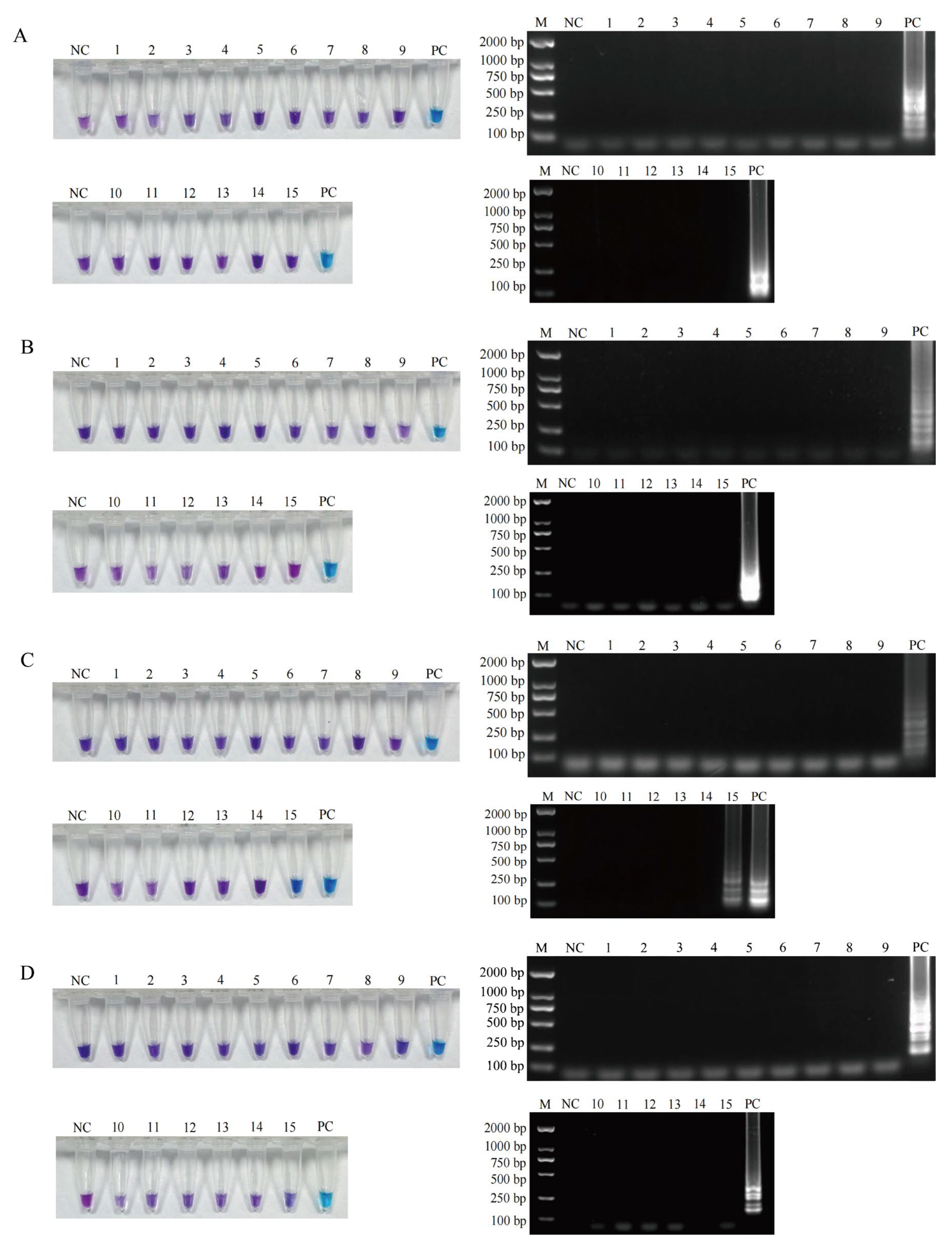
3.2. Specificity of the Established LAMP Assays

Sixteen bacteria, including the four target foodborne pathogens and twelve other bacteria, were used to evaluate the specificity of the established LAMP assays. For the LAMP detection systems of L. monocytogenes, S. aureus, and E. coli O157:H7, only tubes containing the target pathogens exhibited sky blue and the corresponding amplification bands, while all other fifteen bacteria tubes and the negative control sterile water tube remained violet with no amplification bands (Figure 1A,B,D). Thus, the three established LAMP assays achieved species-level discrimination. Both the S. enterica and S. bongori tubes showed positive results (Figure 1C), suggesting that the LAMP method for Salmonella exhibited genus-level specificity.

3.3. Sensitivity of the Established LAMP Methods

The counts of the four foodborne pathogenic pure cultures in the logarithmic growth phase (OD600 = 0.6–0.8) were determined using the dilution plate method, and the concentrations of L. monocytogenes, S. aureus, S. enterica, and E. coli O157:H7 were quantified as 1.8 × 10⁹ CFU/mL, 5.1 × 10⁹ CFU/mL, 1.2 × 10⁹ CFU/mL, and 3.3 × 10⁹ CFU/mL, respectively. Each pathogenic bacterium sample was subjected to 10-fold serial dilution to obtain eight concentration gradients, then all samples were detected by LAMP and PCR simultaneously. The detection limits of the LAMP assays were determined as follows: 1.8 × 101 CFU/mL for L. monocytogenes, 5.1 × 101 CFU/mL for S. aureus, 1.2 × 101 CFU/mL for S. enterica, and 3.3 × 10³ CFU/mL for E. coli O157:H7, which were 100-fold lower than those of PCR for L. monocytogenes and S. aureus, 1000-fold lower for S. enterica, and 10-fold lower for E. coli O157:H7. These results demonstrate that the LAMP assays exhibited 10- to 1000-fold higher sensitivity than conventional PCR for detecting these four foodborne pathogens (Table 3). Moreover, the detection limits of L. monocytogenes, S. aureus, and S. enterica detected by our LAMP systems were in the same order of magnitude as those reported in other existing research with LAMP or LAMP-derivative techniques [28,29,30], confirming the enhanced sensitivity of the established LAMP methods.

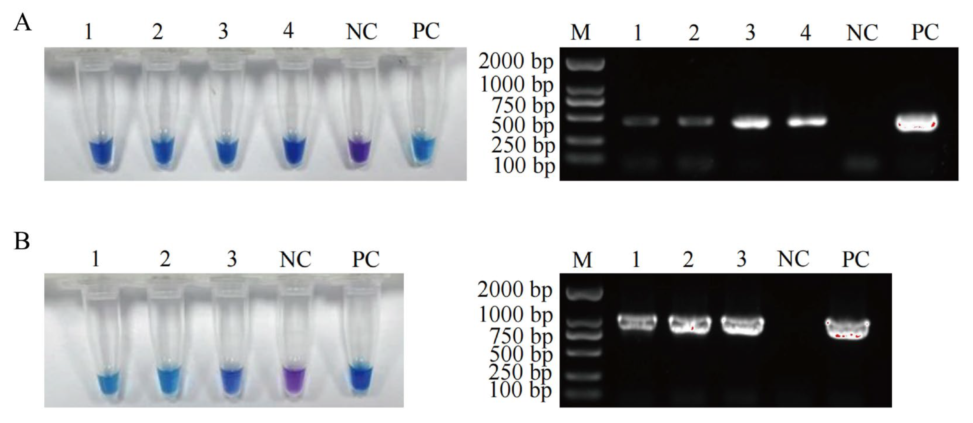
3.4. Detection Performance of the Established LAMP Methods in Meat and Meat Product Samples

The contamination status of four foodborne pathogenic bacteria in 52 samples was analyzed using both the established LAMP detection methods and PCR methods. The results showed that 4 S. enterica-positive samples and 3 E. coli O157:H7-positive samples were detected by LAMP, and the PCR detection results were consistent with these findings (Figure 2). The contamination of pathogenic bacteria in the seven positive samples was further verified by culture-based methods (Figures S8 and S9). These results demonstrate that the accuracies of the designed LAMP methods for detecting L. monocytogenes, S. aureus, S. enterica, and E. coli O157:H7 were 100% (Table 4). Given the limited sample size (n = 52), broader sampling and the inclusion of additional sample types remain necessary to further confirm the accuracy of the developed LAMP methods.
The four S. enterica-positive samples were raw meats purchased from farmers’ markets or mini-markets, including chicken thigh meat, beef, pork, and duck thigh meat. The detection rate for Salmonella in raw livestock meat was 10.54%, while in pork, it reached 17.60% in 2515 raw livestock meat samples collected across 15 provinces in China during 2021 [31]. These results indicate that Salmonella contamination is highly prevalent in raw meat, so enhanced detection and control strategies for Salmonella are urgent. The three E. coli O157:H7-positive samples were cooked meat products bought from mobile vendors, which consisted of marinated pork head meat, marinated chicken legs, and marinated pig trotters. A previous study showed that 5-log reductions of E. coli O157:H7 were achieved in 6.54 min by cooking meatballs to an internal temperature of 85 °C [32]. Therefore, to minimize the risk of E. coli O157:H7 and other pathogens, cooked meat purchased from mobile vendors should be thoroughly reheated before consumption. Notably, none of the four pathogenic bacteria were detected in the 10 raw and 10 cooked meat samples purchased from supermarkets, which may be attributed to the faster turnover cycles and stricter quarantine procedures of meat and meat products sold in large supermarkets.

4. Conclusions

This study successfully established four optimized LAMP assays for the rapid detection of L. monocytogenes, S. aureus, S. enterica, and E. coli O157:H7, with the entire detection process completed within 60 min. Colorimetric changes were observed only for the target pathogens among the 16 tested pathogenic bacteria, demonstrating the high specificity of the LAMP methods. Comparative studies revealed that the LAMP assays exhibited 10-fold to 1000-fold higher sensitivity than conventional PCR for detecting these four foodborne pathogens. Furthermore, the incorporation of HNB enabled the visual interpretation of results without requiring specialized equipment, making the LAMP assays particularly suitable for on-site rapid testing in local food safety regulatory agencies. Practical validation using 52 meat and meat product samples achieved a 100% accuracy, confirming the reliability of the developed LAMP methods and highlighting their strong potential for food safety monitoring applications.

Supplementary Materials

The following supporting information can be downloaded at: https://www.mdpi.com/article/10.3390/foods14132321/s1, Figure S1: Optimization results of Mg2+ concentration for LAMP detection of four pathogenic bacteria. Figure S2: Optimization results of Bst DNA polymerase volume for LAMP detection of four pathogenic bacteria. Figure S3: Optimization results of dNTP Mix volume for LAMP detection of four pathogenic bacteria. Figure S4: Optimization results of the addition ratio of internal and external primers for LAMP detection of four pathogenic bacteria. Figure S5: Optimization results of reaction temperatures for LAMP detection of four pathogenic bacteria. Figure S6: Optimization results of reaction time for LAMP detection of four pathogenic bacteria. Figure S7: Optimization results of added concentration of HNB for LAMP detection of four pathogenic bacteria. Figure S8: Microbial culture results confirming Salmonella positive samples. Figure S9: Microbial culture results confirming E. coli O157:H7 positive samples.

Author Contributions

Conceptualization, X.Z. and L.Q.; methodology, X.Z. and X.L.; formal analysis, X.Z. and X.L.; investigation, X.L., M.Z., and S.W.; data curation, X.L., W.L., and B.R.; writing—original draft preparation, X.L.; writing—review and editing, X.Z. and L.Q. All authors have read and agreed to the published version of the manuscript.

Funding

This work was supported by the Science and Technology Project of Key Laboratory of Food Safety Quick Testing and Smart Supervision Technology for State Market Regulation (No. ZDSYS202310), and the Science and Technology Project of Henan Province (No. 242102311170).

Institutional Review Board Statement

Not applicable.

Informed Consent Statement

Not applicable.

Data Availability Statement

All data generated or analyzed during this study are included in this published article and its Supplementary Materials files.

Conflicts of Interest

Author B.R. was employed by Zhengzhou Zhongdao Biotechnology Co., Ltd. The remaining authors declare that the research was conducted in the absence of any commercial or financial relationships that could be construed as a potential conflict of interest.

References

  1. Stadnik, J. Nutritional value of meat and meat products and their role in human health. Nutrients 2024, 16, 1446. [Google Scholar] [CrossRef] [PubMed]
  2. Ji, J.; Shankar, S.; Royon, F.; Salmieri, S.; Lacroix, M. Essential oils as natural antimicrobials applied in meat and meat products—A review. Crit. Rev. Food Sci. Nutr. 2023, 63, 993–1009. [Google Scholar] [CrossRef] [PubMed]
  3. Shi, C.; Kang, S. Foodborne pathogenic bacteria: Prevalence and control—Volume I. Foods 2024, 13, 1531. [Google Scholar] [CrossRef]
  4. Osek, J.; Lachtara, B.; Wieczorek, K. Listeria monocytogenes—How this pathogen survives in food-production environments? Front. Microbiol. 2022, 13, 866462. [Google Scholar] [CrossRef]
  5. Li, W.; Bai, L.; Fu, P.; Han, H.; Liu, J.; Guo, Y. The epidemiology of Listeria monocytogenes in China. Foodborne Pathog. Dis. 2018, 15, 459–466. [Google Scholar] [CrossRef] [PubMed]
  6. Costa, F.G.; Mills, K.B.; Crosby, H.A.; Horswill, A.R. The Staphylococcus aureus regulatory program in a human skin-like environment. mBio 2024, 15, e0045324. [Google Scholar] [CrossRef] [PubMed]
  7. Cieza, M.Y.R.; Bonsaglia, E.C.R.; Rall, V.L.M.; Santos, M.V.D.; Silva, N.C.C. Staphylococcal enterotoxins: Description and importance in food. Pathogens 2024, 13, 676. [Google Scholar] [CrossRef]
  8. Lu, J.; Wu, H.; Wu, S.; Wang, S.; Fan, H.; Ruan, H.; Qiao, J.; Caiyin, Q.; Wen, M. Salmonella: Infection mechanism and control strategies. Microbiol. Res. 2025, 292, 128013. [Google Scholar] [CrossRef]
  9. Teklemariam, A.D.; Al-Hindi, R.R.; Albiheyri, R.S.; Alharbi, M.G.; Alghamdi, M.A.; Filimban, A.A.R.; Al Mutiri, A.S.; Al-Alyani, A.M.; Alseghayer, M.S.; Almaneea, A.M.; et al. Human Salmonellosis: A continuous global threat in the farm-to-fork food safety continuum. Foods 2023, 12, 1756. [Google Scholar] [CrossRef]
  10. Jiang, L.; Yang, W.; Jiang, X.; Yao, T.; Wang, L.; Yang, B. Virulence-related O islands in enterohemorrhagic Escherichia coli O157:H7. Gut Microbes 2021, 13, 1992237. [Google Scholar] [CrossRef]
  11. Cui, S.; Wei, Y.; Li, C.; Zhang, J.; Zhao, Y.; Peng, X.; Sun, F. Visual loop-mediated isothermal amplification (LAMP) assay for rapid on-site detection of Escherichia coli O157: H7 in milk products. Foods 2024, 13, 2143. [Google Scholar] [CrossRef]
  12. Omer, M.K.; Álvarez-Ordoñez, A.; Prieto, M.; Skjerve, E.; Asehun, T.; Alvseike, O.A. A systematic review of bacterial foodborne outbreaks related to red meat and meat products. Foodborne Pathog. Dis. 2018, 15, 598–611. [Google Scholar] [CrossRef] [PubMed]
  13. Aladhadh, M. A review of modern methods for the detection of foodborne pathogens. Microorganisms 2023, 11, 1111. [Google Scholar] [CrossRef] [PubMed]
  14. Park, D.G.; Ha, E.S.; Kang, B.; Choi, I.; Kwak, J.E.; Choi, J.; Park, J.; Lee, W.; Kim, S.H.; Kim, S.H.; et al. Development and evaluation of a next-generation sequencing panel for the multiple detection and identification of pathogens in fermented foods. J. Microbiol. Biotechnol. 2023, 33, 83–95. [Google Scholar] [CrossRef]
  15. Wang, D.; Chen, Q.; Huo, H.; Bai, S.; Cai, G.; Lai, W.; Lin, J. Efficient separation and quantitative detection of Listeria monocytogenes based on screen-printed interdigitated electrode, urease and magnetic nanoparticles. Food Control 2017, 73, 555–561. [Google Scholar] [CrossRef]
  16. Quintela, I.A.; Vasse, T.; Lin, C.S.; Wu, V.C.H. Advances, applications, and limitations of portable and rapid detection technologies for routinely encountered foodborne pathogens. Front. Microbiol. 2022, 13, 1054782. [Google Scholar] [CrossRef]
  17. Soroka, M.; Wasowicz, B.; Rymaszewska, A. Loop-mediated isothermal amplification (LAMP): The better sibling of PCR? Cells 2021, 10, 1931. [Google Scholar] [CrossRef]
  18. Yang, N.; Zhang, H.; Han, X.; Liu, Z.; Lu, Y. Advancements and applications of loop-mediated isothermal amplification technology: A comprehensive overview. Front. Microbiol. 2024, 15, 1406632. [Google Scholar] [CrossRef]
  19. Novi, V.T.; Meher, A.K.; Abbas, A. Visualization methods for loop mediated isothermal amplification (LAMP) assays. Analyst 2025, 150, 588–599. [Google Scholar] [CrossRef]
  20. Neshani, A.; Zare, H.; Sadeghian, H.; Safdari, H.; Riahi-Zanjani, B.; Aryan, E. A comparative study on visual detection of Mycobacterium tuberculosis by closed tube loop-mediated isothermal amplification: Shedding light on the use of Eriochrome Black T. Diagnostics 2023, 13, 155. [Google Scholar] [CrossRef]
  21. Yan, H.; Xu, B.; Gao, B.; Xu, Y.; Xia, X.; Ma, Y.; Qin, X.; Dong, Q.; Hirata, T.; Li, Z. Comparative analysis of in vivo and in vitro virulence among foodborne and clinical Listeria monocytogenes strains. Microorganisms 2025, 13, 191. [Google Scholar] [CrossRef] [PubMed]
  22. Zhao, Y.; Xia, D.; Ma, P.; Gao, X.; Kang, W.; Wei, J. Advances in the detection of virulence genes of Staphylococcus aureus originate from food. Food Sci. Hum. Wellness 2020, 9, 40–44. [Google Scholar] [CrossRef]
  23. Tian, S.; Wang, C.; Li, Y.; Bao, X.; Zhang, Y.; Tang, T. The impact of SlyA on cell metabolism of Salmonella typhimurium: A joint study of transcriptomics and metabolomics. J. Proteome Res. 2021, 20, 184–190. [Google Scholar] [CrossRef]
  24. Yang, X.; Qi, Y.; Feng, J.; Han, F.; Zhang, P.; Luo, L.; Zheng, Z.; Zhang, W.; Li, Z.; Tang, W. On-site monitoring of Escherichia coli O157:H7 in drinking water based on rapid detection of the rfbE gene at the single copy level. Sens. Actuators B Chem. 2024, 401, 135069. [Google Scholar] [CrossRef]
  25. Oslan, S.N.H.; Yusof, N.Y.; Lim, S.J.; Ahmad, N.H. Rapid and sensitive detection of Salmonella in agro-Food and environmental samples: A review of advances in rapid tests and biosensors. J. Microbiol. Methods 2024, 219, 106897. [Google Scholar] [CrossRef]
  26. Ngwa, G.A.; Schop, R.; Weir, S.; León-Velarde, C.G.; Odumeru, J.A. Detection and enumeration of E. coli O157:H7 in water samples by culture and molecular methods. J. Microbiol. Methods 2013, 92, 164–172. [Google Scholar] [CrossRef]
  27. Logeshwari, R.; Gopalakrishnan, C.; Kamalakannan, A.; Ramalingam, J.; Saraswathi, R. A colorimetric hydroxy naphthol blue based loop-mediated isothermal amplification detection assay targeting the β-tubulin locus of Sarocladium oryzae infecting rice seed. Front. Plant Sci. 2022, 13, 1077328. [Google Scholar] [CrossRef]
  28. Fiore, A.; Treglia, I.; Ciccaglioni, G.; Ortoffi, M.F.; Gattuso, A. Application of a loop-mediated isothermal amplification (LAMP) assay for the detection of Listeria monocytogenes in cooked ham. Foods 2023, 12, 193. [Google Scholar] [CrossRef]
  29. Xu, D.; Zeng, H.; Wu, W.; Liu, H.; Wang, J. Isothermal amplification and CRISPR/Cas12a-system-based assay for rapid, sensitive and visual detection of Staphylococcus aureus. Foods 2023, 12, 4432. [Google Scholar] [CrossRef]
  30. Liu, W.; Zhang, S.; Chen, C.; Jin, J.; Liu, H.; Bian, Z.; Qi, W.; Xie, Y.; Zhang, H. Development of a visual detection method for Salmonella based on loop-mediated isothermal amplification using pyrophosphatase. FEMS Microbiol. Lett. 2023, 370, fnad040. [Google Scholar] [CrossRef]
  31. Ren, X.; Yang, D.; Yang, Z.; Li, Y.; Yang, S.; Li, W.; Qiao, X.; Xue, C.; Chen, M.; Zhang, L.; et al. Prevalence and antimicrobial susceptibility of foodborne pathogens from raw livestock meat in China, 2021. Microorganisms 2024, 12, 2157. [Google Scholar] [CrossRef] [PubMed]
  32. Tosuncuk, Ö.; Bozatli, S.B.; Dikici, A. Investigation of efficient thermal inactivation parameters of Escherichia coli O157:H7 in meatballs by grilling. J. Food Sci. Technol. 2023, 60, 1731–1737. [Google Scholar] [CrossRef] [PubMed]
Figure 1. Specificity analysis of the four established LAMP assays was individually validated for each target foodborne pathogenic bacteria. (A) The specificity results of the LAMP assay for L. monocytogenes, samples 1–3 are S. aureus, S. enterica, and E. coli O157:H7, PC is L. monocytogenes. (B) the specificity results of the LAMP assay for S. aureus, samples 1–3 are L. monocytogenes, S. enterica, and E. coli O157:H7, PC is S. aureus. (C) the specificity results of the LAMP assay for S. enterica, samples 1–3 are L. monocytogenes, S. aureus, and E. coli O157:H7, PC is S. enterica. (D) the specificity results of the LAMP assay for E. coli O157:H7, samples 1–3 are L. monocytogenes, S. aureus, and S. enterica, PC is E. coli O157:H7. Every NC is sterile water as a negative control, and all samples 4–15 are E. fergusonii, S. flexneri, S. sonnei, B. cereus, E. coli, P. aeruginosa, S. xylosus, S. epidermidis, S. capitis, L. innocua, L. ivanovii, and S. bongori. M: DS2000 DNA marker. Violet: negative reaction, sky blue: positive reaction.
Foods 14 02321 g001
Figure 2. Specificity. The detection results of positive samples by LAMP and PCR assays. (A) The detection results of 4 S. enterica positive samples by LAMP and PCR assays, sample 1: raw chicken thigh meat, sample 2: raw beef, sample 3: raw pork, sample 4: raw duck thigh meat, NC: sterile water as negative control, PC: S. enterica as positive control. (B) The detection results of 3 E. coli O157:H7 positive samples by LAMP and PCR assays, sample 1: marinated pork head meat, sample 2: marinated chicken legs, sample 3: marinated pig trotters, NC: sterile water as negative control, PC: E. coli O157:H7 as positive control. M: DS2000 DNA marker. Violet: negative reaction, sky blue: positive reaction.
Foods 14 02321 g002
Table 1. Nucleotide sequence of LAMP and PCR primers.
GenePrimer TypePrimer NameSequence (5′ → 3′)
prfALAMP primerLM-F3GTATTAGCGAGAACGGGAC
LM-B3TTGTTTTTGTAGGGTTTGGAA
LM-FIPGCCAACCGATGTTTCTGTATCAATAATTTACAATACTACAAAGGGGCT
LM-BIPGCAGGCTACCGCATACGTTAAAAGTGCGTAAGATTTTTGCTC
PCR primerLM-FATGAACGCTCAAGCAGAAGAATTCAAA
LM-RTTAATTTAATTTTCCCCAAGTAGCAGGAC
nucLAMP primerSA-F3AACAGTATATAGTGCAACTTCAA
SA-B3CTTTGTCAAACTCGACTTCAA
SA-FIPATGTCATTGGTTGACCTTTGTACATAAATTACATAAAGAACCTGCGA
SA-BIPGTTGATACACCTGAAACAAAGCATCATTTTTTTCGTAAATGCACTTGC
PCR primerSA-FATGACAGAATACTTATTAAGTGCTG
SA-RAAATATTTAATTTCTTTTTTTTCGCTTGTG
slyALAMP primerSE-F3TTCTGATCTGGCACGGTTG
SE-B3GATCCAACGTGCGTACCAG
SE-FIPTGACCCAATGTGTCTGCGTCAACATTTGGCGTGCTCTGATTG
SE-BIPTCATCAATTGCCGCCTGACCACTGCTCAATGCCTATCGCTT
PCR primerSE-FAAGGAGATGAAATTGGAATCGCCA
SE-RTCAATCGTGAGAGTGCAATTCCATA
rfbELAMP primerEC-F3AGCGTTAGGTATATCGGAAG
EC-B3GTTCCATATCACATGGATGTC
EC-FIPTGGCTCCTGTGTATTTTATAGCATTGAGATGAAGTTATTGTTCCAACA
EC-BIPTGAAACTTGGCAAATGTCTGTTAGTAATGGACACACATAATAGCTTTA
PCR primerEC-FATGAAATATATACCAGTTTACCAACC
EC-RCTATTTATCACTATAAAATTCGTTAATAGA
Table 2. Optimized LAMP system components and reaction parameters.
ItemsLAMP System for
L. monocytogenes
LAMP System for
S. aureus
LAMP System for
S. enterica
LAMP System for E. coli O157:H7
10× ThermPol Buffer2.5 μL2.5 μL2.5 μL2.5 μL
MgSO4 (100 mM)1.5 μL1.5 μL1.5 μL1.5 μL
dNTP Mix (10 mM)3.0 μL2.5 μL2.5 μL3.0 μL
F3/B3 primers (10 µM)0.5 μL0.5 μL0.5 μL0.5 μL
FIP/BIP primers (10 µM)4.0 μL4.0 μL4.0 μL4.0 μL
Bst DNA polymerase0.4 μL0.8 μL0.6 μL0.8 μL
HNB (3 mM)1.0 μL0.75 μL0.75 μL0.75 μL
Genomic DNA template100 ng100 ng100 ng100 ng
ddH2OFill up to 25 μLFill up to 25 μLFill up to 25 μLFill up to 25 μL
Reaction temperature64 °C62 °C62 °C61 °C
Reaction time50 min40 min40 min50 min
Table 3. Comparison of the sensitivity of LAMP and conventional PCR for the detection of four pathogenic bacteria.
StrainsLAMP Detection Limit (CFU/mL)PCR Detection Limit (CFU/mL)Fold Change
L. monocytogenes1.8 × 1011.8 × 103100
S. aureus5.1 × 1015.1 × 103100
S. enterica1.2 × 1011.2 × 1041000
E. coli O157:H73.3 × 1033.3 × 10410
Table 4. The detection results of 52 samples.
StrainsTotal SamplesLAMP DetectionAccuracy Rate
Positive SamplesNegative Samples
L. monocytogenes52052100%
S. aureus52052100%
S. enterica52448100%
E. coli O157:H752349100%
Disclaimer/Publisher’s Note: The statements, opinions and data contained in all publications are solely those of the individual author(s) and contributor(s) and not of MDPI and/or the editor(s). MDPI and/or the editor(s) disclaim responsibility for any injury to people or property resulting from any ideas, methods, instructions or products referred to in the content.

