Article

Heterologous Expression of Recombinant Ginseng Tetradecapeptide in Saccharomyces cerevisiae and Evaluation of Its Biological Activity

1
Engineering Research Center of Western Resource Innovation Medicine Green Manufacturing, Ministry·of Education, School of Chemical Engineering, Northwest University, Xi’an 710127, China
2
Shaanxi Key Laboratory of Biomaterials and Synthetic Biology, Shaanxi R&D Center of Biomaterials and Fermentation Engineering, School of Chemical Engineering, Northwest University, Xi’an 710127, China
3
Biotechnology & Biomedicine Research Institute, Northwest University, Xi’an 710127, China
4
Shaanxi Green Bio-Manufacturing Future Industry Research Institute, School of Chemical Engineering, Northwest University, Xi’an 710127, China
*
Authors to whom correspondence should be addressed.
Foods 2025, 14(12), 2049; https://doi.org/10.3390/foods14122049
Submission received: 29 April 2025 / Revised: 7 June 2025 / Accepted: 9 June 2025 / Published: 10 June 2025
(This article belongs to the Special Issue Bioactive Peptides and Probiotic Bacteria: Modulators of Human Health)

Abstract

:
Ginseng peptides, as bioactive components of ginseng, have attracted increasing attention. In this study, a 14-amino acid ginseng peptide was selected and heterologously expressed in Saccharomyces cerevisiae using a multicopy tandem fusion strategy, named 7RS14α. The secondary structure of the recombinant ginseng tetradecapeptide (7RS14α) was analyzed, and a high-glucose model was established in mouse adipocytes to evaluate its biological activity. Transcriptomic profiling was further performed to elucidate its potential mechanisms. Results demonstrated that 7RS14α significantly enhanced glucose uptake in high-glucose model cells, likely by modulating lipid metabolism pathways and insulin signaling cascades, thereby influencing energy homeostasis in adipocytes.

1. Introduction

Diabetes mellitus is a chronic metabolic disease, characterized by persistent hyperglycemia resulting from defects in insulin secretion or action, and has emerged as a major global health threat [1]. It triggers multisystem complications, including diabetic ketoacidosis, vascular dysfunction, and neuropathy. Current therapeutic strategies for diabetes primarily focus on lifestyle interventions, pharmacological treatments (e.g., insulin analogs, metformin), and emerging approaches such as complementary and alternative medicine (CAM) [2]. However, these existing methods often fall short in effectively managing diabetes due to issues like drug dependency, side effects, and incomplete control of blood glucose levels [3]. This highlights the urgent need for novel therapeutic agents and innovative approaches to address the growing burden of diabetes [4].
In the quest for new treatments, natural products have garnered significant attention for their rich content of bioactive components and therapeutic benefits [5]. Hypoglycemic peptides, derived from natural or synthetic bioactive peptides with anti-hyperglycemic properties, are gaining attention as adjunctive therapies due to their low toxicity and multitarget regulatory mechanisms [6]. These peptides, derived from natural sources, such as soybean and bitter melon, or synthesized through various platforms, modulate glucose homeostasis by inhibiting digestive enzymes, mimicking insulin activity, or regulating gut hormone secretion [7]. In 2023, the global market for hypoglycemic peptides took shape, highlighting their clinical and commercial significance in reducing drug dependence and improving the quality of life of diabetic patients [8]. Among a wide variety of natural products, Panax ginseng C.A. Meyer, as a highly regarded medicinal plant, holds significant value for research and application [9]. Documented in classical texts such as Shennong Bencaojing (The Divine Farmer’s Materia Medica) and Huangdi Neijing (The Yellow Emperor’s Inner Canon), ginseng has been used for millennia to enhance recovery, stimulate blood circulation, alleviate stress, and combat fatigue [10].
Modern phytochemical studies have identified over 200 bioactive compounds in ginseng, including triterpenes, saponins (ginsenosides), alkaloids, peptidoglycans, and proteins. While ginsenosides have traditionally been recognized as their primary active constituents, recent research has uncovered the untapped potential of ginseng-derived peptides [11]. In 1980, Japanese researchers first isolated a small peptide from ginseng aqueous extract that inhibited adrenaline-induced lipolysis, hypothesizing its peptide nature based on amino acid composition [12]. Subsequent studies elucidated the sequence of the recombinant ginseng peptide as ETVEIIDSEGGGDA, a 14-amino acid peptide, and characterized its secondary structure [13]. Functional assays demonstrated its dual ability to suppress lipolysis and enhance cellular glucose uptake, positioning it as a promising hypoglycemic agent [14]. In type 2 diabetes mellitus (T2DM) mouse models, its hypoglycemic efficacy was confirmed, highlighting its potential to regulate systemic glucose homeostasis through these mechanisms [15]. This discovery has opened a new frontier in diabetes management, with small-molecule peptides from ginseng emerging as promising candidates for therapeutic development [16]. Despite the high therapeutic potential of ginseng-derived peptides, their industrial production remains constrained by conventional methods such as plant extraction and chemical synthesis [17]. These approaches suffer from significant drawbacks, including excessive costs, low purity, and environmental pollution due to toxic byproducts [18]. Recent advances in synthetic biology offer promising alternatives, with microbial biosynthesis emerging as a cost-effective and environmentally friendly method for producing bioactive peptides [19,20].
While prokaryotic systems (e.g., Escherichia coli, Bacillus subtilis) are widely used, their inability to perform post-translational modifications often leads to protein misfolding and inclusion body formation, complicating downstream purification [21,22].
In contrast, Saccharomyces cerevisiae (strain S288C), the first eukaryote with a fully sequenced genome (1996), stands out as a Generally Recognized as Safe (GRAS) organism with millennia of use in food fermentation (e.g., brewing, baking) [23,24]. At the beginning of the 21st century, most commercial yeast-derived products originated from S. cerevisiae, underscoring its industrial reliability [25]. Its well-characterized genetics, rapid growth in defined media, and compatibility with flexible induction strategies (e.g., galactose-inducible promoters) make it an ideal host for scalable, cost-effective production of bioactive peptides [26,27]. Moreover, the utilization of Saccharomyces cerevisiae as a host system effectively circumvents challenges associated with small peptides, including host protein interference, intracellular proteolytic degradation, and the requirement for proper folding, while simplifying downstream purification processes [28,29]. In addition, utilizing a multi-copy strategy effectively enhances gene expression. For instance, following the implementation of the multicopy tandem strategy, the expression levels of the recombinant ginseng hexapeptide increased by over 5-fold, while those of the beef flavor peptide improved by 2-fold [30].
Based on the aforementioned background, we proposed a genetically engineered method for producing recombinant ginseng peptide using Saccharomyces cerevisiae (BY4741) as the host. By employing a multicopy tandem expression strategy, we aim to enhance the expression and stability of the recombinant ginseng peptides, thereby increasing their yield and therapeutic potential. Furthermore, the biological activity of recombinant ginseng peptides was validated using an insulin-resistant fat cell model. By discussing the possible action mechanisms of recombinant ginseng peptides, we aim to demonstrate their potential as hypoglycemic peptides for application. This innovative approach addresses challenges in ginseng peptide production and offers new avenues for developing novel, safe, and effective treatments for diabetes.

2. Materials and Methods

2.1. Bacterial Strains, Plasmids, and Media

The bacterial strains and plasmids used in this study are listed in Table 1. Escherichia coli (E. coli) was grown in LB medium (lysogeny broth), while Saccharomyces cerevisiae (S. cerevisiae) was cultivated in YPD (yeast extract peptone dextrose) or SD-Ura (Synthetic Defined medium lacking uracil). Media were supplemented with ampicillin (100 μg/mL) or phosphate-buffered saline (PBS) as required by experimental protocols. Restriction enzymes BamHI and EcoRI, along with T4 DNA ligase, were obtained from Takara Biotechnology Co., Ltd. (Dalian, China). The positive control RS14 (recombinant ginseng tetradecapeptide) was chemically synthesized by Sangon Biotech Co., Ltd. (Shanghai, China).

2.2. Reagents and DNA Manipulation Techniques

The procedures for isolating and handling recombinant DNA followed established protocols. Tsingke Biotechnology Co., Ltd., located in Beijing, China, was responsible for carrying out DNA sequencing and synthesis, as detailed in Table 1. Essential molecular biology tools, such as restriction enzymes, were sourced from TaKaRa Biotechnology in Dalian, China. Tiangen Biotech, based in Beijing, China, provided the Prep mini plasmid kit. Furthermore, a kit for preparing sodium dodecyl sulfate-polyacrylamide gel electrophoresis (SDS-PAGE) was acquired from Sangon Biotech Co., Ltd. in Shanghai, China.

2.3. Molecular Construction and Transformation of S. cerevisiae

The digested fragments were ligated into the pYES2 vector, which had been linearized using BamH I and EcoR I, following the treatment of pET28-7RS14α vectors with BamH I and Xba I. The ligated product was then introduced into DH5α cells, and colonies containing the desired construct were identified and designated as pYES2-7RS14α. For yeast transformation, the Lithium Acetate Transformation Method was employed [31]. In this process, a suitable quantity of the positive transformants was mixed with competent S. cerevisiae cells and incubated at 37 °C with continuous shaking for 2 h. The mixture was subsequently plated onto an SD Agar Plate lacking URA (SD-Ura). Successful transformants were further confirmed through colony polymerase chain reaction (PCR) analysis [32].

2.4. Expression of 7RS14α in Flasks

Individual colonies of the genetically modified S. cerevisiae BY4741 strain were picked from SD-Ura agar plates and pre-cultivated for 24 h in 50 mL of SD medium at 30 °C. Subsequently, 2 mL of the freshly grown seed culture was transferred into 100 mL of YPD medium contained in 250 mL shake flasks and incubated at 30 °C until the optical density (OD) reached around 2 [33]. Following this, the culture was subjected to centrifugation, and the supernatant was removed. The resulting cell pellet was then re-suspended in YPG medium (Yeast Extract Peptone Galactose Medium), a step taken to eliminate any residual glucose that might disrupt the galactose induction process. The cells were induced to produce 7RS14α at 28 °C with agitation at 200 rpm, and the progression was monitored at various intervals.

2.5. Protein Detection and Purification

The target protein (7RS14α) was detected using either Western blot (WB) technique or Rapid Flag-tag Detection Kit (Detai Bioengineering, Nanjing, China). In the WB technique, after performing SDS-PAGE electrophoresis, the membrane was incubated with DYKDDDK tag Polyclonal antibody (Proteintech, Wuhan, China), followed by incubation with a corresponding secondary antibody (Rabbit). After thorough washing, the results were visualized through color development. When using the Flag detection test strips, 75 μL of the supernatant from the sample was applied to the loading area of the test strip, and the detection results were read after waiting for ten minutes.
The supernatant containing recombinant ginseng peptide and other heterologous proteins was obtained by centrifugation at 6000× g for 10 min at 4 °C. First, the supernatant was filtered through a 0.22 μm membrane, followed by purification using Anti-DYKDDDDK (Flag) Affinity Gel Anti-Flag (Yeasen Biotechnology, Shanghai, China) with an Affinity Chromatography Column (50 mL) provided by Sangon Biotech Co., Ltd. (Shanghai, China). Subsequently, the elution buffer was collected to obtain the recombinant ginseng protein (7RS14α). Finally, the protein was desalted and concentrated using a dialysis bag or a 15 kDa ultrafiltration tube. The target protein was detected by Western blot (WB). The purified 7RS14α was lyophilized and stored at −40 °C for future use [34].

2.6. Cell Culture, Treatment, and Activity Assay

The 3T3-L1 mouse embryonic fibroblasts were sourced from Shanghai Fuheng Biotechnology Co., Ltd. (Shanghai, China) and cultured in a humidified atmosphere with 5% carbon dioxide at 37 °C. The 3T3-L1 complete medium and the 3T3-L1 cell adipogenic induction differentiation and staining kit were also obtained from Shanghai Fuheng Biotechnology Co., Ltd. The trypsin (containing EDTA) used for digestion was purchased from Beijing Solarbio Science & Technology Co., Ltd. (Beijing, China).
Cell viability was assessed using the Cell Counting Kit-8 (CCK-8) assay, purchased from Abclone Scientific Kit (Wuhan, China), following the manufacturer’s protocol. Approximately 1 × 10⁴ cells were seeded into each well of a 96-well plate. The study was divided into six treatment groups: 0.1 mg/mL, 0.3 mg/mL, 0.5 mg/mL, 0.7 mg/mL, 0.9 mg/mL, and 1.1 mg/mL, along with a blank group and a control group. After 24 h, 10 μL of CCK-8 solution was added to each well, and the plates were incubated at 37 °C for 1–4 h. The absorbance of each well was measured at 450 nm.

2.7. Adipogenic Differentiation of Cells, Oil Red O Staining Observation, and Insulin-Resistant Cell Model Establishment

The 3T3-L1 cells were induced to differentiate into adipocytes following the protocol of the 3T3-L1 cell adipogenic induction differentiation and staining kit. After differentiation, the cells were fixed with paraformaldehyde (Beyotime, Shanghai, China) to preserve their morphology, and then stained with Oil Red O (Fuheng, Shanghai, China) to observe intracellular lipid droplets.
A 3T3-L1 insulin-resistant cell model was established by adding dexamethasone (Dex). After completing adipogenic induction and differentiation, the 3T3-L1 cells were cultured in growth medium containing 1 μmol·L−1 dexamethasone (Dex) for 24–120 h. Cell viability and glucose content were measured every 24 h to determine the optimal duration of dexamethasone treatment [35].

2.8. The Insulin Synergistic Effect on the Insulin-Resistant Cell Model Cells Was Assessed

After inducing differentiation in six-well plates at a density of 5 × 10⁵ cells per well or in 48-well plates at a density of 5 × 10⁴ cells per well, the cells were divided into two control groups, two model groups, and two experimental groups. An additional set of control and model groups included insulin (10−7 mmol/L−1). The experimental groups consisted of model cells treated with 0.1 mg/mL of the target protein and model cells treated with both insulin (10−7 mmol/L−1) and 0.1 mg/mL of the target protein. After 24 h of incubation, the glucose concentration in the supernatant of the 48-well plate cells was measured using a glucose content detection kit, while the triglyceride content in the cell lysates of the six-well plate cells was determined using a triglyceride detection kit. All reagents mentioned above were obtained from the Nanjing Jiancheng Bioengineering Institute.

3. Results

3.1. Molecular Engineering, Heterologous Expression, and Purification of Recombinant Ginseng Polypeptide

The PYES2 plasmid, a commercially available shuttle plasmid containing a GAL1 promoter, was selected to induce the expression of recombinant ginseng peptides upon switching the carbon source to galactose. The 14-amino acid ginseng peptide sequence (ETVEIIDSEGGGDA) was designed to be concatenated head-to-tail seven times. Additionally, the truncated α-factor secretion signal by removing its C-terminal four amino acids was used to ensure extracellular secretion of the target peptide without residual amino acids, thereby minimizing potential interference with peptide bioactivity. The FLAG epitope tag was inserted at the 5′-end of the recombinant peptide sequence to enable rapid detection and facilitate high-purity isolation of the target protein. The construct designated plasmid PYES2-7RS14α is shown in Figure 1A.
The 7RS14-Flag peptide gene sequence fused with the FLAG tag was chemically synthesized and amplified via PCR, as shown in Figure 1B(a), confirming successful amplification of both fragments. These fragments were subsequently ligated into the PYES2 plasmid using homologous recombination. The recombinant plasmid was verified by PCR with primers specific to the PYES2 plasmid’s sequencing sites (Figure 1B(b)). Agarose gel electrophoresis revealed that the recombinant fragment migrated slightly larger than 500 bp, consistent with its theoretical size (576 bp) (Figure 1B(b)). The excised band was subjected to Sanger sequencing, which confirmed 100% identity with the designed nucleotide sequence, validating the successful construction of the recombinant fragment. The recombinant plasmid was then transformed into Saccharomyces cerevisiae obtain recombinant yeast cells. The gene of the recombinant ginseng peptide has been successfully transferred into the Saccharomyces cerevisiae cells (BY4741-PYES2-7RD14α), which is proved by yeast colony PCR (Figure 1B(c)).
The engineered BY4741-pYES2-7RS14α strain was induced in shake flasks for 48 h. Supernatants from 24 h and 48 h fermentation cultures were collected after centrifugation and analyzed for target protein expression using lateral flow FLAG-tag detection. A red band at the test line (T line) indicates the absence of FLAG-tagged protein in the sample. Figure 1C demonstrates the presence of FLAG-tagged protein in both 24 h and 48 h supernatants, confirming successful expression of the target peptide.
Western blot (WB) analysis (Figure 1E) further validated the identity of the peptide, showing a distinct band within the 10–15 kDa range, aligning with the electrophoretic results in Figure 1D. For competitive elution, three concentrations of FLAG peptide solutions (0.1 mg/mL, 0.3 mg/mL, and 0.5 mg/mL) were tested. As shown in Figure 1E, the 0.5 mg/mL FLAG peptide solution achieved optimal elution efficiency, yielding a peptide band between 10 and 15 kDa—consistent with the theoretical size of the recombinant ginseng peptide (10 kDa). ImageJ (v1.8.0) analysis confirmed >95% purity. Approximately 20 mg/L of recombinant ginseng peptide was obtained per liter of fermentation in a 500 mL flask broth after desalting and lyophilization, enabling subsequent functional studies.

3.2. Characterization of Recombinant Ginseng Polypeptides

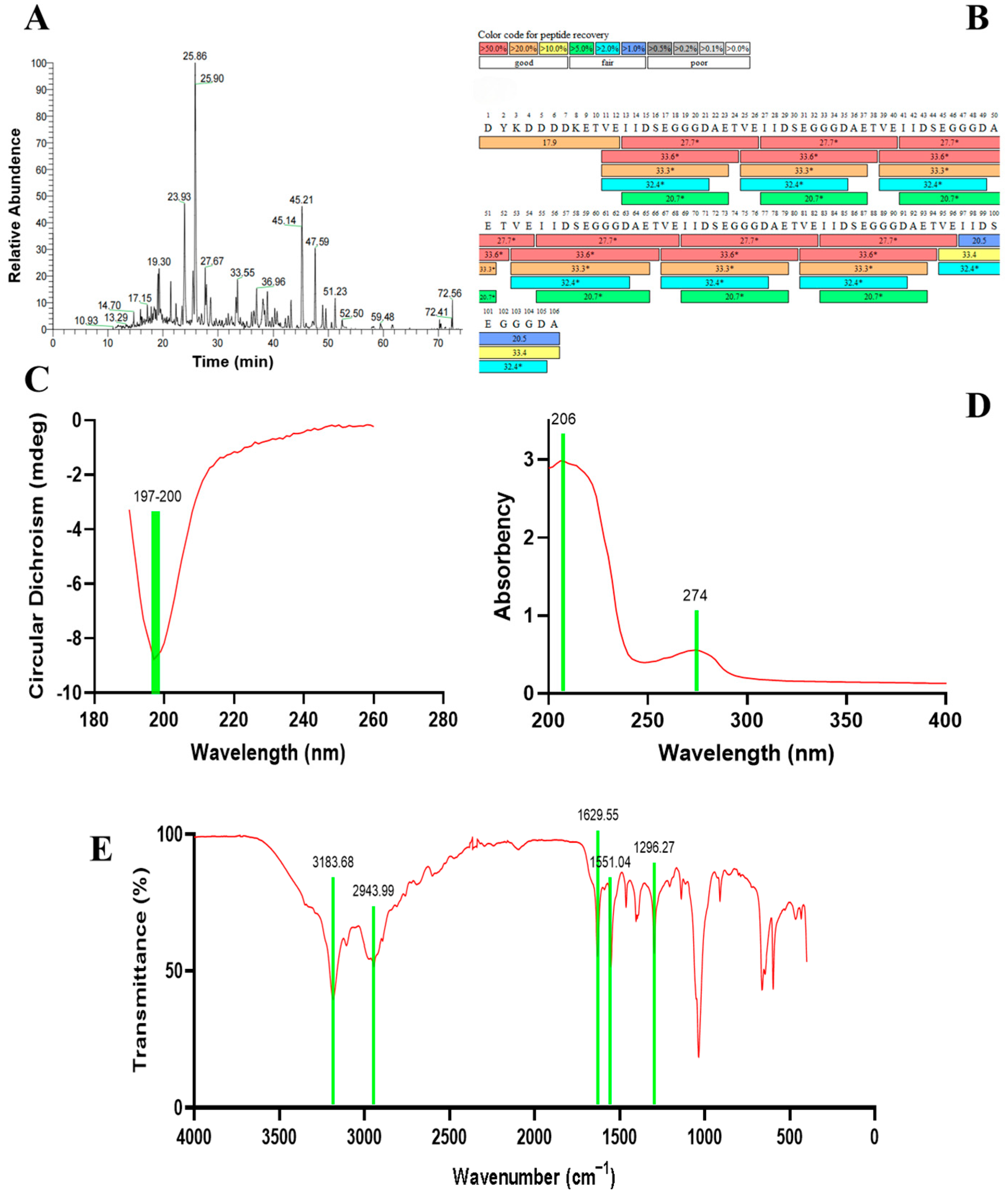
Figure 2A illustrates the peptide profile of the purified recombinant ginseng polypeptide following in-gel digestion and LC-MS/MS analysis, demonstrating efficient enzymatic cleavage. Further validation using Thermo BioPharma Finder (5.0) software for mass spectrometry database searching revealed a 100% sequence coverage alignment between the recombinant polypeptide produced by the engineered strain and the theoretical amino acid sequence (Figure 2B). These results confirm the feasibility of the Saccharomyces cerevisiae engineering system for successful expression of the recombinant ginseng polypeptide.
The far-UV circular dichroism (CD) spectrum of the recombinant ginseng polypeptide (Figure 2C) provides insights into the arrangement of peptide bonds, while secondary structure analysis using CDNN (11.2) software revealed the proportions of α-helix, β-sheet, β-turn, and random coil configurations (Table 2). The ultraviolet (UV) absorption spectrum of the recombinant polypeptide (Figure 2D) exhibited characteristic peaks at 206 nm and 274 nm, which are indicative of peptide bond and aromatic residue contributions, respectively, and can be utilized for quantitative determination. Fourier-transform infrared (FTIR) spectroscopy (Figure 2E) further confirmed structural features, namely absorption bands at 3183 cm−1 (N–H and O–H stretching vibrations), 2943 cm−1 (C–H stretching vibrations), and 1629 cm−1 (β-sheet structure, consistent with CD analysis). Additional peaks at 1551 cm−1 (amide II band, C–N stretching and N–H bending) and 1296 cm−1 (amide III band) further validated the polypeptide’s structural integrity.

3.3. Evaluation of the Biological Activity of Recombinant Ginseng Tetradecapeptide

The earliest identified activity of ginseng tetradecapeptide was its antilipolytic effect on adipocytes, and subsequent studies further discovered its glucose absorption-promoting activity. We believe that recombinant ginseng peptides are likely to also exert activity on adipocytes. Therefore, to evaluate the glucose absorption-promoting activity of recombinant ginseng tetradecapeptide, we attempted to use 3T3-L1 cells, which are the most commonly used cells for studying adipocyte growth and metabolism. First, 3T3-L1 cells were induced to differentiate into adipocytes using a differentiation induction kit. The 3T3-L1 cell line, derived from mouse embryonic fibroblasts, is a preadipocyte line with specific adipogenic differentiation potential and is widely used as a model for studying lipid metabolism. As shown in Figure 3A, significant lipid droplets were observed in cells after 10 days of induction, confirming successful differentiation of 3T3-L1 cells into adipocytes. Next, dexamethasone (Dex) and varying concentrations of insulin were employed to establish the insulin-resistant model. The results shown in Figure 3B reveal that adipocytes treated with 1 μmol/L Dex and 10−8 mmol/L insulin exhibited significantly reduced glucose uptake compared to the control group at 96 h. Notably, Dex-treated adipocytes showed more pronounced differences in glucose uptake relative to controls. Therefore, 1 μmol/L Dex treatment for 96 h was selected to construct the insulin-resistant cell model.
To further confirm that the reduction in cellular glucose uptake was not due to compromised cell viability, the CCK-8 assay was performed on cells treated with varying insulin concentrations and 1 μmol/L Dex. As shown in Figure 3C,D, cell viability was higher in treated cells compared to untreated controls, confirming that the decreased glucose uptake was independent of cell survival. Subsequently, to determine the safe dosage of the recombinant ginseng peptide, mouse adipocytes were cultured in media supplemented with 0.1 mg/mL, 0.3 mg/mL, 0.5 mg/mL, 0.7 mg/mL, 0.9 mg/mL, or 1.1 mg/mL recombinant peptide, and cell viability was assessed. A concentration that neither affected cell growth nor impairs the effect of promoting glucose absorption was prioritized. The results shown in Figure 3E indicate that 0.1 mg/mL recombinant peptide exhibited the most favorable impact on cell viability, and this concentration was selected for subsequent experiments.
Figure 3F compares the effects of recombinant ginseng peptide and insulin on glucose uptake in insulin-resistant model cells. Insulin treatment significantly increased glucose uptake in normal mouse adipocytes, confirming their insulin sensitivity. In contrast, insulin-resistant model cells exhibited reduced basal glucose uptake, which remained elevated even after insulin treatment, indicating insulin resistance. Strikingly, supplementation with recombinant ginseng peptide further increased glucose uptake in the model cells, achieving levels nearly comparable to insulin-treated normal cells. These findings highlight the potential of recombinant ginseng peptides to be applied as hypoglycemic peptides. Concurrently, Figure 3G demonstrates that insulin treatment significantly increased intracellular triglyceride (TG) content in normal adipocytes, whereas model cells showed no response to insulin. However, recombinant ginseng peptide supplementation elevated TG content in model cells, suggesting insulin-mimetic activity and highlighting its potential to modulate insulin-resistant cells.
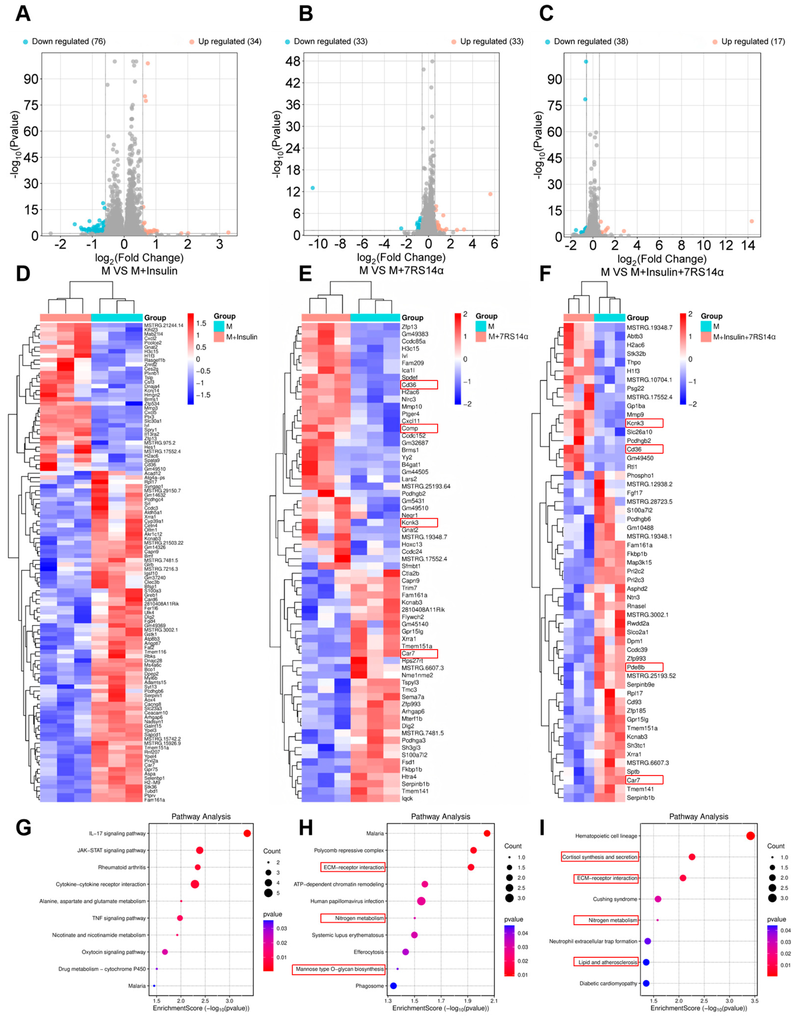
To further investigate the mechanism of the recombinant ginseng peptide, eukaryotic reference-based transcriptomic analysis was performed on insulin-resistant model cells treated with either the recombinant peptide or insulin. Four experimental groups were analyzed: insulin-resistant model cells (M), M treated with insulin (10−7 mmol/L) (M + insulin), M treated with 7RS14α (0.1 mg/mL) (M + 7RS14α), and M treated with both insulin and 7RS14α (M + insulin + 7RS14α), with three biological replicates per group. Volcano plots of differentially expressed genes (DEGs) for comparisons between the M group and the other three groups (M + insulin, M + 7RS14α, M + insulin + 7RS14α) are shown in Figure 4A–C. The x-axis represents log2FoldChange (log2FC), indicating the fold change in gene expression between groups, and the y-axis represents −log10 p-value, reflecting the statistical significance of expression differences. Red and blue dots denote upregulated and downregulated genes, respectively. The limited number of DEGs across all comparisons likely reflects insulin-resistance-induced transcriptional stability. Hierarchical clustering heatmaps of these DEGs are presented in Figure 4D–F. Genes with consistent expression trends in both the M + 7RS14α and M + insulin + 7RS14α groups—Kcnk3, CD36, and Car7—were identified as potential key mediators of the recombinant peptide’s bioactivity.
The Kcnk3 gene, a member of the two-pore potassium channel (K2P) family, regulates cellular membrane potential and excitability, likely influencing ion homeostasis and signaling pathways in mouse adipocytes to modulate metabolic activity, differentiation, and function. Upregulation of Kcnk3 alters the electrophysiological properties of adipocytes, potentially affecting responses to external signals [36]. Concurrently, membrane potential shifts may impact the activity of metabolic enzymes and receptor signaling, such as lipid metabolism-associated enzymes and insulin receptor pathways, suggesting that 7RS14α modulates adipocyte metabolism and energy balance through these mechanisms. The CD36 gene, involved in fatty acid uptake, lipid metabolism, and signaling, may regulate adipocyte energy homeostasis by modulating fatty acid transport, insulin signaling, and inflammation/immune pathways [37]. The Car7 gene catalyzes the conversion of CO2 and H2O into HCO3− and H+, influencing intracellular pH, which is linked to enzyme activity, signal transduction, and oxidative stress. Additionally, HCO3− serves as a carbon source for fatty acid synthesis. In mouse adipocytes, Car7 may affect cellular metabolism and energy balance by altering pH dynamics and fatty acid synthesis [38].
KEGG pathway enrichment analysis of differentially expressed genes across the three experimental groups is shown in Figure 4G–I. The M + insulin group exhibited significant activation of the IL-17 and JAK-STAT signaling pathways, likely attributable to insulin resistance. In the M + 7RS14α group, enriched pathways related to metabolism included ECM–Receptor Interaction, Nitrogen Metabolism, and Mannose Type O-Glycan Biosynthesis. Upregulation of CD36 in the ECM–Receptor Interaction pathway suggests activation of cell surface receptors involved in lipid metabolism, potentially enhancing lipid synthesis [39]. Downregulation of Car7 in the Nitrogen Metabolism pathway implies reduced HCO3− availability, dampening nitrogen metabolism [40]. Upregulation of B4gat1 in the Mannose Type O-Glycan Biosynthesis pathway indicates enhanced O-glycosylation of glycoproteins, which may strengthen insulin receptor O-glycosylation to potentiate insulin signaling [41].

4. Discussion

In the M + insulin + 7RS14α group, key metabolic pathways included cortisol synthesis and secretion, ECM–receptor interaction, nitrogen metabolism, and lipid and atherosclerosis. The ECM–receptor interaction and nitrogen metabolism pathways mirrored those in the M + 7RS14α group. In the cortisol synthesis and secretion pathway, upregulation of Kcnk3 may alter membrane potential to reduce local cortisol utilization, thereby suppressing triglyceride breakdown, while downregulation of Pde8b could elevate cAMP levels, increasing cortisol availability to accelerate lipolysis [42]. Upregulation of CD36 in the Lipid and Atherosclerosis pathway suggests enhanced fatty acid transport and lipid synthesis [43]. Collectively, these findings indicate that 7RS14α modulates energy metabolism in mouse adipocytes by targeting lipid metabolism pathways and insulin signaling cascades.
Diabetes mellitus, a chronic metabolic disorder characterized by hyperglycemia, ranks as the third most prevalent noncommunicable disease globally, following cancer and cardiovascular diseases, posing substantial public health challenges. Current clinical management relies on therapeutics including insulin analogs, insulin secretagogues, α-glucosidase inhibitors, and insulin sensitizers. Existing α-glucosidase inhibitors are primarily derived from microbial metabolites, natural extracts, and synthetic/semi-synthetic compounds [44]. Despite therapeutic advances, suboptimal long-term glycemic control persists in many patients. Concurrently, growing interest in complementary and alternative medicine (CAM), particularly among diabetic populations, established botanical interventions as promising adjunctive therapies [45]. Among botanical CAMs, ginseng has demonstrated notable potential in the management of type 2 diabetes mellitus (T2DM) [46]. In this study, we developed a biosynthetic approach to produce a recombinant ginseng polypeptide, analyzed its secondary structure, validated its glucose-regulatory efficacy in vitro using adipocyte models, and investigated its molecular mechanisms of action. These findings advance the development of this recombinant polypeptide as a novel antidiabetic agent.
The recombinant ginseng peptide yield via Saccharomyces cerevisiae expression remains suboptimal, with ~20 mg/L obtained from fermentation broth. However, molecular-level optimizations (e.g., codon usage bias adjustment, promoter engineering) combined with fermentation process enhancements (e.g., targeted nutrient supplementation, induction timing control) could substantially improve productivity. S. cerevisiae as a host ensures exceptional biosafety and enables direct application of fermentation products, fulfilling compliance requirements for therapeutic/nutraceutical use. Critically, the recombinant polypeptide exhibited 100% sequence identity with the native ginseng tetradecapeptide, confirming fidelity in expression. Secondary structure analysis provided insights into its spatial conformation, establishing a foundation for future structure-activity relationship studies. Functionally, the recombinant polypeptide significantly reduced glucose uptake in insulin-resistant adipocytes, demonstrating mechanisms conducive to glucose stabilization. Transcriptomic profiling further revealed its modulation of lipid metabolic enzymes and insulin signaling pathways, consistent with observed phenotypic effects. These findings collectively validate the therapeutic potential of this recombinant polypeptide for diabetes management while delineating molecular targets for mechanistic exploration and translational development.
Additionally, despite the relatively low yield of recombinant ginseng tetradecapeptide (7RS14α), its production via microbial synthesis demonstrates superior cost-effectiveness compared to plant extraction and chemical synthesis. The material cost for 1 L of fermentation broth is approximately CNY 50, with affinity gel purification adding around CNY 3500, yielding 10–20 mg of recombinant peptide. In contrast, plant extraction requires 1 kg of ginseng powder (costing CNY ~2000) to obtain merely 2 mg of the peptide, alongside complex purification steps involving organic solvents, prolonged timelines, and low overall efficiency. Chemical synthesis, while capable of producing 20 mg of the peptide at a cost of CNY ~2000, involves labor-intensive stepwise amino acid coupling (Table 3). Both plant extraction and chemical synthesis face challenges of significant environmental pollution and procedural complexity. Given the high safety profile of Saccharomyces cerevisiae, future studies could explore direct use of fermentation broth to streamline purification and further reduce costs, enhancing the economic viability of this biosynthetic approach.
Given the extremely high safety of Saccharomyces cerevisiae, fermented products containing heterologous recombinant proteins obtained using S. cerevisiae as a host have the potential to be directly used as products or raw materials. Whether such fermented products can be directly used as products or raw materials is mainly determined by the laws and regulations of different countries and regions. For example, China currently does not allow fermented products produced by genetically engineered strains to be directly used in the food industry. However, fermented products produced by genetically engineered strains can be used in the feed industry after routine industry testing, as well as additional sterilization steps and PCR detection (to ensure no exogenous gene fragments remain). Direct use of fermented products as products or raw materials offers significant economic advantages due to the elimination of separation and purification steps. The genetically engineered strain BY4741-pYES2-7RS14α constructed in this study has the potential for its fermented products to be directly used as products or raw materials. However, to further scale up production, continuous optimization of the medium formulation and fermentation method is required.

Author Contributions

Conceptualization, Y.Q. and C.Z.; methodology, Y.Q. and C.Z.; validation, Y.Q., P.M., P.W. and C.Z.; formal analysis, P.M.; investigation, Y.Q.; resources, Y.Q. and C.Z.; writing—original draft preparation, Y.Q.; writing—review and editing, Y.Q., P.W. and C.Z.; visualization, P.M.; supervision, C.Z.; project administration, C.Z.; funding acquisition, C.Z. All authors have read and agreed to the published version of the manuscript.

Funding

This research was funded by National Key Research and Development Program, grant number 2021YFC2103900.

Institutional Review Board Statement

Not applicable.

Informed Consent Statement

Not applicable.

Data Availability Statement

The original contributions presented in the study are included in the article; further inquiries can be directed to the corresponding author.

Conflicts of Interest

The authors declare no conflicts of interest.

Abbreviations

The following abbreviations are used in this manuscript:
CAMComplementary and Alternative Medicine
T2DMType 2 Diabetes Mellitus
GRASGenerally Recognized as Safe
YPDYeast Extract Peptone Dextrose Medium
SD-UraSynthetic Dropout Medium-Uracil
PBSPhosphate-Buffered Saline
PCRPolymerase Chain Reaction
SDS-PAGESodium Dodecyl Sulfate-PolyAcrylamide Gel Electrophoresis
ODOptical Density
LC-MSLiquid Chromatograph Mass Spectrometer
WBWestern Blot
CDCircular Dichroism Spectroscopy
CDNNCluster-based Discriminative Neural Networks
UVUltraviolet
FTIRFourier-Transform Infrared Spectroscopy
TICTotal I on Chromatogram
DexDexamethasone
CCK-8Cell Counting Kit-8
TGTriglyceride
FCFoldChange
DEGsDifferentially Expressed Genes
KEGGKyoto Encyclopedia of Genes and Genomes
CNYChinese Yuan

References

  1. Cojic, M.; Kocic, R.; Klisic, A.; Kocic, G. The Effects of Vitamin D Supplementation on Metabolic and Oxidative Stress Markers in Patients with Type 2 Diabetes: A 6-Month Follow-Up Randomized Controlled Study. Front. Endocrinol. 2021, 12, 610893. [Google Scholar] [CrossRef] [PubMed]
  2. Rein, M.; Ben-Yacov, O.; Godneva, A.; Shilo, S.; Zmora, N.; Kolobkov, D.; Cohen-Dolev, N.; Wolf, B.-C.; Kosower, N.; Lotan-Pompan, M.; et al. Effects of Personalized Diets by Prediction of Glycemic Responses on Glycemic Control and Metabolic Health in Newly Diagnosed T2DM: A Randomized Dietary Intervention Pilot Trial. BMC Med. 2022, 20, 56. [Google Scholar] [CrossRef]
  3. Bhatt, D.L.; Szarek, M.; Pitt, B.; Cannon, C.P.; Leiter, L.A.; McGuire, D.K.; Lewis, J.B.; Riddle, M.C.; In Zucchi, S.E.; Kosiborod, M.N.; et al. Sotagliflozin in Patients with Diabetes and Chronic Kidney Disease. N. Engl. J. Med. 2021, 384, 129–139. [Google Scholar] [CrossRef]
  4. Zhang, C.-L.; Yang, X.; Meng, X.; Wu, L.; Liu, X.; Gao, J.; Liu, S.; Wu, J.; Huang, D.; Wang, Z.; et al. Discovery of Novel PTP1B Inhibitors with Once—Weekly Therapeutic Potential for Type 2 Diabetes: Design, Synthesis, and In Vitro and In Vivo Investigations of BimBH3 Peptide Analogues. J. Med. Chem. 2023, 66, 3030–3044. [Google Scholar] [CrossRef]
  5. Naufel, M.F.; Telles, M.M.; Hachul, A.C.L.; Santamarina, A.B.; Oyama, L.M.; Coelho, F.M.S.; Ribeiro, E.B.; Pedroso, A.P. Bioactive Natural Products for the Prevention and Treatment of Diabetes Mellitus. Stud. Nat. Prod. Chem. 2020, 67, 161–197. [Google Scholar] [CrossRef]
  6. Acquah, C.; Kodzu Vor, C.K.; Tosh, S.; Agyei, D. Anti-Diabetic Effects of Bioactive Peptides: Recent Advances and Clinical Implications. Crit. Rev. Food Sci. Nutr. 2022, 62, 442–459. [Google Scholar] [CrossRef]
  7. Lu, J.; Zeng, Y.; Hou, W.; Zhang, S.; Li, L.; Luo, X.; Xi, W.; Chen, Z.; Xiang, M. The Soybean Peptide Aglycin Regulates Glucose Homeostasis in Type 2 Diabetic Mice via IR/IRS1 Pathway. J. Nutr. Biochem. 2012, 23, 1449–1457. [Google Scholar] [CrossRef] [PubMed]
  8. Liu, H.; Lv, C.-N.; Lu, J. Panax ginseng C. A. Meyer as a Potential Therapeutic Agent for Organ Fibrosis Disease. Chin. Med. 2020, 15, 124. [Google Scholar] [CrossRef]
  9. Lu, G.; Liu, Z.; Wang, X.; Wang, C. Recent Advances in Panax ginseng C.A. Meyer as a Herb for Anti-Fatigue: An Effects and Mechanisms Review. Foods 2021, 10, 1030. [Google Scholar] [CrossRef]
  10. Loo, S.; Kam, A.; Dutta, B.P.; Zhang, X.; Feng, N.; Sze, S.K.; Liu, C.-F.; Wang, X.; Tam, J.P. Broad-spectrum ginsentides are principal bioactives in unraveling the cure—All effects of ginseng. Acta Pharm. Sin. B 2024, 14, 653–666. [Google Scholar] [CrossRef]
  11. Liu, W.; Liu, Y.-B.; Wang, Z.; Li, W. Research Progress on Chemical Constituents and Transformation Mechanism of Ginseng. Jilin Nongye Daxue Xuebao 2023, 45, 664–670. [Google Scholar] [CrossRef]
  12. Ando, T.; Muraoka, T.; Yamasaki, N.; Okuda, H. Preparation of Anti–lipolytic Substance from Panax ginseng. Planta Med. 1980, 38, 18–23. [Google Scholar] [CrossRef] [PubMed]
  13. Kajiwara, H.; Aso, H.; Okubo, A.; Yamazaki, S. Inhibition of Adrenaline—Induced Lipolysis by Ginseng Polypeptide and Its Modified Peptides. Biosci. Biotechnol. Biochem. 1998, 62, 2065–2067. [Google Scholar] [CrossRef] [PubMed]
  14. Liu, T.; Zhu, C.; Duan, Z.; Ma, P.; Ma, X.; Fan, D. Network Pharmacological Analysis Combined with Experimental Verification to Explore the Effect of Ginseng Polypeptide on the Improvement of Diabetes Symptoms in db/db Mice. J. Agric. Food Chem. 2024, 72, 18537–18551. [Google Scholar] [CrossRef]
  15. Li, M.-H.; Jin, M.-H.; Hu, R.-Y.; Tang, S.; Li, K.-K.; Gong, X.-J.; Sun, Y.-S.; Wang, Y.-P.; Wang, Z.; Li, W. Exploring the Mechanism of Active Components from Ginseng to Manage Diabetes Mellitus Based on Network Pharmacology and Molecular Docking. Sci. Rep. 2023, 13, 793. [Google Scholar] [CrossRef]
  16. Song, M.; Tan, D.; Li, B.; Wang, Y.; Shi, L. Gypenoside Ameliorates Insulin Resistance and Hyperglycemia via the AMPK—Mediated Signaling Pathways in the Liver of Type 2 Diabetes Mellitus Mice. Food Sci. Hum. Wellness 2022, 11, 1347–1354. [Google Scholar] [CrossRef]
  17. Tam, J.P.; Nguyen, G.K.T.; Loo, S.; Wang, S.; Yang, D.W.; Kam, A. Ginsentides: Cysteine and Glycine-rich Peptides from the Ginseng Family with Unusual Disulfide Connectivity. Sci. Rep. 2018, 8, 16201. [Google Scholar] [CrossRef]
  18. Li, C.J.; Yan, X.; Xu, Z.Z.; Wang, Y.; Shen, X.; Zhang, L.; Zhou, Z.H.; Wang, P.P. Pathway elucidation of bioactive rhamnosylated ginsenosides in Panax ginseng and their de novo high-level production by engineered Saccharomyces cerevisiae. Commun. Biol. 2022, 5, 775. [Google Scholar] [CrossRef]
  19. Wu, Z.; Li, Y.; Zhang, L.; Ding, Z.; Shi, G. Microbial production of small peptide: Pathway engineering and synthetic biology. Microb. Biotechnol. 2021, 14, 2257–2278. [Google Scholar] [CrossRef]
  20. Pandi, A.; Adam, D.; Zare, A.; Trinh, V.T.; Schaefer, S.L.; Burt, M.; Erb, T.J. Cell-free Biosynthesis Combined with Deep Learning Accelerates De Novo-development of Antimicrobial Peptides. Nat. Commun. 2023, 14, 7197. [Google Scholar] [CrossRef]
  21. Bhatwa, A.; Wang, W.; Hassan, Y.I.; Abraham, N.; Li, X.-Z.; Zhou, T. Challenges Associated With the Formation of Recombinant Protein Inclusion Bodies in Escherichia coli and Strategies to Address Them for Industrial Applications. Front. Bioeng. Biotech. 2021, 9, 630551. [Google Scholar] [CrossRef] [PubMed]
  22. Xu, K.D.; Tong, Y.; Li, Y.; Tao, J.; Rao, S.Q.; Li, J.H.; Zhou, J.W.; Liu, S. Autoinduction AND Gate inhibits cell lysis to enhance protein production in Bacillus subtilis controlled by population density and cell physiological state. ACS Synth. Biol. 2023, 12, 2742–2752. [Google Scholar] [CrossRef] [PubMed]
  23. Goffeau, A.; Barrell, B.G.; Bussey, H.; Davis, R.W.; Dujon, B.; Feldmann, H.; Oliver, S.G. The Genome Sequence of Saccharomyces cerevisiae: A Platform for Functional Genomics. Science 1996, 274, 546–567. [Google Scholar] [CrossRef] [PubMed]
  24. Chrzanowski, G. Saccharomyces Cerevisiae—An Interesting Producer of Bioactive Plant Polyphenolic Metabolites. Int. J. Mol. Sci. 2020, 21, 7343. [Google Scholar] [CrossRef]
  25. Barbosa, C.; Lage, P.; Vilela, A.; Mendes–Faia, A.; Mendes-Ferreira, A. Phenotypic and Metabolic Traits of Commercial Saccharomyces cerevisiae Yeasts. AMB Express 2014, 4, 39. [Google Scholar] [CrossRef]
  26. Aprodu, I.; Bunea, A.; Socaci, S.A.; Vodnar, D.C. Protease as a Tool for Regulating the Antioxidant Activity and Function of Brewer’s Yeast Protein. Molecules 2024, 29, 1550. [Google Scholar] [CrossRef]
  27. Roberts, N.A.; Suter, B.; Müller, S.; Rudolph, C. Development and optimisation of a defined high cell density yeast medium. Yeast 2020, 37, 336–347. [Google Scholar] [CrossRef]
  28. Tippelt, A.; Nett, M. Saccharomyces cerevisiae as host for the recombinant production of polyketides and nonribosomal peptides. Microb. Cell Fact. 2021, 20, 161. [Google Scholar] [CrossRef]
  29. Oliveira, A.S.; Ferreira, C.; Pereira, J.O.; Pintado, M.E.; Carvalho, A.P. Spent brewer’s yeast (Saccharomyces cerevisiae) as a potential source of bioactive peptides: An overview. Int. J. Biol. Macromol. 2022, 208, 1116–1126. [Google Scholar] [CrossRef]
  30. Xiang, Q.; Qu, L.; Lei, H.; Duan, Z.; Zhu, C.; Yuwen, W.; Ma, X.; Fan, D. Expression of Multicopy Tandem Recombinant Ginseng Hexapeptide in Bacillus subtilis and the Evaluation of Antiaging Activity. J. Agric. Food Chem. 2024, 72, 7266–7278. [Google Scholar] [CrossRef]
  31. Finlayson, S.D.; Fleming, C.; Berry, D.R.; Johnston, J.R. An improved lithium acetate method for yeast transformation. Biotechnol. Tech. 1991, 5, 13–18. [Google Scholar] [CrossRef]
  32. Bonnet, C.; Rigaud, C.; Chanteclaire, E.; Blandais, C.; Tassy-Freches, E.; Rico, C.A.; Javaud, C. PCR on yeast colonies: An improved method for glyco-engineered Saccharomyces cerevisiae. BMC Res. Notes 2013, 6, 201. [Google Scholar] [CrossRef] [PubMed]
  33. Paciello, L.; De Alteriis, E.; Mazzoni, C.; Palermo, V.; Zueco, J.; Parascandola, P. Performance of the auxotrophic Saccharomyces cerevisiae BY4741 as host for the production of IL-1β in aerated fed-batch reactor: Role of ACA supplementation, strain viability, and maintenance energy. Microb. Cell Fact. 2009, 8, 70. [Google Scholar] [CrossRef]
  34. Xing, L.; Wan, X.; Yu, M.T.; He, Y.J.; Wang, Y.; Zhou, T.J.; Liu, X.Y.; Sun, Y.; Luo, J.; Wang, W.J.; et al. A novel whole blood purifier for efficient capture and separation of circulating tumor cells. Biosens. Bioelectron. 2023, 232, 115292. [Google Scholar] [CrossRef]
  35. Deng, X.F.; Chen, B.; Luo, Q.; Zao, X.R.; Liu, H.Z.; Li, Y.Q. Hulless barley polyphenol extract inhibits adipogenesis in 3T3-L1 cells and obesity related-enzymes. Front. Nutr. 2022, 9, 933068. [Google Scholar] [CrossRef]
  36. Sörmann, J.; Schewe, M.; Proks, P.; Jouen-Tachoire, T.; Rao, S.; Riel, E.B.; Agre, K.E.; Begtrup, A.; Dean, J.; Descartes, M. Gain-of-function mutations in KCNK3 cause a developmental disorder with sleep apnea. Nat Genet. 2022, 54, 1534–1543. [Google Scholar] [CrossRef]
  37. Li, J.; Zhang, H. Lipolysis engages CD36 to promote ZBP1—Mediated necroptosis—Impairing lung regeneration in COPD. Cell Rep. Med. 2024, 5, 101732. [Google Scholar] [CrossRef]
  38. Chiesa, R.; Georgiadis, C.; Syed, F.U.; Zhan, H.; Etuk, A.; Gkazi, S.A.; Preece, R.; Ottaviano, G.; Braybrook, T.; Chu, J.; et al. Base-Edited CAR7 T Cells for Relapsed T-Cell Acute Lymphoblastic Leukemia. N. Engl. J. Med. 2023, 389, 899–910. [Google Scholar] [CrossRef]
  39. Nersisyan, S.; Novosad, V.; Giba Ryan, N.E.; Ushkaryov, Y.; Nikulin, S.; Tonevitsky, A. ECM–Receptor Regulatory Network and Its Prognostic Role in Colorectal Cancer. Front. Genet. 2021, 12, 782699. [Google Scholar] [CrossRef]
  40. Choi, S.J.; Lee, Z.; Jeong, E.; Kim, S.; Seo, J.S.; Um, T.; Shim, J.S. Signaling Pathways Underlying Nitrogen Transport and Metabolism in Plants. BMB Rep. 2023, 56, 56–64. [Google Scholar] [CrossRef]
  41. Huang, Y.F.; Aoki, K.; Akase, S.; Ishihara, M.; Liu, Y.S.; Yang, G.; Kizuka, Y.; Mizumoto, S.; Tiemeyer, M.; Gao, X.D.; et al. Global Mapping of Glycosylation Pathways in Human—Derived Cells. Dev. Cell 2021, 56, 1195–1209. [Google Scholar] [CrossRef] [PubMed]
  42. Olson-Manning, C. Elaboration of the Corticosteroid Synthesis Pathway in Primates through a Multistep Enzyme. Mol. Biol. Evol. 2020, 37, 2257–2267. [Google Scholar] [CrossRef] [PubMed]
  43. Khojasteh Malek Mohammad, K.; Bezsonov, E.E.; Rafieian-Kopaei, M. Role of Lipid Accumulation and Inflammation in Atherosclerosis: Focus on Molecular and Cellular Mechanisms. Front. Cardiovasc. Med. 2021, 8, 707529. [Google Scholar] [CrossRef]
  44. Kumar, S.; Narwal, S.; Kumar, V.; Prakash, O. α-Glucosidase Inhibitors from Plants: A Natural Approach to Treat Diabetes. Pharmacogn Rev. 2011, 5, 19–29. [Google Scholar] [CrossRef]
  45. Cho, G.J.; Lee, S.H.; Park, K.S. Therapeutic potential of ginseng and its components for the treatment of diabetes mellitus. J. Ginseng Res. 2020, 44, 563–569. [Google Scholar] [CrossRef]
  46. Shao, J.W.; Jiang, J.L.; Zou, J.J. Therapeutic potential of ginsenosides on diabetes: From hypoglycemic mechanism to clinical trials. J. Funct. Foods 2020, 64, 103630. [Google Scholar] [CrossRef]
Figure 1. Construction, transformation, protein expression and purification of engineered strains. (A) Graphical representation of the key steps in plasmid engineering. (B) Construction and validation of engineered strains: (a) the cloning of α-factor fragment and 7RS14-Flag fragment; (b) PCR validation of recombinant plasmids; (c) colony PCR verification of BY4741-pYES2-7RS14α. (C) Rapid detection of 7RS14α expression using colloidal gold-based FLAG-tag immunoassay kit. (D) Western blot analysis of 7RS14a protein expression. (E) Purification of 7RS14a using FLAG affinity chromatography (Lane 1–2: Flow-through fraction from fermentation broth, Lane 3: wash buffer fraction, Lane 4–6: elution fractions with varying FLAG peptide concentrations (0.1mg/mL,0.3 mg/mL,0.5 mg/mL), Lane 7–8: regeneration wash buffer fraction).
Foods 14 02049 g001
Figure 2. Systematic characterization of bioactive properties in recombinant ginseng-derived polypeptides. (A) Total I on chromatogram (TIC) analysis. (B) Sequence coverage map analysis. * indicates peptide not unique. (C) Circular dichroism (CD) spectroscopic analysis. (D) Ultraviolet-visible (UV–Vis) absorption spectroscopic analysis. (E) Fourier-transform infrared (FTIR) spectroscopic analysis.
Foods 14 02049 g002
Figure 3. Effects of 7RS14α on insulin-resistant model cells. (A) Microscopic evaluation of 3T3-L1 adipocyte differentiation: comparative analysis of pre-and post-adipogenic (Day 10) induction with oil red O staining (10×, red deposits indicate intracellular lipid accumulation). (B) Effects of treatment duration and drug type on the establishment of insulin-resistant cell models (1 μmol/L Dex and 10−6 mmol/L, 10−7 mmol/L, 10−8 mmol/L Insulin). (C) The changes in cell viability of differentiated 3T3-L1 adipocytes, treated with three concentrations of insulin for 120 h (10−6 mmol/L, 10−7 mmol/L, 10−8 mmol/L). (D) The changes in cell viability of differentiated 3T3-L1 adipocytes treated with 1 μmol/L Dex for 120 h. (E) Dose-dependent effects of recombinant ginseng polypeptides on cellular viability in differentiated 3T3-L1 adipocytes. (F) The impact of 7RS14α on glucose uptake in murine adipocytes. (G) The impact of 7RS14α on triglyceride content in murine adipocytes. * p < 0.05 versus model group, ** p < 0.01 versus model group, and *** p < 0.001 versus model group, # p < 0.05 versus control group, #### p < 0.0001 versus control group.
Foods 14 02049 g003
Figure 4. Transcriptomic analysis of 7RS14α effects on insulin-resistant adipocytes (p < 0.05). (AC): Volcano plots of differentially expressed genes (DEGs) for comparisons: M vs. M + insulin, M vs. M + 7RS14α, and M vs. M + insulin + 7RS14α. (DF): Hierarchical clustering heatmaps based on differentially expressed genes (DEGs) for comparisons: M vs. M + insulin, M vs. M + 7RS14α, and M vs. M + insulin + 7RS14α. (GI): Enriched pathway maps based on differentially expressed genes (DEGs) for comparisons: M vs. M + insulin, M vs. M + 7RS14α, and M vs. M + insulin + 7RS14α.
Foods 14 02049 g004
Table 1. Strains and plasmids used in this work.
Strain or PlasmidDescriptionSource/
Reference
BY4741Saccharomyces cerevisiae
S288C-derivative laboratory strain, MATa his3Δ1 leu2Δ0 met15Δ0 ura3Δ0
lab stock
DH5αEscherichia coli
cloning strain
lab stock
pYES2plasmid
E. coliS. cerevisiae shuttle vector, AmpR for E. coli
lab stock
pET28-7RS14E. coli vector, AmpR, pET28 carrying seven replicate fragments of ginseng tetradecapeptide and FLAG tag.Sagon
AmpR, ampicillin resistance; Sagon, Sangon Biotech (Shanghai) Co., Ltd. (Shanghai, China).
Table 2. The secondary structure proportions of recombinant ginseng tetradecapeptide.
NameProtein Secondary Structure
HelixBeta-Pleated SheetBeta-TurnRndm. Coil
AntiparallelParallel
7RS14α8.5%39.5%3.1%19.6%29.4%
Table 3. Comparison of microbial synthesis, plant extraction and chemical synthesis.
NameMicrobial SynthesisPlant ExtractionChemical Synthesis
material costCNY 50 CNY 2000 CNY 2000
timea weekmore than a weekmore than a week
yield10–20 mg recombinant ginseng peptide2 mg ginseng peptide20 mg ginseng peptide
Disclaimer/Publisher’s Note: The statements, opinions and data contained in all publications are solely those of the individual author(s) and contributor(s) and not of MDPI and/or the editor(s). MDPI and/or the editor(s) disclaim responsibility for any injury to people or property resulting from any ideas, methods, instructions or products referred to in the content.