Article

Benzyl Isothiocyanate and Resveratrol Synergistically Alleviate Dextran Sulfate Sodium-Induced Colitis in Mice

1
School of Food Science and Technology, Dalian Polytechnic University, No. 1, Qinggongyuan, Dalian 116034, China
2
College of Food Science and Engineering, Jilin University, Changchun 130062, China
3
Department of Inorganic Nonmetallic Materials Engineering, No. 1, Qinggongyuan, Dalian 116034, China
*
Author to whom correspondence should be addressed.
Foods 2024, 13(13), 2078; https://doi.org/10.3390/foods13132078
Submission received: 8 May 2024 / Revised: 12 June 2024 / Accepted: 25 June 2024 / Published: 1 July 2024

Abstract

:
The aim of our study was to investigate whether the combination of benzyl isothiocyanate (BITC) and resveratrol (RES) has a synergistic effect on the inhibition of inflammation in colitis. The results revealed that the BITC and RES combination (BITC_RES) was more effective than either substance alone at significantly alleviating the symptoms of dextran sodium sulfate (DSS)-induced colitis in mice, including the prevention of colon shortening and loss of body weight, a reduction in the disease activity index, and prevention of colon damage. Similarly, compared with the DSS group, BITC_RES reduced myeloperoxidase (MPO) and inducible nitric oxide synthase (iNOS) levels in the mouse colon by 1.4–3.0-fold and 1.4-fold, respectively. In addition, the combination of BITC and RES upregulated the inflammatory factor IL-10 by 1.3- and 107.4-fold, respectively, compared to the individual BITC and RES groups, whereas the proinflammatory factors, including TNF-α, IL-1β, and IL-6, were downregulated by 1.1–7.4-, 0.7–3.6-, and 0.6–2.6-fold, respectively, in the BITC_RES group compared with the individual groups. Gut microbiome analysis indicated that BITC_RES remodeled the structure of gut bacteria at the phylum, family, and genus levels, upregulating the abundance of the phylum Bacteroidetes and the family Muribaculaceae and the genus norank_f_Muribaculaceae and downregulating the abundance of the phylum Firmicutes. Significant correlations between the relative levels of these proinflammatory cytokines and changes in the gut microbiota were found using Pearson’s correlation analysis. BITC and RES exhibited synergistic effects by reshaping the gut microbiota and modulating the level of serum cellular inflammatory factors, thus exerting a protective effect against colitis.

Graphical Abstract

1. Introduction

The main symptoms of ulcerative colitis (UC) are diarrhea, abdominal pain, and weight loss [1,2,3]. Although UC is prevalent worldwide, its pathogenesis is yet to be thoroughly explored. Several factors, including cytosolic inflammatory factors, the gut microbiome, the gut immune system, and the environment [4,5], have been implicated in the progression of UC. The severity of UC relief is related to the antioxidant capacity of the administered substance [6]. Natural products with good antioxidant properties, including polyphenols [7] and isothiocyanates [8], can relieve UC without side effects. Therefore, the use of natural products for UC mitigation is highly important.
Resveratrol (3,5,4′-trihydroxy-trans-stilbene, RES) is a natural polyphenolic compound with a broad spectrum of bioactive health-promoting properties, including anti-inflammatory, antioxidative, and anti-UC effects [9]. Resveratrol has been shown to suppress UC progression in studies using mouse models [10]. RES can reduce the severity of colon damage, inflammation, and mortality in mice and may affect multiple pathways, including cytokine expression, redox status, and cellular signaling in the gut [11]. Importantly, synergistic effects allow lower concentrations of drugs to be used in combination with each other to achieve the same effect as higher concentrations alone, reducing the concentration of drugs used and thus providing a better balance [12]. Previous studies have reported synergistic antioxidant effects of RES and curcumin and synergistic reductions in hypertension in animal models [13]. Polygonum cuspidatum Siebold & Zucc extracts inhibit the NF-kappa B signaling pathway and exert their anti-colitic effects through the synergistic action of polysaccharides, RESs, or rhodopsin [14]. Like other isothiocyanates, benzyl isothiocyanate (BITC) has been shown to have significant pharmacological activities, including anti-inflammatory, antimicrobial [15], and antioxidant activities [16]. In addition, allyl isothiocyanate improves dextran sodium sulfate (DSS)-induced UC in mice by increasing tight junction and mucin expression [8]. It has been reported that phenethyl isothiocyanate treatment attenuates the symptoms of DSS-induced UC in mice [17]. We previously found that BITC and RES synergistically inhibited the growth of Listeria monocytogenes by inhibiting biofilm formation, cell membrane disruption and ROS generation [12]. However, no studies have reported synergistic effects of BITC and RES on UC. Therefore, it is necessary to study the synergistic effects of BITC and RES in relieving UC.
The gut microbiota potentially plays a role in the pathogenesis of UC [18]. RES improves enteritis by targeting the colon flora and inflammatory signaling pathways [19]. It also regulates the gut microbiota by inducing Tregs and inhibiting Th17 cells to prevent the development of colitis in mice [20]. By enhancing the ratio of Firmicutes to Bacteroidetes, suppressing the growth of Enterococcus faecalis, and improving the abundance of Lactobacillus and Bifidobacterium, resveratrol has been reported to reduce colon dysbiosis [21]. RES and Lactococcus lactis have potential synergistic effects on regulating the colon flora [22]. In addition, sulforaphane (a kind of natural isothiocyanate) alters the microbiota and attenuates colitis severity in DSS-induced UC in mice [23]. ITC-rich moringa seed extract modulates the gut microbiota and thus fat in model mice [24]. Therefore, it is necessary to alleviate DSS-induced colitis by investigating the synergistic modulatory effects of BITC and RES on the colon flora.
Therefore, the aim of this study was to investigate the synergistic mitigating effect of BITC and RES on colitis. First, antioxidant tests were conducted with different ratios of BITC and RES to determine the optimal ratio of synergistic antioxidants. The synergistic effects of BITC and RES on DSS-induced colitis were subsequently assessed by evaluating myeloperoxidase (MPO) and inducible nitric oxide synthase (iNOS) levels; the levels of inflammatory factors, including IL-10, TNF-α, IL-1β and IL-6; and changes in the colon flora.

2. Materials and Methods

2.1. Materials

BITC (purity > 99%) was acquired from Sigma Aldrich (St. Louis, MI, USA). Resveratrol (RES, 99%) was acquired from Shanghai Aladdin Biochemical Co., Ltd. (Shanghai, China). Anhydrous ethanol was procured from the Damao Chemical Factory (Tianjin, China). All the other chemicals used were of analytical grade.

2.2. Antioxidant Activity

2.2.1. DPPH Radical Scavenging Activity Assay

A previously described method was used to determine the DPPH scavenging capacity of the samples [25], with some modifications. BITC and RES were dissolved in ethanol at ratios of 1:1, 1:2, 1:3, 1:4, 1:5, 5:1, 4:1, 3:1, and 2:1. Briefly, 2 mL of 100 µM DPPH ethanol solution was mixed with equal amounts of BITC and RES and then incubated at room temperature in the dark for synergistic antioxidant testing. After 30 min, the absorbance at 517 nm was measured. The DPPH• scavenging percentage was estimated using the following equation:
D P P H   s c a v e n g i n g   p e r c e n t a g e % = A c A s A c × 100%
where Ac is the absorbance of the control reaction and As is the absorbance of the test sample.

2.2.2. ABTS•+ Scavenging Activity Assay

The method of Floegel et al. was used for the ABTS•+ radical scavenging assay [26], with slight modifications. BITC and RES were dissolved in ethanol at ratios of 1:1, 1:2, 1:3, 1:4, 1:5, 5:1, 4:1, 3:1, and 2:1. In brief, ABTS was dissolved in water to prepare the ABTS stock solution. The ABTS stock solution was reacted with the same volume of potassium persulfate for 12–16 h at room temperature in the dark. BITC and RES were dissolved in absolute ethanol. BITC, RES, and BITC_RES were reacted with the ABTS•+ solution in the dark. After 6 min of reaction, the absorbance was measured at 734 nm using a UV-visible spectrophotometer. To calculate the percentage of ABTS•+ radicals, the following equation was used:
A B T S + s c a v e n g i n g   p e r c e n t a g e % = A c A s A c × 100%
where Ac is the absorbance of the control reaction and As is the absorbance of the test sample.

2.3. Experimental Animals

Male BALB/c mice (5 weeks) were purchased from Liaoning Changsheng Biological Co., Ltd., Benxi, Liaoning, China. All mice were housed in the SPF standard animal house of the Animal Experiment Center of Dalian University of Technology at a temperature of 25 ± 2 °C, a humidity of 60 ± 5%, and a 24-h period of day and night alternation (license number DLPU2023033). All animals were fed regularly according to the IACUC-approved protocol for laboratory animals prior to the experiments. The animals were fed ad libitum with deionized water and standard dietary materials, and the bedding was changed once every 3–5 days.

2.4. DSS-Induced Colitis in Mice

A total of 50 specific pathogen-free male BALB/c mice (5 weeks of age, weight: 20 ± 0.5 g) were randomized into the control, DSS, BITC, RES, and BITC-RES groups (n = 6 per group). BITC and RES were administered intragastrically according to the body weights of the mice. According to Ibrahim et al. [27], 30 mg/kg/d RES was administered intragastrically. In our preliminary experiment, BITC and RES had a strong synergistic antioxidant effect at a 1:1 ratio. Therefore, the dose of BITC in the selected group was also 30 mg/kg/d. In the BITC group and the RES group, the mice were given BITC (30 mg/kg/d) and RES (30 mg/kg/d), and in the BITC_RES group, the mice were given BITC-RES (15 mg/kg/d + 15 mg/kg/d) for 14 days. Starting from the eighth day, 3.5% DSS was given ad libitum to all groups except the healthy control group for 7 days to induce colitis. The mice were fasted for 12 h before euthanasia. The serum, intestine and colon contents of all the mice were collected and stored at −80 °C. Some of the intestine was fixed in paraformaldehyde for further analysis.

2.5. Assessment of the Disease Activity Index (DAI)

The DAI was collected from the first day of DSS treatment and scored according to weight loss (scored as 0, no change; 1, <5% loss; 2, 5–10% loss; 3, 10–20% loss; and 4, >20% loss), stool consistency (scored as 0, normal; 2, loose stool; and 4, diarrhea), and fecal blood (scored as 0, none; 2, blood; and 4, gross bleeding) [28]. The final macroscopic result for each animal was the sum of these three separate degrees.

2.6. Histological Analysis

The colon tissues were dissected and stained with hematoxylin and eosin (H&E) to identify pathological changes in the distal colon tissues of the mice [29]. First, mouse colon and colon tissues were washed in flowing water for 4 h, embedded, and sectioned at 5 μm. The treated tissues were then placed on slides. The slides were dried, deparaffinized, and embedded in H&E. The degree of colon tissue damage in each group was assessed, and the tissues were photographed by a fluorescence microscope (Nikon, Eclipse Ti, Japan).

2.7. Inducible Nitric Oxide Synthase (iNOS) Level and Myeloperoxidase (MPO) Activity Assay

Briefly, for iNOS measurements, a 0.1 g/mL tissue homogenate was prepared with saline and centrifuged at 3000 rpm for 10 min, after which the supernatants were obtained. iNOS production was measured using a nitric oxide synthase assay kit (Nanjing Jiancheng Bioengineering Institute, Nanjing, China) according to the manufacturer’s instructions by evaluating the total volume of reaction liquid, sampling volume, colorimetric light path, reaction time, and tissue homogenate protein concentration. A BCA protein test kit (Tiangen Biotech, Beijing, China) was used to measure the protein concentration. Colon tissue was accurately weighed, an MPO test kit (Nanjing Jiancheng Bioengineering Institute, Nanjing, China) reagent was added as a buffer solution, and 0.05 g/mL tissue homogenate was prepared according to a weight: volume ratio of 1:19. The MPO levels were measured with a mouse MPO test kit according to the manufacturer’s protocol.

2.8. Determination of the Serum Proinflammatory Cytokine Concentration

After 12 h of fasting, blood samples were taken from the retroorbital sinuses at the end of the study and centrifuged at 2000× g for 15 min. Mouse IL-6, IL-1β, and TNF-α level and IL-10 enzyme-associated immunosorbent test (ELISA) kits (Neobioscience Technology, Shenzhen, China) were used to quantify the serum IL-6, IL-1β, TNF-α, and IL-10 concentrations via ELISA.

2.9. Microbial Analysis

After collection, the fecal samples were stored at −80 °C. DNA was extracted from the total microbial community according to the instructions of the E.Z.N.A.® Soil DNA Kit (Omega Biotek, Norcross, GA, USA). PCR amplification of the V3–V4 variable region of the 16S rRNA gene was conducted with 338F (ACTCCTACGGGGAGGCAGCAG) and 806R (GGACTACHVGGGGTWTCTAAT) following the following amplification procedure: 27 cycles of pre-denaturation at 95 °C for 3 min. Then, the mixture was stably heated at 72 °C for 10 min and finally stored at 10 °C (PCR instrument: ABI GeneAmp® Model 9700, Applied Biosystems, Foster City, CA, United States). The PCR system included 4 μL of 5× TransStart FastPfu Buffer, 2.5 mM dNTPs, 0.8 μL of upstream primer (5 µM), 0.8 μL of downstream primer (5 µM), 0.4 μL of TransStart FastPfu DNA polymerase, and 10 ng of template DNA, and had a final volume of 20 μL. Library construction was performed using the NEXTFLEX Rapid DNA-Seq Kit (BIOO Scientific, Austin, TX, USA).

2.10. Statistical Analysis

Statistical analysis of the data (from at least three separate experiments) was performed via one-way ANOVA with Duncan’s test for comparison of individual means via SPSS software (v19.0, IBM, Inc., Armonk, NY, USA). p < 0.05 was considered to indicate statistical significance.

3. Results and Discussion

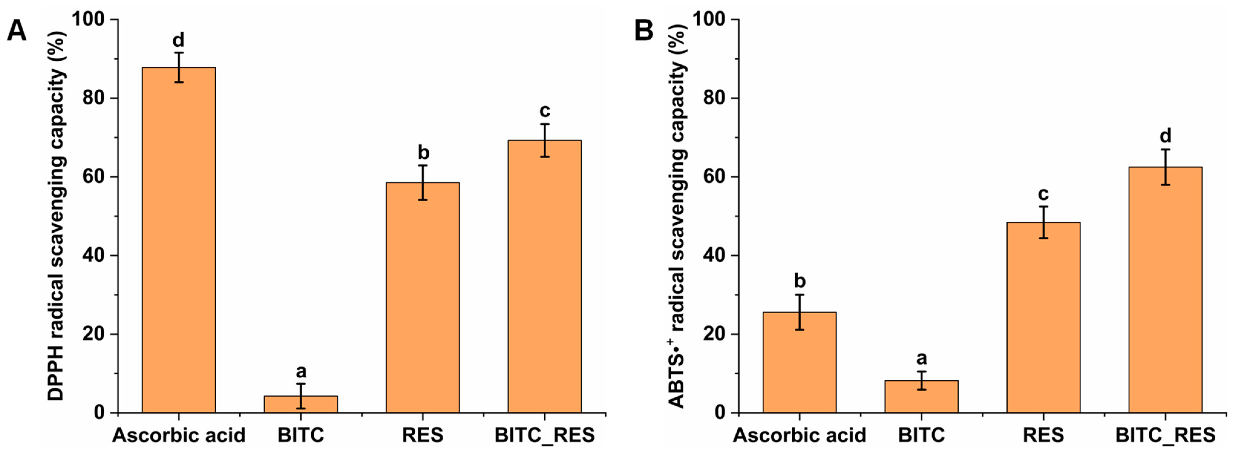
3.1. Synergistic Antioxidant Activities of BITC and RES

The antioxidant activities of BITC, RES, and BITC_RES were determined using DPPH and ABTS free radical scavenging activity tests. Finally, the optimal 1:1 ratio of BITC to RES was chosen for the following experiments: BITC (30 μg/mL), RES (30 μg/mL) and BITC_RES (15 μg/mL + 15 μg/mL). DPPH radical scavenging was 69.3% for the mixed BITC and RES samples, which was greater than the 4.3% for the BITC group and 58.5% for the RES group (Figure 1A). However, the level in the ascorbic acid group was significantly greater than that in the BITC_RES group. In addition, the same synergistic effect was observed for ABTS. The ABTS free radical scavenging rate in the BITC_RES group was 1.3-fold to 7.6-fold greater than that in the BITC, ascorbic acid, and RES groups (Figure 1B). These results suggest that BITC and RES have synergistic antioxidant effects. Therefore, we chose an equal concentration ratio to further determine the synergistic effect of BITC and RES on DSS-induced colitis.

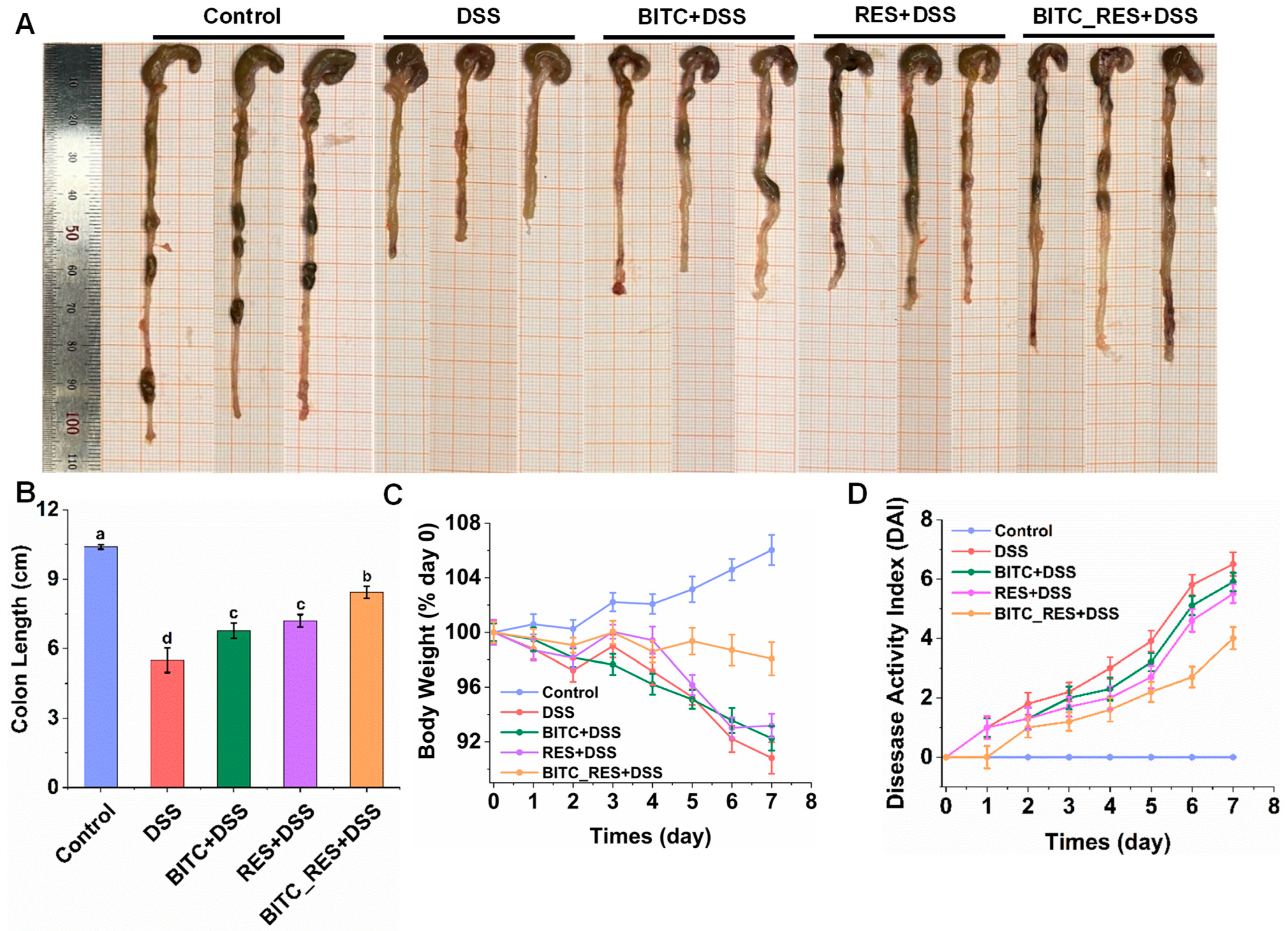
3.2. Synergistic Effects of BITC and RES on DSS-Induced Colitis Symptom Severity

To determine the synergistic effect of BITC and RES on preventing UC, we tested DSS-induced colitis in mice. After DSS treatment, the mice lost weight and developed diarrhea and even hematochezia. The colon length, body weight, and DAI are shown in Figure 2. The control colon tissue was normal in color and thickness, with no congestion, edema or adhesion to surrounding tissue (Figure 2A). The colons of the mice in the DSS group appeared edematous and significantly shorter, with a length of 5.50 ± 0.53 cm (Figure 2A,B). However, mice in the BITC_RES group exhibited particularly significant relief of colonic shortening, with a length of 8.43 ± 0.25 cm, which was 1.17–1.25 times greater than that in the BITC and RES alone groups (p < 0.05). In addition, the body weight of the normal mice increased with increasing length of the experimental cycle, which was a natural increase of approximately 106.03% (Figure 2C). The body weights of the mice in the other groups showed different degrees of continuous decline, and the decreasing trend was obvious from the fifth day onward. By the seventh day, the mice in the DSS-induced group exhibited a greater decrease in body weight, down to approximately 90.82% of their starting weight. The BITC and RES groups showed a similar decreasing trend to the DSS group, with body weights decreasing to approximately 92.25% and 93.1%, respectively. However, the weight loss of the BITC_RES-treated mice was significantly reduced, and the body weight decreased to only 98.07%, which was markedly different from that of the other treatment groups. The DAI score is a direct reflection of disease status. As is shown in Figure 2D, the results showed that DAI was dramatically increased in DSS-induced mice to 6.50 ± 0.40 compared to the control group. The BITC and RES groups reduced the DAI scores, and, in particular, the BITC_RES group significantly reduced the DAI score by 2.5 relative to the DSS group (p < 0.05). These findings are consistent with previous research [8,10,30,31], demonstrating that ITC and RES prevent the progression of colitis. Therefore, the combined effect of BITC and RES was greater than that of either alone, further suggesting a synergistic effect of BITC and RES.

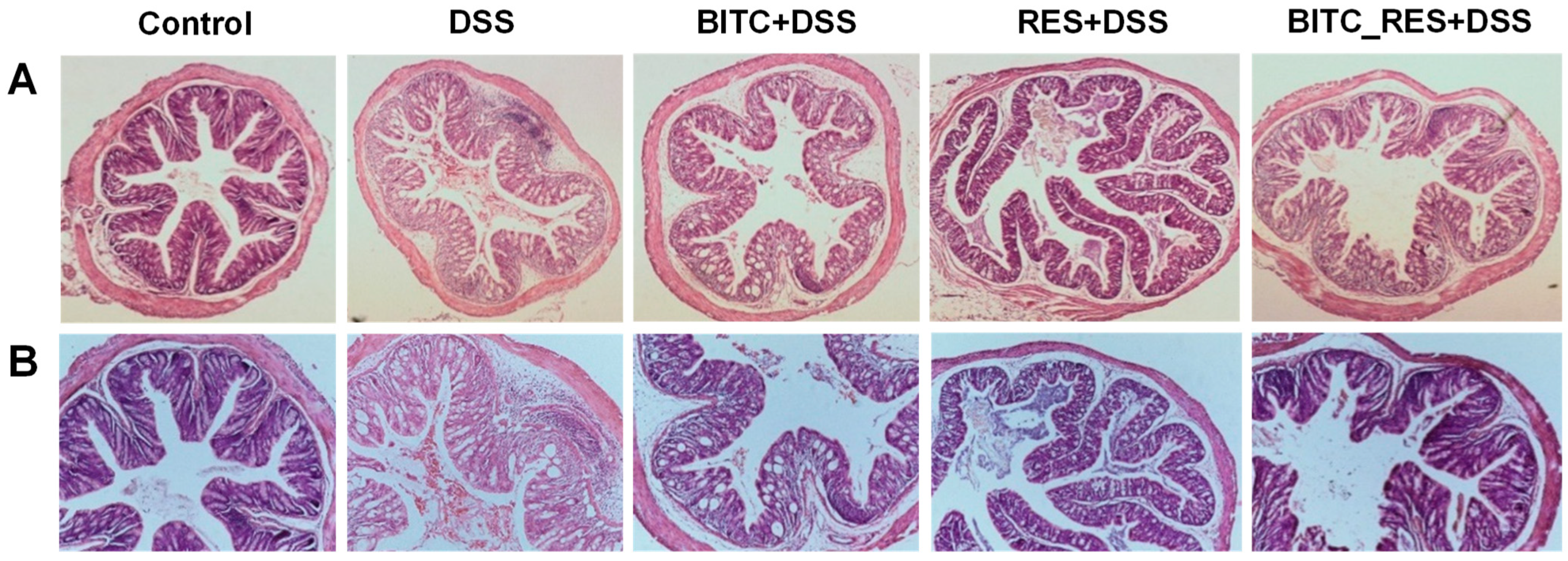
3.3. Synergistic Effects of BITC and RES on Colonic Microinjury in DSS-Induced Colitis

We observed changes in the microstructure of the colon in five groups of mice to further study the effect of BITC in combination with RES on colitis (Figure 3). In the control group, the colonic mucosa was intact and continuous, and the glands were regularly arranged and structured. In the DSS group, the colonic tissues were severely lesioned, the epithelial cells of the colonic tissues had detached, the crypts and the structure of the cups had basically disappeared and were disorganized, the submucosal layer was more severely affected, and some areas were congested and severely ulcerated. Compared with that in the model group, inflammatory cell infiltration gradually decreased in the BITC and RES groups. However, the colonic mucosal tissue of the BITC_RES group was more similar to that of the control group, and BITC_RES had a greater preventive effect on UC than did BITC or RES alone. It has been reported that the combination of an anthocyanin-rich extract of black rice and rosemarinic acid inhibits DSS-induced colitis-induced colon damage to varying degrees compared to treatment alone [32]. Therefore, these results suggest that BITC and RES have synergistic effects on DSS-induced colitis-related colonic injury.

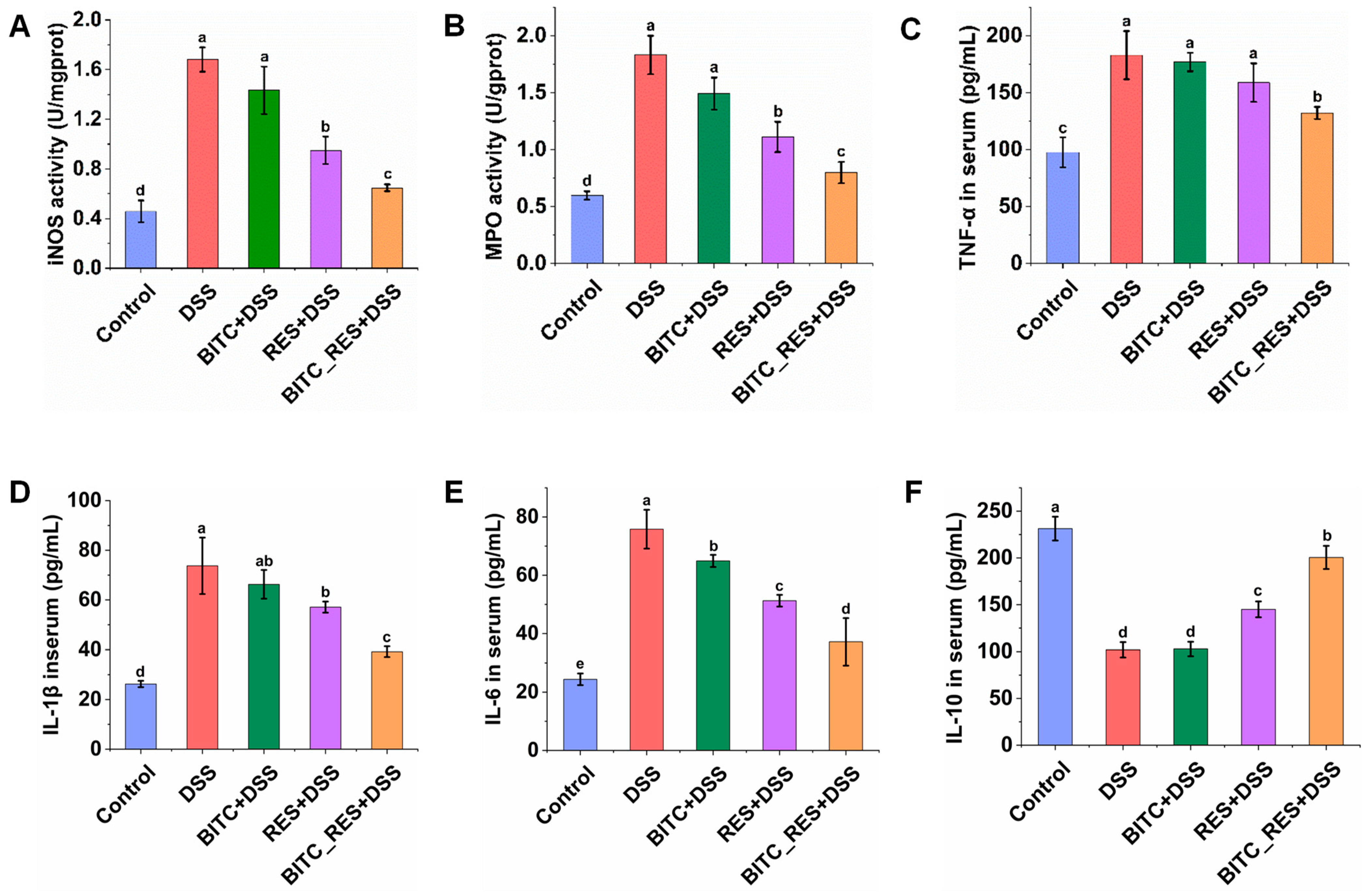
3.4. Synergistic Effects of BITC and RES on iNOS and MPO Levels in Colonic Tissues and Cytokine Level in Serum

Excess NO produced by iNOS through direct cytotoxicity, neutrophil activation, vasodilation, and peroxynitrite radical formation could worsen the clinicopathological features of colitis [33]. As shown in Figure 4A, the iNOS level in the control group was 0.46 ± 0.09 U/mg protein, while in the DSS group, it was markedly increased to 1.68 ± 0.10 U/mg protein (p < 0.05). Among all the intervention groups, iNOS levels were significantly lower in the BITC_RES and RES groups than in the DSS group, and the former reduced iNOS levels by 1.4 times more than did the latter. There was no significant difference in the BITC group. In addition, compared with that in the control group, the level of MPO in the DSS group decreased from 1.83 ± 0.17 U/g protein to 0.60 ± 0.04 U/g protein. The RES and BITC_RES treatments attenuated this response, and the MPO level in the BITC_RES group was similar to that in the control group (Figure 4B). Similarly, BITC_RES had synergistic effects on the MPO level, with MPO values of 1.49, 1.11, and 0.80 (Figure 4B). Our findings are consistent with previous studies that have shown natural products to be more effective in combination with each other than alone for preventing colitis and lowering NO and MPO levels [32,34]. In addition, TNF-α, IL-1β, and IL-6 are typical proinflammatory cytokines; IL-10 is a classical anti-inflammatory cytokine; and these cytokines are closely linked with the formation and progression of UC [35,36]. Therefore, a potential way to alleviate UC is to restore the balance of these cytokines. The serum cytokine levels were measured to further assess the extent of inflammation (Figure 4C–F). The serum levels of TNF-α, IL-1β, and IL-6 in the control group ranged from 24.4–97.6 pg/mL. Compared with those in the control group, the serum levels of TNF-α, IL-1β, and IL-6 were significantly elevated to 73.7–182.8 pg/mL in the DSS model mice. There was no significant difference in the TNF-α level between the BITC-only group and the RES-alone group compared to that in the DSS group (Figure 4C), but the TNF-α level in the BITC_RES group was significantly lower (50.64 pg/mL). Similarly, BITC_RES cotreatment reduced the level of IL-1β and IL-6 more significantly than BITC or RES alone, as shown in Figure 4D,E (p < 0.05). A trend in the opposite direction was observed for the level of the anti-inflammatory cytokine IL-10 (Figure 4F). Although the significant attenuation of colonic tissues’ TNF-α, IL-1β, and IL-6 levels with RES in the DSS group was consistent with the findings of earlier studies [10,37], the modifying effects of BITC on serum TNF-α, IL-1β, and IL-6 have not been previously studied. BITC metabolites revealed that benzylamine was the major metabolite and there were lesser amounts of benzoic acid, benzaldehyde, N,N′-di-benzylurea, and N,N′-di-benzylthiourea [38]. Benzylamine substituents play an important role in antioxidant and anti-inflammatory properties [39]. Therefore, the anti-inflammatory effects of BITC may be related to its metabolites. Previous studies have shown that the polysaccharides resveratrol and rhodopsin have synergistic effects on the level of inflammatory factors, which in turn alleviate colitis [14]. Therefore, BITC and RES synergistically regulate the level of inflammatory factors.

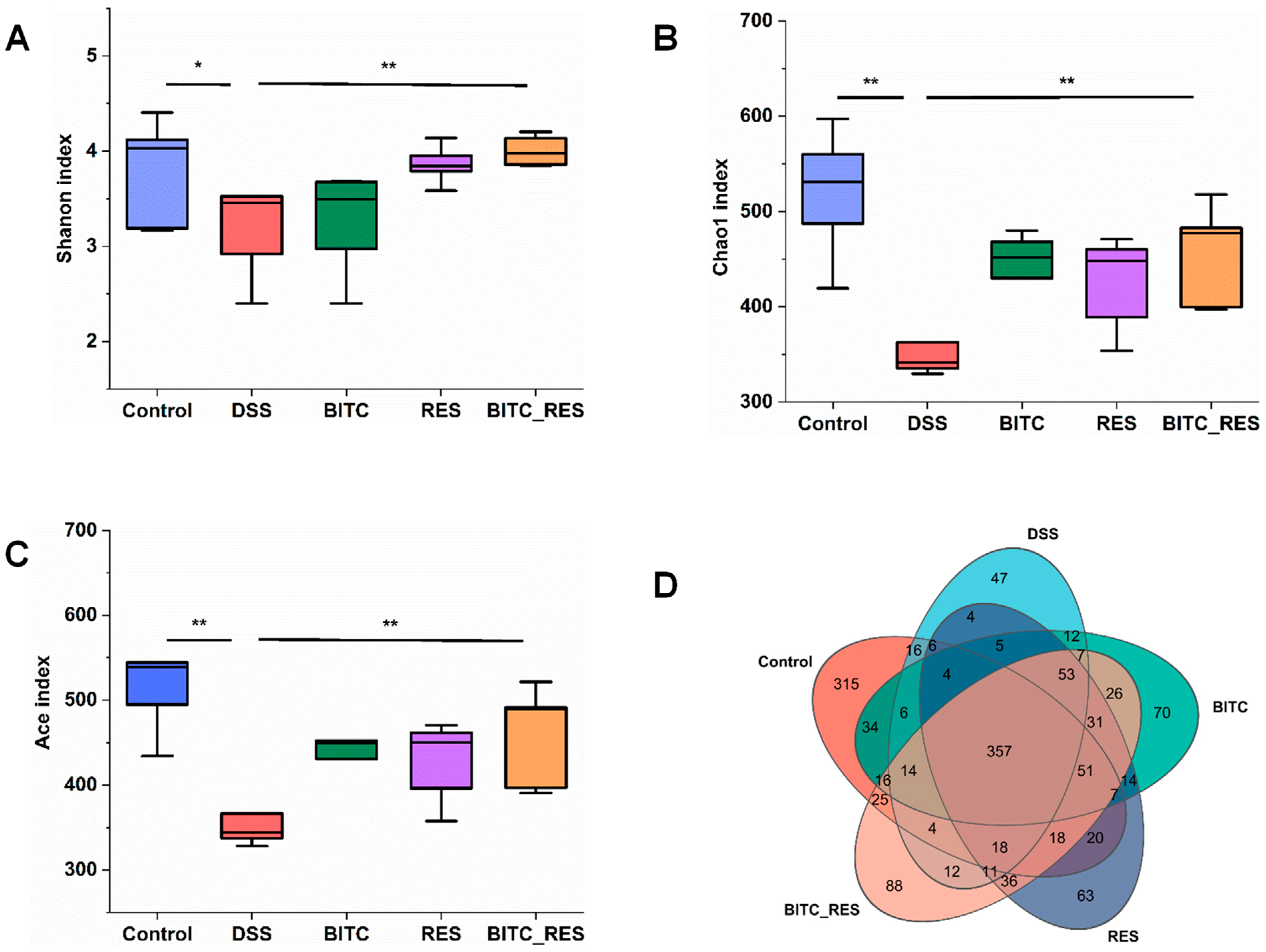
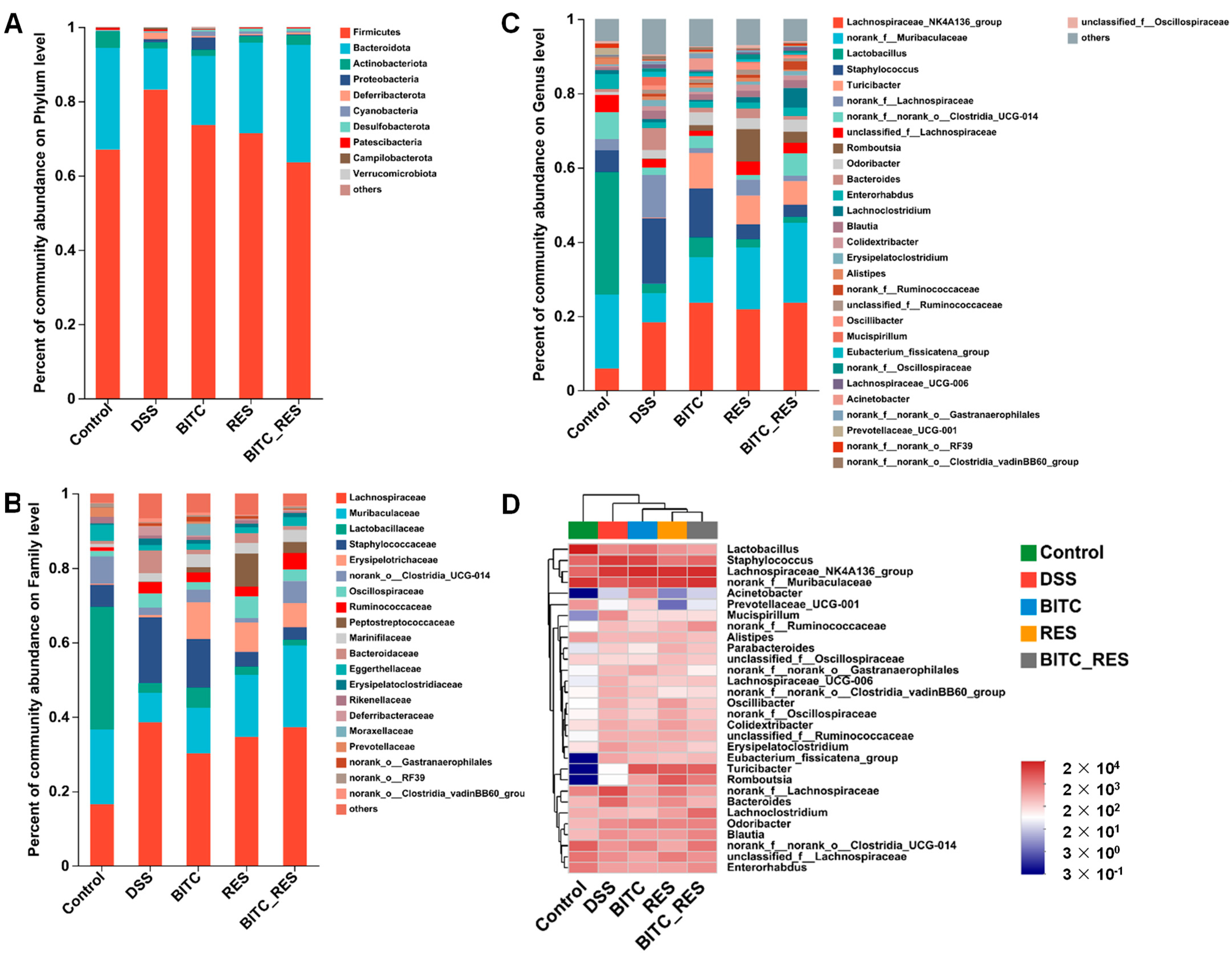
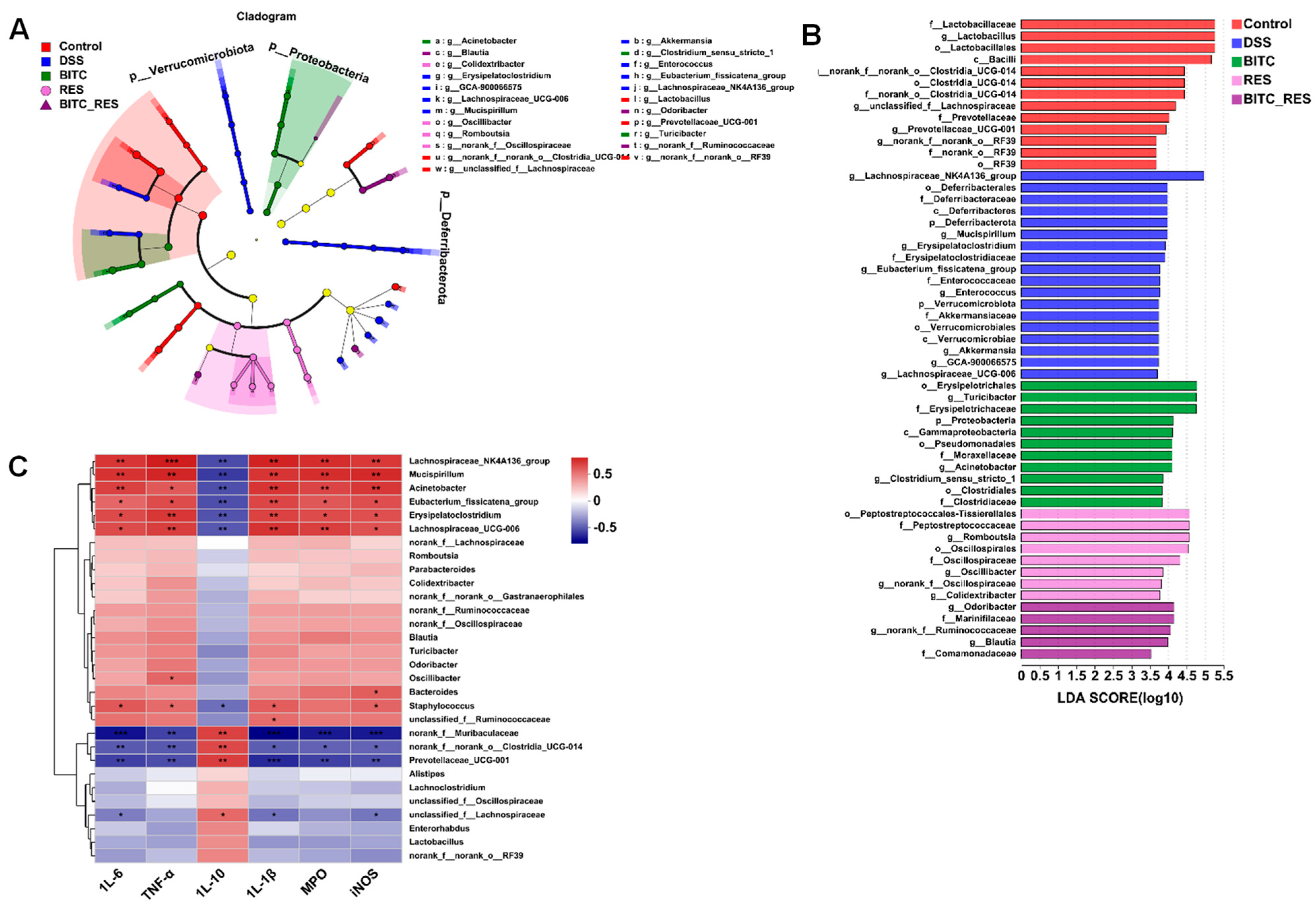
3.5. Synergistic Effects of BITC and RES on the Colon Microbiota of Mice with DSS-Induced Colitis

The gut microbiome is a complex community of microbes that has an essential impact on the health of the host. UC is strongly linked with the onset and progression of dysbiosis [40]. In this study, we investigated whether BITC and RES have synergistic regulatory effects on the colon flora to ameliorate DSS-induced colonic inflammation in mice. Previous studies have shown that BITC and RES can ameliorate disease by regulating the composition of the gut flora [24,30]. The number of operational taxonomic units (OTUs), Shannon diversity, Chao1 index and Ace index were assessed because they are widely used measures of alpha diversity, indicating the depth of sequence coverage and community diversity within assays [41]. As shown in Figure 5A–C, the α diversity, which included the Shannon, Chao1 and Ace indices, was significantly lower in the DSS group than in the control group. In addition, 357 OTUs were identified as being shared between the different groups, as shown in the Venn diagram. The numbers of specific OTUs in the control group, DSS group, BITC group, RES group, and BITC-RES group were 315, 47, 70, 63, and 88, respectively (Figure 5D). These findings indicate that species diversity was reduced by DSS treatment, while BITC-RES treatment restored species richness more significantly than did BITC or RES treatment alone.
To understand which taxa BITC and RES may influence, we assessed the relative abundances of the dominant taxa within and between treatment groups. As shown in Figure 6A, Firmicutes, Bacteroidetes, Actinobacteria, Proteobacteria, and Deferribacterota were identified at the phylum level in five different groups, with Firmicutes and Bacteroidetes being the two most abundant bacterial phyla. The pathogenesis and regulation of enteritis are closely related to the structure of the colon microbiota [42]. For example, the ratio of Firmicutes to Bacteroidetes is associated with the degree of inflammation in patients with colitis [43]. In the DSS-group, there was an increase in the relative abundance of Firmicutes and a decrease in the relative abundance of Bacteroidetes; a similar conclusion was reached by Li et al. [44]. In contrast, BITC-RES was effective at reversing the Firmicutes-to-Bacteroidetes ratio, as assessed by BITC and RES treatments alone, which was consistent with the findings of previous studies [45]. Bacteroidetes are the largest gram-negative phylum in the microbiome of the gastrointestinal tract and are typically beneficial for hosts when confined to the gastrointestinal tract [46]. Earlier studies have shown that the average level of Bacteroidetes in the UC group was significantly lower than that in the control group [47]. RES also increased Bacteroidetes and decreased Firmicutes in DSS-induced colitis mice and alleviated symptoms, in line with our studies [48]. At the family level, there was a significant enrichment of the beneficial bacteria Muribaculaceae, Lactobacillaceae, and Lachnospiraceae in the control group (Figure 6B). Treatment with DSS resulted in a significant decrease in these OTUs and an increase in the relative abundance of harmful bacteria belonging to Staphylococcaceae and Bacteroidaceae. After RES and BITC_RES treatment, the abundance of beneficial bacteria increased, and the abundance of pathogenic bacteria decreased. Once we had identified the roles of BITC and RES in regulating colon microbial structure at both the phylum and family levels, we sought to identify which bacterium was likely responsible for their effect on chronic colitis in mice. In addition, at the genus level, community abundance histograms and heatmaps were generated to examine the compositions of the communities in the different groups (Figure 6C,D). Muribaculaceae are considered anti-inflammatory bacteria for use in evaluating the effectiveness of Lactobacillus plantarum Q7 for preventing ulcerative colitis [49]. Muribaculaceae was strongly reduced in the DSS model group compared with the control group and increased in abundance in the BITC and RES treatment groups compared with the BITC- and RES-alone treatment groups. Liu et al. showed that Muribaculaceae outperformed the other bacteria in the consumption of inulin, resistant starch and polysaccharides and in the production of propionate [50,51]. According to Wang et al., Muribaculaceae improved the levels of short-chain fatty acids when given undigested pea protein [52]. BITC_RES administration resulted in the upregulation of Staphylococcus and Turicibacter and the downregulation of Bacteroides iparison to those in the model group. In conclusion, BITC_RES treatment enhanced the diversity of beneficial bacteria and reduced the diversity of harmful bacteria relative to those in the BITC and RES groups, further demonstrating the synergistic effect of BITC and RES in ameliorating colitis-related inflammation.
The abundance of the gut microbiota at the phylum and genus levels was also assessed by LEfSe (Figure 7A,B). Odoribacter was the major genus in BITC_RES. Odoribacter belongs to the order Bacteroidales and is a wide short-chain fatty acid-producing constituent of the human gut microbiota [53]. Phenotypic analysis has shown that members of Bacteroidales possess a very broad spectrum of sugar degradation capabilities, with some strains capable of degrading tens of different complex carbohydrates [54]. To determine possible associations between the gut microbial composition and pro- and anti-inflammatory cytokines, we examined Spearman’s correlation coefficients among the 30 most abundant gut microbiomes at the genus level and six indicators of colon inflammation (Figure 7C). IL-10 was positively related to the abundance of norank_f_Muribaculaceae, norank_f__norank_o__Clostridia_UCG-014, Prevotellaceae_UCG-001, and unclassified_f__Lachnospiraceae and negatively related to the diversity of Bacteroides and Mucispirillum. Conversely, the levels of inflammatory cytokines (IL-6, IL-1β, and TNF-α) and inflammatory enzymes (iNOS and MPO) correlated positively with the diversity of Bacteroides and Mucispirillum and negatively with Lachnospiraceae_NK4A136_group, Lachnospiraceae_UCG-006, and others. The pathogenic genera Bacteroides and Mucispirillum were also found to induce inflammation in mice by Sheng et al. and Herp et al. [55,56]. Furthermore, Fei et al. reported that combined treatment modified the abundance and metabolism of microorganisms in the gut, specifically affecting the levels of short-chain fatty acids and anti-inflammatory-associated molecules [57]. In conclusion, BITC_RES better reshaped the colon’s bacterial community, altered the levels of inflammatory factors in mice, and consequently improved colonic inflammatory symptoms.

4. Conclusions

BITC and RES were found to have synergistic antioxidant effects. BITC_RES increased colon length, alleviated weight loss, decreased DAI values, increased levels of the proinflammatory factor IL-10, and decreased levels of several proinflammatory cytokines, including TNF-α, IL-1β, and IL-6, compared with the BITC and RES groups. In addition, BITC-RES was effective at reversing the Firmicutes-to-Bacteroidetes ratio, as assessed by the BITC and RES groups alone. Therefore, the synergies of BITC and RES can be applied to UC. However, due to the high volatility of BITC and the low bioavailability of RES, it is necessary to study the codelivery system of both.

Author Contributions

Conceptualization, G.Z. and J.L.; methodology, J.L; software, Q.Z. and J.B.; validation, J.B.; formal analysis, J.L. and G.Z.; investigation, Q.Z.; resources, J.L. and Q.Z.; data curation, J.L.; writing—original draft preparation, J.L.; writing—review & editing, J.L. and G.Z.; visualization, H.H. (Hongshun Hao); supervision, H.H. (Hongman Hou) and H.H. (Hongshun Hao); project administration, H.H. (Hongman Hou) and G.Z.; funding acquisition, G.Z. All authors discussed the results and approved the final manuscript. All authors have read and agreed to the published version of the manuscript.

Funding

This work was supported financially by the National Key R and D Program of China (2022YFD210050103).

Institutional Review Board Statement

All animals were fed regularly according to the IACUC-approved protocol for laboratory animals prior to the experiments. Ethical approval for animal use was obtained from the Ethic Committee of Laboratory Animal Ethics Committee of Dalian Polytechnic University (Approval Code: DLPU2023033, Approval Date: 23 March 2023).

Informed Consent Statement

Not applicable.

Data Availability Statement

The original contributions presented in the study are included in the article, further inquiries can be directed to the corresponding author.

Conflicts of Interest

All authors declare no conflicts of interest.

Abbreviations

BITCbenzyl isothiocyanate
RESresveratrol
BITC_RESbenzyl isothiocyanate and resveratrol
MPOmyeloperoxidase
DSSdextran sodium sulfate
NOSnitric oxide synthase
DAIdisease activity index
OUToperational taxonomic unit

References

  1. Alsoud, D.; Verstockt, B.; Fiocchi, C.; Vermeire, S. Breaking the therapeutic ceiling in drug development in ulcerative colitis. Lancet Gastroenterol. Hepatol. 2021, 6, 589–595. [Google Scholar] [CrossRef]
  2. Li, D.; Feng, Y.; Tian, M.; Ji, J.F.; Hu, X.S.; Chen, F. Gut Microbiota-derived inosine from dietary barley leaf supplementation attenuates colitis through PPARγ signaling activation. Microbiome 2021, 9, 83. [Google Scholar] [CrossRef]
  3. Sun, L.; Ouyang, J.; Zeng, F.; Wu, S.Z. An AIEgen-based oral-administration nanosystem for detection and therapy of ulcerative colitis via 3D-MSOT/NIR-II fluorescent imaging and inhibiting NLRP3 inflammasome. Biomaterials 2022, 283, 121468. [Google Scholar] [CrossRef] [PubMed]
  4. Fei, F.; Yan, S.; Li, X. Catechins and their therapeutic benefits to inflammatory bowel disease. Molecules 2017, 22, 484. [Google Scholar] [CrossRef] [PubMed]
  5. Fonseca-Camarillo, G.; Yamamoto-Furusho, J.K. Immunoregulatory pathways involved in inflammatory bowel disease. Inflamm. Bowel Dis. 2015, 21, 2188–2193. [Google Scholar] [CrossRef]
  6. Fatani, A.J.; Alrojayee, F.S.; Parmar, M.Y.; Abuohashish, H.M.; Ahmed, M.M.; Al-Rejaie, S.S. Myrrh attenuates oxidative and inflammatory processes in acetic acid-induced ulcerative colitis. Exp. Ther. Med. 2016, 12, 730–738. [Google Scholar] [CrossRef]
  7. Baur, J.A.; Sinclair, D.A. Therapeutic potential of resveratrol: The in vivo Evidence. Nat. Rev. Drug Discov. 2006, 5, 493–506. [Google Scholar] [CrossRef]
  8. Min, K.; Seungho, C.; Sun, K.; Yoon, Y.; Kang, J.H.; Oh, S. Allyl isothiocyanate ameliorates dextran sodium sulfate-induced colitis in mouse by enhancing tight junction and mucin expression. Int. J. Mol. Sci. 2018, 19, 2025. [Google Scholar] [CrossRef]
  9. Ramachandra, D.P.; Sudheer, P. Self-micro emulsifying drug delivery via intestinal lymphatics: A lucrative approach to drug targeting. Pharm. Nanotechnol. 2023, 11, 238–246. [Google Scholar] [CrossRef]
  10. Mayangsari, Y.; Suzuki, T. Resveratrol ameliorates intestinal barrier defects and inflammation in colitic mice and intestinal cells. J. Agric. Food Chem. 2018, 66, 12666–12674. [Google Scholar] [CrossRef]
  11. Cui, X.; Jin, Y.; Hofseth, A.B.; Pena, E.; Habiger, J.; Chumanevich, A.; Poudyal, D.; Nagarkatti, M.; Nagarkatti, P.S.; Singh, U.P. Resveratrol Suppresses Colitis and Colon Cancer Associated with Colitis. Cancer Prev. Res. 2010, 3, 549–559. [Google Scholar] [CrossRef] [PubMed]
  12. Li, Z.L.; Wu, H.Y.; Liu, J.N.; Hao, H.S.; Bi, J.R.; Hou, H.M.; Zhang, G.L. Synergistic effects of benzyl isothiocyanate and resveratrol against Listeria monocytogenes and their application in chicken meat preservation. Food Chem. 2023, 419, 135984. [Google Scholar] [CrossRef] [PubMed]
  13. Liu, F.; Ma, D.; Luo, X.; Zhang, Z.; He, L.; Gao, Y.; Mcclements, D.J. Fabrication and characterization of protein-phenolic conjugate nanoparticles for co-delivery of curcumin and resveratrol. Food Hydrocoll. 2018, 79, 450–461. [Google Scholar] [CrossRef]
  14. Liu, B.H.; Li, S.D.; Sui, X.D.; Guo, L.Y.; Liu, X.M.; Li, H.M.; Gao, L.M.; Cai, S.S.; Li, Y.R.; Wang, T.T.; et al. Root Extract of Polygonum cuspidatum Siebold & Zucc. Ameliorates DSS-induced Ulcerative colitis by affecting NF-kappaB signaling pathway in a mouse model via synergistic effects of polydatin, resveratrol, and emodin. Front. Pharmacol. 2018, 9, 347. [Google Scholar]
  15. Cuggino, S.G.; Bascon-Villegas, I.; Rincon, F.; Maria, A.; Guiomar, P.; Javier, M.; Cristina, P.C.; Fernando, P.R. Modelling the combined effect of chlorine, benzyl isothiocyanate, exposure time and cut size on the reduction of Salmonella in fresh-cut lettuce during washing process. Food Microbiol. 2020, 86, 103346. [Google Scholar] [CrossRef] [PubMed]
  16. Okulicz, M.; Hertig, I. Benzyl isothiocyanate disturbs lipid metabolism in rats in a way independent of its thyroid impact following in vivo long-term treatment and in vitro adipocytes studies. J. Physiol. Biochem. 2013, 69, 75–84. [Google Scholar] [CrossRef] [PubMed]
  17. Liu, Y.; Dey, M. Dietary phenethyl isothiocyanate protects mice from colitis associated colon cancer. J. Turbul. 2017, 90, 1908. [Google Scholar] [CrossRef] [PubMed]
  18. Manichanh, C.; Rigottier-Gois, L.; Bonnaud, E.; Gloux, K.; Pelletier, E.; Frangeul, L.; Nalin, R.; Jarrin, C.; Chardon, P.; Marteau, P.; et al. Reduced diversity of faecal microbiota in Crohn’s disease revealed by a metagenomic approach. Dig. World Core Med. J. 2006, 55, 205–211. [Google Scholar]
  19. Li, M.; Li, P.; Tang, R.; Lu, H. Resveratrol and its derivates improve inflammatory bowel disease by targeting gut microbiota and inflammatory signaling pathways. Food Sci. Hum. Wellness 2022, 11, 22–31. [Google Scholar] [CrossRef]
  20. Alrafas, H.R.; Busbee, P.B.; Nagarkatti, M.; Nagarkatti, P.S. Resveratrol modulates the gut microbiota to prevent murine colitis development through induction of Tregs and suppression of Th17 cells. J. Leukoc. Biol. 2019, 106, 1. [Google Scholar] [CrossRef]
  21. Qiao, Y.; Sun, J.; Xia, S.; Tang, X.; Shi, Y.H.; Le, G.W. Effects of resveratrol on gut microbiota and fat storage in a mouse model with high-fat-induced obesity. Food Funct. 2014, 5, 1241–1249. [Google Scholar] [CrossRef]
  22. Lelis, D.D.F.; Souza, L.A.A.; Brito, R.V.J.; Andrade, M.C.; Nobre, S.A.W.; Guimaraes, A.L.S.; de Paula, A.M.B.; de Lima, J.P.; Hilzendeger, A.M. Lactococcus lactis and resveratrol decrease body weight and increase benefic gastrointestinal microbiota in mice. Protein Pept. Lett. 2021, 28, 761–768. [Google Scholar]
  23. Zhang, Y.; Tan, L.; Li, C.; Wu, H.; Ran, D.; Zhang, Z.Y. Sulforaphane alter the microbiota and mitigate colitis severity on mice ulcerative colitis induced by DSS. AMB Express 2020, 10, 119. [Google Scholar] [CrossRef] [PubMed]
  24. Jaja-Chimedza, A.; Zhang, L.; Khea, W.; Brittany, L.G.; Peter, K.; Kristin, M.; Richar, C.; Susan, N.; Michael, S.J. A dietary isothiocyanate-enriched moringa (Moringa oleifera) seed extract improves glucose tolerance in a high-fat-diet mouse model and modulates the gut microbiome. J. Funct. Foods 2018, 47, 376–385. [Google Scholar] [CrossRef] [PubMed]
  25. Jøraholmen, M.W.; Škalko-Basnet, N.; Acharya, G.; Basnet, P. Resveratrol-loaded liposomes for topical treatment of the vaginal inflammation and infections. Eur. J. Pharm. Sci. 2015, 79, 112–121. [Google Scholar]
  26. Floegel, A.; Kim, D.O.; Chung, S.J.; Koo, S.L.; Chun, O.K. Comparison of ABTS/DPPH assays to measure antioxidant capacity in popular antioxidant-rich US foods. J. Food Compos. Anal. 2011, 24, 1043. [Google Scholar] [CrossRef]
  27. Ibrahim, A.; Al-Hizab, F.A.; Abushouk, A.I.; Abdel-Daim, M.M. Nephroprotective effects of benzyl isothiocyanate and resveratrol against cisplatin-induced oxidative stress and inflammation. Front. Pharmacol. 2018, 9, 1268. [Google Scholar] [CrossRef] [PubMed]
  28. Kanwal, S.; Joseph, T.P.; Aliya, S.; Song, S.Y.; Saleem, M.; Nisar, M.A.; Wang, Y.; Meyiah, A.; Ma, Y.F.; Xin, Y. Attenuation of DSS induced colitis by Dictyophora indusiata polysaccharide (DIP) via modulation of gut microbiota and inflammatory related signaling pathways. J. Funct. Foods 2019, 64, 103641. [Google Scholar] [CrossRef]
  29. Kong, Y.; Yan, T.; Tong, Y.; Deng, H.T.; Tan, C.; Wan, M.Z.; Wang, M.Y.; Meng, X.J.; Wang, Y.H. Gut microbiota modulation by polyphenols from aronia melanocarpa of LPS-induced liver diseases in rats. J. Agric. Food Chem. 2021, 69, 3312–3325. [Google Scholar] [CrossRef] [PubMed]
  30. Li, F.; Han, Y.; Cai, X.; Gu, M.; Sun, J.; Qi, C.; Goulette, T.; Song, M.Y.; Li, Z.Z.; Xiao, H. Dietary resveratrol attenuated colitis and modulated gut microbiota in dextran sulfate sodium-treated mice. Food Funct. 2019, 11, 1063–1073. [Google Scholar] [CrossRef]
  31. Larrosa, M.J.; Yañéz-Gascón, M.; Selma, M.A.V.; González-Sarrías, A.; Toti, S.; Cerón, J.J.; Tomás-Barberán, F.; Dolara, P.; Dolara, J.C. Effect of a low dose of dietary resveratrol on colon microbiota, inflammation and tissue damage in a DSS-induced colitis rat model. J. Agric. Food Chem. 2009, 57, 2211. [Google Scholar] [CrossRef]
  32. Zhao, L.; Zhang, Y.; Liu, G.; Hao, S.; Wang, C.; Wang, Y. Black rice anthocyanin-rich extract and rosmarinic acid, alone and in combination, protect against DSS-induced colitis in mice. Food Funct. 2018, 9, 2796–2808. [Google Scholar] [CrossRef] [PubMed]
  33. Kolios, G.; Valatas, V.; Ward, S.G. Nitric oxide in inflammatory bowel disease: A universal messenger in an unsolved puzzle. Immunology 2005, 113, 427–437. [Google Scholar] [CrossRef] [PubMed]
  34. Xu, B.; Huang, S.; Chen, Y. Synergistic effect of combined treatment with baicalin and emodin on DSS-induced colitis in mouse. Phytother. Res. 2021, 35, 5708–5719. [Google Scholar]
  35. Aaron, S.M.; Jesus, D.L.; Yamamoto-Furusho, K. Role of cytokines in inflammatory bowel disease. World J. Gastroenterol. 2008, 14, 9. [Google Scholar]
  36. Chen, Y.; Su, W.; Tie, S.; Cui, W.; Yu, X.Y.; Zhang, L.J.; Hua, Z.; Tan, M. Orally deliverable sequence-targeted astaxanthin nanoparticles for colitis alleviation. Biomaterials 2023, 293, 121976. [Google Scholar] [CrossRef] [PubMed]
  37. Yao, J.; Wang, J.Y.; Liu, L.; Zeng, W.S.; Li, Y.X.; Xun, A.Y.; Zhao, L.; Jia, C.H.; Feng, J.L.; Wei, X.X.; et al. Polydatin ameliorates DSS-induced colitis in mice through inhibition of nuclear factor-kappaB activation. Planta Medica 2011, 77, 421–427. [Google Scholar] [CrossRef] [PubMed]
  38. Goosen, T.C.; Mills, D.E.; Hollenberg, P.F. Effects of benzyl isothiocyanate on rat and human cytochromes P450: Identification of metabolites formed by P450 2B1. J. Pharmacol. Exp. Ther. 2011, 296, 198–206. [Google Scholar]
  39. Wong, K.W.; Teh, S.S.; Law, K.P.; Ismail, I.S.; Sato, K.; Mase, N.Y.; Mah, S.H. Synthesis of benzylated amine-substituted xanthone derivatives and their antioxidant and anti-inflammatory activities. Arch. Pharm.-Chem. Life Sci. 2023, 356, 2200418. [Google Scholar] [CrossRef] [PubMed]
  40. Zheng, L.; Wen, X.L. Gut microbiota and inflammatory bowel disease: The current status and perspectives. World J. Clin. Cases 2021, 9, 13. [Google Scholar] [CrossRef]
  41. Odamaki, T.; Kato, K.; Sugahara, H.; Hashikur, N.; Takahashi, S.; Xiao, J.; Abe, F.; Osawa, R. Age-related changes in gut microbiota composition from newborn to centenarian: A cross-sectional study. BMC Microbiol. 2016, 16, 90. [Google Scholar] [CrossRef]
  42. Liu, G.; Wen, X.; Ding, S.J.; Jiang, H.M.; Ma, Y.; Wang, H.; Fang, J. Effects of IRW and IQW on oxidative stress and gut microbiota in dextran sodium sulfate-induced colitis. Cell. Physiol. Biochem. Int. J. Exp. Cell. Physiol. Biochem. Pharmacol. 2018, 51, 441–451. [Google Scholar] [CrossRef] [PubMed]
  43. Guo, M.; Li, Z. Polysaccharides isolated from Nostoc commune Vaucher inhibit colitis-associated colon tumorigenesis in mice and modulate gut microbiota. Food Funct. 2019, 10, 6873–6881. [Google Scholar] [CrossRef] [PubMed]
  44. Li, X.; Ouyang, W.; Jiang, Y.; Lin, Q.R.; Peng, X.N.; Hu, H.; Ye, Z.M.; Liu, G.; Yu, Y.G. Dextran-sulfate-sodium-induced colitis-ameliorating effect of aqueous phyllanthus emblica L. Extract through regulating colonic cell gene expression and gut microbiomes. J. Agric. Food Chem. 2023, 71, 6999–7008. [Google Scholar] [CrossRef] [PubMed]
  45. Tan, C.; Wang, M.; Kong, Y.; Wan, M.Z.; Deng, H.T.; Tong, Y.Q.; Lyu, C.M.; Meng, X.J. Anti-inflammatory and intestinal microbiota modulation properties of high hydrostatic pressure treated cyanidin-3-glucoside and blueberry pectin complexes on dextran sodium sulfate-induced ulcerative colitis mice. Food Funct. 2022, 68, 12295–12309. [Google Scholar]
  46. Moissl-Eichinger, C.; Willing, B.P.; Burke, C.M.; Lukiw, W. Bacteroides fragilis lipopolysaccharide and inflammatory signaling in alzheimers disease. Front. Microbiol. 2017, 7, 1544. [Google Scholar]
  47. Zhou, Y.; Zhi, F. Lower level of bacteroides in the gut microbiota is associated with inflammatory bowel disease: A meta-analysis. BioMed Res. Int. 2016, 2016, 5828959. [Google Scholar] [CrossRef] [PubMed]
  48. Yin, X.H.; Liao, W.Y.; Li, Q.R.; Zhang, H.M.; Liu, Z.H.; Zheng, X.J.; Zheng, L.; Feng, X. Interactions between resveratrol and gut microbiota affect the development of hepatic steatosis: A fecal microbiota transplantation study in high-fat diet mice. J. Funct. Foods 2020, 67, 103883. [Google Scholar] [CrossRef]
  49. Hao, H.; Zhang, X.; Tong, L.; Liu, Q.Q.; Liang, X.; Bu, Y.S.; Gong, P.; Liu, T.J.; Zhang, L.W.; Xia, Y.; et al. Effect of extracellular vesicles derived from Lactobacillus plantarum Q7 on gut microbiota and ulcerative colitis in mice. Front. Immunol. 2021, 12, 777147. [Google Scholar] [CrossRef]
  50. Liu, H.; Liao, C.; Wu, L.; Tang, J.; Chen, J.; Lei, C.; Zheng, L.; Zhang, C.; Liu, Y.Y.; Xavier, J. Ecological dynamics of the gut microbiome in response to dietary fiber. ISME J. 2022, 16, 2040–2055. [Google Scholar] [CrossRef]
  51. Smith, B.; Miller, R.; Schmidt, T. Muribaculaceae genomes assembled from metagenomes suggest genetic drivers of differential response to acarbose treatment in mice. MSphere 2021, 6, e0085121. [Google Scholar] [CrossRef]
  52. María, A.M.; Ramos, S. Impact of dietary flavanols on microbiota, immunity and inflammation in metabolic diseases. Nutrients 2021, 13, 850. [Google Scholar] [CrossRef] [PubMed]
  53. Hiippala, K.; Barreto, G.; Burrello, C.; Diaz-Basabe, A.; Satokari, R. Novel odoribacter splanchnicus strain and its outer membrane vesicles exert immunoregulatory effects in vitro. Front. Microbiol. 2020, 11, 575455. [Google Scholar] [CrossRef] [PubMed]
  54. Flint, H.J.; Scott, K.P.; Duncan, S.H.; Louis, P.; Forano, E. Microbial degradation of complex carbohydrates in the gut. Gut Microbes 2012, 3, 289–306. [Google Scholar] [CrossRef] [PubMed]
  55. Sheng, K.; Xu, Y.; Kong, X.; Wang, J.; Zha, X.; Wang, Y. Probiotic Bacillus cereus alleviates dextran sulfate sodium-induced colitis in mice through improvement of the intestinal barrier function, anti-Inflammation, and gut microbiota modulation. J. Agric. Food Chem. 2021, 49, 69. [Google Scholar] [CrossRef] [PubMed]
  56. Herp, S.; Durai Raj, A.C.; Salvado Silva, M.; Woelfel, S.; Stecher, B. The human symbiont Mucispirillum schaedleri: Causality in health and disease. Med. Microbiol. Immunol. 2021, 210, 173–179. [Google Scholar] [CrossRef]
  57. Fei, Y.; Zhang, S.; Han, S.; Qiu, B.; Lu, Y.M.; Huang, W.X.; Li, F.; Chen, D.Y.; Berglund, B.; Xiao, H. The role of dihydroresveratrol in enhancing the synergistic effect of Ligilactobacillus salivarius liol and resveratrol in ameliorating colitis in mice. Research 2022, 16, 9863845. [Google Scholar]
Figure 1. Synergistic antioxidant activities of BITC and RES. DPPH (A) and ABTS•+ (B) radical scavenging capacities. BITC, benzyl isothiocyanate. RES, resveratrol. Different superscript letters in the graph (a, b, c, and d) indicate significant differences (p < 0.05).
Foods 13 02078 g001
Figure 2. Synergistic effects of BITC and RES on DSS-induced colitis symptom severity. (A) Image of the colon length. (B) Colon length. (C) Percentage of initial body weight. (D) Disease activity index score. The data are presented as the mean ± standard error of the mean (n = 6). Different superscript letters in the graph (a, b, c, and d) indicate significant differences (p < 0.05).
Foods 13 02078 g002
Figure 3. Synergistic effects of BITC and RES on colonic microinjury in DSS-induced colitis. Hematoxylin and eosin staining of representative inflamed areas of colon sections ((A), magnification ×40; (B), magnification ×100).
Foods 13 02078 g003
Figure 4. Synergistic effects of BITC and RES on iNOS and MPO levels in colonic tissues and cytokine levels in serum. (A) iNOS, (B) MPO, (C) TNF-α, (D) IL-1β, (E) IL-6, and (F) IL-10. The data are expressed as the mean ± SD (n = 6). Bars with different superscripts letters are significantly different.
Foods 13 02078 g004
Figure 5. Diversity of the gut microbiota among the control, DSS, BITC, RES, and BITC_RES groups (n = 5). α-Diversity analysis of the (A) Shannon, (B) Chao1, and (C) Ace indices. (D) Venn diagram of shared and unique species at the OTU level. * p < 0.05, ** p < 0.01.
Foods 13 02078 g005
Figure 6. The percent community abundance histogram of the gut microbiota in the control, DSS, BITC, RES, and BITC_RES groups. (A) Phylum level, (B) family level, and (C) genus level. A cluster heatmap of species richness is shown (D).
Foods 13 02078 g006
Figure 7. Microbial taxa discrepancies among the control, DSS, BITC, RES, and BITC_RES groups. Linear discriminant analysis (LDA) revealed scores > 3.5 and adjusted p values < 0.05. (A) Cladogram and histogram (B,C) correlation analysis (p < 0.05 and |R2| > 0.7, * p < 0.05, ** p < 0.01, *** p < 0.001) based on Spearman’s rank order correlation between the 30 most abundant gut microorganisms at the genus level and biochemical criterion.
Foods 13 02078 g007
Disclaimer/Publisher’s Note: The statements, opinions and data contained in all publications are solely those of the individual author(s) and contributor(s) and not of MDPI and/or the editor(s). MDPI and/or the editor(s) disclaim responsibility for any injury to people or property resulting from any ideas, methods, instructions or products referred to in the content.

