Article

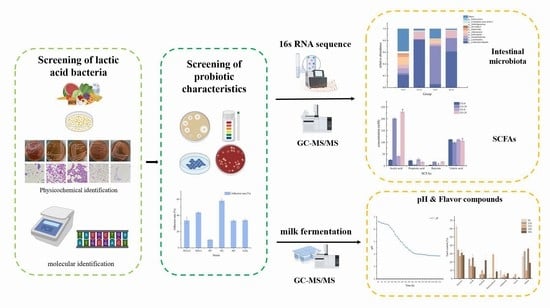
Screening, Identification and Physiological Characteristics of Lactobacillus rhamnosus M3 (1) against Intestinal Inflammation

College of Food Science and Technology, Hunan Agricultural University, Changsha 410128, China
*
Author to whom correspondence should be addressed.
Foods 2023, 12(8), 1628; https://doi.org/10.3390/foods12081628
Submission received: 9 March 2023 / Revised: 30 March 2023 / Accepted: 11 April 2023 / Published: 12 April 2023
(This article belongs to the Special Issue Probiotics: Selection, Cultivation, Evaluation and Application)

Abstract

:
The probiotic role of lactic acid bacteria (LAB) in regulating intestinal microbiota to promote human health has been widely reported. However, the types and quantities of probiotics used in practice are still limited. Therefore, isolating and screening LAB with potential probiotic functions from various habitats has become a hot topic. In this study, 104 strains of LAB were isolated from and identified in traditionally fermented vegetables, fresh milk, healthy infant feces, and other environments. The antibacterial properties—resistance to acid, bile salts, and digestive enzymes—and adhesion ability of the strains were determined, and the biological safety of LAB with better performance was studied. Three LAB with good comprehensive performance were obtained. These bacteria had broad-spectrum antibacterial properties and good acid resistance and adhesion ability. They exhibited some tolerance to pig bile salt, pepsin, and trypsin and showed no hemolysis. They were sensitive to the selected antibiotics, which met the required characteristics and safety evaluation criteria for probiotics. An in vitro fermentation experiment and milk fermentation performance test of Lactobacillus rhamnosus (L. rhamnosus) M3 (1) were carried out to study its effect on the intestinal flora and fermentation performance in patients with inflammatory bowel disease (IBD). Studies have shown that this strain can effectively inhibit the growth of harmful microorganisms and produce a classic, pleasant flavor. It has probiotic potential and is expected to be used as a microecological agent to regulate intestinal flora and promote intestinal health. It can also be used as an auxiliary starter to enhance the probiotic value of fermented milk.

Graphical Abstract

1. Introduction

Changes in the structure and quantity of intestinal microflora greatly impact human health and the occurrence of diseases. The gut microbiome is known as the ‘invisible endocrine organ’, the place where the human body digests food and absorbs nutrients. LAB, as one of the intestinal bacteria, are widely distributed and can be isolated from the environment, food, and human and animal gastrointestinal tracts [1]. Among them, strains with probiotic properties have high resistance to acid and bile salts, can improve immunity, and have antibacterial, antioxidant, and free radical scavenging activities. They can improve gastrointestinal function, regulate intestinal flora, and promote the apoptosis of human cancer cells [2,3]. Probiotics are defined as living microorganisms that can bring health benefits to the host when taken in sufficient quantities [4]. With the continuous discovery and exploration of the probiotic properties of LAB, many strains have been proven effective and safe, and have been developed for general use.
IBD is an inflammatory state of the colon and small intestine caused by an immune response disorder. It is one of the common gastrointestinal diseases, which include Crohn’s disease and ulcerative colitis, usually accompanied by diarrhea, pain, fatigue, constipation, and other symptoms. Epidemiological studies have shown that the incidence of IBD is on the rise worldwide and may greatly impact human health and life. However, the exact cause of the disease is still unclear. Factors such as susceptibility genes, the immune system, and external environmental stimuli may lead to the occurrence of IBD [5]. Recently, the imbalance in intestinal flora has become an IBD research hotspot; microbial imbalance has been shown to be closely associated with allergy, eczema, IBD, and irritable bowel syndrome [6]. Harmful microorganisms not only affect food quality and safety but also, to some extent, threaten the life and health of the host. Antibiotics are commonly used in clinical practice to prevent and treat bacterial infections. However, because of the improper use of antibiotics, bacterial drug resistance has been gradually increasing, compounding the difficulty of controlling bacterial infections. Probiotics have been shown to inhibit the growth and colonization of pathogenic bacteria by inducing the host to produce organic acids, bacteriocins, and other substances; alter the intestinal microbial community; and improve the body’s intestinal function. To strengthen the intestinal barrier by maintaining tight junctions and inducing mucin production, mediated immune regulation may mediate the secretion of cytokines and immunoglobulins through signaling pathways such as Nuclear Factor kappa-B, affecting the proliferation and differentiation of immune cells or epithelial cells. LAB play a crucial role in regulating the composition and metabolism of intestinal microflora and intestinal immune function. This makes them beneficial in preventing and treating IBD and other diseases [6,7]. LAB with antibacterial ability can be used as probiotics to partly replace antibiotics, avoid bacterial resistance, and improve human and animal health [8]. However, probiotics’ production and survival in the human gastrointestinal tract are complex processes. Probiotics should survive in the presence of gastric acid and bile, and have the ability to colonize and play a role in the intestine [9]. Therefore, being nontoxic, harmless, nonpathogenic, acid and bile resistant, and able to proliferate in the presence of bile are the essential criteria for potential probiotic strains. Therefore, to screen out potential probiotics, it is a necessity and a priority to identify and evaluate their physiological characteristics and safety. Previous studies have shown that naturally fermented foods and dairy products are important sources of probiotic strains. In addition, probiotics for human use are best sourced from humans because such microorganisms may be more competitive and better adapted to human microenvironment conditions than probiotic strains from other sources. In this study, LAB with probiotic potential were isolated and identified from traditionally fermented vegetables, fresh milk, healthy infant feces, and other habitats. The inhibition of pathogenic bacteria, acid and bile salt resistance, adhesion ability, safety, intestinal flora regulation ability, and fermentation performance were evaluated in vitro. The aim was to obtain new probiotic resources and lay a foundation for subsequent research and application.

2. Materials and Methods

2.1. Sample Collection

The sources of fermented vegetables were 3 fermented bamboo shoots from Yiyang, Hunan Province, and 9 fermented vegetables sold in Tongdao County, Huaihua City, Hunan Province. Three feces samples we obtained from obese people and three from infants and children (0–3 years old), collected from schools. Raw milk sources produced 3 samples from the Hunan Institute of Animal Husbandry.

2.2. Reagents

The reagents and materials used are shown in Table 1:

2.3. Isolation and Purification of Bacterial Strains

On an ultra-clean bench, 25 g or 25 mL of the sample was placed in 225 mL of sterile saline containing glass beads and shaken well in a constant temperature shaker at 37 °C. The sample solution was diluted gradually, and 100 μL of the appropriate diluent was coated on an MRS agar plate and anaerobically cultured at 37 °C for 24–48 h. Single colonies were picked from countable MRS agar plates and repeatedly streaked on MRS agar medium to further purify the colonies. The purified bacteria were transferred to MRS broth at 37 °C for 12 h, mixed with 50% glycerol by volume at 7:3, and stored at −20 °C. Colonies were scribed in MRS agar medium slant, cultured at 37 °C until the growth of colonies was detected, preserved at 4 °C for identification, and placed in glycerol preservation at −80 °C.

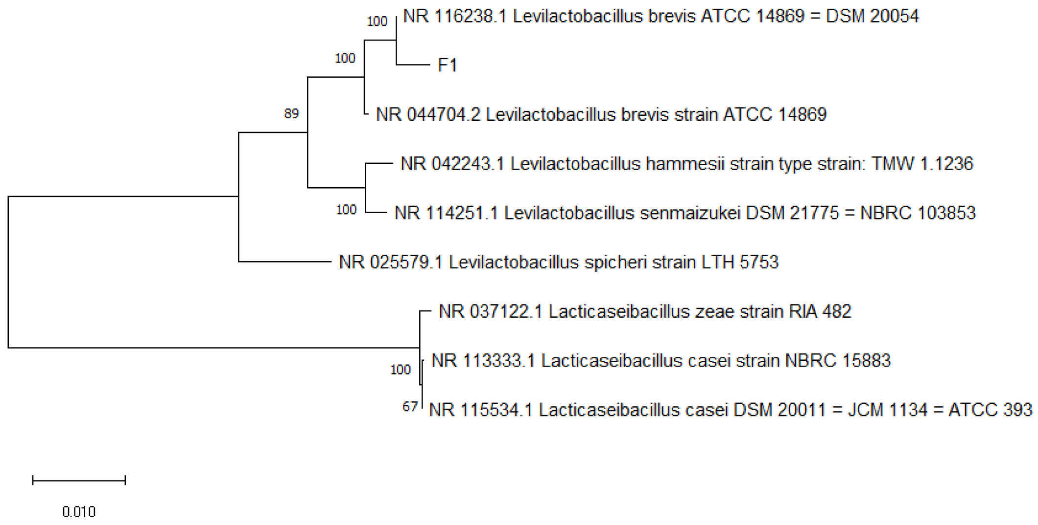
2.4. Identification of LAB

Strain identification generally clarifies the taxonomic relationship between microbial populations through morphological characteristics, physical and chemical properties, genetic evolution, and other aspects used to confirm the identity of strains. The physiological and biochemical characteristics of the screened strains were detected according to the ‘Manual for Systematic Identification of Common Bacteria’ (eighth edition) and ‘Berger’s Manual of Bacteria’ [10]. DNA was extracted from Gram-positive and catalase-negative strains using a bacterial genomic DNA extraction kit. The 16 S rDNA gene was amplified, and the universal primers of bacteria were selected for amplification primers: the forward primer 27F: 5′-AGAGTTTGATCCTGGCTCAG-3′ and reverse primer 1492R: 5′-AAGGAGGTGATCCAGCC-3′. The PCR reaction mixture was prepared. After the amplification reaction, the PCR product was separated by electrophoresis, stained, and checked on the ultraviolet illuminator. The clear target band was selected and sent to Sangon Biotech (Shanghai, China) Co., Ltd., for gene sequencing. The obtained sequence was subjected to the BLAST program for sequence homology alignment. BioEdit sequence alignment was used to edit forward and reverse sequences, and the isolates were identified according to the sequence. Phylogenetic trees were constructed using Mega-x software.

2.5. Antibacterial Experiment

The Oxford cup method [11] was used to screen LAB with active antibacterial substances, and common pathogenic bacteria in food were selected as indicator bacteria. Selected indicator bacteria were S. aureus ATCC 6538, L. monocytogenes ATCC 19115, E. coli CGMCC 9181, Pseudomonas aeruginosa, and Bacillus subtilis. The indicator bacteria were activated in LB broth for three generations, cultured at 37 °C for 24–36 h; 106 CFU/mL was taken as the concentration of indicator bacteria suspension, and the indicator bacteria were stored in a refrigerator. The screened LAB were activated in an MRS broth medium for three generations and then inoculated in MRS broth with 1% inoculation. After anaerobic culture at 37 °C for 48 h and 8 min centrifugation at 10,000 r/min, the fermentation supernatant was taken to obtain the crude extract of LAB. A total of 200 μL indicator bacteria was coated on the LB agar medium, and the sterile Oxford cup was buckled on the LB agar medium with a specific strength. Then, 200 μL crude extract of LAB was added to the Oxford cup, and standard MRS was performed as a blank control. Three experiments were repeated for each LAB. The plate was transferred to a constant temperature and humidity incubator and cultured at 37 °C. After 24 h, the diameter of the inhibition zone was measured accurately with a vernier caliper wherever the inhibition zone was observed.

2.6. Determination of LAB Tolerance

2.6.1. Acids Resistance Test

The activated LAB were cultured in MRS liquid medium in a 37 °C anaerobic environment for 24 h, centrifuged at 4 °C, 10,000× g for 8 min, and washed twice with sterile PBS buffer (pH 7.2) or 0.9% sterile saline. The bacteria were resuspended in sterile MRS broth medium at pH 2.0 and 3.0 and incubated anaerobically at 37 °C for 3 h. Samples were taken at 0 h and 3 h for dilution, respectively, and 100 μL of the appropriate gradient dilution was spread and inoculated on MRS agar medium and incubated under anaerobic conditions for 24 to 48 h. The standard MRS broth in which the cultures were inoculated was used as a positive control [12]. To calculate its survival rate, we used the formula:
Survival rate (%) = LogNt (CFU)/LogN0 (CFU) × 100%
where Nt is the number of viable bacteria after treatment and N0 is the number of added live bacteria.

2.6.2. Bile Salt Resistance Experiment

In order to detect the LAB tolerance of bile in a low pH environment (growth and survival at pH 2.0), MRS broth medium with 0.2% and 0.3% pig bile salt concentration was prepared and sterilized using a 0.22 μm filter membrane. The activated bacterial liquid was mixed and centrifuged at 4 °C, 10,000× g for 8 min, and the organisms were washed twice with saline buffer. Bacteria were resuspended in different concentrations of bile salt medium, and positive control groups were established. Samples were taken at 0 h and 3 h after incubation for gradient dilution, and 100 μL of the appropriate gradient dilutions was inoculated on MRS agar medium. The survival rate was calculated using the formula described for acid tolerance assay [13].

2.6.3. Simulated Gastrointestinal Fluid Tolerance Experiment

Probiotics need to tolerate not only lower pH and bile salts but also pepsin and trypsin in the gastrointestinal fluid to colonize and function in the human body. The artificial gastric and intestinal fluids were prepared with reference to the literature [14,15]. To simulate gastric fluid, hydrochloric acid was diluted to adjust the pH to 2.0 and 3.0, 10 g of pepsin was added, and 1000 mL of water was used to fix the volume. To simulate intestinal fluid, 6.8 g of potassium dihydrogen phosphate solution dissolved in water was used, and the pH was adjusted to 6.8 with NaOH solution. A total of 10 g of pancreatin was dissolved in water, and 1000 mL of water was added to fix the volume. The simulated gastric and intestinal fluids were filtered with a 0.22 μm filter membrane for sterilization and set aside.
The bacterial suspension was inoculated into the simulated gastric fluid, incubated anaerobically at 37 °C, and the survival rate was calculated by sampling and counting at 0 h and 3 h. At the same time, 1 mL of simulated gastric fluid containing bacteria was added into 9 mL of simulated intestinal fluid and incubated anaerobically at 37 °C, and the survival rate was calculated by sampling and counting at 0 h and 3 h in the simulated intestinal fluid. The formula for calculating the survival rate was described in the acid resistance test.

2.7. Determination of Adhesion Ability of LAB

The complete medium (fetal bovine serum: Burke modified Eagle high-glucose medium = 1:9) was prepared. The Caco-2 cells preserved in liquid nitrogen were quickly thawed in a 37 °C water bath, transferred into a cell culture flask containing a complete medium, blown evenly, and cultured in a 5% CO2 incubator at 37 °C. The medium was changed according to the cell growth. When the cell fusion degree reached 80–90%, the cells were subcultured and digested with 0.25% trypsin solution. The cells were used in the experiment after 3 passages. The cells were resuspended in a complete medium to a 5 × 10⁴/mL concentration and inoculated into 96-well plates. The cells were cultured until the cell monolayer covered the plate holes.
The weighted bacteria with a concentration of 1 × 10⁸ CFU/mL was suspended in a complete medium containing 10% serum. The medium in the perforated plate was sucked, the sterile PBS buffer preheated at 37 °C was slowly washed twice, 200 μL of LAB suspension was added to co-culture with cells, and a cell-free blank group was prepared. All were incubated in a carbon dioxide incubator for 4 h. After incubation, the suspension containing the cell pores was removed and washed with preheated sterile PBS buffer 3 times, and the non-adherent bacteria were washed away. The cells were digested with 0.25% trypsin, digested, and mixed. A total of 100 μL of the mixture (the suspension was taken directly from the blank group) was selected and spread on MRS agar medium for culture. The adhesion rate was counted and calculated with the formula described in the acid resistance test.

2.8. In Vitro Safety Experiment

2.8.1. Hemolytic Test

Hemolysis is the rupture of erythrocytes and escape of hemoglobin under the action of hemolytic toxins and other physicochemical factors, and the grades of hemolysis can be divided into: complete hemolysis (β hemolysis), partial hemolysis (α hemolysis), and no hemolysis (γ hemolysis) [16]. The screened strains were scribed on Columbia blood agar plates and incubated anaerobically at 37 °C for 24 h. The colonies and blood agar plate morphology were observed, and S. aureus was used as a positive control.

2.8.2. Antibiotic Sensitivity Test

The antibiotic susceptibility of the screened strains was assessed by the paper diffusion method, and the susceptibility of 13 antibiotics, including Clindamycin, Streptomycin, Gentamicin, Kanamycin, Vancomycin, Tetracycline, Chloramphenicol, Ampicillin, Metronidazole, Bacitracin, Cotrimoxazole, Neomycin, and Ofloxacin, was tested by the paper diffusion method [17]. The LAB solution of 200 μL MRS broth was cultured for 24 h, evenly coated on MRS agar medium, and left to wait for the plate to dry. Sterile antibiotic paper sheets were attached to the plate with the center of each paper sheet greater than 24 mm apart and the paper sheets greater than 15 mm from the inner edge of the plate. Three to four paper sheets were attached to each dish placed at 4 °C for 30 min for antibiotic diffusion and then incubated anaerobically at 37 °C for 24 to 48 h. The diameter of the inhibition zone was measured precisely with vernier calipers, and the criteria for determining drug resistance were taken from the Executive Standard for Antimicrobial Drug Susceptibility Testing [18], shown in Table 2.

2.9. In Vitro Fermentation and Detection

2.9.1. In Vitro Fermentation

  • Sample collection: This study was approved by the Biomedical Research Ethics Committee of the Hunan Agricultural University (Protocol Code: Ethical Review 2022 No. 117, 10 November 2022). All participants provided written informed consent. Fecal samples were collected from 8 donors with diarrhea symptoms, all of whom had no underlying diseases and had not taken antibiotics within 3 months before collection. The collected samples were mixed with the same amount of feces and frozen in a refrigerator at −80 °C until use;
  • Preparation of basal medium [19]: The medium composition (/L) was yeast extract 2.0 g, peptone 2.0 g, sodium chloride 0.1 g, potassium dihydrogen phosphate 0.04 g, sodium bicarbonate 2.0 g, calcium chloride 0.01 g, magnesium sulfate 0.01 g, hemin 50 mg, vitamin K 10 μL, bile salt 0.5 g, L-cysteine 0.5 g, and resazurin 1.0 mg. The basic medium was divided into test tubes and sterilized at 121 °C for 20 min. After sterilization, the medium was removed and placed in an anaerobic incubator for 18–24 h to ensure the container was anaerobic before the experiment. The expected color of the basic medium in the anaerobic environment was light yellow, and the medium was to turn red rapidly in the presence of oxygen;
  • In vitro fermentation: The feces were taken out and mixed evenly in a sterile container, and sterile normal saline with a weight of 9 times the weight of the feces was added to mix thoroughly. The gauze was filtered to obtain a 10% fully mixed fecal suspension. Then, 10% fecal suspension was added to each test tube. The experimental group received 10% LAB suspension with a concentration of about 108 CFU/mL, and the same amount of sterile water was added to the control group. The samples were cultured in an anaerobic incubator at 37 °C for 24 h, and the samples were collected aseptically at 0 and 24 h, respectively. The samples were taken out and immediately placed in ice water to stop fermentation and then stored at −80 °C.

2.9.2. DNA Extraction and Sequencing

Three groups of parallel samples of each bacteria were mixed evenly, and the genomic DNA of the samples was extracted by the CTAB method. The purity and concentration of total DNA were detected by 0.8% agarose gel electrophoresis. PCR amplification was performed using specific primers with Barcode and high-fidelity DNA polymerase, forward primer 515F (5′-GTGCCAGCMGCCGCGGTAA-3′), and reverse primer 806R (5′-GGACTACHVGGGTWTCTAAT-3′). Each sample was repeated 3 times to ensure all PCR reactions terminated in the linear amplification period. After PCR, the PCR products of the same sample were mixed and detected by 2% agarose gel electrophoresis. The detection condition was 5 V/cm and the detection time was 20 min. The target fragment was cut and recovered. The target DNA fragments were recovered by elution with TE buffer, and the PCR amplification products were quantified by the QuantiFluor TM-ST blue fluorescence quantitative system. The library was constructed using the NEB Next® Ultra TM DNA Library Prep Kit according to the mixing ratio, and the quality and sequencing were performed by Agilent Bioanalyzer 2100 and Qubit. The double-ended sequencing results were spliced by FLASH, and the quality control screening was performed to obtain high-quality target sequences. Subsequent bioinformatics operations were completed using QIIME, Usearch, etc., and statistics and mapping were mainly completed through R.

2.9.3. Detection of PH Value and Determination of Short-Chain Fatty Acids (SCFAs)

The pH values of the fermentation culture at 0 h and 24 h were measured using a handheld pH meter. Each sample was measured 3 times, and the mean and standard deviation were calculated by repeated measurements.
The content of SCFA was determined by gas chromatography (GC), referring to a reported method [20], with some modifications. The fermentation broth was mixed with 25% metaphosphoric acid solution with a 4:1 ratio, vortexed for 1 min, centrifuged at 8000 r/min for 4 min, and filtered by 0.22 μm organic microporous membrane. DB-FFAP gas chromatographic column (30 m × 250 μm × 5 μm) was used. The carrier gas was N2, and the flow rate was 0.8 mL/min. The auxiliary gas was H2. The FID detector temperature was 270 °C, the injection port temperature was 250 °C, the split ratio was 5:1, and the injection volume was 1 μL at 220 °C. Mass spectrometry (MS) conditions were: voltage of 70 eV, ion source temperature of 230 °C, mass scanning range of m/z 28–300, and electron ionization (EI) mode. The mass spectra of the compounds were compared with the NIST 20. L mass spectrometry database, and compounds with a matching degree greater than 80% were extracted for analysis.

2.10. Study on the Milk Fermentation Performance

LAB with better comprehensive performance were selected for milk fermentation, and their pH and volatile flavor substances were measured and analyzed to explore the fermentation performance of the screened LAB.

2.10.1. Preparation of Fermented Milk

Activation of bacteria: After anaerobic culture at 37 °C for 24 h, the activated bacterial solution was mixed and centrifuged at 4 °C and 10,000× g for 8 min, then washed twice in the saline buffer. The collected cells were resuspended in saline to prepare a bacterial suspension, and the concentration of LAB reached about 109 CFU/mL.
Preparation of fermented milk: Whole milk powder was added to distilled water at 50 °C, heated to 60 °C, stirred for 15 min, then placed in a water bath at 50 °C for 30 min. After hydration, the sample was heated again to 65 °C, 20 MPa homogenization, sterilized at 95 °C for 5 min, then immediately cooled in an ice water bath, to about 37 °C and inoculated with 1% inoculum. The fermentation was completed at 4 °C after 12 h.

2.10.2. PH Changes

For PH changes, 150 mL of pure milk was added to a 300 mL conical flask. After pasteurization, milk was cooled, inoculated with 1% inoculation amount, and fermented at 37 °C. From the start of the inoculation, the pH value of fermented milk was measured every 2 h using a handheld pH meter.

2.10.3. Determination and Analysis of Volatile Flavor Compounds

In sample pretreatment, 6 mL of fermented milk was added to the sample bottle, and the extraction head was aged at 250 °C for 5 min at the inlet, then vertically inserted into the sample bottle to extract the flavor compounds of fermented milk. The extraction conditions were 60 °C temperature, 300 r/min rotation speed of magnetic stirrer, and 50 min extraction time [21].
The extraction head was inserted vertically into the forward sample port on the machine and desorbed for 5 min. The initial gas chromatography conditions were a temperature of 35 °C and a time duration 5 min; then, the temperature was increased to 140 °C at a rate of 5 °C/min and maintained for 2 min. It was raised again to 10 °C/min 250 °C. The carrier gas was high-purity helium (He), the carrier gas flow rate was 1.0 mL/min, and the injection was not split. Mass spectrometry conditions were electron energy 70 eV, ion source temperature 230 °C, ionization mode EI, and mass scanning range m/z 35–500. The emission current was 100 μA, and the detection voltage was 1.4 k V. Qualitative and comparative analyses were conducted, and data were exported for CAS number query.

2.11. Statistical Analysis

All tests were conducted in triplicate, and the results are shown as mean ± standard deviation. SPSS statistical analysis software (IBM SPSS Statistics 23) was used for analysis. The data were evaluated by one-way ANOVA, analyzed, and plotted using Origin 2018, and Duncan’s multiple range test was used to analyze the significant differences.

3. Results and Discussion

3.1. Analysis of Basic Physiological Characteristics of LAB Strains

A total of 228 strains of bacteria were isolated and purified from traditionally fermented vegetables, 108 strains of bacteria were obtained from feces, and 68 strains of bacteria were obtained from raw cow’s milk. After morphological observation, Gram staining, microscopic examination, and the identification of physiological and biochemical characteristics, we obtained 134 strains of bacteria that conformed to the basic physiological characteristics of LAB. The colony morphology of these strains was round and shiny, with a central protrusion and flush edges, creamy white or off-white in color, 0.5–1.5 mm in diameter, Gram-positive, and all negative for hydrogen peroxide contact enzyme, and their microscopic examination showed a single pair of rods or a spherical structure with no movement and no spores (Figure 1).

3.2. Molecular Identification Results and Analysis

The genome extraction, PCR amplification, and sequencing of 134 strains of suspected LAB were carried out, and the gene sequences of the strains were compared using BLAST, and phylogenetic tree was established (Figure 2). The results indicated that 103 strains of LAB were obtained in this screening, and some results are indicated in Table 3.
Among these strains, 53 LAB strains were isolated from traditionally fermented vegetables and screened, including 22 strains of Lactobacillus plantarum (L. plantarum) and 31 strains of Lactobacillus brevis (L. brevis). The other obtained LAB were relatively small or single in number. This may be related to the composition of nutrients in traditionally fermented vegetables and the lower pH environment and because L. plantarum and L. brevis may be the dominant strains in the screened fermented vegetables. Twenty-nine strains of LAB were isolated from feces samples, including fifteen strains of Enterococci, eight strains of Lactobacillus paracasei (L. paracasei), two strains of L. brevis, one strain of Bifidobacterium crudilactis (B. crudilactis), one strain of L. plantarum, one strain of Pediococcus pentosaceus (P. pentosaceus), one strain of Westerella sibiricus, and one strain of Streptococcus salivarius (S. salivarius). Twenty-one strains of LAB were isolated from raw milk, including two strains of L. rhamnosus, six strains of L. paracasei, two strains of Bifidobacterium thermophilus, four strains of B. crudilactis, two strains of P. pentosaceus, one strain of Lactobacillus parabuchneri (L. parabuchneri), one strain of Lactobacillus buchneri, and two strains of Enterococcus faecium (E. faecium).

3.3. Antibacterial Experiment Results and Analysis

Probiotics can secrete organic acids, polysaccharides, antimicrobial peptides, and other substances to inhibit the growth of other microorganisms [22]. In this experiment, the representatives of Gram-positive bacteria: S. aureus, Listeria, and Bacillus subtilis, and Gram-negative bacteria: Pseudomonas aeruginosa and E. coli were used as indicator bacteria to detect the antibacterial activity of the screened LAB. A total of 101 strains were excluded because of repetitive LAB; 72 strains of LAB were subjected to the bacteriostatic test, excluding LAB without a bacteriostatic ring and a smaller bacteriostatic ring, leaving 16 strains of LAB with a better comprehensive bacteriostatic level.
The diameter of the inhibition zone is shown in Table 4, and the morphology of Antibacterial circle is shown in Figure 3.The crude extracts of 16 strains of LAB showed certain antibacterial effects. They have good inhibition levels against S. aureus and Pseudomonas aeruginosa, and the diameter of the inhibition zone can reach more than 12.00 mm. Among them, L. paracasei M16 had the best inhibitory effect on S. aureus and Bacillus subtilis, reaching 19.51 ± 0.49 mm and 17.44 ± 0.58 mm, respectively. L. rhamnosus M3 (2) had the best inhibitory effect on Listeria, reaching 18.05 ± 0.23 mm; L. paracasei M5 had the best inhibitory effect on E. coli and could reach 14.52 ± 0.26 mm. P.pentosaceus M19 had a good inhibitory effect on Pseudomonas aeruginosa, and the inhibition zone size reached 21.07 ± 0.19 mm. The results showed a significant difference in the inhibitory effect of varied strains on different indicator bacteria, and the antibacterial effect of different strains varied, pointing to the physiological characteristics of the strain and whether it produced bacteriocin.

3.4. Acid Resistance Test Results and Analysis

The effectiveness of probiotics depends on their microbial quality, resistance to harsh gastric environments, and functional characteristics. The human digestive tract is complex, and probiotics need to have a certain ability to survive in its environment [23]. Probiotics must survive for 1.5–2 h at pH 3.0. The viability of microorganisms in simulated gastrointestinal environments is one of the effective indicators for evaluating probiotics. In this experiment, 16 strains of LAB with good antibacterial activity were treated with MRS broth medium with pH 2 and pH 3, and incubated in the anaerobic environment at 37 °C to simulate the acidic conditions of human gastric juice. The results showed that the 16 strains of LAB had good tolerance to acidic conditions at pH 3, but there was a significant difference in tolerance at pH 2. Among the sixteen strains, M5, M3 (1), M2, M7, and M21 (2) showed good acid resistance, so these five LAB were selected for subsequent experiments. Acid resistance results are shown in Table 5.

3.5. Bile Salt Tolerance Test Results and Analysis

Bile acids play an important role in digesting food and regulating the balance of intestinal microbial communities. The bile salt concentration in the small intestine of healthy people is between 0.3 and 3.0 g/L. High concentrations of bile salts can antagonize the growth of probiotics, change the permeability of the cell membrane, decompose membrane proteins, and cause cell rupture and death [24]. Five strains of acid-tolerant LAB were tolerated at 0.2% and 0.3% bile salts for 3 h, and the concentrations still reached 106 CFU/mL, they all reach the critical density of live bacteria for functional properties of bacteria, as shown in Table 6. The results showed that when the bile salt content was 0.2%, the survival rates of the five strains were more than 80%, and M3 (1) and M7 had a certain growth trend; when the bile salt content was 0.3%, the survival rates of the five strains were significantly different. Respectively, the survival rates of M3 (1) and M5 were 92.58 ± 10.02 a and 88.49 ± 0.82 c.

3.6. Simulated Gastrointestinal Fluid Tolerance Results and Analysis

The biochemical characteristics of the human gastrointestinal environment are very complex. They contain a variety of different enzymes, which can affect the activity of microbial surface proteins and polysaccharides, and consequently affect the adhesion, colonization, and physiological function of microorganisms in the intestine. The ability of probiotics to survive in the gastrointestinal tract can determine whether LAB can normally play a probiotic role in the human body. In this experiment, five strains of LAB were treated with artificially configured gastric and intestinal juices, and were incubated in an anaerobic environment at 37 °C to simulate the probiotics’ digestive environment in the gastrointestinal tract upon entering the human body to evaluate their viability. As shown in Table 7, the tolerance of M2 and M7 to pepsin in the simulated gastric fluid was poor, failing to reach the viable cell density of 106 CFU/mL, which was consistent with the acid resistance results. The five strains of LAB had good tolerance to trypsin in the simulated intestinal fluid, and the survival rates of M5, M3 (1), and M21 (2) reached more than 95%, 97.89%, 98.63%, and 98.60% respectively.

3.7. Adhesion Experiment

One of the necessary conditions for the long-term physiological function of probiotics in the host intestine is the ability to adhere to and colonize the host intestinal mucosal epithelial cells. They play a role after forming a stable flora in certain parts of the intestine, and adhesion is considered a key step in colonization [25]. At present, the in vitro cell adhesion model is widely used by researchers to evaluate the adhesion of LAB. Caco-2 is a colon cancer cell that can exhibit similar morphology and function to mature intestinal epithelial cells in vitro. It has high flexibility, high repeatability, and low cost, so it is often used as an in vitro model to evaluate the adhesion ability of probiotics [26].
Five strains of LAB with good tolerance were subjected to adhesion experiments, and the L. rhamnosus model strain L. rhamnosus GG was used as a positive control. The adhesion results are shown in Figure 4.
It can be seen from the figure that several strains of LAB had certain adhesion abilities with L. rhamnosus GG as the positive control (adhesion rate was 34.1 ± 1.4%). Most of the LAB had a similar adhesion rate to L. rhamnosus GG, and the adhesion rate of M2 reached 58.2%. The adhesion rates of M21 (2), M3 (1), and M5 were 33.8 ± 4%, 43.7 ± 1.2%, and 33.5 ± 2.4%, respectively. The adhesion rate of M7 was only 10.0%. However, different studies have obtained different results in adhesion ability; therefore, the adhesion experiment can only be used as a reference index, with more in-depth research still needed to clarify the bacterial adhesion ability.

3.8. In Vitro Safety Experiment

From the above experiments, it can be concluded that LAB M5, M3 (1), and M21 (2) had a good comprehensive tolerance to low pH, bile salt, and simulated gastrointestinal fluid, and they exhibited some adhesion ability.

3.8.1. Hemolytic Analysis

Hemolysis is the rupture of red blood cells and the escape of hemoglobin in response to hemolytic toxins and other physicochemical factors. The brownish green agar around the colony indicates α-hemolysis, a transparent circle around the colony points to β-hemolysis, and no change in the agar around the colony means γ-hemolysis (there is no hemolysis). Our results showed that the three strains of LAB were all γ-hemolytic and had no hemolytic effect.

3.8.2. Drug Susceptibility Test Results and Analysis

Antibiotic resistance is an important index parameter for probiotic safety evaluation. The resistance of probiotics can help them better cope with different conditions in the intestine. For example, when the body receives antibiotic treatment, probiotics with good tolerance can colonize effectively, avoid bacterial death, play their role in maintaining the balance of intestinal flora, and promote its recovery and stability [27]. However, with the continuous abuse of antibiotics, more and more LAB are becoming drug resistant or carrying drug resistance genes, which poses a potential safety hazard to human health. The drug-resistant LAB may introduce drug resistance genes to the intestine, allowing other microorganisms in the intestine to obtain drug resistance genes. When these microorganisms are pathogenic, they may affect the therapeutic results; when LAB containing drug resistance genes are potentially pathogenic, antibiotic treatment can also cause additional problems [28].
The results of the drug sensitivity test are shown in Figure 5 and Table 8. The three strains of LAB were sensitive to tetracycline, chloramphenicol, ampicillin, novobiocin, and clindamycin, and resistant to streptomycin, gentamicin, kanamycin, vancomycin, metronidazole, and bacitracin. Most lactobacilli are not sensitive to aminoglycoside antibiotics (such as streptomycin and gentamicin) but are sensitive to chloramphenicol. The three strains of LAB are consistent with the drug resistance results of most lactobacilli [29].

3.9. In Vitro Fermentation Test

3.9.1. The Effect of M3 (1) on Intestinal Flora

Intestinal flora is closely related to host health. The interaction between microorganisms and hosts can stabilize certain internal environments of the human body. A disruption in the balance of microorganisms in the body may result in host metabolic dysfunction and chronic inflammation, leading to UC, irritable bowel syndrome, food allergy, gastritis and peptic ulcer, cardiovascular disease, and gastrointestinal cancer [30]. In order to study the effect of M3 (1) on the intestinal flora of IBD patients, we used high-throughput sequencing analysis and QIME software to obtain the bacterial composition and abundance of each sample at each classification level. The community composition analysis diagram visually illustrates the community structure at the phylum level (Figure 6a) and the genus level (Figure 6b). As shown in the figure, all groups were composed mainly of Proteobacteria, Firmicutes, and Bacteroidetes at the phylum level, while the proportion of Actinobacteria is very small, which was similar to cases reported in enteritis patients. At 0 h of in vitro fermentation, Proteobacteria and Firmicutes were significantly different between the control group (CG) and the experimental group (EG) with M3 (1). The proportion of Firmicutes in the EG group was significantly higher than in the CG group, and the relative abundance reached 82%. Proteobacteria accounted for 31% and 10% of the CG group and EG group, respectively. With the progress of fermentation, the proportion of Proteobacteria increased rapidly and became the dominant flora. At 24 h of in vitro fermentation, the relative abundance of the CG group reached 95%, and that of the EG group was 73%. At this time, the Firmicutes of the EG group were significantly higher than in the of CG group (p < 0.05), accounting for 21%, which was about five times that of the CG group. The low expression of Lactobacillus, Bifidobacterium, and fecal bacilli in fecal samples, and the abnormally rapid growth of Proteobacteria, mean that the host intestinal flora was unbalanced, a result that can be used as a potential diagnostic criterion for enteritis diseases and prove the reliability of this experimental sample. Therefore, adding M3 (1) can, to some extent, inhibit the growth of Proteobacteria in the intestinal environment of IBD patients, minimize their increase, and improve the abundance of Firmicutes.
In a further comparison of the intestinal microflora composition in the fermentation broth, shown in the figure, at the genus level, the intestinal microflora was mainly composed of Escherichia coli -Shigella (E. coli/Shigella), Lactobacillus, Enterobacter, Akkermansia, and Bacteroides. E. coli/Shigella, Akmannia, Subdoligranulum, Enterobacter, and Bacteroides levels in the CG group were higher than in the EG group, while Lactobacillus was higher in the EG group, which was related to the addition of M3 (1). The differences at the genus level are consistent with the trend of the analysis results at the gate level, further verifying the experiment’s depth and credibility. It can be seen from Figure 6b that with the progress of fermentation, E. coli/Shigella dominated the sample and reached 82% at 24 h in the CG group. Some E. coli/Shigella affect the intestinal mucosal barrier function of the host, which is a critical microorganism leading to diarrhea [31]. However, in the EG group, the relative abundance of E. coli/Shigella was only 62%, 20% lower than in the CG group, which effectively inhibited the E. coli/Shigella. Lactobacillus can inhibit the growth of Salmonella typhimurium, enterohemorrhagic E. coli, and Shigella by secreting acidic substances such as lactic acid or nonacidic bacteriocins [32]. In the host, it can also inhibit the growth of intestinal pathogenic microorganisms by competing for nutrients and colonization sites.
In summary, M3 (1) has a significant inhibitory effect on the harmful microorganism E. coli/Shigella in the intestine without affecting the indigenous microorganisms such as Enterobacter, and to some extent, it promotes the growth of Lactobacillus. It has a positive regulatory effect on the intestinal flora of IBD patients.

3.9.2. Correlation Analysis between pH and SCFAs

It can be seen from the figure that as the fermentation progressed, the formation of acidic substances led to a change in the fermentation medium pH, and the pH of the in vitro fermentation broth changed significantly. The initial pH was about 7.2. After 24 h of fermentation, the pH decreased significantly. The EG group was significantly lower than the CG group because of the growth and metabolism of the intestinal flora. The fermentation of probiotics such as LAB produced a large amount of short-chain fatty acids, which reduced the pH value and indirectly reflected the changes in intestinal microflora.
SCFAs are formed by intestinal microbes fermenting dietary fiber, carbohydrates, and other plant nutrients in the intestine, and are an essential energy source for peripheral tissues such as intestinal microbes and colonic epithelium [33]. The catabolism of carbohydrates by human intestinal microorganisms mainly produces three SCFAs: acetic acid, propionic acid, and butyric acid [34]. To understand the effect of M3 (1) on the production of SCFAs by fecal microorganisms in IBD patients, this study analyzed the fecal mixture of four groups at each fermentation time point using GC-MS, with acetic acid (C4) as a standard for detecting acetic acid, propionic acid, butyric acid, caproic acid, and valeric acid. The results showed that the SCFAs detected in different groups of feces were mainly acetic acid, propionic acid, butyric acid, and valeric acid. The proportion of acetic acid was the highest, followed by valeric acid, then propionic acid, and the proportion of butyric acid was the lowest. The type of SCFAs and the concentration produced during fermentation are shown in Figure 7.
After 24 h of fermentation, the concentrations of acetic acid and butyric acid in the two groups increased significantly, while the concentrations of propionic acid and valeric acid decreased slightly. Compared with the CG group, the concentration of acetic acid and valeric acid in the EG group increased significantly (p < 0.05), and the concentration of propionic acid was significantly higher than that in the CG group at 24 h of fermentation. The measured concentration of butyric acid was similar in the two treatments, and the EG group was slightly higher than the CG group. The concentration of SCFAs was positively correlated with the change in pH value. It has been reported that SCFAs are beneficial to human health. The acetic acid content in the four SCFAs were the highest, followed by valeric acid. Acetic acid is usually produced by a variety of microorganisms and reaches its highest concentration in the intestinal lumen [35]. Propionic acid is the primary metabolite of Bacteroides, which can affect the metabolism of the host liver and cholesterol [36], reduce serum and cholesterol levels [37], and prevent diet-induced obesity. Butyric acid is mainly produced by the metabolism of Bacteroidetes and Firmicutes, which is closely related to intestinal health [38]. It is the energy source of colonic epithelium and indirectly affects the metabolism of sugars and lipids by participating in gluconeogenesis, ketone body formation, and triglyceride synthesis [39]. There was no significant difference in the concentration of butyric acid between the EG and CG groups, which was consistent with the analysis of the proportion of Enterobacter at the genus level. Valeric acid is produced by the metabolism of microorganisms such as Lactobacillus and Bacteroides, and can be produced by the fermentation of dietary fiber by intestinal microorganisms. Valeric acid can affect immune regulation, reduce the body’s inflammatory response, improve blood pressure, and reduce the occurrence of eczema [40]. At 24 h of fermentation, the valeric acid content in the EG group increased significantly, which was consistent with the increase in Lactobacillus proportion.
These results suggest that the production of SCFAs such as acetic acid may change the pH of the intestinal environment and affect the intestinal microflora. The intervention of M3 (1) can change the intestinal flora, promote the production of SCFAs such as acetic acid and propionic acid, reduce the pH environment, reduce the colonization of harmful microorganisms, improve intestinal health, and reduce the occurrence of intestinal inflammation.

3.10. Flavor Analysis

The M3 (1) strain with better comprehensive ability was used for the milk fermentation test, and its pH and volatile flavor substances were detected and analyzed.

3.10.1. pH Changes

The pH value of milk was recorded every two hours form the access to M3 (1). The results are shown in Figure 8. It can be seen from the figure that with the extension of the access time of LAB, the pH of fermented milk decreased significantly, and it entered the end of fermentation at about the 42 h point. As the pH of the fermented milk decreased, microorganisms consumed sugars and fatty acids in the early stages of fermentation to produce low-molecular-weight acids [41], which reduced the pH to the end of fermentation.

3.10.2. Analysis of Volatile Components

The unique flavor of yogurt is composed of a complex mixture of lactic acid and aromatic compounds. Free amino acids produced by protein decomposition after inoculating LAB can be converted into flavor compounds, including ammonia, amines, aldehydes, phenols, indoles, and alcohols [42]. M3 (1)-fermented milk conforms to the fermentation properties of L. rhamnosus and brings a good flavor to the milk. There are significant differences in volatile metabolites at different time points. A total of 41 volatile flavor substances were detected by GC-MS (Table 9), including esters (3), ketones (8), aldehydes (4), acids (3), alcohols (3), hydrocarbons (11), and other compounds (11). Among them, the most volatile flavor substances in the fermented milk samples were hydrocarbons, ketones, acids, and alcohols as the main flavor contributors (Figure 9).
Ketones are the main volatile organic compounds in fermented milk at all stages. Eight ketones were identified, among which 2-nonanone and 2-heptanone were the most abundant, which significantly affected the flavor of M3 (1)-fermented milk and provided its cream flavor [43]. Acetoin was found at 36 h and 48 h of fermentation. L. rhamnosus can metabolize lactose and citrate to produce diacetyl, and further oxidize to produce acetoin [44]. The production of acetoin can add pleasant smells such as butter and caramel to fermented milk [45]. No acids were detected in unfermented milk, meeting the quality standards. As the fermentation progressed, the acids in milk increased significantly. These acids are usually derived from lipolysis, proteolysis, or lactose fermentation [46]. Acetic acid is a common flavor substance in fermented milk, which has a significant influence on the taste of fermented milk. L. rhamnosus belongs to heterogeneous fermentation; in the heterogeneous fermentation pathway, glucose and some galactose molecules in milk can be converted into glyceryl 3-phosphate and acetyl CoA by phosphorylase. Some acetyl CoA can be reduced to ethanol by NADH [47]. Under oxidation conditions, a large amount of acetyl can be converted into acetic acid [48]. A high concentration of acetic acid has a spicy and irritating odor, which has a negative effect on fermented milk. This may be related to the termination of LAB fermentation and the occurrence of esterification [49], making the flavor softer. In addition to the common acetic acid, caproic and octanoic acids were also detected at 36 h of fermentation. These two acids can give the fermented milk a cheese, caramel, and floral flavor, increasing the characteristic flavor of M3 (1)-fermented milk. In addition to the main volatile organic compounds that affect the flavor of regular yogurt [50], we found that nonanal and heptanal were produced in milk fermented with M3 (1), which may be formed by the metabolic decarboxylation of amino acids such as methionine and phenylalanine by M3 (1), and can also be formed by the secondary oxidation of fatty acids [51]. They increase fermented milk’s sweet, floral, citrus, and grass aroma [52]. Esters are odor-active compounds that can be formed in the reaction of alcohols with organic acids and may also be produced by coupling with CoA [53]. M3 (1)-fermented milk mainly produces isocyanates, octyl formate, and methyl formate. Although its mass fraction is not high, it significantly contributes to the taste of fermented milk and produces its fruit flavor [54].

4. Discussion

One hundred and three strains of LAB were isolated from different samples. They were identified as 14 species: L. plantarum, L. brevis, Staphylococcus sp., L. paracasei, P. pentosaceus, E. avium, B. breve, B. crudilactis, S. salivarius, E. faecium, W. cibaria, L. rhamnosus, B. psychraerophilum, and L. parabuchneri. The results showed that the species of LAB isolated from different sources differed because of the samples’ nutritional composition and pH environment.
The antibacterial ability, acid and bile salt resistance, and simulated gastrointestinal fluid resistance of the same strain isolated from different sources were also different. The screened LAB had a good inhibitory effect on the growth of S. aureus and Pseudomonas aeruginosa, which could reach 12.00 mm. The inhibitory effect on other strains differed. The five strains M5, M3 (1), M2, M7, and M21 (2) had good acid resistance. The survival rate of M3 (1) and M5 incubated in 0.3 % bile salt for 3 h was above 85 %. M5 and M3 (1) showed good tolerance to simulated gastrointestinal fluid, and their survival rate was higher than 95%.
The adhesion rate of M3 (1) was higher than that of L. rhamnosus GG, and their comprehensive evaluation was better. Therefore, basic research on in vitro fermentation and the application of M3 (1) was carried out, and it was found that M3 (1) could change the intestinal flora of IBD patients. Without affecting other probiotics, it could effectively inhibit the growth of E. coli/Shigella, promote the production of SCFAs such as acetic acid and propionic acid, and reduce the pH of the internal environment. Milk fermented with M3 (1) could produce a classic and pleasant flavor. Therefore, L. rhamnosus M3 (1) is expected to be used in microecological preparation and probiotic auxiliary starter cultures, which can survive and colonize in the digestive system, effectively inhibit the growth of harmful microorganisms in the intestine, regulate the intestinal flora, promote intestinal health, and enhance the probiotic value of fermented milk.
Finding LAB with probiotic functions from different environments can enrich the LAB resource library. The genetic, physiological, metabolic, and fermentation characteristics of LAB from different sources were systematically studied, and the functional characteristics of different strains were clarified. For example, L. plantarum Z1 and Z2 strains obtained from fermented vegetables had poor antibacterial ability compared with single tolerance ability but had an outstanding inhibitory ability against Bacillus subtilis. In the fermentation process, it can effectively inhibit the growth of Bacillus and improve the flavor and fermentation performance of fermented vegetables, which provides a new opportunity for preserving and safely controlling heat-sensitive foods.
Probiotic LAB are an important part of the larger healthcare context. Their biodiversity can be used as an important scientific basis to conduct targeted research and development of probiotics, synbiotics, metazoans, and other related health products, and to obtain abundant LAB resources. At the same time, LAB derived from traditionally fermented foods and healthy humans have relatively safe biological characteristics. They have broad application potential as biological expression vectors for functional components and chassis cells for synthetic biology. Therefore, constructing and developing LAB germplasm resources is a common mission of scientific and technological workers with common interests in this field.

Author Contributions

Conceptualization, J.J. and Z.L.; methodology, writing—original draft preparation, J.J.; software, visualization, data curation, H.M. and S.Z.; project administration, writing—review and editing, funding acquisition, Z.L. and Y.W.; methodology, suggestions on revision, Z.W. and K.L. All authors have read and agreed to the published version of the manuscript.

Funding

This research was funded by the Key Research Program of Hunan Province (Department of Science and Technology of Hunan Province, 2022NK2040), and project support of Hunan Province Agricultural Industry Technology system (Department of Agriculture and Rural Affairs of Hunan Province, 2022-67).

Institutional Review Board Statement

The study was conducted in accordance with the Declaration of Helsinki, and approved by the Biomedical Research Ethics Committee of Hunan Agricultural University (protocol code: Ethics Review Section 2022 No.117, 10 November 2022).

Informed Consent Statement

Informed consent was obtained from all subjects involved in the study.

Data Availability Statement

The datasets used and analyzed during the current study are available from the corresponding author on reasonable request.

Acknowledgments

We wish to thank the Key Research Program of Hunan Province (2022NK2040) and project support of Hunan Province Agricultural Industry Technology system (2022-67).

Conflicts of Interest

The authors declare no conflict of interest.

References

  1. Barakat, O.S.; Ibrahim, G.; Tawfik, N.; El-Kholy, W.; El-Rab, G.D. Identification and probiotic characteristics of Lactobacillus strains isolated from traditional Domiati cheese. Int. J. Microbiol. Res. 2011, 3, 59. [Google Scholar]
  2. Liu, H.; Hou, C.; Wang, G.; Jia, H.; Yu, H.; Zeng, X.; Thacker, P.A.; Zhang, G.; Qiao, S. Lactobacillus reuteri I5007 modulates intestinal host defense peptide expression in the model of IPEC-J2 cells and neonatal piglets. Nutrients 2017, 9, 559. [Google Scholar] [CrossRef] [PubMed] [Green Version]
  3. Edlund, C.; Nord, C.E. Effect on the human normal microflora of oral antibiotics for treatment of urinary tract infections. J. Antimicrob. Chemother. 2000, 46, 41–48. [Google Scholar] [CrossRef] [PubMed] [Green Version]
  4. Hill, C.; Guarner, F.; Reid, G.; Gibson, G.R.; Merenstein, D.J.; Pot, B.; Morelli, L.; Canani, R.B.; Flint, H.J.; Salminen, S. Expert consensus document: The International Scientific Association for Probiotics and Prebiotics consensus statement on the scope and appropriate use of the term probiotic. Nat. Rev. Gastroenterol. Hepatol. 2014, 11, 506–514. [Google Scholar] [CrossRef] [PubMed] [Green Version]
  5. Eichele, D.D.; Kharbanda, K.K. Dextran sodium sulfate colitis murine model: An indispensable tool for advancing our understanding of inflammatory bowel diseases pathogenesis. World J. Gastroenterol. 2017, 23, 6016. [Google Scholar] [CrossRef]
  6. Simon, E.; Călinoiu, L.F.; Mitrea, L.; Vodnar, D.C. Probiotics, prebiotics, and synbiotics: Implications and beneficial effects against irritable bowel syndrome. Nutrients 2021, 13, 2112. [Google Scholar] [CrossRef]
  7. Jiang, J.; Li, K.; Xiao, Y.; Zhong, A.; Tang, J.; Duan, Y.; Li, Z. Limosilactobacillus reuteri Regulating Intestinal Function: A Review. Fermentation 2023, 9, 19. [Google Scholar] [CrossRef]
  8. Wang, J.; Feng, J.; Liu, S.; Cai, Z.; Song, D.; Yang, L.; Nie, G. The probiotic properties of different preparations using Lactococcus lactis Z-2 on intestinal tract, blood and hepatopancreas in Cyprinus carpio. Aquaculture 2021, 543, 736911. [Google Scholar] [CrossRef]
  9. Kolaček, S.; Hojsak, I.; Berni Canani, R.; Guarino, A.; Indrio, F.; Orel, R.; Pot, B.; Shamir, R.; Szajewska, H.; Vandenplas, Y. ESPGHAN working Group for Probiotics and Prebiotics. Commercial probiotic products: A call for improved quality control. A position paper by the ESPGHAN working Group for Probiotics and Prebiotics. J. Pediatr. Gastroenterol. Nutr. 2017, 65, 117–124. [Google Scholar] [CrossRef]
  10. Dong, X.; Cai, M. Common bacterial system identification manual. Science 2001, 354–357. [Google Scholar]
  11. Monteiro, C.R.; do Carmo, M.S.; Melo, B.O.; Alves, M.S.; Dos Santos, C.I.; Monteiro, S.G.; Bomfim, M.R.Q.; Fernandes, E.S.; Monteiro-Neto, V. In vitro antimicrobial activity and probiotic potential of Bifidobacterium and Lactobacillus against species of Clostridium. Nutrients 2019, 11, 448. [Google Scholar] [CrossRef] [Green Version]
  12. Feng, T.; Wang, J. Oxidative stress tolerance and antioxidant capacity of lactic acid bacteria as probiotic: A systematic review. Gut Microbes 2020, 12, 1801944. [Google Scholar] [CrossRef]
  13. Liu, W.; Chen, M.; Duo, L.; Wang, J.; Guo, S.; Sun, H.; Menghe, B.; Zhang, H. Characterization of potentially probiotic lactic acid bacteria and bifidobacteria isolated from human colostrum. J. Dairy Sci. 2020, 103, 4013–4025. [Google Scholar] [CrossRef]
  14. Kuerman, M.; Bao, Y.; Guo, Y.; Guo, M. Effects of prebiotic carbohydrates on the growth promotion and cholesterol-lowering abilities of compound probiotics in vitro. LWT 2020, 118, 108703. [Google Scholar] [CrossRef]
  15. Sui, Y.; Liu, J.; Liu, Y.; Wang, Y.; Xiao, Y.; Gao, B.; Zhu, D. In vitro probiotic characterization of Lactobacillus strains from fermented tangerine vinegar and their cholesterol degradation activity. Food Biosci. 2021, 39, 100843. [Google Scholar] [CrossRef]
  16. Yasmin, I.; Saeed, M.; Khan, W.A.; Khaliq, A.; Chughtai, M.F.J.; Iqbal, R.; Tehseen, S.; Naz, S.; Liaqat, A.; Mehmood, T. In vitro probiotic potential and safety evaluation (hemolytic, cytotoxic activity) of Bifidobacterium strains isolated from raw camel milk. Microorganisms 2020, 8, 354. [Google Scholar] [CrossRef] [Green Version]
  17. Kim, H.; Kim, J.-S.; Kim, Y.; Jeong, Y.; Kim, J.-E.; Paek, N.-S.; Kang, C.-H. Antioxidant and probiotic properties of Lactobacilli and Bifidobacteria of human origins. Biotechnol. Bioprocess Eng. 2020, 25, 421–430. [Google Scholar] [CrossRef]
  18. Hindler, J.F.; Munro, S. Antimicrobial susceptibility testing. Clin. Microbiol. Proced. Handb. 2010. [Google Scholar] [CrossRef]
  19. Robinson, C.D.; Auchtung, J.M.; Collins, J.; Britton, R.A. Epidemic Clostridium difficile strains demonstrate increased competitive fitness compared to nonepidemic isolates. Infect. Immun. 2014, 82, 2815–2825. [Google Scholar] [CrossRef] [PubMed] [Green Version]
  20. Zhou, W.; Yan, Y.; Mi, J.; Zhang, H.; Lu, L.; Luo, Q.; Li, X.; Zeng, X.; Cao, Y. Simulated digestion and fermentation in vitro by human gut microbiota of polysaccharides from bee collected pollen of Chinese wolfberry. J. Agric. Food Chem. 2018, 66, 898–907. [Google Scholar] [CrossRef] [PubMed]
  21. Settachaimongkon, S.; Nout, M.R.; Fernandes, E.C.A.; Hettinga, K.A.; Vervoort, J.M.; van Hooijdonk, T.C.; Zwietering, M.H.; Smid, E.J.; van Valenberg, H.J. Influence of different proteolytic strains of Streptococcus thermophilus in co-culture with Lactobacillus delbrueckii subsp. bulgaricus on the metabolite profile of set-yoghurt. Int. J. Food Microbiol. 2014, 177, 29–36. [Google Scholar] [CrossRef]
  22. Liu, Q.; Yu, Z.; Tian, F.; Zhao, J.; Zhang, H.; Zhai, Q.; Chen, W. Surface components and metabolites of probiotics for regulation of intestinal epithelial barrier. Microb. Cell Factories 2020, 19, 23. [Google Scholar] [CrossRef] [PubMed] [Green Version]
  23. Vecchione, A.; Celandroni, F.; Mazzantini, D.; Senesi, S.; Lupetti, A.; Ghelardi, E. Compositional quality and potential gastrointestinal behavior of probiotic products commercialized in Italy. Front. Med. 2018, 5, 59. [Google Scholar] [CrossRef] [Green Version]
  24. Sharma, D.; Kapila, S.; Kapila, R. Bile salt tolerance and adhesion mechanism of probiotic bacteria. In Agricultural Science: Research and Reviews; Bhumi Publishing: Kolhapur, India, 2013; Volume II, p. 20. ISBN 978-93-91768-12-6. [Google Scholar]
  25. Nickzad, A.; Déziel, É. The involvement of rhamnolipids in microbial cell adhesion and biofilm development–an approach for control? Lett. Appl. Microbiol. 2014, 58, 447–453. [Google Scholar] [CrossRef] [Green Version]
  26. Ding, X.; Hu, X.; Chen, Y.; Xie, J.; Ying, M.; Wang, Y.; Yu, Q. Differentiated Caco-2 cell models in food-intestine interaction study: Current applications and future trends. Trends Food Sci. Technol. 2021, 107, 455–465. [Google Scholar] [CrossRef]
  27. Das, D.J.; Shankar, A.; Johnson, J.B.; Thomas, S. Critical insights into antibiotic resistance transferability in probiotic Lactobacillus. Nutrition 2020, 69, 110567. [Google Scholar] [CrossRef] [PubMed]
  28. Li, T.; Teng, D.; Mao, R.; Hao, Y.; Wang, X.; Wang, J. A critical review of antibiotic resistance in probiotic bacteria. Food Res. Int. 2020, 136, 109571. [Google Scholar] [CrossRef]
  29. Coman, M.; Verdenelli, M.; Cecchini, C.; Belà, B.; Gramenzi, A.; Orpianesi, C.; Cresci, A.; Silvi, S. Probiotic characterization of Lactobacillus isolates from canine faeces. J. Appl. Microbiol. 2019, 126, 1245–1256. [Google Scholar] [CrossRef] [PubMed]
  30. Martin, F.P.J.; Wang, Y.; Sprenger, N.; Yap, I.K.; Lundstedt, T.; Lek, P.; Rezzi, S.; Ramadan, Z.; Van Bladeren, P.; Fay, L.B. Probiotic modulation of symbiotic gut microbial–host metabolic interactions in a humanized microbiome mouse model. Mol. Syst. Biol. 2008, 4, 157. [Google Scholar] [CrossRef] [Green Version]
  31. Chen, L.; Xu, Y.; Chen, X.; Fang, C.; Zhao, L.; Chen, F. The maturing development of gut microbiota in commercial piglets during the weaning transition. Front. Microbiol. 2017, 8, 1688. [Google Scholar] [CrossRef] [PubMed] [Green Version]
  32. Coconnier, M.H.; Bernet, M.F.; Chauvière, G.; Servin, A.L. Adhering heat-killed human Lactobacillus acidophilus, strain LB, inhibits the process of pathogenicity of diarrhoeagenic bacteria in cultured human intestinal cells. J. Diarrhoeal Dis. Res. 1993, 11, 235–242. Available online: https://www.jstor.org/stable/23498285 (accessed on 1 April 2023).
  33. Wong, J.M.; De Souza, R.; Kendall, C.W.; Emam, A.; Jenkins, D.J. Colonic health: Fermentation and short chain fatty acids. J. Clin. Gastroenterol. 2006, 40, 235–243. [Google Scholar] [CrossRef] [PubMed]
  34. Louis, P.; Scott, K.P.; Duncan, S.H.; Flint, H.J. Understanding the effects of diet on bacterial metabolism in the large intestine. J. Appl. Microbiol. 2007, 102, 1197–1208. [Google Scholar] [CrossRef] [PubMed]
  35. Louis, P.; Flint, H.J. Formation of propionate and butyrate by the human colonic microbiota. Environ. Microbiol. 2017, 19, 29–41. [Google Scholar] [CrossRef] [Green Version]
  36. Venter, C.S.; Vorster, H.H.; Cummings, J.H. Effects of dietary propionate on carbohydrate and lipid metabolism in healthy volunteers. Am. J. Gastroenterol. (Springer Nat.) 1990, 85, 549–553. [Google Scholar] [PubMed]
  37. Hosseini, E.; Grootaert, C.; Verstraete, W.; Van de Wiele, T. Propionate as a health-promoting microbial metabolite in the human gut. Nutr. Rev. 2011, 69, 245–258. [Google Scholar] [CrossRef]
  38. Willing, B.P.; Dicksved, J.; Halfvarson, J.; Andersson, A.F.; Lucio, M.; Zheng, Z.; Järnerot, G.; Tysk, C.; Jansson, J.K.; Engstrand, L. A pyrosequencing study in twins shows that gastrointestinal microbial profiles vary with inflammatory bowel disease phenotypes. Gastroenterology 2010, 139, 1844–1854.e1841. [Google Scholar] [CrossRef] [PubMed]
  39. Donohoe, D.R.; Holley, D.; Collins, L.B.; Montgomery, S.A.; Whitmore, A.C.; Hillhouse, A.; Curry, K.P.; Renner, S.W.; Greenwalt, A.; Ryan, E.P. A Gnotobiotic Mouse Model Demonstrates That Dietary Fiber Protects against Colorectal Tumorigenesis in a Microbiota-and Butyrate-Dependent MannerFiber–Microbiota–Butyrate Axis in Tumor Suppression. Cancer Discov. 2014, 4, 1387–1397. [Google Scholar] [CrossRef] [PubMed] [Green Version]
  40. Onyszkiewicz, M.; Gawrys-Kopczynska, M.; Sałagaj, M.; Aleksandrowicz, M.; Sawicka, A.; Koźniewska, E.; Samborowska, E.; Ufnal, M. Valeric acid lowers arterial blood pressure in rats. Eur. J. Pharmacol. 2020, 877, 173086. [Google Scholar] [CrossRef] [PubMed]
  41. Shiratsuchi, H.; Shimoda, M.; Imayoshi, K.; Noda, K.; Osajima, Y. Volatile flavor compounds in spray-dried skim milk powder. J. Agric. Food Chem. 1994, 42, 984–988. [Google Scholar] [CrossRef]
  42. Chen, C.; Zhao, S.; Hao, G.; Yu, H.; Tian, H.; Zhao, G. Role of lactic acid bacteria on the yogurt flavour: A review. Int. J. Food Prop. 2017, 20, S316–S330. [Google Scholar] [CrossRef] [Green Version]
  43. Vazquez-Landaverde, P.A.; Velazquez, G.; Torres, J.; Qian, M. Quantitative determination of thermally derived off-flavor compounds in milk using solid-phase microextraction and gas chromatography. J. Dairy Sci. 2005, 88, 3764–3772. [Google Scholar] [CrossRef] [PubMed] [Green Version]
  44. Ott, A.; Germond, J.-E.; Baumgartner, M.; Chaintreau, A. Aroma comparisons of traditional and mild yogurts: Headspace gas chromatography quantification of volatiles and origin of α-diketones. J. Agric. Food Chem. 1999, 47, 2379–2385. [Google Scholar] [CrossRef]
  45. Coolbear, T.; Weimer, B.C.; Wilkinson, M. Lactic acid bacteria: Lactic acid bacteria in flavor development. In Encyclopedia of Dairy Sciences: Second Edition; Elsevier Inc.: Amsterdam, The Netherlands, 2011; pp. 160–165. [Google Scholar] [CrossRef]
  46. Delgado, F.J.; González-Crespo, J.; Cava, R.; García-Parra, J.; Ramírez, R. Characterisation by SPME–GC–MS of the volatile profile of a Spanish soft cheese PDO Torta del Casar during ripening. Food Chem. 2010, 118, 182–189. [Google Scholar] [CrossRef]
  47. Axelsson, L. Lactic acid bacteria: Classification and physiology. Food Sci. Technol. N. Y. Marcel Dekker 2004, 139, 1–66. [Google Scholar]
  48. Tsai, Y.K.; Lin, T.H. Sequence, organization, transcription and regulation of lactose and galactose operons in Lactobacillus rhamnosus TCELL-1. J. Appl. Microbiol. 2006, 100, 446–459. [Google Scholar] [CrossRef] [PubMed]
  49. Dalié, D.; Deschamps, A.; Richard-Forget, F. Lactic acid bacteria–Potential for control of mould growth and mycotoxins: A review. Food Control 2010, 21, 370–380. [Google Scholar] [CrossRef]
  50. Cheng, H. Volatile flavor compounds in yogurt: A review. Crit. Rev. Food Sci. Nutr. 2010, 50, 938–950. [Google Scholar] [CrossRef]
  51. Smit, G.; Smit, B.A.; Engels, W.J. Flavour formation by lactic acid bacteria and biochemical flavour profiling of cheese products. FEMS Microbiol. Rev. 2005, 29, 591–610. [Google Scholar] [CrossRef]
  52. Curioni, P.; Bosset, J. Key odorants in various cheese types as determined by gas chromatography-olfactometry. Int. Dairy J. 2002, 12, 959–984. [Google Scholar] [CrossRef]
  53. Acree, T.; Barnard, J.; Cunningham, D. A procedure for the sensory analysis of gas chromatographic effluents. Food Chem. 1984, 14, 273–286. [Google Scholar] [CrossRef]
  54. Aryana, K.J.; Olson, D.W. A 100-Year Review: Yogurt and other cultured dairy products. J. Dairy Sci. 2017, 100, 9987–10013. [Google Scholar] [CrossRef] [Green Version]
Figure 1. Some bacterial colony morphology similar to LAB, through microscopic examination. The selected bacteria were Gram-positive, hydrogen-peroxide-negative, and non-motile bacteria. M7 and FU were single-rod-shaped, A3 and Z1 were rod-shaped, and M2 was cocci.
Foods 12 01628 g001
Figure 2. F1 gene development tree.
Foods 12 01628 g002
Figure 3. Antibacterial effect of M16 on S. aureus.
Foods 12 01628 g003
Figure 4. Adhesion rate of LAB.
Foods 12 01628 g004
Figure 5. Sensitivity of M5 to novobiocin (NOV).
Foods 12 01628 g005
Figure 6. (a): Phylum-level species distribution; (b): Genus-level species distribution.
Foods 12 01628 g006
Figure 7. pH and SCFA concentration during in vitro fermentation (p < 0.05).
Foods 12 01628 g007
Figure 8. The trend of pH of M3 (1)-fermented milk.
Foods 12 01628 g008
Figure 9. Changes in components of milk during fermentation.
Foods 12 01628 g009
Table 1. Main reagents and materials.
Name of Material or ReagentSource or Manufacturer
Staphylococcus aureus (S. aureus) ATCC 6538Center for Microbial Species Preservation, Chinese Academy of Sciences (Beijing, China)
Listeria monocytogenes (L. monocytogenes) ATCC 19115
Escherichia coli (E. coli) CGMCC 9181
Pseudomonas aeruginosaLaboratory of Food Science and Technology College of Hunan Agricultural University (Changsha, China)
Bacillus subtilis
Soybean peptone, tryptoneThermo Fisher Oxoid (Shanghai, China)
MRS agar medium, MRS broth, technical agar powderGuangdong Huankai Microbial Technology Co., Ltd. (Guangzhou, China)
Blood agar plate, Gram staining kit
Pig bile salt
PepsinBeijing Soleibao Technology Co., Ltd. (Beijing, China)
Sensitivity paperHangzhou Microbial Reagent Co., Ltd. (Hangzhou, China)
DNA Bacterial Extraction KitHangzhou Beiwo Medical Technology Co., Ltd. (Hangzhou, China)
TrypsinSinopharm Chemical Reagent Co., Ltd. (Shanghai, China)
Fetal calf serumBiological Industries Co., Ltd. (Guangzhou, China)
Dulbecco’s modified eagle medium high glucose medium
Trypsin-EDTA
Short-chain fatty acid standardShanghai Macklin Biochemical Co., Ltd. (Shanghai, China)
Table 2. Drug resistance judgment standard reference.
ItemDrug CategoryDrug Content on Paper (μg/Piece)Evaluation Criteria (mm)
SIR
ClindamycinLincomycin class2.0≥21.0015.00–20.00≤14.00
StreptomycinAminoglycosides10.0≥15.0012.00–14.00≤11.00
GentamicinAminoglycosides10.0≥15.0013.00–14.00<12.00
KanamycinAminoglycosides30.0≥18.0014.00–17.00≤13.00
VancomycinPolypeptide Class30.0>17.0014.00–17.00<14.00
TetracyclineTetracycline class30.0≥19.0015.00–18.00≤14.00
Chloramphenicolβ-Lactams30.0≥18.0013.00–17.00≤12.00
AmpicillinPenicillin class10.0≥17.0014.00–16.00≤13.00
MetronidazoleNitroimidazole class5.0≥16.0011.00–15.00≤10.00
BacitracinPolypeptide Class0.4≥10.00-<10.00
CotrimoxazoleSulfonamides25.0≥16.0010.00–16.00<10.00
NeomycinCoumarin Class30.0≥17.0013.00–16.00<12.00
OfloxacinQuinolones5.0≥16.0013.00–15.00≤12.00
S (sensitive): recommended doses achieve good inhibition; I (intermediary): strains of antimicrobial drugs are close to the levels achievable in blood and tissues, but the therapeutic efficiency response rate may be lower than that of sensitive strains; R (resistant): normal doses have no significant inhibitory effect.
Table 3. Partial comparison results of 16SrRNA sequence homology of screening isolates from different sources.
StrainSourceStrain NameStrain NumberLogin NumberSimilarity%
E5Fermented bamboo shootsL. plantarumL. plantarum strain NRRL B-14768NR_042394.199.66%
F9Fermented bamboo shootsL. plantarumL. plantarum strain 3335MT613628.199.86%
C2Fermented bamboo shootsL. plantarumL. plantarum strain BCH-2KX388383.199.86%
A1Fermented bamboo shootsL. plantarumL. plantarum strain JCM 1149NR_115605.199.79%
G3Fermented bamboo shootsL. brevisL. brevis strain 23MH681601.199.86%
C4Fermented bamboo shootsL. brevisL. Brevis strain ATCC 14869NR_044704.299.52%
D10Fermented vegetablesL. brevisL. brevis strain ATCC 14869NR_044704.299.45%
F10Fermented vegetablesL. brevisL. brevis strain NWAFU1541MG551215.199.86%
D6Fermented cowpeasL. plantarumL. plantarum strain 3784MT538636.199.86%
D11Fermented cowpeasL. plantarumL. plantarum strain thankcomeLP1MZ045749.199.86%
G5Fermented bamboo shootsL. plantarumL. plantarum strain TMPC 33321OM265411.1100.00%
G7Fermented bamboo shootsL. plantarumL. plantarum strain LAB-12MW928452.199.93%
F11(2)Fermented vegetablesL. brevisL. brevis strain 1997MT597799.199.80%
B11Fermented vegetablesL. plantarumL. plantarum strain Sourdough_B11MG754629.199.86%
C7Pickled ChiliL. plantarumL. plantarum strain MLG4-3-1MT473372.1100.00%
C1Fermented bamboo shootsL. brevisL. brevis strain 1997MT597799.199.86%
Fa16Infant fecesL. paracaseiL. paracasei strain HBUAS52231MH472956.192.55%
Fb5Feces of obese peopleEnterococcus avium (E. avium)E. avium strain MG4610ON631294.196.73%
Fa17Infant fecesBifidobacterium breve (B. breve)B. breve strain 1176MT573641.192.27%
Fa1Infant fecesL. paracaseiL. paracasei strain 5601MT510433.197.93%
F2Feces of obese peopleL. paracaseiL. paracasei strain 6582MT463826.198.24%
F5Feces of obese peopleB. crudilactisB. crudilactis strain C4/12BMG639889.196.30%
F3Feces of obese peopleS. salivariusS. salivarius strain 1740MT597596.196.28%
F9Feces of obese peopleE. faeciumE. faecium strain CDCP37MT814619.195.54%
F6Feces of obese peopleWeissella cibaria (W. cibaria) W. cibaria strain M.D.W.YAN2-10JF690894.197.42%
F8Feces of obese peopleE. faeciumE. faecium isolate NSYEU047802.196.17%
M7Raw milkL. rhamnosusL. rhamnosus strain 2949MT611888.196.08%
M3(1)Raw milkL. rhamnosusL. rhamnosus strain YIT 0105 (=ATCC 7469)AB008211.197.06%
M2Raw milkP.pentosaceusP.pentosaceus s strain F8S2KF245570.197.62%
M19Raw milkP.pentosaceusP.pentosaceus strain 6359MT463768.197.04%
M8(1)Raw milkL. parabuchneriL. parabuchneri strain B3BL14GMW700852.197.64%
M14Raw milkB. crudilactisB. crudilactis strain C4/12B strain C4/12BMG639889.197.17%
M9Raw milkBifidobacterium psychraerophilum (B. psychraerophilum)B. psychraerophilum strain DA-50BKJ128206.196.26%
M6Raw milkL. paracaseiL. paracasei strain 6493MT515921.197.91%
M5Raw milkL. paracaseiL. paracasei strain 4888MT505631.197.53%
M3(2)Raw milkL. paracaseiL. paracasei strain 6522MT515950.197.70%
M2(2)Raw milkB. crudilactisB. crudilactis strain C4/12BMG639889.196.70%
M8(2)Raw milkL. parabuchneriL. parabuchneri strain 2012MT604605.198.49%
Table 4. Inhibition results of the screened LAB (top 16 strains).
StrainS. aureus (mm)L. monocytogenes (mm)E. coli (mm)Bacillus subtilis (mm)Pseudomonas aeruginosa (mm)
M1619.51 ± 0.4910.97 ± 0.49-17.44 ± 0.5817.69 ± 1.60
M619.45 ± 0.51-10.32 ± 0.0010.44 ± 0.1418.17 ± 0.47
FU17.23 ± 1.5510.16 ± 0.0010.37 ± 0.2112,70 ± 0.0018.80 ± 0.06
F217.57 ± 0.2711.36 ± 0.00-9.70 ± 0.1614.79 ± 0.89
M11 (2)16.99 ± 0.3311.02 ± 0.349.23 ± 0.4112.55 ± 3.7717.46 ± 0.24
M717.02 ± 0.0013.79 ± 0.138.02 ± 0.00-15.37 ± 0.27
M516.13 ± 0.0711.37 ± 0.0514.52 ± 0.2612.16 ± 1.8918.59 ± 0.49
M3 (2)16.02 ± 0.0018.05 ± 0.238.93 ± 0.11-18.68 ± 0.48
M1915.56 ± 0.4614.25 ± 0.33--21.07 ± 0.19
M1214.83 ± 0.3715.42 ± 0.10-16.21 ± 1.3116.54 ± 0.06
M21 (2)15.10 ± 0.04--15.12 ± 1.1420.45 ± 2.61
M3 (1)15.04 ± 0.0614.48 ± 0.189.60 ± 0.2013.41 ± 1.3920.09 ± 0.21
M12 (1)14.87 ± 0.3715.75 ± 0.27-11.77 ± 0.2114.99 ± 0.49
Z213.11 ± 0.6111.63 ± 0.27-15.53 ± 0.5514.33 ± 1.35
D213.57 ± 0.2311.75 ± 0.29--12.05 ± 0.71
Z112.84 ± 0.189.79 ± 0.29-16.05 ± 0.8918.80 ± 0.10
Oxford cup size: inner diameter 6 mm, outer diameter 7 mm, table: - indicates that the inhibition circle is less than 1 mm, negligible, inhibition is negative.
Table 5. 16 strains of LAB acid resistance results.
StrainpH = 2PH = 3
0 h (l g CFU/mL)3 h (l g CFU/mL)0 h (l g CFU/mL)3 h (l g CFU/mL)
M3 (1)6.53 ± 0.090 a5.56 ± 0.100 c6.26 ± 0.055 a6.28 ± 0.078 b
M57.23 ± 0.416 cd7.11 ± 0.090 f7.28 ± 0.072 c7.27 ± 0.046 h
M3 (2)7.25 ± 0.092 cdND a7.12 ± 0.115 c6.80 ± 0.118 f
M76.75 ± 0.075 b4.20 ± 0.020 b7.15 ± 0.070 c6.37 ± 0.076 c
M167.18 ± 0.072 cdND a7.24 ± 0.049 c6.63 ± 0.058 e
M67.79 ± 0.011 eND a7.19 ± 0.061 c6.87 ± 0.012 f
M11 (2)6.43 ± 0.117 aND a6.64 ± 0.036 b6.23 ± 0.006 bc
M27.31 ± 0.021 d6.28 ± 0.062 d7.35 ± 0.105 c7.02 ± 0.015 g
M196.81 ± 0.067 bND a6.64 ± 0.288 b6.19 ± 0.055 b
M12 (1)6.41 ± 0.031 aND a6.35 ± 0.104 a6.23 ± 0.055 bc
M21 (2)7.12 ± 0.093 c6.68 ± 0.387 e7.23 ± 0.110 c6.21 ± 0.025 g
Z17.13 ± 0.105 cND a7.29 ± 0.077 c6.20 ± 0.025 b
Z27.26 ± 0.164 cdND a7.34 ± 0.115 c6.21 ± 0.058 bc
D27.13 ± 0.095 aND a6.37 ± 0.066 a6.21 ± 0.081 b
F26.84 ± 0.095 cND a6.77 ± 0.110 b5.63 ± 0.031 a
FU6.98 ± 0.053 bND a6.61 ± 0.061 b6.41 ± 0.031 d
ND: Not detected. In the same column value, different shoulder letters indicate significant difference (p < 0.05).
Table 6. Bile salt tolerance of LAB.
Strain0.20%0.30%
0 h (l g CFU/mL)3 h (l g CFU/mL)0 h (l g CFU/mL)3 h (l g CFU/mL)
M3 (1)6.32 ± 0.023 a6.04 ± 0.012 a6.33 ± 0.036 a6.32 ± 0.080 a
M27.30 ± 0.067 d7.28 ± 0.056 d6.61 ± 0.034 b6.34 ± 0.024 b
M76.49 ± 0.015 b6.49 ± 0.004 b7.49 ± 0.010 c6.84 ± 0.034 b
M57.10 ± 0.007 c7.08 ± 0.003 c7.11 ± 0.031 d7.06 ± 0.027 c
M21 (2)7.17 ± 0.052 c7.11 ± 0.000 c7.06 ± 0.554 e7.48 ± 0.005 d
In the same column value, different shoulder letters indicate significant difference (p < 0.05).
Table 7. Tolerance of LAB simulated gastrointestinal fluid.
StrainSimulated Gastric Juice (pH = 2)Simulated Intestinal Fluid (pH = 6.8)
0 h (l g CFU/mL)3 h (l g CFU/mL)0 h (l g CFU/mL)3 h (l g CFU/mL)
M58.65 ± 0.006 a6.46 ± 0.012 c5.23 ± 0.035 c5.12 ± 0.017 c
M3 (1)8.79 ± 0.014 d6.33 ± 0.010 b5.11 ± 0.024 d5.04 ± 0.113 d
M21 (2)9.80 ± 0.018 c7.23 ± 0.018 d5.01 ± 0.017 b4.94 ± 0.017 c
M28.07 ± 0.032 a4.27 ± 0.002 a3.51 ± 0.001 a2.87 ± 0.130 b
M78.09 ± 0.042 aND e3.48 ± 0.011 a2.65 ± 0.171 a
ND: Not detected. The same column numerical shoulder letters showed significant differences (p < 0.05).
Table 8. Drug sensitivity test results.
ItemSize of Inhibition Zone of M3 (1) (mm)Sensitive LevelSize of Inhibition Zone of M21 (2) (mm)Sensitive LevelSize of Inhibition Zone of M5 (mm)Sensitive Level
Clindamycin31.53 ± 0.34S29.06 ± 0.58S18.35 ± 0.97I
Streptomycin9.47 ± 0.52R7.00 ± 0.00R7.94 ± 0.58R
Gentamicin11.29 ± 0.43R9.967 ± 0.613R10.29 ± 0.99R
Kanamycin9.95 ± 0.62R10.69 ± 0.27R7.00 ± 0.00R
Vancomycin7.00 ± 0.00R7.00 ± 0.00R7.00 ± 0.00R
Tetracycline34.73 ± 0.87S21.90 ± 0.62S17.91 ± 0.33S
Chloramphenicol33.74 ± 0.23S31.43 ± 0.02S23.76 ± 1.56S
Ampicillin22.70 ± 0.50S16.92 ± 0.16I27.88 ± 0.86S
Metronidazole7.00 ± 0.00R7.00 ± 0.00R8.90 ± 0.78R
Bacitracin7.00 ± 0.00R7.00 ± 0.00R7.00 ± 0.00R
Cotrimoxazole7.00 ± 0.00R7.00 ± 0.00R13.31 ± 0.43I
Neomycin32.34 ± 0.16S16.89 ± 0.14I23.47 ± 2.37S
Ofloxacin17.50 ± 0.02S8.16 ± 0.27R7.00 ± 0.00R
As shown in the notes to Table 2 above.
Table 9. Five fermentation stages of fermented milk were detected by GC-MS.
NONameFormulaCASRelative Amount (%)
0 h24 h36 h48 h56 h
12-NonanoneC9H18O821-55-68.928 ± 0.833 ab10.495 ± 0.265 b8.897 ± 0.881 ab8.3663 ± 0.969 a10.528 ± 1.221 b
22-Butanone, 3-methyl-C5H10O563-80-41.674 ± 1.176 bNDND0.472 ± 0.041 aND
3Pyrolo[3,2-d]pyrimidin-2,4(1H,3H)-dioneC6H5N3O265996-50-1NDNDND0.477 ± 0.049 bND
42-UndecanoneC11H22O112-12-9NDND2.087 ± 0.363 cND1.433 ± 0.492 b
5IsophoroneC9H14O78-59-1NDND3.003 ± 0.444 bND4.67 ± 0.459 c
61-Pentanone, 1-(4-methylphenyl)-C12H16O1671-77-8NDND0.433 ± 0.335 bNDND
7AcetoinC4H8O2513-86-0NDNDND0.480 ± 0.052 b0.367 ± 0.169 b
82-HeptanoneC7H14O110-43-051.697 ± 0.624 d24.010 ± 1.297 c12.437 ± 1.325 a21.320 ± 2.236 b11.193 ± 1.376 a
9Acetic acidC2H4O264-19-7ND23.368 ± 0.830 c9.790 ± 0.840 b25.107 ± 3.583 c14.278 ± 12.378 bc
10Hexanoic acidC6H12O2142-62-1NDND2.868 ± 0.636 bNDND
11Octanoic acidC8H16O2124-07-2NDND2.457 ± 0.289 bNDND
122-HeptanolC7H16O543-49-7ND4.980 ± 4.888 b9.680 ± 9.183 c5.100 ± 6.227 b20.243 ± 16.089 d
132-NonanolC9H20O628-99-9NDNDNDND1.827 ± 0.129b
14LongifoleneC15H24475-20-71.310 ± 0.156 c0.723 ± 0.136 bND0.677 ± 0.038 b0.813 ± 0.095 b
15Benzene, 1-methyl-3-(1-methylethyl)-C10H14535-77-3NDND0.897 ± 0.055 cND0.423 ± 0.146 b
16Benzene, 1,2,4,5-tetramethyl-C10H1495-93-2NDND9.323 ± 0.280 bNDND
17Benzene, 1-ethyl-2,4-dimethyl-C10H14874-41-9NDND3.700 ± 0.252 bND4.137 ± 0.623 b
18Benzene, pentamethyl-C11H16700-12-9NDND3.547 ± 0.407 cND1.130 ± 0.308 b
19Benzene, 1,2,3,5-tetramethyl-C10H14527-53-7NDND8.500 ± 0.440 bNDND
20Benzene, 2-ethyl-1,4-dimethyl-C10H141758-88-9NDND2.430 ± 0.234 bND0.577 ± 0.096 c
21Benzene, 1-methyl-4-propyl-C10H141074-55-1NDND0.457 ± 0.012 bNDND
222,4-Di-tert-butylphenolC14H22O96-76-4NDNDNDND0.590 ± 0.020 b
23Benzene, 1-ethyl-3,5-dimethyl-C10H14934-74-7NDNDNDND0.920 ± 0.485 b
242-PropenalC3H4O107-02-8NDNDND0.600 ± 0.044ND
25Benzaldehyde, 3,4-dimethyl-C9H10O5973-71-7NDND2.957 ± 2.561 bND5.510 ± 0.907 c
26DecanalC10H20O112-31-2NDND0.783 ± 0.035 bNDND
27NonanalC9H18O124-19-69.957 ± 3.669 b1.857 ± 0.136 a1.133 ± 0.162 a1.817 ± 1.436 a0.793 ± 0.121 a
28Hydrogen isocyanateCHNO75-13-8ND0.531 ± 0.036 b5.0827 ± 0.173 cNDND
29Formic acid, octyl esterC9H18O2112-32-3ND1.170 ± 0.363 bNDNDND
30Methyl formateC2H4O2107-31-3NDNDND0.050 ± ND a1.173 ± 0.197 b
31Oxime-, methoxy-phenyl-_C8H9NO21000222-86-626.417 ± 2.709 c31.740 ± 1.834 d8.430 ± 0.559 a34.863 ± 0.953 d18.093 ± 4.510 b
32Dodecane, 4,6-dimethyl-C14H3061141-72-8ND0.203 ± 0.077 bND0.271 ± 0.061 bND
33Nonane, 4,5-dimethyl-C11H2417302-23-7ND0.107 ± 0.035 bNDND0.153 ± 0.021 c
34Naphthalene, 1,2,3,5,6,7,8,8a-octahydro-1,8a-dimethyl-7-(1-methylethenyl)-, [1R-(1.alpha.,7.beta.,8a.alpha.)]-C15H244630-7-3ND0.773 ± 0.068 bNDNDND
35Benzene, 1-ethyl-4-(1-methylethyl)-C11H164218-48-8NDND0.623 ± 0.055 bNDND
36Benzene, 1-ethyl-2,4,5-trimethyl-C11H1617851-27-3NDNDNDND0.587 ± 0.032 b
372-Oxo-4-phenyl-6-(4-chlorophenyl)-1,2-dihydropyrimidineC16H11ClN2O24030-13-5NDNDND0.363 ± 0.015 bND
383,5-DimethylbenzylhexylamineC15H25N1000491-60-9NDND0.090 ± 0.082 bNDND
39CyclobutanecarbohydrazideC5H10N2O1000489-21-7NDND0.140 ± 0.030 bNDND
401,2-Benzenediol, O-(4-ethylbenzoyl)-O’-propargyloxycarbonyl-C19H16O51000329-75-1NDND0.153 ± 0.023 bNDND
412-Butanol, 3-methyl-C5H12O598-75-4NDNDND0.473 ± 0.410 bND
ND: not detected. a–d Different letters in peer data indicate significant differences between groups (p < 0.05).
Disclaimer/Publisher’s Note: The statements, opinions and data contained in all publications are solely those of the individual author(s) and contributor(s) and not of MDPI and/or the editor(s). MDPI and/or the editor(s) disclaim responsibility for any injury to people or property resulting from any ideas, methods, instructions or products referred to in the content.

