
Anti-Inflammatory, Anti-Diabetic, Anti-Oxidant and Cytotoxicity Assays of South African Herbal Teas and Bush Tea Blends

 , ,
, ,
Abstract
:1. Introduction
2. Materials and Methods
2.1. Plant Material
2.2. Extraction
2.3. Assays
2.3.1. Cytotoxicity Assay
2.3.2. Anti-Oxidant Assays
The 2,2-Azino-Bis (3-Ethylbenzothiazoline-6-sulfonic Acid) Diammonium Salt (Abts) Anti-Oxidant Assay
The 2,2-Diphenyl-1-picrylhydrazyl (DPPH) Anti-Oxidant Scavenging Assay
2.3.3. Anti-Inflammatory Assays
2.3.4. Anti-Diabetic Assays
2.4. Data Presentation and Statistical Analysis
3. Results and Discussion
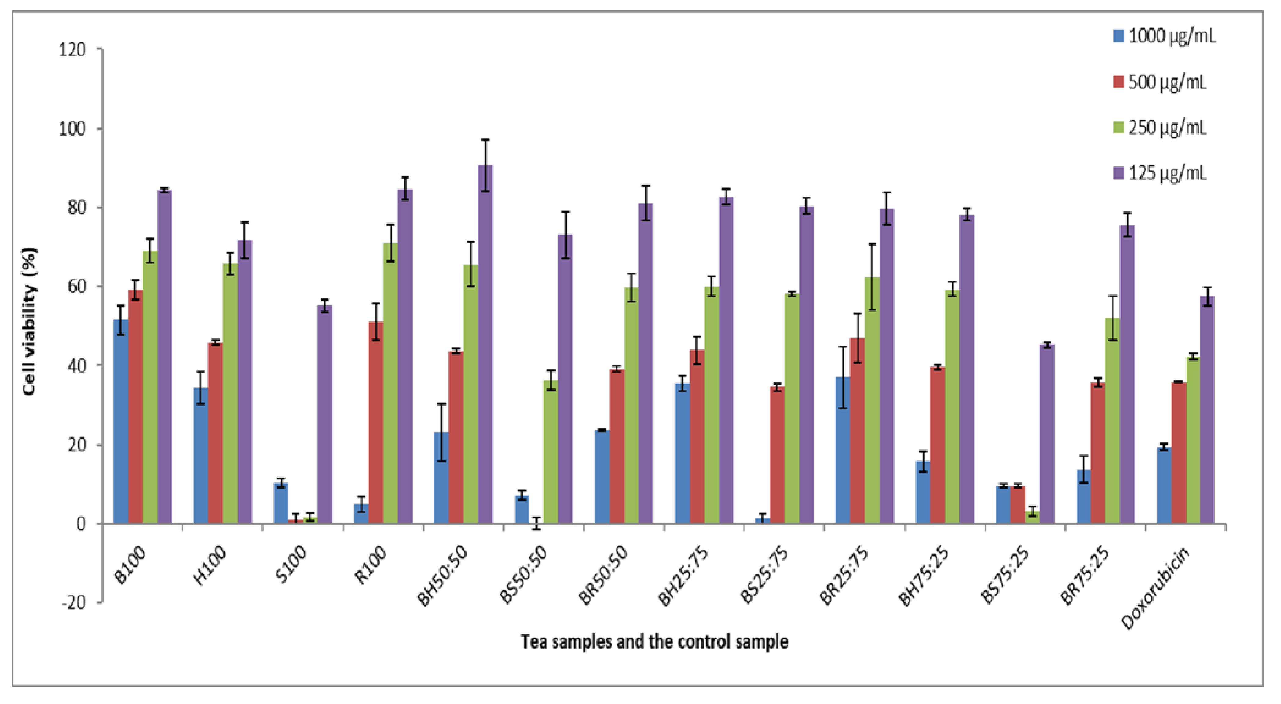
3.1. Cytotoxicity Assays
3.2. Anti-Oxidant Activities
3.2.1. The 2,2-Diphenyl-1-picrylhydrazyl Radical Scavenging Method
3.2.2. The 2,2-Azino-Bis (3-Ethylbenzothiazoline-6-sulfonic Acid) Diammonium Salt Decolourisation Assay
3.3. Anti-Inflammatory (15-Lipoxygenase) Activity
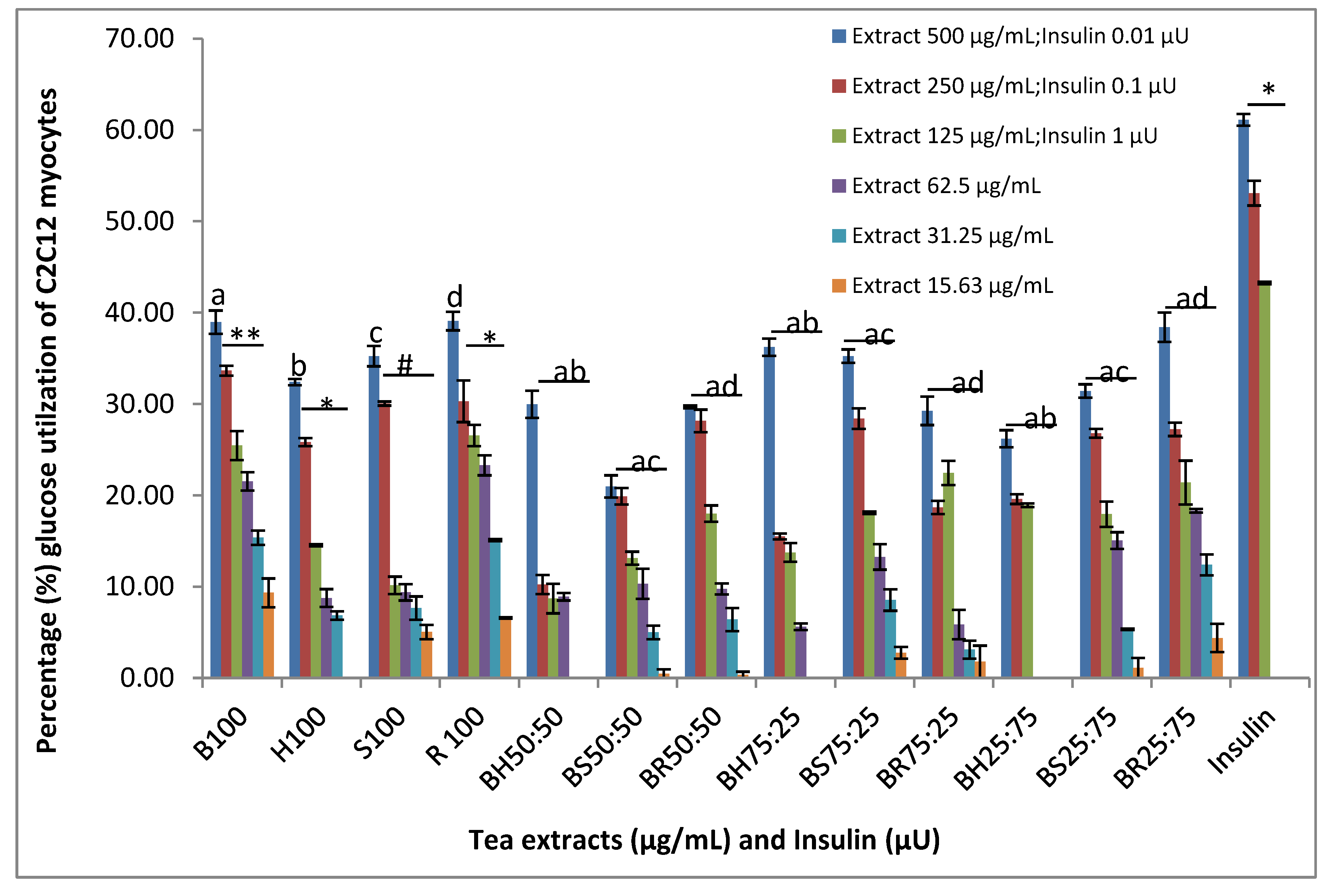
3.4. Anti-Diabetic Assay
4. Conclusions
Author Contributions
Funding
Institutional Review Board Statement
Informed Consent Statement
Data Availability Statement
Conflicts of Interest
References
- Dube, P.; Meyer, S.; Marnewick, J.L. Antimicrobial and anti-oxidant activities of different solvent extracts from fermented and green honeybush (Cyclopia intermedia) plant material. South Afr. J. Bot. 2016, 110, 184–193. [Google Scholar] [CrossRef]
- Baba, H.; Ohtsuka, Y.; Haruna, H.; Lee, T.; Nagata, S.; Maeda, M.; Yamashiro, Y.; Shimizu, T. Studies of anti-inflammatory effects of Rooibos tea in rats. Pediatrics Int. 2009, 51, 700–704. [Google Scholar] [CrossRef] [PubMed]
- Lerotholi, L.; Chaudhary, S.K.; Combrinck, S.; Viljoen, A. Bush tea (Athrixia phylicoides): A review of the traditional uses, bioactivity and phytochemistry. South Afr. J. Bot. 2016, 110, 4–17. [Google Scholar] [CrossRef]
- Mathivha, L.P.; Thibane, V.S.; Mudau, F.N. Anti-diabetic and anti-proliferative activities of herbal teas, Athrixia phylicoides DC and Monsonia burkeana Planch. ex Harv, indigenous to South Africa. Br. Food J. 2019, 121, 964–974. [Google Scholar] [CrossRef] [Green Version]
- Kawano, A.; Nakamura, H.; Hata, S.-I.; Minakawa, M.; Miura, Y.; Yagasaki, K. Hypoglycemic effect of aspalathin, a rooibos tea component from Aspalathus linearis, in type 2 diabetic model db/db mice. Phytomedicine 2009, 16, 437–443. [Google Scholar] [CrossRef] [PubMed]
- Mudau, F.N.; Araya, H.T.; Toit, E.S.D.; Soundy, P.; Olivier, J. Bush Tea (Athrixia phylicoides DC.) as an Alternative Herbal and Medicinal Plant in Southern Africa:Opportunity for Commercialisation. Med. Aromat. Plant Sci. Biotechnol. 2007, 1, 70–73. [Google Scholar]
- Mudau, F.N.; Mariga, I.K. Bush Tea as a Herbal Beverage and Medicinal Plant in South Africa. In Tea in Health and Disease Prevention, 1st ed.; Elsevier: Amsterdam, The Netherlands, 2013; pp. 183–189. [Google Scholar] [CrossRef]
- Tshivhandekano, I.; Ntushelo, K.; Ngezimana, W.; Tshikalange, T.E.; Mudau, F.N. Chemical compositions and antimicrobial activities of Athrixia phylicoides DC. (bush tea), Monsonia burkeana (special tea) and synergistic effects of both combined herbal teas. Asian Pac. J. Trop. Med. 2014, 7 (Suppl. 1), S448–S453. [Google Scholar] [CrossRef] [Green Version]
- Rampedi, I.; Olivier, J. The development path of rooibos tea—A review of patterns and lessons learnt for the commercialisation of other indigenous teas in South Africa. Int. J. Afr. Renaiss. Stud. Multi- Inter- Transdiscipl. 2008, 3, 5–20. [Google Scholar] [CrossRef]
- Joubert, E.; Joubert, M.E.; Bester, C.; de Beer, D.; de Lange, J.H. Honeybush (Cyclopia spp.): From local cottage industry to global markets—The catalytic and supporting role of research. South Afr. J. Bot. 2011, 77, 887–907. [Google Scholar] [CrossRef]
- Marnewick, J.L. Rooibos and Honeybush: Recent Advances in Chemistry, Biological Activity and Pharmacognosy. Afr. Nat. Plant Prod. New Discov. Chall. Chem. Qual. 2009, 1021, 277–294. [Google Scholar] [CrossRef]
- Steenkamp, V.; Fernandes, A.C.; van Rensburg, C.E.J.; Jäger, A.K. Anti-oxidant scavenging potential of South African export herbal teas. South Afr. J. Bot. 2004, 70, 660–663. [Google Scholar] [CrossRef] [Green Version]
- Piljac-Žegarac, J.; Šamec, D.; Piljac, A.; Preedy, V.R.; Piljac-Žegarac, J.; Šamec, D.; Piljac, A.; Preedy, V.R.; Mehra, A.; Lynch, P.; et al. Herbal Teas: A Focus on Antioxidant Properties. In Tea in Health and Disease Prevention, 1st ed.; Preedy, V.R., Ed.; Elsevier: Amsterdam, The Netherlands, 2013; pp. 129–140. [Google Scholar] [CrossRef]
- Joubert, E.; Schultz, H. Production and quality aspects of rooibos tea and related products. A review. J. Appl. Bot. Food Qual. 2012, 80, 138–144. Available online: http://pub.jki.bund.de/index.php/JABFQ/article/view/2173 (accessed on 15 June 2017).
- von Gadow, A.; Joubert, E.; Hansmann, C.F. Comparison of the anti-oxidant activity of aspalathin with that of other plant phenols of rooibos tea (Aspalathus linearis), α-tocopherol, BHT, and BHA. J. Agric. Food Chem. 1997, 45, 632–638. [Google Scholar] [CrossRef]
- Huang, S.-S.; Su, S.-Y.; Chang, J.-S.; Lin, H.-J.; Wu, W.-T.; Deng, J.-S.; Huang, G.-J. Anti-oxidants, anti-inflammatory, and anti-diabetic effects of the aqueous extracts from Glycine species and its bioactive compounds. Bot. Stud. 2016, 57, 38–49. [Google Scholar] [CrossRef] [Green Version]
- Chilelli, N.C.; Burlina, S.; Lapolla, A. AGEs, rather than hyperglycemia, are responsible formicrovascular complications in diabetes: A “glycoxidation-centric” point of view, Nutrition. Metab. Cardiovasc. Dis. 2013, 23, 913–919. [Google Scholar] [CrossRef]
- Shojaii, A.; Goushegir, A.; Dabaghian, F.H.; Abdollahi, M.; Huseini, H.F. Herbs and herbal preparations for glycemic control in diabetes mellitus (a systematic review). J. Med. Plant Res. 2011, 5, 3846–3855. Available online: http://www.scopus.com/inward/record.url?eid=2-s2.0-80051932573&partnerID=40&md5=c13a94f5ee9456a9a5217c67d2dfde2f (accessed on 4 May 2017).
- Shahbazian, H. World Diabetes Day. J. Ren. Inj. Prev. 2013, 2, 123–124. [Google Scholar] [CrossRef]
- Chellan, N.; Muller, C.J.F.; de Beer, D.; Joubert, E.; Page, B.J.; Louw, J. An in vitro assessment of the effect of Athrixia phylicoides DC. aqueous extract on glucose metabolism. Eur. J. Integr. Med. 2012, 19, 730–736. [Google Scholar] [CrossRef]
- Najafian, M.; Najafian, B.; Najafian, Z. The Effect of Aspalathin on Levels of Sugar and Lipids in Streptozotocin-Induced Diabetic and Normal Rats. Zahedan J. Res. Med. Sci. 2016, 18, e4963. [Google Scholar] [CrossRef] [Green Version]
- Joubert, E.; Gelderblom, W.C.A.; Louw, A.; de Beer, D. South African herbal teas— Aspalathus linearis, Cyclopia spp. and Athrixia. J. Ethnopharmacol. 2008, 119, 376–412. [Google Scholar] [CrossRef]
- Chellan, N.; Joubert, E.; Strijdom, H.; Roux, C.; Louw, J.; Muller, C.J.F. Aqueous extract of unfermented honeybush (Cyclopia maculata) attenuates stz-induced diabetes and β-cell cytotoxicity. Planta Med. 2014, 80, 622–629. [Google Scholar] [CrossRef] [PubMed]
- Miura, T.; Ichiki, H.; Hashimoto, I.; Iwamoto, N.; Kato, M.; Kubo, M.; Ishihara, E.; Komatsu, Y.; Okada, M.; Ishida, T.; et al. Anti-diabetic activity of a xanthone compound, mangiferin., Phytomedicine. Int. J. Phytother. Phytopharm. 2001, 8, 85–87. [Google Scholar] [CrossRef]
- Adebayo, S.A.; Dzoyem, J.P.; Shai, L.J.; Eloff, J.N. The anti-inflammatory and anti-oxidant activity of 25 plant species used traditionally to treat pain in southern African. BMC Complementary Altern. Med. 2015, 15, 159–169. [Google Scholar] [CrossRef] [Green Version]
- Padayachee, K. The Phytochemistry and Biological Activities of Athrixia Phylicoides. 2011. Available online: http://wiredspace.wits.ac.za/bitstream/handle/10539/10667/%281%29.pdf?sequence=1 (accessed on 24 April 2015).
- Eloff, J.N. Which extractant should be used for the screening and isolation of antimicrobial components from plants? J. Ethnopharmacol. 1998, 60, 1–8. [Google Scholar] [CrossRef]
- Mosmann, T. Rapid colorimetric assay for cellular growth and survival: Application to proliferation and cytotoxicity assays. J. Immunol. Methods 1983, 65, 55–63. [Google Scholar] [CrossRef]
- Re, R.; Pellegrini, N.; Proteggente, A.; Pannala, A.; Yang, M.; Rice-Evans, C. Anti-oxidant activity applying an improved ABTS radical cation decolorisation assay. Free Radic. Biol. Med. 1999, 26, 1231–1237. [Google Scholar] [CrossRef]
- Brand-Wiliams, C.; Cuvelier, W.; Berset, M.E.; Brand-Williams, W.; Cuvelier, M.E.; Berset, C. Use of a free radical method to evaluate anti-oxidant activity. Food Sci. Technol. 1995, 28, 25–30. [Google Scholar] [CrossRef]
- Apak, R.; Özyürek, M.; Güçlü, K.; Çapanoğlu, E. Anti-oxidant Activity/Capacity Measurement. 2. Hydrogen Atom Transfer (HAT)-Based, Mixed-Mode (Electron Transfer (ET)/HAT), and Lipid Peroxidation Assays. J. Agric. Food Chem. 2016, 64, 1028–1045. [Google Scholar] [CrossRef]
- Pinto, M.D.C.; Tejeda, A.; Duque, A.L.; Macías, P. Determination of lipoxygenase activity in plant extracts using a modified ferrous oxidation-xylenol orange assay. J. Agric. Food Chem. 2007, 55, 5956–5959. [Google Scholar] [CrossRef]
- Olaokun, O.O.; Mcgaw, L.J.; van Rensburg, I.J.; Eloff, J.N.; Naidoo, V. Anti-diabetic activity of the ethyl acetate fraction of Ficus lutea (Moraceae) leaf extract: Comparison of an in vitro assay with an in vivo obese mouse model. BMC Complementary Altern. Med. 2016, 16, 110. [Google Scholar] [CrossRef] [Green Version]
- Yin, J.; Gao, Z.; Liu, D.; Liu, Z.; Ye, J. Berberine improves glucose metabolism through induction of glycolysis. Am. J. Physiol. Endocrinol. Metab. 2008, 294, E148–E156. [Google Scholar] [CrossRef] [Green Version]
- Hamidi, M.R.; Jovanova, B.; Panovska, T.K. Toxicological evaluation of the plant products using Brine Shrimp (Artemia salina L.) model. Maced. Pharm. Bull. 2014, 60, 9–18. [Google Scholar] [CrossRef]
- McGaw, L.J.; Steenkamp, V.; Eloff, J.N. Evaluation of Athrixia bush tea for cytotoxicity, anti-oxidant activity, caffeine content and presence of pyrrolizidine alkaloids. J. Ethnopharmacol. 2007, 110, 16–22. [Google Scholar] [CrossRef] [PubMed] [Green Version]
- Kiyohara, H.; Matsumoto, T.; Yamada, H. Combination Effects of Herbs in a Multi-herbal Formula: Expression of Juzen-taiho-to’s Immuno-modulatory Activity on the Intestinal Immune System. Evid. Based Complementary Altern. Med. ECAM 2004, 1, 83–91. [Google Scholar] [CrossRef] [Green Version]
- Malongane, F.; McGaw, L.J.; Mudau, F.N. Topic: Chemical compositions and mineral content of four selected South African herbal teas and the synergistic response of combined teas. Br. Food J. 2020, 122, 2769–2785. [Google Scholar] [CrossRef]
- Mamphiswana, N.D.; Mashela, P.W.; Mdee, L.K. Distribution of total phenolics and anti-oxidant activity in fruit, leaf, stem and root of Monsonia burkeana. Afr. J. Agric. Res. 2010, 5, 2570–2575. Available online: http://www.academicjournals.org/AJAR (accessed on 6 July 2016).
- Enko, J. Influence of the interactions between tea (Camellia sinensis) extracts and ascorbic acid on their anti-oxidant activity: Analysis with interaction indexes and isobolograms. Food Addit. Contam. 2015, 32, 1234–1242. [Google Scholar] [CrossRef]
- Hemmati, S.; Seradj, H. Justicidin B: A promising bioactive lignan. Molecules 2016, 21, 820. [Google Scholar] [CrossRef]
- Malongane, F.; McGaw, L.J.; Nyoni, H.; Mudau, F.N. Metabolic profiling of four South African herbal teas using high resolution liquid chromatography-mass spectrometry and nuclear magnetic resonance. Food Chem. 2018, 257, 90–100. [Google Scholar] [CrossRef]
- Srisook, K.; Buapool, D.; Boonbai, R.; Simmasut, P.; Charoensuk, Y.; Srisook, E. Anti-oxidant and anti-inflammatory activities of hot water extract from Pluchea indica Less. herbal tea. J. Med. Plants Res. 2012, 6, 4077–4081. [Google Scholar] [CrossRef]
- van Wyk, B.E.E. A review of Khoi-San and Cape Dutch medical ethnobotany. J. Ethnopharmacol. 2008, 119, 331–341. [Google Scholar] [CrossRef] [PubMed]
- Kim, Y.H.; Oh, T.W.; Park, E.; Yim, N.H.; Park, K.i.; Cho, W.K.; Ma, J.Y. Anti-inflammatory and anti-apoptotic effects of Acer palmatum Thunb. Extract, KIOM-2015EW, in a hyperosmolar-stress-induced in vitro dry eye model. Nutrients 2019, 10, 12–14. [Google Scholar] [CrossRef] [Green Version]
- Akiyama, S.; Katsumata, S.I.; Suzuki, K.; Ishimi, Y.; Wu, J.; Uehara, M. Dietary hesperidin exerts hypoglycemic and hypolipidemic effects in streptozotocin-induced marginal type 1 diabetic rats. J. Clin. Biochem. Nutr. 2010, 46, 87–92. [Google Scholar] [CrossRef] [PubMed] [Green Version]


| Tea Name and Positive Control (1 µg/mL) | Tea Code | DPPH ± SD (IC50) | ABTS ± SD (IC50) |
|---|---|---|---|
| Bush tea | B100 | 20.82 ± 3.06 | 4.46 ± 2.79 |
| Honeybush tea | H100 | 54.98 ± 2.11 | 23.03 ± 2.70 |
| Special tea | S100 | 2.74 ± 1.91 | 1.05 ± 1.34 |
| Rooibos tea | R100 | 6.49 ± 2.40 | 2.77 ± 2.74 |
| Bush tea 50%:Honeybush tea 50% | BH50:50 | 10.97 ± 1.12 | 6.25 ± 4.19 |
| Bush tea 50%:Special tea 50% | BS50:50 | 1.33 ± 0.21 | 0.39 ± 0.54 |
| Bush tea 50%:Rooibos tea 50% | BR50:50 | 7.74 ± 3.54 | 4.22 ± 3.13 |
| Bush tea 25%:Honeybush tea 75% | BH25:75 | 6.28 ± 3.52 | 7.44 ± 2.19 |
| Bush tea 25%:Special tea 75% | BS25:75 | 0.62 ± 0.68 | 0.26 ± 0.36 |
| Bush tea 25%:Rooibos tea 75% | BR25:75 | 3.74 ± 0.592 | 2.11 ± 1.90 |
| Bush tea 75%:Honeybush tea 25% | BH75:25 | 4.62 ± 0.38 | 3.03 ± 2.24 |
| Bush tea 75%:Special tea 25% | BS75:25 | 6.62 ± 1.25 | 2.88 ± 1.83 |
| Bush tea 75%:Rooibos tea 25% | BR75:25 | 4.66 ± 1.02 | 0.99 ± 1.03 |
| Vitamin C | 1.05 ± 0.59 | 1.17 ± 0.62 | |
| Trolox | 1.52 ± 0.98 | 1.16 ± 0.25 |
| Tea Name and Positive Control (1 mg/mL) | Tea Code | 15-LOX ± SD (IC50) |
|---|---|---|
| Bush tea | B100 | >100 |
| Honeybush tea | H100 | >100 |
| Special tea | S100 | 6.54 ± 0.84 |
| Rooibos tea | R100 | >100 |
| Bush tea 50%:Honeybush tea 50% | BH50:50 | >100 |
| Bush tea 50%:Special tea 50% | BS50:50 | 8.22 ± 3.21 |
| Bush tea 50%:Rooibos tea 50% | BR50:50 | >100 |
| Bush tea 25%:Honeybush tea 75% | BH25:75 | >100 |
| Bush tea 25%:Special tea 75% | BS25:75 | 8.98 ± 1.19 |
| Bush tea 25%:Rooibos tea 75% | BR25:75 | >100 |
| Bush tea 75%:Honeybush tea 25% | BH75:25 | >100 |
| Bush tea 75%:Special tea 25% | BS75:25 | 64.77 ± 1.50 |
| Bush tea 75%:Rooibos tea 25% | BR75:25 | >100 |
| Quercetin | 24.65 |
Publisher’s Note: MDPI stays neutral with regard to jurisdictional claims in published maps and institutional affiliations. |
© 2022 by the authors. Licensee MDPI, Basel, Switzerland. This article is an open access article distributed under the terms and conditions of the Creative Commons Attribution (CC BY) license (https://creativecommons.org/licenses/by/4.0/).
Share and Cite
Malongane, F.; McGaw, L.J.; Olaokun, O.O.; Mudau, F.N. Anti-Inflammatory, Anti-Diabetic, Anti-Oxidant and Cytotoxicity Assays of South African Herbal Teas and Bush Tea Blends. Foods 2022, 11, 2233. https://doi.org/10.3390/foods11152233
Malongane F, McGaw LJ, Olaokun OO, Mudau FN. Anti-Inflammatory, Anti-Diabetic, Anti-Oxidant and Cytotoxicity Assays of South African Herbal Teas and Bush Tea Blends. Foods. 2022; 11(15):2233. https://doi.org/10.3390/foods11152233
Chicago/Turabian StyleMalongane, Florence, Lyndy Joy McGaw, Oyinlola Oluwunmi Olaokun, and Fhatuwani Nixwell Mudau. 2022. "Anti-Inflammatory, Anti-Diabetic, Anti-Oxidant and Cytotoxicity Assays of South African Herbal Teas and Bush Tea Blends" Foods 11, no. 15: 2233. https://doi.org/10.3390/foods11152233
APA StyleMalongane, F., McGaw, L. J., Olaokun, O. O., & Mudau, F. N. (2022). Anti-Inflammatory, Anti-Diabetic, Anti-Oxidant and Cytotoxicity Assays of South African Herbal Teas and Bush Tea Blends. Foods, 11(15), 2233. https://doi.org/10.3390/foods11152233