Article

CsCuAOs and CsAMADH1 Are Required for Putrescine-Derived γ-Aminobutyric Acid Accumulation in Tea

1
College of Horticulture, Nanjing Agricultural University, Nanjing 210095, China
2
Department of Plant Science, University of Manitoba, 66 Dafoe Road, Winnipeg, MB R3T 2N2, Canada
3
Tea Research Institute, Guizhou Provincial Academy of Agricultural Sciences, Guiyang 417100, China
*
Author to whom correspondence should be addressed.
Foods 2022, 11(9), 1356; https://doi.org/10.3390/foods11091356
Submission received: 27 March 2022 / Revised: 28 April 2022 / Accepted: 4 May 2022 / Published: 6 May 2022
(This article belongs to the Special Issue Tea Product: Processing, Extraction, Quality and Health)

Abstract

:
Polyamines are a potential source of γ-aminobutyric acid (GABA) in plants under abiotic stress. However, studies on GABA enrichment in tea mostly focus on the GABA shunt, while the correlation between polyamine degradation and GABA formation in tea is largely unknown. In this study, tea plants responded to exogenous putrescine, resulting in a significant increase in GABA content, while the glutamate level did not change. At the same time, five copper-containing amine oxidase (CuAO) and eight aminoaldehyde dehydrogenase (AMADH) genes involved in the putrescine-derived GABA pathway were identified from the Tea Plant Information Archive. Expression analysis indicated that CsCuAO1, CsCuAO3 as well as CsAMADH1 were induced to play an important function in response to exogenous putrescine. Thus, the three genes were cloned and the catalytic efficiency of soluble recombinant proteins was determined. CsCuAOs and CsAMADH1 exhibited indispensable functions in the GABA production from putrescine in vitro. Subcellular localization assays indicated that CsAMADH1 was localized in plastid, while both CsCuAO1 and CsCuAO3 were localized in peroxisome. In addition, the synergistic effects of CsCuAOs and CsAMADH1 were investigated by a transient co-expression system in Nicotiana benthamiana. Our data suggest that these three genes regulate the accumulation of GABA in tea by participating in the polyamine degradation pathway and improve the content of GABA in tea to a certain extent. The results will greatly contribute to the production of GABA tea.

1. Introduction

γ-Aminobutyric acid (GABA) is a special four-carbon non-protein amino acid, which plays an important role involving plant growth and development [1,2]. GABA acts as an inhibitory neurotransmitter in animals, which can reduce blood pressure, relieve insomnia, depression, epilepsy and seizures [3,4,5]. The anabolism of GABA in higher plants mainly comes from the glutamate, which is catalyzed by glutamate decarboxylase (GAD), followed by GABA transaminase and succinic semialdehyde dehydrogenase before entering the tricarboxylic acid cycle [6,7]. In addition, polyamines are degraded by diamine oxidase (DAO) to form 4-aminobutyraldehyde (4-ABAL) as intermediates for GABA formation [8], which also contributes to GABA enrichment in plants besides the GABA shunt pathway. Putrescine usually undergoes a two-step reaction, including DAO or a polyamine oxidase (PAO) catalytic process, and is followed by aminoaldehyde dehydrogenase (AMADH) to form GABA in coping with external stress in plants [9,10,11].
In dicotyledonous plants, copper-containing amine oxidase (CuAO) could catalyze the putrescine into 4-ABAL, spermidine into 1,3-diaminopropane, as well as 1,3-diaminopropane into 3-aminopropionaldehyde (APAL) [12,13]. The oxidation of 4-ABAL and pyrroline is generally considered to be catalyzed by the same enzyme, NAD+-dependent AMADH [7], which leads to GABA biosynthesis. The preliminary evidence reported that plant AMADH activity is usually determined by substrate-dependent NADH production, which is based on crude protein extracts that convert 4-ABAL into GABA [14].
CuAO is a homodimer enzyme, not only for putrescine but also for cadaverine [15]. The molecular weight of CuAOs ranged from 70 to 90 kDa, containing a copper ion and a 2,4,5-trihydroxyphenylalanine quinone cofactor, which passes through the active site [15]. Although the overall primary sequence identities of CuAOs from different sources are usually not high (<25%) [16], most of the 33 amino acid residues near the catalytic site are completely conserved [17,18,19]. Arabidopsis thaliana carries ten CuAO-encoding genes, and four of them (ATAO1 and AtCuAO1-3) have been identified. The gene expressions were regulated differently by development, injury and hormone or elicitor processing. The localization of CuAO protein is also different, as AtCuAO1 and TAO1 are located in exosomes, while AtCuAO2 and AtCuAO3 are peroxisomes [20,21,22]. Two genes encoding CuAO (NtMPO1 and NtMPO2) in Nicotiana Tobacum, play important roles in the biosynthesis of pyrrolidine alkaloids [23,24].
Plant AMADHs exhibited the biosynthesis function of betaine aldehyde dehydrogenase, which was localized in the chloroplast [25]. Basmati or jasmine rice lacks functional 4-ABAL dehydrogenase protein and acetylates ABAL (or its cyclic pyrroline) to accumulate 2-acetylpyrroline, which is the effective ingredient of rice flavor [26]. Moreover, there are two putative AMADH genes (AtALDH10A8 and AtALDH10A9) in Arabidopsis, but recombinant AtALDH10A9 can only be produced and purified in the presence of a precursor that causes the reduction of NAD+ [27,28].
To investigate the key enzymes of the polyamine degradation pathway of GABA accumulation, the gene expression of CuAOs and AMADHs in the tea plant by exogenous putrescine was determined. CsCuAOs and CsAMADH1 exhibited indispensable functions in the GABA production from putrescine in vitro. Moreover, the synergistic effects of CsCuAOs and CsAMADH1 were also verified by Agrobacterium-mediated co-expression in Nicotiana benthamiana leaves. Our data suggested that the CsCuAO1, CsCuAO3 and CsAMADH1 participate in the polyamine degradation pathway to form GABA, which is conducive to the accumulation of GABA in tea.

2. Materials and Methods

2.1. Plant Materials and Treatments

Nicotiana benthamiana and tea plants (Camellia sinensis var. zhongcha108) were used in this study. N. benthamiana plants were grown in an artificial climate incubator at 26 °C under a 16 h light (600 μmol·m−2·s−1)/8 h dark photocycle. Tea plants were grown under a cycle of 16 h light (25 °C 600 μmol·m−2·s−1)/8 h dark (20 °C). The tea plants were divided into four groups, including CK, putrescine spraying and anaerobic treatment, respectively. For the CK samples, plants were sprayed with clean water every day, and samples were taken after 0, 1, 3 and 5 days. For putrescine spraying, 5 mM putrescine spraying was applied to plants, and samples were taken after 1 day. For anaerobic, tea leaves were sealed in a bag and all the air was pumped out by vacuum, and finally, samples were taken after 8 h. The collected samples were quickly dropped into liquid nitrogen and stored in a −80 °C freezer until further analysis.

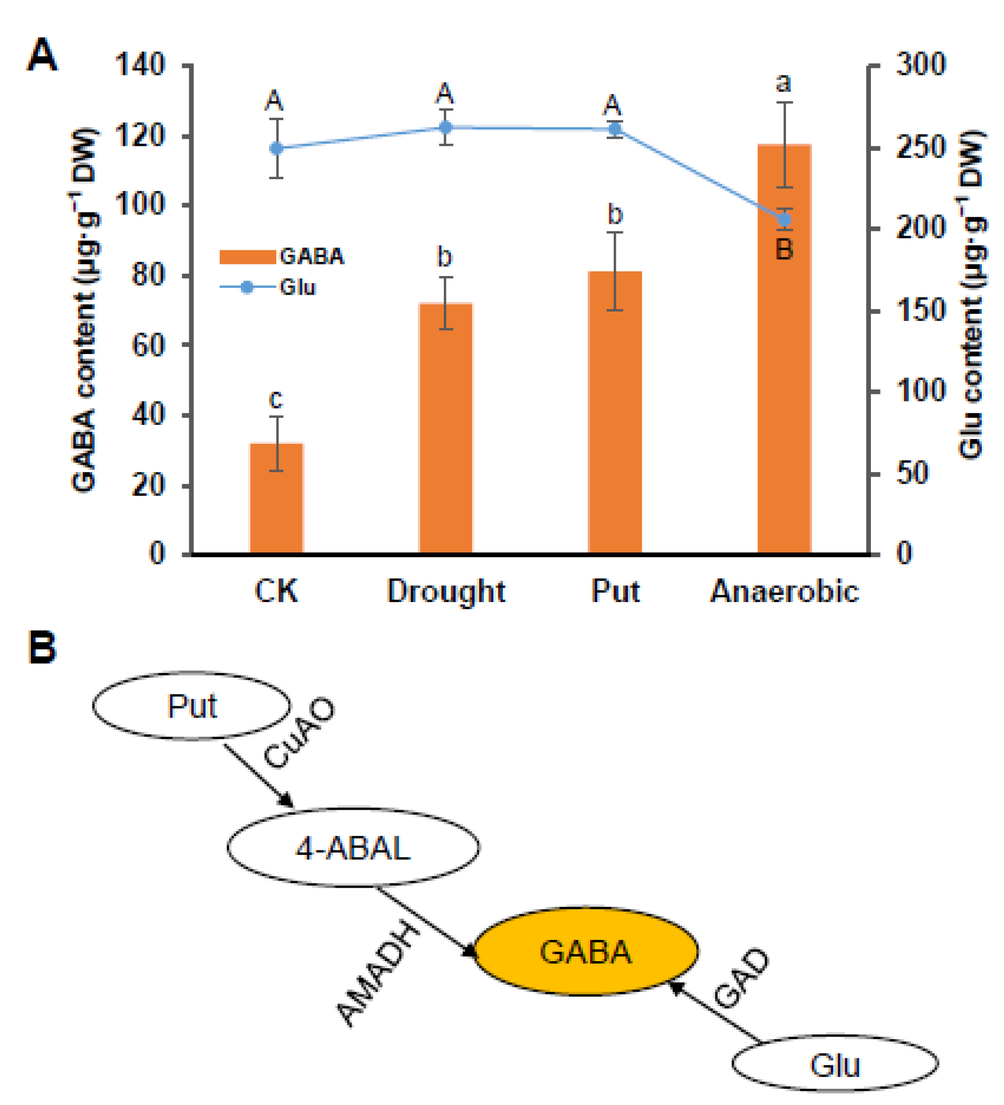
2.2. Determination of GABA and Glutamate Contents

A total of 0.2 g sample was ground and placed into a 10 mL centrifuge tube, 2 mL of 0.02 M HCl were then added before incubating at 4 °C for 8 h. The extracted solution was centrifuged under 4 °C at 14,000× g for 15 min, then 2 mL of supernatant was transferred into a new 10 mL tube. After adding 4% sulfosalicylic acid in the same volume, the extract solution was filtered by a 0.22 μm organic filter, and then the contents of GABA and glutamate were determined by an amino acid composition analyzer (Hitachi L-8900, Osaka, Japan). The contents of GABA and glutamate were obtained by calculating the peak area, which was compared to the standard solution.

2.3. Determination of the Putrescine Content

Putrescine content was detected by high-performance liquid chromatography (HPLC) as described by Zhu et al. [29] with a little modification. Briefly, samples were homogenized with 5% pre-cooling perchloric acid, and the homogenates were centrifuged at 12,000× g for 20 min under 4 °C. The supernatant was mixed with 2 M NaOH and benzoyl chloride and incubated at 37 °C for 30 min. Samples were completely mixed with diethylether and then centrifuged at 3000× g for 10 min at 4 °C for phase separation. The organic solvent phase was evaporated and dissolved with 0.5 mL methanol, followed by HPLC detection (C18 column, 15 cm × 0.39 cm × 4 μm).

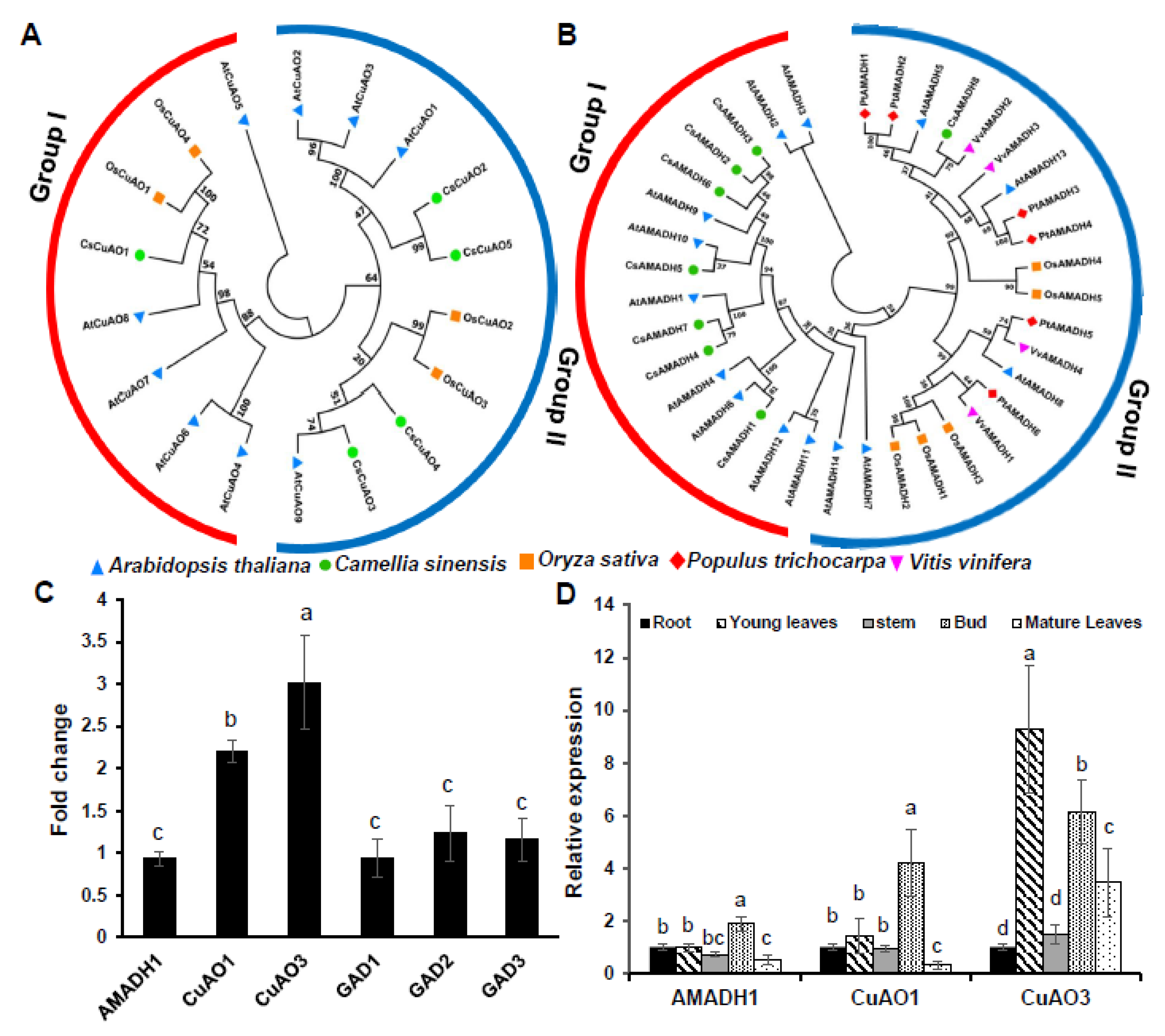
2.4. Phylogenetic Tree Construction of Tea Plant CuAO and AMADH Gene Family

The AMADH and CuAO protein sequences of A. thaliana, Populus tomentosa and Vitis vinifera were derived from the plant transcription factor database, PlantTFDB (http://planttfdb.cbi.pku.edu.cn/, accessed on 15 November 2019). The rice AMADH and CuAO protein sequences were obtained from the Rice Genome Annotation Project (http://rice.plantbiology.msu.edu/index.shtml, accessed on 15 November 2019). The HMMER software was employed for specific domain searching from the tea plant genome database (http://tpia.teaplant.org/, accessed on 15 November 2019) with the default parameter E-value < 1 × 10⁻⁵. Pfam and PROSITE were used to verify the AMADH and CuAO domain. MEGA7.0 software, with default parameters, was used for constructing a neighbor-junction (NJ) phylogenetic tree. The prediction of amino acid sequence features and the motifs of the amino acid sequence was conducted by the ProtParam tool (https://web.expasy.org/protparam/, accessed on 15 November 2019) and MEME (http://meme-suite.org/tools/meme, accessed on 15 November 2019), respectively.

2.5. Gene Expression Analysis

Total RNA was isolated and then reverse transcribed using a Plant Total RNA isolation Kit Plus (Foregene biotech Co. Ltd., Chengdu, China) and a HiScript II Q RT SuperMix (Vazyme biotech Co. Ltd., Nanjing, China) according to the manufacturer’s instruction, respectively. The quantitative real-time PCR assays (Bio-Rad, Houston, TX, America) were performed in the public platform of the laboratory of the College of Horticulture, Nanjing Agricultural University. The reagent used in the qRT-PCR experiment is ChamQ Universal SYBR qPCR Master Mix (Vazyme biotech Co. Ltd., Nanjing, China). The relative gene expressions were calculated using the 2−ΔΔCT method, in which Csβ-actin was selected as the internal control. The primer pairs used in this study were listed in Table S1.

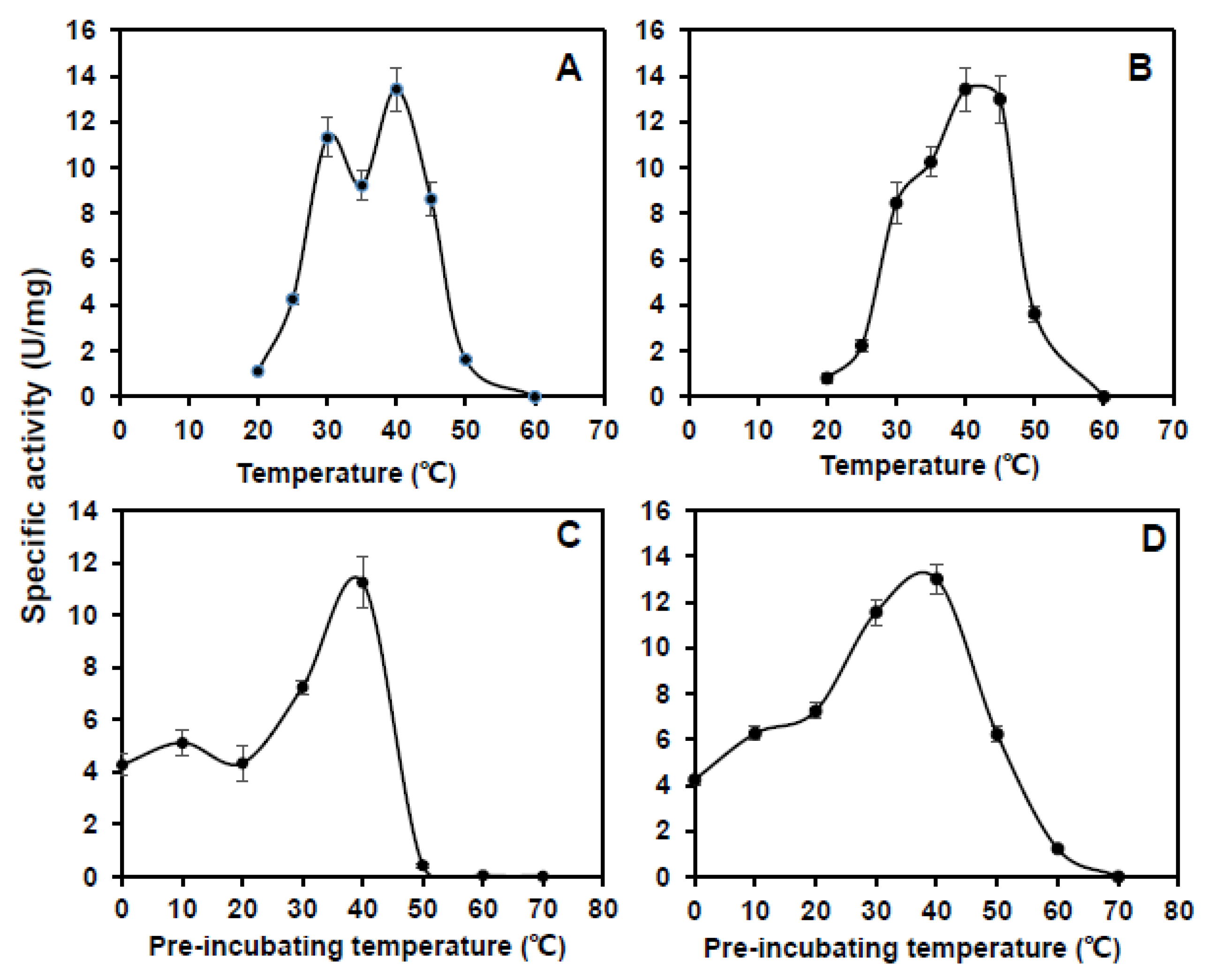
2.6. Purification of CsCuAO1, CsCuAO3 and CsAMADH1 In Vitro

CsCuAO1, CsCuAO3 and CsAMADH1 from tea leaves (Camellia sinensis var. zhongcha108) were cloned by specific primer pairs, and are listed in Table S1. The followed protocol was used: denature for 2 min at 94 °C; followed 35 cycles of the sequence: 30 s at 94 °C, 30 s at 56 °C, and 1 min at 68 °C for annealing. The PCR product was purified, and restriction enzymes were cut and ligated into a pGEX-4T-1 vector (CWBio Co. Ltd., Shanghai, China).

2.7. CsCuAOs and CsAMADHs Activities Assay

CsCuAOs activities were determined as described by Tipping and McPherson [30], with a little modification. Samples were homogenized in 0.1 M potassium phosphate buffer (pH 6.5) and then centrifuged at 15,000× g for 15 min at 4 °C. The supernatant was transferred to a new tube for enzyme reaction, 4-aminoantipyrine/N, N-dimethylaniline chromogenic solution, and 0.1 mL horseradish peroxidase were added. The reaction was initiated by adding 0.02 M putrescine solution, then detected the absorbance at 555 nm by a spectrophotometer (Hitachi UH4150, Osaka, Japan).
For CsAMADH1 activity, the crude enzymes were extracted according to the method described by Petrivalský et al. [31]. The samples were homogenized using 0.1 M potassium phosphate buffer (pH 8.0, including 0.005 M DTT, 0. 1 mM EDTA and 10% sucrose). The enzyme extracts were transferred to a new tube, followed by adding 0.1 M potassium phosphate buffer (pH 8.0), 0.001 M NAD+. The reaction was initiated by adding 0.1 mM of 4-ABAL, then the absorbance at 340 nm by a spectrophotometer was detected.
To investigate the Km value for 4-ABAL and GABA formation, a series of precursors from 0.1 mM to 100 mM were set for the enzyme reaction solution.

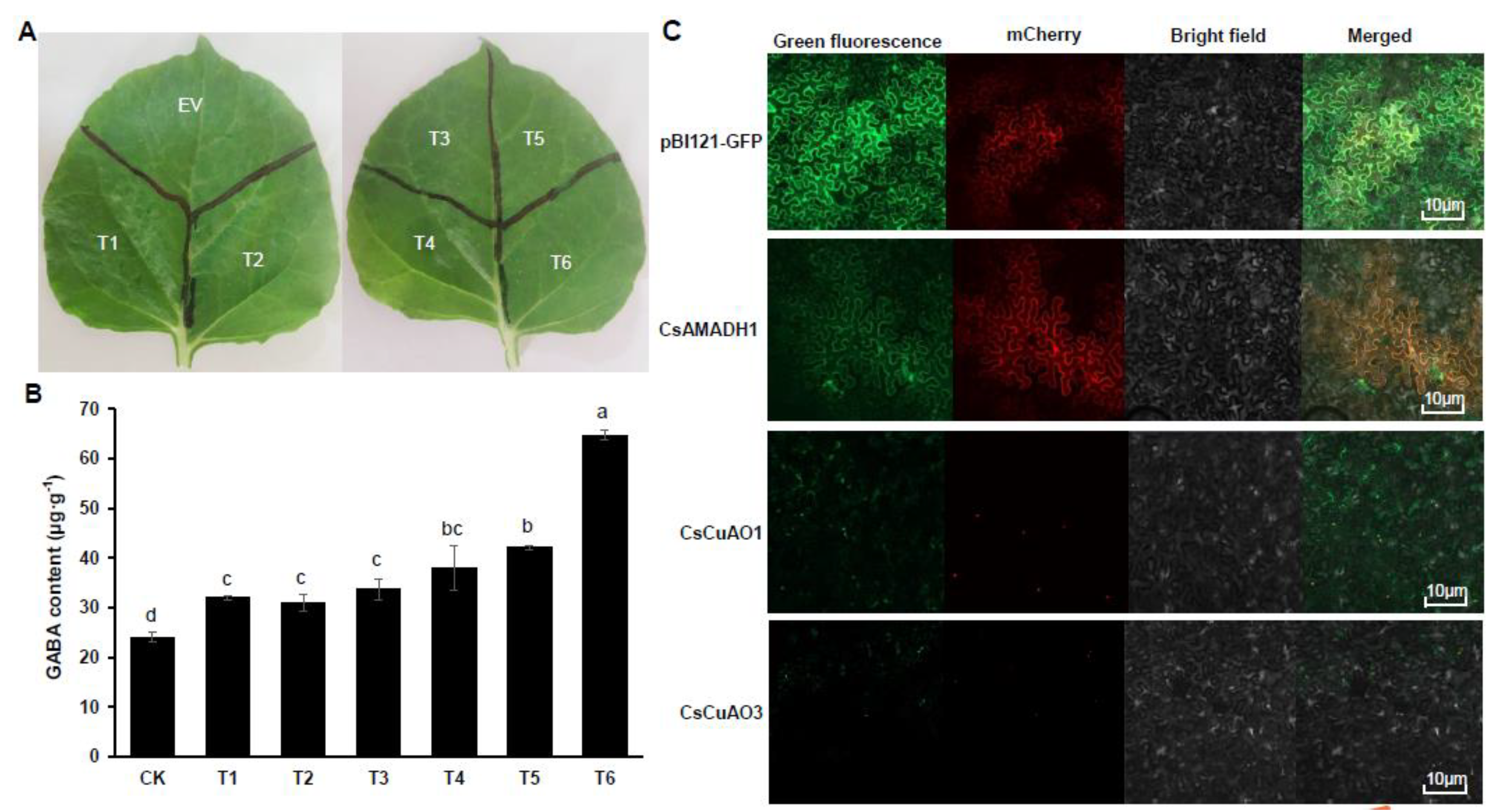
2.8. Subcellular Localization Analysis of CsCuAO1, CsCuAO3 and CsAMADH1

The recombinant proteins of CsCuAO1-GFP, CsCuAO3-GFP and CsAMADH-GFP transformed into Agrobacterium tumefaciens strain GV3101 cells, the vector pBI121-GFP was used as a control [32]. The transient expressed N. benthamiana plants were put in a plant incubator in the dark overnight and followed a 16 h light (600 μmol·m−2·s−1)/8 h dark photocycle for two days. Finally, N. benthamiana leaves were detected using an LSM800 ultra high-resolution confocal microscopy imaging system (Zeiss Co., Oberkochen, Germany).

2.9. Statistical Analysis

The data were calculated using Excel (Microsoft Office 2016, Seattle, WA, USA) from three replicates. Both ANOVA and Duncan’s test were employed for significance analysis.

3. Results

3.1. Changes in GABA, Glutamate, Putrescine Contents under Different Treatments

The GABA levels in leaves increased significantly after putrescine spraying and anaerobic treatment, compared with the control tea plants (Figure 1A). Interestingly, the content of glutamate exhibited different trends under both two treatments. The glutamate contents had no significant change compared with the control group under putrescine spraying treatment. However, for anaerobic treatment, the glutamate content decreased significantly. There are two pathways in GABA synthesis: one is the GABA shunt regulated by GADs, and the other is the polyamine degradation pathway, which is divided into two steps, catalyzed by CuAOs and AMADHs, respectively (Figure 1B).

3.2. Phylogenetic Analysis of Tea Plant CuAO and AMADH Gene Family and Gene Expression

Five CuAO and eight AMADH genes were strictly identified from the Tea Plant Information Archive. Phylogenetic tree analysis showed that CsCuAOs’ family can be divided into two groups (Figure 2A) and the CsAMADHs family also has two groups (Figure 2B). The detailed sequences and conserved motifs of CsCuAOs and CsAMADHs were analyzed (Figure S1).
Interestingly, the expression of CsAMADH1 and CsGADs did not increase obviously by putrescine application, but the expression of CsCuAO1 and CsCuAO3 increased significantly, which the fold change value of CsCuAO1 and CsCuAO3 was 2.2 and 3, respectively (Figure 2C). Furthermore, there were no apparent changes in the expression of CsGADs under the putrescine spaying (Figure 2C). For tissue-specific analysis, the expression of CsAMADH1 and CsCuAO1 showed the highest expression in buds and the lowest in mature leaves, while the expression of CsCuAO3 was highly expressed in young leaves, buds and mature leaves, and the expression pattern showed the highest in young leaves while the lowest in roots in the tea plant (Figure 2D).

3.3. Purification and Enzyme Kinetics of CsCuAO1, CsCuAO3 and CsAMADH1

The CsCuAO1, CsCuAO3 and CsAMADH1 proteins with the GST tag were isolated and purified, and the sizes of these proteins were 79.6, 74.8 and 55.0 kDa, respectively (Figure 3A). Lineweaver–Burk plots (Figure 3B–D), calculated from the linear formula (1/U against 1/[S]), revealed the Vmax values for the converting of CsCuAO1 and CsCuAO2 to putrescine were 16.9 and 21.1 μmol·mg−1·min−1, respectively. The Vmax values for the conversion of CsAMADH1 to 4-ABAL was 17.2 μmol·mg−1·min−1. Moreover, the Km values of CsCuAO1, CsCuAO3 and CsAMADH1 were 21.9, 15.8 and 25.7 Mm, which indicated that the substrate-binding affinity of the three enzymes was definitely discrepant.
The maximum enzyme activity of CsCuAO1 and CsCuAO3 was detected at pH 5.5 (Figure 4A,B). The performance of pH stability analysis revealed that CsCuAO1 activity was restored after a 12 h treatment under pH 4.5–7.5, while the enzyme activity disappeared under treatment at pH 3.5. CsCuAO3 activity was gradually restored at pH 4.0–8.0 and was eliminated at pH 3.0, and the pH stability of CsCuAO3 was more stable than CsCuAO1 (Figure 4C,D). The most active temperature for both CsCuAO1 and CsCuAO3 was 40 °C (Figure 5A,B). Additionally, the detection of thermal stability revealed that pre-incubation temperatures of more than 40 °C decreased the activity of both CsCuAOs (Figure 5C,D), especially CsCuAO1, which is almost completely inactivated when the pre-incubation temperature reaches 50 °C. These results implied that the enzyme activity of CsCuAO3 was more stable than CsCuAO1, and the temperature had a greater effect than that of pH.

3.4. Assays of CsCuAO1, CsCuAO3 and CsAMADH1 Enzyme Activity on GABA Production In Vitro

In order to identify whether CsCuAOs and CsAMADHs have synergistic effects, we carried out validation experiments on the three proteins in vitro and treated them with seven groups (CK and T1–T6) with putrescine as the substrate (Figure 6A). The GABA content, as the reaction product, was determined by an amino acid analyzer (Figure 6B). What can be obviously seen, was that there is no GABA production in either the CK group or the T1, T2 and T3 groups. However, GABA was produced in the groups of T4, T5, T6, and the GABA content of T6 was much higher than T4 and T5 (Figure 6C). These results indicated that CsCuAOs and CsAMADH1 were indispensable for the GABA production from putrescine in vitro.

3.5. Transient Transformation Expression in Nicotiana Benthamiana

Based on the in vitro results, we further carried out the validation of three genes in vivo—the Agrobacterium-mediated Nicotiana Benthamiana transient assay (Figure 7A), and GABA in leaves was detected after agro-infiltration for three days. The results suggested that the GABA level in N. Benthamiana leaves increased by agro-infiltration with a single gene, while the content of GABA in leaves could increase more by agro-infiltration with two-step genes simultaneously (Figure 7B). In fact, the GFP fusion subcellular localization analysis indicated that CsAMADH1 was localized in plastid, while both CsCuAO1 and CsCuAO3 were localized in peroxisome (Figure 7C).

4. Discussion

GABA is considered to be the major amino acid synthesized under anaerobic conditions, which are formed by glutamate decarboxylation based on GAD function [33,34]. The GABA level was increased significantly, and the content of glutamate decreased, indicating that the increased GABA under hypoxia is dominated by the GABA shunt pathway (Figure 1A) [35,36]. However, GABA content increased but glutamate did not change under the putrescine spraying treatment, which indicates that the GABA shunt did not respond to the induction of exogenous putrescine, and the increase in the GABA content is through polyamine degradation. For gene expression patterns, the putrescine degradation pathway genes, including CsCuAO1, CsCuAO3 and CsAMADH1 could respond to exogenous putrescine (Figure 2D). However, in the GABA shut pathway genes, the expression of CsGADs was only induced under anaerobic stress (Figure S2). Subcellular localization results indicated that CsAMADH1 was localized in plastid, and both CsCuAO1 and CsCuAO3 were localized in peroxisome (Figure 5C). In Arabidopsis, the localization of CuAOs protein was different, which AtCuAO1 was localized in ectoplast, while AtCuAO2 and AtCuAO3 were found in peroxidases [16,37,38]. Taken together, CsCuAO1, CsCuAO3 and CsAMADH1 could respond to the induction of exogenous putrescine and regulated the polyamine degradation pathway to increase the content of GABA in tea. Under anaerobic conditions, the significant increase in GABA content is dominated by the GABA shunt and its key genes CsGADs.
Our previous studies have revealed that 1/4 of GABA produced in tea leaves under anoxia comes from polyamine (predominantly putrescine) degradation [35]. In order to further study the functions and characteristics of these three genes and their enzyme synthesis, we purified their recombinant proteins and carried out a series of function verification experiments in vivo and in vitro. In the present study, we found that the substrate-binding affinity of CsCuAO3 was greater than that of CsCuAO1, as the Km value of CsCuAO3 was lower than CsCuAO1 (Figure 3). In addition, CsCuAO1, CsCuAO3 and CsAMADH1 exhibited very high enzyme activity in vitro and in vivo played for GABA formation (Figure 6 and Figure 7). It is reported that polyamines are degraded by DAO to form 4-ABAL intermediates, followed by AMADH catalyzation, which is another way for GABA enrichment in plants [39]. DAO takes putrescine as substrate in cowpea seedlings, in which Km and Vmax are 0.15 mM and 0.065 mol·min−1, respectively, while the oxidation activity of spermidine and spermine is only 16% and 38% of putrescine (Petrivalský et al., 2007). In this study, we revealed that the synergistic effects of CsCuAOs and CsAMADH1 were shown by a transient co-expression system (Figure 7). Therefore, the present study demonstrated that these three genes are involved in GABA production through polyamine degradation in the tea plant.
It appears that there may exist a totally different pathway between the accumulation of GABA levels and the response to putrescine spraying treatment or hypoxia stress. Thus, we studied the accumulation of GABA in tea from the perspective of the polyamine degradation pathway and clarified its mechanism to a certain extent. The results showed that the CsCuAOs and CsAMADH1 were key genes in the polyamine degradation pathway. However, GADs were considered the dominant genes for regulating GABA formation under anaerobic conditions, in which the expression levels of CsGAD2 and CsGAD3 increased significantly, 3.5- and 2.4-fold, respectively (Mei et al., 2016; Figure S2).

5. Conclusions

The functions of three key enzymes involved in GABA production from the polyamine degradation pathway were analyzed in this study. The three genes employed by the putrescine-derived GABA accumulation in the tea plant were firstly reported. Our data showed that CsCuAO1, CsCuAO3 and CsAMADH1 were the key genes involved in GABA production in the tea polyamine degradation pathway and it was driven by the combined synthesis of CsCuAO1, CsCuAO3 and CsAMADH1, which will have a great contribution to the production of GABA Tea.

Supplementary Materials

The following supporting information can be downloaded at: https://www.mdpi.com/article/10.3390/foods11091356/s1, Figure S1: Schematic drawing of CsAMADH and CsCuAO family members; Figure S2: CsGADs were induced under hypoxia treatment, but not CsCuAOs and CsAMADH1. The lowercase letters present over the columns indicate significant differences among different genes (p < 0.05). Table S1: Primers used in this study.

Author Contributions

Conceptualization, X.Z. and W.F.; methodology, Z.Z. and Y.M.; software, Y.C. (Yu Cao); validation, F.L.; formal analysis, X.Y.; investigation, K.Z. and Y.D.; resources, Q.S.; data curation, K.Z. and Y.D.; writing—original draft preparation, K.Z.; writing—review and editing, Y.C. (Yiwen Chen); visualization, W.F.; supervision, X.Z. and W.F.; project administration, X.Z. and W.F.; funding acquisition, X.Z. All authors have read and agreed to the published version of the manuscript.

Funding

This research was funded by The National Natural Science Foundation of China (32160729, 31870680), the Science and Technology support project of Guizhou Province ([2020]1Y004, ZK [2021]154), the China Agriculture Research System of MOF and MARA (CARS-19), the Changzhou Agriculture Science and Technology Support Program (CE20212017), the Jiangsu Belt and Road innovation cooperation project (BZ2019012), and the Priority Academic Program Development of Jiangsu Higher Education Institutions.

Institutional Review Board Statement

Not applicable.

Informed Consent Statement

Not applicable.

Data Availability Statement

All data are available in the manuscript or the Supplemental Materials.

Acknowledgments

We would like to thank the Central Lab in College of Horticulture for providing the LSM800 confocal microscopy imaging system and Bio-Rad IQ5 Real Time PCR System.

Conflicts of Interest

The authors declare that there is no conflict of interest.

References

  1. Alharbi, B.; Hunt, J.D.; Dimitrova, S.; Spadafora, N.D.; Cort, A.P.; Colombo, D.; Müller, C.T.; Ghuge, S.A.; Davoli, D.; Cona, A.; et al. Mutation of Arabidopsis Copper-Containing Amine Oxidase Gene AtCuAOδ Alters Polyamines, Reduces Gibberellin Content and Affects Development. Int. J. Mol. Sci. 2020, 21, 7789. [Google Scholar] [CrossRef] [PubMed]
  2. Bouché, N.; Fromm, H. GABA in plants: Just a metabolite? Trends Plant Sci. 2004, 9, 110–115. [Google Scholar] [CrossRef] [PubMed]
  3. Li, D.; Pan, H. Role of gamma-Aminobutyric Acid (GABA)A and GABAB receptors in paraventricular nucleus in control of sympathetic vasomotor tone in hypertension. J. Pharmacol. Exp. Ther. 2007, 320, 615–626. [Google Scholar] [CrossRef] [PubMed] [Green Version]
  4. Benson, K.L.; Bottary, R.; Schoerning, L.; Baer, L.; Gonenc, A.; Jensen, J.E.; Winkelman, J.W. 1H MRS Measurement of Cortical GABA and Glutamate in Primary Insomnia and Major Depressive Disorder: Relationship to Sleep Quality and Depression Severity. J. Affect. Disord. 2020, 274, 624–631. [Google Scholar] [CrossRef]
  5. Ting Wong, C.G.; Bottiglieri, T.; Snead, O.C., III. GABA, γ-hydroxybutyric acid, and neurological disease. Ann. Neurol. 2003, 54, S3–S12. [Google Scholar] [CrossRef]
  6. Chi, Z.; Dai, Y.; Cao, S.; Wei, Y.; Shao, X.; Huang, X.; Xu, F.; Wang, H. Exogenous calcium chloride (CaCl2) promotes γ-aminobutyric acid (GABA) accumulation in fresh-cut pears. Postharvest Biol. Technol. 2020, 174, 111446. [Google Scholar] [CrossRef]
  7. Lee, J.; Rudell, D.R.; Davies, P.J.; Watkins, C.B. Metabolic changes in 1-methylcyclopropene (1-MCP)-treated ‘Empire’ apple fruit during storage. Metabolomics 2012, 8, 742–753. [Google Scholar] [CrossRef]
  8. Moschou, P.; Wu, J.; Cona, A.; Tavladoraki, P.; Angelini, R.; Roubelakis-Angelakis, K.A. The polyamines and their catabolic products are significant players in the turnover of nitrogenous molecules in plants. J. Exp. Bot. 2012, 63, 5003–5015. [Google Scholar] [CrossRef] [Green Version]
  9. Federico, R.; Angelini, R. Polyamine catabolism in plants. In Biochemistry and Physiology of Polyamines in Plants; Slocum, R.D., Flores, H.E., Eds.; CRC Press: Boca Raton, FL, USA, 1991; pp. 41–56. [Google Scholar]
  10. Fincato, P.; Moschou, P.; Spedaletti, V.; Tavazza, R.; Angelini, R.; Federico, R.; Roubelakis-Angelakis, K.A.; Tavladoraki, P. Functional diversity inside the Arabidopsis polyamine oxidase gene family. J. Exp. Bot. 2010, 62, 1155–1168. [Google Scholar] [CrossRef]
  11. Podlešáková, K.; Ugena, L.; Spíchal, L.; Doležal, K.; De Diego, N. Phytohormones and polyamines regulate plant stress responses by altering GABA pathway. New Biotechnol. 2018, 48, 53–65. [Google Scholar] [CrossRef]
  12. Renault, H.; Roussel, V.; El Amrani, A.; Arzel, M.; Renault, D.; Bouchereau, A.; Deleu, C. The Arabidopsis pop2-1mutant reveals the involvement of GABA transaminase in salt stress tolerance. BMC Plant Biol. 2010, 10, 20. [Google Scholar] [CrossRef] [PubMed] [Green Version]
  13. Shelp, B.J.; Bozzo, G.G.; Trobacher, C.P.; Zarei, A.; Deyman, K.L.; Brikis, C.J. Hypothesis/review: Contribution of putrescine to 4-aminobutyrate (GABA) production in response to abiotic stress. Plant Sci. 2012, 193–194, 130–135. [Google Scholar] [CrossRef] [PubMed]
  14. Xing, S.G.; Jun, Y.B.; Hau, Z.W.; Liang, L.Y. Higher accumulation of gamma-aminobutyric acid induced by salt stress through stimulating the activity of diamine oxidases in Glycine max (L.) Merr. roots. Plant Physiol. Biochem. 2007, 45, 560–566. [Google Scholar] [CrossRef] [PubMed]
  15. Zarei, A.; Trobacher, C.P.; Cooke, A.R.; Meyers, A.J.; Hall, J.C.; Shelp, B.J. Apple Fruit Copper Amine Oxidase Isoforms: Peroxisomal MdAO1 Prefers Diamines as Substrates, Whereas Extracellular MdAO2 Exclusively Utilizes Monoamines. Plant Cell Physiol. 2014, 56, 137–147. [Google Scholar] [CrossRef] [Green Version]
  16. Planas-Portell, J.; Gallart, M.; Tiburcio, A.F.; Altabella, T. Copper-containing amine oxidases contribute to terminal polyamine oxidation in peroxisomes and apoplast of Arabidopsis thaliana. BMC Plant Biol. 2013, 13, 109. [Google Scholar] [CrossRef] [Green Version]
  17. Cona, A.; Rea, G.; Angelini, R.; Federico, R.; Tavladoraki, P. Functions of amine oxidases in plant development and defence. Trends Plant Sci. 2006, 11, 80–88. [Google Scholar] [CrossRef]
  18. Flores, H.E.; Filner, P. Polyamine catabolism in higher plants: Characterization of pyrroline dehydrogenase. Plant Growth Regul. 1985, 3, 277–291. [Google Scholar] [CrossRef]
  19. Shelp, B.J.; Mullen, R.T.; Waller, J. Compartmentation of GABA metabolism raises intriguing questions. Trends Plant Sci. 2012, 17, 57–59. [Google Scholar] [CrossRef]
  20. Díaz-Sanchez, Á.; González-Segura, L.; Mújica-Jiménez, C.; Rudino-Pinera, E.; Montiel, C.; Martínez-Castilla, L.P.; Muñoz-Clares, R.A. Amino Acid Residues Critical for the Specificity for Betaine Aldehyde of the Plant ALDH10 Isoenzyme Involved in the Synthesis of Glycine Betaine. Plant Physiol. 2012, 158, 1570–1582. [Google Scholar] [CrossRef] [Green Version]
  21. Kopečný, D.; Končitíková, R.; Tylichová, M.; Vigouroux, A.; Moskalíková, H.; Soural, M. Plant ALDH10 family: Identifying critical residues for substrate specificity and trapping a thiohemiacetal intermediate. J. Biol. Chem. 2013, 288, 9491–9507. [Google Scholar] [CrossRef] [Green Version]
  22. Riveros-Rosasa, H.; González-Segura, L.; Julián-Sánchez, A.; Díaz-Sánchez, Á.G.; Muñoz-Clares, R.A. Structural determinants of substrate specificity in aldehyde dehydrogenases. Chem.-Biol. Interact. 2013, 202, 51–61. [Google Scholar] [CrossRef] [PubMed]
  23. Heim, W.G.; Sykes, K.A.; Hildreth, S.B.; Sun, J.; Lu, R.-H.; Jelesko, J.G. Cloning and characterization of a Nicotiana tabacum methylputrescine oxidase transcript. Phytochem. 2007, 68, 454–463. [Google Scholar] [CrossRef] [PubMed]
  24. Katoh, A.; Shoji, T.; Hashimoto, T. Molecular Cloning of N-methylputrescine Oxidase from Tobacco. Plant Cell Physiol. 2007, 48, 550–554. [Google Scholar] [CrossRef] [PubMed]
  25. Weigel, P.; Weretilnyk, E.A.; Hanson, A.D. Betaine Aldehyde Oxidation by Spinach Chloroplasts. Plant Physiol. 1986, 82, 753–759. [Google Scholar] [CrossRef] [PubMed] [Green Version]
  26. Bradbury, L.M.T.; Gillies, S.A.; Brushett, D.J.; Waters, D.L.E.; Henry, R.J. Inactivation of an aminoaldehyde dehydrogenase is responsible for fragrance in rice. Plant Mol. Biol. 2008, 68, 439–449. [Google Scholar] [CrossRef] [Green Version]
  27. Missihoun, T.D.; Schmitz, J.; Klug, R.; Kirch, H.-H.; Bartels, D. Betaine aldehyde dehydrogenase genes from Arabidopsis with different sub-cellular localization affect stress responses. Planta 2010, 233, 369–382. [Google Scholar] [CrossRef]
  28. Muñoz-Clares, R.A.; Riveros-Rosas, H.; Garza-Ramos, G.; González-Segura, L.; Mújica-Jiménez, C.; Julián-Sánchez, A. Exploring the evolutionary route of the acquisition of betaine aldehyde dehydrogenase activity by plant ALDH10 enzymes: Implications for the synthesis of the osmoprotectant glycine betaine. BMC Plant Biol. 2014, 14, 149. [Google Scholar] [CrossRef] [Green Version]
  29. Zhu, X.; Li, Q.; Hu, J.; Wang, M.; Li, X. Molecular Cloning and Characterization of Spermine Synthesis Gene Associated with Cold Tolerance in Tea Plant (Camellia sinensis). Appl. Biochem. Biotechnol. 2015, 177, 1055–1068. [Google Scholar] [CrossRef]
  30. Tipping, A.J.; McPherson, M. Cloning and Molecular Analysis of the Pea Seedling Copper Amine Oxidase. J. Biol. Chem. 1995, 270, 16939–16946. [Google Scholar] [CrossRef] [Green Version]
  31. Petřivalský, M.; Brauner, F.; Luhová, L.; Gagneul, D.; Šebela, M. Aminoaldehyde dehydrogenase activity during wound healing of mechanically injured pea seedlings. J. Plant Physiol. 2007, 164, 1410–1418. [Google Scholar] [CrossRef]
  32. Li, J.; Duan, Y.; Han, Z.; Shang, X.; Zhang, K.; Zou, Z.; Ma, Y.; Li, F.; Fang, W.; Zhu, X. Genome-Wide Identification and Expression Analysis of the NRAMP Family Genes in Tea Plant (Camellia sinensis). Plants 2021, 10, 1055. [Google Scholar] [CrossRef] [PubMed]
  33. Mei, X.; Xu, X.; Yang, Z. Characterization of two tea glutamate decarboxylase isoforms involved in GABA production. Food Chem. 2019, 305, 125440. [Google Scholar] [CrossRef] [PubMed]
  34. Reggiani, R.; Cantu, C.A.; Brambilla, I.; Bertani, A. Accumulation and Interconversion of Amino Acids in Rice Roots under Anoxia. Plant Cell Physiol. 1988, 29, 981–987. [Google Scholar] [CrossRef]
  35. Liao, J.; Wu, X.; Xing, Z.; Li, Q.; Duan, Y.; Fang, W.; Zhu, X. γ-Aminobutyric Acid (GABA) Accumulation in Tea (Camellia sinensis L.) through the GABA Shunt and Polyamine Degradation Pathways under Anoxia. J. Agric. Food Chem. 2017, 65, 3013–3018. [Google Scholar] [CrossRef]
  36. Mei, X.; Chen, Y.; Zhang, L.; Fu, X.; Wei, Q.; Grierson, D.; Zhou, Y.; Huang, Y.; Don, G.; Yang, Z. Dual mechanisms regulating glutamate decarboxylases and accumulation of gamma-aminobutyric acid in tea (Camellia sinensis) leaves exposed to multiple stresses. Sci. Rep. 2016, 6, 23685. [Google Scholar] [CrossRef] [Green Version]
  37. Angelini, R.; Cona, A.; Federico, R.; Fincato, P.; Tavladoraki, P.; Tisi, A. Plant amine oxidases “on the move”: An update. Plant Physiol. Biochem. 2010, 48, 560–564. [Google Scholar] [CrossRef]
  38. Reumann, S.; Quan, S.; Aung, K.; Yang, P.; Manandhar-Shrestha, K.; Holbrook, D.; Linka, N.; Switzenberg, R.; Wilkerson, C.G.; Weber, A.P.; et al. In-Depth Proteome Analysis of Arabidopsis Leaf Peroxisomes Combined with in Vivo Subcellular Targeting Verification Indicates Novel Metabolic and Regulatory Functions of Peroxisomes. Plant Physiol. 2009, 150, 125–143. [Google Scholar] [CrossRef] [Green Version]
  39. Moller, S.; McPherson, M. Developmental expression and biochemical analysis of theArabidopsis atao1gene encoding an H2O2-generating diamine oxidase. Plant J. 1998, 13, 781–791. [Google Scholar] [CrossRef]
Figure 1. The accumulation profiles of GABA under various different treatments. (A) The quantitative analysis of GABA and Glu under drought, anaerobic, Put spraying and mechanical damage. Data represent the mean value ± standard deviation; means with different letters are significantly different from each other (p ≤ 0.05). Capital letters and lowercase letters represent Glu and GABA, respectively. (B) A schematic drawing of the GABA biosynthesis pathway. Glu, glutamate; Put, putrescine.
Foods 11 01356 g001
Figure 2. The expression profiles of CsCuAO and CsAMADH family members. Phylogenetic tree showing clustering of CsCuAO (A) and CsAMADH (B) family members from C. sinensis and other plant species. (C) The fold change of three genes and GADs expression by putrescine spraying treatment. The lowercase letters present over the column indicate significant differences by treatment (p < 0.05) (D) The expression patterns of three genes in various organs. The lowercase letters present over the column indicate significant differences among different organs (p < 0.05).
Foods 11 01356 g002
Figure 3. Properties of CsCuAO1, CsCuAO3 and CsAMADH1. (A) The sodium dodecyl sulfate polyacrylamide gel electrophoresis analysis of the recombinant GST-fusion protein. Lane 1: empty vector; Lane 2: CsCuAO3; Lane 3: CsCuAO1; Lane 4: CsAMADH1; Lane 5: protein molecular weight marker. Lineweaver–Burk plot of CsCuAO1 (B), CsCuAO3 (C) and CsAMADH1 (D).
Foods 11 01356 g003
Figure 4. Activity and stability of CsCuAOs under different pH conditions. Activity of CsCuAO1 (A) and CsCuAO3 (B) was determined between pH = 3 to pH = 8. Stability of CsCuAO1 (C) and CsCuAO3 (D) was detected by incubating with a series of pH buffers for 12 h at 4 °C and then assayed the enzyme activity at pH = 5.5 and pH = 5.0, respectively. CK, the enzyme stored at −80 °C, was used as the control.
Foods 11 01356 g004
Figure 5. Activity and stability of CsCuAOs under temperature. Activity of CsCuAO1 (A) and CsCuAO3 (B) was determined between 20–60 °C. Stability of CsCuAO1 (C) and CsCuAO3 (D) was detected by incubating at a series of temperatures for 30 min, and then assayed the enzyme at 40 °C, respectively.
Foods 11 01356 g005
Figure 6. Assays of CsCuAO1, CsCuAO3 and CsAMADH1 enzyme activity on GABA production in vitro. (A) A simplified scheme showing the enzyme reaction. (B) Representative amino acid chromatograms for enzymatic reactions with substrate. (C) GABA accumulated in different enzyme reactions. The lowercase letters present over the columns indicate significant differences among different treatments (p < 0.05).
Foods 11 01356 g006
Figure 7. Transient assays of CsCuAO1, CsCuAO3 and CsAMADH1 in N. benthamiana leaves. (A) The phenotypes of N. benthamiana leaves by Agrobacteria infiltration harbor the respective plasmids after 1 day. (B) GABA accumulated in leaves at 3 days after agro-infiltrations. The lowercase letters present over the columns indicate significant differences among different treatments (p < 0.05). (C) The subcellular localization of GFP fusion proteins of CsCuAO1, CsCuAO3 and CsAMADH1. The vector pBI121-GFP was used as control, and the mcherry was used as plastid and peroxisome, respectively. Scale bars were 10 μM.
Foods 11 01356 g007
Publisher’s Note: MDPI stays neutral with regard to jurisdictional claims in published maps and institutional affiliations.

