
Natural Methoxyphenol Compounds: Antimicrobial Activity against Foodborne Pathogens and Food Spoilage Bacteria, and Role in Antioxidant Processes


Abstract
:1. Introduction
2. Materials and Methods
2.1. Reagents
2.2. Phytochemicals
2.3. Bacterial Strains and Growth Conditions
2.4. Antibacterial Activity
2.4.1. Microbial Susceptibility Assay
2.4.2. MIC Assay
2.5. Antioxidant Activity
2.5.1. DPPH Assay
2.5.2. ABTS Assay
2.5.3. TBARS Assay
2.5.4. ORAC Assay
2.6. Statistical Analysis
3. Results
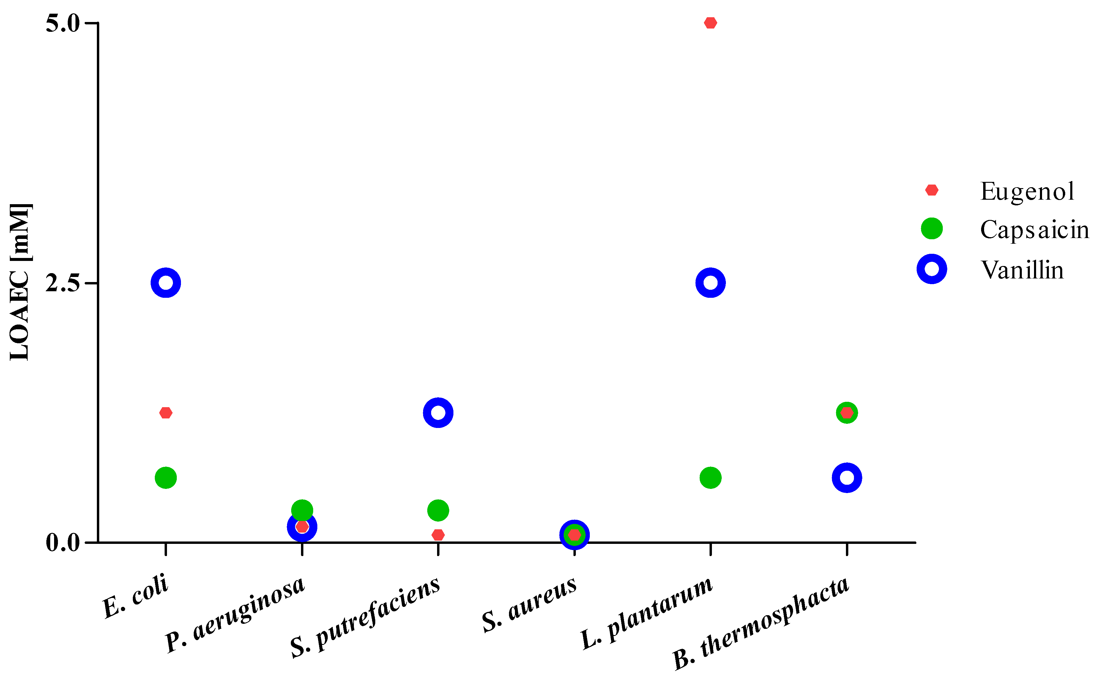
3.1. Antibacterial Activity
3.2. Antioxidant Activity
4. Discussion
5. Conclusions
Supplementary Materials
Author Contributions
Funding
Institutional Review Board Statement
Informed Consent Statement
Data Availability Statement
Acknowledgments
Conflicts of Interest
References
- Mohammad, A.; Mohd, I. Prospects of Medicinal Plants Derived Nutraceuticals: A Re-emerging New Era of Medicine and Health Aid. Prog. Chem. Biochem. Res. 2019, 2, 150–169. [Google Scholar]
- AlAli, M.; Alqubaisy, M.; Aljaafari, M.N.; AlAli, A.O.; Baqais, L.; Molouki, A.; Abushelaibi, A.; Lai, K.; Lim, S.E. Nutraceuticals: Transformation of Conventional Foods into Health Promoters/Disease Preventers and Safety Considerations. Molecules 2021, 26, 2540. [Google Scholar] [CrossRef] [PubMed]
- Prakash, B.; Kujur, A.; Singh, P.P.; Kumar, A.; Yadav, A. Plants-Derived Bioactive Compounds as Functional Food Ingredients and Food Preservative. J. Nutr. Food Sci. 2017, 1, 1–7. [Google Scholar]
- EFSA. Panel on Food Additives and Nutrient Sources added to Food (ANS). Scientific Opinion on the reevaluation of Butylated hydroxytoluene BHT (E 321) as a food additive. EFSA J. 2012, 10, 2588. [Google Scholar]
- Allam, S.S.M. Antioxidant efficiency of some common traditional Egyptian beverages. Riv. Ital. Delle Sostanze Grasse 2007, 84, 94–103. [Google Scholar]
- Pramod, K.; Ansari, S.H.; Ali, J. Eugenol: A natural compound with versatile pharmacological actions. Nat. Prod. Commun 2010, 5, 1999–2006. [Google Scholar] [CrossRef] [PubMed] [Green Version]
- Lavorgna, M.; Orlo, E.; Nugnes, R.; Piscitelli, C.; Russo, C.; Isidori, M. Capsaicin in Hot Chili Peppers: In Vitro Evaluation of Its Antiradical, Antiproliferative and Apoptotic Activities. Plant Foods Hum. Nutr. 2019, 74, 164–170. [Google Scholar] [CrossRef]
- Singletary, K. Vanilla Potential Health Benefits. Nutr. Today 2020, 55, 186–196. [Google Scholar] [CrossRef]
- Ngarmsak, M.; Delaquis, P.; Toivonen, P.; Ngarmsak, T.; Ooraikul, B.; Mazza, G. Antimicrobial activity of vanillin against spoilage microorganisms in stored fresh-cut mangoes. J. Food Prot. 2006, 69, 1724–1727. [Google Scholar] [CrossRef]
- Devi, K.P.; Nisha, S.A.; Sakthivel, R.; Pandian, S.K. Eugenol (an essential oil of clove) acts as an antibacterial agent against Salmonella typhi by disrupting the cellular membrane. J. Ethnopharmacol. 2010, 130, 107–115. [Google Scholar] [CrossRef]
- Cava-Roda, R.M.; Taboada-Rodríguez, A.; Valverde-Franco, M.T.; Marín-Iniesta, F. Antimicrobial Activity of Vanillin and Mixtures with Cinnamon and Clove Essential Oils in Controlling Listeria monocytogenes and Escherichia coli O157:H7 in Milk. Food Bioprocess Technol. 2012, 5, 2120–2131. [Google Scholar] [CrossRef]
- Marini, E.; Magi, G.; Mingoia, M.; Pugnaloni, A.; Facinelli, B. Antimicrobial and Anti-Virulence Activity of Capsaicin Against Erythromycin-Resistant, Cell-Invasive Group A Streptococci. Front. Microbiol. 2015, 6, 1281. [Google Scholar] [CrossRef] [Green Version]
- Ballester-Costa, C.; Sendra, E.; Fernández-López, J.; Pérez-Álvarez, J.A.; Viuda-Martos, M. Assessment of antioxidant and antibacterial properties on meat homogenates of essential oils obtained from four thymus species achieved from organic growth. Foods 2017, 6, 59. [Google Scholar] [CrossRef] [Green Version]
- Ng, K.R.; Lyu, X.; Mark, R.; Chen, W.N. Antimicrobial and antioxidant activities of phenolic metabolites from flavonoid-producing yeast: Potential as natural food preservatives. Food Chem. 2019, 270, 123–129. [Google Scholar] [CrossRef] [PubMed]
- Gupta, D.; Gupta, R.K. Bioprotective properties of Dragon’s blood resin: In vitro evaluation of antioxidant activity and antimicrobial activity. BMC Complement. Altern. Med. 2011, 11, 13. [Google Scholar] [CrossRef] [PubMed] [Green Version]
- Mith, H.; Duré, R.; Delcenserie, V.; Zhiri, A.; Daube, G.; Clinquart, A. Antimicrobial activities of commercial essential oils and their components against food-borne pathogens and food spoilage bacteria. Food Sci. Nutr. 2014, 2, 403–416. [Google Scholar] [CrossRef] [PubMed] [Green Version]
- Kolarević, S.; Milovanović, D.; Avdović, M.; Oalđe, M.; Kostić, J.; Sunjog, K.; Nikolić, B.; Knežević-Vukčević, J.; Vuković-Gačić, B. Optimisation of the microdilution method for detection of minimum inhibitory concentration values in selected bacteria. Bot. Serbica 2016, 40, 29–36. [Google Scholar]
- Lavorgna, M.; Iacovino, R.; Russo, C.; Di Donato, C.; Piscitelli, C.; Isidori, M. A New Approach for Improving the Antibacterial and Tumor Cytotoxic Activities of Pipemidic Acid by Including It in Trimethyl-β-cyclodextrin. Int. J. Mol. Sci. 2019, 20, 416. [Google Scholar] [CrossRef] [Green Version]
- Nikolić, B.; Vasiljević, B.; Mitić-Ćulafić, D.; Lesjak, M.; Vuković-Gačić, B.; Mimica, D.N.; Knežević-Vukčević, J. Screening of the antibacterial effect of Juniperus sibirica and Juniperus sabina essential oils in a microtitre platebased MIC assay. Bot. Serbica 2016, 40, 43–48. [Google Scholar]
- Brand-Williams, W.; Cuvelier, M.E.; Berset, C. Use of a free radical method to evaluate antioxidant activity. LWT Food Sci. Technol. 1995, 28, 25–30. [Google Scholar] [CrossRef]
- Lavorgna, M.; Pacifico, S.; Nugnes, R.; Russo, C.; Orlo, E.; Piccolella, S.; Isidori, M. Theobromacacao Criollo var. Beans: Biological Properties and Chemical Profile. Foods 2021, 10, 571. [Google Scholar] [CrossRef]
- Sroka, Z.; Cisowski, W. Hydrogen peroxide scavenging, antioxidant and anti-radical activity of some phenolic acids. Food Chem. Toxicol. 2003, 41, 753–758. [Google Scholar] [CrossRef]
- Gillespie, K.M.; Chae, J.M.; Ainsworth, E.A. Rapid measurement of total antioxidant capacity in plants. Nat. Protoc. 2007, 2, 867–870. [Google Scholar] [CrossRef]
- Sharpe, E.; Bradley, R.; Frasco, T.; Jayathilaka, D.; Marsh, A.; Andreescu, S. Metal oxide based multisensor array and portable database for field analysis of antioxidants. Sens. Actuators B Chem. 2014, 193, 552–562. [Google Scholar] [CrossRef] [Green Version]
- Fernández-Ramírez, M.D.; Smid, E.J.; Abee, T.; Nierop-Groot, M.N. Characterisation of biofilms formed by Lactobacillus plantarum WCFS1 and food spoilage isolates. Int. J. Food Microbiol. 2015, 207, 23–29. [Google Scholar] [CrossRef]
- Qiu, J.; Feng, H.; Lu, J.; Xiang, H.; Wang, D.; Dong, J.; Wang, J.; Wang, X.; Liu, J.; Deng, X. Eugenol reduces the expression of virulence-related exoproteins in Staphylococcus aureus. Appl. Environ. Microbiol. 2010, 76, 5846–5851. [Google Scholar] [CrossRef] [PubMed] [Green Version]
- Kalia, N.P.; Mahajan, P.; Mehra, R.; Nargotra, A.; Sharma, J.P.; Koul, S.; Khan, I.A. Capsaicin, a novel inhibitor of the NorA efflux pump, reduces the intracellular invasion of Staphylococcus aureus. J. Antimicrob. Chemother. 2012, 67, 2401–2408. [Google Scholar] [CrossRef] [PubMed] [Green Version]
- Qiu, J.; Niu, X.; Wang, J.; Xing, Y.; Leng, B.; Dong, J.; Li, H.; Luo, M.; Zhang, Y.; Dai, X.; et al. Capsaicin protects mice from community-associated methicillin-resistant Staphylococcus aureus pneumonia. PLoS ONE 2012, 7, e33032. [Google Scholar] [CrossRef] [PubMed] [Green Version]
- Oyedemi, B.O.; Kotsia, E.M.; Stapleton, P.D.; Gibbons, S. Capsaicin and gingerol analogues inhibit the growth of efflux-multidrug resistant bacteria and R-plasmids conjugal transfer. J. Ethnopharmacol. 2019, 245, 111871. [Google Scholar] [CrossRef] [PubMed]
- Mourtzinos, I.; Konteles, S.; Kalogeropoulos, N.; Karathanos, V.T. Thermal oxidation of vanillin affects its antioxidant and antimicrobial properties. Food Chem. 2009, 114, 791–797. [Google Scholar] [CrossRef]
- Bevilacqua, A.; Corbo, M.R.; Senigaglia, M. In Vitro Evaluation of the Antimicrobial Activity of Eugenol, Limonene, and Citrus Extract against Bacteria and Yeasts, Representative of the Spoiling Microflora of Fruit Juices. J. Food Prot. 2010, 73, 888–894. [Google Scholar] [CrossRef]
- Ouwehand, A.C.; Tiihonen, K.; Kettunen, H.; Peuranen, S.; Schulze, H.; Rautonen, N. In vitro effects of essential oils on potential pathogens and beneficial members of the normal microbiota. Vet. Med. 2010, 55, 71–78. [Google Scholar] [CrossRef] [Green Version]
- Filgueiras, C.T.; Vanetti, M.C.D. Effect of eugenol on growth and listeriolysin o production by Listeria monocytogenes. Braz. Arch. Biol. Technol. 2006, 49, 405–409. [Google Scholar] [CrossRef] [Green Version]
- Di Pasqua, R.; Betts, G.; Hoskins, N.; Edwards, M.; Ercolini, D.; Mauriello, G. Membrane toxicity of antimicrobial compounds from essential oils. J. Agric. Food Chem. 2007, 55, 4863–4870. [Google Scholar] [CrossRef] [PubMed]
- Hyldgaard, M.; Mygind, T.; Meyer, R.L. Essential oils in food preservation: Mode of action, synergies, and interactions with food matrix components. Front. Microbiol. 2012, 3, 1–24. [Google Scholar] [CrossRef] [Green Version]
- Das, B.; Mandal, D.; Dash, S.K.; Chattopadhyay, S.; Tripathy, S.; Dolai, D.P.; Dey, S.K.; Roy, S. Eugenol provokes ROS-mediated membrane damage-associated antibacterial activity against clinically isolated multidrug-resistant Staphylococcus aureus strains. Infect. Dis. Res. Treat. 2016, 9, 11–19. [Google Scholar]
- Kurita, S.; Kitagawa, E.; Kim, C.H.; Momose, Y.; Iwahashi, H. Studies on the Antimicrobial Mechanisms of Capsaicin Using Yeast DNA Microarray. Biosci. Biotechnol. Biochem. 2002, 66, 532–536. [Google Scholar] [CrossRef] [Green Version]
- Fitzgerald, D.J.; Stratford, M.; Gasson, M.J.; Narbad, A. The potential application of vanillin in preventing yeast spoilage of soft drinks and fruit juices. J. Food Prot. 2004, 6, 391–395. [Google Scholar] [CrossRef] [PubMed]
- Nazzaro, F.; Fratianni, F.; De Martino, L.; Coppola, R.; De Feo, V. Effect of essential oils on pathogenic bacteria. J. Pharm. 2013, 6, 1451–1474. [Google Scholar] [CrossRef]
- Arfa, A.B.; Combes, S.; Preziosi-Belloy, L.; Gontard, N.; Chalier, P. Antimicrobial activity of carvacrol related to its chemical structure. Lett. Appl. Microbiol. 2006, 43, 149–154. [Google Scholar] [CrossRef]
- Zhang, Y.; Wang, Y.; Zhu, X.; Cao, P.; Wei, S.; Lu, Y. Antibacterial and antibiofilm activities of eugenol from essential oil of Syzygium aromaticum (L.) Merr. & L. M. Perry (clove) leaf against periodontal pathogen Porphyromonas gingivalis. Microb. Pathog. 2017, 113, 396–402. [Google Scholar] [PubMed]
- Pinheiro, P.F.; Parreira Menini, L.A.; Bernardes, C.P.; Saraiva, S.H.; Carneiro, J.W.M.; Costa, V.A.; Arruda, R.T.; Lage, M.R.; Gonçalves, P.M.; de Oliveira Bernardeès, C.; et al. Semisynthetic Phenol Derivatives Obtained from Natural Phenols: Antimicrobial Activity and Molecular Properties. J. Agric. Food Chem. 2018, 66, 323–330. [Google Scholar] [CrossRef]
- Adeboye, P.T.; Bettiga, M.; Olsson, L. The chemical nature of phenolic compounds determines their toxicity and induces distinct physiological responses in Saccharomyces cerevisiae in lignocellulose hydrolysates. AMB Express 2014, 4, 46. [Google Scholar] [CrossRef] [Green Version]
- Kemegne, G.A.; Mkounga, P.; Ngang, J.J.E.; Kamdem, S.L.S.; Nkengfack, A.E. Antimicrobial structure activity relationship of five anthraquinones of emodine type isolated from Vismia laurentii. BMC Microbiol. 2017, 17, 41. [Google Scholar] [CrossRef] [Green Version]
- Hu, Q.; Zhou, M.; Wei, S. Progress on the antimicrobial activity research of clove oil and eugenol in the food antisepsis field. J. Food Sci. 2018, 83, 1476–1483. [Google Scholar] [CrossRef] [PubMed] [Green Version]
- Catherine, A.; Deepika, H.; Negi, P.S. Antibacterial activity of eugenol and peppermint oil in model food systems. J. Essent. Oil Res. 2012, 24, 481–486. [Google Scholar] [CrossRef]
- Jeyakumar, G.E.; Lawrencel, R. Mechanisms of bactericidal action of eugenol against Escherichia coli. J. Herb. Med. 2021, 26, 100406. [Google Scholar] [CrossRef]
- Teixeira, B.; Marques, A.; Ramos, C.; Neng, N.R.; Nogueirac, J.M.F.; Saraiva, J.A.; Nunes, M.L. Chemical composition and antibacterial and antioxidant properties of commercial essential oils. Ind. Crop. Prod. 2013, 43, 587–595. [Google Scholar] [CrossRef]
- Manrique, Y.; Suriyarak, S.; Gibis, M.; Schmidt, H.; Weiss, J. Survival of spoilage bacteria subjected to sequential eugenol and temperature treatments. Int. J. Food Microbiol. 2016, 218, 6–16. [Google Scholar] [CrossRef]
- Takahashi, H.; Nakamura, A.; Fujino, N.; Sawaguchi, Y.; Sato, M.; Kuda, T.; Kimura, B. Evaluation of the antibacterial activity of allyl isothiocyanate, clove oil, eugenol and carvacrol against spoilage lactic acid bacteria. LWT Food Sci. Technol. 2021, 145, 111263. [Google Scholar] [CrossRef]
- Gu, H.; Yang, Z.; Yu, W.; Xu, K.; Fu, Y. Antibacterial Activity of Capsaicin against Sectional Cariogenic Bacteria. Pak. J. Zool. 2019, 51, 681–687. [Google Scholar] [CrossRef]
- Tayseer, I.; Aburjai, T.; Abu-Qatouseh, L.; AL-Karabieh, N.; Ahmed, W.; Al-Samydai, A. In vitro Anti-Helicobacter pylori Activity of Capsaicin. J. Pure Appl. Microbiol. 2020, 14, 279–286. [Google Scholar] [CrossRef] [Green Version]
- Mellegård, H.; Stalheim, T.; Hormazabal, V.; Granum, P.E.; Hardy, S.P. Antibacterial activity of sphagnum acid and other phenolic compounds found in Sphagnum papillosum against food-borne bacteria. Lett. Appl. Microbiol. 2009, 49, 85–90. [Google Scholar] [CrossRef]
- Chen, J.; Yang, J.; Ma, L.; Li, J.; Shahzad, N.; Kim, C.K. Structure-antioxidant activity relationship of methoxy, phenolic hydroxyl, and carboxylic acid groups of phenolic acids. Sci. Rep. 2020, 10, 2611. [Google Scholar] [CrossRef]
- Noguchi, N.; Niki, E. Phenolic antioxidants: A rationale for design and evaluation of novel antioxidant drug for atherosclerosis. Free Radic. Biol. Med. 2000, 28, 1538–1546. [Google Scholar] [CrossRef]
- Floegel, A.; Kim, D.O.; Chung, S.J.; Koo, S.I.; Chun, O.K. Comparison of ABTS/DPPH assays to measure antioxidant capacity in popular antioxidant-rich US foods. J. Food Compos. Anal. 2011, 24, 1044–1048. [Google Scholar] [CrossRef]
- Dorman, H.J.D.; Surai, P.; Deans, S.G. In vitro antioxidant activity of a number of plant essential oils and phytoconstituents. J. Essent. Oil Res. 2000, 12, 241–248. [Google Scholar] [CrossRef]
- Dorman, H.J.D.; Figueiredo, A.C.; Barroso, J.G.; Deans, S.G. In vitro evaluation of antioxidant activity of essential oils and their components. Flavour Frag. J. 2000, 15, 12–16. [Google Scholar] [CrossRef]
- Galano, A.; León-Carmona, J.R.; Alvarez-Idaboy, J.R. Influence of the environment on the protective effects of guaiacol derivatives against oxidative stress: Mechanisms, kinetics, and relative antioxidant activity. J. Phys. Chem. 2012, 116, 7129–7137. [Google Scholar] [CrossRef] [PubMed]
- Selles, S.M.A.; Kouidri, M.; Belhamiti, B.T.; Amrane, A.A. Chemical composition, in-vitro antibacterial and antioxidant activities of Syzygium aromaticum essential oil. J. Food Meas. Charact. 2020, 14, 2352–2358. [Google Scholar] [CrossRef]
- Galano, A.; Martínez, A. Capsaicin, a tasty free radical scavenger: Mechanism of action and kinetics. J. Phys. Chem. B 2012, 116, 1200–1208. [Google Scholar] [CrossRef] [PubMed]
- Tai, A.; Sawano, T.; Yazama, F.; Ito, H. Evaluation of antioxidant activity of vanillin by using multiple antioxidant assays. Biochim. Biophys. Acta 2011, 1810, 170–177. [Google Scholar] [CrossRef] [PubMed]
- Roginsky, V.; Lissi, E.A. Review of methods to determine chainbreaking antioxidant activity in food. Food Chem. 2005, 92, 235–254. [Google Scholar] [CrossRef]
- Ahmed, M.; Pickova, J.; Ahmad, T.; Liaquat, M.; Farid, A.; Jahangi, M. Oxidation of lipids in foods. Sarhad J. Agric. 2016, 32, 231–238. [Google Scholar] [CrossRef]
- Gulcin, I. Antioxidant Activity of Eugenol: A Structure–Activity Relationship Study. J. Med. Food 2011, 14, 975–985. [Google Scholar] [CrossRef] [PubMed]


| Chemical Structure | Molecular Formula | Molar Mass (g/mol) | Chemical Name |
|---|---|---|---|
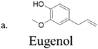 | C10H12O2 | 164.2 | 4-Allyl-2-methoxyphenol |
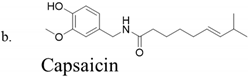 | C18H27NO3 | 305.4 | N-(4-Hydroxy-3-methoxybenzyl)-8- methylnon-trans-6-enamide |
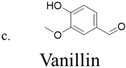 | C8H8O3 | 152.1 | 4-Hydroxy 3-methoxybenzaldehyde |
| Bacterial Strain | IC50 | ||
|---|---|---|---|
| Eugenol | Capsaicin | Vanillin | |
| E. coli | 2.70 a,b (2.39–3.06) | 4.79 a,b (3.46–6.63) | 5.87 b,c (4.72–7.31) |
| P. aeruginosa | 2.19 a,b (1.76–2.72) | 1.21 a (0.92–1.60) | 9.23 c,d (6.99–12.20) |
| S. putrefaciens | 1.11 a (0.97–1.28) | 4.28 a,b (3.29–5.55) | 2.60 a,b (2.50–2.71) |
| S. aureus | 0.75 a (0.60–0.93) | 0.68 a (0.64–0.72) | 1.38 a (1.17–1.63) |
| L. plantarum | 25.20 c (24.01–26.45) | >10 | 32.31 e (30.20–34.57) |
| B. thermosphacta | 5.02 b (4.65–5.42) | 7.77 b (7.18–8.39) | 11.38 d (10.46–12.37) |
| Bacterial Strain | Eugenol | Capsaicin | Vanillin | |||
|---|---|---|---|---|---|---|
| MIC | MBC | MIC | MBC | MIC | MBC | |
| E. coli | 12.5 | 25 | >12.5 | >12.5 | 12.5 | 25 |
| P. aeruginosa | 12.5 | 25 | >12.5 | >12.5 | 25 | 50 |
| S. putrefaciens | 3.125 | 6.25 | 12.5 | >12.5 | 6.25 | 12.5 |
| S. aureus | 6.25 | 12.5 | 12.5 | >12.5 | 12.5 | 25 |
| L. plantarum | 50 | 100 | >12.5 | >12.5 | 100 | >100 |
| B. thermosphacta | 12.5 | 25 | >12.5 | >12.5 | 25 | 50 |
| IC50 | TEAC | ||||
|---|---|---|---|---|---|
| DPPH | ABTS | TBARS | DPPH | ABTS | |
| Trolox | 0.016 (0.014–0.019) | 0.013 (0.011–0.015) | - | - | - |
| α-Tocopherol | - | - | 0.013 (0.011–0.014) | - | - |
| Eugenol | 0.152 a,*** (0.140–0.165) | 0.012 a (0.010–0.014) | 0.024 a (0.019–0.030) | 0.105 | 1.083 |
| Capsaicin | 0.090 a,* (0.088–0.092) | 0.016 a (0.014–0.017) | 0.198 b,*** (0.154–0.251) | 0.178 | 0.812 |
| Vanillin | 3.199 b,*** (2.638–3.879) | 5.566 b,*** (4.663–6.642) | 0.802c,*** (0.752–0.855) | 0.005 | 0.002 |
| IC50 | ORAC Value (TE) | |
|---|---|---|
| Trolox | 10.83 (10.33–11.35) | - |
| Eugenol | 4.11 a,** (3.91–4.32) | 2.12 ± 0.08 a |
| Capsaicin | 5.81 a,** (5.40–6.24) | 1.60 ± 0.25 a |
| Vanillin | 6.15 a,* (5.53–6.83) | 1.81 ± 0.19 a |
Publisher’s Note: MDPI stays neutral with regard to jurisdictional claims in published maps and institutional affiliations. |
© 2021 by the authors. Licensee MDPI, Basel, Switzerland. This article is an open access article distributed under the terms and conditions of the Creative Commons Attribution (CC BY) license (https://creativecommons.org/licenses/by/4.0/).
Share and Cite
Orlo, E.; Russo, C.; Nugnes, R.; Lavorgna, M.; Isidori, M. Natural Methoxyphenol Compounds: Antimicrobial Activity against Foodborne Pathogens and Food Spoilage Bacteria, and Role in Antioxidant Processes. Foods 2021, 10, 1807. https://doi.org/10.3390/foods10081807
Orlo E, Russo C, Nugnes R, Lavorgna M, Isidori M. Natural Methoxyphenol Compounds: Antimicrobial Activity against Foodborne Pathogens and Food Spoilage Bacteria, and Role in Antioxidant Processes. Foods. 2021; 10(8):1807. https://doi.org/10.3390/foods10081807
Chicago/Turabian StyleOrlo, Elena, Chiara Russo, Roberta Nugnes, Margherita Lavorgna, and Marina Isidori. 2021. "Natural Methoxyphenol Compounds: Antimicrobial Activity against Foodborne Pathogens and Food Spoilage Bacteria, and Role in Antioxidant Processes" Foods 10, no. 8: 1807. https://doi.org/10.3390/foods10081807
APA StyleOrlo, E., Russo, C., Nugnes, R., Lavorgna, M., & Isidori, M. (2021). Natural Methoxyphenol Compounds: Antimicrobial Activity against Foodborne Pathogens and Food Spoilage Bacteria, and Role in Antioxidant Processes. Foods, 10(8), 1807. https://doi.org/10.3390/foods10081807