Article

Application of Whole Genome Sequencing to Understand Diversity and Presence of Genes Associated with Sanitizer Tolerance in Listeria monocytogenes from Produce Handling Sources

by
Rebecca N. Bland
1,
Jared D. Johnson
2,
Joy G. Waite-Cusic
2,
Alexandra J. Weisberg
3,
Elizabeth R. Riutta
3,
Jeff H. Chang
3 and
Jovana Kovacevic
1,*
1
Food Innovation Center, Oregon State University, Portland, OR 97209, USA
2
Department of Food Science and Technology, Oregon State University, Corvallis, OR 97331, USA
3
Department of Botany and Plant Pathology, Oregon State University, Corvallis, OR 97331, USA
*
Author to whom correspondence should be addressed.
Foods 2021, 10(10), 2454; https://doi.org/10.3390/foods10102454
Submission received: 15 September 2021 / Revised: 9 October 2021 / Accepted: 11 October 2021 / Published: 14 October 2021
(This article belongs to the Section Foodomics)

Abstract

:
Recent listeriosis outbreaks linked to fresh produce suggest the need to better understand and mitigate L. monocytogenes contamination in packing and processing environments. Using whole genome sequencing (WGS) and phenotype screening assays for sanitizer tolerance, we characterized 48 L. monocytogenes isolates previously recovered from environmental samples in five produce handling facilities. Within the studied population there were 10 sequence types (STs) and 16 cgMLST types (CTs). Pairwise single nucleotide polymorphisms (SNPs) ranged from 0 to 3047 SNPs within a CT, revealing closely and distantly related isolates indicative of both sporadic and continuous contamination events within the facility. Within Facility 1, we identified a closely related cluster (0–2 SNPs) of isolates belonging to clonal complex 37 (CC37; CT9492), with isolates recovered during sampling events 1-year apart and in various locations inside and outside the facility. The accessory genome of these CC37 isolates varied from 94 to 210 genes. Notable genetic elements and mutations amongst the isolates included the bcrABC cassette (2/48), associated with QAC tolerance; mutations in the actA gene on the Listeria pathogenicity island (LIPI) 1 (20/48); presence of LIPI-3 (21/48) and LIPI-4 (23/48). This work highlights the potential use of WGS in tracing the pathogen within a facility and understanding properties of L. monocytogenes in produce settings.

1. Introduction

Listeria monocytogenes is a Gram-positive environmental pathogen, frequently associated with food processing environments [1,2,3]. Recent listeriosis outbreaks involving fresh produce have demonstrated serious foodborne illness risks for this food category [4,5,6,7,8]. Most notably, in 2011, a listeriosis outbreak linked to contaminated cantaloupe caused 147 illnesses and 33 deaths. Food and Drug Administration (FDA) investigations revealed that a lack of hygienic design of washing equipment was a likely cause for contamination and cross contamination of the implicated product [9]. With natural reservoirs in agricultural growing environments, the risk of L. monocytogenes in fresh produce packing houses and processing facilities is a challenge that the industry must address [10,11]. In many fresh produce facilities, growers are faced with a large volume of incoming raw product directly from the field and must handle it as quickly as possible to maintain optimum quality. Depending on the type of produce, this may include diverse operations such as shaking, sorting, trimming, washing, waxing, and often chilling. Some facilities will handle one or a few different crops from few sources, while others will handle many different crops from many different growers. Control of L. monocytogenes in fresh produce facilities is almost entirely reliant on cleaning and sanitation; however, many of these facilities do not have hygienically designed equipment nor a robust environmental monitoring program to mitigate and monitor their risks.
In facilities handling freshly harvested produce, there will likely be frequent detection of Listeria. This makes determining appropriate corrective actions difficult as positive samples may be due to transient Listeria coming in with product that will be effectively mitigated by cleaning and sanitation; or positive samples may be from a persistent Listeria population that would require investigation to find the harborage site [12].
Testing methods for environmental swabs used by the industry are often rapid screening methods [13,14,15,16] or standard culture methods (ISO 11290 [17] and BAM [18]) that report the results as positive or negative for either Listeria spp. or L. monocytogenes. Historically, the industry has been most interested in obtaining a result as quickly as possible; therefore, most results are presumptive and not confirmed. While this approach provides immediately actionable results, it fails to yield confirmed isolates that can be characterized to watch for meaningful trends to find persistent problems. Classification can range in cost and ease of data analysis from serotyping to whole genome sequencing (WGS). While WGS analysis can provide the highest level of discrimination, the food industry has been reluctant to embrace this approach due to fear of regulatory access and use of these data to support increased regulation or increased investigations. Less discriminatory and simpler characterization may have utility in monitoring for persistent L. monocytogenes. To effectively evaluate the suitability of each characterization option, these analytical tools need to be applied to a set of diverse isolates from different food commodity processing environments and geographic locations.
Our previous investigation of the prevalence of Listeria spp. and L. monocytogenes in produce processing facilities in the Pacific Northwest region of the United States [19] has yielded a set of isolates that supports this characterization. The objective of the current study was to evaluate the suitability of genotypic analyses (MLST, cgMLST, whole genome single nucleotide polymorphism [SNP], and pangenome analyses) to classify and differentiate L. monocytogenes isolates. Phenotypic stress response to a commercial quaternary ammonium compound (cQAC) was used in parallel to further understand and differentiate the highly related isolates in produce facility environments.

2. Materials and Methods

2.1. Whole Genome Sequencing and Data Assembly

L. monocytogenes isolates (n = 48) were previously isolated from environmental samples collected from produce handling and processing facilities in the Pacific Northwest region of the United States between May 2018 and April 2019. Isolate source and initial characterization are described by Jorgensen et al. [19,20]. Isolates were stored at −80 °C in tryptic soy broth (TSB; Neogen, Lansing, MI, USA) supplemented with 25% (wt/vol) glycerol. Prior to use, isolates were resuscitated by streaking onto tryptic soy agar (TSA; Neogen, Lansing, MI, USA) supplemented with 0.6% Yeast Extract (YE; Fisher, Hampton, NH, USA), followed by 24 h incubation at 37 °C.
Genomic DNA of the 48 isolates was extracted using the Qiagen DNeasy Blood and Tissue Kit (Qiagen; Hilden, Germany) according to manufacturer’s instructions for Gram-positive bacterial DNA. Quality was measured using the HS dsDNA assay kit (Fisher; Waltham, MA, USA) on the Qubit fluorometer (Fisher; Waltham, MA, USA). The quality of genetic material was determined using the nanodrop spectrophotometer (Fisher; Waltham, MA, USA). DNA libraries were prepared at Oregon State University Center for Quantitative Life Sciences (Corvallis, OR, USA) using the PlexWell kit (seqWell; Beverly, MA, USA) according to the manufacturer’s instructions. Libraries were sequenced using 2 × 150-bp paired-end sequencing on the Illumina HiSeq platform.
Raw reads were quality checked with FastQC (v 0.11.9; http://www.bioinformatics.babraham.ac.uk/projects/fastqc, accessed on 11 January 2021), followed by low-quality trimming using Trimmomatic (v 0.39; accessed on 5 May 2020 [21]). Trimmed reads were de novo assembled using SPAdes (v 3.14.1 [22]) optimized with Unicycler (v 0.4.8; accessed on 3 March 2021 [23]). Resulting assembly files were assessed for quality (Table S1) and annotated with Prokka (v 1.12; accessed on 5 May 2020 [24]).

2.2. Core Genome Multilocus Sequence Typing (cgMLST)

Core genome MLST (cgMLST) was performed in the BIGSdb-Lm platform, using a scheme consisting of 1748 conserved core genes [25]. After comparing isolates to cgMLST profiles already in the database, assemblies of all isolates were submitted to the Listeria Pasteur database to receive cgMLST type (CT) assignments.

2.3. Detection and Assessment of Antimicrobial Resistance, Stress Survival, and Pathogenicity Genes

All isolates were screened for presence or absence of antimicrobial resistance genes using NCBI BLASTN [26] with a minimum nucleotide identity and alignment length coverage of 80% (tetR, tnpABC, qacH, qacC, emrE, emrC, and bcrABC). Resistance determinants to the heavy metal cadmium were also assessed (cadA1 and cadA2). Genomic investigation of virulence genes and genetic elements was carried out by screening for presence/absence and mutation within Listeria pathogenicity islands (LIPI-1, LIPI-3, LIPI-4) and internalin A (inlA). Similarly, presence/absence and mutations in stress survival islet SSI-1 was also evaluated in all isolates. Accession numbers for reference strains used for screening and alignments of all genes and genetic elements can be found in Supplementary Materials Table S2.

2.4. Whole Genome SNP and Pangenome Analyses

Single nucleotide polymorphisms (SNPs) were called as described by Weisberg, et al. [27] with slight modifications. Briefly, FastANI (v 1.1) was used to calculate pairwise average nucleotide identity (ANI) among isolates and confirm species-level grouping (i.e., L. monocytogenes; >95% ANI) [28]. Raw reads were mapped to a representative reference sequence within the group (WRLP472) using BWA mem (v 0.7.17; accessed on 20 August 2021 [29]). Alignments were annotated, sorted, and duplicate reads identified with Picard tools (v 2.0.1; accessed on 20 August 2021 [30]). Graphtyper (v 2.6.2; accessed on 20 August 2021) was run on the dataset with the default parameters [31]. SNPs were filtered using vcffilter in vcflib (v 1.0.0; accessed on 20 August 2021 [32]) with the options ‘-f “ABHet < 0.0 | ABHet > 0.33” -f “ABHom < 0.0 | ABHom > 0.97” -f “MaxAASR > 0.4” -f “MQ > 30”. SNP calls annotated as “FAIL” or “heterozygous” were filtered to “no-call”. Bcftools (v 1.3; accessed on 20 August 2021) was used to convert filtered SNP calls to fasta format [33]. The bitwise.dist function from the R package poppr (v 2.9.2; accessed on 20 August 2021) was used to construct pairwise SNP distance tables from the fasta alignments [34]. The R package poppr was used to construct and visualize a minimum spanning network.
Pangenome analysis was conducted with Panaroo (v 1.2.8; accessed on 20 August 2021 [35]) using Prokka annotations as input to obtain the core and accessory genome of the 48 L. monocytogenes isolates. Accessory genomes of each CT and isolates were determined using a custom python script (https://github.com/johnjare/panaroo_scripts/tree/main/population_genes; accessed on 25 July 2021).

2.5. Growth in the Presence of Quaternary Ammonium Compound (QAC) Sanitizer

Growth of 48 L. monocytogenes isolates in the presence or absence of a commercial quaternary ammonium compound (cQAC; Professional Lysol No Rinse Sanitizer; EPA registration 675-30; Reckitt Benckiser, Parsippany, NJ, USA) was evaluated. Prior to use, isolates were resuscitated by streaking onto TSA-YE (Neogen), followed by 24 h incubation at 37 °C. Stock solution of the cQAC sanitizer was prepared in accordance with the manufacturer’s recommended concentration (MRC; 200 ppm), filter sterilized and stored for up to one week at 4 °C. Cultures were added to TSB supplemented with yeast extract (TSB-YE) containing 1.56 ppm cQAC at an initial inoculum of approximately 7 log CFU/mL in a final volume of 10 mL. An aliquot (200 µL) of each culture in sanitizer was transferred to a sterile 96-well plate (VWR; Radnor, PA, USA), in duplicate. Plates were incubated at 30 °C in a SpectraMax plate reader (Molecular Devices) and OD600 was measured at 30 min interval for 24 h with shaking (5 s) prior to each measurement. The OD600 data were fitted to growth curves to obtain the lag phase duration (LPD), maximum growth rate (MGR), and maximum density (OD) using the DMFit 3.0 Excel add-in program (ComBase; Computational Microbiology Research Group, Institute of Food Research, Colney, Norwich, UK), based on the models of Baranyi and Roberts [36]. All statistical analyses were performed in JMP (Version 16.0.0, SAS Institute Inc., Cary, NC, USA). One-way ANOVA was performed on lag phase and growth rate data to evaluate differences between isolates and cgMLST types when exposed to sub-lethal concentrations of cQAC. Tukey-Kramer HSD was used as a post-hoc test to determine significant differences between change in lag phase and growth rate by strain and cgMLST types. Students t-test was used to evaluate differences between growth parameters of control (without sanitizer) and 1.56 ppm of the sanitizer compound.

3. Results

3.1. General Genome Characteristics
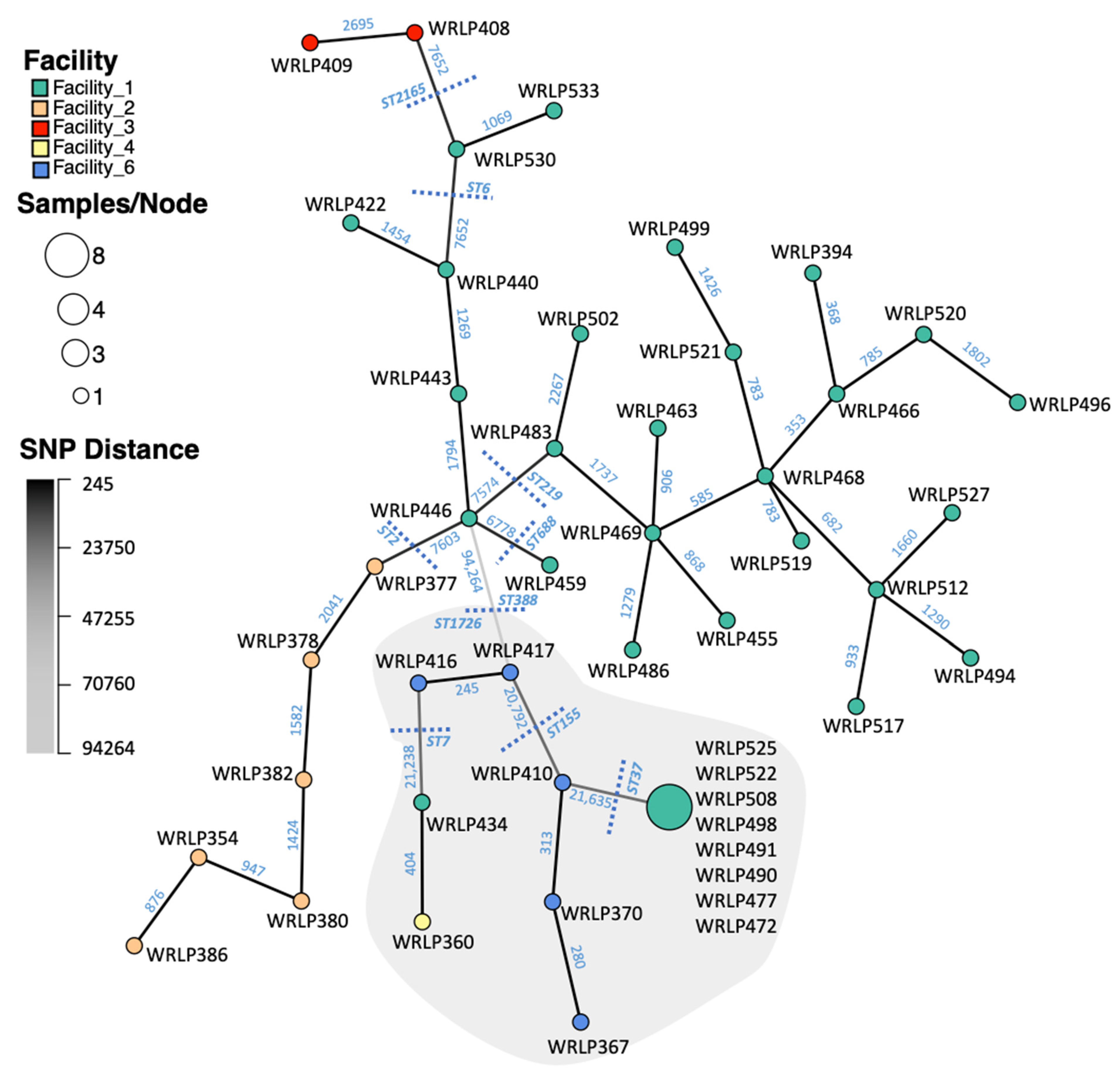
L. monocytogenes isolates (n = 48) were grouped into 10 MLST profiles (ST) and were associated with 10 clonal complexes (CCs) (Table 1). Lineage I isolates included six CCs, while four CCs were observed among lineage II isolates. Core genome MLST (cgMLST) further delineated the 48 isolates into 16 cgMLST types (CT; Figure 1). The BIGSdb-Lm typing scheme defines cgMLST types as groups of profiles that differ by up to seven allelic types out of the 1748 loci, and sub-lineages (SL) differing by up to 150. The SL assignments were all in agreement with the classic MLST typing scheme determined and previously published that considers just seven housekeeping genes; therefore, ST and cgMLST types will be used to describe isolates [20,37].
Listeria pathogenicity islands LIPI-1, LIPI-3, and LIPI-4 were found amongst the isolates in this study (Table 1). LIPI-1, which harbors six of the virulence genes associated with listeriosis infection (prfA, plcA, hly, mpl, actA, and plcB), was present in all isolates. In all isolates belonging to CC345/ST2165 and CC4/ST219, the actA gene had a 35 amino acid in-frame deletion at nucleotide position 793 [38]. Previously reported deletions in the prfA gene leading to attenuated virulence were not observed [39]. LIPI-3 were absent from all lineage II (serotype 1/2a or 3a) isolates, as well as CC2/ST2, CC388/ST388, and CC345/ST2165 within lineage I. LIPI-3 (llsG, llsH, llsX, llsB, llsY, llsD, llsP, llsA), which codes for hemolytic and cytotoxic factors important in gastrointestinal colonization [40], was present in 21/48 isolates. Isolates belonging to CC4, CC6, and CC668 from Facility 1 harbored LIPI-3. LIPI-4 was only present in a subset of lineage I isolates (ST219, ST388, and ST688) within CC4, CC388, and CC688. Isolates harboring both LIPI-3 and 4 belonged to CC4/ST219 and CC688/ST688.

3.2. SNP Analyses

Single nucleotide polymorphisms (SNPs) of whole-genome sequences (WGS) were used to characterize the relationship amongst the isolates within and between the produce processing facilities (Figure S1). The minimum spanning network (MSN) in Figure 2 illustrates the relative similarity between isolates of the largest ANI species group, with nodes (representing genotypes) connected by a branch to the node that has the fewest SNP differences (i.e., most similar). Isolates of the two serotypes (1/2a and 4b) were seen to cluster together based on having relatively few SNP differences. The branch connecting WRLP446 and WRLP417 is light grey, signifying a large number of SNP differences (94,264) at the breakpoint between lineage I and II in the tree. Isolates belonging to the same ST and CC are clustered within the tree.
The genomes of the 48 L. monocytogenes isolates differed by as few as 0 SNPs and up to 120,331 SNPs (WRLP386, lineage I; and WRLP508, lineage II). Within each CT, SNP differences ranged from 0 to 3047 SNPs. Within each CT, pairwise SNP differences between isolates ranged from 0 to 2 (CT9492); 280 to 415 (CT9497); 245 (CT9495); 2069 to 2196 (CT9488); 1069 (CT9494); 1269 to 1887 (CT9493); to 353 to 3047 (CT9489). Isolates within a facility differed by 0 to 118,686 SNPs (Facility 1); 876 to 2818 SNPs (Facility 2); 2695 SNPs (Facility 3; only two isolates); and 245 to 22,863 SNPs (Facility 6). The only ST found within multiple facilities was ST7, isolated from both Facility 1 (WRLP434) and Facility 4 (WRLP360). These isolates had a total of 404 pairwise SNP differences and belonged to two different CTs based on core genome allelic differences.

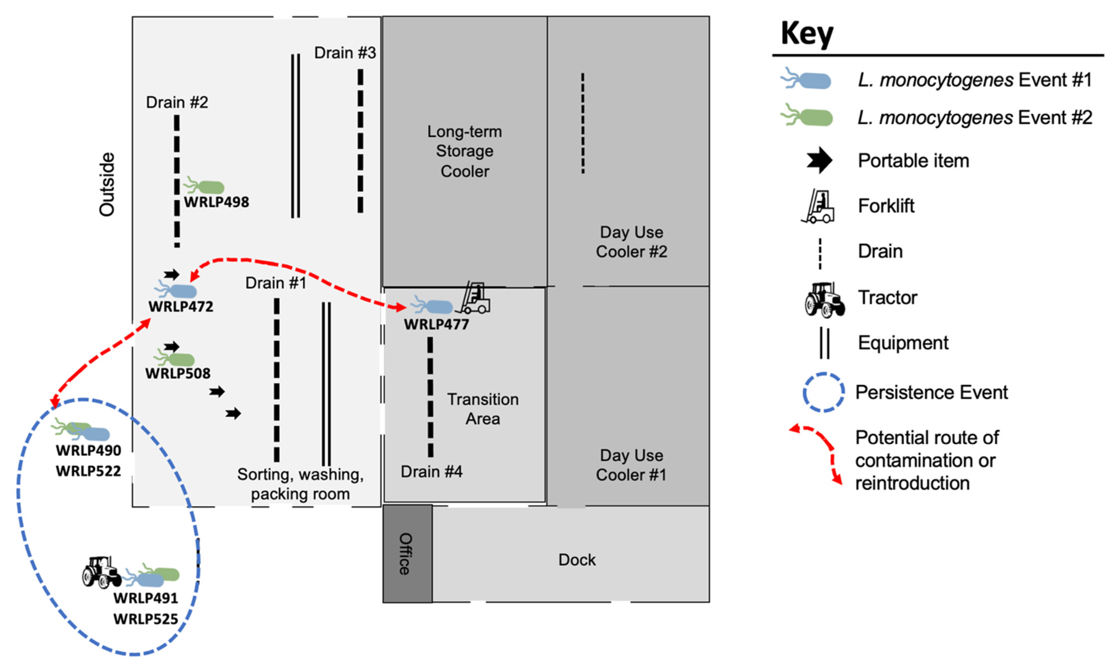
3.3. Highly Related CC37 Cluster in Facility 1

Within Facility 1, SNP analysis revealed high similarity amongst CC37 (ST37), with a maximum of two SNP differences seen between any two isolates in the ST (Figure 2). Isolates belonging to ST37 originated from samples taken during two different sampling events roughly one-year apart (Figure 3). Within this CC, identical isolates (0 pairwise SNPs; WRLP491, WRLP525) were found on a tractor tire immediately outside of the facility collected during two separate sampling events. Of the samples taken from across the facility on mobile elements, inside a drain, and on a forklift tire, 3/4 (WRLP508, WRLP472, and WRLP477) isolates had no pairwise SNPs amongst themselves and compared to the isolates recovered outside the facility on the tractor tire (Figure 3). The remaining three isolates, found immediately outside of the facility and in a drain inside the facility, had from one to two pairwise SNP differences between other isolates within the CC (WRLP490, WRLP498, and WRLP522).

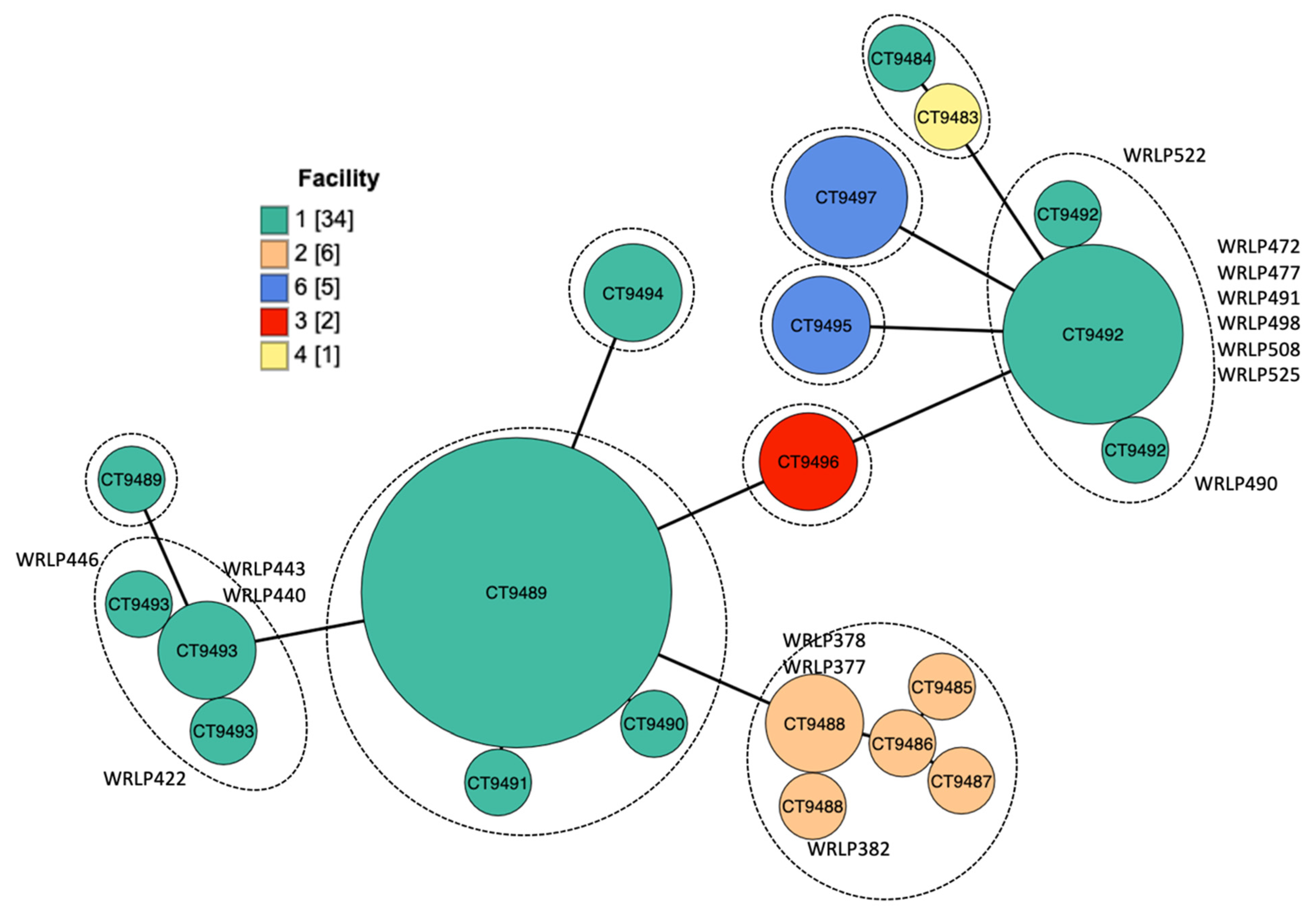
3.4. Accessory Genome Analysis

The 48 isolates, recovered from five different facilities, shared a core genome of 2391 genes and a pangenome of 9861 genes. The accessory genome, reflecting genes unique to a single isolate or group, was evaluated for each ST/CC, as well as for individual isolates within their respective ST/CC (Table 2). The number of genes in the accessory genome of a CC ranged from 3 to 103 genes. When isolates belonging to the same CC were compared, the number of their accessory genes ranged from 1 to 683 genes. Isolates within CC2 (WRLP354, WRLP377, WRLP378, WRLP380, WRLP382, and WRLP386) had a nearly identical accessory genome with only 1 to 12 accessory gene differences.

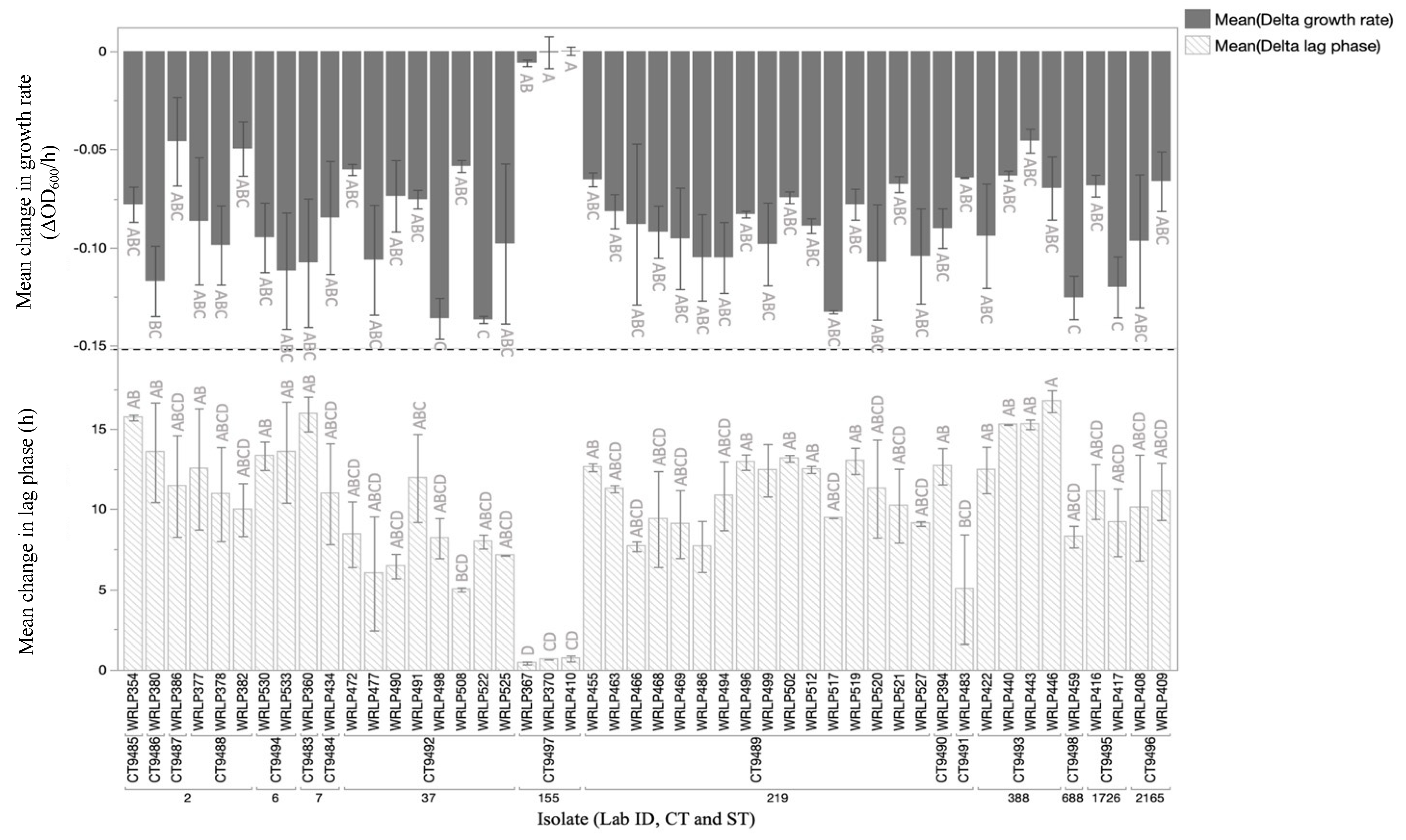
3.5. Phenotypic Analyses and Presence of Genetic Elements Assocaited with QAC Tolerance

When isolates were grown in the presence of cQAC at 1.56 ppm, the lag phase duration and growth rate were significantly impacted for 45/48 L. monocytogenes strains (Figure 4). These 45 isolates all showed significantly longer lag phase durations, and significantly slower growth rates compared to growth in the absence of the sanitizer (p < 0.05; student’s t-test). The growth of three strains, all belonging to CC155/CT9497, was only minimally influenced by the presence of cQAC, with no significant reduction in growth rate for any of the isolates within this CC (WRLP367, WRLP370, WRLP410; p < 0.05 student’s t-test). Genetic determinants of QAC tolerance that may explain the phenotype observed, such as specific efflux pumps (i.e., bcrABC), were only found in 2/48 isolates (WRLP370, WRLP410). The two isolates in which the bcrABC cassette was found belonged to the same CC (CC155) and CT (CT9489) and were both recovered from Facility 6. Notably, the third isolate demonstrating QAC tolerance (WRLP367) did not possess the bcrABC cassette despite being a member of the same CC/CT and also being isolated from the same facility. Stress survival islet 1 (SSI-1: lmo0444, lmo0445, lmo0446, lmo0447, lmo0448) was found in 4/48 isolates. Of these four isolates, three belonged to CC155 and exhibited phenotypic tolerance towards cQAC (no significant change in growth rate). The SSI-1 harbored by each of the isolates within CC155 possessed an insertion of 4 bp at nucleotide position 2571 within lmo0444 resulting in a premature stop codon (PMSC). No insertion was found within the only other isolates harboring SSL-1 (WRLP360, CC7). Isolates within CC155 had 61 unique genes compared to other isolates within lineage II and 39 unique genes when compared to the entire isolate set. Each isolate within the ST had between 103 and 536 genes unique to the other isolates belonging to CC155. Gene predictions of the genes within the accessory genome of CC155 revealed that there are several transcriptional regulators, as well as genes predicted to code for membrane components (Table S3).
The high similarity amongst isolates in ST37/CT9492 (WRLP472, WRLP477, WRLP490, WRLP491, WRLP498, WRLP508, WRLP522, and WRLP525) from Facility 1 suggested its potential persistence; therefore, the evaluation of sanitizer tolerance was of particular interest. None of these strains were particularly tolerant to cQAC (Figure 4).
The majority (68%) of L. monocytogenes isolates in this study carried genes associated with resistance towards the heavy metal cadmium (cadA1 and cadA2) (Table 1). One or both cadmium resistance determinants were found in all lineage I isolates, and were absent from all lineage II isolates.

4. Discussion

The risk of L. monocytogenes in fresh produce remains a concern due to its natural ecology in agricultural environments and the lack of a kill step in produce production. While public health and regulatory agencies have embraced WGS for outbreak investigations, the use of this technology to determine the biodiversity and assist in the understanding environmental sampling data in produce environments is not frequently employed by the industry. The L. monocytogenes isolates used in this study were obtained through environmental sampling of produce processing and handling facilities in the Pacific Northwest region of the United States [19]. While the limited number of isolates used in this study makes it difficult to draw conclusions about the region or industry as a whole, the data provide insight into the diversity, relatedness, and virulence potential of L. monocytogenes within produce operations and highlight the levels of discrimination attainable through the use of WGS.
Initial grouping of the isolates using MLST and cgMLST resulted in 10 ST/CCs and 16 CTs, respectively. Three most frequently found ST/CCs included CC4/ST219, CC37/ST37, CC2/ST2. These CCs were also common among French isolates recovered from both food and clinical sources in the period from 2005 to 2013 [41]. The size of the core genome within our isolate population (2391 genes) is in line with the size of other reported core genomes for L. monocytogenes (2322 to 2562 genes) [39,42,43,44,45,46]. The accessory genome in the present study, which generally encompasses a larger pool of genes compared to the core genome, included 7470 genes. A recent study that looked at the pangenome of L. monocytogenes from soil samples across the United States suggested that pangenome openness is the consequence of adaptive evolutionary processes that enable bacteria to survive in variable environments [45]. This study found that the pangenome and especially accessory genes of L. monocytogenes evolve at a faster rate than the core genome is seen to diversify [45]. The authors suggest that because of this difference in diversification rate, the accessory genome can be used as a further measure of diversity within a set of isolates [45]. Within our isolate set, a number of CC37 isolates possessing 0–2 pairwise SNP differences appeared to be identical or highly similar (Figure 2). However, the accessory genome for each isolate within this CC differed by 94–210 genes (Table 2). The variance in cQAC tolerance as well as the differences in antibiotic resistance profiles (Table 1) also suggest that these strains behave differently. While these differences are not explained by SNPs, the accessory genome of each isolate within CC37 suggests that these genes may be giving rise to the phenotypic differences observed. Further investigation is necessary to understand the variation in predicted gene function of these isolates and to infer what advantage they may be contributing to survival. The isolation of these highly similar CC37 isolates immediately outside the facility from multiple sampling events and dissemination across the facility (Figure 3), suggests continuous contamination into the processing environment and routes of dissemination throughout the facility. Isolation of related isolates (WRLP491 and WRLP525) from the tractor tire over two different sampling events is not unexpected as any Listeria contamination within the facility is most likely coming from the growing environment, which for these facilities was relatively local. All sampling events of isolates included in this study were conducted during production. Therefore, the presence of closely related isolates, such as in CC37 could be due to recurring contamination from a field, recurring introduction into the facility from an outside harborage site, or a potential persistence event within the facility.
In the present study, both cadA1 and cadA2, genes associated with cadmium resistance [47], were found in all lineage I isolates and 68% of total isolates. Heavy metals such as cadmium can be used in various pesticide products (including sanitizers) and therefore are commonly found in soils, and agricultural environments. Bacterial populations within these environments, such as L. monocytogenes, often possess genetic elements to combat this stressor [47,48]. These genes are commonly found on mobile genetic elements, such as plasmids (e.g., pLM80) or transposons (e.g., Tn5422), and their proximity in coding regions to QAC tolerance determinants (e.g., bcrABC) has led to speculation that the two may be associated. However, there are no data to suggest that the presence of one (e.g., QAC tolerance determinant) influences the other (i.e., cadmium tolerance determinant) or vice versa [49,50]. In the present study all ST2 and ST2165 isolates possessed both cadA1 and cadA2, whereas isolates belonging to ST219, ST388, and ST6 were found to only have cadA2 (31/48). The presence of cad did not seem to impact QAC tolerance. In evaluating isolates from food products, clinical cases, and environmental samples for QAC and heavy metal resistance genes, Gelbicova, et al. [48] found that cadmium resistance determinants were more frequently found across the samples compared to QAC tolerance genes (27.8% vs. 7%). While the prevalence rate of cadmium resistance and QAC tolerance genes was similar in the present sample set, the combination of both gene types was not found in any one isolate (i.e., isolates with bcrABC did not have either cadA1 or cadA2). The presence of these resistance determinants, especially those carried on mobile elements, is of particular concern due to possible horizontal gene transfer in the population and between species [50].
To better understand the behavior of our isolates in the presence of cQAC we exposed them to a sublethal concentration of cQAC. Three CC155 isolates had no significant decrease in growth rate and two isolates had no significant increase in lag phase duration (WRLP367, WRLP410), suggesting increased tolerance to cQAC (Figure 4). This phenotypic tolerance is in part explained by the presence of the bcrABC cassette found in WRLP370 and WRLP410 and is in line with previous reports of this element conferring increased tolerance to benzalkonium chloride and commercial QAC products [49,51,52,53,54,55]. Initially found to be harbored in an outbreak strain from the 1998-1999 listeriosis outbreak in hot dogs, Elhanafi, et al. [51] and Dutta, et al. [49] described a three-gene cassette, with bcrA and bcrB coding for small multidrug resistance (SMR) transporters and bcrC as a transcriptional regulator. Elhanafi, et al. [51] showed that the transcription levels of bcrABC are higher in cultures exposed to subinhibitory concentrations of QAC than in cultures grown without QAC. Dutta, et al. [49] noted a strong association of benzalkonium chloride tolerance in isolates that harbor this element (70/71 strains). The tolerance of both WRLP370 and WRLP410 to cQAC can in part be explained by the presence of the bcrABC cassette. However, WRLP367 behaved similarly to the other two isolates within CC155, but it lacked any known genetic elements associated with QAC tolerance. Other studies have also reported QAC tolerance in the absence of common genetic elements associated with QAC tolerance, such as bcrABC, qacH, qacC, or emrE [56,57]. In some of these strains, general efflux pumps found across the species, such as mdrL and lde, and modifications to reduce the cell membrane permeability have been proposed to be contributing factors to QAC tolerance, thought they are not consistently reported [58,59,60,61].
The presence of SSI-1, associated with increased tolerance to low pH and high salt stress, has also been described in isolates with phenotypic tolerance to QAC [62]. Particularly, the occurrence of this islet within CC155 has been previously reported [3,39,56]. The high prevalence of CC155 among food, food processing and clinical cases, indicates its importance and warrants further exploration [39,41]. Notably, we observed the 4 bp deletion within lmo0444 (SSI-1), resulting in a PMSC in three isolates that exhibited phenotypic tolerance to cQAC (WRLP367, WRLP370, WRLP410), whereas the only other isolate that harbored the SSI-1 (WRLP360; CC7/ST7) encoded the full-length gene. Ryan, et al. [62] speculated through in silico analysis that lmo0444 encodes a hypothetical protein with a reovirus attachment protein domain and possesses homology with a phage infection protein. Further analysis is needed to elucidate the effect of this deletion on the function of the gene and impact on the islet as a whole on resulting stress tolerance. Additionally, Keeney, et al. [63] reported that the presence of SSI-1 was strongly correlated with biofilm forming ability as shown by increased adherence in adhesion assays. Other research has suggested that genetic determinants associated with QAC tolerance confer tolerance, rather than resistance, to sanitizers used within food processing environments [64]. However, the impact of biofilm formation on persistence within a facility and increased tolerance towards QAC has been described [65]. It is likely that the combination of a set of genes conferring increased biofilm formation in combination with a sanitizer stress efflux system may confer increased overall survival within a facility.
When considering the virulence potential of L. monocytogenes, intact inlA and pathogenicity islands have been accepted as common virulence markers [66,67,68,69]. No PMSCs in inlA were detected among any of the 48 tested isolates suggesting fully functional genes. Previous literature has reported a relatively high rate of mutations leading to PMSC across lineage II isolates (~30%), whereas linage I is more likely to harbor a full-length gene [66,67,68]. The disproportional representation of lineage I isolates within the present data set may be in part responsible for the lack of mutations seen here. Nightingale, et al. [68] illustrated that invasion efficacies of strains with an inlA PMSC were reduced (from −3.6 to −4.24) compared to those of full-length genes (from 0.52 to 0.89). While no PMSCs were detected in the isolates studied here, a 9 bp deletion within inlA was found in WRLP530 and WRLP533 of CC6/ST6. This mutation was initially reported by Kovacevic, et al. [70] and has since been reported by Kanki, et al. [71], Smith, et al. [72], and Raschle, et al. [42]. Raschle, et al. [42] found 2/25 isolates from surface water in Switzerland possessing this 9 bp deletion, both of which also belonged to CC6 and were the only two CC6 isolates within the sample set. Similarly, Smith, et al. [72] found this mutation in the only isolate belonging to CC6 within a sample set from produce and produce processing environments. Kovacevic, et al. [70] and Kanki, et al. [71] both found that isolates with this deletion had equivalent or increased invasion efficacy to isolates encoding the full-length inlA. While the mutations seen within this gene may not be leading to a reduction in virulence, Keeney, et al. [63] found that this mutation was correlated with a significant decrease in the ability to form biofilms. The combination of increased ability to survive within a food processing facility and increased ability to cause disease in the event of consumption of contaminated food product is of particular concern.
Further evidence of virulence potential was assessed by screening for four major pathogenicity islands (LIPI1-4), associated with genes that increase the ability of L. monocytogenes to cause infection in a human host. Listeria pathogenicity island 1 (LIPI-1) is widely conserved across the species, harboring genes largely responsible for phagosome escape from host cells and cell-to-cell spread. Specifically, the actA gene within LIPI-1 is responsible for the formation of an actin tail, assisting in cell-to-cell spread [73,74]. The 35 amino acid actA deletion seen across the majority of lineage I isolates in our study has previously been described in other L. monocytogenes isolates [38,75,76]. This deletion was first identified in a dairy isolate by Jiang, et al. [38]. They reported similar invasion efficacy for both control and deletion strains in HeLa cell models, but a defect in cell-to-cell spread was observed in a plaque-forming assay (L929 cell line) [38]. A study by Holch, et al. [75] used a similar assay to evaluate cell-to-cell spread in isolates possessing this deletion from food, clinical and environmental sources. Conversely, they did not see that the deletion influenced the plaque formation associated with cell-to-cell spread. The deletion allele only possesses three of four protein-rich repeats within the proline rich region of the gene. Smith, et al. [73] detailed the effect of deletions of these repeats as being largely responsible for motility [73]. Further analysis is needed to determine if similar virulence potential is observed in the strains within the present study. The occurrence of this deletion within isolates otherwise associated with increased virulence (lineage I, CC4, LIPI-3, and LIPI-4), highlight the genomic complexities found within produce environments.
Other pathogenicity islands, LIPI-3 and LIPI-4 were found in 44% (21/48) and 48% (23/48) of our isolates, respectively. Both islands were seen across all CC4 and CC688 isolates (19/48). The prevalence of these islands in lineage I CTs and more specifically LIPI-4 in CC4 is in line with what is reported in the literature [42,69]. A recent study by Maury, et al. [69] found that the six genes on LIPI-4 are associated with enhanced invasion, central nervous system and maternal-neonatal listeriosis and are strongly associated with CC4. While most virulence genes are well conserved across L. monocytogenes, the presence of pathogenicity islands LIPI-3 and LIPI-4, both of which are strongly associated with lineage I isolates, are not always present across the species [40,41,77]. Their prevalence within the present set of data suggests their increased virulence potential.

5. Conclusions

By evaluating a set of L. monocytogenes previously isolated from produce processing facilities we were able to examine the utility of various genomic analyses on their ability to differentiate isolates within produce environments in aiding our understanding of contamination events. Within environments that contain closely related strains, a deeper level of analysis, likely beyond MLST and even cgMLST, may be needed to differentiate potential persistent or recurring strains from those that are transient. Whole genome SNP analysis and pangenome analysis resulted in similar classifications that could separate “nearly identical” (i.e., likely persistent) and “clearly different” (i.e., likely transient) strains, which is a necessary level of interpretation for optimizing the use of environmental monitoring programs. The identification of a “nearly identical” (likely persistent) population in one facility identified a potential route of contamination and suggested the presence of a harborage site in an outdoor high traffic area that seems to be a source of indoor contamination on more than one processing day.
While SNP analysis was useful in discriminating between isolates, and it is generally considered as an important tool in epidemiological traceback and outbreak investigations, it did not fully provide insight into isolate properties and differences, such as those seen through phenotypic responses and the accessory genome. Our data show that fully exploiting WGS data and considering virulence profiles, SNPs, and accessory genomes can lead to a greater understanding of closely related populations that may be missed otherwise. By pairing genotypic data with some basic phenotypic analysis in the form of susceptibility profiling to antimicrobials is suggestive of genomic differences and helps guide and confirm risk management decisions.

Supplementary Materials

The following are available online at https://www.mdpi.com/article/10.3390/foods10102454/s1, Table S1: Assembly statistics resulting from SPAdes de novo assemblies, Table S2: NCBI Accession numbers used to screen for antimicrobials tolerance determinants and virulence genes, Table S3: GoTerm analysis of select CC155 accessory genes annotated as DNA binding, transcriptional regulator or membrane component, Figure S1: Pairwise SNP matrix.

Author Contributions

Conceptualization, J.G.W.-C. and J.K.; Data curation, R.N.B. and E.R.R.; Formal analysis, R.N.B., J.D.J., A.J.W., J.H.C., J.G.W.-C. and J.K.; Funding acquisition, J.K.; Methodology, R.N.B., J.D.J., A.J.W., J.G.W.-C. and J.K.; Project administration, J.K.; Supervision, A.J.W., J.H.C., J.G.W.-C. and J.K.; Validation, A.J.W.; Visualization, R.N.B. and A.J.W.; Writing—original draft, R.N.B.; Writing—review & editing, J.H.C., J.G.W.-C. and J.K. All authors have read and agreed to the published version of the manuscript.

Funding

This research was funded in part by the Oregon Specialty Crop Block Grant Program, grant number ODA-5010-GR, and the OSU Agricultural Research Foundation, grant number ARF#9148A.

Institutional Review Board Statement

Not applicable.

Informed Consent Statement

Not applicable.

Data Availability Statement

Not applicable.

Acknowledgments

The authors would like to thank The Center for Quantitative Life Sciences at Oregon State University (Corvallis, OR, USA) for conducting the WGS and bioinformatics training.

Conflicts of Interest

The authors declare no conflict of interest.

References

  1. Tompkin, R. Control of Listeria monocytogenes in the food-processing environment. J. Food Prot. 2002, 65, 709–725. [Google Scholar] [CrossRef] [PubMed]
  2. Hurley, D.; Luque-Sastre, L.; Parker, C.T.; Huynh, S.; Eshwar, A.K.; Nguyen, S.V.; Andrews, N.; Moura, A.; Fox, E.M.; Jordan, K. Whole-genome sequencing-based characterization of 100 Listeria monocytogenes isolates collected from food processing environments over a four-year period. mSphere 2019, 4, e00252-19. [Google Scholar] [CrossRef] [Green Version]
  3. Chen, Y.; Chen, Y.; Pouillot, R.; Dennis, S.; Xian, Z.; Luchansky, J.B.; Porto-Fett, A.C.S.; Lindsay, J.A.; Hammack, T.S.; Allard, M.; et al. Genetic diversity and profiles of genes associated with virulence and stress resistance among isolates from the 2010–2013 interagency Listeria monocytogenes market basket survey. PLoS ONE 2020, 15, e0231393. [Google Scholar] [CrossRef]
  4. Angelo, K.; Conrad, A.; Saupe, A.; Dragoo, H.; West, N.; Sorenson, A.; Barnes, A.; Doyle, M.; Beal, J.; Jackson, K. Multistate outbreak of Listeria monocytogenes infections linked to whole apples used in commercially produced, prepackaged caramel apples: United States, 2014–2015. Epidemiol. Infect. 2017, 145, 848–856. [Google Scholar] [CrossRef] [PubMed] [Green Version]
  5. CDC. Multistate Outbreak of Listeriosis Linked to Frozen Vegetables (Final Update). Available online: https://www.cdc.gov/listeria/outbreaks/frozen-vegetables-05-16/index.html (accessed on 10 September 2021).
  6. CDC. Multistate Outbreak of Listeriosis Linked to Packaged Salads Produced at Springfield, Ohio Dole Processing Facility (Final Update). Available online: https://www.cdc.gov/listeria/outbreaks/bagged-salads-01-16/index.html (accessed on 10 September 2021).
  7. CDC. Outbreak of Listeria Infections Linked to Enoki Mushrooms (Final Update). Available online: https://www.cdc.gov/listeria/outbreaks/enoki-mushrooms-03-20/index.html (accessed on 10 September 2021).
  8. Gaul, L.K.; Farag, N.H.; Shim, T.; Kingsley, M.A.; Silk, B.J.; Hyytia-Trees, E. Hospital-acquired listeriosis outbreak caused by contaminated diced celery—Texas, 2010. Clin. Infect. Dis. 2013, 56, 20–26. [Google Scholar] [CrossRef] [PubMed]
  9. FDA. Memorandum to the File on the Environmental Assessment; FDA: Silver Spring, MD, USA, 2012. [Google Scholar]
  10. Zhu, Q.; Gooneratne, R.; Hussain, M.A. Listeria monocytogenes in fresh produce: Outbreaks, prevalence and contamination levels. Foods 2017, 6, 21. [Google Scholar] [CrossRef] [Green Version]
  11. Freitag, N.E.; Port, G.C.; Miner, M.D. Listeria monocytogenes—From saprophyte to intracellular pathogen. Nat. Rev. Microbiol. 2009, 7, 623–628. [Google Scholar] [CrossRef]
  12. Ferreira, V.; Wiedmann, M.; Teixeira, P.; Stasiewicz, M.J. Listeria monocytogenes persistence in food-associated environments: Epidemiology, strain characteristics, and implications for public health. J. Food Prot. 2014, 77, 150–170. [Google Scholar] [CrossRef]
  13. Wendorf, M.; Feldpausch, E.; Pinkava, L.; Luplow, K.; Hosking, E.; Norton, P.; Biswas, P.; Mozola, M.; Rice, J. Validation of the ANSR® Listeria Method for Detection of Listeria spp. in Environmental Samples. J. AOAC Int. 2019, 96, 1414–1424. [Google Scholar] [CrossRef]
  14. Benesh, D.L.; Crowley, E.S.; Bird, P.M. 3M™ Tecra™ Listeria Visual Immunoassay: AOAC Official MethodsSM 995.22 and 2002.09. J. AOAC Int. 2019, 96, 218–224. [Google Scholar] [CrossRef]
  15. Benesh, D.L.; Crowley, E.S.; Bird, P.M. 3M™ Petrifilm™ Environmental Listeria Plate. J. AOAC Int. 2019, 96, 225–228. [Google Scholar] [CrossRef] [PubMed]
  16. Rapid Microbiology. Listeria Detection and Identification Methods in Foods. Available online: https://www.rapidmicrobiology.com/test-method/listeria-detection-and-identification-methods (accessed on 8 October 2021).
  17. International Organization for Standardization (ISO). Microbiology of the food chain—Horizontal Method for the Detection and Enumeration of Listeria monocytogenes and of Listeria spp.—Part 1: Detection Method. Available online: https://www.iso.org/standard/60313.html/ (accessed on 8 October 2021).
  18. Hitchins, A.D.; Jinneman, K.; Chen, Y. BAM Chapter 10: Detection of Listeria monocytogenes in Foods and Environmental Samples, and Enumeration of Listeria monocytogenes in Foods. In Bacteriological Analytical Manual (BAM); U.S. Food and Drug Administration (FDA): Silver Spring, MD, USA, 2017. [Google Scholar]
  19. Jorgensen, J.; Waite-Cusic, J.; Kovacevic, J. Prevalence of Listeria spp. in produce handling and processing facilities in the Pacific Northwest. Food Microbiol. 2020, 90, 103468. [Google Scholar]
  20. Jorgensen, J.; Bland, R.; Waite-Cusic, J.; Kovacevic, J. Diversity and antimicrobial resistance of Listeria spp. and L. monocytogenes clones from produce handling and processing facilities in the Pacific Northwest. Food Control 2021, 123, 107665. [Google Scholar] [CrossRef]
  21. Bolger, A.M.; Lohse, M.; Usadel, B. Trimmomatic: A flexible trimmer for Illumina sequence data. Bioinformatics 2014, 30, 2114–2120. [Google Scholar] [CrossRef] [Green Version]
  22. Bankevich, A.; Nurk, S.; Antipov, D.; Gurevich, A.A.; Dvorkin, M.; Kulikov, A.S.; Lesin, V.M.; Nikolenko, S.I.; Pham, S.; Prjibelski, A.D.; et al. SPAdes: A new genome assembly algorithm and its applications to single-cell sequencing. J. Comput. Biol. 2012, 19, 455–477. [Google Scholar] [CrossRef] [Green Version]
  23. Wick, R.R.; Judd, L.M.; Gorrie, C.L.; Holt, K.E. Unicycler: Resolving bacterial genome assemblies from short and long sequencing reads. PLoS Comput. Biol. 2017, 13, e1005595. [Google Scholar] [CrossRef] [Green Version]
  24. Seemann, T. Prokka: Rapid prokaryotic genome annotation. Bioinformatics 2014, 30, 2068–2069. [Google Scholar] [CrossRef] [PubMed]
  25. Moura, A.; Criscuolo, A.; Pouseele, H.; Maury, M.M.; Leclercq, A.; Tarr, C.; Björkman, J.T.; Dallman, T.; Reimer, A.; Enouf, V. Whole genome-based population biology and epidemiological surveillance of Listeria monocytogenes. Nat. Microbiol. 2016, 2, 16185. [Google Scholar] [CrossRef]
  26. National Center for Biotechnology Information (NCBI). Basic Local Alignment Search Tool (BLAST). Standard Nucleotide BLAST (blastn). Available online: https://blast.ncbi.nlm.nih.gov/Blast.cgi (accessed on 8 October 2021).
  27. Weisberg, A.J.; Davis, E.W., 2nd; Tabima, J.; Belcher, M.S.; Miller, M.; Kuo, C.H.; Loper, J.E.; Grunwald, N.J.; Putnam, M.L.; Chang, J.H. Unexpected conservation and global transmission of agrobacterial virulence plasmids. Science 2020, 368, eaba5256. [Google Scholar] [CrossRef]
  28. Jain, C.; Rodriguez, R.L.; Phillippy, A.M.; Konstantinidis, K.T.; Aluru, S. High throughput ANI analysis of 90K prokaryotic genomes reveals clear species boundaries. Nat. Commun. 2018, 9, 5114. [Google Scholar] [CrossRef] [Green Version]
  29. Li, H.; Durbin, R. Fast and accurate short read alignment with Burrows-Wheeler transform. Bioinformatics 2009, 25, 1754–1760. [Google Scholar] [CrossRef] [Green Version]
  30. Broad Institute. Picard Toolkit. Available online: http://broadinstitute.github.io/picard (accessed on 8 October 2021).
  31. Eggertsson, H.P.; Jonsson, H.; Kristmundsdottir, S.; Hjartarson, E.; Kehr, B.; Masson, G.; Zink, F.; Hjorleifsson, K.E.; Jonasdottir, A.; Jonasdottir, A.; et al. Graphtyper enables population-scale genotyping using pangenome graphs. Nat. Genet. 2017, 49, 1654–1660. [Google Scholar] [CrossRef] [PubMed]
  32. Garrison, E.; Kronenberg, Z.N.; Dawson, E.T.; Pedersen, B.S.; Prins, P. Vcflib and tools for processing the VCF variant call format. bioRxiv 2021. [Google Scholar] [CrossRef]
  33. Li, H.; Handsaker, B.; Wysoker, A.; Fennell, T.; Ruan, J.; Homer, N.; Marth, G.; Abecasis, G.; Durbin, R.; 1000 Genome Project Data Processing Subgroup. The Sequence Alignment/Map format and SAMtools. Bioinformatics 2009, 25, 2078–2079. [Google Scholar] [CrossRef] [PubMed] [Green Version]
  34. Kamvar, Z.N.; Tabima, J.F.; Grunwald, N.J. Poppr: An R package for genetic analysis of populations with clonal, partially clonal, and/or sexual reproduction. PeerJ 2014, 2, e281. [Google Scholar] [CrossRef] [PubMed] [Green Version]
  35. Tonkin-Hill, G.; MacAlasdair, N.; Ruis, C.; Weimann, A.; Horesh, G.; Lees, J.A.; Gladstone, R.A.; Lo, S.; Beaudoin, C.; Floto, R.A.; et al. Producing polished prokaryotic pangenomes with the Panaroo pipeline. Genome Biol. 2020, 21, 180. [Google Scholar] [CrossRef]
  36. Baranyi, J.; Roberts, T.A. A dynamic approach to predicting bacterial growth in food. Int. J. Food Microbiol. 1994, 23, 277–294. [Google Scholar] [CrossRef]
  37. Ragon, M.; Wirth, T.; Hollandt, F.; Lavenir, R.; Lecuit, M.; Le Monnier, A.; Brisse, S. A new perspective on Listeria monocytogenes evolution. PLoS Pathog. 2008, 4, e1000146. [Google Scholar] [CrossRef] [Green Version]
  38. Jiang, L.L.; Xu, J.J.; Chen, N.; Shuai, J.B.; Fang, W.H. Virulence phenotyping and molecular characterization of a low-pathogenicity isolate of Listeria monocytogenes from cow’s milk. Acta Biochim. Biophys. Sin. 2006, 38, 262–270. [Google Scholar] [CrossRef] [Green Version]
  39. Wagner, E.; Zaiser, A.; Leitner, R.; Quijada, N.M.; Pracser, N.; Pietzka, A.; Ruppitsch, W.; Schmitz-Esser, S.; Wagner, M.; Rychli, K. Virulence characterization and comparative genomics of Listeria monocytogenes sequence type 155 strains. BMC Genom. 2020, 21, 847. [Google Scholar] [CrossRef]
  40. Cotter, P.D.; Draper, L.A.; Lawton, E.M.; Daly, K.M.; Groeger, D.S.; Casey, P.G.; Ross, R.P.; Hill, C. Listeriolysin S, a novel peptide haemolysin associated with a subset of lineage I Listeria monocytogenes. PLoS Pathog. 2008, 4, e1000144. [Google Scholar] [CrossRef] [Green Version]
  41. Maury, M.M.; Bracq-Dieye, H.; Huang, L.; Vales, G.; Lavina, M.; Thouvenot, P.; Disson, O.; Leclercq, A.; Brisse, S.; Lecuit, M. Hypervirulent Listeria monocytogenes clones’ adaption to mammalian gut accounts for their association with dairy products. Nat. Commun. 2019, 10, 2488. [Google Scholar] [CrossRef] [Green Version]
  42. Raschle, S.; Stephan, R.; Stevens, M.J.A.; Cernela, N.; Zurfluh, K.; Muchaamba, F.; Nuesch-Inderbinen, M. Environmental dissemination of pathogenic Listeria monocytogenes in flowing surface waters in Switzerland. Sci. Rep. 2021, 11, 9066. [Google Scholar] [CrossRef]
  43. Pirone-Davies, C.; Chen, Y.; Pightling, A.; Ryan, G.; Wang, Y.; Yao, K.; Hoffmann, M.; Allard, M.W. Genes significantly associated with lineage II food isolates of Listeria monocytogenes. BMC Genom. 2018, 19, 1–11. [Google Scholar] [CrossRef]
  44. Den Bakker, H.C.; Desjardins, C.A.; Griggs, A.D.; Peters, J.E.; Zeng, Q.; Young, S.K.; Kodira, C.D.; Yandava, C.; Hepburn, T.A.; Haas, B.J. Evolutionary dynamics of the accessory genome of Listeria monocytogenes. PLoS ONE 2013, 8, e67511. [Google Scholar] [CrossRef] [PubMed] [Green Version]
  45. Liao, J.; Guo, X.; Weller, D.L.; Pollak, S.; Buckley, D.H.; Wiedmann, M.; Cordero, O.X. Nationwide genomic atlas of soil-dwelling Listeria reveals effects of selection and population ecology on pangenome evolution. Nat. Microbiol. 2021, 6, 1021–1030. [Google Scholar] [CrossRef]
  46. Hilliard, A.; Leong, D.; O’Callaghan, A.; Culligan, E.P.; Morgan, C.A.; DeLappe, N.; Hill, C.; Jordan, K.; Cormican, M.; Gahan, C.G. Genomic characterization of Listeria monocytogenes isolates associated with clinical listeriosis and the food production environment in Ireland. Genes 2018, 9, 171. [Google Scholar] [CrossRef] [PubMed] [Green Version]
  47. Mullapudi, S.; Siletzky, R.M.; Kathariou, S. Diverse cadmium resistance determinants in Listeria monocytogenes isolates from the turkey processing plant environment. Appl. Environ. Microbiol. 2010, 76, 627–630. [Google Scholar] [CrossRef] [PubMed] [Green Version]
  48. Gelbicova, T.; Florianova, M.; Hluchanova, L.; Kalova, A.; Korena, K.; Strakova, N.; Karpiskova, R. Comparative analysis of genetic determinants encoding cadmium, arsenic, and benzalkonium chloride resistance in Listeria monocytogenes of human, food, and environmental origin. Front. Microbiol. 2020, 11, 599882. [Google Scholar] [CrossRef]
  49. Dutta, V.; Elhanafi, D.; Kathariou, S. Conservation and distribution of the benzalkonium chloride resistance cassette bcrABC in Listeria monocytogenes. Appl. Environ. Microbiol. 2013, 79, 6067–6074. [Google Scholar] [CrossRef] [Green Version]
  50. Katharios-Lanwermeyer, S.; Rakic-Martinez, M.; Elhanafi, D.; Ratani, S.; Tiedje, J.; Kathariou, S. Coselection of cadmium and benzalkonium chloride resistance in conjugative transfers from nonpathogenic Listeria spp. to other Listeriae. Appl. Environ. Microbiol. 2012, 78, 7549–7556. [Google Scholar] [CrossRef] [PubMed] [Green Version]
  51. Elhanafi, D.; Dutta, V.; Kathariou, S. Genetic characterization of plasmid-associated benzalkonium chloride resistance determinants in a Listeria monocytogenes strain from the 1998–1999 outbreak. Appl. Environ. Microbiol. 2010, 76, 8231–8238. [Google Scholar] [CrossRef] [Green Version]
  52. Jiang, X.; Yu, T.; Liang, Y.; Ji, S.; Guo, X.; Ma, J.; Zhou, L. Efflux pump-mediated benzalkonium chloride resistance in Listeria monocytogenes isolated from retail food. Int. J. Food Microbiol. 2016, 217, 141–145. [Google Scholar] [CrossRef] [PubMed]
  53. Meier, A.B.; Guldimann, C.; Markkula, A.; Pontinen, A.; Korkeala, H.; Tasara, T. Comparative phenotypic and genotypic analysis of Swiss and Finnish Listeria monocytogenes isolates with respect to benzalkonium chloride resistance. Front. Microbiol. 2017, 8, 397. [Google Scholar] [CrossRef]
  54. Cherifi, T.; Carrillo, C.; Lambert, D.; Miniai, I.; Quessy, S.; Lariviere-Gauthier, G.; Blais, B.; Fravalo, P. Genomic characterization of Listeria monocytogenes isolates reveals that their persistence in a pig slaughterhouse is linked to the presence of benzalkonium chloride resistance genes. BMC Microbiol. 2018, 18. [Google Scholar] [CrossRef] [PubMed]
  55. Cooper, A.L.; Carrillo, C.D.; DeschEnes, M.; Blais, B.W. Genomic markers for quaternary ammonium compound resistance as a persistence indicator for Listeria monocytogenes contamination in food manufacturing environments. J. Food Prot. 2021, 84, 389–398. [Google Scholar] [CrossRef]
  56. Ebner, R.; Stephan, R.; Althaus, D.; Brisse, S.; Maury, M.; Tasara, T. Phenotypic and genotypic characteristics of Listeria monocytogenes strains isolated during 2011–2014 from different food matrices in Switzerland. Food Control. 2015, 57, 321–326. [Google Scholar] [CrossRef] [Green Version]
  57. Ortiz, S.; Lopez, V.; Martinez-Suarez, J.V. The influence of subminimal inhibitory concentrations of benzalkonium chloride on biofilm formation by Listeria monocytogenes. Int. J. Food Microbiol. 2014, 189, 106–112. [Google Scholar] [CrossRef]
  58. Jiang, X.; Yu, T.; Xu, Y.; Wang, H.; Korkeala, H.; Shi, L. MdrL, a major facilitator superfamily efflux pump of Listeria monocytogenes involved in tolerance to benzalkonium chloride. Appl. Microbiol. Biotechnol. 2019, 103, 1339–1350. [Google Scholar] [CrossRef]
  59. Rakic-Martinez, M.; Drevets, D.A.; Dutta, V.; Katic, V.; Kathariou, S. Listeria monocytogenes strains selected on ciprofloxacin or the disinfectant benzalkonium chloride exhibit reduced susceptibility to ciprofloxacin, gentamicin, benzalkonium chloride, and other toxic compounds. Appl. Environ. Microbiol. 2011, 77, 8714–8721. [Google Scholar] [CrossRef] [Green Version]
  60. Romanova, N.A.; Wolffs, P.F.; Brovko, L.Y.; Griffiths, M.W. Role of efflux pumps in adaptation and resistance of Listeria monocytogenes to benzalkonium chloride. Appl. Environ. Microbiol. 2006, 72, 3498–3503. [Google Scholar] [CrossRef] [Green Version]
  61. To, M.S.; Favrin, S.; Romanova, N.; Griffiths, M.W. Postadaptational resistance to benzalkonium chloride and subsequent physicochemical modifications of Listeria monocytogenes. Appl. Environ. Microbiol. 2002, 68, 5258–5264. [Google Scholar] [CrossRef] [PubMed] [Green Version]
  62. Ryan, S.; Begley, M.; Hill, C.; Gahan, C.G. A five-gene stress survival islet (SSI-1) that contributes to the growth of Listeria monocytogenes in suboptimal conditions. J. Appl. Microbiol. 2010, 109, 984–995. [Google Scholar] [CrossRef]
  63. Keeney, K.; Trmcic, A.; Zhu, Z.; Delaquis, P.; Wang, S. Stress survival islet 1 contributes to serotype-specific differences in biofilm formation in Listeria monocytogenes. Lett. Appl. Microbiol. 2018, 67, 530–536. [Google Scholar] [CrossRef] [PubMed]
  64. Cruz, C.D.; Fletcher, G.C. Assessing manufacturers’ recommended concentrations of commercial sanitizers on inactivation of Listeria monocytogenes. Food Control 2012, 26, 194–199. [Google Scholar] [CrossRef]
  65. Pang, X.; Wong, C.; Chung, H.-J.; Yuk, H.-G. Biofilm formation of Listeria monocytogenes and its resistance to quaternary ammonium compounds in a simulated salmon processing environment. Food Control 2019, 98, 200–208. [Google Scholar] [CrossRef]
  66. Orsi, R.H.; den Bakker, H.C.; Wiedmann, M. Listeria monocytogenes lineages: Genomics, evolution, ecology, and phenotypic characteristics. Int. J. Med. Microbiol. 2011, 301, 79–96. [Google Scholar] [CrossRef]
  67. Van Stelten, A.; Simpson, J.; Ward, T.; Nightingale, K. Revelation by single-nucleotide polymorphism genotyping that mutations leading to a premature stop codon in inlA are common among Listeria monocytogenes isolates from ready-to-eat foods but not human listeriosis cases. Appl. Environ. Microbiol. 2010, 76, 2783–2790. [Google Scholar] [CrossRef] [Green Version]
  68. Nightingale, K.; Ivy, R.; Ho, A.; Fortes, E.; Njaa, B.L.; Peters, R.; Wiedmann, M. inlA premature stop codons are common among Listeria monocytogenes isolates from foods and yield virulence-attenuated strains that confer protection against fully virulent strains. Appl. Environ. Microbiol. 2008, 74, 6570–6583. [Google Scholar] [CrossRef] [Green Version]
  69. Maury, M.M.; Tsai, Y.-H.; Charlier, C.; Touchon, M.; Chenal-Francisque, V.; Leclercq, A.; Criscuolo, A.; Gaultier, C.; Roussel, S.; Brisabois, A. Uncovering Listeria monocytogenes hypervirulence by harnessing its biodiversity. Nat. Genet. 2016, 48, 308. [Google Scholar] [CrossRef] [Green Version]
  70. Kovacevic, J.; Arguedas-Villa, C.; Wozniak, A.; Tasara, T.; Allen, K.J. Examination of food chain-derived Listeria monocytogenes strains of different serotypes reveals considerable diversity in inlA genotypes, mutability, and adaptation to cold temperatures. Appl. Environ. Microbiol. 2013, 79, 1915–1922. [Google Scholar] [CrossRef] [Green Version]
  71. Kanki, M.; Naruse, H.; Taguchi, M.; Kumeda, Y. Characterization of specific alleles in InlA and PrfA of Listeria monocytogenes isolated from foods in Osaka, Japan and their ability to invade Caco-2 cells. Int. J. Food Microbiol. 2015, 211, 18–22. [Google Scholar] [CrossRef]
  72. Smith, A.; Hearn, J.; Taylor, C.; Wheelhouse, N.; Kaczmarek, M.; Moorhouse, E.; Singleton, I. Listeria monocytogenes isolates from ready to eat plant produce are diverse and have virulence potential. Int. J. Food Microbiol. 2019, 299, 23–32. [Google Scholar] [CrossRef] [Green Version]
  73. Smith, G.A.; Theriot, J.A.; Portnoy, D.A. The tandem repeat domain in the Listeria monocytogenes ActA protein controls the rate of actin-based motility, the percentage of moving bacteria, and the localization of vasodilator-stimulated phosphoprotein and profilin. J. Cell Biol. 1996, 135, 647–660. [Google Scholar] [CrossRef] [PubMed] [Green Version]
  74. Travier, L.; Guadagnini, S.; Gouin, E.; Dufour, A.; Chenal-Francisque, V.; Cossart, P.; Olivo-Marin, J.C.; Ghigo, J.M.; Disson, O.; Lecuit, M. ActA promotes Listeria monocytogenes aggregation, intestinal colonization and carriage. PLoS Pathog. 2013, 9, e1003131. [Google Scholar] [CrossRef] [PubMed] [Green Version]
  75. Holch, A.; Gottlieb, C.T.; Larsen, M.H.; Ingmer, H.; Gram, L. Poor invasion of trophoblastic cells but normal plaque formation in fibroblastic cells despite actA deletion in a group of Listeria monocytogenes strains persisting in some food processing environments. Appl. Environ. Microbiol. 2010, 76, 3391–3397. [Google Scholar] [CrossRef] [PubMed] [Green Version]
  76. Poimenidou, S.V.; Dalmasso, M.; Papadimitriou, K.; Fox, E.M.; Skandamis, P.N.; Jordan, K. Virulence gene sequencing highlights similarities and differences in sequences in Listeria monocytogenes serotype 1/2a and 4b strains of clinical and food origin from 3 different geographic locations. Front. Microbiol. 2018, 9, 1103. [Google Scholar] [CrossRef] [PubMed]
  77. Vilchis-Rangel, R.E.; Espinoza-Mellado, M.D.R.; Salinas-Jaramillo, I.J.; Martinez-Pena, M.D.; Rodas-Suarez, O.R. Association of Listeria monocytogenes LIPI-1 and LIPI-3 marker llsX with invasiveness. Curr. Microbiol. 2019, 76, 637–643. [Google Scholar] [CrossRef]
Figure 1. Minimum spanning tree of core genome MLST (cgMLST) profiles. Unique profiles are represented by nodes, the size of each node reflects the number of isolates that share an identical cgMLST profile. CT represented by different nodes have up to seven allelic differences. Colors are representative of the facility number from which the isolate was found. The length of the branch between nodes is proportional to the number of allelic differences between profiles. The dashed circles groupings of the nodes represent the sublineages which have up to 150 allelic differences.
Foods 10 02454 g001
Figure 2. Minimum spanning network (MSN) of whole genome SNPs identified amongst 48 Listeria monocytogenes genomes. Nodes represent genotypes of isolates with <3 pairwise SNP differences and node size is proportional to the number of isolates of that genotype. Node color indicates the produce facility where each strain was sourced. Branch color and bales indicate the number of pairwise SNPs between genotypes (i.e., darker colors indicate fewer differences). Dashed lines illustrate where ST groupings fall in relation to the minimum spanning network. Grey background shows isolates belonging to lineage II (serotype 1/2a or 3a) whereas a white background shows isolates belonging to lineage I (serotype 4b, 4d, or 4e).
Foods 10 02454 g002
Figure 3. Sampling locations of isolates belonging to CC37 found throughout the facility, with 0–2 SNP differences over two different sampling events. Blue circle depicts suspected source of persistent L. monocytogenes and red arrows show a potential route of contamination into the facility.
Foods 10 02454 g003
Figure 4. Average change in growth rate (ΔOD600/h; dark grey bars) and lag phase duration (h; striped bars) in the presence of 1.56 ppm commercial quaternary ammonium compound-based sanitizer compared to the growth rate and lag phase duration in the absence of the sanitizer. Isolates are clustered by multilocus sequence types (ST) and core genome MLST types (CT). Error bars represent standard error (n = 2). Isolates with different letter above (change in lag phase) or below (change in growth rate) their respective bar are significantly different based on the means comparisons of all pairs using ANOVA with Tukey-Kramer HSD at p < 0.05.
Foods 10 02454 g004
Table 1. Distribution of antimicrobial resistance genes and virulence factors across the isolates.
Isolate IDLineage aSTCCcgMLSTFac.#bcrABCSSI-1 bcadA TypeLIPI1 cLIPI3LIPI4inlAdAMR
Profile e
WRLP360277CT94834-+-+--+CLI
WRLP434277CT94841---+--+CLI
WRLP47223737CT94921---+--+PEN
WRLP47723737CT94921---+--+PEN
WRLP49023737CT94921---+--+CLI, PEN
WRLP49123737CT94921---+--+CLI, PEN
WRLP49823737CT94921---+--+
WRLP50823737CT94921---+--+
WRLP52223737CT94921---+--+
WRLP52523737CT94921---+--+
WRLP3672155155CT94976--+--+CLI
WRLP3702155155CT94976+-+--+CLI
WRLP4102155155CT94976+-+--+CIP, CLI
WRLP41721726452CT94956---+--+CHL, CIP, NOV
WRLP41621726452CT94956---+--+CHL, CIP, PEN
WRLP354122CT94852--A1, A2+--+CLI
WRLP380122CT94862--A1, A2+--+AMP, CIP, PEN
WRLP386122CT94872--A1, A2+--+CLI, PEN
WRLP377122CT94882--A1, A2+--+
WRLP378122CT94882--A1, A2+--+CLI
WRLP382122CT94882--A1, A2+--+CIP, CLI
WRLP530166CT94941--A2++-ΔCLI
WRLP533166CT94941--A2++-ΔCLI
WRLP49912194CT94891--A2+++
WRLP52112194CT94891--A2+++CLI
WRLP45512194CT94891--A2+++CLI, PEN
WRLP46312194CT94891--A2+++CLI, PEN
WRLP46612194CT94891--A2+++CLI, PEN
WRLP46812194CT94891--A2+++CLI, PEN, NOV
WRLP46912194CT94891--A2+++CLI, PEN, NOV
WRLP48612194CT94891--A2+++CLI, PEN
WRLP49412194CT94891--A2+++CLI, PEN
WRLP49612194CT94891--A2+++
WRLP50212194CT94891--A2+++
WRLP51212194CT94891--A2+++
WRLP51712194CT94891--A2+++CLI
WRLP51912194CT94891--A2+++CLI
WRLP52012194CT94891--A2+++CLI, PEN
WRLP52712194CT94891--A2+++CLI
WRLP39412194CT94901--A2+++CLI
WRLP48312194CT94911--A2+++PEN, NOV
WRLP4221388388CT94931--A2+-++CLI
WRLP4401388388CT94931--A2+-++CLI
WRLP4431388388CT94931--A2+-++AMP, CLI
WRLP4461388388CT94931--A2+-++CLI
WRLP4591688688CT94981- A2++++CLI, NOV
WRLP40812165345CT94963--A1, A2--+CIP, CLI, NOV
WRLP40912165345CT94963--A1, A2--+CIP, CLI
a Serotypes of each lineage include lineage I: 4b, 4d, or 4e and lineage II: 1/2a or 3a. b + presence of SSI-1; Δ internal 4 bp insertion in lmo0444 resulting in a premature stop codon (PMSC). c + presence of LIPI-1; Δ 35 amino acid in frame deletion on actA. d + full length inlA, Δ internal deletion of 9 bp at nucleotide position 2212. e Resistance profiles previously determined by Jorgensen et al. (2021): AMP = ampicillin, CHL = chloramphenicol, CIP = ciprofloxacin, CLI = clindamycin, PEN = penicillin, NOV = novobiocin. All isolates were resistant to cefoxitin and nalidixic acid, blank rows indicate resistance to only these antibiotics.
Table 2. Evaluation of accessory genomes between L. monocytogenes isolates (n = 48). The number of unique genes indicates the number of genes that were present in all genomes within the grouping (strain, ST, or lineage), but absent outside the respective grouping.
Isolate Grouping
(Lineage/Serogroup; Sequence Type; Strain ID)
No. Unique Genes a
Lineage I/Serogroup 4b, 4d, 4e11
    ST720
WRLP36062
WRLP43416
    ST3749
WRLP472118
WRLP477143
WRLP490108
WRLP491141
WRLP49896
WRLP50894
WRLP522139
WRLP525210
    ST15539
WRLP367103
WRLP370239
WRLP410536
    ST172647
WRLP416406
WRLP41746
Lineage II/Serogroup 1/2a or 3a104
    ST236
WRLP35412
WRLP3771
WRLP3787
WRLP3801
WRLP3824
WRLP38613
    ST621
WRLP530509
WRLP533308
    ST2193
WRLP39422
WRLP45541
WRLP46372
WRLP46626
WRLP46830
WRLP46926
WRLP483110
WRLP48675
WRLP494101
WRLP49670
WRLP49964
WRLP5021
WRLP51252
WRLP51767
WRLP51983
WRLP52056
WRLP52146
WRLP52751
    ST3889
WRLP422212
WRLP440129
WRLP443173
WRLP446683
    ST68819
WRLP459nd b
    ST2165103
WRLP408157
WRLP409530
a Number of genes unique to lineage and serogroup, sequence type or individual strains within each level of grouping. b nd, no data as there was only one isolate from this ST and comparison between isolates of the same ST was not possible.
Publisher’s Note: MDPI stays neutral with regard to jurisdictional claims in published maps and institutional affiliations.

