Article

Arsenates of Divalent Metals Comprising Arsenic Acid—An Update

Institute for Chemical Technologies and Analytics, Division of Structural Chemistry, TU Wien, Getreidemarkt 9/164-SC, A-1060 Vienna, Austria
Inorganics 2019, 7(10), 122; https://doi.org/10.3390/inorganics7100122
Submission received: 6 September 2019 / Revised: 30 September 2019 / Accepted: 1 October 2019 / Published: 9 October 2019
(This article belongs to the Special Issue Oxido Compounds)

Abstract

:
Divalent metal oxidoarsenates(V) with compositions M(H2AsO4)2(H3AsO4)2 (M = Mg, Mn, Co, Ni), M(HAsO4)(H3AsO4)(H2O)0.5 (M = Mn, Cd) and Zn(HAsO4)(H3AsO4) were obtained from solutions containing an excess of arsenic acid. Single crystal X-ray diffraction revealed isotypism of the M(H2AsO4)2(H3AsO4)2 (M = Mg, Mn, Co, Ni) structures with the known Cu and Zn members of this series whereas M(HAsO4)(H3AsO4)(H2O)0.5 (M = Mn, Cd) and Zn(HAsO4)(H3AsO4) crystallize in novel structure types. The two isotypic M(HAsO4)(H3AsO4)(H2O)0.5 (M = Mn, Cd) structures are closely related with that of Zn(HAsO4)(H3AsO4). Both comprise undulating centrosymmetric [ 1 MO4/2O2/1] chains that share corners with HAsO42 tetrahedra and H3AsO4 tetrahedra to build up layers extending along (001). Intermediate water molecules (occupancy 0.5) link adjacent layers in the water-containing compound whereas the linkage in the Zn-compound is mediated by weak hydrogen bonding interactions between the layers. Results of a quantitative comparison between all known structures of the M(H2XO4)2(H3XO4)2 (M = Mg, Mn, Co, Ni, Cu, Zn; X = P, As) series as well as between the two M(HAsO4)(H3AsO4)(H2O)0.5 (M = Mn, Cd) structures are presented.

1. Introduction

Inorganic phosphates or arsenates of divalent metals with the corresponding free acid as structure units are restricted to a handful of compounds. Most probably, Zn(H2PO4)2(H3PO4)2 was one of the first of such phases ever reported [1], however without giving structural details at that time. More than three decades later, the crystal structure of Co(H2PO4)2(H3PO4)2 was determined, providing full details of the hydrogen bonding scheme in the structure [2], and later reported as being isotypic with the zinc phase [3]. Approximately at the same time, the first arsenate phase CdH10(AsO4)4 (or Cd(H2AsO4)2(H3AsO4)2) was structurally determined but without localization of H positions [4], claiming isotypism of the Cd member with structures of the series M(H2AsO4)2(H3AsO4)2 (M = Mg, Mn, Co, Ni, Cu and Zn). However, the isotypic relationship between CdH10(AsO4)4 and the M(H2AsO4)2(H3AsO4)2 series was questioned some years later during structure determination of the copper(II) phase Cu(H2AsO4)2(H3AsO4)2 [5]. Next to the previously reported Zn representative [6], the two structures are the only members of the M(H2AsO4)2(H3AsO4)2 series for which detailed structure data, including H atom positions, have been determined so far.
The current study was devoted to crystallize other members of the M(H2AsO4)2(H3AsO4)2 series (M = Mg, Mn, Co, Ni) to achieve detailed structural data from single crystal X-ray diffraction with the purpose to prove isotypism and to quantify structural relationships between them with the aid of the program compstru [7]. Next to the four M(H2AsO4)2(H3AsO4)2 (M = Mg, Mn, Co, Ni) members, three other arsenates with additional arsenic acid moieties, viz. M(HAsO4)(H3AsO4)(H2O)0.5 (M = Mn, Cd) and Zn(HAsO4)(H3AsO4), were obtained during this study for the first time and were structurally characterized by single crystal X-ray diffraction.

2. Results and Discussion

2.1. Crystal Structures of the M(H2AsO4)2(H3AsO4)2 Series (M = Mg, Mn, Co, Ni)

The M(H2AsO4)2(H3AsO4)2 (M = Mg, Mn, Co, Ni) crystal structures are isotypic with Co(H2PO4)2(H3PO4)2 [2], Cu(H2AsO4)2(H3AsO4)2 [5] and Zn(H2AsO4)2(H3AsO4)2 [6]. Since for all of the latter compounds a detailed structure description has already been given, only the most important features of this structure type are briefly depicted here.
The divalent metal cations M are located on an inversion center (Wyckoff position 1a). They are surrounded by six oxygen atoms in the form of a slightly distorted octahedron. Four oxygen atoms (O7, O5 and their symmetry-related counterparts) belong to the O atoms of the H2AsO4 group (As2) and make up the equatorial plane, while the H3AsO4 group (As1) provides two oxygen atoms (O3 and its symmetry-related counterpart) in axial positions with the longest M–O distance in the octahedron (Figure 1; Table 1).
Individual M–O bond lengths and their mean values are typical for the corresponding metal cation. In accordance with the largest ionic radius of Mn of all four MII cations (M = Mg, Mn, Co, Ni) [8], Mn exhibits the longest M–O bonds. Neighboring metal cations are linked into chains along (100) by bridging H2AsO4 groups; the H3AsO4 group is attached to the chains and has no bridging character. An intricate network of strong to medium-strong hydrogen bonds (Table 2) connects the chains into layers extending along (010) (Figure 1) whereby all O atoms that do not bond to the M cations carry a hydrogen atom. Hydrogen bonds of remarkable strengths between the layers do not exist.

2.2. Crystal Structures of M(HAsO4)(H3AsO4)(H2O)0.5 (M = Mn, Cd) and Zn(HAsO4)(H3AsO4)

The M(HAsO4)(H3AsO4)(H2O)0.5 (M = Mn, Cd) and Zn(HAsO4)(H3AsO4) compounds appeared as the first crystallization products in the strongly acidic aqueous solutions and subsequently converted into the M(H2AsO4)2(H3AsO4)2 compounds after a few days, or to koritnigite (ZnHAsO4·H2O) for the Zn compound. This behavior indicates a dynamic equilibrium between the double and single deprotonated HAsO42 and H2AsO4 anions and fully protonated H3AsO4. Apparently, in the first crystallization stage the less acidic M(HAsO4)(H3AsO4)(H2O)0.5 (M = Mn, Cd) compounds form, and in the subsequent crystallization stage the higher acidic M(H2AsO4)2(H3AsO4)2 compounds. This behavior, however, is reversed for the Zn compounds where the less acidic phase ZnHAsO4·H2O formed as the second crystallization product. Therefore, a clear trend cannot be noticed for these systems.
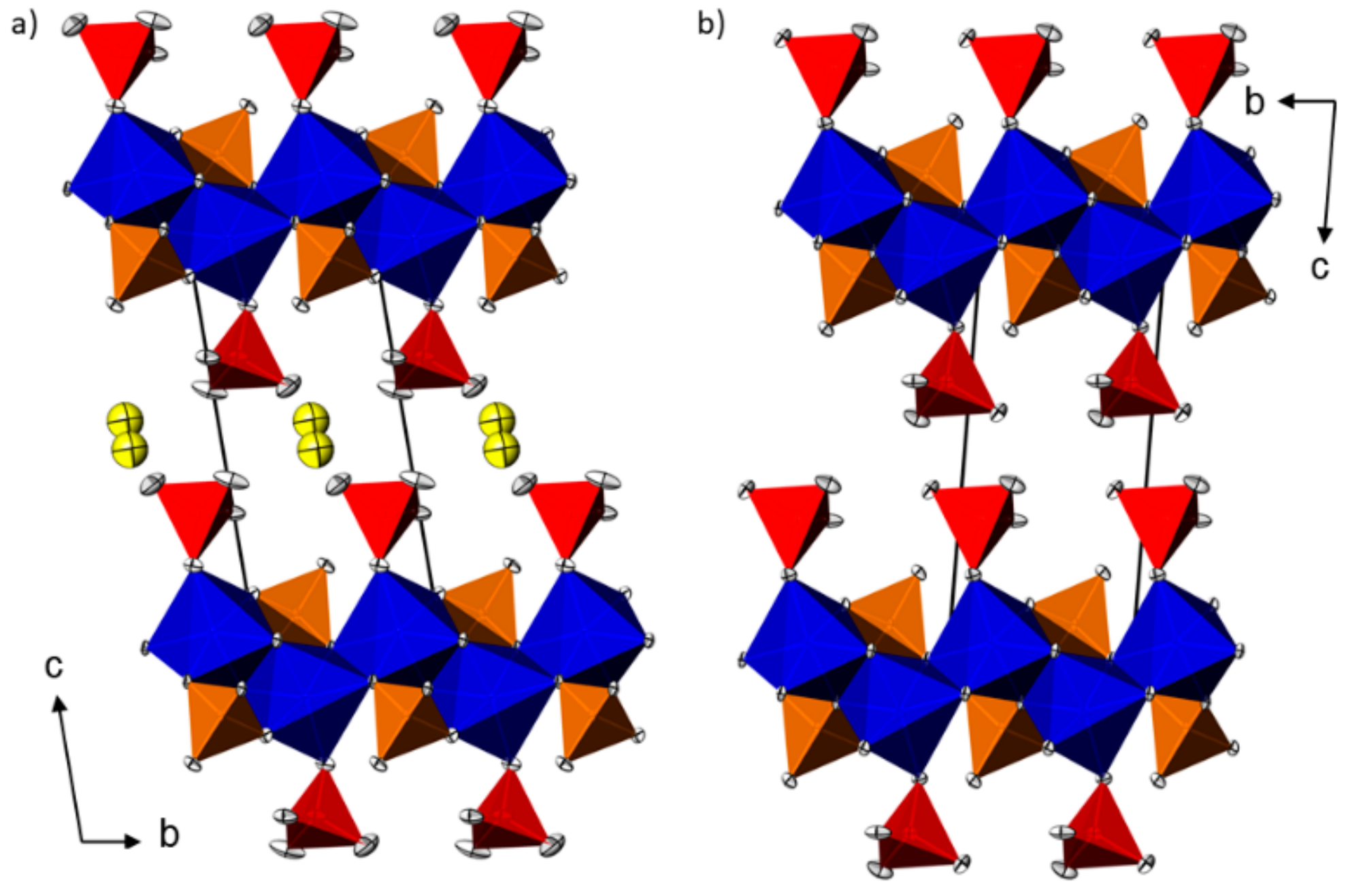
The two M(HAsO4)(H3AsO4)(H2O)0.5 (M = Mn, Cd) crystal structures are isotypic. All atoms in the asymmetric unit are located on general positions. The MII cations exhibit a considerably distorted octahedral coordination environment, with bond lengths between 2.14 and 2.23 Å in the Mn structure and between 2.24 and 2.32 Å in the Cd structure. In contrast to the M(H2AsO4)2(H3AsO4)2 structure type (M:As ratio = 4) where isolated MO6 octahedra are linked by bridging dihydrogenarsenate groups into chains, the MO6 octahedra in the M(HAsO4)(H3AsO4)(H2O)0.5 structure type (M:As ratio 1:2) share edges under formation of undulating centrosymmetric [ 1 MO4/2O2/1] chains running parallel (010). As expected, the Cd–O bonds are longer (on average about 0.09 Å) than the Mn–O bonds. As1O4 tetrahedra share two corners with the M cation within the [ 1 MO4/2O2/1] chain, and one corner with an M cation in an adjacent chain, thus bridging the chains into layers extending along (001). As2O4 tetrahedra share only one O atom with the chain and make up the outer boundary of the layers. A disordered water molecule is situated approximately at c/2 and links neighboring layers along (001) (Figure 2a).
The crystal structure of Zn(HAsO4)(H3AsO4) is closely related to that of the water-containing layered crystal structure type of M(HAsO4)(H3AsO4)(H2O)0.5. A relation between the crystal structures of M(HAsO4)(H3AsO4)(H2O)0.5 and Zn(HAsO4)(H3AsO4) is apparent from the similar topological arrangement of structure units (Figure 2) and similar lengths of the unit cell axes, with a ≈ 5.0, b ≈ 5.4, c ≈ 13.4 Å. However, the unit cell angles in the two types of structures differ considerably, with all three angles <90° for the Zn structure and all three angles >90° in the Mn and Cd structures. Since it is not possible to transform one of the unit cells into a setting that has comparable axes and angles (all >90° or all <90°) to the other unit cell, the given reduced cells (Table 3) with ab ≤ c and either with all angles >90° or <90° were used.
The general set-up within the (001) layers in the two crystal structures is the same. Since Zn has a smaller ionic radius in comparison with Mn and Cd [8], the Zn–O distances are the shortest in the [ 1 MO4/2O2/1] chains. The main difference between the crystal structures is related to the missing water molecule in Zn(HAsO4)(H3AsO4). Here adjacent layers are directly stacked along [001] (Figure 2b).

2.3. Hydrogen Bonding Schemes

In contrast to the M(H2AsO4)2(H3AsO4)2 crystal structures where the localization of all hydrogen atoms and thus interpretation of the hydrogen bonds were unproblematic (Table 2), a clear assignment of the hydrogen bonding scheme was not possible for the M(HAsO4)(H3AsO4)(H2O)0.5 and Zn(HAsO4)(H3AsO4) structures. Therefore, the bond valence method [9] was used to assign those O atoms in the three M(HAsO4)(H3AsO4)(H2O)0.5 and Zn(HAsO4)(H3AsO4) structures that most probably carry a hydrogen atom. For all MII–O bonds, the values of Brese and O’Keeffe [10] were applied, and for AsV–O bonds the values by Gangé and Hawthorne [11]. Results of the bond valence sum (BVS) calculations are listed in Table 1 and indicate the following atoms as the most probable donor groups, because they have the lowest bond valence sums (≤1.3 valence units) of all oxygen atoms: O1 bonded to As1, and O4, O6 and O8 bonded to As2. All other O atoms have considerably higher bond valence sums between 1.5 and 2.0 valence units.
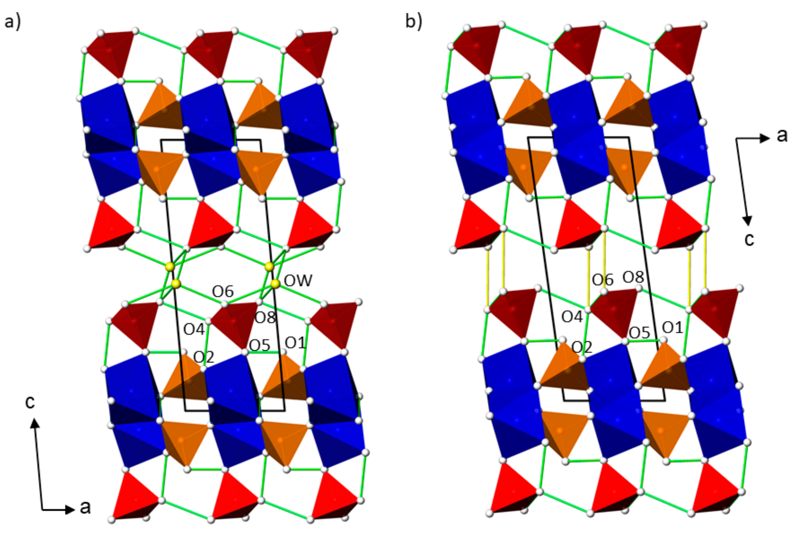
In analogy with the crystal-chemical features of M(H2AsO4)2(H3AsO4)2 where the OH groups of the AsO4 tetrahedra do not bond to the metal cations, all four assigned OH groups are bonded solely to arsenic as part of an AsO4 tetrahedron and also show the longest As–O bonds in the two types of tetrahedra. Therefore, As1 represents a HAsO4 anion and As2 the fully protonated acid, in accordance with charge neutrality of the overall structure. Analysis of the O···O distances around the four OH groups/(disordered) water molecules revealed hydrogen bonds with possible acceptor atoms listed in Table 2. This includes a very strong hydrogen bond between O4 and O2 with an O···O distance of ≈2.44 Å, and other strong hydrogen bonds between 2.60 and 2.65 Å for D···A contacts. The formation of these hydrogen bonds is also reflected in the BVS values of the acceptor O atoms that do not carry a hydrogen atom themselves. O2 and O5 are considerably undersaturated (BVS between 1.5 and 1.8 valence units) but are the acceptor atoms of the strongest hydrogen bonds (Table 2). On the other hand, oxygen atoms O3 and O7 are saturated with BVS values of 2.0 valence units and do not take part in any hydrogen bonding interaction. In case the water molecule is involved in hydrogen bonding, the hydrogen bonds become weaker (D···A: 2.60–2.92 Å). The weakest hydrogen bond is developed in Zn(HAsO4)(H3AsO4) between O6 and O4 (3.07 Å), connecting two adjacent layers long [001]. Supposed hydrogen bonds for the M(HAsO4)(H3AsO4)(H2O)0.5 and Zn(HAsO4)(H3AsO4) structures are illustrated in Figure 3. The various possibilities for hydrogen bonding in the two types of structures make it seem likely that parts of the hydrogen atoms are disordered and thus could not unambiguously be located in difference Fourier syntheses.

2.4. AsO4 Tetrahedra in the Sructures

The averaged As–O bond lengths for each AsO4 tetrahedron in the refined structures are collated in Table 1. Individual averaged values scatter only slightly (range 1.677 to 1.689 Å), and the overall mean of the 14 independent tetrahedral AsO4 groups in the seven structures is 1.685 Å, a value in very good agreement with that of 1.687 Å reported in literature [12]. Very recently, a statistical evaluation of As–O bond lengths in AsO43, HAsO42, H2AsO4 and H3AsO4 groups was published [13], revealing the following average values: 1.667(18) Å for As–O bonds to nonprotonated O atoms, 1.728(19) Å for As–OH bonds in HAsO42 groups, 1.714 (12) Å for As–OH bonds in H2AsO4 groups and 1.694(16) Å for As–OH bonds in H3AsO4 groups. These values are in very good agreement with corresponding values for averaged As–OH bond lengths in the four M(H2AsO4)2(H3AsO4)2 structures: M = Mg, 1.698 Å for the H3AsO4 group and 1.714 Å for the H2AsO4 group; M = Mn, 1.695 and 1.710 Å; M = Co, 1.698 and 1.713 Å; M = Ni, 1.701 and 1.713 Å. On the other hand, comparison of the averaged As–OH bond lengths [13] with those of the assigned HAsO42 and H3AsO4 groups in the structures of M(HAsO4)(H3AsO4)(H2O)0.5 and Zn(HAsO4)(H3AsO4) shows some subtle differences. Both literature values for As–OH in a HAsO42 (1.728(19) Å) and a H3AsO4 group (1.694(16) Å) are slightly larger than corresponding values in the three structures (Mn = 1.708, 1.688 Å; Cd = 1.714, 1.686 Å, Zn = 1.710, 1.686 Å). Nevertheless, individual values match the literature values within the single standard deviation of the latter. These slight differences might also indicate some disorder of the hydrogen atoms, as already suspected in Section 2.3.

2.5. Comparison of the Crystal Structures

For a quantitative comparison of the isotypic crystal structures within the M(H2XO4)2(H3XO4)2 series (M = Mg, Mn, Co, Ni, Cu; X = As, P) and between the two isotypic structures of M(HAsO4)(H3AsO4)(H2O)0.5 (M = Mn, Cd), respectively, the program compstru [7] available at the Bilbao Crystallographic Server [14] was used. Due to different treatments of H atoms in the various refinements, e.g., by constrains/restrains regarding O–H bond lengths, the comparisons do not include hydrogen atoms.
Zn(H2AsO4)2(H3AsO4)2 was chosen as a reference to which all structures in the M(H2XO4)2(H3XO4)2 series were compared. As can be seen from the numerical details of the comparisons compiled in Table 4, the crystal structures of Zn(H2AsO4)2(H3AsO4)2 and its Mg, Co, and Ni analogues show a very high similarity (Δ < 0.02) due to similar ionic radii of the four metal cations. Except for the Mg member for which data were recorded at −173 °C, all other crystals were measured at room temperature. The minor effect of the temperature on M–O and As–O bond lengths is neglected. Mn with its greater ionic radius leads to a somewhat larger difference (Δ = 0.033). Likewise, substitution of the arsenate groups by smaller phosphate tetrahedra (Co–P) affects the displacements between comparable atomic pairs. Although individual displacement values reach up to 0.17 Å for some O atoms, Δ amounts to only 0.037 and thus indicates a high similarity of the two structures. The most remarkable change, however, pertains to the Cu member (Δ = 0.112). Here the highest displacement is nearly 0.7 Å for atom O3 that represents the axially bound O atom in the MO6 octahedron. Due to the Jahn–Teller effect associated with the copper(II) cation, the CuO6 octahedron distorts under considerable elongation of the axial Cu–O3 bond.
The compstru program was also used to check the relationship between the M(H2XO4)2(H3XO4)2 structure type and the crystal structure of CdH10(AsO4)4. The latter crystallizes likewise in space group P−1 with one formula unit, has the metal cation situated on an inversion center (Wyckoff position 1a) and exhibits similar lattice parameters, a = 5.69(5), b = 7.42(4), c = 8.60(6) Å, α = 105.17(12), β = 95.13(5), γ = 91.85(8)° [4]. The evaluation of structural similarity revealed very large displacements between corresponding atomic pairs. For example, the two types of As atoms in the crystal structures have displacements of 1.71 and 1.97 Å, respectively, and for some oxygen atom displacements >2.0 Å were calculated. Hence the crystal structures of CdH10(AsO4)4 and M(H2XO4)2(H3XO4)2 cannot be considered as isotypic, as already suggested in [5], but can be regarded as isopointal [15].
Comparison of the two isotypic M(HAsO4)(H3AsO4)(H2O)0.5 structures again shows a high degree of similarity between them. Notable differences with displacements between 0.1 and 0.2 Å affect atom pairs O5 and O6 of the H3AsO4 tetrahedron, and the water molecule (Table 4).

3. Materials and Methods

3.1. Synthesis and Crystal Growth

The chemicals used (MnCO3, CdCO3, ZnO, Mg(OH)2, CoCO3·xH2O, NiCO3, H3AsO4) were of pro analysi quality. The target compounds were prepared by treating the corresponding metal carbonate, oxide or hydroxide with an excess of concentrated arsenic acid (80 wt %); the employed molar ratio H3AsO4:metal precursor was ≈10:1. The solid metal precursors were added in small portions to the acid and warmed (ca. 70 °C) until a clear solution was obtained overnight. In case the metal precursor dissolution was incomplete, the suspension was filtered through a G4 glass frit. All solutions were then left at room temperature in a desiccator filled with concentrated sulfuric acid until the first crystals appeared. The crystallization process lasted at least about one week; yields were not determined.
All grown crystals of the M(H2AsO4)2(H3AsO4)2 (M = Mg, Co, Ni) phases had a plate-like form and were directly taken out of the mother liquor. Cd(HAsO4)(H3AsO4)(H2O)0.5 crystallized in form of thin plates and was the first crystallisation product from the solution. After a few days, these crystals converted in the mother liquor into coarse-crystalline CdH10(AsO4)4 [4]. Likewise, Mn(HAsO4)(H3AsO4)(H2O)0.5 crystals were the first crystallization product and converted over the course of a few days into Mn(H2AsO4)2(H3AsO4)2. Zn(HAsO4)(H3AsO4) crystals appeared with an unspecific form together with synthetic koritnigite (ZnHAsO4·H2O [17]; platy crystals). The amount of the latter phase increased in the remaining mother liquor over time.

3.2. Single Crystal X-ray Diffraction

Since all crystals appeared to be highly hygroscopic, they were immediately immersed in perfluorinated oil (Fomblin® Y, Sigma-Aldrich, Taufkirchen, Germany) and optically preselected under a polarizing microscope. The diffraction studies (Mo Kα radiation; Bruker SMART CCD or APEX-II CCD diffractometer; Bruker-AXS Inc. (Madison, WI, USA) followed standard procedures, including absorption corrections based on a multi-scan approach [18,19]. All crystal structures were solved with SHELXT [20] and refined with SHELXL [21]. The diffraction pattern of Ni(H2AsO4)2(H3AsO4)2 revealed a two-domain crystal (arbitrarily intergrown). Intensity data of this crystal were assigned to the two different domains and further processed as a HKLF-5 file; the ratio of the two domains refined to a value of 0.57:0.43. For better comparison, atomic coordinates and atom labels of all M(H2AsO4)2(H3AsO4)2 (M = Mg, Mn, Co, Ni) structures were finally adapted to the structure data of Zn(H2AsO4)2(H3AsO4)2 [6]. Likewise, the two new M(HAsO4)(H3AsO4)(H2O)0.5 (M = Mn, Cd) structures are described with comparable atomic coordinates and atom labels. In this structure type, conspicuous electron density was observed around an inversion center after modelling the M(HAsO4)(H3AsO4) framework. This electron density was assigned to the O atom of a water molecule of crystallization, disordered around an inversion center. Because the symmetry-related water molecules cannot be occupied at the same time, the occupancy of the water molecule was constrained to 0.5. For the final model, this atom was refined with an isotropic displacement parameter. In the closely related structure of Zn(HAsO4)(H3AsO4), no such additional electron density was found.
For the M(H2AsO4)2(H3AsO4)2 (M = Mg, Mn, Co, Ni) crystal structures, all hydrogen atoms could clearly be located from difference Fourier syntheses. For refinement, their H–O distances were restrained to 0.90(1) Å, together with a common Uiso(H) parameter. For the crystal structures of M(HAsO4)(H3AsO4)(H2O)0.5 (M = Mn, Cd) and Zn(HAsO4)(H3AsO4), an unambiguous assignment of hydrogen atoms was not possible. Hence, their positions are not included in the final models. Graphical representations of the crystal structures were performed with ATOMS for Windows [22].
Details of numerical values of the data collections and structure refinements are gathered in Table 3 and Supplementary Materials. Further details of the crystal structure investigations may be obtained from The Cambridge Crystallographic Data Centre (CCDC) on quoting the depository numbers listed at the end of Table 3. The data can be obtained free of charge via www.ccdc.cam.ac.uk/structures.

4. Conclusions

The oxidoarsenates(V) M(H2AsO4)2(H3AsO4)2 (M = Mg, Mn, Co, Ni), M(HAsO4)(H3AsO4)(H2O)0.5 (M = Mn, Cd) and Zn(HAsO4)(H3AsO4) were crystallized from highly acidic solutions. The results of structure refinements of isotypic M(H2XO4)2(H3XO4)2 (M = Mg, Mn, Co, Ni; X = As) compounds from single crystal X-ray data supplement the knowledge on this structure type. Quantitative comparisons between all known crystal structures in this series with M = Mg, Mn, Co, Ni, Cu, Zn; X = As; P revealed a high similarity with the exception for the (Cu,As) member. The crystal structure of the latter shows considerable distortions due to the Jahn–Teller effect of Cu(II), leading to a displacement of up to 0.7 Å between related atomic pairs. It was also shown that the isoformular compound CdH10(AsO4)4 (= Cd(H2AsO4)2(H3AsO4)2) is not isotypic with the M(H2XO4)2(H3XO4)2 series, but isopointal. The crystal structures of the two isotypic M(HAsO4)(H3AsO4)(H2O)0.5 (M = Mn, Cd) compounds and of Zn(HAsO4)(H3AsO4) are closely related. They comprise of M(HAsO4)(H3AsO4) layers that are linked through hydrogen-bonded water molecules in the water-containing structures, or directly in the water-free structure. As–O distances in the two types of tetrahedra (HAsO42; H3AsO4) suggest disorder of the hydrogen atoms that could not be located in the present study.

Supplementary Materials

The following are available online at https://www.mdpi.com/2304-6740/7/10/122/s1, the CIFs and the checkCIF output files for M(H2AsO4)2(H3AsO4)2 (M = Mg, Mn, Co, Ni), M(HAsO4)(H3AsO4)(H2O)0.5 (M = Mn, Cd) and Zn(HAsO4)(H3AsO4).

Funding

This research received no external funding.

Acknowledgments

The X-ray centre of the TU Wien is acknowledged for providing access to the single-crystal and powder X-ray diffractometers.

Conflicts of Interest

The author declares no conflict of interest.

References

  1. Salmon, J.E.; Terrey, H. The systems zinc oxide–phosphoric oxide–water and manganous oxide–phosphoric oxide–water at temperatures between 25° and 100°. J. Chem. Soc. 1950, 2813–2824. [Google Scholar] [CrossRef]
  2. Herak, R.; Prelesnik, B.; Čurić, M.; Djurić, S. The crystal structure of Co(H2PO4)2·2H3PO4. Z. Kristallogr. 1983, 164, 25–30. [Google Scholar] [CrossRef]
  3. Averbuch-Pouchot, M.T.; Durif, A. Topics in Phosphate Chemistry; World Scientific: Singapore, 1996; p. 346. [Google Scholar]
  4. Boudjada, A.; Durif, A.; Guitel, J.C. Structure d’un orthoarsenate acide de cadmium: CdH10(AsO4)4. Acta Crystallogr. Sect. B 1980, B36, 133–135. [Google Scholar] [CrossRef]
  5. Tran Qui, D.; Chiadmi, M. Structure de Décahydrogénotétraarsenate de Cuivre et Isotypie dans la Série MIIH10(AsO4)4 (MII = Mg, Mn, Co, Ni, Cu et Zn). Acta Crystallogr. Sect. C 1986, C42, 391–393. [Google Scholar] [CrossRef]
  6. Sure, S.; Guse, W. The crystal structure of zinc-decahydrogen-tetraarsenate ZnH10(AsO4)4. Neues Jahrb. Mineral. Monatsh. 1989, 9, 401–409. [Google Scholar]
  7. De la Flor, G.; Orobengoa, D.; Tasci, E.; Perez-Mato, J.M.; Aroyo, M.I. Comparison of structures applying the tools available at the Bilbao Crystallographic Server. J. Appl. Crystallogr. 2016, 49, 653–664. [Google Scholar] [CrossRef]
  8. Shannon, R.D. Revised effective ionic radii and systematic studies of interatomic distances in halides and chalcogenides. Acta Crystallogr. Sect. A 1976, A32, 751–767. [Google Scholar] [CrossRef]
  9. Brown, I.D. The Chemical Bond in Inorganic Chemistry: The Bond Valence Model; Oxford University Press: Oxford, UK, 2002. [Google Scholar]
  10. Brese, N.E.; O’Keeffe, M. Bond-valence parameters for solids. Acta Crystallogr. Sect. B 1991, B47, 192–197. [Google Scholar] [CrossRef]
  11. Gagné, O.C.; Hawthorne, F.C. Comprehensive derivation of bond-valence parameters for ion pairs involving oxygen. Acta Crystallogr. Sect. B 2015, B71, 562–578. [Google Scholar] [CrossRef] [PubMed]
  12. Gagné, O.C.; Hawthorne, F.C. Bond-length distributions for ions bonded to oxygen: Metalloids and post-transition metals. Acta Crystallogr. Sect. B 2018, B74, 63–78. [Google Scholar] [CrossRef]
  13. Schwendtner, K.; Kolitsch, U. Three new acid M+ arsenates and phosphates with multiply protonated As/PO4 groups. Acta Crystallogr. Sect. C 2019, C75, 1134–1141. [Google Scholar] [CrossRef] [PubMed]
  14. Aroyo, M.I.; Perez-Mato, J.M.; Capillas, C.; Kroumova, E.; Ivantchev, S.; Madariaga, G.; Kirov, A.; Wondratschek, H. Bilbao Crystallographic Server I: Databases and crystallographic computing programs. Z. Kristallogr. 2006, 221, 15–27. [Google Scholar] [CrossRef]
  15. Lima-de-Faria, J.; Hellner, E.; Liebau, F.; Makovicky, E.; Parthé, E. Nomenclature of inorganic structure types. Report of the International Union of Crystallography Commission on Crystallographic Nomenclature Subcommittee on the Nomenclature of Inorganic Structure Types. Acta Crystallogr. Sect. A 1990, A46, 1–11. [Google Scholar] [CrossRef]
  16. Gelato, L.M.; Parthé, E. STRUCTURE TIDY—A computer program to standardize crystal structure data. J. Appl. Crystallogr. 1987, 20, 139–143. [Google Scholar] [CrossRef]
  17. Keller, P.; Hess, H.; Riffel, H. Die Kristallstruktur von Koritnigit, Zn[H2O|HOAsO3]. Neues Jahrb. Mineral. Abh. 1980, 138, 316–332. [Google Scholar]
  18. Krause, L.; Herbst-Irmer, R.; Sheldrick, G.M.; Stalke, D. Comparison of silver and molybdenum microfocus X-ray sources for single-crystal structure determination. J. Appl. Crystallogr. 2015, 48, 3–10. [Google Scholar] [CrossRef] [PubMed] [Green Version]
  19. Sheldrick, G.M. TWINABS; University of Göttingen: Göttingen, Germany, 2012. [Google Scholar]
  20. Sheldrick, G.M. SHELXT—Integrated space-group and crystal-structure determination. Acta Crystallogr. Sect. A 2015, A71, 3–8. [Google Scholar] [CrossRef] [PubMed]
  21. Sheldrick, G.M. Crystal structure refinement with SHELXL. Acta Crystallogr. Sect. C 2015, C71, 3–8. [Google Scholar]
  22. Dowty, E. Atoms for Windows; Version 6.3; Shape Software: Kingsport, TN, USA, 2006. [Google Scholar]
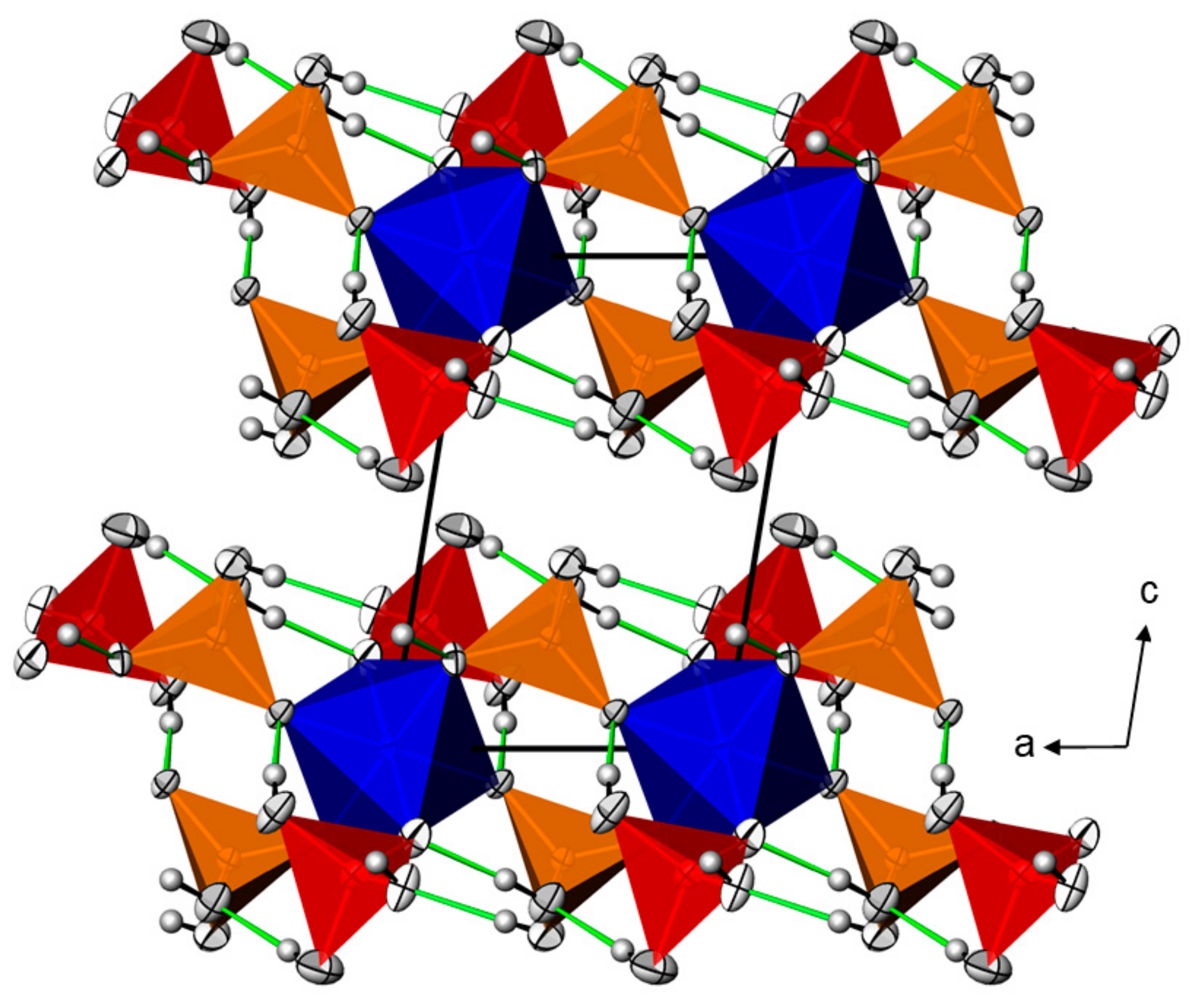
Figure 1. Representative for the M(H2XO4)2(H3XO4)2 series, the crystal structure of Mn(H2AsO4)2(H3AsO4)2 is given in a projection along (010). Displacement ellipsoids are drawn at the 90% probability level. H2AsO4 tetrahedra (As2) are orange, H3AsO4 tetrahedra (As1) are red, MnO6 octahedra are blue; H atoms are displayed as grey spheres of arbitrary radius. O···H hydrogen bonds are shown as green lines.
Inorganics 07 00122 g001
Figure 2. (a) Representative for the two M(HAsO4)(H3AsO4)(H2O)0.5 (M = Mn, Cd) compounds, the crystal structure of Mn(H2AsO4)2(H3AsO4)2 is given in a projection along (100); (b) the crystal structure of Zn(HAsO4)(H3AsO4) in a projection along (100). Displacement ellipsoids are drawn at the 50% probability level. HAsO42 tetrahedra (As1) are orange, H3AsO4 tetrahedra (As2) are red, and MO6 octahedra are blue. Water molecules are yellow, with both possible positions (occupancy 0.5) shown.
Inorganics 07 00122 g002
Figure 3. (a) Possible hydrogen bonding schemes in the crystal structures of Mn(HAsO4)(H3AsO4)(H2O)0.5 ((a); projection along [010]) and Zn(HAsO4)(H3AsO4) ((b); projection along [010]). Hydrogen bonds with D···A contacts < 3.0 Å are given as green lines, and those > 3.0 Å as yellow lines. Color code as in Figure 2.
Inorganics 07 00122 g003
Table 1. Selected bond lengths/Å, angles/° with estimated standard deviations in parentheses, and bond valence parameters (BVS)/valence units. Averaged values (av.) are given in the last line of each column.
Mn(HAsO4)(H3AsO4)(H2O)0.5 Cd(HAsO4)(H3AsO4)(H2O)0.5 Zn(HAsO4)(H3AsO4)
Mn1O32.149(2) Cd1O32.242(3) Zn1O32.031(3)
O52.162(3) O52.253(4) O72.048(3)
O72.186(3) O72.273(3) O22.123(4)
O72.202(3) O72.307(3) O52.127(4)
O22.229(3) O22.307(3) O72.180(4)
O32.229(3) O32.314(3) O32.204(4)
av.2.19 av.2.28 av.2.12
As1O71.666(3) As1O71.663(3) As1O71.669(3)
O31.673(3) O31.672(3) O31.673(3)
O21.698(3) O21.696(3) O21.690(4)
O11.708(3) O11.714(4) O11.710(4)
av.1.686 av.1.686 av.1.689
As2O51.654(3) As2O51.649(4) As2O51.668(4)
O41.666(3) O41.670(4) O41.673(4)
O61.692(5) O61.686(7) O81.687(4)
O81.705(5) O81.702(6) O61.698(5)
av.1.680 av.1.677 av.1.682
BVS
Mn1 2.03, As1 5.01, As2 5.11, O1 1.18, O2 1.52, O3 1.98, O4 1.33, O5 1.73, O6 1.23, O7 2.00, O8 1.19. Cd1 2.16, As1 5.01, As2 5.15, O1 1.16, O2 1.55, O3 2.03, O4 1.31, O5 1.78, O6 1.25, O7 2.04, O8 1.20. Zn1 1.98, As1 5.02, As2 5.07, O1 1.17, O2 1.56, O3 1.97 O4 1.30, O5 1.64, O6 1.21, O7 1.99, O8 1.25.
Mg(H2AsO4)2(H3AsO4)2 Mn(H2AsO4)2(H3AsO4)2 Co(H2AsO4)2(H3AsO4)2
Mg1O72.0680(13)Mn1O72.1629(9)Co1O72.050(3)
O52.0853(14) O52.1707(9) O52.146(3)
O32.1205(14) O32.2120(11) O32.146(4)
av.2.09 av.2.18 av.2.11
As1O31.6484(14) As1O31.6458(10) As1O31.650(3)
O11.6852(14) O11.6831(10) O11.687(3)
O41.6983(16) O41.6983(11) O41.696(3)
O21.7096(15) O21.7037(12) O21.711(4)
av.1.685 av.1.683 av.1.686
As2O51.6562(15) As2O51.6552(9) As2O51.663(3)
O71.6579(14) O71.6561(9) O71.665(3)
O61.7119(14) O61.7076(11) O61.709(4)
O81.7161(15) O81.7131(11) O81.717(4)
av.1.686 av.1.683 av.1.689
Ni(H2AsO4)2(H3AsO4)2
Ni1O72.037(3)
O52.097(3)
O32.117(3)
av.2.08
As1O31.644(3)
O11.694(3)
O41.696(3)
O21.712(3)
av.1.689
As2O51.656(3)
O71.664(3)
O61.713(3)
O81.713(3)
av.1.687
Table 2. Details of the hydrogen bonding geometry/Å, °.
DHAD–H H⋯ADAD–H⋯A
Mg(H2AsO4)2(H3AsO4)2
O2H1O80.890(10)1.918(18)2.769(2)160(4)
O1H2O70.898(10)1.620(11)2.517(2)174(4)
O4H3O50.895(10)1.758(14)2.634(2)167(4)
O6H4O10.884(10)1.96(2)2.760(2)149(4)
O8H5O30.893(10)1.686(11)2.577(2)174(4)
Mn(H2AsO4)2(H3AsO4)2
O2H1O80.890(10)1.921(11)2.8018(16)170(3)
O1H2O70.898(10)1.654(13)2.5358(14)167(4)
O4H3O50.895(10)1.759(12)2.6452(15)170(3)
O6H4O10.884(10)1.932(12)2.7968(14)166(3)
O8H5O30.893(10)1.723(13)2.6007(14)167(4)
Co(H2AsO4)2(H3AsO4)2
O2H1O80.898(10)1.94(3)2.812(5)163(8)
O1H2O70.901(10)1.69(3)2.564(5)162(8)
O4H3O50.898(10)1.75(3)2.616(5)162(7)
O6H4O10.896(10)1.94(3)2.799(4)159(7)
O8H5O30.899(10)1.71(2)2.592(5)167(8)
Ni(H2AsO4)2(H3AsO4)2
O2H1O80.897(10)1.92(3)2.762(4)155(6)
O1H2O70.898(10)1.629(17)2.516(4)169(6)
O4H3O50.899(10)1.75(3)2.610(4)159(6)
O6H4O10.897(10)1.90(2)2.755(4)159(6)
O8H5O30.900(10)1.658(14)2.553(4)172(7)
Mn(HAsO4)(H3AsO4)(H2O)0.5
O1 O5 2.635(4)
O4 O2 2.458(5)
O8 O4 2.633(6)
O6 OW 2.61(2)
O8 OW 2.64(2)
OW O8 2.90(2)
OW O6 2.92(2)
Cd(HAsO4)(H3AsO4)(H2O)0.5
O1 O5 2.625(5)
O4 O2 2.470(5)
O8 O4 2.670(7)
O6 OW 2.75(4)
O8 OW 2.60(4)
OW O6 2.84(4)
OW O8 2.94(4)
Zn(HAsO4)(H3AsO4)
O1 O5 2.610(6)
O4 O2 2.429(6)
O8 O4 2.666(6)
O6 O4 2.962(7)
O6 O4 3.071(7)
Table 3. Details of X-ray data collections and crystal structure refinements.
CompoundMn(HAsO4)-(H3AsO4)(H2O)0.5Cd(HAsO4)(H3AsO4)-(H2O)0.5Zn(HAsO4)-(H3AsO4)Mg(H2AsO4)2-(H3AsO4)2Mn(H2AsO4)2-(H3AsO4)2Co(H2AsO4)2-(H3AsO4)2Ni(H2AsO4)2-(H3AsO4)2
MR345.82403.28347.24590.07620.70624.69624.47
Temp./°C232323–173232323
Radiation; λ— Mo Kα; 0.71073 —
DiffractometerAPEXII CCD SMART CCD APEXII CCD APEXII CCDAPEXII CCD APEXII CCD APEXII CCD
Crystal size/mm30.48 × 0.18 × 0.020.20 × 0.15 × 0.020.12 × 0.12 × 0.010.10 × 0.06 × 0.010.12 × 0.09 × 0.020.12 × 0.06 × 0.010.09 × 0.06 × 0.04
Crystal color; formlight-pink; platecolorless; platecolorless; fragmentcolorless; platelight-pink; plateviolet; plateyellow; plate
Space group P1P1P1P1P1P1P1
Formula units, Z2221111
a4.9750(10)5.0188(9)4.9187(3)5.4558(3)5.5602(2)5.495(3)5.4297(7)
b5.4747(11)5.6180(10)5.2357(3)7.3180(4)7.4100(3)7.394(4)7.3308(9)
c13.603(3)13.734(2)12.8459(8)8.3382(5)8.4276(4)8.330(5)8.2795(10)
α98.86(3)99.254(3)83.987(3)100.231(2)100.110(2)100.604(15)100.356(5)
β93.63(3)93.756(3)81.286(3)98.614(2)98.578(2)97.550(14)98.088(5)
γ99.09(3)98.845(3)80.117(3)93.022(2)92.744(2)92.858(12)92.982(5)
V3360.02(13)376.02(12)321.09(3)322.84(3)337.03(2)328.8(3)319.95(7)
μ/mm−110.96611.64814.05410.39910.80811.37911.869
X-ray Dens./g·cm−33.1903.5623.5923.0353.0583.155 3.241
Range θminθmax3.87–30.003.02–31.063.96–30.983.79–41.892.80–41.522.81–31.002.53–36.71
Range h−6→6−7→7−7→6−10→10−10→10−7→7−9→8
k−7→3−7→8−7→7−13→13−13→14−10→10−12→12
l−19→19−19→19−18→18−15→15−15→15−12→110→13
Meas. refl.467744307650199861784145823000
Indep. refl.2087232620114436484320573000
Obs.refl. [I > 2σ(I)]1831199616003262376715682566
Ri0.02870. 02870.04610.06560.02850.0364-
Abs. corr.SADABSSADABSSADABSSADABSSADABSSADABSTWINABS
Trans. coef. Tmin; Tmax0.526; 0.7480. 443; 0.6630.480; 0.7470.531; 0.7480.479; 0.7480.592; 0.7470.257; 0.439
Number of parameters104104100113113113114
R[F2 > 2σ(F2)]0.03320.03500.03800.03530.02640.03800.0387
wR2(F2 all)0.10530.09500.09940.07120.05380.08500.0880
Goof1.13241.0411.0321.0191.0101.0371.082
CSD number1951017195101319510191951015195101619510141951018
Table 4. Numerical details from the comparisons of the crystal structure of Zn(H2AsO4)2(H3AsO4)2 a with isotypic structures in the M(H2XO4)2(H3XO4)2 series (M = Mg, Mn, Co, Ni, Cu; X = As, P), and between Mn(HAsO4)(H3AsO4)(H2O)0.5 and Cd(HAsO4)(H3AsO4)(H2O)0.5 using the compstru program [7].
Zn(H2AsO4)2(H3AsO4)2 versus M(H2AsO4)2(H3AsO4)2
M1MgMn(Co,P) b(Co,As)NiCu c
Atom, atomic displacement |u|/Å
Zn1/M1000000
As1/As1(P1)0.01110.02890.07460.00770.02000.3198
As2/As2(P2)0.01760.04340.08520.00410.01220.1569
O10.02420.07370.16460.01340.03940.2641
O20.02410.03050.16410.01650.02850.3981
O30.02950.05000.11570.02720.05080.6942
O40.02530.03530.08890.02060.01840.2552
O50.03860.04970.14970.01850.01290.0974
O60.02300.03840.06690.01000.03370.2862
O70.00610.08070.16120.00510.02660.3203
O80.02950.01650.16160.02240.03080.3185
degree of lattice distortion (S) 0.00490.00780.01760.00300.00460.0243
arithmetic mean (dav)0.02180.04260.11740.01390.02600.2963
measure of similarity (Δ)0.0140.0330.0370.0170.0110.112
Mn(HAsO4)(H3AsO4)(H2O)0.5versus Cd(HAsO4)(H3AsO4)(H2O)0.5
Atom, atomic displacement |u|/Å
Mn1/Cd10.0576
As10.0523
As20.0816
O10.0540
O20.0835
O30.0492
O40.0645
O50.1206
O60.1899
O70.0747
O80.0497
OW0.1435
degree of lattice distortion (S) 0.0095
arithmetic mean (dav)0.0851
measure of similarity (Δ)0.037
a Lattice parameters: a = 5.460(2), b = Å 7.389(1), c = 8.347(1) Å, α =100.83(4), β = 97.90(4), γ = 92.89(3)°. b Standardized [16] lattice parameters: a = 5.286(2), b = 7.195(4), c = 8.121(4) Å, α = 101.10(4), β = 96.72(3), γ = 92.97(3)°. c Lattice parameters: a = 5.392(3) Å, b = 7.632(4) Å, c = 8.298(5) Å, α = 105.87(4)°, β = 97.63(3)°, γ = 93.6(1)°.