1. Introduction
Two-dimensional (2D) van der Waals (vdW) ferromagnets have emerged as a promising platform for spintronics due to their intrinsic long-range magnetic order persisting down to the monolayer limit, high Curie temperatures, and tunable magnetic properties [
1,
2,
3,
4,
5,
6,
7]. Among these materials, Fe
3GaTe
2 stands out for its robust room-temperature ferromagnetism (
TC > 300 K), strong perpendicular magnetic anisotropy (PMA), and metallic conductivity, making it attractive for next-generation spintronic devices [
5,
8]. Thickness-dependent magnetic behavior is a hallmark of 2D vdW magnets, where reduced dimensionality can significantly alter exchange interactions, magnetic anisotropy, and domain configurations [
9,
10,
11,
12]. However, systematic studies of hysteresis loops and magnetic domain structures in Fe
3GaTe
2 films remain limited, hindering a comprehensive understanding of dimensional confinement effects.
Molecular beam epitaxy (MBE) enables precise layer-by-layer growth of high-quality 2D vdW films with atomic-scale thickness control, as demonstrated in related systems such as Fe
3GeTe
2 and Fe
5GeTe
2 [
6,
13,
14,
15,
16,
17]. Combined with magneto-optical Kerr effect (MOKE) microscopy, which offers high spatial resolution for imaging magnetic domains and measuring hysteresis loops [
4,
5,
18,
19], MBE-grown films provide an ideal platform to probe thickness-dependent magnetism. Unlike Lorentz Transmission Electron Microscopy (LTEM), which loses sensitivity in the ultrathin limit, MOKE microscopy remains highly effective for probing the monolayer regime. In this work, we report the MBE growth of Fe
3GaTe
2 films from one to four unit cells (u.c.) and elucidate the critical influence of dimensional confinement on their magnetic properties.
2. Results
To evaluate the crystalline quality and verify the layer stacking sequence of the MBE-grown samples, detailed structural characterization was performed.
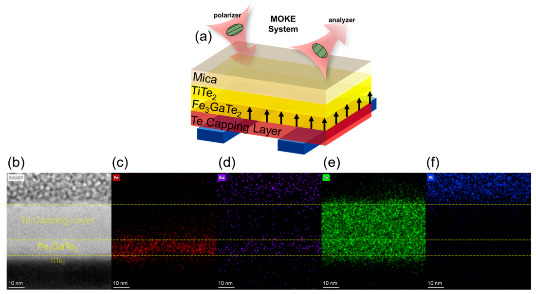
Figure 1a illustrates the schematic design of the heterostructure, which comprises a mica substrate, the Fe
3GaTe
2 magnetic layer, and a non-magnetic Te capping layer to prevent oxidation. The polar MOKE geometry is also depicted, where the incident light interacts with the perpendicular magnetization (black arrows). In our measurements, the MOKE signal was collected in a backside polar configuration through the transparent F-mica substrate. The F-mica substrate is non-magnetic and therefore does not generate a hysteretic Kerr contribution. Its possible optical influence is mainly limited to a field-independent background and a reduction in reflected intensity, which do not affect the extracted coercive fields or the comparison of domain evolution among films with different thicknesses.
The microscopic structure was visualized using cross-sectional HAADF-STEM, as shown in
Figure 1b. Detailed contrast analysis reveals a sharply defined layered structure with atomically flat interfaces between the Fe
3GaTe
2 layer and the adjacent Te layers. No obvious interdiffusion or threading dislocations are observed, indicating high structural integrity arising from the precise control of MBE growth parameters.
To further confirm the chemical composition and spatial distribution of the elements, energy-dispersive X-ray spectroscopy (EDS) mapping was conducted on the same cross-section. As presented in
Figure 1c–e, the elemental signals for Fe, Ga, and Te are uniform and coincide perfectly with the layered structure observed in the STEM image. Specifically, Fe (
Figure 1c) and Ga (
Figure 1d) are strictly confined to the intermediate Fe
3GaTe
2 layer, while Te (
Figure 1e) is distributed across the active and capping layers, consistent with the designed nominal structure. The platinum (Pt) signal observed in
Figure 1f originates from the protective layer deposited during the focused ion beam (FIB) sample preparation process and is not part of the device structure. These structural and elemental analyses support the formation of high-quality epitaxial Fe
3GaTe
2 films with composition close to the nominal Fe
3GaTe
2 stoichiometry. The assignment of the Fe
3GaTe
2 phase is supported by the calibrated Fe:Ga:Te flux ratio during MBE growth, in situ RHEED evolution indicating layer-by-layer epitaxy, the designed unit cell-controlled thickness, and the HAADF-STEM-resolved layered structure [
6]. EDS mapping is used here to confirm elemental co-localization rather than to determine crystallographic phase. Importantly, within the examined STEM field of view, no extended structural defects are directly resolved. However, localized growth-related variations such as atomic steps, point defects, local strain fluctuations, or nanoscale compositional variations may exist below the spatial resolution or outside the sampling volume of the present measurement.
To investigate the magnetic properties at the 2D limit, systematic MOKE measurements were performed on the ultrathin (1 u.c.) Fe
3GaTe
2 film. For the 1 u.c. sample [
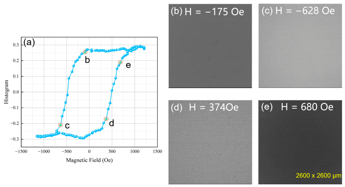
Figure 2a], the polar MOKE hysteresis loop exhibits a finite coercivity and a slightly slanted magnetic transition, providing evidence of room-temperature ferromagnetism with perpendicular magnetic anisotropy (PMA) in the monolayer limit. The magnetization reversal mechanism was further elucidated by directly imaging the domain evolution, as shown in
Figure 2b–e. Upon sweeping the magnetic field from negative saturation (approximately −1200 Oe) to positive saturation (approximately 1200 Oe), the MOKE contrast across the entire field of view changes uniformly. No localized domain nucleation or domain wall propagation was observed within the optical resolution limit. This near-single-domain-like reversal switching behavior, coupled with the slightly sheared loop, suggests that the ultrathin film functions as a near-single-domain system where reversal occurs via continuous rotation, likely because strong exchange interactions dominate over magnetostatic energy at the 2D limit.
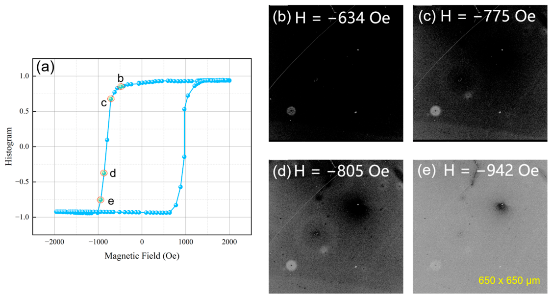
As the film thickness increases to 2 u.c., a distinct magnetic evolution is observed. In sharp contrast to the monolayer, the hysteresis loop of the 2 u.c. film [
Figure 3a] exhibits a remarkably perfect square shape with a highly abrupt magnetic transition and a significantly enhanced coercivity (~1000 Oe). This increase in coercivity with thickness suggests an enhancement of domain wall pinning effects, likely induced by the increased interaction volume or subtle structural defects in thicker films.
The domain imaging in
Figure 3b–e reveals a strikingly different reversal mechanism compared to the monolayer sample. Instead of near-single-domain-like switching, the reversal process is governed by the nucleation and growth of complex magnetic domains. Specifically, at intermediate fields (e.g.,
Figure 3b,c), distinct circular features with white contrast emerge against the dark background. These are identified as magnetic bubble domains. As the field increases, these bubbles expand and eventually merge to complete the magnetization reversal (
Figure 3e).
To gain a dynamic understanding of this reversal mechanism, we tracked the step-by-step domain evolution corresponding to the hysteresis loop, as detailed in
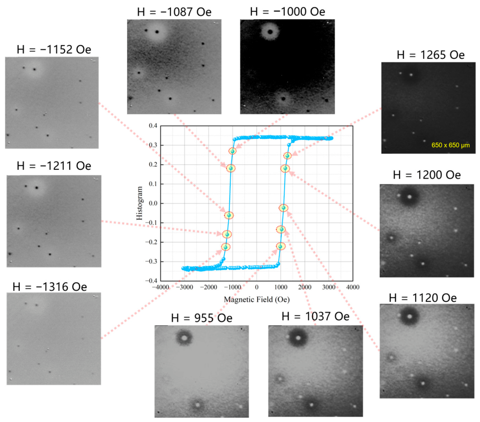
Figure 4. The central panel displays the macroscopic M-H loop for the 4 u.c. film, while the surrounding MOKE images capture the microscopic domain states at specific magnetic fields.
Starting from the positive saturation state and sweeping towards negative fields (left panels), the film initially maintains a uniform gray contrast. As the opposing field reaches −1152 Oe, sporadic dark spots begin to emerge. These are the nucleation centers of the “down” magnetic domains. As the field strength further increases to −1087 Oe and −1000 Oe, these dark nuclei rapidly expand into distinct, circular magnetic bubble domains with diameters ranging from several hundred nanometers to the micrometer scale. The circular symmetry of these bubbles indicates a highly isotropic domain wall energy in the film plane, a hallmark of high-quality systems with perpendicular magnetic anisotropy.
Conversely, during the sweep from negative back to positive saturation (right panels), a symmetric reversal process is observed. At H = 955 Oe, bright circular domains (representing “up” magnetization) nucleate within the dark background. As the field increases to 1037 Oe and 1120 Oe, these bright bubbles proliferate and expand. Notably, the positions where bubbles first nucleate appear to be spatially pinned, suggesting that the reversal is triggered by local inhomogeneities or lattice defects acting as preferential nucleation sites.
This specific “nucleation and growth” mode explains the increased coercivity observed in the 4 u.c. films compared to the 1 u.c. limit. Unlike the near-single-domain-like rotation seen in the monolayer, the reversal here requires overcoming the energy barrier for domain wall depinning and propagation. The formation of these stable bubble domains also hints at the competition between the exchange interaction (favoring uniform alignment) and the dipolar interaction (favoring domain fragmentation) as the film thickness increases.
To quantify the thickness-dependent magnetic evolution, key parameters extracted from room-temperature MOKE loops are summarized in
Table 1.
HC is obtained from linear interpolation of the zero-crossing point, and
Mr/
Ms is defined as the zero-field normalized Kerr signal.
3. Discussion
The significant differences in both magnetic hysteresis and domain evolution among Fe
3GaTe
2 samples of varying thicknesses are primarily attributed to the thickness-dependent modulation of magnetic anisotropy. According to magnetic theory, magnetic anisotropy is a key factor determining magnetization behavior, and its magnitude is closely related to dimensionality, crystal structure, and internal stress [
2]. In this study, as the thickness of Fe
3GaTe
2 increases from 1 u.c. to 4 u.c., the coercivity exhibits a notable increase. This phenomenological trend is opposite to that typically observed in mechanically exfoliated vdW magnets, where out-of-plane coercivity is often largest at the strict 2D limit and decreases drastically in thicker, bulk-like crystals due to the unhindered propagation of complex magnetic domains [
6]. Mechanically exfoliated Fe
3GaTe
2 flakes obtained from bulk crystals can retain high intrinsic crystalline quality and therefore provide an important reference for fundamental studies. However, the final thickness and lateral size of exfoliated flakes are governed by the stochastic cleavage process rather than by direct experimental design. In practice, suitable flakes must be obtained through repeated exfoliation and subsequently identified by optical microscopy and AFM thickness measurements. This trial-and-error procedure is inefficient, particularly when targeting a specific unit cell thickness or approaching the monolayer limit, and is less suitable for systematic thickness-dependent studies or scalable device fabrication. In contrast, the MBE approach used here allows the Fe
3GaTe
2 thickness to be designed and controlled through calibrated deposition rates and growth time, enabling reproducible preparation of 1, 2, and 4 u.c. continuous films. Together with in situ RHEED monitoring, the sharp interfaces and homogeneous Fe/Ga/Te distributions observed by HAADF-STEM and EDS confirm the high structural and chemical quality of the MBE-grown films. Thus, while mechanical exfoliation remains valuable for probing intrinsic properties, MBE offers a more controllable and scalable pathway toward thickness-engineered Fe
3GaTe
22 films for spintronic applications.
Additionally, the interplay between thermodynamic phase transitions, magnetostatic energy, and defect-induced pinning must be carefully evaluated. In the ultrathin limit (1 u.c.), strong perpendicular magnetic anisotropy restricts the system to a single-domain state. However, because the monolayer is closer to its
, severe thermal fluctuations at room temperature significantly soften its magnetic order. This thermal softening allows the macroscopic magnetization to reverse via a continuous, near-single-domain-like rotation without overcoming a large domain wall nucleation barrier, thereby resulting in a relatively small coercive field [
2]. According to Ref. [
6], the Curie temperatures of epitaxial Fe
3GaTe
2 films increase with thickness, with
values of approximately 345 K, 376 K, and 388 K for 1 u.c., 2 u.c., and 4 u.c. films, respectively, and up to approximately 420 K for thicker 9 u.c. films. Thus, at room temperature, the 1 u.c. film is much closer to its
than the thicker films. The reduced distance from
enhances thermal fluctuations and weakens the effective magnetic order parameter, which naturally accounts for the more slanted hysteresis loop and the softer, near-single-domain-like reversal observed in the monolayer film. By contrast, the 2 u.c. and 4 u.c. films are further below their
, allowing more robust perpendicular magnetic order and a sharper switching behavior.
Nevertheless, it is worth noting that the specific concentration and spatial distribution of Fe ions within the Fe
3GaTe
2 lattice may also influence the hysteresis loops. This factor warrants further investigation combined with more precise quantitative experimental data. This observation aligns with previous studies on other 2D magnetic materials, such as Cr
2Ge
2Te
6, suggesting that thickness-dependent magnetic anisotropy is a universal characteristic of 2D magnetic systems [
4].
While this dimensional domain evolution (from single to multi-domain) is fundamentally similar to the magnetostatic energy competition model widely recognized in exfoliated vdW nanosheets, the distinct coercivity enhancement trend stems directly from the specific kinetics of MBE growth. Unlike nearly defect-free single crystals obtained by mechanical exfoliation, the layer-by-layer MBE growth of thicker continuous films inevitably accumulates slight interfacial strains, atomic step edges, and point defects (as suggested by the spatially pinned domain nucleation observed in MOKE imaging [
10]). The cross-sectional STEM results do not reveal extended structural defects, suggesting that the films are structurally highly uniform at the probed scale. Therefore, the pinning centers inferred from magnetization reversal are not attributed to directly observed defects. Instead, they are likely associated with localized inhomogeneities indirectly evidenced by MOKE imaging, where bubble-domain nucleation occurs at reproducible spatial positions. Such pinning sites may originate from subtle growth-induced variations, including atomic steps, point defects, or local strain fluctuations.
These localized structural inhomogeneities act as highly effective pinning centers for domain walls. During the reversal process of the 2–4 u.c. films, once the magnetic bubble domains nucleate to minimize the long-range dipolar energy, their domain walls immediately encounter these deep pinning potentials. Consequently, a significantly larger external magnetic field is required to depin the walls and drive the subsequent abrupt domain expansion (as evidenced by our dynamic MOKE measurements). This powerful defect-induced pinning mechanism dominates the magnetic reversal dynamics in thicker MBE-grown layers, perfectly resolving the paradox of why the formation of complex multi-domain structures is accompanied by an anomalously enhanced coercive field compared to the thermally softened monolayer limit. Based on the calibrated optical scale, the optically resolved bubble-like domains typically have diameters on the order of approximately 0.5–3 μm, depending on the applied field and the stage of reversal. The smallest resolved domains are close to the optical resolution limit, whereas larger bubbles expand to the micrometer scale before merging during magnetization reversal.
4. Materials and Methods
The Fe
3GaTe
2 thin films were grown on fluorophlogopite-mica (F-mica) substrates using an Omicron molecular beam epitaxy (MBE) system in an ultra-high-vacuum environment. After standard cleaning and in situ annealing at 430 °C for 30 min, the substrates were cooled to a growth temperature of 380 °C. Ga (99.999%) and Te (99.999%) were deposited via standard Knudsen cells, while Fe (99.99%) was evaporated using an electron-beam evaporator. To precisely control the stoichiometry (Fe:Ga:Te = 3:1:2), the Fe deposition rate was calibrated to 0.3 Å/min, with Ga and Te fluxes adjusted accordingly. Under these conditions, the deposition time for a single unit cell (1 u.c., ~8.1 Å) was 27 min. Samples of 2 u.c. and 4 u.c. thicknesses were obtained by extending the deposition time to 54 and 108 min, respectively. In situ reflection high-energy electron diffraction (RHEED) was employed to monitor the layer-by-layer growth, and a protective bilayer of 2 monolayers of GaTe and 20 nm Te was capped on all samples to prevent ambient degradation prior to ex situ measurements [
6].
The crystalline quality and interfacial structure of the 4 u.c. sample were characterized via high-angle annular dark-field scanning transmission electron microscopy (HAADF-STEM) using a 200 kV transmission electron microscope (FEI Talos F200S). Cross-sectional lamellas (~100 nm thick) were prepared using a focused ion beam (FIB, FEI SCIOS2) system with a protective Pt capping layer. Energy-dispersive X-ray spectroscopy (EDS) mapping was performed [
5,
6,
10]. Both Talos F200S (S/TEM) and Scios 2 (DualBeam FIB-SEM) instruments are designed, engineered and manufactured at Thermo Fisher’s FEI facilities split between Hillsboro (OR, USA, core electronics & ion column R&D) and Eindhoven (The Netherlands, full system assembly, electron optics and final testing).
Magnetic properties and domain structures were investigated at room temperature using a custom-built magneto-optical Kerr effect (MOKE) microscope (TuoTuo TTT-02 Kerr Microscope G4.). The MOKE measurements were performed using a polar configuration with a light source wavelength of 635 nm. The optical system provides an estimated spatial resolution of approximately 500 nm. In hysteresis-loop measurements, the magnetic field was swept with a step size defined as the field increment between successive data points (field step: as specified in each loop measurement). The effective acquisition rate corresponds to a scanning time of 300 ms per data point, and each image was recorded with an exposure time of 100 ms. All MOKE measurements were performed in a backside polar geometry through the transparent F-mica substrate. The substrate is non-magnetic and does not contribute a hysteretic Kerr signal. Its influence is limited to a field-independent optical background and reduced reflected intensity, which do not affect the extracted coercive fields or the thickness-dependent domain evolution. Hysteresis loops were obtained by sweeping an external out-of-plane magnetic field (up to ± 10 kOe) while recording the spatially averaged Kerr rotation [
9]. For domain imaging, the samples were measured under specific localized applied fields to capture the microstate evolution [
8]. To eliminate dynamic optical noise, signals were enhanced through pixel-by-pixel background subtraction and multiple averaging routines.




 ,
,