Article

A Biohybrid Catalyst for Cross-Coupling Reactions That Contains Pd/P.yeei@ORMOSIL

by
Olga A. Kamanina
1,*,
Vitaliy N. Soromotin
1,
Pavel V. Rybochkin
1,
Nina M. Ivanova
2,
Anton N. Zvonarev
3 and
Vasilina V. Farofonova
4
1
Research and Development Department, Tula State University, Pr. Lenina 92, Tula 300012, Russia
2
Zelinsky Institute of Organic Chemistry, Russian Academy of Sciences, Leninsky Pr. 47, Moscow 119991, Russia
3
G.K. Skryabin Institute of Biochemistry and Physiology of Microorganisms, Russian Academy of Sciences, Pushchino 142290, Russia
4
Institute for Biological Instrumentation of the Russian Academy of Sciences, Pushchino 142290, Russia
*
Author to whom correspondence should be addressed.
Inorganics 2026, 14(4), 117; https://doi.org/10.3390/inorganics14040117

Submission received: 25 March 2026 / Revised: 16 April 2026 / Accepted: 17 April 2026 / Published: 20 April 2026
(This article belongs to the Special Issue Multifunctional Composites and Hybrid Materials)

Abstract

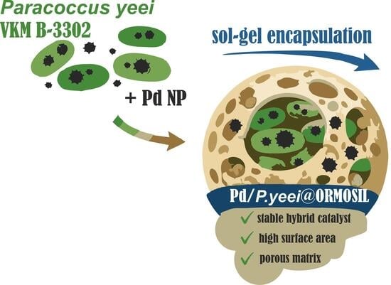
This study demonstrates the feasibility of encapsulating Paracoccus yeei VKM B-3302 cells, which contain palladium nanoparticles, within an organosilicon matrix synthesized using the sol–gel method. The resulting organosilicon material is characterized by a well-developed porous structure and a high specific surface area, ensuring the formation of a catalytic system with accessible active sites. Kinetic studies of the Mizoroki–Heck reaction showed that, although encapsulating the Pd/P. yeei catalyst in an organosilicon matrix slightly decreases its initial reaction rate, it increases the selectivity of the process and reduces the leaching of the active metal during repeated use. These results suggest the potential of encapsulating microorganisms containing metal nanoparticles in organosilicon materials to create stable hybrid catalytic systems.

Graphical Abstract

1. Introduction

In recent decades, extensive research has focused on the ability of bacteria, fungi, and yeasts to interact with metal ions and form nanoparticles on the cell surface as well as intracellularly [1,2,3,4,5]. These processes are of significant interest for the development of biocatalytic systems [1], environmentally friendly water purification technologies [6], sensor platforms [7,8], and antimicrobial materials [4].
A characteristic feature of microorganisms is the presence of numerous functional groups on the cell wall surface, including carboxyl, hydroxyl, amine, and phosphate groups [9]. These reactive sites can bind metal ions and participate in their subsequent reduction and stabilization. Consequently, nanoparticles of various metals (e.g., gold, silver, platinum, and palladium) can form on or within cells. Biogenic nanoparticles have several advantages over their synthetic counterparts, including milder synthesis conditions, biocompatibility, and specific morphology.
Despite the promise of such systems, their practical application is limited in several ways. One of the key challenges is the instability of the nanoparticles formed with the involvement of microorganisms [10,11,12]. Under operating conditions, particularly in liquid media and flow-through systems, these nanoparticles can desorb from the cell surface and enter the solution [13,14]. This results in the gradual loss of the material’s functional properties and can also lead to secondary environmental contamination with free nanoparticles. Consequently, there is a need to develop approaches that stabilize the resulting hybrid nanocatalysts. One of the most promising solutions is to encapsulate microbial cells in porous inert materials. Of the various types of such materials, organosilicon materials obtained through sol-gel processes are of particular interest. They are characterized by high chemical stability, controllable porosity and the ability to modify the surface functionality.
Using organosilicon materials allows for the creation of hybrid biomaterials, in which microbial cells and nanoparticles are embedded within an inorganic network structure. This prevents the cells and metal nanoparticles from leaching out. Systems based on encapsulated microorganisms and nanoparticles have significant potential applications in various fields of science and technology. One of the most promising areas is biocatalysis. Metal nanoparticles, including palladium-based ones, exhibit high catalytic activity in cross-coupling reactions. At the same time, conventional systems such as Pd/C, while highly efficient, often operate via “release-and-catch” mechanisms and may suffer from metal leaching under reaction conditions [15,16,17,18]. In this context, the development of alternative catalyst architectures that enable more controlled formation and stabilization of palladium species is of particular interest. The biologically mediated synthesis of Pd nanoparticles, followed by their immobilization within an organosilicon matrix, offers a distinct approach to catalyst design, providing spatial confinement of the active phase and potentially improved resistance to metal loss. Importantly, the aim of this work is not to outperform classical Pd/C systems but rather to explore the structure–performance relationships in such bio-derived hybrid materials and to assess their potential as an alternative catalytic platform. However, the catalytic properties of hybrid systems containing encapsulated microorganisms and metal nanoparticles in these reactions have not yet been adequately investigated. This study aims to examine the selectivity and catalytic performance of such hybrid catalysts in the Mizoroki–Heck reaction.

2. Results and Discussions

Previously [19], our research team developed a rapid method of forming palladium (Pd) nanoparticles on Paracoccus yeei VKM B-3302 bacterial cells. The resulting catalyst exhibited high activity in the Mizoroki–Heck reaction [20]. However, significant leaching of the Pd nanoparticles from the support was also observed [21]. The immobilisation of biocatalysts in organosilicon matrices is known to have a positive effect on operational stability during repeated use in reactions [22], as well as increasing the overall service life of such systems [23].
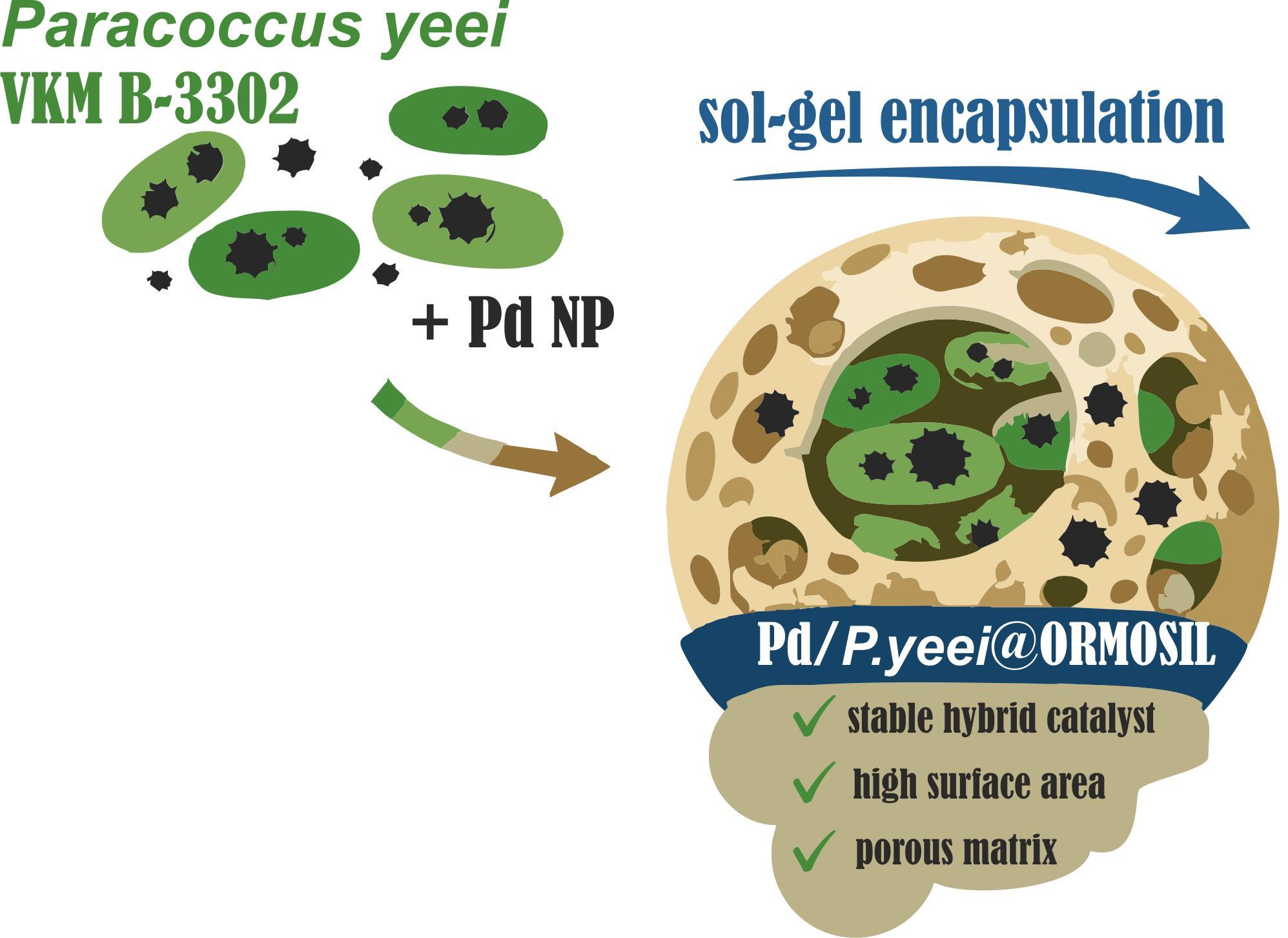
To improve the stability and reusability of Metall/bacteria catalysts, it is possible to coat their outer surface with an organic-inorganic hybrid silica shell, for example, using tetraethyl orthosilicate and 3-aminopropyltriethoxysilane as silica precursors by the silica coating method [24,25,26]. For P. yeei bacteria, encapsulation in an organosilicon material (organomodified silicate—ORMOSIL) prepared by the sol-gel method has been demonstrated [27,28]. Using an initial mass ratio of 3/1/43/44 (P. yeei/PVA/MTES/TEOS), we produced a Pd/P.yeei@ORMOSIL catalyst with a specific surface area of 359 m2/g (Langmuir model) and 312 m2/g (BET model). This is due to the significant proportion of meso- and macropores present (Figure 1).
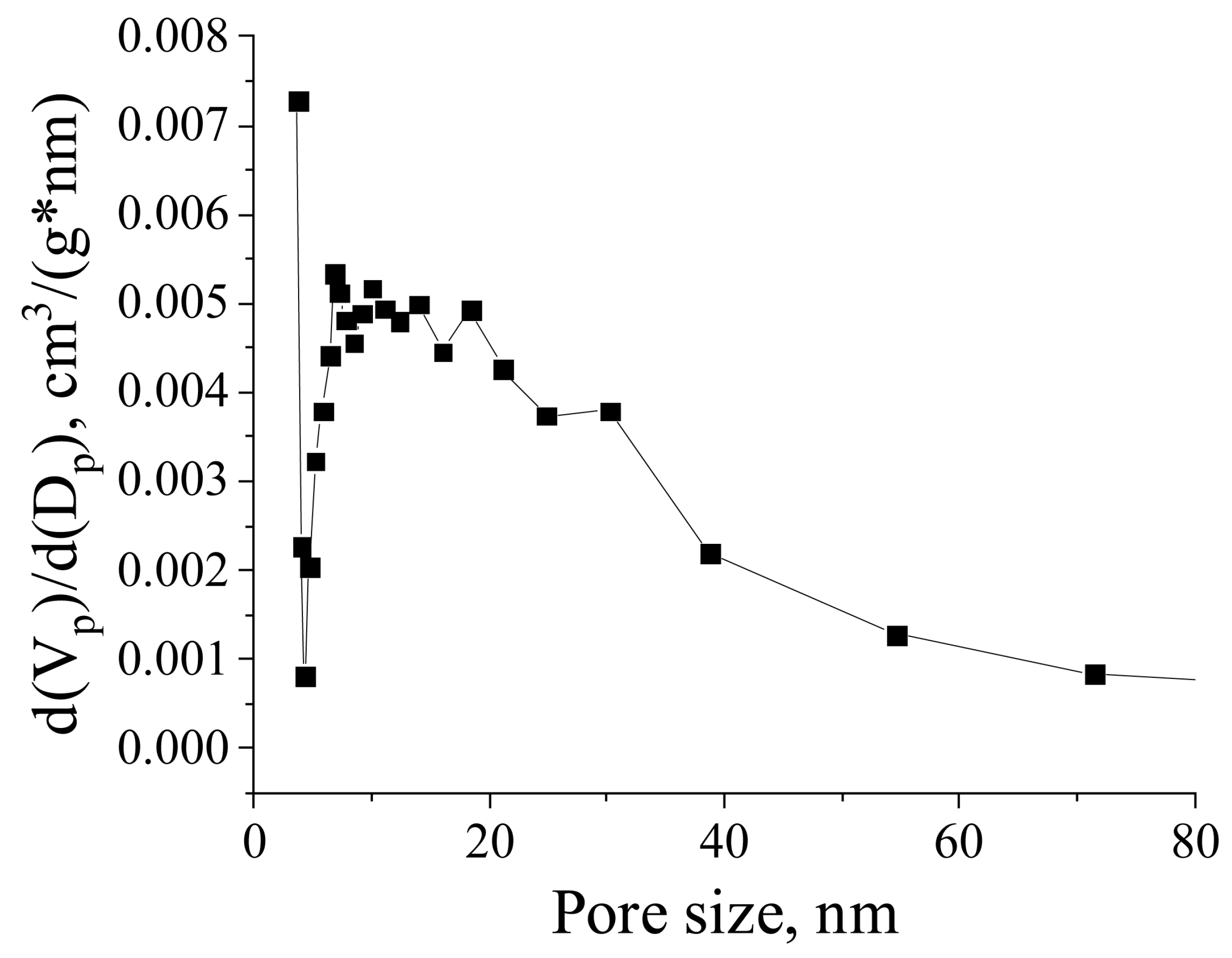
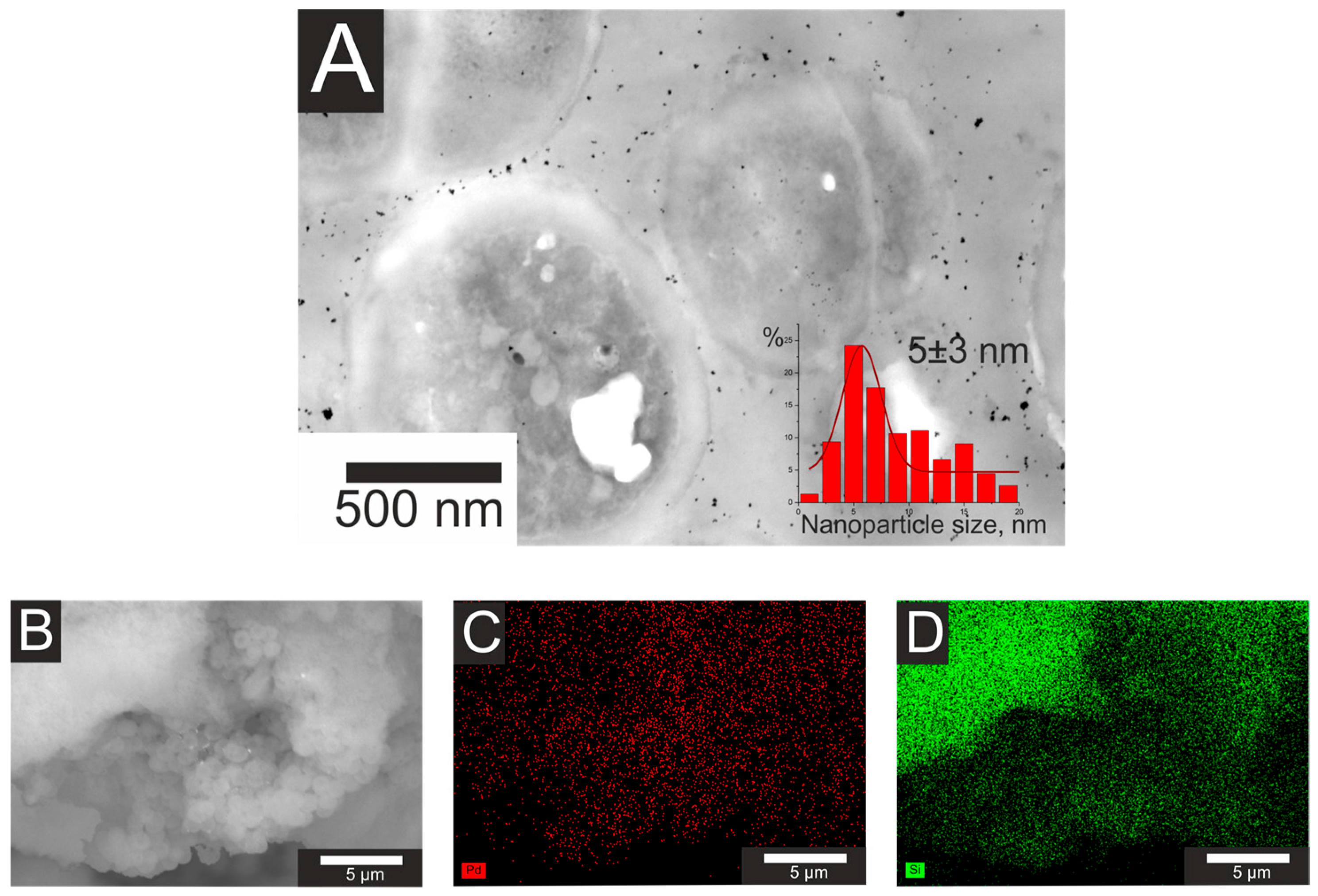
The Pd/P. yeei@ORMOSIL catalyst was characterized by transmission electron microscopy (TEM). Ultrathin section revealed palladium nanoparticles distributed both intracellularly and within the surrounding organosilicon matrix (Figure 2). It appears that some of the palladium nanoparticles that were weakly bound to, or not bound to, the cell surface were absorbed onto the ORMOSIL surface during the catalyst preparation process.
The average size of the palladium (Pd) nanoparticles was 5 ± 3 nm, comparable to that of the Pd/P. yeei precursor catalyst (3 ± 1 nm, [21]). The EDX element map (Figure 2B,C) shows the presence of Pd-rich/Si-poor regions alongside Si-rich/Pd-depleted areas. Such spatial heterogeneity is consistent with the anticipated morphology of the material, where palladium is localized within or on bacterial cells encapsulated in the organosilicon (ORMOSIL) matrix. Therefore, the catalyst comprises palladium nanoparticles situated both within and outside the P. yeei bacteria, with both the metal and the cells encapsulated in an organosilicon material. The Pd content of the Pd/P.yeei@ORMOSIL catalyst was quantified by inductively coupled plasma optical emission spectrometry (ICP-OES) at 0.61 ± 0.07 wt%, compared to 4.4 wt% for the initial Pd/P. yeei biohybrid [21].
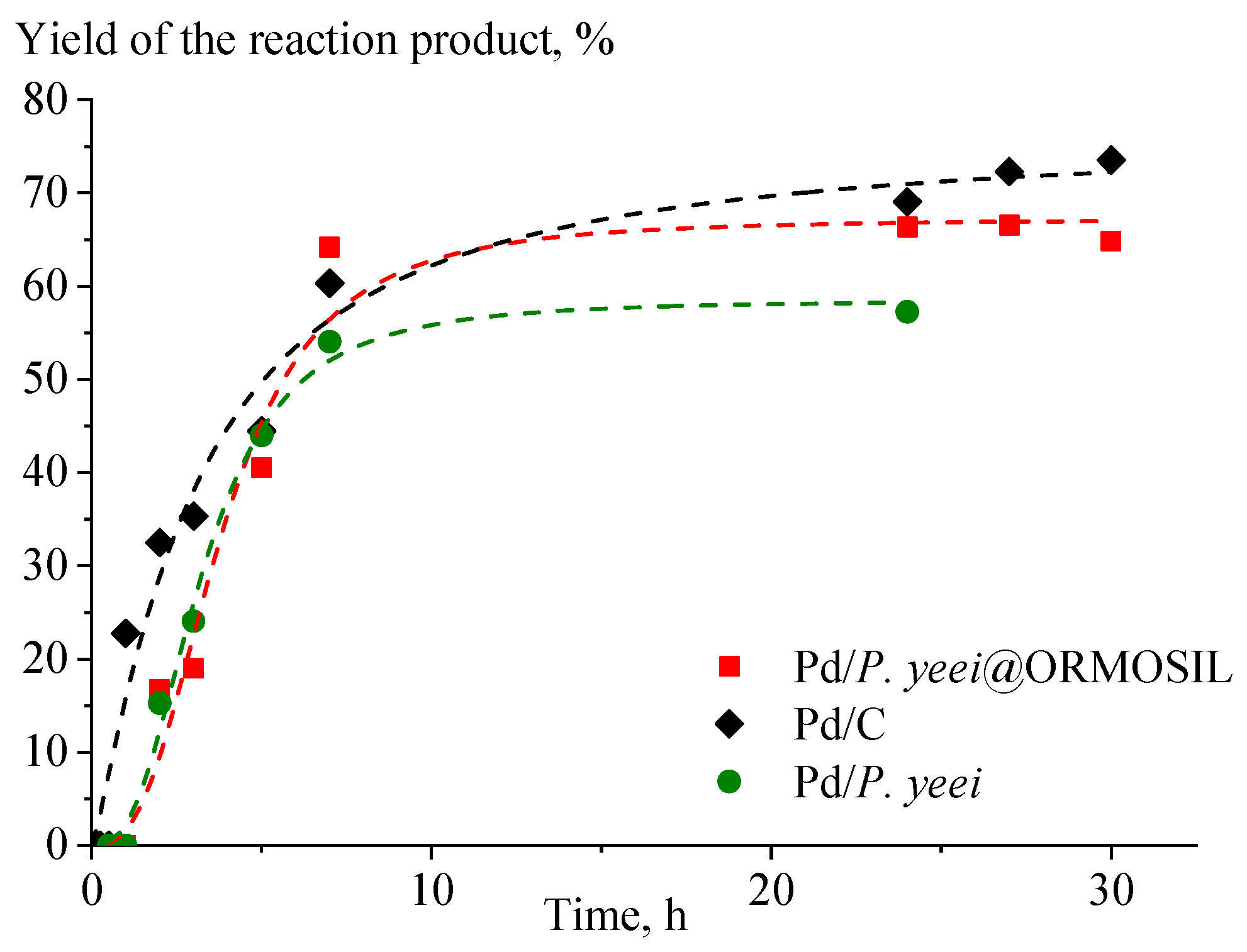
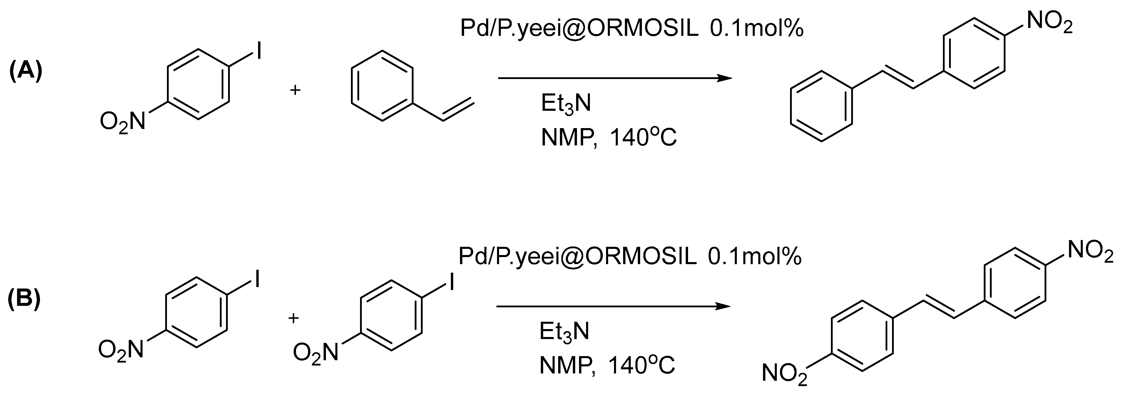
In the Pd/P. yeei@ORMOSIL catalyst, the Pd/P. yeei biohybrid is encapsulated within a porous organosilicon matrix. While the Pd nanoparticles remain distributed on and within the bacterial cells (Figure 2), the surrounding matrix may impose diffusion limitations or steric constraints that affect catalytic performance. To evaluate the impact of encapsulation on catalytic activity, the kinetics of the Mizoroki–Heck reaction were compared for the Pd/P. yeei@ORMOSIL catalyst and the initial Pd/P. yeei biohybrid under identical conditions. Kinetic experiments were performed at 140 °C (Figure 3). The conversion of the aryl halide and the yield of the target reaction product (Scheme 1A) were monitored over time, with the reaction carried out in N-methylpyrrolidone (NMP) using triethylamine (Et3N) as a base and a catalyst loading of 0.1 mol% of Pd/P. yeei@ORMOSIL. During these experiments, the yield of the byproduct (4,4′-dinitrobiphenyl) was determined by 1H NMR spectroscopy (Scheme 1B).
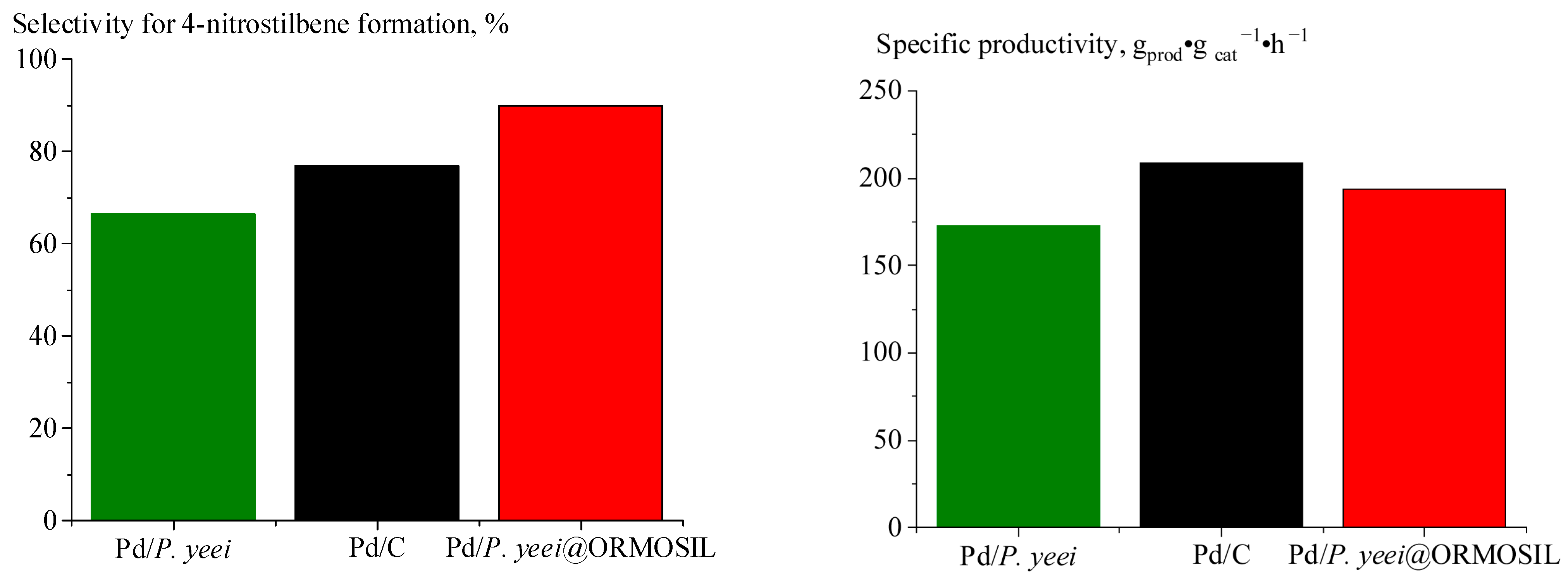
The average initial product formation rate was lower for Pd/P. yeei@ORMOSIL than for Pd/C and Pd/P. yeei (Table 1, Figure 4). The initial rate was defined as the tangent to the initial segment of the Cprod = f(t) curve (Figure 3). This decrease is attributed to the additional requirement for the substrate to diffuse through the organosilicon matrix. The maximum TOF value is observed for Pd/C (104 h−1), whereas for Pd/P. yeei it is lower (78 h−1). Encapsulation in ORMOSIL increased TOF to 86 h−1, reflecting greater catalytic activity relative to the non-encapsulated catalyst. The maximum TON value is observed for the Pd/C catalyst; for Pd/P.yeei@ORMOSIL and Pd/P. yeei, this value is lower. Although the conventional Pd/C catalyst exhibited the highest reaction rate and conversion among the catalysts tested, it demonstrated lower selectivity for the desired product. In contrast, the highest selectivity was achieved using the Pd/P. yeei@ORMOSIL bio-hybrid.
The increase in selectivity is likely due to the formation of an organosilicon matrix, which provides a specific microenvironment around the palladium nanoparticles, thereby limiting the occurrence of side reactions. In the 1-iodo-4-nitrobenzene and styrene reaction system (Scheme 1A), the adsorption and desorption kinetics of reactants at the organosilicon matrix surface critically govern the diffusion of reactants to the active palladium nanoparticles. On porous silicon dioxide, 1-iodo-4-nitrobenzene molecules are effectively retained due to hydrogen bonding and dipole-induced forces with surface silanol groups (Si–OH). This is because the nitro group is highly polar, giving 1-iodo-4-nitrobenzene a dipole moment of approximately 4.2 D, whereas styrene has a significantly lower dipole moment of approximately 0.13–0.18. Consequently, 1-iodo-4-nitrobenzene diffuses more slowly through the pores to the Pd catalytic centers than styrene does. According to the Mizoroki–Heck reaction mechanism, the initial stage of the catalytic cycle involves the oxidative addition of an aryl halide to palladium. Therefore, in the system under consideration, the adsorption and desorption kinetics of 1-iodo-4-nitrobenzene on the organosilicon matrix surface slow its diffusion through the pores, resulting in a lower local concentration of the aryl halide near the palladium nanoparticles. This effect is exacerbated further by the use of aryl halide (1 equiv.) in stoichiometric deficit relative to styrene (1.2 equiv.). Consequently, the average initial reaction rate decreases (see Table 1) and the progression of side reactions is limited, particularly the dimerization of 1-iodo-4-nitrobenzene (see Scheme 1B). This leads to an increase in the selectivity of target product formation.
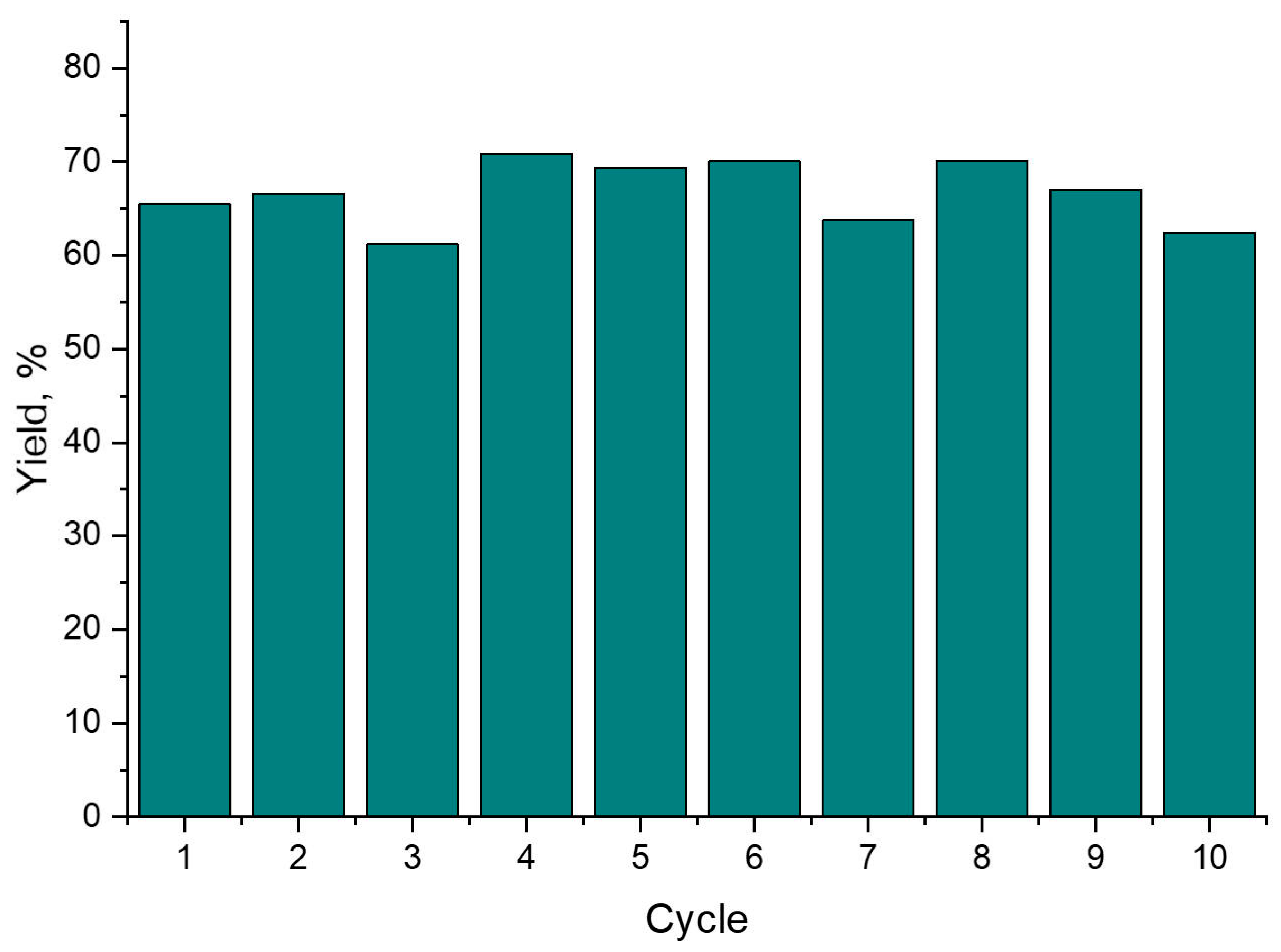
The stability test was conducted according to the method described in [29] over ten consecutive cycles. In accordance with the “Fresh Start” approach, the reaction was repeated after the catalytic reaction had been completed and the yield had been determined using NMR and GC-MS. For this, fresh portions of substrates, base and solvent were loaded (Figure 5).
Incorporating the Pd/P. yeei catalyst into an organosilicon material enables a high product yield to be maintained for at least 10 consecutive cycles. This stability is likely due to reduced palladium loss during the reaction and/or a lower tendency for Pd nanoparticle agglomeration, ensured by the organosilicate shell. Palladium leaching after ten consecutive cycles in the Mizoroki–Heck reaction was assessed using ICP. The Pd content decreased from 0.61 wt% to 0.51 wt% for the Pd/P.yeei@ORMOSIL catalyst (a 16% loss from the initial value), whereas the decrease in Pd content was 23% over 5 cycles when using Pd/P. yeei [19]. These results clearly demonstrate reduced leaching for the biohybrid catalyst encapsulated in an organosilicate shell.
Therefore, despite a slight decrease in the initial reaction rate, the encapsulated catalyst performs comparably to commercial Pd/C. Overall, the results suggest that the Pd/P.yeei@ORMOSIL catalyst is a promising catalytic system, offering high selectivity and stability while efficiently utilising active palladium nanoparticles.

3. Materials and Methods

3.1. Conditions for Culturing Microorganisms and Plotting Growth Curves

Paracoccus yeei VKM B-3302 (All-Russian Collection of Microorganisms, VKM) was cultured in lysogeny broth (LB) with the following composition: 10 g/L peptone (Molekula Ltd., Durham, UK), 10 g/L NaCl (≥99%, Helikon, Moscow, Russia), 5 g/L yeast extract (Bio Springer, Maisons-Alfort, France). The bacterial culture was grown in 750 mL Erlenmeyer flasks containing 200 mL of culture medium at 28 °C and agitated at 7 g using a Bios BSI-2 shaker incubator (Guangzhou, China). Upon completion of cultivation, the cells were harvested by centrifugation at 5700× g for 10 min using a MWP-351R centrifuge (Lower Silesian Voivodeship, Poland) into pre-weighed Eppendorf-type microtubes. The supernatant was decanted. The cell biomass was used immediately after cultivation, without storage [30].

3.2. Immobilisation of the Biocomposite in an Organosilicon Matrix

The synthesis of palladium zero-valent (Pd(0)) nanoparticles via a biologically mediated approach involving metabolically active Paracoccus yeei VKM B-3302 microbial cells was carried out using a method similar to that described in [21]. The resulting catalyst was then immobilised in an organosilicon material prior to drying. The organosilicon material was synthesised using a method similar to that described in [31], with methyltriethoxysilane (MTES, 1.53 mol/dm3) and tetraethoxysilane (TEOS, 1.28 mol/dm3) (both Sigma-Aldrich, St. Louis, MO, USA) in a 1:1 volume ratio relative to the initial suspension. A structure-controlling agent (polyvinyl alcohol grade 16/1, Polymer LLC, Moscow, Russia) at 5 wt% was added, catalysed by sodium fluoride (RusChem, St Petersburg, Russia) at 20 mmol/dm3. At this stage, the microbial cells remain structurally integrated within the forming matrix; however, subsequent drying (48 h at room temperature (25 °C) and atmospheric pressure) leads to loss of cell viability, resulting in a bio-derived, non-living hybrid material used in the catalytic reaction.

3.3. Determination of Surface Area and Pore Distribution by Low-Temperature Nitrogen Adsorption

A sample weight was placed in a pre-weighed quartz cuvette, which was installed in the Beckman Coulter™ SA-Prep™ sample preparation instrument (Coulter Corporation, Brea, CA, USA), where drying was performed in a nitrogen stream for 60 min at a temperature up to +60 °C. Next, the sample was placed in the degassing port of the Beckman Coulter™ SA 3100™ instrument (Coulter Corporation, USA; hereinafter SA 3100™). Degassing conditions: temperature +60 °C, degassing time—60 min, vacuum—0.001 mm Hg. After the specified time had elapsed and the cuvette had cooled, it was weighed with an accuracy of (±0.0005). The cuvette containing the sample was placed in the analytical port of the SA 3100TM instrument. The surface characteristics of the samples (specific surface area, porosity) were determined based on the nitrogen adsorption value at a constant temperature. To determine the sample type (micro-, meso-, or macroporous), measurements were performed in the relative pressure range of 0–0.995 and the pore size distribution range of 3 to 200 nm. Based on the obtained isotherm and porosity data, a conclusion was drawn regarding the sample type.

3.4. Reaction Products Were Analysed by Gas Chromatography–Mass Spectrometry (GC–MS)

For the quantitative analysis, a series of standard solutions of the target product (trans-stilbene) and the starting aryl halide (iodobenzene) were prepared in acetonitrile, with the concentration of the internal standard (diphenylacetylene) kept constant. Prior to analysis, samples of the reaction mixtures were diluted 1000-fold with an internal standard solution (diphenylacetylene, 0.0001 mmol/L) in acetonitrile, then passed through a 0.43 μm, 25 mm diameter syringe filter. The prepared samples were analysed using a Maestro-α quadrupole instrument (Interlab, Moscow, Russia), with sample injection via a Crystallux-4000M gas chromatograph (Meta-Chrom, Yoshkar-Ola, Russia). A ZB-5ms column (length = 30 m, inner diameter = 0.25 mm, phase thickness = 0.25 μm; Phenomenex, Torrance, CA, USA) was used for this analysis. The initial temperature was set to 50 °C (pressure 0.353 atm) with a heating rate of 10 °C/min up to 80 °C. This was then followed by a heating rate of 20 °C/min up to a final temperature of 310 °C (pressure 1.576 atm). The ionisation energy was 70 mV.

3.5. Determination of the Palladium (Pd) Content in a Catalyst by Inductively Coupled Plasma Mass Spectrometry

The sample was dissolved in a specified volume of concentrated nitric acid and the elemental content of the resulting solution was quantified using a Varian 710-ES inductively coupled plasma optical emission spectrometer (ICP-OES) (Agilent, Santa Clara, CA, USA) under the following conditions: plasma power 1.20 kW, plasma flow rate 15.0 L/min and axial flow rate 1.5 L/min.

3.6. Registration of Nuclear Magnetic Resonance Spectra

Nuclear magnetic resonance spectra were recorded on a Bruker AVANCE DRX 500 spectrometer (Munich, Germany) at a frequency of 500.13 MHz (1H) and on a Bruker Fourier 300 HD spectrometer at 300.1 MHz (1H), using the residual solvent peak as an internal standard. The spectra were processed using MestReNova software.

3.7. Transmission Electron Microscopy (TEM) Measurement

Biocomposite sections were prepared using the following method: The sample was fixed in a 2% solution of glutaraldehyde in 0.05 M cacodylate buffer solution (pH 7.2) for one hour at 4 °C. The sample was then washed three times with 0.05 M cacodylate buffer solution (pH 7.2). The dehydrated sample in agar blocks was then coated with Epon 812 epoxy resin and sectioned using a Reichert-Jung UltraCut E ultramicrotome (Vienna, Austria).
Before the measurements, the samples were deposited on 3 mm grids. The sample morphology was studied using a Hitachi HT7700 (Tokyo, Japan) transmission electron microscope. Images were acquired in bright-field TEM mode at an accelerating voltage of 100 kV. The nanoparticle size distribution was determined via ImageJ 1.54g software. The DLgram [32] cloud service for microscopic image analysis is used to determine the size distribution of nanoparticles.

3.8. Scanning Electron Microscopy (SEM) Measurement

Before measurements the sample was mounted on a 25 mm aluminum specimen stub, fixed by conductive carbon tape and coated with a 30 nm film of carbon. The observations were carried out using a Hitachi Regulus8230 (Japan) field-emission scanning electron microscope (FE-SEM). Images were acquired in backscattered electron mode (compositional contrast) at a 5 kV accelerating voltage. EDS-SEM studies were carried out using a Bruker Quantax 400 EDS system (USA) equipped with an XFlash 6|60 detector at a 20 kV accelerating voltage.

4. Conclusions

This study demonstrates the feasibility of encapsulating Paracoccus yeei VKM B-3302 cells within a palladium nanoparticle-containing organosilicon matrix produced using the sol-gel method. The synthesized organosilicon material has a developed porous structure and a high specific surface area, ensuring the formation of a catalytic system with accessible active sites. The final Pd/P.yeei@ORMOSIL catalyst contains 0.61 wt% palladium. Kinetic studies of the Mizoroki–Heck reaction revealed that, compared to the Pd/C and Pd/P. yeei systems, encapsulation of the biogenic catalyst results in a slight decrease in the initial reaction rate, which is likely due to diffusion limitations arising during substrate transport into the organosilicon matrix. However, the use of an organosilicon matrix is shown to increase reaction selectivity. The catalyst productivity remains comparable to that of commercial Pd/C. Organosilicon materials have been shown to prevent the leaching of the active metal during repeated use. Therefore, encapsulating microorganisms with palladium nanoparticles in organosilicon materials is a promising method for creating hybrid biomaterials.

Author Contributions

Conceptualization, O.A.K. and V.N.S.; methodology, V.N.S., P.V.R. and O.A.K.; software, O.A.K. and P.V.R.; validation, V.N.S. and O.A.K.; formal analysis, P.V.R., A.N.Z., V.V.F. and N.M.I.; investigation, O.A.K., P.V.R. and V.N.S.; resources, O.A.K.; data curation, V.N.S.; writing—original draft preparation, V.N.S., P.V.R. and O.A.K.; writing—review and editing, V.N.S., P.V.R. and O.A.K.; visualization, N.M.I. and O.A.K.; supervision, O.A.K.; project administration, O.A.K.; funding acquisition, O.A.K. All authors have read and agreed to the published version of the manuscript.

Funding

This work was supported by the Russian Science Foundation, grant no. 24-73-10013, https://rscf.ru/en/project/24-73-10013/ (accessed on 14 April 2026).

Data Availability Statement

The raw data supporting the conclusions of this article will be made available by the authors on request.

Acknowledgments

The authors thank Valentine P. Ananikov for helpful discussions. Electron Microscopy Core Facilities of the PSCBR RAS (http://www.ckp-rf.ru/ckp/670266/ (accessed on 14 April 2026)).

Conflicts of Interest

The authors declare no conflicts of interest.

Abbreviations

The following abbreviations are used in this manuscript:
BETBrunauer–Emmett–Teller
BJHBarrett-Joyner-Halenda
ICP-OESOptical Emission Spectrometer with Inductively Coupled Plasma
GH-MSgas chromatography–mass spectrometry
LBlysogeny broth
MTESmethyltriethoxysilane
N-MPN-methylpyrrolidone
NMRNuclear magnetic resonance
ORMOSILorganomodified silicate
PVApolyvinyl alcohol
TEMtransmission electron microscope
TEOStetraethoxysilane
TOFTurnover Frequency
TONTurnover Number
VKMall-Russian Collection of Microorganisms

References

  1. Saravanan, A.; Kumar, P.S.; Karishma, S.; Vo, D.N.; Jeevanantham, S.; Yaashikaa, P.R.; George, C.S. A Review on Biosynthesis of Metal Nanoparticles and Its Environmental Applications. Chemosphere 2021, 264, 128580. [Google Scholar] [CrossRef]
  2. Iravani, S.; Varma, R.S. Bacteria in Heavy Metal Remediation and Nanoparticle Biosynthesis. ACS Sustain. Chem. Eng. 2020, 8, 5395–5409. [Google Scholar] [CrossRef]
  3. Khandel, P.; Shahi, S.K. Mycogenic Nanoparticles and Their Bio—Prospective Applications: Current Status and Future Challenges. J. Nanostruct. Chem. 2018, 8, 369–391. [Google Scholar] [CrossRef]
  4. Mishra, A.; Tripathy, S.K.; Yun, S. Bio-Synthesis of Gold and Silver Nanoparticles from Candida Guilliermondii and Their Antimicrobial Effect Against Pathogenic Bacteria. J. Nanosci. Nanotechnol. 2011, 11, 243–248. [Google Scholar] [CrossRef]
  5. Egan-Morriss, C.; Lloyd, J.R.; Kimber, R.L.; Powell, N.A. Biotechnological Synthesis of Pd-Based Nanoparticle Catalysts. Nanoscale Adv. 2022, 4, 654–679. [Google Scholar] [CrossRef]
  6. Patel, A.; Enman, J.; Gulkova, A.; Guntoro, P.I.; Dutkiewicz, A.; Ghorbani, Y.; Rova, U.; Christakopoulos, P.; Matsakas, L. Integrating Biometallurgical Recovery of Metals with Biogenic Synthesis of Nanoparticles. Chemosphere 2021, 263, 128306. [Google Scholar] [CrossRef]
  7. Selvakumar, R.; Seethalakshmi, N.; Thavamani, P.; Naidu, R.; Megharaj, M. Recent Advances in Synthesis of Inorganic Nano/Microstructures Using Microbial Biotemplates and Their Applications. RSC Adv. 2014, 4, 52156–52169. [Google Scholar] [CrossRef]
  8. Banerjee, K.; Rai, V.R. A Review on Mycosynthesis, Mechanism, and Characterization of Silver and Gold Nanoparticles. BioNanoScience 2017, 8, 17–31. [Google Scholar] [CrossRef]
  9. Jiang, W.; Saxena, A.; Song, B.; Ward, B.B.; Beveridge, T.J.; Myneni, S.C.B. Elucidation of Functional Groups on Gram-Positive and Gram-Negative Bacterial Surfaces Using Infrared Spectroscopy. Langmuir 2004, 20, 11433–11442. [Google Scholar] [CrossRef] [PubMed]
  10. Moore, T.L.; Rodriguez-lorenzo, L.; Hirsch, V.; Balog, S.; Urban, D.; Jud, C.; Rothen-Rutishauser, B.; Lattuada, M.; Petri-Fink, A. Nanoparticle Colloidal Stability in Cell Culture Media and Impact on Cellular Interactions. Chem. Soc. Rev. 2015, 44, 6287–6305. [Google Scholar] [CrossRef] [PubMed]
  11. Xie, C.; Zhang, J.; Ma, Y.; Ding, Y.; Zhang, P.; Zheng, L.; Chai, Z.; Zhao, Y.; Zhang, Z.; He, X. Bacillus subtilis Causes Dissolution of Ceria Nanoparticles at the Nano—Bio Interface. Environ. Sci. Nano 2019, 6, 216–223. [Google Scholar] [CrossRef]
  12. Praetorius, A.; Badetti, E.; Brunelli, A.; Clavier, A.; Gallego-Urrea, J.A.; Gondikas, A.; Hassellöv, D.M.; Hofmann, T.; Mackevica, A.; Marcomini, A.; et al. Strategies for Determining Heteroaggregation Attachment Efficiencies of Engineered Nanoparticles in Aquatic Environments. Environ. Sci. Nano 2020, 7, 351–367. [Google Scholar] [CrossRef]
  13. Kutralam-Muniasamy, G.; Perez-Guevara, F. Recombinant Surface Engineering to Enhance and Expand the Potential of Biologically Produced Nanoparticles: A Review. Process Biochem. 2016, 59, 4–17. [Google Scholar] [CrossRef]
  14. Yang, Y.; Vance, M.; Tou, F.; Tiwari, A.; Liu, M.; Hochella, M.F. Nanoparticles in Road Dust from Impervious Urban Surfaces: Distribution, Identification, and Environmental Implications. Environ. Sci. Nano 2016, 3, 534–544. [Google Scholar] [CrossRef]
  15. Pavia, C.; Giacalone, F.; Bivona, L.A.; Salvo, A.M.P.; Petrucci, C.; Strappaveccia, G.; Vaccaro, L.; Aprile, C.; Gruttadauria, M. Evidences of Release and Catch Mechanism in the Heck Reaction Catalyzed by Palladium Immobilized on Highly Cross-Linked-Supported Imidazolium Salts. J. Mol. Catal. A Chem. 2014, 387, 57–62. [Google Scholar] [CrossRef]
  16. Prima, D.O.; Kulikovskaya, N.S.; Galushko, A.S.; Mironenko, R.M.; Ananikov, V.P. Transition Metal ‘Cocktail’-Type Catalysis. Curr. Opin. Green Sustain. Chem. 2021, 31, 100502. [Google Scholar] [CrossRef]
  17. Egorov, M.P.; Lee, V.Y.; Alabugin, I.V. Cocktail of Catalysts: A Dynamic Advance in Modern Catalysis. Chemistry 2025, 7, 109. [Google Scholar] [CrossRef]
  18. Beletskaya, I.P.; Ananikov, V.P. Transition-Metal-Catalyzed C–S, C–Se, and C–Te Bond Formations via Cross-Coupling and Atom-Economic Addition Reactions. Achievements and Challenges. Chem. Rev. 2022, 122, 16110–16293. [Google Scholar] [CrossRef]
  19. Rybochkin, P.V.; Perchikov, R.N.; Karlinskii, B.Y.; Kamanina, O.A.; Arlyapov, V.A.; Kashin, A.S.; Ananikov, V.P. Aerobic Bacteria-Supported Biohybrid Palladium Catalysts for Efficient Cross-Coupling Reactions. J. Catal. 2023, 429, 115238. [Google Scholar] [CrossRef]
  20. Soromotin, V.N.; Rybochkin, P.V.; Kamanina, O.A. A Study of the Kinetics of the Mizoroki—Heck Reaction on the Pd/P. Yeei Catalyst. Kinet. Catal. 2025, 66, 521–527. [Google Scholar] [CrossRef]
  21. Kamanina, O.; Rybochkin, P.; Borzova, D.; Soromotin, V.N.; Galushko, A.; Kashin, A.S.; Ivanova, N.; Zvonarev, A.; Suzina, N.; Holicheva, A.; et al. Sustainable Catalysts in a Short Time: Harnessing Bacteria for Swift Palladium Nanoparticle Production. Nanoscale 2025, 17, 5289–5300. [Google Scholar] [CrossRef]
  22. Carturan, G.; Campostrini, R.; Diré, S.; Scardi, V.; De Alteriis, E. Inorganic Gels for Immobilization of Biocatalysts: Inclusion of Invertase-Active Whole Cells of Yeast (Saccharomyces cerevisiae) into Thin Layers of SiO2 Gel Deposited on Glass Sheets. J. Mol. Catal. 1989, 57, L13–L16. [Google Scholar] [CrossRef]
  23. Inama, L.; Diré, S.; Carturan, G.; Cavazza, A. Entrapment of Viable Microorganisms by SiO2 Sol-Gel Layers on Glass Surfaces: Trapping, Catalytic Performance and Immobilization Durability of Saccharomyces Cerevisiae. J. Biotechnol. 1993, 30, 197–200. [Google Scholar] [CrossRef]
  24. Guo, J.; Amini, S.; Lei, Q.; Ping, Y.; Agola, J.O.; Wang, L.; Zhou, L.; Cao, J.; Franco, S.; Noureddine, A.; et al. Robust and Long-Term Cellular Protein and Enzymatic Activity Preservation in Biomineralized Mammalian Cells. ACS Nano 2022, 16, 2164–2175. [Google Scholar] [CrossRef]
  25. Zhang, Y.; Yue, X.; Zhang, S.; Kong, W.; Zhu, R.; Zhou, L.; Gao, J.; Jiang, Y.; Liu, Y. Ambient Synthesis of Single-Atom Catalysts on Catalytically Active Cells for Chemoenzymatic Cascades. Nat. Commun. 2026, 17, 2935. [Google Scholar] [CrossRef]
  26. Lei, Q.; Guo, J.; Kong, F.; Cao, J.; Wang, L.; Zhu, W.; Brinker, C.J. Bioinspired Cell Silicification: From Extracellular to Intracellular. J. Am. Chem. Soc. 2021, 143, 6305–6322. [Google Scholar] [CrossRef]
  27. Kamanina, O.; Arlyapov, V.; Rybochkin, P.; Lavrova, D.; Podsevalova, E.; Ponamoreva, O. Application of Organosilicate Matrix Based on Methyltriethoxysilane, PVA and Bacteria Paracoccus Yeei to Create a Highly Sensitive BOD. 3 Biotech 2021, 11, 331. [Google Scholar] [CrossRef]
  28. Kamanina, O.A.; Saverina, E.A.; Rybochkin, P.V.; Arlyapov, V.A.; Vereshchagin, A.N.; Ananikov, V.P. Preparation of Hybrid Sol-Gel Materials Based on Living Cells of Microorganisms and Their Application in Nanotechnology. Nanomaterials 2022, 12, 1086. [Google Scholar] [CrossRef] [PubMed]
  29. Mironenko, R.M.; Saybulina, E.R.; Trenikhin, M.V.; Izmailov, R.R.; Lotsman, K.A.; Rodygin, K.S.; Ananikov, V.P. Comparing Separation vs. Fresh Start to Assess Reusability of Pd/C Catalyst in Liquid-Phase Hydrogenation. ChemCatChem 2021, 13, 3656–3661. [Google Scholar] [CrossRef]
  30. Kamanina, O.A.; Rybochkin, P.V.; Soromotin, V.N. Use of Microorganisms to Recover Metals from Water Bodies and Create Catalysts on Their Basis. Proc. Univ. Appl. Chem. Biotechnol. 2025, 15, 495–502. [Google Scholar] [CrossRef]
  31. Kamanina, O.A.; Lantsova, E.A.; Rybochkin, P.V.; Arlyapov, V.A.; Saverina, E.A.; Kulikovskaya, N.S.; Perepukhov, A.M.; Vereshchagin, A.N.; Ananikov, V.P. “3-in-1” Hybrid Biocatalysts: Association of Yeast Cells Immobilized in a Sol–Gel Matrix for Determining Sewage Pollution. ACS Appl. Mater. Interfaces 2023, 15, 47779–47789. [Google Scholar] [CrossRef] [PubMed]
  32. Matveev, A.V.; Nartova, A.V.; Sankova, N.N.; Okunev, A.G. DLgram Cloud Service for Deep-Learning Analysis of Microscopy Images. Microsc. Res. Tech. 2024, 87, 991–998. [Google Scholar] [CrossRef] [PubMed]
Figure 1. Size distribution of pores in an organosilicon material containing P. yeei cells, as determined by the Barrett-Joyner-Halenda (BJH) model.
Inorganics 14 00117 g001
Figure 2. (A) Transmission electron microscope (TEM) image of an ultrathin section of the Pd/P.yeei@ORMOSIL. Inset: size distribution of Pd nanoparticles, (B) Scanning electron microscopy (SEM) image of the Pd/P.yeei@ORMOSIL, (C,D) EDX element maps showing the spatial distribution of silicon and palladium in the prepared Pd/P.yeei@ORMOSIL.
Inorganics 14 00117 g002
Scheme 1. Formation of the main product (A) and by-product (B) of the Mizoroki–Heck reaction.
Inorganics 14 00117 sch001
Figure 3. Reaction product yield as a function of time for Pd/C (1 wt%), Pd/P. yeei (1 wt%) [20] and Pd/P.yeei@ORMOSIL (0.61 wt%) catalysts, with a loading of 0.1 mol% and a temperature of 140 °C.
Inorganics 14 00117 g003
Figure 4. Selectivity for the formation of 4-nitrostilbene and the specific productivity of the Pd/C, Pd/P. yeei [20] and Pd/P.yeei@ORMOSIL catalysts.
Inorganics 14 00117 g004
Figure 5. Reaction product yield based on reusability testing results.
Inorganics 14 00117 g005
Table 1. Mizoroki-Heck reaction parameters for the Pd/P.yeei@ORMOSIL catalyst.
Pd/C [20]Pd/P. yeei [20]Pd/P. yeei@ORMOSIL
Maximal rate of formation of the product, μmol h−1 mL−126 2220
TON732543603
TOF, ч−11047886
Conversion, %958167
Selectivity for 4-nitrostilbene formation, %776790
Specific productivity, gprod·gcat−1·h−1208173194
Disclaimer/Publisher’s Note: The statements, opinions and data contained in all publications are solely those of the individual author(s) and contributor(s) and not of MDPI and/or the editor(s). MDPI and/or the editor(s) disclaim responsibility for any injury to people or property resulting from any ideas, methods, instructions or products referred to in the content.