Article

Hematite Nanoparticles Synthesized by Green Route: Characterization, Anticancer and Antioxidant Activities

1
Department of Chemistry, College of Sciences, King Khalid University, Abha P.O. Box 9004, Saudi Arabia
2
Center for Innovation and Entrepreneurship, Imam Mohammad Ibn Saud Islamic University (IMSIU), Riyadh 11623, Saudi Arabia
3
Department of Industrial Chemistry, First Technical University Ibadan, Ibadan 200243, Oyo State, Nigeria
4
Materials Science Innovation and Modelling (MaSIM) Research Focus Area, Faculty of Natural and Agricultural Science, North-West University (Mafikeng Campus), Private Bag X2046, Mmabatho 2735, South Africa
*
Author to whom correspondence should be addressed.
Inorganics 2025, 13(5), 167; https://doi.org/10.3390/inorganics13050167
Submission received: 13 March 2025 / Revised: 27 April 2025 / Accepted: 6 May 2025 / Published: 15 May 2025

Abstract

Recently, attention has shifted towards the green synthesis of nanoparticles using plant extracts rich in phytochemicals like phenols and flavonoids, offering an alternative method that avoids harmful chemicals and enables large-scale, low-cost production. This study introduces a straightforward and eco-friendly approach to synthesizing hematite α-Fe2O3 nanoparticles utilizing an aqueous extract of Musa paradisiaca. The variation in the calcination temperature resulted in the formation of nanoparticles presented as Fe2O3 (1), Fe2O3 (2), and Fe2O3 (3), obtained at 650, 750, and 900 °C for 4 h, respectively. This variation allowed for an investigation into the impact of different reaction temperatures on the structural and optical properties of the nanoparticles. Structural analysis was conducted using X-ray diffraction (XRD), while scanning electron microscopy (SEM) and transmission electron microscopy (TEM) were employed to examine morphology. Optical properties were assessed via UV-vis spectroscopy, revealing a reduction in the energy band gap (from 2.5 to 1.87 eV), attributed to an increase in crystallite size resulting from longer calcination temperatures (650–900 °C). A biological assay was carried out to evaluate the antioxidant and anticancer potentials of the nanoparticles. Both Fe2O3 (1) and Fe2O3 (2) with IC50 values of 46.84 and 46.14 µg/mL, respectively, showed similar antioxidant potentials, while peel extract exhibited the least activity with an IC50 of 79.26 µg/mL. The nanoparticles, peels, and 5-FU (used as standard) showed a stronger inhibitory effect on the Human Embryonic Kidney (HEK) 293 cells compared to the HeLa cells. This implies that the HEK 293 cells might be more susceptible to the drug samples and a lower concentration might even be sufficient to achieve the inhibition of normal cell proliferation. These results indicate a better therapeutic window with a lesser inhibitory effect compared to standard drugs used as controls.

1. Introduction

In recent years, the use of different parts of plants, such as leaves, fruits, seeds, bark, and peels, in the synthesis of nanoparticles (known as biogenic, green, or phyto-mediated synthesis) has continued to grow and attract research interests. This interest is spurred by the several advantages that green synthesis has over physical and chemical processes. For example, using natural materials eliminates the need for harmful chemicals and harsh reaction conditions [1,2]. The biochemical approach offers flexible benefits over the chemical approach, including lower cytotoxicity, eco-friendly nature, reduced environmental footprint during synthesis, and minimal or no harmful waste production [3]. Therefore, it has emerged as a good alternative for the preparation of nanomaterials, specifically when they are meant for environmental or biomedical use. Several applications are made possible by the exceptional features of metal oxide nanoparticles, including ZnO, TiO2, MgO, and CuO. In different processes, including oxidation, reduction, and photocatalysis, they operate as catalysts or catalyst supports [4]. For example, metal oxide nanoparticles are widely used in environmental remediation to purify water by breaking down organic contaminants and removing heavy metals. TiO2 nanoparticles are particularly used for the photocatalytic destruction of contaminants [5]. They are also useful in biological applications for antibacterial coatings, imaging, drug delivery, and biosensing because of their biocompatibility and reactivity [6]. Moreover, their employment in gas sensors, biosensors, and chemical sensors is supported by their sensitivity to gases and biomolecules [7]. Additionally, their unique optical and electrical properties make them suitable for optoelectronics and photovoltaics, including solar cells, LEDs, and smart windows [8].
Due to the synthesis conditions, the major challenge associated with biogenic synthesis is to prepare nanoparticles with well-defined morphology [9,10]. This is critical because, within the nanometric domain, both the shape and size of nanoparticles are important. A reduction in particle size with an increase in mixing speed has been reported for iron nanoparticles prepared using an extract of Prunus avium stem [11]. Similarly, the extract of Hibiscus rosa sinensis flower has been used to produce size-reduced iron oxide nanoparticles [12].
Metal oxide nanoparticles have benefited immensely from green synthesis compared to other nanomaterial compounds since they do not suffer corrosion and are relatively stable in the air. Iron oxide is one of the important metal oxides that is widely studied due to its magnetic properties, biocompatibility, and easy surface functionalization [13,14]. It exists in different forms. It has different forms, including magnetite, maghemite, hematite, and wurtzite [15]. Due to its magnetic and electronic properties, iron oxide nanoparticles obtained from Abutilon indicum leaf extract have been reported to potentially serve as a therapeutic agent for a wide range of human diseases [16]. The notable biomedical properties of green-synthesized iron oxide nanoparticles using A. indicum and Nicotiana plumbaginifolia leaf extract and their promising applications in the field of medicine have been reported [16,17]. Magnetite (Fe3O4), maghemite (γ-Fe2O3), and hematite (α-Fe2O3) are the main forms that are widely studied due to their fascinating features and unique physicochemical properties [15,18]. These properties are further improved when iron oxides are reduced to the nanoscale due to quantum size phenomena, thus resulting in particles with outstanding chemical stability, optical, magnetic, structural, and electrical properties [19,20].
Several studies have described the advancement of iron oxide nanoparticles in a wide range of applications such as catalysis [21], magnetic recording media [22], lithium-ion batteries [23], and biomedical [24,25]. The use of iron oxide nanoparticles for biomedical applications has garnered a lot of interest specifically in the fields of antimicrobial [26,27], anticancer [28,29], antidiabetic [30], antitumor [31], antioxidant [32,33], and wound-dressing applications [34,35]. For the broad biomedical application of these nanoparticles, crystal size control and shape modifications are very important because these features define the chemical and physical properties of the nanoparticles [36].
The phenomenal advancement of nanotechnology in recent years has ushered in a new approach to the treatment of diseases such as tumors. Studies show that cancer claims more lives than the combined statistics of death from malaria, tuberculosis, and AIDS [37]. Hence, the growing research activities on the use of nanoparticles to overcome these challenges offer unique alternatives to the treatment process. Nanoparticles attack cancerous cells and exhibit a higher degree of harm compared to normal cells. Their cytotoxic effects are associated with the reactive oxygen species (ROS), activation of caspase-3, permeabilization of the mitochondrial outer membrane, and cleavage of DNA [38,39]. Apoptosis can be triggered through internal or extrinsic pathways, and nanomaterials can induce apoptosis through various mechanisms [40]. Similarly, nanoparticles possess good antioxidant properties, and this is attributed to their increased surface-to-volume ratio [41,42]. Antioxidants are compounds that inhibit the oxidation of molecules, playing a crucial role in safeguarding cells from harm caused by unstable free radicals in biological contexts. They disrupt the chain reactions initiated by free radicals, halting their oxidation process. By doing so, antioxidants prevent oxidative damage, lowering the risk of diseases, cancer, and aging [43,44].
Musa paradisiaca, commonly known as plantain, is an herbaceous plant that is extensively cultivated in Southeast Asia and the western Pacific regions. The inflorescence of the plant, also known as the flower cluster, is a staple vegetable in the southern parts of Asia, particularly in regions of southern India, Malaysia, and Taiwan, and is also widely used in various African countries. In traditional medicine, plantain has been used to treat dysentery, menorrhagia, and diabetes [45]. According to Arun et al. [46], plantain is considered to be a potent antidiabetic plant. Plantain inflorescence was also reported to be a potential source of phytochemicals with promising health benefits. The qualitative phytochemical analysis of Musa paradisiaca extract confirmed the presence of biomolecules such as flavonoids, phenols, tannins, quinones, flavonoids, saponins, and phytosterols [47].
The objective of the current study is to examine the potential and efficacy of Fe2O3 NPs synthesized using an extract of plantain peels as an anticancer and antioxidant agent. While the green synthesis of Fe2O3 nanoparticles (NPs) is indeed a well-explored area, our study introduces a novel approach utilizing a unique plant extract that has not been reported previously for Fe2O3 NPs synthesis. Furthermore, we systematically correlate the synthesis conditions with the resulting morphology/size distribution and their impact on the anticancer and antioxidant efficiency, which has not been extensively addressed in previous reports. Thus, the importance of our study lies not only in the green synthesis of Fe2O3 NPs but also in providing a deeper understanding of how reaction temperature influences their functional properties, thereby contributing new insights to the design of sustainable nanomaterials for anticancer and antioxidant applications.

2. Results and Discussion

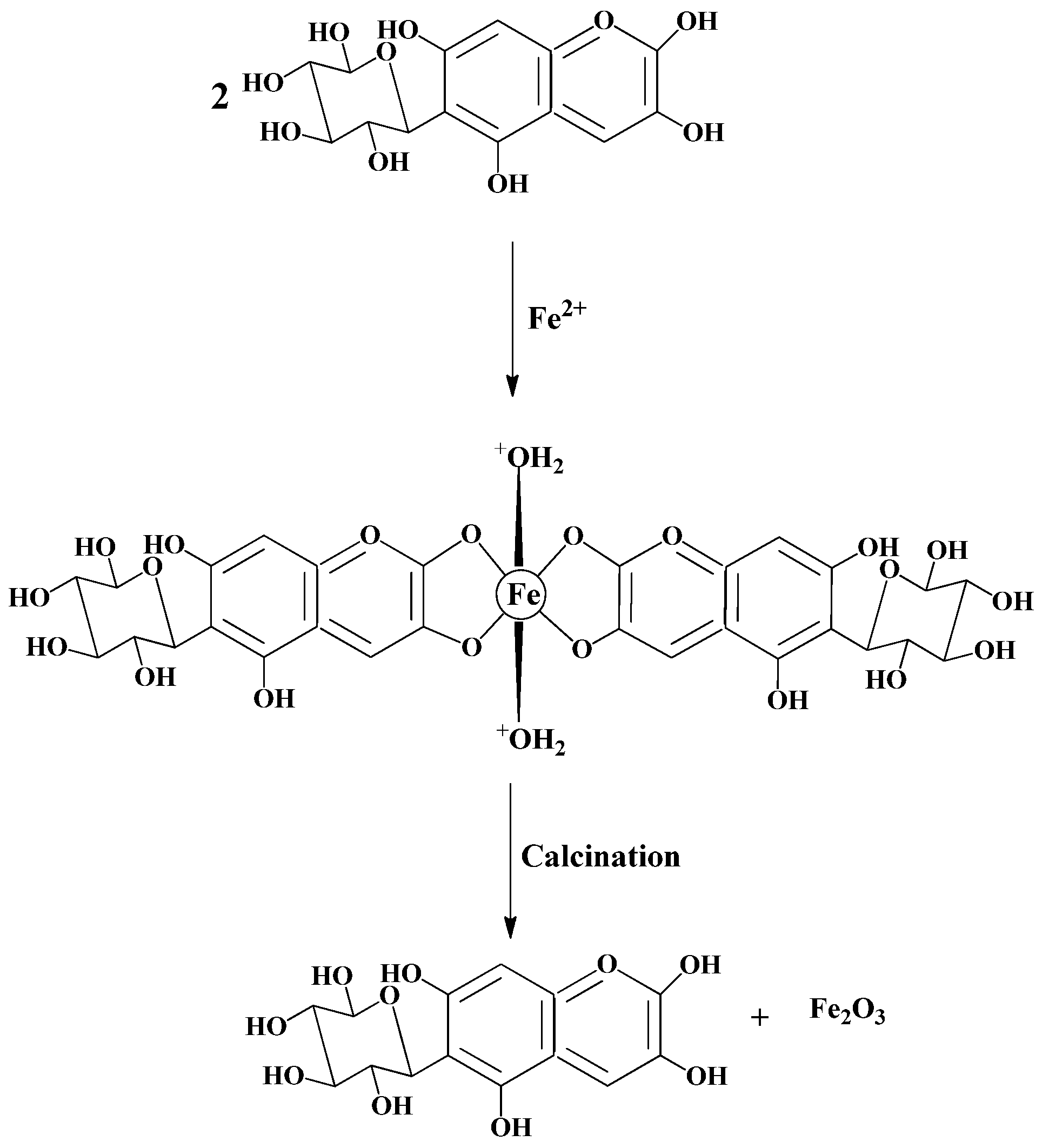
2.1. Synthesis Process

The synthesis process involves iron(III) salt (Fe3+) as a precursor and plant extract as the source of -COOH, -OH, CHO, and CN. Sodium hydroxide solution helps drive the reaction in the forward direction. pH is directly related to the stability of nanoparticles. The change in pH can alter the double-layer properties that can directly influence the zeta potential of the system, because each type of nanoparticles is stable near the isoelectric point. In the current synthesis reaction, the ferric nitrate breaks down into Fe3+ and (3NO3), then the phyto-components present in the leaf extract of Musa paradisiaca induce the formation of Fe2O3 NPs [48]. The formation of the Fe2O3 nanoparticles proceeded according to the general mechanism given in Scheme 1 [49].
Similar to the formation process of metal oxides, thermal energy is needed to remove the water, oxidize the metal, and improve the crystallinity. The increase in crystallinity has been reported to increase the kinetic energy of the electrons and reduce the band gap energy [50,51]. Calcination temperature and the duration of the calcination process determine the amount of energy imparted on the nanoparticles. A higher calcination temperature and longer reaction time imply more thermal energy being absorbed by the nanoparticles. The elevated thermal energy causes Fe2O3 nanoparticles to lose their surface energy. Hence, thermal energy functions as a driving force, influencing the nanoparticle surface’s interaction with adjacent particles.
From a thermodynamic perspective, when two particles come into contact, atomic transport occurs between them during calcination. The phenomenon is widely recognized as the Ostwald ripening process. These particles interact, reducing their boundary energy, and grain boundary migration takes place, moving from one particle to its neighboring particles through surface diffusion [52]. Throughout this process, one particle rotates towards another particle to align their crystal planes. Eventually, this process results in the formation of a new, larger particle. These new particles are denser than their initial counterparts [52]. Consequently, Fe2O3 nanoparticles subjected to higher calcination temperatures and longer reaction times exhibit increased X-ray density.

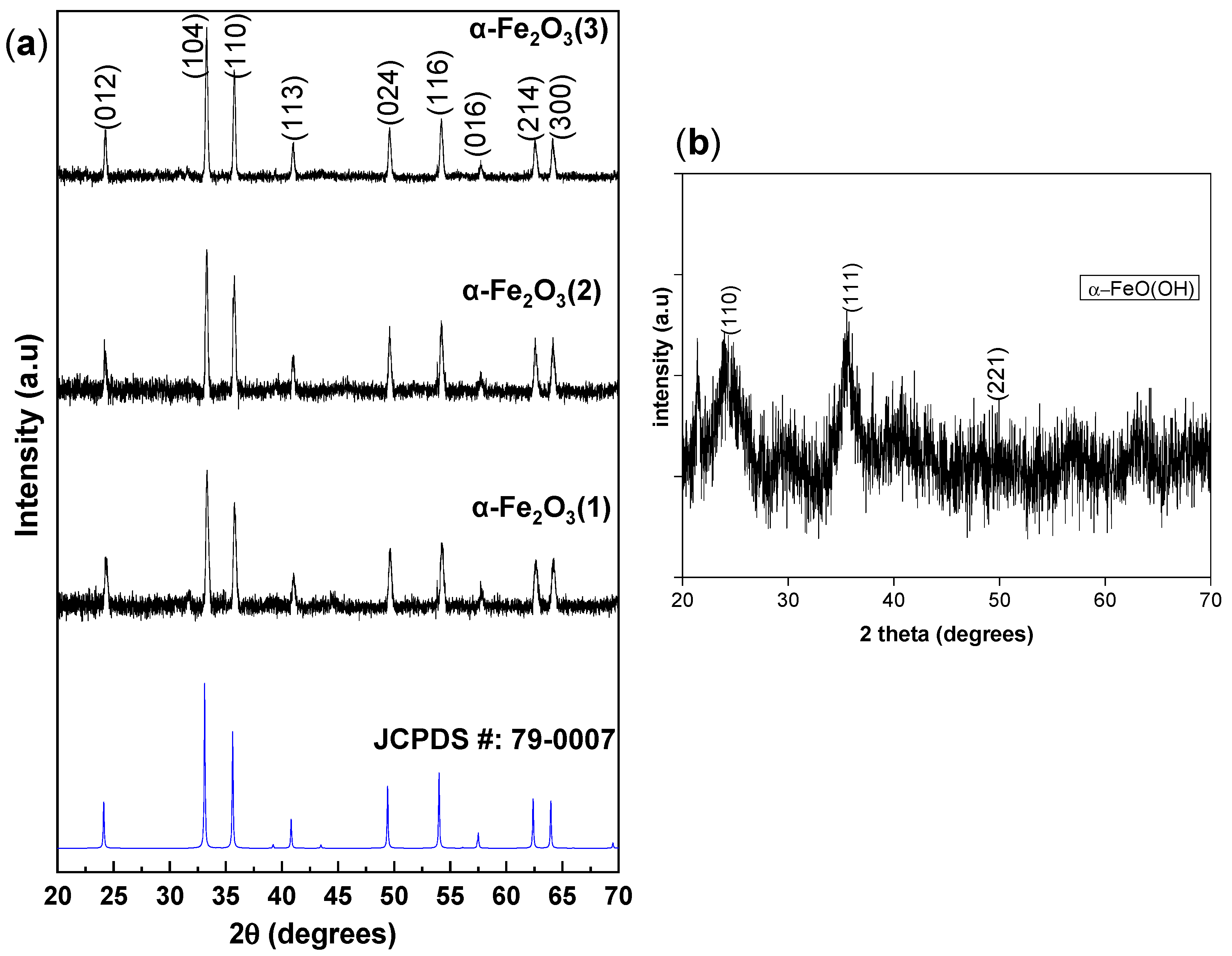
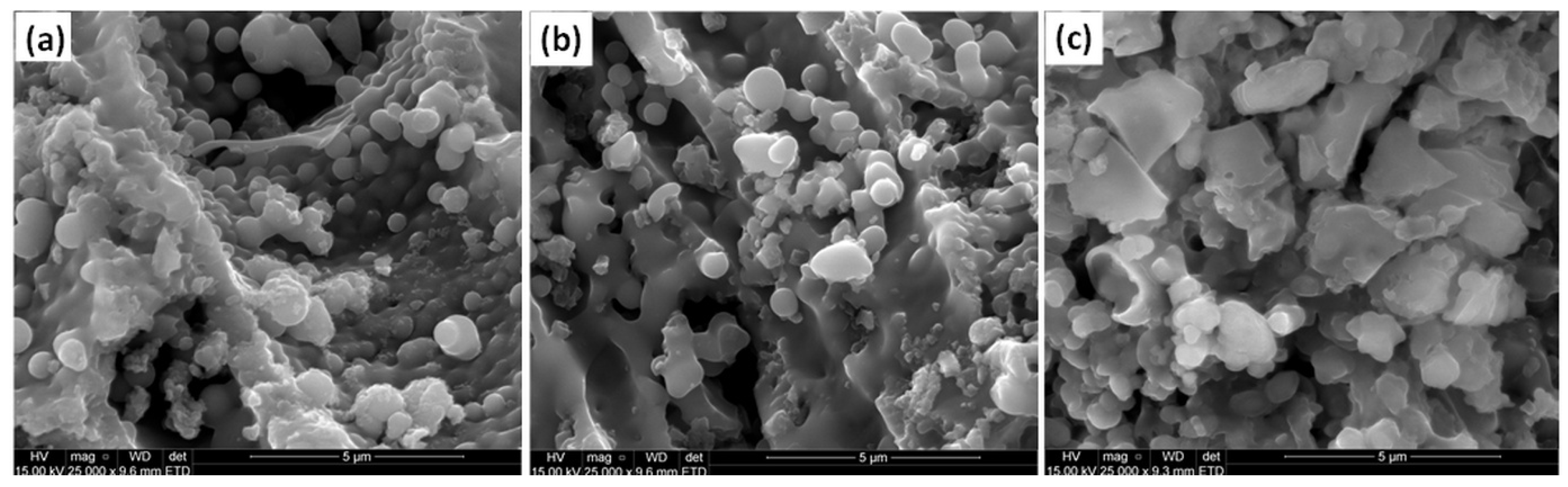
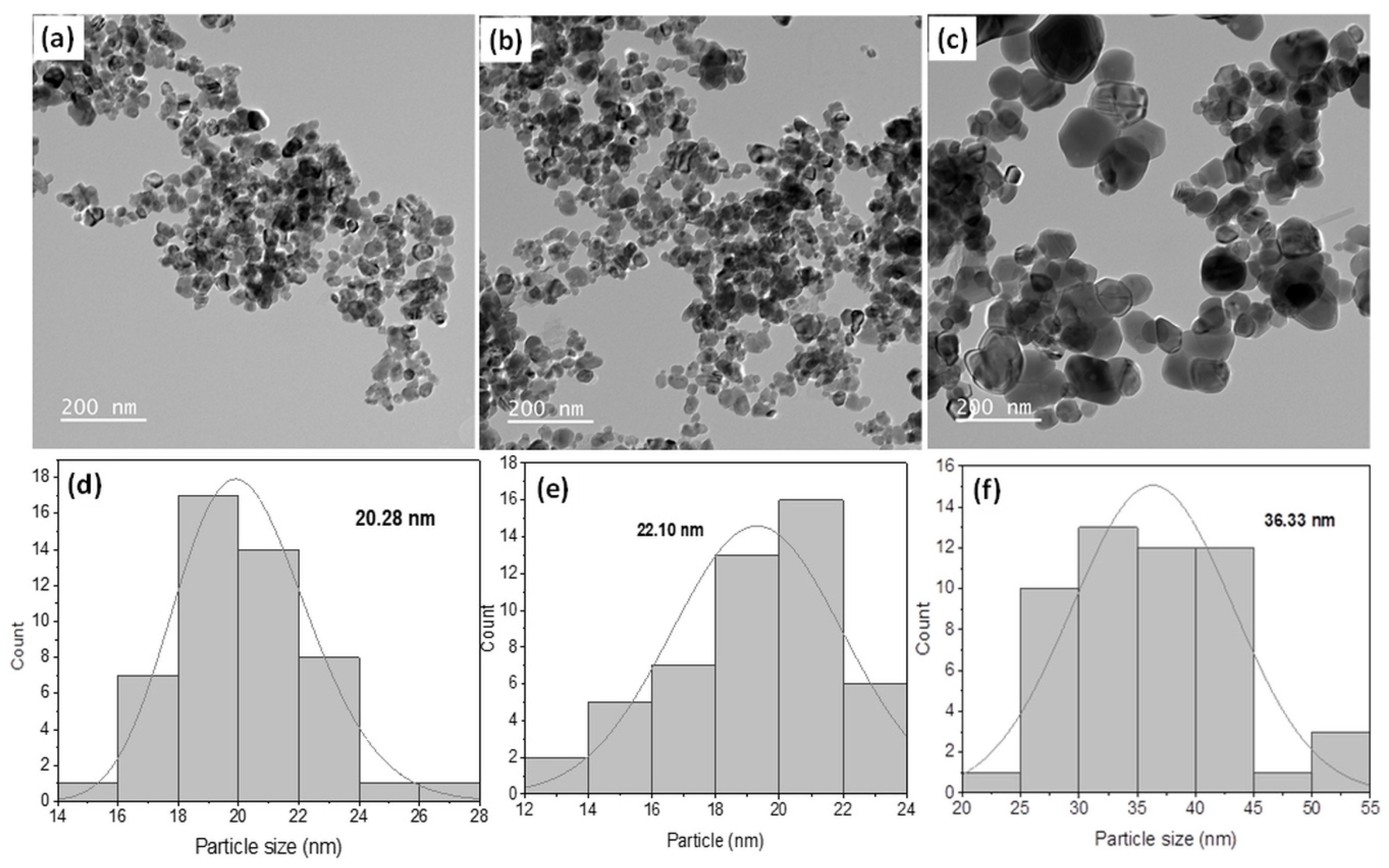
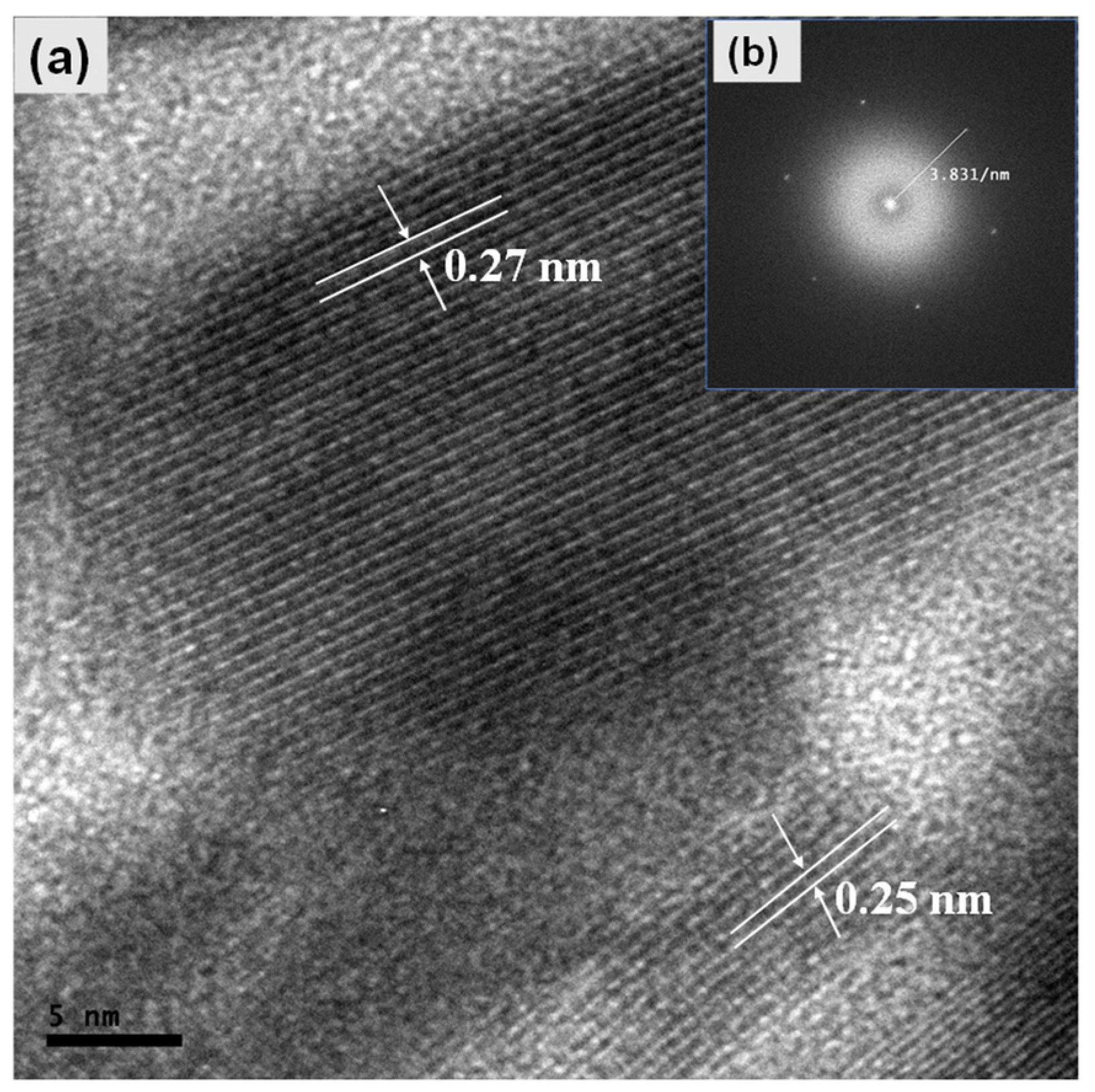
2.2. Microstructural and Morphological Properties

The purity and crystallinity of the obtained nanoparticles were ascertained by powder X-ray diffraction (XRD) measurement. Figure 1a presents the diffraction patterns of the nanoparticles at different calcination temperatures of 650, 750, and 900 °C, respectively. All the patterns show the diffraction peaks that correspond well to the (012), (104), (110), (113), (024), (116), (214), and (300) planes of the rhombohedral structure of α-Fe2O3 phase (JCPDS card No: 79–0007) [53]. Irrespective of the calcination temperature, they all belong to the same crystal structure and space group of R-3c. The lattice constants are α = 5.0285 Å and c = 13.7360 Å [54]. The sample calcined at temperatures below 650 °C showed poor crystallinity (Figure 1b) with no distinct peak observed. The (110), (111), and (221) peaks of α-FeO(OH) could be observed, indicating that this hydroxide is the intermediate product, and at a temperature above 650 °C, the FeOOH is converted to α-Fe2O3 hematite phase. This implies that the samples calcined below 650 °C were amorphous with no crystalline growth. There are no peaks that are associated with other crystalline phases of Fe2O3, including the β-Fe2O3, ε-Fe2O3, and γ-Fe2O3, or oxides such as Fe3O4 (magnetite) and FeO(OH) in the diffraction patterns, indicating the high purity of the α-Fe2O3 nanoparticles.
The average crystallite size (D) of the Fe2O3 NPs was estimated using Scherrer’s Equation (1) [55]:
D = K λ β C o s   θ
Here, λ represents the wavelength of the X-ray (1.5406 Å), K is a shape factor represented as 0.9, θ is the diffraction angle in degrees, and β represents the full width at half maxima (FWHM) of the peaks of interest [43]. In the current study, the two most intense peaks of (104) and (110) were considered for the estimation of the crystallite size to give 25.20, 30.50, and 38.30 nm for the Fe2O3 (1), Fe2O3 (2), and Fe2O3 (3) NPs, respectively. This confirms that higher particle growth is associated with calcination time.
SEM was used to explore the morphological characteristics, and Figure 2a–c illustrates the morphology of the synthesized Fe2O3 (1), Fe2O3 (2), and Fe2O3 (3) samples, respectively. The figures reveal spherical shapes with some observable agglomeration of grains with irregular shapes as the calcination time increased. The formation of the powder seems to have occurred through the coalescence of grains, possibly accounting for the irregular spherical shapes and aggregated particles observed in the final grains of the samples calcinated at 900 °C. Similar observations are made in previous studies involving the hydrothermal synthesis of α-Fe2O3 [56,57].
TEM was further used to examine the particle size and morphology of the biosynthesized nanoparticles (Figure 3). A quasi-spherically shaped nanoparticles with an average diameter of 20.28, 28.10, and 36.33 nm could be observed in samples Fe2O3 (1), Fe2O3 (2), and Fe2O3 (3), respectively. Similar morphologies were earlier reported for single-crystalline hematite α-Fe2O3 via the hydrothermal method [58,59]. Figure 4 shows the corresponding high-resolution TEM (HRTEM), and in the inset of Figure 4 is the fast Fourier transform (FFT), which shows it to be α-Fe2O3. The distinct, well-defined crystalline patterns reveal two different interplanar spacings of 0.25 and 0.27 nm, corresponding to the (1 1 0) and (1 0 4) planes of α-Fe2O3, respectively.

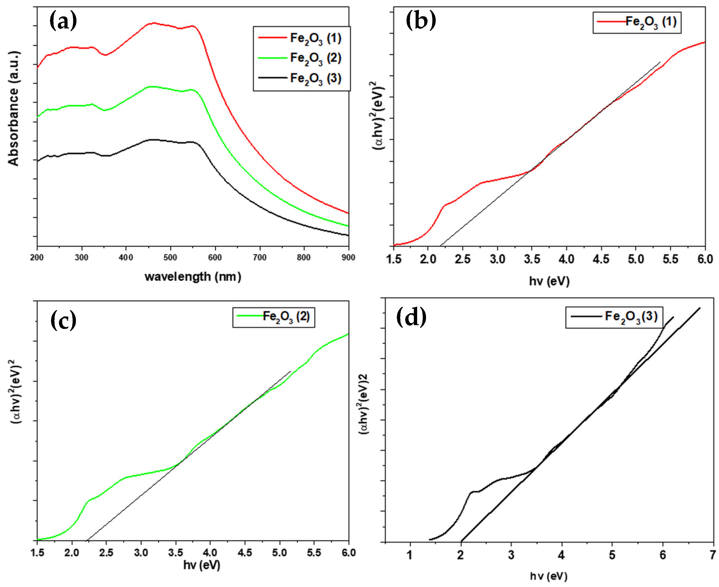
2.3. Optical Characterization

To explore the impact of calcination time on the optical characteristics of the Fe2O3 nanoparticles, UV-vis spectroscopy analysis was conducted at room temperature within the 200–900 nm range for all calcined samples (Figure 5). The optical band gap values for these samples, processed at various calcination times, were calculated from the absorption spectra utilizing the Tauc Equation (2) [60].
αhυ = A(hυ = Eg)n
where h is Planck’s constant (6.626 × 10⁻³⁴ Js), n is an exponential factor for the electronic transition (n = ½, for indirect band, n = 2, for direct band), and α is the absorption coefficient.
α-Fe2O3 is a direct band-gap-semiconducting material; hence, a plot of the values of (αhν)2 versus photon energy (hν) is presented in Figure 5b–d for Fe2O3 (1), Fe2O3 (2), and Fe2O3 (3), respectively. The optical band gap at different calcination times was deduced by the linear extrapolation of (αhυ)2 versus hυ to the energy axis [61]. A decrease in the optical band gap from 2.5 eV in Fe2O3 (1) to 2.0 eV in Fe2O3 (3) was observed as the calcination temperature increased. This reduction in the energy band gap with temperature increase can be attributed to the increase in particle size and the improvement in crystallinity, as confirmed by XRD analysis. The rationale behind this phenomenon lies in the larger particle size, which leads to a higher number of atoms constituting a particle. Consequently, the valence and conduction electrons become more strongly attracted to the ions at the core of the particles, thereby causing a decrease in the band gap of the particle.

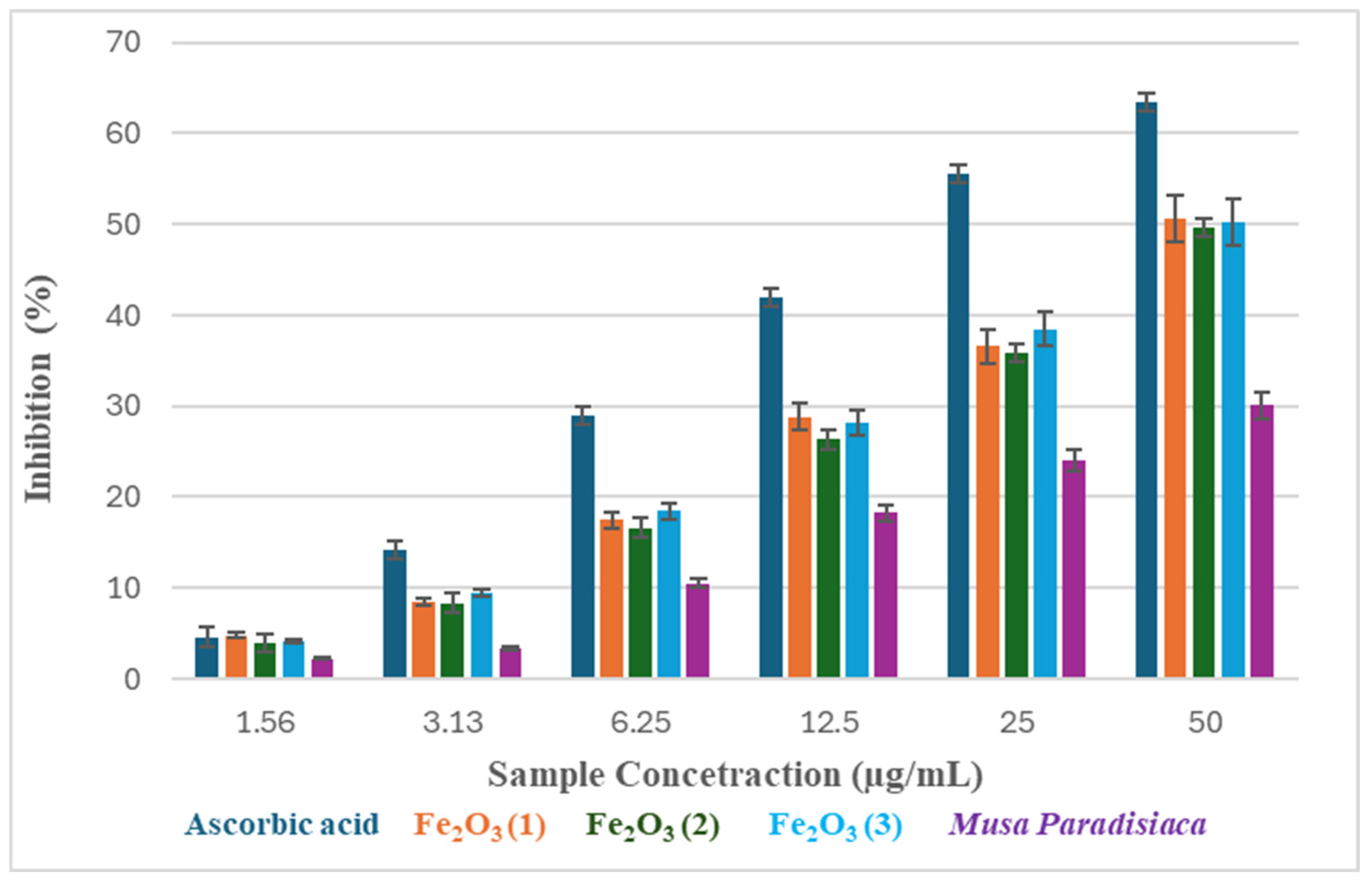
2.4. Antioxidant and Anticancer Studies

The antioxidant activity of green-synthesized Fe2O3 nanoparticles and peels of Musa paradisiaca was evaluated using the DPPH radical inhibition assay. The result of the percentage inhibition is presented in Table 1 and Figure 6. DPPH is a well-known nitrogen-centered radical with a deep purple color [62,63]. Antioxidants donate electrons or hydrogen atoms to the radical to neutralize the DPPH, forming a yellow-colored non-radical DPPH [62,64,65]. As observed in this study, the nanoparticles and peels demonstrated a concentration-dependent inhibition of DPPH radicals. The potency of the nanoparticles and extracts was measured based on the concentration of the sample that inhibited 50% of the radical (IC50). Ascorbic acid, used as a standard, with an IC50 value of 30.17 µg/mL showed the best antioxidant activity, while Fe2O3 (1) and Fe2O3 (2) with IC50 values of 46.84 and 46.14 µg/mL, respectively, showed similar antioxidant potentials. The least antioxidant activity was observed for the peel extract, which had an IC50 value of 79.26 µg/mL. Previous studies show that Musa paradisiaca peels are rich in extractable polyphenols and tannins that are effective as radical scavengers [66]. It was demonstrated that the peel is a good antioxidant, with its polar components contributing to the observed effect [67]. Overall observation in this study agrees with other studies on Musa paradisiaca peel as a potent antioxidant source [66,67]. Reports are only available for biosynthesized zinc oxide nanoparticles from plantain peel, and the results demonstrate the biological potential of the nanoparticles [68]. Antioxidants from plants are good reducing and stabilizing agents, able to enhance metal oxide nanoparticle synthesis from metal salts as precursors [69]. The high polyphenolic and tannins previously reported in Musa paradisiaca peel could favor the bio-fabrication of the iron oxide nanoparticles, with subsequent improved DPPH radical inhibition observed in this study.
The results in Table 2 and Figure 7 provide the inhibitory efficacies of the iron oxide nanoparticles on cell growth and proliferation of HEK 293 (Human Embryonic Kidney cells) and HeLa (Cervical Cancer) cell lines. As observed in the result, variation in the IC50 values across the samples and cell lines reflects their behavior with different cell lines. In addition, the results from the study show that the viability of the cell lines decreases with increasing concentration of the samples, indicating a concentration-dependent efficacy of the samples. General observation showed that the iron oxide nanoparticles, peels, and 5-FU might have a stronger inhibitory effect on the HEK 293 cells compared to the HeLa cells. By implication, the HEK 293 cells might be more susceptible to the drug samples, and a lower concentration might even be sufficient to achieve the inhibition of normal cell proliferation. Furthermore, Fe2O3 (2) and Fe2O3 (3), on the other hand, showed better efficacy against the HeLa cell line than HEK 293, implying that subtle variations in the preparation or composition of the nanoparticles can significantly influence the biological response, and consequently, the anticancer efficacy. Asimeng et al. [70] reported a concentration-dependent inhibition of HeLa cell proliferation after treatment with iron oxide nanoparticles synthesized from maize leaves. It was observed from the study that better anticancer efficacies were observed for particles with smaller sizes. The effectiveness of the smaller size particles against cancer cells could be attributed to the ability of the nanoparticles to penetrate pores more effectively due to their ability to go through the blood–brain barrier [71]. The same nanoparticles synthesized using Terminalia catappa showed similar inhibition of HEK 293 and HeLa cell lines with IC50 values of 44.12 and 34.94 μg, respectively [72]. Overall observation in this study is the cytotoxic effects of the nanoparticles and Musa paradisiaca peel on the normal cell (HEK 293); however, modification of the bio-fabrication methods might result in better observation. Although nanoparticles are known for their various biomedical applications, toxicity to normal cell lines such as HEK-293 has been reported [73]. This toxicity is associated with nanoparticle size, dose, agglomeration, and the route of administration [73]. Based on the IC50 values recorded in this study, it is clear that the nanoparticles and Musa paradisiaca peel extracts were more toxic to HEK-293 cells compared to HeLa cells, whereas the Fe2O3 demonstrated better selective toxicity by inhibiting the cancer cell line more than the normal cell line, thus indicating a better therapeutic window [59,69,74].
Fe2O3 nanoparticles can cause antitumor effects in several ways, such as by producing reactive oxygen species (ROS), upsetting the cellular redox equilibrium, and causing mitochondrial malfunction, which in turn triggers apoptosis [75,76]. Furthermore, ferroptosis, a controlled type of cell death marked by iron-dependent lipid peroxidation, may be involved [77]. Given the intrinsic iron content of Fe2O3 nanoparticles, it is plausible that when cancer cells internalize them, intracellular iron levels rise, causing lipid ROS buildup and Fenton-type reactions that may lead to ferroptosis [78]. Ferroptosis may contribute to the initiation of oxidative stress and mitochondrial impairment, thereby activating apoptotic pathways [79,80].

3. Materials and Methods

3.1. Chemicals

High-purity (99%) ferric nitrate nonahydrate Fe(NO3)3·9 H2O and sodium hydroxide (NaOH) were purchased from Merck, Riyadh, Saudi Arabia, and used as received. Ethanol was procured from ACE Chemicals, Riyadh, Saudi Arabia.

3.2. Collection and Preparation of Aqueous Plant Extract

Musa paradisiaca (plantain) was sourced locally from a farm and properly identified. The peels were removed, properly cleaned with water, and then chopped into smaller bits. An electric blender (TOSCANA U 2500 W 2-in-1 Electric Blender Multi-Function, China) was used to blend about 500 g in 1000 mL of distilled water. The obtained pulp was filtered via a muslin cloth and was kept for a few minutes to allow the settling of the sediments, and the supernatant obtained was then filtered through a Whatman No 1 filter paper with a pore size of 25 µm.

3.3. Synthesis of α-Fe2O3 Nanoparticles

A 100 mL aqueous solution of ferric nitrate nonahydrate Fe(NO3)3·9 H2O was heated to 80 °C, and 50 mL of the extract of Musa paradisiaca was added dropwise. The pH of the solution was adjusted to 8 with a few drops of a solution of sodium hydroxide (NaOH) and stirred for 30 min. The product was poured into a Teflon-lined stainless-steel reactor and placed in a muffle furnace with the temperature maintained at 150 °C for 6 h [81]. The obtained product was centrifuged at 4300 rpm for 20 min, followed by thorough rinsing with distilled water and finally with ethanol. The crude precipitate was divided into 3 portions, which were transferred to different crucibles and calcinated at 650, 750, and 900 °C for 4 h to afford samples denoted as Fe2O3 (1), Fe2O3 (2), and Fe2O3 (3).

3.4. Characterization

All sample characterizations were carried out at room temperature and ambient conditions. No control over pressure or humidity was necessary except if stated otherwise. Ultraviolet-visible spectroscopy (UV–Vis, Merck Prove 300, by Merck KGaA, Darmstadt, Germany) was used for optical characteristics, X-ray diffraction (a Bruker D8 Advance X-ray diffractometer, from Bruker AXS GmbH, Karlsruhe, Germany) was used for crystallographic information and phase information. The external morphology was studied using a field emission scanning electron microscope (FESEM, FEI Inspect F50 produced in Eindhoven, The Netherlands) and transmission electron microscope (TEM, FEI Tecnai G2 20 S-Twin produced in Eindhoven, The Netherlands) for internal morphology, high-resolution imaging, and selected area diffraction.

3.5. Biological Activity

3.5.1. Antioxidant Activity Measurement Using DPPH Scavenging Assay

The antioxidant activity of nanoparticles has been extensively measured using the DPPH scavenging assay [82,83]. Scavenging of (1,1-diphenyl-2-picrylhydrazyle) (DPPH) was utilized to assess the free radical scavenging activity of the green synthesized Fe2O3 nanoparticles. Six different concentrations (1.56–50 µg/mL) of the nanoparticles were prepared and to these different concentrations was added 1 mL of 0.1 mM DPPH in 100 percent ethanol. The solutions were analyzed after a 30 min incubation period at room temperature in the dark. The measurement was conducted by sampling 250 μL of each solution into a 96-well microplate in triplicates and measuring the absorbance at 517 nm in comparison to a blank. Ascorbic acid and plantain peels were used as positive and negative controls. The percentage of free radical scavenging activity of the DPPH was estimated using Equation (3) [84]:
DPPH   scavenging   activity   ( % ) = A o       A t       A o
where Ao represents the absorbance of the control at 30 min and At is the absorbance of Fe2O3 (1), Fe2O3 (2), and Fe2O3 (3).

3.5.2. Cytotoxicity Activity Measurement Using MTT Assay of the Nanoparticles

Human embryonic kidney 293 (HEK 293) and human cervical carcinoma (HeLa) cells were obtained from the ATCC in Manassas, USA. These cell lines were cultured in 25 cm2 tissue culture flasks using EMEM (Lonza BioWhittaker, Verviers, Belgium) supplemented with 10% fetal bovine serum, 100 U/mL penicillin, and 100 mg/mL streptomycin. To assess cell viability in HeLa and HEK 293 cell lines, the MTT assay was conducted in 96-well plates, seeding 2.5 × 10² cells per well in 100 mL of EMEM. The cells were incubated at 37 °C overnight, and the media were replaced. Subsequently, samples at different concentrations (10, 25, 50, and 100 µg) were added. After another overnight incubation at 37 °C, the medium was replaced, and the samples were added again at varying concentrations. The prepared solutions were then incubated for 48 h at 37 °C, followed by the MTT assay. Additionally, 5-fluorouracil (5FU) was used as a standard alongside untreated cells. In the assay, 50% of the MTT reagent (5 mg/mL in PBS) was replaced and incubated for 4 h at 37 °C. Afterwards, 100 mL of DMSO was used to dissolve the insoluble formazan crystals, and the absorbance was measured at 570 nm using a Mindray MR-96A microplate reader (Vacutec, Hamburg, Germany), with DMSO as the blank. The entire test was performed in triplicate.

4. Conclusions

Pure single-phase hematite α-Fe2O3 nanoparticles were successfully synthesized by an environmentally friendly route using the waste peels of Musa paradisiaca. The calcination temperature was varied, and the goal of determining the optimal conditions for obtaining high-quality nanoparticles while conserving energy was achieved. Reaction temperature affected the structure, size, and optical properties of the nanoparticles. The particles’ crystallinity was found to improve with calcination temperature. Furthermore, calcinating the powder at higher temperature facilitated boundary migration and the movement of one particle to its neighboring particles through surface diffusion impurity. Anticancer and antioxidant assays of the green synthesized nanoparticles showed that they display a concentration-dependent efficiency. Both Fe2O3 (1) and Fe2O3 (2) demonstrated comparable antioxidant potentials with IC50 values of 46.84 and 46.14 µg/mL, respectively, while the peel extract showed the lowest activity with an IC50 of 79.26 µg/mL. The nanoparticles, peels, and 5-FU (used as a standard) exhibited a stronger inhibitory effect on HEK 293 cells compared to HeLa cells. This suggests that HEK 293 cells may be more susceptible to the drug samples, and a lower concentration might suffice to inhibit normal cell proliferation, thus indicating a better therapeutic window.

Author Contributions

Conceptualization, writing—original draft preparation, S.E. and H.F.; methodology, O.E.O. and S.E.; validation, O.E.O., S.E., H.F. and D.C.O.; formal analysis, O.E.O., S.E., H.F. and D.C.O.; writing—review and editing, H.F. and D.C.O.; funding acquisition, H.F. All authors have read and agreed to the published version of the manuscript.

Funding

This work was supported and funded by the Deanship of Scientific Research at Imam Mohammad Ibn Saud Islamic University (IMSIU) (grant number IMSIU-DDRSP2501).

Institutional Review Board Statement

Not applicable.

Informed Consent Statement

Not applicable.

Data Availability Statement

The original contributions presented in this study are included in the article. Further inquiries can be directed to the corresponding author.

Acknowledgments

Authors acknowledge the support and funding of the Deanship of Scientific Research at Imam Mohammad Ibn Saud Islamic University (IMSIU).

Conflicts of Interest

The authors declare no conflicts of interest.

References

  1. Begum, N.A.; Mondal, S.; Basu, S.; Laskar, R.A.; Mandal, D. Biogenic synthesis of Au and Ag nanoparticles using aqueous solutions of Black Tea leaf extracts. Colloids Surf. B Biointerfaces 2009, 71, 113–118. [Google Scholar] [CrossRef] [PubMed]
  2. Singaravelu, G.; Arockiamary, J.; Kumar, V.G.; Govindaraju, K. A novel extracellular synthesis of monodisperse gold nanoparticles using marine alga, Sargassum wightii Greville. Colloids Surf. B Biointerfaces 2007, 57, 97–101. [Google Scholar] [CrossRef] [PubMed]
  3. Sani, A.; Murad, A.; Hassan, D.; Channa, G.M.; El-Mallul, A.; Medina, D.I. Photo-catalytic and biomedical applications of one-step, plant extract-mediated green-synthesized cobalt oxide nanoparticles. Environ. Sci. Pollut. Res. 2023, 30, 20736–20745. [Google Scholar] [CrossRef] [PubMed]
  4. Chavali, M.S.; Nikolova, M.P. Metal oxide nanoparticles and their applications in nanotechnology. SN Appl. Sci. 2019, 1, 607. [Google Scholar] [CrossRef]
  5. Joshi, N.; Pandey, D.K.; Mistry, B.G.; Singh, D.K. Metal oxide nanoparticles: Synthesis, properties, characterization, and applications. In Nanomaterials: Advances and Applications; Singh, D.K., Singh, S., Singh, P., Eds.; Springer Nature: Singapore, 2023; pp. 103–144. [Google Scholar]
  6. Murthy, S.; Effiong, P.; Fei, C.C. 11—Metal oxide nanoparticles in biomedical applications. In Metal Oxide Powder Technologies; Al-Douri, Y., Ed.; Elsevier: Amsterdam, The Netherlands, 2020; pp. 233–251. [Google Scholar]
  7. Yoon, Y.; Truong, P.L.; Lee, D.; Ko, S.H. Metal-oxide nanomaterials synthesis and applications in flexible and wearable sensors. ACS Nanosci. Au 2022, 2, 64–92. [Google Scholar] [CrossRef]
  8. Nowsherwan, G.A.; Ali, Q.; Ali, U.F.; Ahmad, M.; Khan, M.; Hussain, S.S. Advances in organic materials for next-generation optoelectronics: Potential and challenges. Organics 2024, 5, 520–560. [Google Scholar] [CrossRef]
  9. Iravani, S. Green synthesis of metal nanoparticles using plants. Green Chem. 2011, 13, 2638–2650. [Google Scholar] [CrossRef]
  10. Kharissova, O.V.; Dias, H.V.; Kharisov, B.I.; Pérez, B.O.; Pérez, V.M. The greener synthesis of nanoparticles. Trends Biotechnol 2013, 31, 240–248. [Google Scholar] [CrossRef]
  11. Şahin, A.; Altınsoy, Ş.; Kızılbey, K. An approach for cationic dyes removal from wastewater: Green synthesis of iron nanoparticles using Prunus avium stems extracts. Kuwait J. Sci. 2024, 51, 100226. [Google Scholar] [CrossRef]
  12. Buarki, F.; AbuHassan, H.; Al Hannan, F.; Henari, F. Green synthesis of iron oxide nanoparticles using Hibiscus rosa sinensis flowers and their antibacterial activity. J. Nanotechnol. 2022, 2022, 5474645. [Google Scholar] [CrossRef]
  13. Keshta, B.E.; Gemeay, A.H.; Kumar Sinha, D.; Elsharkawy, S.; Hassan, F.; Rai, N.; Arora, C. State of the art on the magnetic iron oxide nanoparticles: Synthesis, functionalization, and applications in wastewater treatment. Results Chem. 2024, 7, 101388. [Google Scholar] [CrossRef]
  14. Wu, W.; He, Q.; Jiang, C. Magnetic iron oxide nanoparticles: Synthesis and surface functionalization strategies. Nanoscale Res. Lett. 2008, 3, 397. [Google Scholar] [CrossRef] [PubMed]
  15. Roca, A.G.; Gutiérrez, L.; Gavilán, H.; Brollo, M.E.F.; Veintemillas-Verdaguer, S.; del Puerto Morales, M. Design strategies for shape-controlled magnetic iron oxide nanoparticles. Adv. Drug Deliv. Rev. 2019, 138, 68–104. [Google Scholar] [CrossRef]
  16. Panneerselvam, C.; Alshehri, M.A.; Saif, A.; Faridi, U.; Khasim, S.; Mohammedsaleh, Z.M.; Parveen, H.; Omer, N.; Alasmari, A.; Mukhtar, S. Green synthesis of Abutilon indicum (L) derived iron oxide (FeO) nanoparticles with excellent biological, anticancer and photocatalytic activities. Polyhedron 2024, 257, 117022. [Google Scholar] [CrossRef]
  17. Duan, Y.-T.; Soni, K.; Patel, D.; Choksi, H.; Sangani, C.B.; Saeed, W.S.; Ameta, K.L.; Ameta, R.K. Green synthesis of iron oxide nanoparticles using Nicotiana plumbaginifolia and their biological evaluation. J. Mol. Liq. 2024, 396, 123985. [Google Scholar] [CrossRef]
  18. Ali, A.; Zafar, H.; Zia, M.; ul Haq, I.; Phull, A.R.; Ali, J.S.; Hussain, A. Synthesis, characterization, applications, and challenges of iron oxide nanoparticles. Nanotechnol. Sci. Appl. 2016, 9, 49–67. [Google Scholar] [CrossRef]
  19. Tringides, M.C.; Jałochowski, M.; Bauer, E. Quantum size effects in metallic nanostructures. Phys. Today 2007, 60, 50–54. [Google Scholar] [CrossRef]
  20. Li, Q.; Kartikowati, C.W.; Horie, S.; Ogi, T.; Iwaki, T.; Okuyama, K. Correlation between particle size/domain structure and magnetic properties of highly crystalline Fe3O4 nanoparticles. Sci. Rep. 2017, 7, 9894. [Google Scholar] [CrossRef]
  21. Veisi, H.; Ozturk, T.; Karmakar, B.; Tamoradi, T.; Hemmati, S. In situ decorated Pd NPs on chitosan-encapsulated Fe3O4/SiO2-NH2 as magnetic catalyst in Suzuki-Miyaura coupling and 4-nitrophenol reduction. Carbohydr. Polym. 2020, 235, 115966. [Google Scholar] [CrossRef]
  22. Tang, N.; Zhong, W.; Jiang, H.; Wu, X.; Liu, W.; Du, Y. Nanostructured magnetite (Fe3O4) thin films prepared by sol–gel method. J. Magn. Magn. Mater. 2004, 282, 92–95. [Google Scholar] [CrossRef]
  23. He, C.; Wu, S.; Zhao, N.; Shi, C.; Liu, E.; Li, J. Carbon-encapsulated Fe3O4 nanoparticles as a high-rate lithium ion battery anode material. ACS Nano 2013, 7, 4459–4469. [Google Scholar] [CrossRef] [PubMed]
  24. Shete, P.; Patil, R.; Tiwale, B.; Pawar, S. Water dispersible oleic acid-coated Fe3O4 nanoparticles for biomedical applications. J. Magn. Magn. Mater. 2015, 377, 406–410. [Google Scholar] [CrossRef]
  25. Liu, H.-L.; Ko, S.P.; Wu, J.-H.; Jung, M.-H.; Min, J.H.; Lee, J.H.; An, B.H.; Kim, Y.K. One-pot polyol synthesis of monosize PVP-coated sub-5 nm Fe3O4 nanoparticles for biomedical applications. J. Magn. Magn. Mater. 2007, 310, e815–e817. [Google Scholar] [CrossRef]
  26. Basnet, P.; Larsen, G.K.; Jadeja, R.P.; Hung, Y.-C.; Zhao, Y. α-Fe2O3 nanocolumns and nanorods fabricated by electron beam evaporation for visible light photocatalytic and antimicrobial applications. ACS Appl. Mater. Interfaces 2013, 5, 2085–2095. [Google Scholar] [CrossRef]
  27. Jana, T.; Pal, A.; Mandal, A.; Sarwar, S.; Chakrabarti, P.; Chatterjee, K. Photocatalytic and antibacterial performance of α-Fe2O3 nanostructures. ChemistrySelect 2017, 2, 3068–3077. [Google Scholar] [CrossRef]
  28. Al-Tememe, E.; Algalal, H.M.A.A.; Abodood, A.A.F.; Mohammed, K.A.; Khamees, E.J.; Zabibah, R.S.; Abed, A.S. Anticancer and Antimicrobial activity of PVA/Fe2O3/TiO2 hybrid nanocomposite. Int. J. Nanosci. 2022, 21, 2250018. [Google Scholar] [CrossRef]
  29. Yoonus, J.; Resmi, R.; Beena, B. Evaluation of antibacterial and anticancer activity of green synthesized iron oxide (α-Fe2O3) nanoparticles. Mater. Today Proc. 2021, 46, 2969–2974. [Google Scholar] [CrossRef]
  30. Athithan, A.S.; Jeyasundari, J.; Jacob, Y. Biological synthesis, physico-chemical characterization of undoped and Co doped α-Fe2O3 nanoparticles using tribulus terrestris leaf extract and its antidiabetic, antimicrobial applications. Adv. Nat. Sci. Nanosci. Nanotechnol. 2021, 12, 045003. [Google Scholar] [CrossRef]
  31. Ansari, M.A.; Asiri, S.M.M. Green synthesis, antimicrobial, antibiofilm and antitumor activities of superparamagnetic γ-Fe2O3 NPs and their molecular docking study with cell wall mannoproteins and peptidoglycan. Int. J. Biol. Macromol. 2021, 171, 44–58. [Google Scholar] [CrossRef]
  32. Bhattacharya, K.; Gogoi, B.; Buragohain, A.; Deb, P. Fe2O3/C nanocomposites having distinctive antioxidant activity and hemolysis prevention efficiency. Mater. Sci. Eng. C 2014, 42, 595–600. [Google Scholar] [CrossRef]
  33. Shams, S.; Khan, A.U.; Yuan, Q.; Ahmad, W.; Wei, Y.; Khan, Z.U.H.; Shams, S.; Ahmad, A.; Rahman, A.U.; Ullah, S. Facile and eco-benign synthesis of Au@ Fe2O3 nanocomposite: Efficient photocatalytic, antibacterial and antioxidant agent. J. Photochem. Photobiol. B Biol. 2019, 199, 111632. [Google Scholar] [CrossRef] [PubMed]
  34. Morsy, R.; Hosny, M.; Reicha, F.; Elnimr, T. Development and characterization of multifunctional electrospun ferric oxide-gelatin-glycerol nanofibrous mat for wound dressing applications. Fibers Polym. 2016, 17, 2014–2019. [Google Scholar] [CrossRef]
  35. Harandi, F.N.; Khorasani, A.C.; Shojaosadati, S.A.; Hashemi-Najafabadi, S. Surface modification of electrospun wound dressing material by Fe2O3 nanoparticles incorporating Lactobacillus strains for enhanced antimicrobial and antibiofilm activity. Surf. Interfaces 2022, 28, 101592. [Google Scholar] [CrossRef]
  36. Lee, J.; Kwon, S.G.; Park, J.-G.; Hyeon, T. Size dependence of metal–insulator transition in stoichiometric Fe3O4 nanocrystals. Nano Lett. 2015, 15, 4337–4342. [Google Scholar] [CrossRef]
  37. Moulana Kareem, M.; Hari Babu, M.; Vijaya Lakshmi, G. Anticancer, antibacterial, antioxidant, and photo-catalytic activities of eco-friendly synthesized Ni nanoparticles. Inorg. Chem. Commun. 2023, 148, 110274. [Google Scholar] [CrossRef]
  38. Mahendran, D.; Kavi Kishor, P.; Geetha, N.; Manish, T.; Sahi, S.; Venkatachalam, P. Efficient antibacterial/biofilm, anti-cancer and photocatalytic potential of titanium dioxide nanocatalysts green synthesised using Gloriosa superba rhizome extract. J. Exp. Nanosci. 2021, 16, 11–30. [Google Scholar] [CrossRef]
  39. Venugopal, N.; Saiprakash, P.; Jayalakshmi, M.; Ram Reddy, Y.; Rao, M.M. A study on the effect of nanosized tin oxide on the electrochemical performance of nanosized nickel hydroxide in alkali solution. J. Exp. Nanosci. 2013, 8, 684–693. [Google Scholar] [CrossRef]
  40. Sagadevan, S.; Vennila, S.; Muthukrishnan, L.; Gurunathan, K.; Oh, W.C.; Paiman, S.; Mohammad, F.; Al-Lohedan, H.A.; Jasni, A.H.; Fatimah, I. Exploring the therapeutic potentials of phyto-mediated silver nanoparticles formed via Calotropis procera (Ait.) R. Br. root extract. J. Exp. Nanosci. 2020, 15, 217–231. [Google Scholar] [CrossRef]
  41. Balraj, B.; Senthilkumar, N.; Potheher, I.V.; Arulmozhi, M. Characterization, antibacterial, anti-arthritic and in-vitro cytotoxic potentials of biosynthesized magnesium oxide nanomaterial. Mater. Sci. Eng. B 2018, 231, 121–127. [Google Scholar] [CrossRef]
  42. Sharma, R.P.; Raut, S.D.; Kadam, A.S.; Mulani, R.M.; Mane, R.S. In-vitro antibacterial and anti-biofilm efficiencies of chitosan-encapsulated zinc ferrite nanoparticles. Appl. Phys. A 2020, 126, 824. [Google Scholar] [CrossRef]
  43. Anu, K.; Hemalatha, J. Magnetic and electrical conductivity studies of zinc doped cobalt ferrite nanofluids. J. Mol. Liq. 2019, 284, 445–453. [Google Scholar] [CrossRef]
  44. Jyothish, B.; Jacob, J. Synthesis and characterization of Ni2+ and Al3+ doped zinc ferrite nanoparticles for antibacterial, antioxidant, and anticancer (MCF-7) analysis. Chem. Phys. Impact 2023, 6, 100209. [Google Scholar] [CrossRef]
  45. Eleazu, C.; Okafor, P. Use of unripe plantain (Musa paradisiaca) in the management of diabetes and hepatic dysfunction in streptozotocin induced diabetes in rats. Interv. Med. Appl. Sci. 2015, 7, 9–16. [Google Scholar] [CrossRef]
  46. Fu, J.; Tu, S.; Yi, G.; Wang, J.; Sheng, O.; Zhang, W. Plantain flour—A beneficial material for the organ and transcriptional profile of kidney of diabetic rats. J. Funct. Foods 2023, 110, 105817. [Google Scholar] [CrossRef]
  47. Khalid, R.; Din, M.I.; Hussain, Z. Eco-friendly synthesis of copper oxide nanomaterial by using Musa paradisiaca leaves extract and their slow pyrolysis or catalytic reduction activities. Next Nanotechnol. 2024, 6, 100041. [Google Scholar] [CrossRef]
  48. Yadav, P.; Manori, S.; Chamoli, P.; Raina, K.K.; Shukla, R.K. Microwave assisted green synthesis of γ-Fe2O3 nanoparticles and their application for photodegradation of ternary dye mixture. J. Mater. Sci. Mater. Electron. 2023, 34, 1065. [Google Scholar] [CrossRef]
  49. Al-Ruqeishi, M.S.; Mohiuddin, T.; Al-Saadi, L.K. Green synthesis of iron oxide nanorods from deciduous Omani mango tree leaves for heavy oil viscosity treatment. Arab. J. Chem. 2019, 12, 4084–4090. [Google Scholar] [CrossRef]
  50. Marcorius, A.; Sulaeman, U.; Afif, M.; Nurfiah, S.; Khanifudin, K.; Afifah, K. The enhanced photocatalytic properties of silver phosphate synthesized under mangosteen peel extract solution. J. Teknol. 2021, 84, 21–27. [Google Scholar] [CrossRef]
  51. Hamid, A.; Haq, S.; Ur Rehman, S.; Akhter, K.; Rehman, W.; Waseem, M.; Ud Din, S.; Hafeez, M.; Khan, A.; Shah, A. Calcination temperature-driven antibacterial and antioxidant activities of fumaria indica mediated copper oxide nanoparticles: Characterization. Chem. Pap. 2021, 75, 4189–4198. [Google Scholar] [CrossRef]
  52. Indrayana, I.; Tjuana, L.; Tuny, M.; Kurnia. Nanostructure and optical properties of Fe3O4: Effect of calcination temperature and dwelling time. J. Phys. Conf. Ser. 2019, 1341, 082044. [Google Scholar] [CrossRef]
  53. Hjiri, M. Highly sensitive NO2 gas sensor based on hematite nanoparticles synthesized by sol–gel technique. J. Mater. Sci. Mater. Electron. 2020, 31, 5025–5031. [Google Scholar] [CrossRef]
  54. Archana, V.; Joseph Prince, J.; Kalainathan, S. Simple one-step leaf extract-assisted preparation of α-Fe2O3 nanoparticles, physicochemical properties, and its sunlight-driven photocatalytic activity on methylene blue dye degradation. J. Nanomater. 2021, 2021, 8570351. [Google Scholar] [CrossRef]
  55. Apte, S.K.; Naik, S.D.; Sonawane, R.S.; Kale, B.B.; Baeg, J.O. Synthesis of nanosize-necked structure α- and γ-Fe2O3 and its photocatalytic activity. J. Am. Ceram. Soc. 2007, 90, 412–414. [Google Scholar] [CrossRef]
  56. Hung, C.M.; Hoa, N.D.; Van Duy, N.; Van Toan, N.; Le, D.T.T.; Van Hieu, N. Synthesis and gas-sensing characteristics of α-Fe2O3 hollow balls. J. Sci. Adv. Mater. Devices 2016, 1, 45–50. [Google Scholar] [CrossRef]
  57. Zeng, Q.Z.; Ma, S.Y.; Jin, W.X.; Yang, H.M.; Chen, H.; Ge, Q.; Ma, L. Hydrothermal synthesis of monodisperse α-Fe2O3 hollow microspheroids and their high gas-sensing properties. J. Alloys Compd. 2017, 705, 427–437. [Google Scholar] [CrossRef]
  58. Yang, Y.; Ma, H.; Zhuang, J.; Wang, X. Morphology-controlled synthesis of hematite nanocrystals and their facet effects on gas-sensing properties. Inorg. Chem. 2011, 50, 10143–10151. [Google Scholar] [CrossRef]
  59. Deepa, P.R.; Vandhana, S.; Jayanthi, U.; Krishnakumar, S. Therapeutic and toxicologic evaluation of anti-lipogenic agents in cancer cells compared with non-neoplastic cells. Basic Clin. Pharmacol. Toxicol. 2012, 110, 494–503. [Google Scholar] [CrossRef]
  60. Tahir, M.; Fakhar-e-Alam, M.; Atif, M.; Mustafa, G.; Ali, Z. Investigation of optical, electrical and magnetic properties of hematite α-Fe2O3 nanoparticles via sol-gel and co-precipitation method. J. King Saud Univ.-Sci. 2023, 35, 102695. [Google Scholar] [CrossRef]
  61. Raja, K.; Mary Jaculine, M.; Jose, M.; Verma, S.; Prince, A.A.M.; Ilangovan, K.; Sethusankar, K.; Jerome Das, S. Sol–gel synthesis and characterization of α-Fe2O3 nanoparticles. Superlattices Microstruct. 2015, 86, 306–312. [Google Scholar] [CrossRef]
  62. Dumore, N.S.; Mukhopadhyay, M. Antioxidant properties of aqueous selenium nanoparticles (ASeNPs) and its catalysts activity for 1, 1-diphenyl-2-picrylhydrazyl (DPPH) reduction. J. Mol. Struct. 2020, 1205, 127637. [Google Scholar] [CrossRef]
  63. Ogunjinmi, O.E.; Ogunjinmi, S.O.; Alayande, S.O.; Nwoke, E.O.; Adedosu, T.A. Evaluation of antioxidant activities of Celosia trigyna (Linn) extracts african extinction vegetable. Science 2020, 8, 102–106. [Google Scholar]
  64. Adegbola, P.; Fadahunsi, O.S.; Alabi, A.A. Comparative antioxidant study of ripe and unripe plantain and the qualitative assessment of some food oil extracts. Ann. Food Sci. Technol. 2018, 19, 758–765. [Google Scholar]
  65. Adenike, A.A.; Adegbola, P.; Fadahunsi, O.S. Antioxidant property and GCMS profile of oil extracted from Cocos nucifera using a fermentation method. BioTechnol. J. Biotechnol. Comput. Biol. Bionanotechnol. 2019, 100, 349–358. [Google Scholar] [CrossRef]
  66. Agama-Acevedo, E.; Sañudo-Barajas, J.; Vélez De La Rocha, R.; González-Aguilar, G.; Bello-Perez, L.A. Potential of plantain peels flour (Musa paradisiaca L.) as a source of dietary fiber and antioxidant compound. CyTA-J. Food 2016, 14, 117–123. [Google Scholar] [CrossRef]
  67. Arun, K.; Persia, F.; Aswathy, P.; Chandran, J.; Sajeev, M.; Jayamurthy, P.; Nisha, P. Plantain peel-a potential source of antioxidant dietary fibre for developing functional cookies. J. Food Sci. Technol. 2015, 52, 6355–6364. [Google Scholar] [CrossRef]
  68. Imade, E.E.; Ajiboye, T.O.; Fadiji, A.E.; Onwudiwe, D.C.; Babalola, O.O. Green synthesis of zinc oxide nanoparticles using plantain peel extracts and the evaluation of their antibacterial activity. Sci. Afr. 2022, 16, e01152. [Google Scholar] [CrossRef]
  69. Jiang, L.; Sun, G.; Zhou, Z.; Sun, S.; Wang, Q.; Yan, S.; Li, H.; Tian, J.; Guo, J.; Zhou, B. Size-controllable synthesis of monodispersed SnO2 nanoparticles and application in electrocatalysts. J. Phys. Chem. B 2005, 109, 8774–8778. [Google Scholar] [CrossRef]
  70. Asimeng, B.O.; Nyankson, E.; Efavi, J.K.; Nii Amarkai, A.; Manu, G.P.; Tiburu, E. Characterization and inhibitory effects of magnetic iron oxide nanoparticles synthesized from plant extracts on HeLa cells. Int. J. Biomater. 2020, 2020, 2630735. [Google Scholar] [CrossRef]
  71. Teleanu, D.M.; Chircov, C.; Grumezescu, A.M.; Volceanov, A.; Teleanu, R.I. Impact of nanoparticles on brain health: An up to date overview. J. Clin. Med. 2018, 7, 490. [Google Scholar] [CrossRef]
  72. Elemike, E.E.; Nna, P.J.; Ikenweke, C.; Onwudiwe, D.; Omotade, E.T.; Singh, M. Synthesis, characterization, anti-cancer and antimicrobial studies of iron oxide nanoparticles mediated by Terminalia catappa (Indian almond) leaf extract. Inorg. Chem. Commun. 2023, 155, 111048. [Google Scholar] [CrossRef]
  73. Liu, X.; Shan, K.; Shao, X.; Shi, X.; He, Y.; Liu, Z.; Jacob, J.A.; Deng, L. Nanotoxic effects of silver nanoparticles on normal HEK-293 cells in comparison to cancerous HeLa cell line. Int. J. Nanomed. 2021, 16, 753–761. [Google Scholar] [CrossRef] [PubMed]
  74. Bethu, M.S.; Netala, V.R.; Domdi, L.; Tartte, V.; Janapala, V.R. Potential anticancer activity of biogenic silver nanoparticles using leaf extract of Rhynchosia suaveolens: An insight into the mechanism. Artif. Cells Nanomed. Biotechnol. 2018, 46, 104–114. [Google Scholar] [CrossRef] [PubMed]
  75. Sankaranarayanan, S.A.; Thomas, A.; Revi, N.; Ramakrishna, B.; Rengan, A.K. Iron oxide nanoparticles for theranostic applications—Recent advances. J. Drug Deliv. Sci. Technol. 2022, 70, 103196. [Google Scholar] [CrossRef]
  76. Estelrich, J.; Busquets, M.A. Iron oxide nanoparticles in photothermal therapy. Molecules 2018, 23, 1567. [Google Scholar] [CrossRef]
  77. Dixon, S.J.; Lemberg, K.M.; Lamprecht, M.R.; Skouta, R.; Zaitsev, E.M.; Gleason, C.E.; Patel, D.N.; Bauer, A.J.; Cantley, A.M.; Yang, W.S.; et al. Ferroptosis: An iron-dependent form of nonapoptotic cell death. Cell 2012, 149, 1060–1072. [Google Scholar] [CrossRef]
  78. Chen, X.; Yu, C.; Kang, R.; Tang, D. Iron Metabolism in Ferroptosis. Front. Cell Dev. Biol. 2020, 8, 590226. [Google Scholar] [CrossRef]
  79. Stockwell, B.R.; Friedmann Angeli, J.P.; Bayir, H.; Bush, A.I.; Conrad, M.; Dixon, S.J.; Fulda, S.; Gascón, S.; Hatzios, S.K.; Kagan, V.E.; et al. Ferroptosis: A regulated cell death nexus linking metabolism, redox biology, and disease. Cell 2017, 171, 273–285. [Google Scholar] [CrossRef]
  80. Sun, X.; Ou, Z.; Chen, R.; Niu, X.; Chen, D.; Kang, R.; Tang, D. Activation of the p62-Keap1-NRF2 pathway protects against ferroptosis in hepatocellular carcinoma cells. Hepatology 2016, 63, 173–184. [Google Scholar] [CrossRef]
  81. Wan, L.; Yan, S.; Wang, X.; Li, Z.; Zou, Z. Solvothermal synthesis of monodisperse iron oxides with various morphologies and their applications in removal of Cr (VI). CrystEngComm 2011, 13, 2727–2733. [Google Scholar] [CrossRef]
  82. Zamani, M.; Delfani, A.M.; Jabbari, M. Scavenging performance and antioxidant activity of γ-alumina nanoparticles towards DPPH free radical: Spectroscopic and DFT-D studies. Spectrochim. Acta Part A Mol. Biomol. Spectrosc. 2018, 201, 288–299. [Google Scholar] [CrossRef]
  83. García-López, J.I.; Zavala-García, F.; Olivares-Sáenz, E.; Lira-Saldívar, R.H.; Díaz Barriga-Castro, E.; Ruiz-Torres, N.A.; Ramos-Cortez, E.; Vázquez-Alvarado, R.; Niño-Medina, G. Zinc oxide nanoparticles boosts phenolic compounds and antioxidant activity of Capsicum annuum L. during germination. Agronomy 2018, 8, 215. [Google Scholar] [CrossRef]
  84. Saiyed, T.A.; Adeyemi, J.O.; Singh, M.; Okafor, S.N.; Onwudiwe, D.C. Synthesis, characterization, and biological evaluation of some metal complexes containing N and S donor atoms. Results Chem. 2023, 6, 101052. [Google Scholar] [CrossRef]
Scheme 1. Mechanism for the synthesis of α-Fe2O3 NPs using Musa paradisiaca leaf extract.
Inorganics 13 00167 sch001
Figure 1. XRD pattern of the green synthesized (a) Fe2O3 (1), Fe2O3 (2), Fe2O3 (3), and (b) α-FeO(OH) NPs using extract from Musa paradisiaca peels.
Inorganics 13 00167 g001
Figure 2. SEM images of the green synthesized (a) Fe2O3 (1), (b) Fe2O3 (2), (c) Fe2O3 (3), mediated with the extract of Musa paradisiaca peels.
Inorganics 13 00167 g002
Figure 3. TEM images of the green synthesized (a) Fe2O3 (1), (b) Fe2O3 (2), (c) Fe2O3 (3), and their respective particle size distribution histogram (df).
Inorganics 13 00167 g003
Figure 4. (a) HRTEM images and (b) Fast Fourier transform (FFT) patterns of Fe2O3 nanoparticles.
Inorganics 13 00167 g004
Figure 5. (a) overlapped UV-vis spectra of the green synthesized Fe2O3 (1), Fe2O3 (2), Fe2O3 (3), and their respective Tauc plot (bd).
Inorganics 13 00167 g005
Figure 6. Antioxidant activity (IC50) of green-synthesized Fe2O3 nanoparticles and Musa paradisiaca peel waste.
Inorganics 13 00167 g006
Figure 7. Anticancer activity (IC50) of green synthesized Fe2O3 (1), Fe2O3 (2), and Fe2O3 (3) NPs and Musa paradisiaca peel waste on (a) HEK and (b) Hela Cell line.
Inorganics 13 00167 g007
Table 1. Antioxidant activity (%) of green synthesized Fe2O3 nanoparticles and plantain peel waste.
Test SamplesSample Concentrations (µg/mL)IC50 (µg/mL)
1.563.136.2512.52550
Ascorbic acid4.62 ± 0.02214.16 ± 0.03028.95 ± 0.02142.04 ± 0.02055.51 ± 0.05263.50 ± 0.04430.17
Fe2O3 (1)4.82 ± 0.0428.44 ± 0.02317.48 ± 0.04128.80 ± 0.02536.56 ± 0.05150.63 ± 0.31046.84
Fe2O3 (2)3.95 ± 0.0398.36 ± 0.07416.63 ± 0.05226.30 ± 0.05535.80 ± 0.03149.63 ± 0.64046.14
Fe2O3 (3)4.11 ± 0.0269.46 ± 0.03318.43 ± 0.03428.20 ± 0.05538.49 ± 0.03450.23 ± 0.06547.04
Plantain peels2.21 ± 0.0183.34 ± 0.04010.52 ± 0.02418.29 ± 0.03324.09 ± 0.07430.11 ± 0.04779.26
Table 2. Anticancer activity of green synthesized Fe2O3 (1), Fe2O3 (2), and Fe2O3 (3) NPs and plantain peel waste.
Cell LinesTest SamplesSample Concentration (µg/mL)IC50 (µg/mL)
102550100
HEK 2935-FU72.14 ± 0.06253.54 ± 0.03344.30 ± 0.06132.23 ±0.05447.64
Fe2O3 (1)55.23 ± 0.04142.34 ± 0.05235.22 ± 0.06224.51 ± 0.05511.93
Fe2O3 (2)48.24 ± 0.01434.64 ± 0.02426.21 ± 0.06120.10 ± 0.04617.03
Fe2O3 (3)42.34 ± 0.01523.72 ± 0.05618.35 ± 0.03213.15 ± 0.05448.40
Plantain peel65.65 ± 0.04352.33 ± 0.02323.52 ± 0.03614.40 ± 0.06026.68
HeLa5-FU80.40 ± 0.2071.94 ± 0.06058.85± 0.3245.45 ± 0.04083.39
Fe2O3 (1)65.45 ± 0.01254.24 ± 0.05238.34 ± 0.04125.11 ± 0.06136.56
Fe2O3 (2)53.43 ± 0.04238.28 ± 0.03222.64 ± 0.05416.54 ± 0.0150.84
Fe2O3 (3)58.32 ± 0.06244.51 ± 0.02130.02 ± 0.01521.02 ± 0.05516.63
Plantain peel76.63 ± 0.03268.47 ± 0.07152.52 ± 0.02818.62 ± 0.07152.49
Disclaimer/Publisher’s Note: The statements, opinions and data contained in all publications are solely those of the individual author(s) and contributor(s) and not of MDPI and/or the editor(s). MDPI and/or the editor(s) disclaim responsibility for any injury to people or property resulting from any ideas, methods, instructions or products referred to in the content.

