Article

Visible-Light Photocatalytic Degradation of Methylene Blue by Yb3+-Doped 3D Nanosheet Arrays BiOI Anchored on High-Chloride Fly Ash Composites

1
School of Chemistry and Material Science, Guangdong University of Education, Guangzhou 510730, China
2
Research Center for Environmental Materials and Resource Utilization, Guangdong University of Education, Guangzhou 510730, China
*
Authors to whom correspondence should be addressed.
These authors contributed equally to this work.
Inorganics 2025, 13(5), 147; https://doi.org/10.3390/inorganics13050147
Submission received: 21 March 2025 / Revised: 25 April 2025 / Accepted: 2 May 2025 / Published: 6 May 2025

Abstract

A Yb3+-doped BiOI 3D nanosheet array composite was successfully fabricated through a solvothermal deposition strategy on flexible carbon cloth (CC). This composite was subsequently integrated with high-chlorine fly ash (FA) blocks to form the Yb-BiOI/CC/FA hybrid material. Comprehensive characterization was performed using multiple analytical techniques for crystalline phase identification, morphological analysis, valence state, band structure evaluation, and charge carrier separation assessment. Electrochemical measurements were conducted to evaluate the material’s electronic properties. Experimental results demonstrated superior photocatalytic performance under visible light irradiation, with the Yb-BiOI/CC/FA composite achieving 52.87% methylene blue degradation efficiency. The reaction rate constant of this modified nanomaterial was approximately 2.1 times higher than that of pristine BiOI/CC/FA. Radical trapping experiments revealed that superoxide radicals (·O2) served as the predominant oxidative species. This study presents a dual-benefit strategy for environmental remediation by simultaneously achieving sustainable waste valorization of industrial byproducts (FA) and developing high-efficiency photocatalytic materials. The successful integration of rare-earth metal modification with substrate engineering provides valuable insights for designing advanced photocatalytic systems for pollutant degradation.

Graphical Abstract

1. Introduction

Water constitutes the fundamental basis of life, yet global water resources face severe contamination from domestic waste, pesticides, organic dyes, and heavy metal discharges, making water remediation an urgent worldwide priority [1,2]. Semiconductor photocatalysis has emerged as a promising solution for pollutant degradation [3,4]. However, conventional photocatalysts suffer from critical limitations including rapid electron-hole recombination, insufficient redox capacity, and operational instability [5,6]. These challenges underscore the imperative need for strategic modifications to enhance photocatalytic efficiency through material engineering [7,8,9].
Semiconductor photocatalysis emerges as an eco-friendly solution for degrading toxic organic dyes, offering advantages like cost-effectiveness and minimal secondary pollution. Photocatalysts can be categorized into three generations: first-generation single-component materials (e.g., TiO2, ZnO) limited by UV dependency and charge recombination; second-generation heterojunction systems (e.g., WO3/NiWO4, C3N4/Ag3VO4) enhancing visible-light activity via charge separation; and third-generation immobilized catalysts (e.g., FTO/WO3-ZnO) addressing post-separation challenges [10]. Key mechanisms include Z-scheme and p–n junction pathways, while performance is influenced by pH, light intensity, and catalyst-dye ratios [11].
Rare earth elements demonstrate exceptional potential in photocatalytic enhancement through their unique 4f/5d electronic configurations. The partially filled 4f orbitals enable inter-orbital electron transitions [12,13], endowing up-conversion capabilities that transform long-wavelength radiation into higher-energy photons, thereby broadening spectral utilization [14]. Moreover, rare earth doping facilitates bandgap engineering and creates charge transfer pathways via integration into host lattices [15,16,17]. Notable advances include Ce/TiO2 achieving 96% NH3 conversion through optimized calcination [18], and Nd/ZnO outperforming commercial TiO2 in phenol degradation [19]. Particularly, Yb3+/Ho3+ co-doped Bi2WO6 demonstrated remarkable 99.8% RhB degradation within 20 min via synergistic infrared-to-visible up-conversion and charge separation effects [20]. The phase transformation and synergistic enhancement in La/Ce-Bi2O3 further validate the superiority of dual rare-earth doping strategies [21]. CeO2@TiO2 core–shell catalysts demonstrate phase-dependent activity: rutile-rich structures excel in CO oxidation, while Fe-doped anatase/brookite composites achieve rapid dye degradation via Fenton-like processes, offering dual environmental remediation capabilities [22].
Coal fly ash (FA), a predominant solid waste from thermal power plants, presents dual environmental challenges, as it is airborne particulate matter and an aquatic toxicant. Its valorization as a photocatalytic support combines waste management with environmental remediation. While FA’s inherent porosity enables effective pollutant adsorption, conventional FA-supported composites (e.g., N-doped TiO2/FA [23]) suffer from catalyst embedding and quantum efficiency loss due to inadequate active site exposure.
Current photocatalytic systems face practical limitations in powder agglomeration and recyclability, particularly when addressing high-chlorine FA wastewater treatment [24,25]. In this work, we introduce an innovative “sticker-type” strategy featuring: (1) Solvothermal synthesis of Yb3+-doped BiOI on flexible carbon cloth (CC) for enhanced charge separation through bandgap engineering; (2) modular integration with FA-cement substrates enabling practical deployment in rural/urban waterways. This architecture combines bandgap expansion, structural stability, and facile recovery, addressing both material efficiency and engineering applicability.

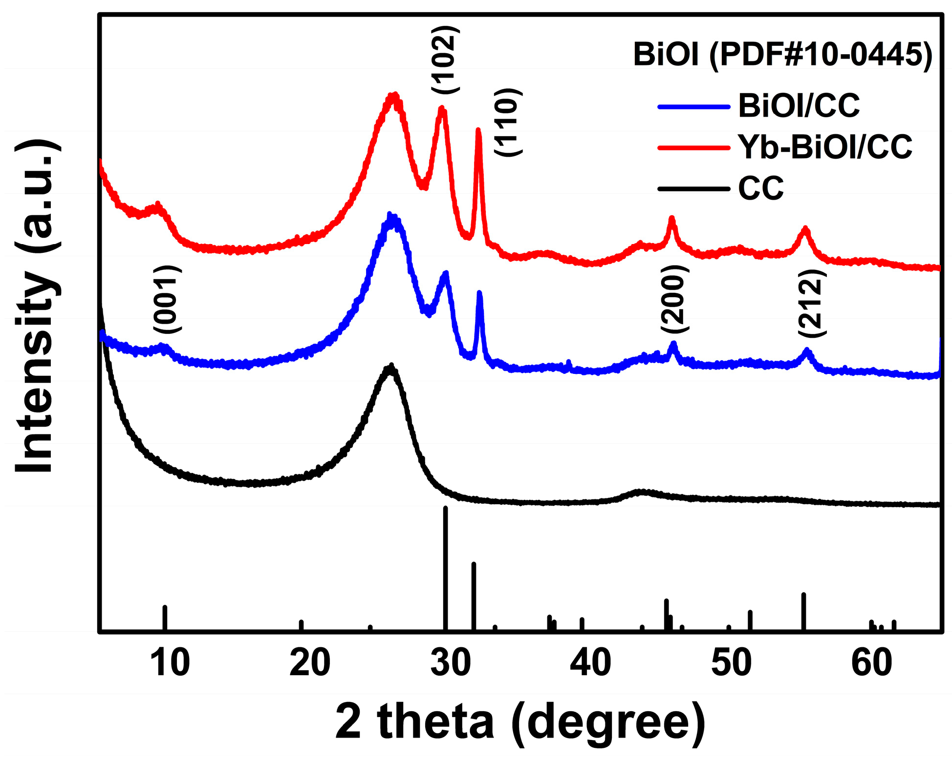
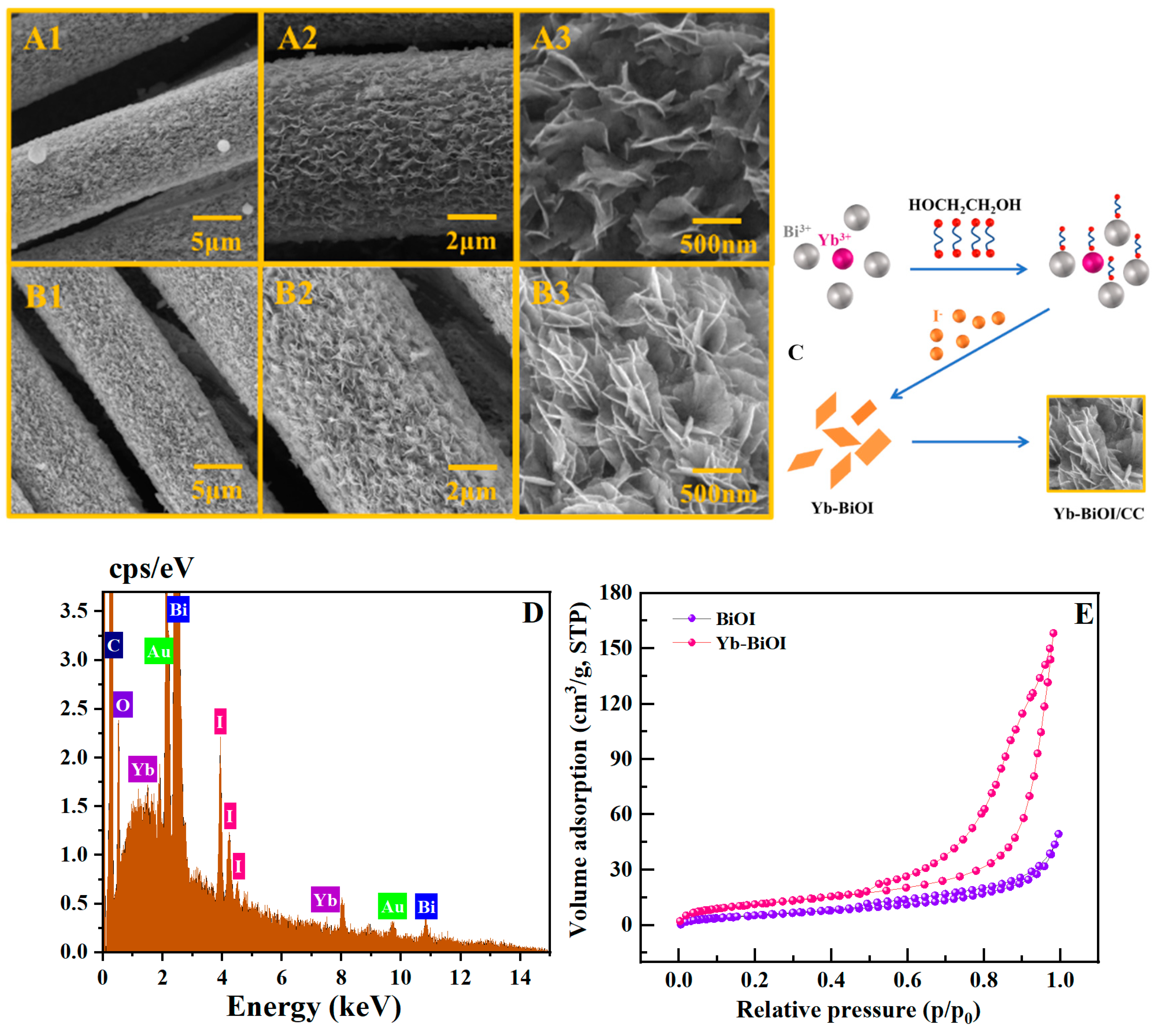
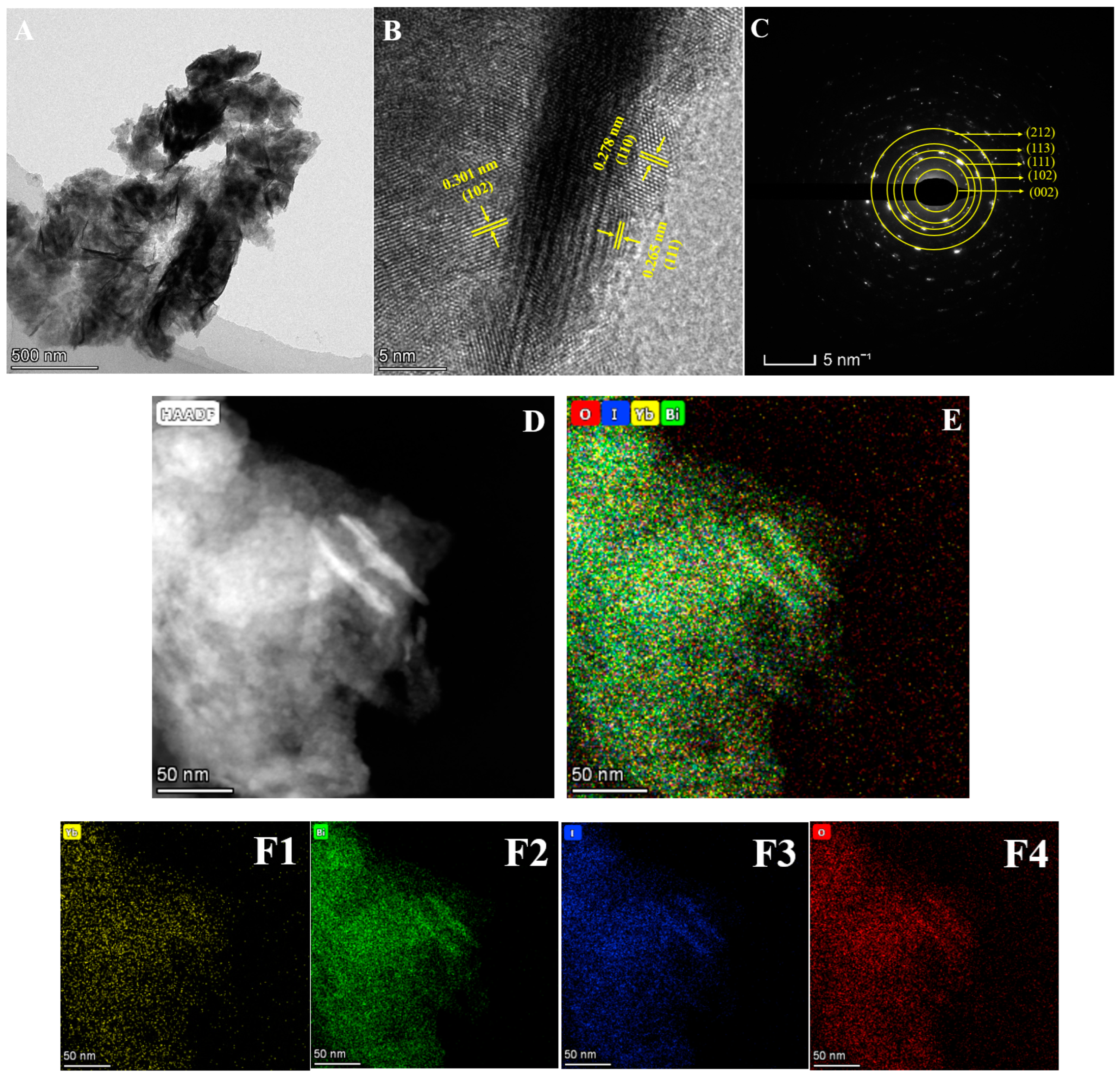
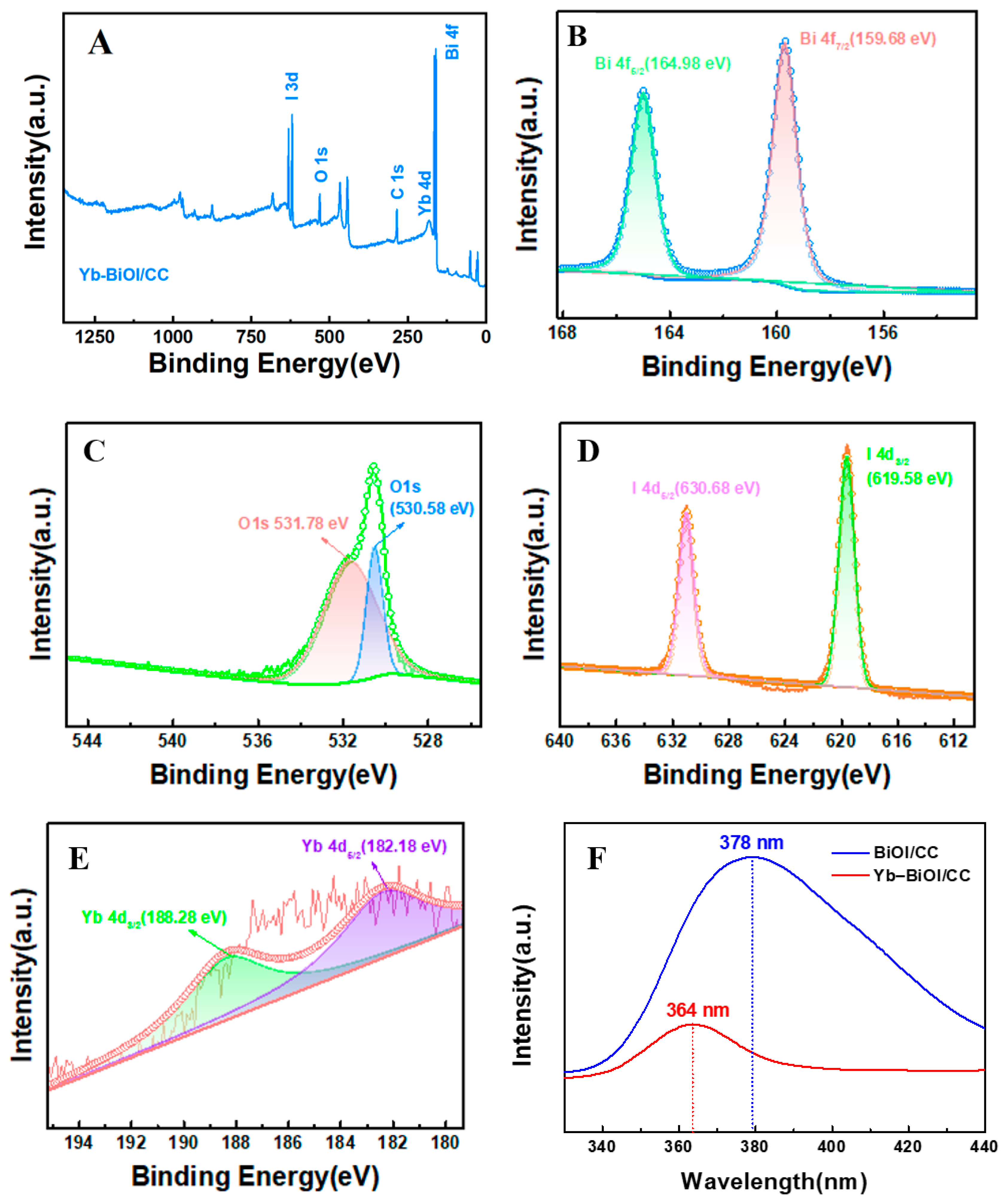
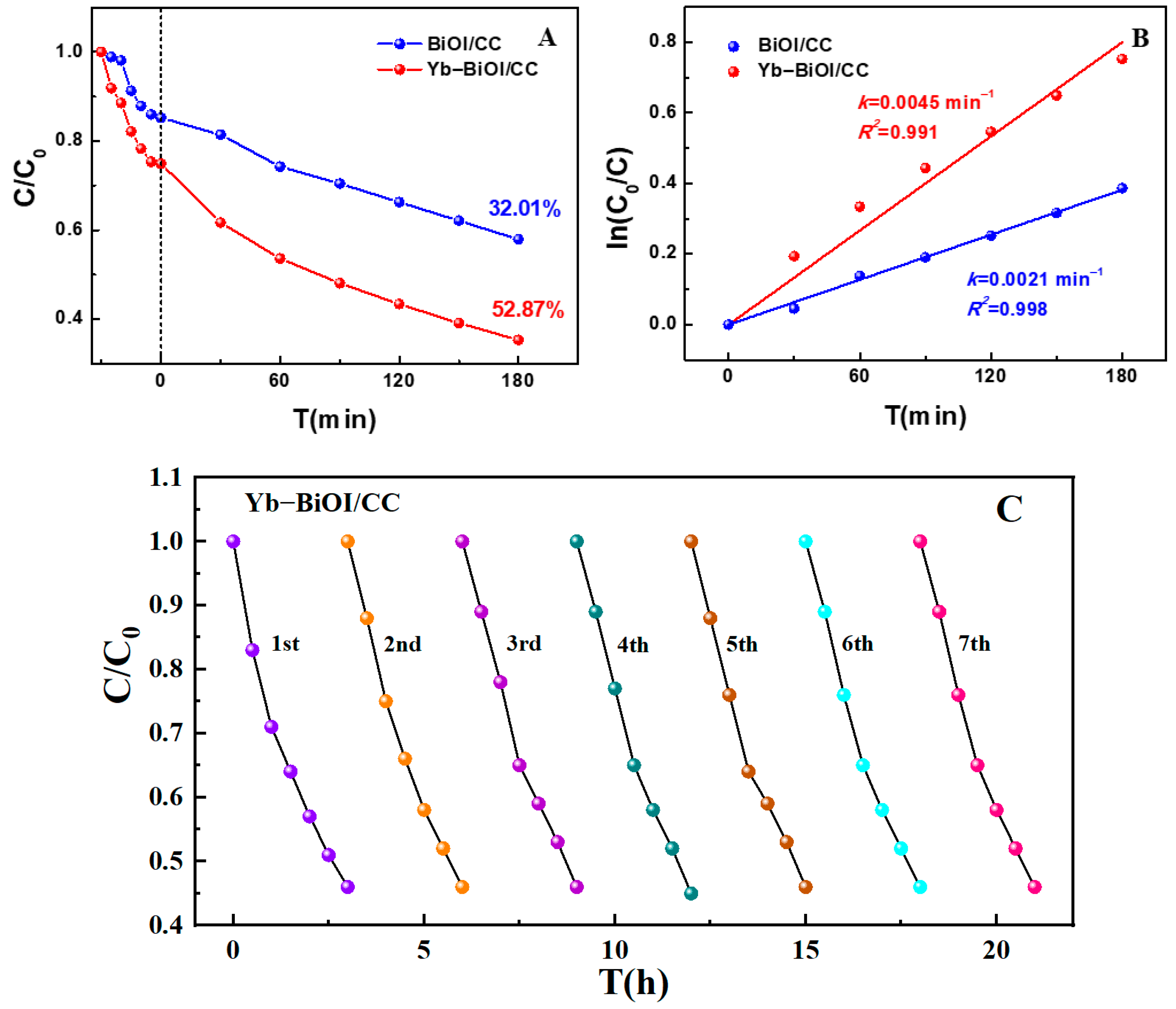
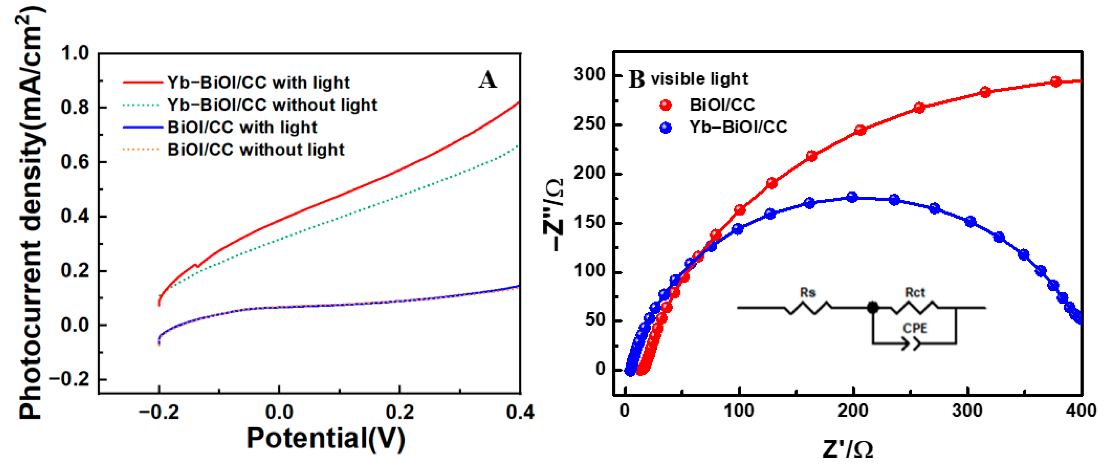
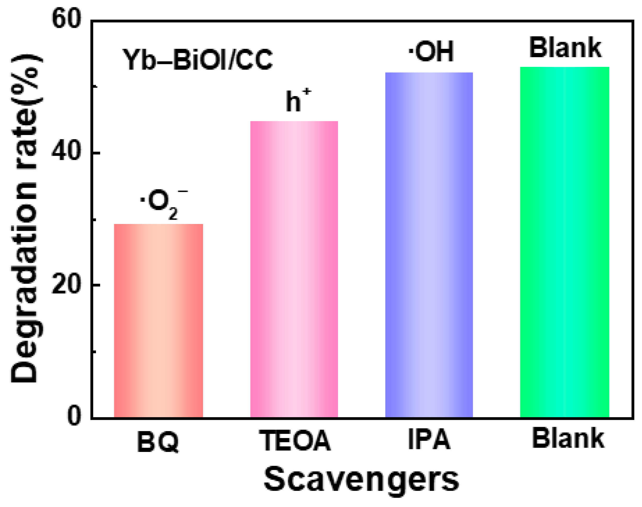
2. Results and Discussion

Figure 1 presents the X-ray powder diffraction (XRD) patterns of three materials. The characteristic peak of pristine carbon cloth (CC) is observed at 2θ = 26°. Other distinct diffraction peaks at 9.3°, 30°, 32.8°, 45.4°, and 55° correspond to the (001), (102), (110), (200), and (212) crystallographic planes of tetragonal BiOI (PDF No. 10-0445), respectively. Notably, no additional peaks emerge after Yb3+ doping, which is attributed to the low doping concentration (below the XRD detection limit). The preserved diffraction profile confirms that the crystalline structure of BiOI remains intact after Yb3+ incorporation, indicating successful lattice integration without inducing phase transformation or secondary phase formation. Additionally, the average crystallite size (D), dislocation density (δ), and strain (ε) of the nanomaterials were calculated using the Scherrer equation and its derived relationships:
D h k l = K λ β cos θ ,   δ = 1 D 2 ,   ε = β cos θ 4
where D represents the crystallite size, K denotes the shape factor (typically 0.89), λ is the X-ray wavelength (0.154 nm for Cu Kα radiation), and θ corresponds to the Bragg angle. The calculated average crystallite sizes for BiOI and Yb-BiOI were determined to be 23.49 nm and 20.08 nm, demonstrating an inverse correlation between crystallite dimensions and peak broadening, further confirming enhanced lattice defects (δ from 0.0018 to 0.0025 nm2) and strain (ε from 0.084 to 0.098) with crystallite refinement. The observed strain elevation in Yb-doped samples indicates lattice distortion arising from both size-induced strain and ytterbium incorporation-induced lattice mismatch, collectively suggesting a potential reduction in crystallinity.
Figure 2 displays comparative scanning electron microscopy (SEM) images of BiOI/CC and Yb-BiOI/CC composites. As revealed in Figure 2A, the pristine BiOI/CC exhibits uniformly distributed crumpled nanosheets with dense growth along carbon fiber surfaces. Remarkably, Yb3+ doping induces significant morphological evolution (Figure 2B), yielding vertically aligned and interwoven ultrathin nanoflakes (≈15 nm thickness) with enhanced structural compactness. This architectural evolution creates porosity while maintaining intimate interfacial contact with the carbon fiber. The vertically oriented nanoarchitecture provides enlarged active surface area and charge transfer pathways. The structural advantages effectively promote both surface redox reactions and bulk charge separation, thereby synergistically enhancing photocatalytic degradation efficiency.
Figure 2C schematically illustrates the formation mechanism of Yb-BiOI/CC photocatalyst through a three-stage growth strategy: (1) Precursor Coordination: Bi3+ ions form ethylene glycol-mediated coordination complexes of Bi(OCH2CH2OH)2 in the solvothermal system. (2) Crystallization: Subsequent reaction with I induces anisotropic growth of 2D BiOI nanosheets. (3) Substrate Assembly: The nanosheets self-assemble into interpenetrating architectures on carbon cloth through Ostwald ripening-driven epitaxial growth. The chemical reaction equations for the process are as follows:
Bi(NO3)3·5H2O → Bi3+ + 3NO3 + 5H2O
Yb(NO3)3·5H2O → Yb3+ + 3NO3 + 5H2O
KI → K+ + I
Bi3+ + Yb3+ + I + H2O → [Bi1−xYbxO]+I
Critical modification occurs upon introducing Yb(NO3)3·5H2O, where Yb3+ ions selectively substitute Bi3+ sites in the BiOI lattice (ionic radii: Yb3+ 0.86 Å vs. Bi3+ 0.96 Å). This isovalent doping enables precise bandgap engineering. Concurrently, the conductive CC substrate establishes an electron expressway to accelerate photogenerated electron transfer and suppress charge recombination through Schottky barrier effects.
Figure 2D presents the energy-dispersive X-ray spectroscopy (EDS) of Yb-doped BiOI. The spectrum confirms the presence of characteristic peaks corresponding to Yb, Bi, I, O, Au, and C. The detection of Yb3+ signals provides direct evidence for the successful incorporation of Yb3+ ions into the BiOI lattice. Notably, the Au signal originates from the gold sputter coating applied for sample conductivity enhancement during SEM-EDS measurements, while the C signal arises from the carbon cloth substrate. Quantitative analysis of the normalized elemental weight percentages (wt%) reveals the following composition: Bi (70.34%), I (22.32%), O (6.18%), and Yb (1.16%). This stoichiometric distribution aligns well with the structural framework of Yb-doped BiOI materials.
Figure 2E displays the nitrogen adsorption–desorption isotherms of the nanomaterials measured at 77 K. According to the International Union of Pure and Applied Chemistry (IUPAC) classification standards, pristine BiOI exhibits an H4-type hysteresis loop characterized by parallel and nearly horizontal adsorption–desorption branches, indicative of slit-shaped pores formed by interpenetrating nanosheets. In contrast, Yb-BiOI demonstrates a distinct H3-type hysteresis loop, where the adsorption branch fails to plateau near saturation pressure, suggesting wedge-shaped pores generated by loosely stacked lamellar particles. Pore size distribution analysis reveals a significant increase in average pore diameter from 12.09 nm (BiOI) to 22.95 nm (Yb-BiOI), accompanied by a 1.79-fold enhancement in specific surface area from 23.79 m2/g (BiOI) to 42.67 m2/g (Yb-BiOI). This structural evolution confirms that Yb3+ doping effectively optimizes textural properties by expanding pore dimensions and increasing accessible surface sites, thereby promoting reactant diffusion and active site exposure in photocatalytic processes.
Figure 3 presents comprehensive transmission electron microscopy (TEM) analyses elucidating the structural and compositional characteristics of the Yb-BiOI nanocomposite. As shown in the low-magnification TEM image (Figure 3A), the material exhibits a hierarchical architecture composed of vertically stacked ultrathin nanosheets, which facilitates light harvesting. High-resolution TEM (HRTEM, Figure 3B) reveals distinct lattice fringes with interplanar spacings of 0.301 nm, 0.265 nm, and 0.278 nm, corresponding to the (102), (111), and (110) crystallographic planes of tetragonal BiOI (PDF No. 10-0445), respectively. The coexistence of multiple lattice orientations confirms the polycrystalline nature of the material. This observation is further verified by the selected-area electron diffraction (SAED) pattern (Figure 3C), displaying characteristic concentric diffraction rings indexed to the (002), (102) and (111). In addition, the nanosheet morphology is demonstrated in the high-angle annular dark-field (HAADF) TEM image (Figure 3D), which shows the two-dimensional sheet-like structure. Elemental mapping (Figure 3E,F) displays the homogeneous spatial distribution of Yb, Bi, I, and O throughout the nanocomposite, which confirms the successful doping of Yb into the BiOI material. The doping uniformity ensures optimal exposure of catalytically active sites while maintaining efficient charge transport pathways.
Figure 4 presents the optical characterization and band structure analysis of BiOI/CC and Yb-BiOI/CC composites. The UV-Vis diffuse reflectance spectra (UV-Vis DRS) in Figure 4A reveal distinct absorption edges at 663 nm (BiOI/CC) and 600 nm (Yb-BiOI/CC), demonstrating a significant blue shift (Δλ = 63 nm) upon Yb3+ incorporation. This blue-shifted phenomenon might originate from crystal field modulation, that is, the smaller ionic radius of Yb3+ (0.86 Å) compared to Bi3+ (0.96 Å) induces lattice distortion and unit cell contraction, thus enhancing quantum confinement effects which widens the bandgap. Tauc plot analysis (Figure 4B) shows the corresponding bandgap widening from 1.67 eV (pristine BiOI/CC) to 1.87 eV (Yb-BiOI/CC), representing a 10.6% enhancement in the oxidation potential. The enlarged bandgap aligns with the increased valence band maximum (VBM) position, which strengthens oxidative capacity by elevating the hole potential [26].
Mott–Schottky (M–S) analysis was employed to investigate the electrolyte–electrode interfacial band structure, carrier density (Nd), Debye length (LD), and depletion layer width (Wd) [27]. The space charge capacitance (Csc) as a function of applied potential (Eapp) was calculated using Equation (2).
1 C s c 2 = 2 e ε 0 ε r A 2 N d × E a p p l E F B k T e
where Csc is the charge layer capacitance, e is the elementary charge (e = 1.602 × 10⁻¹⁹ C), ε0 is the vacuum permittivity (ε0 = 8.854 × 10⁻¹² F/m), εr is the relative permittivity of the material (εr = 6.85 for BiOI [28]), k is the Boltzmann constant (k = 1.38 × 10⁻²³ J/K), T is the testing temperature (T = 298 K), Eappl is the electrode potential, EFB is the flat-band potential, Nd is the carrier density, and Wd is the space charge layer (depletion layer) width. At room temperature (kT/e ≈ 25 mV), the kT/e term is negligible compared to (EapplEFB).
Figure 4C,D reveal positive slopes in the M-S plots for both BiOI/CC and Yb-BiOI/CC, confirming their n-type semiconductor characteristics. The linear portion of the slope was extrapolated to determine the respective flat-band potentials (EFB), which shifted negatively from −0.82 V (BiOI/CC) to −0.93 V (Yb-BiOI/CC), indicating a Fermi level upshift that facilitates valence band electron excitation for superoxide radical (·O2) generation via oxygen reduction, thereby enhancing oxidative dye degradation.
To further elucidate the enhanced Fermi level and reduced charge carrier recombination within the photocatalytic material, the carrier density, Debye length (LD), and depletion width (Wd) were analyzed using the equations described below.
N d = 2 e ε 0 ε r A 2 1 d d E a p p l 1 C s c 2
L D = ε 0 ε r k T e 2 N d
W d = 2 ε 0 ε r E a p p l E F B e N d
E C B = E V B E g
The calculated carrier density (Nd) exhibited a substantial increase from 8.70 × 1019 cm−3 (BiOI/CC) to 6.62 × 1020 cm−3 (Yb-BiOI/CC), demonstrating a remarkable 7.6-fold enhancement. Concurrently, the Debye length (LD) decreased from 0.334 nm to 0.121 nm, while the depletion layer width (Wd) was reduced from 8.45 nm to 3.26 nm. The narrower depletion layer in Yb-BiOI/CC nanomaterials corresponds to a shorter charge carrier diffusion length, which effectively suppresses bulk recombination and significantly enhances photocatalytic activity compared to pristine BiOI/CC. Given the direct proportionality between Wd and LD, this structural optimization facilitates rapid charge carrier transport and separation across the depletion layer in Yb-BiOI/CC, where the minimized diffusion length ensures efficient utilization of photogenerated carriers prior to recombination [29].
Moreover, the value of EFB also corresponds to the Fermi level due to negligible band bending at the EFB potential. In n-type and p-type semiconductors, the Fermi level aligns with the conduction band (ECB) and valence band (EVB) edges, respectively [30]. Therefore, the valence band potentials were subsequently calculated using Equation (8), resulting in values of +0.85 V and +0.94 V, respectively. Based on the determined Eg, ECB, and EVB values from DRS-Tauc and Mott–Schottky analyses, the corresponding band energy diagrams are presented in Figure 4E. Compared to BiOI, the valence band of Yb-BiOI is raised, the conduction band is lowered, and the bandgap is widened. As a result, doping with Yb3+ ions enhances the redox capability of the photogenerated electrons and holes in the material. These experimental observations demonstrate that the elevated carrier density promotes charge pair generation and accelerates carrier diffusion kinetics, ultimately boosting photocatalytic performance through enhanced charge separation efficiency and interfacial redox activity.
X-ray photoelectron spectroscopy (XPS) was employed to investigate the elemental composition and chemical states of the composite. The survey spectrum (Figure 5A) confirms the presence of Bi, O, I, Yb, and C as primary constituents. High-resolution spectra were deconvoluted to analyze specific electronic states. In particular, Bi 4f spectrum (Figure 5B) exhibited characteristic doublets at 164.98 eV (Bi 4f5/2) and 159.68 eV (Bi 4f7/2), verifying the dominant Bi3+ oxidation state, which is consistent with Bi–O bonding in the crystalline matrix [31]. O 1s spectrum (Figure 5C) was deconvoluted two resolved peaks at 530.58 eV (lattice oxygen in Bi-O) and 531.78 eV (surface hydroxyl groups), indicating dual oxygen functionalities [32]. I 4d spectrum (Figure 5D) exhibited peaks at 630.68 eV (I 4d5/2) and 619.58 eV (I 4d3/2), which confirmed iodide species (I) within the layered BiOI structure. Yb 4d spectrum (Figure 5E) showed spin–orbit splitting into Yb 4d3/2 (188.28 eV) and Yb 4d5/2 (182.18 eV), which demonstrates successful Yb3+ incorporation without metallic clustering [33]. These results suggest the effective Yb3+ substitution in the BiOI lattice and surface hydroxyl groups enhancing interfacial redox reactions.
The photoluminescence (PL) intensity of materials under light excitation directly correlates with charge carrier recombination dynamics. Reduced PL intensity signifies suppressed electron-hole recombination, thereby prolonging carrier lifetime. Figure 5F displays the PL spectra of BiOI/CC and Yb-BiOI/CC composites under 239 nm excitation. The BiOI/CC exhibited an emission peak centered at 378 nm, while the Yb-BiOI/CC displayed a blue-shifted emission at 364 nm. Notably, Yb-BiOI/CC exhibits significantly diminished emission intensity within the 320–440 nm range compared to BiOI/CC. This suppression might originate from Yb3+-induced trap states that redirect carriers toward surface redox reactions rather than radiative recombination [34]. Furthermore, the blue-shifted emission maximum (Δλ = 14 nm) in Yb-BiOI/CC suggests activated up-conversion luminescence through Yb3+ 4f energy levels, enabling efficient conversion of low-energy photons to higher-energy light. This dual mechanism of recombination inhibition and spectral up-conversion synergistically enhances photocatalytic degradation efficiency.
The photocatalytic degradation performance of BiOI/CC/FA and Yb-BiOI/CC/FA composites was evaluated through visible-light-driven methylene blue (MB) decomposition. As shown in Figure 6A, before irradiation, the nanomaterials were stirred during 30 min in the dark to establish an adsorption–desorption equilibrium. Under visible-light irradiation for 3 h, the MB degradation efficiencies reached 32.01% (BiOI/CC) and 52.87% (Yb-BiOI/CC), demonstrating a 1.65-fold enhancement for the Yb3+-doped composite. This improvement stems from Yb-BiOI photocatalyst widened bandgap (+0.20 eV), which modulates the energy band positions by elevating the valance band edge to +0.94 V vs. NHE and lowering the conduction band edge to −0.93 V, thereby strengthening oxidative capacity through the generation of ·O2 radical and hole. Figure 6B presents the photocatalytic degradation kinetics of BiOI/CC and Yb-BiOI/CC nanomaterials. The degradation profiles adhere to pseudo-first-order reaction kinetics, with fitted rate constants (k) of 0.00212 min−1 for BiOI/CC and 0.00445 min−1 for Yb-BiOI/CC, demonstrating a 2.1-fold enhancement in degradation efficiency through Yb3+ doping. This improvement correlates with the optimized charge separation efficiency in the modified material. Cyclic stability tests (Figure 6C) reveal that Yb-BiOI/CC retains 96% of its initial methylene blue degradation efficiency after seven consecutive cycles. Moreover, the nanomaterial does not require centrifugation for recovery. Hence, the Yb-BiOI/CC exhibits excellent stability and operational convenience for the visible-light photocatalytic degradation. A wide range of photocatalysts have demonstrated effective photocatalytic degradation of methylene blue dye, as shown in Table 1.
Figure 7A displays the linear sweep voltammetry (LSV) curves of BiOI/CC and Yb-BiOI/CC composites under visible-light irradiation, evaluating their photoelectrochemical response. In the dark condition, the pristine BiOI/CC exhibits a relatively flat curve within the potential range of −0.20 V to +0.40 V. Upon light illumination, the LSV curve of BiOI/CC nearly overlaps with its dark-state counterpart, displaying negligible photocurrent density (less than 0.17 mA/cm2 at +0.40 V), indicating the rapid recombination of photogenerated electron-hole pairs. In contrast, Yb-BiOI/CC demonstrates a gradual current increase with applied bias in the dark, while under illumination, it achieves a significantly enhanced photocurrent density (up to 0.81 mA/cm2 at +0.40 V). This marked improvement implies that doping with the Yb3+ ions facilitates the effective separation of charge carriers and prolongs the lifetime of electron-hole pairs. Figure 7B presents the electrochemical impedance spectroscopy (EIS) of BiOI/CC and Yb-BiOI/CC under visible-light irradiation. The Yb3+-doped composite exhibits a significantly smaller semicircle diameter in the high-frequency region, indicating reduced charge transfer resistance. Equivalent circuit fitting reveals that Yb-BiOI/CC achieves a 52.4% lower charge transfer resistance (Rct = 318 Ω) compared to BiOI/CC (Rct = 668 Ω), demonstrating enhanced interfacial charge separation efficiency. Additionally, the carbon cloth provides an effective pathway for the transport of photogenerated charge carriers, further improving the photocatalytic activity of the nanomaterials.
Figure 8 illustrates the radical trapping experiments for methylene blue (MB) degradation over Yb-BiOI/CC under visible light. Isopropanol (IPA), benzoquinone (BQ), and triethanolamine (TEOA) were employed as selective scavengers for hydroxyl radicals(·OH), superoxide radicals (·O2), and photogenerated holes (h+), respectively [47]. The degradation efficiency exhibited minimal variation upon IPA addition, whereas significant suppression was observed with BQ (23.61% inhibition) and TEOA (8.15% inhibition). This sequence of inhibition (BQ > TEOA > IPA) identifies ·O2 as the predominant reactive species and h+ as a secondary contributor. The dominance of ·O2 originates from Yb3+-enhanced electron transfer kinetics, where photogenerated electrons efficiently reduce adsorbed O2 to ·O2 (O2 + e → ·O2), while h+ directly oxidize MB molecules.

3. Materials and Methods

3.1. Materials

Fly ash, cement powder, and bismuth nitrate pentahydrate were obtained from Xilong Scientific Co., Ltd. (Shantou, China). Potassium iodide was purchased from Qingdao Tuohai Iodine Products Co., Ltd. (Qingdao, China). Ytterbium nitrate pentahydrate was obtained from Aladdin Reagent (Shanghai) Co., Ltd. (Shanghai, China). Ethylene glycol was provided by Xingping Fuchen Chemical Technology Co., Ltd. (Xianyang, China). Methylene blue was purchased from Shanghai Macklin Biochemical Technology Co., Ltd. (Shanghai, China). Sodium sulfate was supplied by Tianjin Damao Chemical Reagent Factory (Tianjin, China). All other reagents were of analytical grade and used without further purification. Deionized water (>18.4 MΩ cm−1) was used for all solution preparation.

3.2. Instrumentations and Characterizations

The phase composition was investigated by X-ray diffraction (XRD, Bruker D8 Advance diffractometer, Bruker, Karlsruhe, Germany). The morphology and elemental distribution were confirmed by field-emission scanning electron microscopy (FE-SEM, MIRA3 LMU microscopy, Brno, Czech Republic) and Transmission Electron Microscope (TEM, Tecnai F20, FEI Company, Hillsboro, OR, USA). The elemental composition was determined by energy dispersive X-ray spectroscopy (EDS, Bruker XFlash7, Bruker, Germany). The specific surface area and pore size distribution were measured by Brunauer–Emmett–Teller specific surface area analysis (BELSORP-max fully automated surface area and porosity analyzer, MicrotracBEL Corp., Osaka, Japan). The valence state was tested by X-ray Photon-electron Spectroscopy (XPS, ESCALAB 250Xi, Thermo Fisher Scientific Inc., Waltham, MA, USA). The optical properties were determined through UV-Vis diffuse reflectance spectrum (DRS, UV2600 spectrophotometer, Shimadzu, Kyoto, Japan) and photoluminescence spectroscopy (PL, RF-5301PC, Shimadzu, Japan). The photoelectrochemical performance was measured by CS electrochemical workstation (Wuhan Corrtest Instrument Corp., Ltd., Wuhan, China).

3.3. Pretreatment of Carbon Cloth and Preparation of Yb-BiOI/CC Photocatalytic Material

Flexible carbon cloth (CC) was cut into 1 cm × 3 cm strips and sequentially ultrasonicated in acetone, absolute ethanol, and deionized water (10 min each) at room temperature to remove surface contaminants. The cleaned CC was dried at 60 °C for subsequent use.
A precursor solution was prepared by dissolving 1.5 mmol bismuth nitrate pentahydrate [Bi(NO3)3·5H2O] and 0.03 mmol ytterbium nitrate pentahydrate [Yb(NO3)3·5H2O] (Yb:Bi molar ratio = 2%) in 35 mL ethylene glycol under magnetic stirring for 3 h. Subsequently, 10 mL ethylene glycol containing 1.5 mmol potassium iodide (KI) was added dropwise to the mixture under vigorous stirring. Pretreated CC strips, immobilized on glass slides, were inclined against the wall of a 50 mL Teflon-lined stainless-steel autoclave. The mixed solution was transferred into the autoclave and reacted at 150 °C for 12 h. Reaction finished, the CC-supported product was thoroughly rinsed with deionized water and dried at 60 °C to obtain Yb-BiOI/CC.

3.4. Preparation of Yb-BiOI/CC/FA

Fly ash (FA) and cement powder were homogenized at a 1:9 mass ratio via mechanical stirring. Deionized water (40 wt% of solids) was gradually added to form a slurry, followed by 5 min of additional mixing. The slurry was cast into a custom-made square mold, compacted with a stainless-steel plate, and laminated with prefabricated Yb-BiOI/CC strips. The rigid composite was formed for 7 days with twice-daily water to spray, as shown in Figure 9.

3.5. Photocatalytic Performance Testing

Two Yb-BiOI/CC/FA strips (1 cm × 3 cm) were vertically mounted in a double-layer quartz cuvette containing 50 mL methylene blue (MB, 5 mg/L). Dark adsorption–desorption equilibrium was established via 30 min magnetic stirring. Visible-light irradiation was provided by a 300 W Xe lamp with a UV cutoff filter. During degradation, the reactor was water-cooled and magnetically stirred (120 rpm). Aliquots were collected at 30 min intervals, and MB concentration was monitored at 664 nm using UV-Vis spectroscopy.

3.6. Photoelectrochemical Performance Measurement

A three-electrode system was employed: Yb-BiOI/CC/FA (2 cm2 working electrode), Pt wire (counter electrode), and saturated calomel electrode (SCE, reference electrode), immersed in 0.5 M Na2SO4 electrolyte. Linear sweep voltammetry and impedance were measured under visible-light illumination using an electrochemical workstation.

4. Conclusions

In summary, a three-dimensional nanosheet array composite photocatalyst (Yb-BiOI/CC/FA) was successfully fabricated through a solvothermal method. Compared with pristine BiOI/CC/FA, the Yb3+-modified composite demonstrates optimized band structure characteristics, where the widened bandgap (+0.20 eV) enhances the redox potential of photogenerated electrons and holes. Yb3+ doping significantly reduces interfacial charge transfer resistance by 52.4% under visible light, effectively suppressing carrier recombination while promoting separation efficiency. These synergistic effects collectively elevate the photocatalytic degradation capacity, achieving a 1.65-fold enhancement in methylene blue removal efficiency after 180 min visible light irradiation. Radical trapping experiments confirm that superoxide radicals (·O2) serve as the predominant reactive species. The modular “sticker-type” configuration enables facile catalyst recovery and demonstrates practical potential in rural/urban waterway remediation applications, combining sustainable waste valorization with environmental purification.

Author Contributions

Conceptualization, L.J. and S.Q.; methodology, L.J. and S.Q.; formal analysis, L.J., S.Q., R.L. and X.L.; resources, J.Y. and L.X.; data curation, L.J., S.Q., R.L. and X.L.; writing—original draft preparation, L.J. and S.Q.; writing—review and editing, D.Z. and X.G.; supervision, D.Z. and X.G.; project administration, D.Z. and X.G.; funding acquisition, D.Z. and X.G. All authors have read and agreed to the published version of the manuscript.

Funding

This research is funded by Research and Innovation Team for Wastewater Treatment and Monitoring of Guangdong University of Education (No: 2024KYCXTD016), Guangdong Provincial Department of Education Key Area Special Project (2022ZDZX4037, 2024ZDZX2087, 2024ZDZX4063), Guangzhou Science and Technology Program Project (Grant No. 202201011099) and College Students’ Innovative Entrepreneurial Training Plan Program (No: S202414278023, 202414278095).

Institutional Review Board Statement

Not applicable.

Informed Consent Statement

Not applicable.

Data Availability Statement

The original contributions presented in this study are included in the article. Further inquiries can be directed to the corresponding authors.

Conflicts of Interest

The authors declare no conflicts of interest.

References

  1. Al-Wasidi, A.S.; AlReshaidan, S. Enhanced Removal of Rhodamine b Dye from Aqueous Media via Adsorption on Facilely Synthesized Zinc Ferrite Nanoparticles. Inorganics 2024, 12, 191. [Google Scholar] [CrossRef]
  2. Al-Wasidi, A.S.; Shah, R.K.; Abdelrahman, E.A.; Mabrouk, E.-S.M. Facile Synthesis of CuFe2O4 Nanoparticles for Efficient Removal of Acid Blue 113 and Malachite Green Dyes from Aqueous Media. Inorganics 2024, 12, 143. [Google Scholar] [CrossRef]
  3. Pereira, M.F.G.; Nascimento, M.M.; Cardoso, P.H.N.; Oliveira, C.Y.B.; Tavares, G.F.; Araújo, E.S. Preparation, Microstructural Characterization and Photocatalysis Tests of V5+-Doped TiO2/WO3 Nanocomposites Supported on Electrospun Membranes. Inorganics 2022, 10, 143. [Google Scholar] [CrossRef]
  4. Varadi, A.; Leostean, C.; Stefan, M.; Popa, A.; Toloman, D.; Pruneanu, S.; Tripon, S.; Macavei, S. Fe3O4-ZnO:V Nanocomposites with Modulable Properties as Magnetic Recoverable Photocatalysts. Inorganics 2024, 12, 119. [Google Scholar] [CrossRef]
  5. Ruan, X.W.; Hu, H.; Che, G.B.; Zhou, P.J.; Liu, C.B.; Dong, H.J. Fabrication of Z-Scheme γ-Bi2MoO6/Bi12GeO20 Heterostructure for Visible-Light-Driven Photocatalytic Degradation of Organic Pollutants. Appl. Surf. Sci. 2020, 499, 143668. [Google Scholar] [CrossRef]
  6. Guan, G.M.; Shao, Y.X.; Xu, Z.L.; Liu, L.P.; Xu, W.Q.; Jiang, L.C. The Preparation and Photo-Electrocatalytic Properties of ZnO@Zn/TiO2 3D Array Composite. Ind. Water Treat. 2022, 42, 130–135. [Google Scholar]
  7. Chang, S.M.; Liu, W.S. Surface Doping Is More Beneficial than Bulk Doping to the Photocatalytic Activity of Vanadium-Doped TiO2. Appl. Catal. B Environ. 2011, 101, 333–342. [Google Scholar] [CrossRef]
  8. Mukherji, A.; Seger, B.; Lu, G.Q.; Wang, L.Z. Nitrogen Doped Sr2Ta2O7 Coupled with Graphene Sheets as Photocatalysts for Increased Photocatalytic Hydrogen Production. ACS Nano 2011, 5, 3483–3492. [Google Scholar] [CrossRef]
  9. Liu, D.H.; Huang, J.F.; Tao, X.W.; Wang, D. One-Step Synthesis of C-Bi2WO6 Crystallites with Improved Photocatalytic Activities under Visible Light Irradiation. RSC Adv. 2015, 5, 66464–66470. [Google Scholar] [CrossRef]
  10. Anwer, H.; Mahmood, A.; Lee, J.; Kim, K.H.; Park, J.W.; Yip, A.C.K. Photocatalysts for Degradation of Dyes in Industrial Effluents: Opportunities and Challenges. Nano Res. 2019, 12, 955–972. [Google Scholar] [CrossRef]
  11. Rafiq, A.; Ikram, M.; Ali, S.; Niaz, F.; Khan, M.; Khan, Q.; Maqbool, M. Photocatalytic Degradation of Dyes Using Semiconductor Photocatalysts to Clean Industrial Water Pollution. Ind. Eng. Chem. 2021, 97, 111–128. [Google Scholar] [CrossRef]
  12. Villabona-Leal, E.G.; López-Neira, J.P.; Pedraza-Avella, J.A.; Pérez, E.; Meza, O. Screening of Factors Influencing the Photocatalytic Activity of TiO2: Ln (Ln=La, Ce, Pr, Nd, Sm, Eu and Gd) in the Degradation of Dyes. Comput. Mater. Sci. 2015, 107, 48–53. [Google Scholar] [CrossRef]
  13. Bhethanabotla, V.C.; Russell, D.R.; Kuhn, J.N. Assessment of Mechanisms for Enhanced Performance of Yb/Er/Titania Photocatalysts for Organic Degradation: Role of Rare Earth Elements in the Titania Phase. Appl. Catal. B Environ. 2017, 202, 156–164. [Google Scholar] [CrossRef]
  14. Adhikari, R.; Gyawali, G.; Cho, S.H.; Narro-García, R.; Sekino, T.; Lee, W.S. Er3+/Yb3+ Co-Doped Bismuth Molybdate Nanosheets Upconversion Photocatalyst with Enhanced Photocatalytic Activity. J. Solid State Chem. 2014, 209, 74–81. [Google Scholar] [CrossRef]
  15. Liu, T.; Tan, G.Q.; Zhao, C.C.; Xu, C.; Su, Y.N.; Wang, Y.; Ren, H.J.; Xia, A.; Shao, D.; Yan, S.M. Enhanced Photocatalytic Mechanism of the Nd-Er Co-Doped Tetragonal BiVO4 Photocatalysts. Appl. Catal. B Environ. 2017, 213, 87–96. [Google Scholar] [CrossRef]
  16. Hassan, M.S.; Amna, T.; Yang, O.-B.; Kim, H.-C.; Khil, M.-S. TiO2 Nanofibers Doped with Rare Earth Elements and Their Photocatalytic Activity. Ceram. Int. 2012, 38, 5925–5930. [Google Scholar] [CrossRef]
  17. Mehtab, A.; Ahmed, J.; Alshehri, S.M.; Mao, Y.B.; Ahmad, T. Rare Earth Doped Metal Oxide Nanoparticles for Photocatalysis: A Perspective. Nanotechnology 2022, 33, 142001. [Google Scholar] [CrossRef]
  18. Lee, S.M.; Lee, H.H.; Hong, S.C. Influence of Calcination Temperature on Ce/TiO2 Catalysis of Selective Catalytic Oxidation of NH3 to N2. Appl. Catal. A Gen. 2014, 470, 189–198. [Google Scholar] [CrossRef]
  19. Sin, J.C.; Lam, S.M.; Lee, K.T.; Mohamed, A.R. Preparation of Rare Earth-Doped ZnO Hierarchical Micro/Nanospheres and Their Enhanced Photocatalytic Activity under Visible Light Irradiation. Ceram. Int. 2014, 40, 5431–5440. [Google Scholar] [CrossRef]
  20. Li, H.; Hao, H.S.; Jin, S.S.; Guo, W.H.; Hu, X.F.; Hou, H.M.; Zhang, G.L.; Yan, S.; Gao, W.Y.; Liu, G.S. Synthesis of Yb3+/Ho3+ Co-Doped Bi2WO6 Upconversion Photocatalyst with Highly Improved Visible Light Photocatalytic Activity. Catal. Commun. 2017, 97, 60–64. [Google Scholar] [CrossRef]
  21. Xue, S.S.; He, H.B.; Fan, Q.Z.; Yu, C.L.; Yang, K.; Huang, W.Y.; Zhou, Y.; Xie, Y. La/Ce-Codoped Bi2O3 Composite Photocatalysts with High Photocatalytic Performance in Removal of High Concentration Dye. J. Environ. Sci. 2017, 60, 70–77. [Google Scholar] [CrossRef] [PubMed]
  22. Zagaynov, I.V.; Liberman, E.Y.; Prikhodko, O.P.; Kon’kova, T.V. Catalytic Activity of CeO2@TiO2 for Environmental Protection. New J. Chem. 2024, 48, 2842. [Google Scholar] [CrossRef]
  23. Ma, Z.W.; Li, C. Research on Fly Ash Cenospheres-Supported Photocatalyst for the Degradation of Dye Wastewater. Ind. Water Treat. 2015, 35, 65–68. [Google Scholar]
  24. He, M.Q.; Li, W.B.; Xia, J.X.; Xu, L.; Di, J.; Xu, H.; Yin, S.; Li, H.M.; Li, M.N. The Enhanced Visible Light Photocatalytic Activity of Yttrium-Doped BiOBr Synthesized via a Reactable Ionic Liquid. Appl. Surf. Sci. 2015, 331, 170–178. [Google Scholar] [CrossRef]
  25. Huo, Y.N.; Zhang, J.; Miao, M.; Jin, Y. Solvothermal Synthesis of Flower-Like BiOBr Microspheres with Highly Visible-Light Photocatalytic Performances. Appl. Catal. B Environ. 2012, 111–112, 334–341. [Google Scholar] [CrossRef]
  26. Jiang, L.C.; Gao, X.Y.; Chen, S.L. Oxygen-Deficient WO3/TiO2/CC Nanorod Arrays for Visible-Light Photocatalytic Degradation of Methylene Blue. Catalysts 2021, 11, 1349. [Google Scholar] [CrossRef]
  27. Chowdhury, A.; Balu, S.; Yang, T.C.-K. Construction of α-Fe2O3-NPs@AgVO3-NRs Z-scheme heterojunction: An efficient photo(electro)catalyst for Cr(VI) reduction and oxygen evolution reactions under visible-light. J. Environ. Chem. Eng. 2023, 11, 109769. [Google Scholar] [CrossRef]
  28. Dai, W.W.; Zhao, Z.Y. Electronic Structure and Optical Properties of BiOI as a Photocatalyst Driven by Visible Light. Catalysts 2016, 6, 133–148. [Google Scholar] [CrossRef]
  29. Mishra, N.S.; Saravanan, P. Z-scheme promoted heterojunction photocatalyst (Ag@AgVO3/rGO/CeVO4) with improved interfacial charge transfer for efficient removal of aqueous organics irradiated under LED light. Chemosphere 2023, 310, 136896. [Google Scholar] [CrossRef]
  30. Balu, S.; Venkatesvaran, H.; Wang, C.-C.; Juan, J.C.; Yang, T.C.-K. Synthesis of Sulfonic Acid-Functionalized g-C3N4/BiOI Bifunctional Heterojunction for Enhanced Photocatalytic Removal of Tartrazine and PEC Oxygen Evolution Reaction. Inorganics 2024, 12, 243. [Google Scholar] [CrossRef]
  31. Wang, W.D.; Huang, F.Q.; Lin, X.P.; Yang, J.H. Visible-light-responsive photocatalysts xBiOBr–(1-x)BiOI. Catal. Commun. 2008, 9, 8–12. [Google Scholar] [CrossRef]
  32. Wang, H.; Yong, D.Y.; Chen, S.C.; Jiang, S.L.; Zhang, X.D.; Shao, W.; Zhang, Q.; Yan, W.S.; Pan, B.C.; Xie, Y. Oxygen-vacancy-mediated exciton dissociation in BiOBr for boosting charge-carrier-involved molecular oxygen activation. J. Am. Chem. Soc. 2018, 140, 1760–1766. [Google Scholar] [CrossRef] [PubMed]
  33. Ohno, Y. XPS studies of the intermediate valence state of Yb in (YbS)1.25CrS2. J. Electron. Spectrosc. Relat. Phenom. 2008, 165, 1–4. [Google Scholar] [CrossRef]
  34. Tricoire, M.; Sroka, W.; Rajeshkumar, T.; Scopelliti, R.; Sienkiewicz, A.; Maron, L.; Mazzanti, M. Multielectron Redox Chemistry of Ytterbium Complexes Reaching the +1 and Zero Formal Oxidation States. J. Am. Chem. Soc. 2025, 147, 1162–1171. [Google Scholar] [CrossRef]
  35. Ullah, R.; Dutta, J. Photocatalytic Degradation of Organic Dyes With Manganese-Doped ZnO Nanoparticles. J. Hazard. Mater. 2008, 156, 194–200. [Google Scholar] [CrossRef]
  36. Shen, W.Z.; Li, Z.J.; Wang, H.; Liu, Y.H.; Guo, Q.J.; Zhang, Y.L. Photocatalytic Degradation for Methylene Blue Using Zinc Oxide Prepared By Codeposition and Sol–Gel Methods. J. Hazard. Mater. 2008, 152, 172–175. [Google Scholar] [CrossRef]
  37. Pawinrat, P.; Mekasuwandumrong, O.; Panpranot, J. Synthesis of Au–ZnO and Pt–ZnO Nanocomposites By One-Step Flame Spray Pyrolysis and Its Application for Photocatalytic Degradation of Dyes. Catal. Commun. 2009, 10, 1380–1385. [Google Scholar] [CrossRef]
  38. Muruganandhm, M.; Chen, I.S.; Wu, J.J. Effect of Temperature on the Formation of Macroporous ZnO Bundles and Its Application In Photocatalysis. J. Hazard. Mater. 2009, 172, 700–706. [Google Scholar] [CrossRef]
  39. Yan, H.W.; Hou, J.B.; Fu, Z.P.; Yang, B.F.; Yang, P.H.; Liu, K.P.; Wen, M.W.; Chen, Y.J.; Fu, S.Q.; Li, F.Q. Growth and Photocatalytic Properties of One-Dimensional ZnO Nanostructures Prepared By Thermal Evaporation. Mater. Res. Bull. 2009, 44, 1954–1958. [Google Scholar] [CrossRef]
  40. Uddin, M.T.; Nicolas, Y.; Qlivier, C.; Toupance, T.; Servant, L.; Muller, M.M.; Kleebe, H.J.; Ziegler, J.; Jaegermann, W. Nanostructured SnO2-ZnO Heterojunction Photocatalysts Showing Enhanced Photocatalytic Activity for the Degradation of Organic Dyes. Inorg. Chem. 2012, 51, 7764–7773. [Google Scholar] [CrossRef]
  41. Soltani, N.; Saion, E.; Yunus, W.M.M.; Erfani, M.; Navasery, M.; Bahmanrokh, G.; Rezaee, K. Enhancement of Visible Light Photocatalytic Activity of ZnS and CdS Nanoparticles Based on Organic and Inorganic Coating. Appl. Surf. Sci. 2014, 290, 440–447. [Google Scholar] [CrossRef]
  42. Peng, Y.; Yu, P.P.; Zhou, H.Y.; Xu, A.W. Synthesis of BiOI/Bi4O5I2/Bi2O2CO3 p–n–p Heterojunctions with Superior Photocatalyticactivities. New J. Chem. 2015, 39, 8321–8328. [Google Scholar] [CrossRef]
  43. Singh, S.; Khare, N. Reduced Graphene Oxide Coupled CdS/CoFe2O4 Ternary Nanohybrid with Enhanced Photocatalytic Activity and Stability: A Potential Role of Reduced Graphene Oxide as A Visible Light Responsive Photosensitizer. RSC Adv. 2015, 5, 96562. [Google Scholar] [CrossRef]
  44. Zhu, C.S.; Zhang, L.; Jiang, B.; Zheng, J.T.; Hu, P.; Li, S.J.; Wu, M.B.; Wu, W.T. Fabrication of Z-Scheme Ag3PO4/MoS2 Composites with Enhanced Photocatalytic Activity and Stability for Organic Pollutant Degradation. Appl. Surf. Sci. 2016, 377, 99–108. [Google Scholar] [CrossRef]
  45. Ayu, D.G.; Gea, S.; Andriayani; Junita, D.; Telaumbanua, J.; Piliang, A.F.R.; Harahap, M.; Yen, Z.H.; Goei, R.; Tok, A.I.Y. Photocatalytic Degradation of Methylene Blue Using N-Doped ZnO/Carbon Dot (N-ZnO/CD) Nanocomposites Derived from Organic Soybean. ACS Omega 2023, 8, 14965–14984. [Google Scholar] [CrossRef]
  46. Mohammed, W.; Matalkeh, M.; Soubaihi, R.M.A.; Elzatahry, A. Visible Light Photocatalytic Degradation of Methylene Blue Dye and Pharmaceutical Wastes over Ternary NiO/Ag/TiO2 Heterojunction. ACS Omega 2023, 8, 40063–40077. [Google Scholar] [CrossRef]
  47. An, H.; Lin, B.; Xue, C.; Yan, X.Q.; Dai, Y.Z.; Wei, J.J.; Yang, G.D. Formation of BiOI/g-C3N4 Nanosheet Composites with High Visible-Light-Driven Photocatalytic Activity. Chin. J. Catal. 2018, 39, 654–663. [Google Scholar] [CrossRef]
Figure 1. XRD pattern of CC, BiOI/CC and Yb-BiOI/CC nanocomposites.
Inorganics 13 00147 g001
Figure 2. SEM images of the samples: (A1A3) BiOI/CC, (B1B3) Yb-BiOI/CC, (C) formation mechanism, (D) EDS of Yb-BiOI/CC, (E) BET.
Inorganics 13 00147 g002
Figure 3. Yb-BiOI/CC of TEM (A), HR-TEM (B), SEAD (C), HAADF (D), overlayed mapping (E), and individual elemental map (F1F4).
Inorganics 13 00147 g003
Figure 4. UV–visible diffuse reflection spectra (A) and calculated bandgaps (B), Mott–Schottky of BiOI (C), Yb-BiOI (D), and schematic diagram of energy band (E).
Inorganics 13 00147 g004
Figure 5. XPS of Yb-BiOI/CC: (A) survey spectra (blue); (B) Bi 4f (green: Bi 4f5/2, orange: Bi 4f7/2); (C) O 1s (pink: Bi-O, blue: OH-); (D) I 4d (purple: I 4d5/2, green: I 4d3/2); (E) Yb 4d (green: Yb 4d3/2, purple: Yb 4d5/2) and (F) fluorescence spectrum (blue: BiOI/CC, red: Yb-BiOI/CC).
Inorganics 13 00147 g005
Figure 6. (A) Visible light photocatalytic degradation; (B) quasi-first-order kinetic curve; (C) cyclic degradation curves of MB.
Inorganics 13 00147 g006
Figure 7. Linear sweep voltammetry (A) and electrochemical impedance spectrum (B) of the BiOI/CC and Yb-BiOI/CC.
Inorganics 13 00147 g007
Figure 8. The active species scavenging tests.
Inorganics 13 00147 g008
Figure 9. Preparation process of Yb-BiOI/CC/FA composite photocatalytic material.
Inorganics 13 00147 g009
Table 1. Comparison of degradation rates of methylene blue by different photocatalysts.
Order Photocatalyst Dyes Light Time
(min)
Degradation Rate References
1Mn-doped ZnOMBVisible60100%[35]
2ZnO-SiMBVisible6090%[36]
3ZnOMBUV18096%[37]
4ZnOMBUV6039.7%[38]
5Au-ZnOMBUV6071%[39]
6SnO2-ZnOMBUV2088%[40]
7CdS/ZnSMBVisible36075%[41]
8BiOI/Bi4O5I2/Bi2O2CO3MBSolar light4597%[42]
9GO/CdS/CoFe2O4MBVisible12082%[43]
10Ag3PO4/MoS2MBVisible6098.2%[44]
11WO3/TiO2/CCMBVisible 18068%[26]
12N-ZnO/CDsMBUV6058.2%[45]
13NiO/Ag/TiO2MBVisible 6093.15%[46]
14Yb3+-BiOI/CCMBVisible 18052.87%This work
Disclaimer/Publisher’s Note: The statements, opinions and data contained in all publications are solely those of the individual author(s) and contributor(s) and not of MDPI and/or the editor(s). MDPI and/or the editor(s) disclaim responsibility for any injury to people or property resulting from any ideas, methods, instructions or products referred to in the content.

