Article

Ex-Post Evaluation of Computational Forecast Accuracy: The Potassium Ion Coordination in a Catecholic Compound

by
Giuseppe M. Lombardo
1 and
Francesco Punzo
2,*
1
Dipartimento di Scienze Chimiche, Università degli Studi di Catania, 95125 Catania, Italy
2
Dipartimento di Scienze del Farmaco e della Salute, Sezione Chimica, Università degli Studi di Catania, 95125 Catania, Italy
*
Author to whom correspondence should be addressed.
Inorganics 2025, 13(12), 385; https://doi.org/10.3390/inorganics13120385
Submission received: 24 October 2025 / Revised: 16 November 2025 / Accepted: 21 November 2025 / Published: 24 November 2025
(This article belongs to the Special Issue State-of-the-Art Inorganic Chemistry in Italy)

Abstract

In the present work we report a detailed description of the structural features of the protocatechuic potassium salt. The X-ray data collection evidenced a complex structure where there is a struggle between the catecholic and carboxylic functions to coordinate the potassium ion. The experimental data have been analyzed on the basis of various different Molecular Dynamics approaches, and they were compared to previous structural experimental and computational data on similar compounds. An improvement for the Dreiding Force Field for what concerns the K+ ion has been implemented to achieve the computational results.

1. Introduction

The protocatechuic acid (PCA), or 3,4 dihydroxybenzoic acid, belongs to the vast set of humic substances (HSs). They are characterized by the presence of aromatic or polyaromatic backbones functionalized by the contemporary presence of carboxylic and phenolic groups which enables their typical behaviour as polyprotic acids. HSs are involved in many biodegradation processes and often form complexes with ions [1,2,3,4].
Due to this ability, this class of compounds plays a key role in potential environmental chemistry applications, as they provide a natural route for the complexation of heavy metals [5,6,7,8,9].
In previous works, after having solved and refined the caffeic acid (CA) structure [10], we focused on its potassium salt [11]. It resulted in an intriguing and challenging hybrid co-crystal and not a “straight” co-crystal [12]. The structure, in fact, was characterized by the concomitant presence, in a 1:1 ratio [13] of a fully protonated, as well as a deprotonated, CA moiety alternately on the carboxylic and on the catecholic function, with a six-fold coordination of a K+ ion and a molecule of water. This was the first and still the only crystal structure of the caffeic acid coordinated with a metal [12].
As compared with the above-mentioned CA salts, the present structure recalls the six-fold geometry, although severely distorted and it can be considered a typical salt, where both the catecholic and the carboxylic functions contribute equally to the K+ ion coordination. Furthermore, and more specifically, this experimental structure vaguely reminds the computed structure of the CA potassium salt described in our previous work [12], at least by considering the groups involved in the coordination, and confirms our computer predictions [11].
We therefore focused on the energetics and the fine structural features of this potassium salt, by analyzing them by using several different Molecular Dynamics (MD) simulations based on both quantum-mechanics density functional theory (DFT) and empirical force fields (FF) with two different functional and parametrical schemes, the Universal and Dreiding Force Fields [14]. We extended our analyses up to very fine details such as the Atomic Displacement Parameters (adps) which measure the mean-square displacement amplitude of an atom about its equilibrium position in a crystal. This approach enables the analysis of constraints and preferential directions of displacement at the atomic level on one hand, thus offering the opportunity to have an additional point of view to interpret and understand the structural and mechanical properties of the compounds, even at the macroscopical level on the other. Interestingly, all the MD results provided very similar results, which are in very good agreement with the experimental data, thus confirming the quality of our approach already tested on a broad variety of substrates [15,16,17,18,19,20,21].

2. Results and Discussion

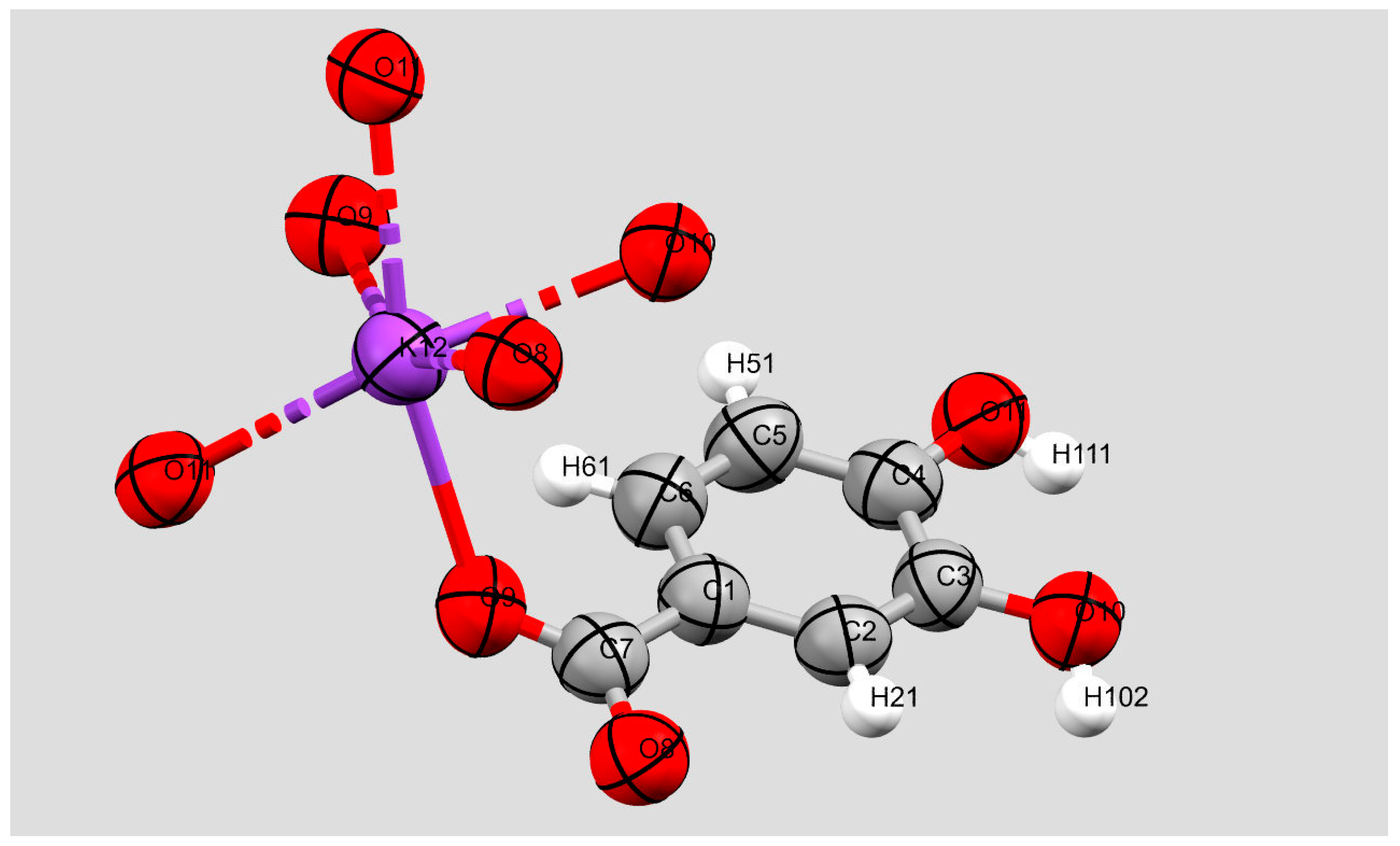
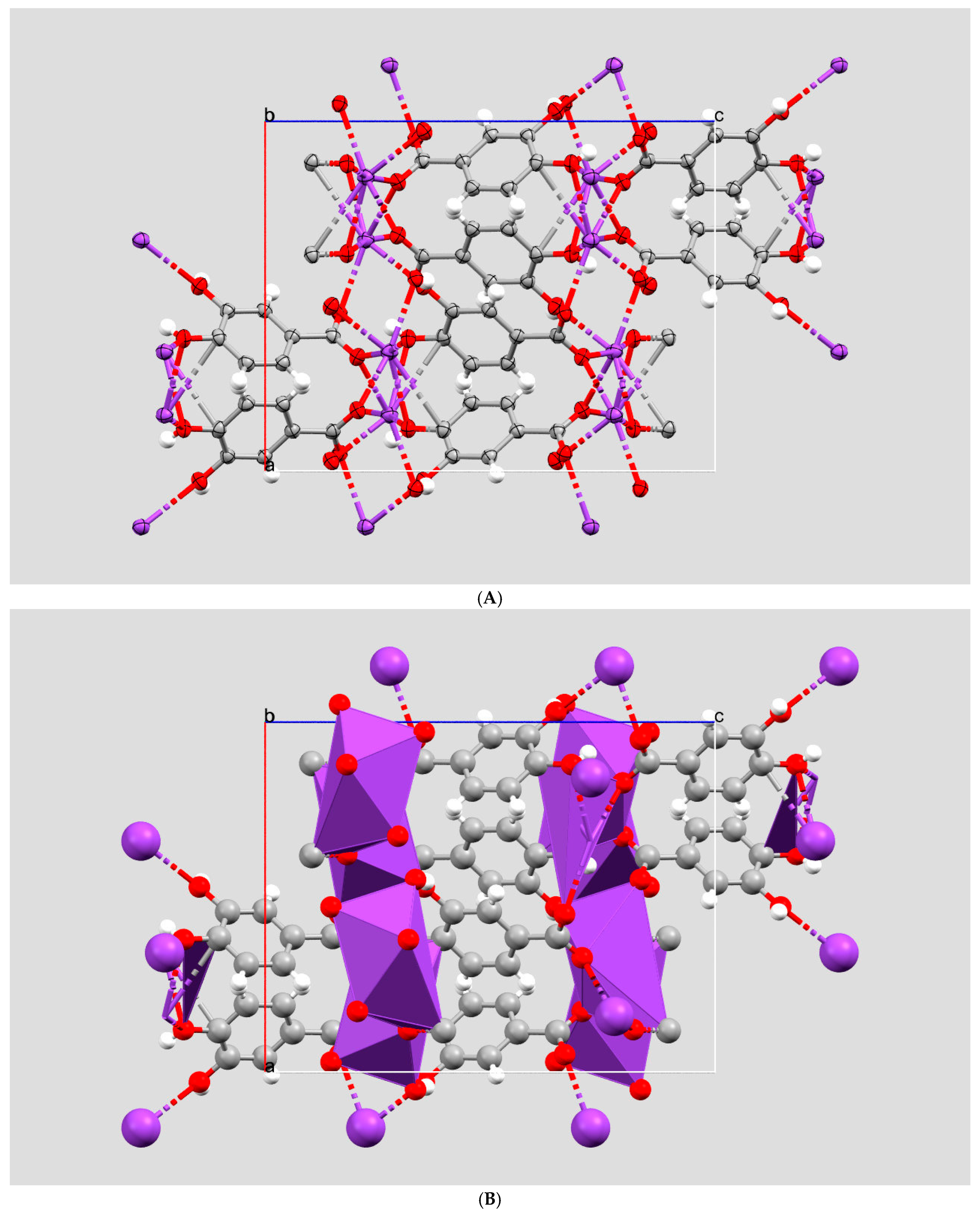
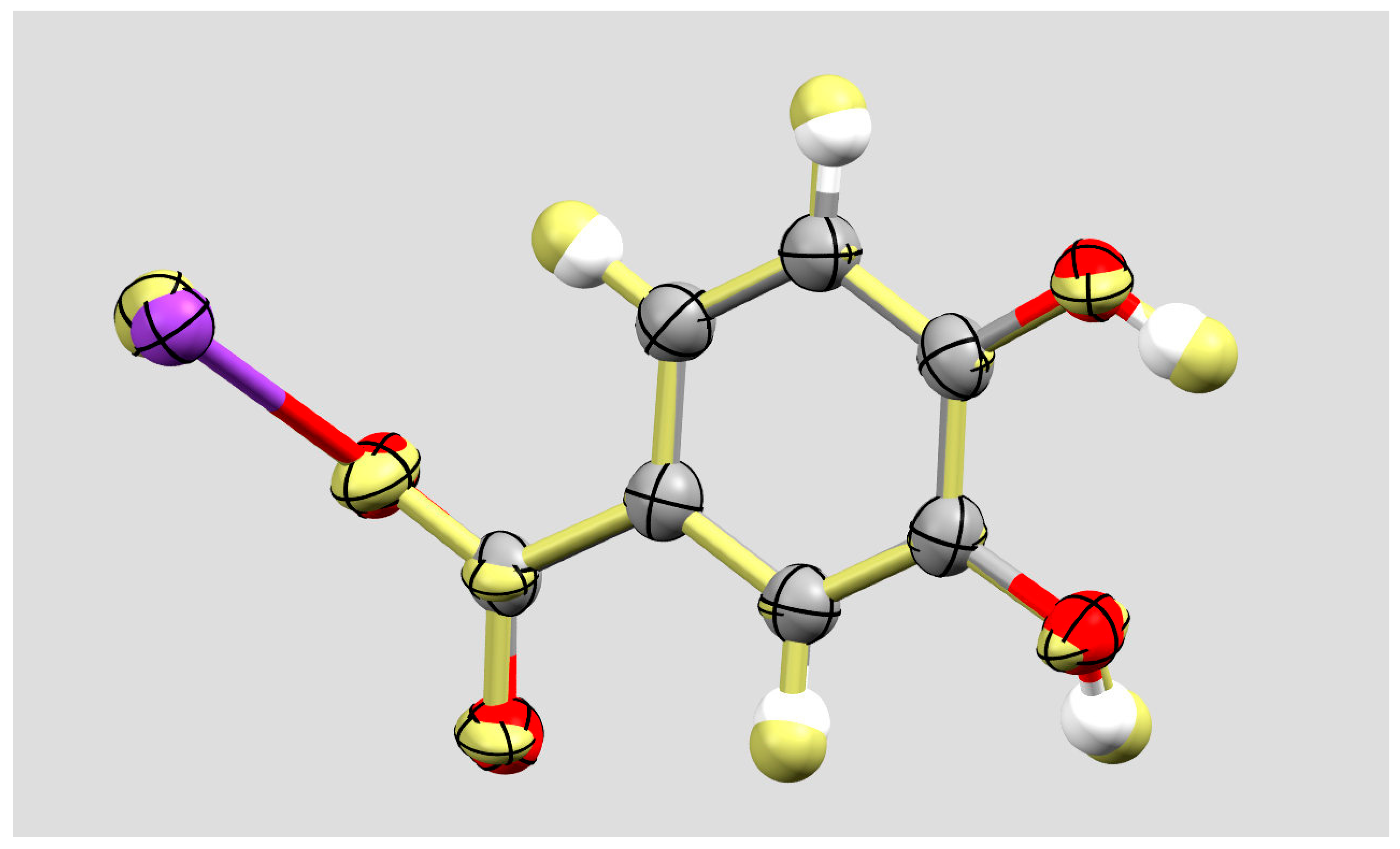
PCA potassium salt (PCAK, Figure 1 and Figure 2) crystallizes in the Pbca space group with an orthorhombic lattice. Its unit cell parameters are reported in Table 1, together with the ones inferred by the different computational approaches. There are eight molecular moieties in the unit cell (Z = 8), and only one formula unit in the asymmetric cell (Z′ = 1). The structure does not show any solvent accessible void and provides a 90.7% packing index [22] which reflects a fairly efficient packing.
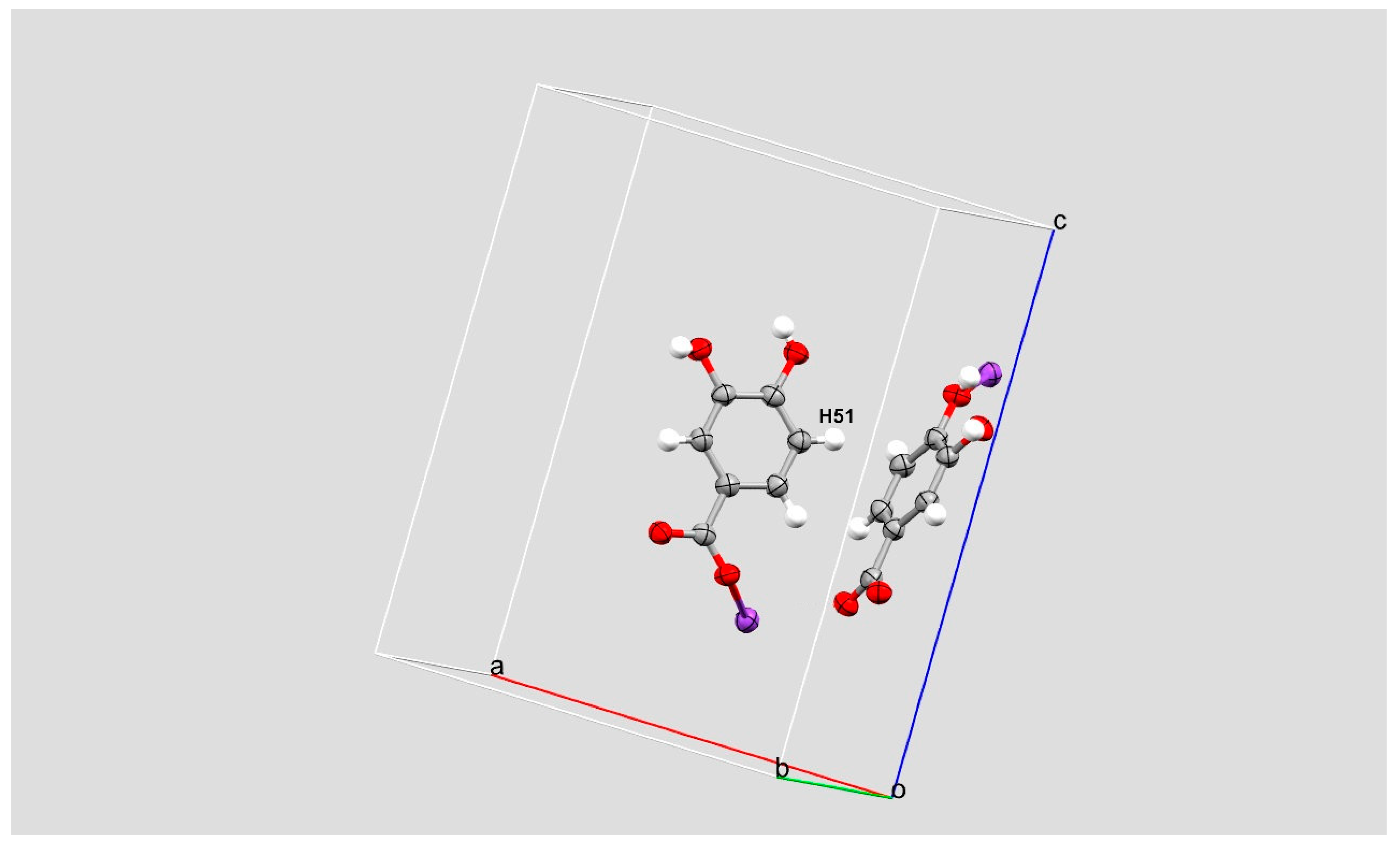
The whole setup is shown in Figure 3A,B.
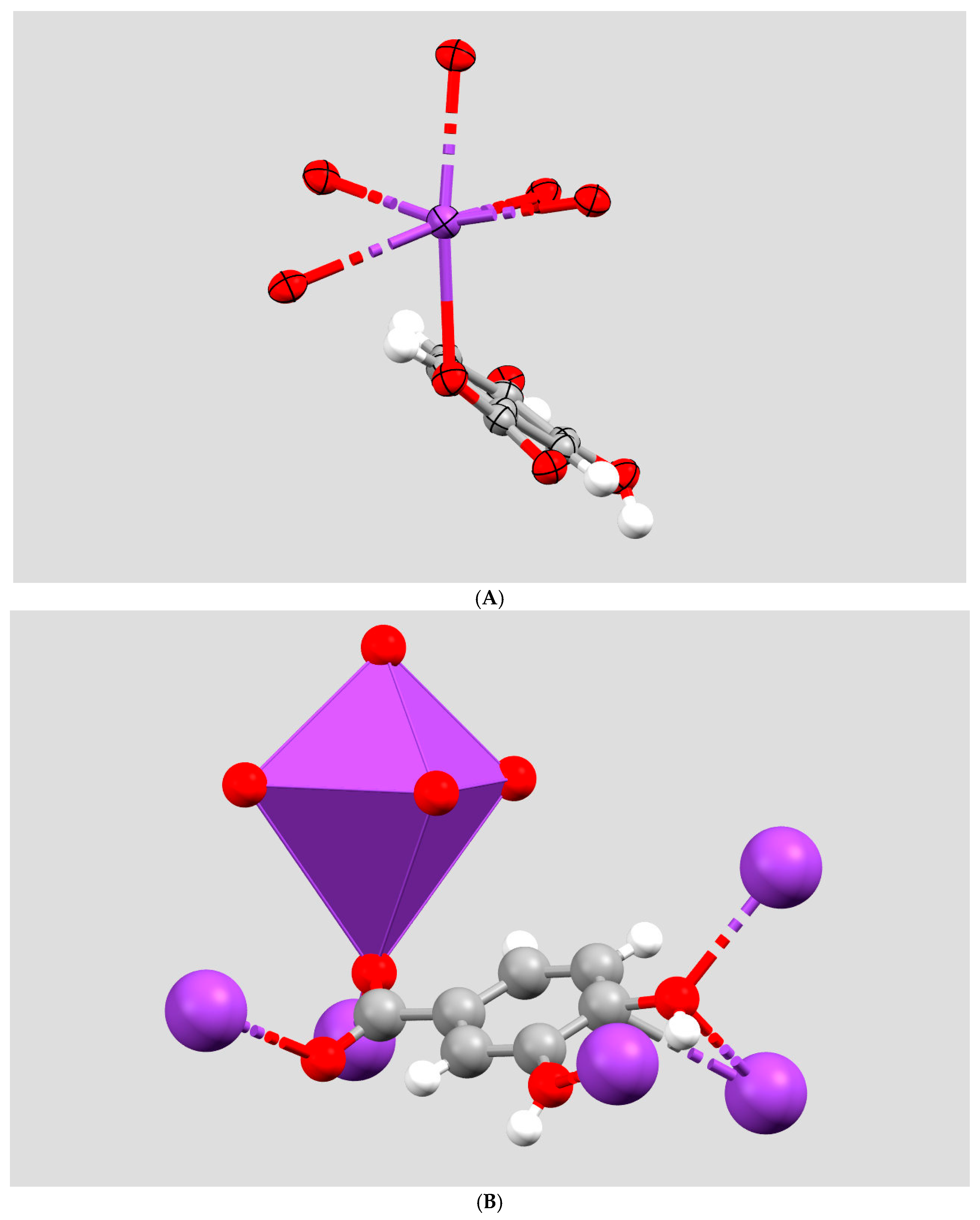
The molecular packing is, as expected, characterized by the competitive coordination of the potassium (K) ion by means of the catecholic and/or the carboxylic function. They both succeed in the K coordination, giving rise to a distorted hexacoordinated structure with a warped octahedral shape. The amount of the deviation from a perfect Platonic octahedron can be quantified accordingly to ref. [24] by an octahedral volume of 22.156 Å3, a quadratic elongation of 1.160 and an angle variance of 453.54 deg2 (Figure 4A,B).
The structure building block shows several inter- and intramolecular hydrogen bonds. They are reported in Table 2.
The structure is further stabilized by the presence of π-stacking and T-shaped interactions which are reported in Table 3. This cooperative work evidenced in the unit cell represents a strong and mostly isotropic network of decent interactions that contribute to the overall stabilization of the structure.
To extend our analysis to very fine details such as the adps, we compared the experimental data, inferred by means of an X-ray data collection of the diffraction of the PCAK, with the ones computed with different in silico approaches. We used Molecular Dynamics to simulate whether the computed unit cell could resemble the experimental one. The method is described in the Materials and Methods section and follows a robust approach already successfully used for a broad variety of compounds. It is based on the initial relaxation of the symmetry to a P1 (triclinic). As a consequence, the structure is free to float over the energy surface without any particular constraint. The simulation validated that the symmetry setup—orthorhombic in our case—is confirmed by checking the relative cell angles, that were free to change but remained constant during the simulation. This was achieved by optimizing the original input file by means of a DFT approach and several MM- and FF-based ones. No matter what the origin of the input .cif file was, it gave rise to similar results for all the different approaches. Interestingly, as described in the Computational Details, an improvement to the original K+ data for the Dreiding Force Field was provided in order to achieve these results. For this purpose, we implemented the original FF with data already published in the literature for what concerns K+ ion [23].
The results are reported in Table 1 for what concerns the unit cell parameters. They show a strong consistency between the experimental data—inferred by means of X-ray diffraction—and the computed ones.
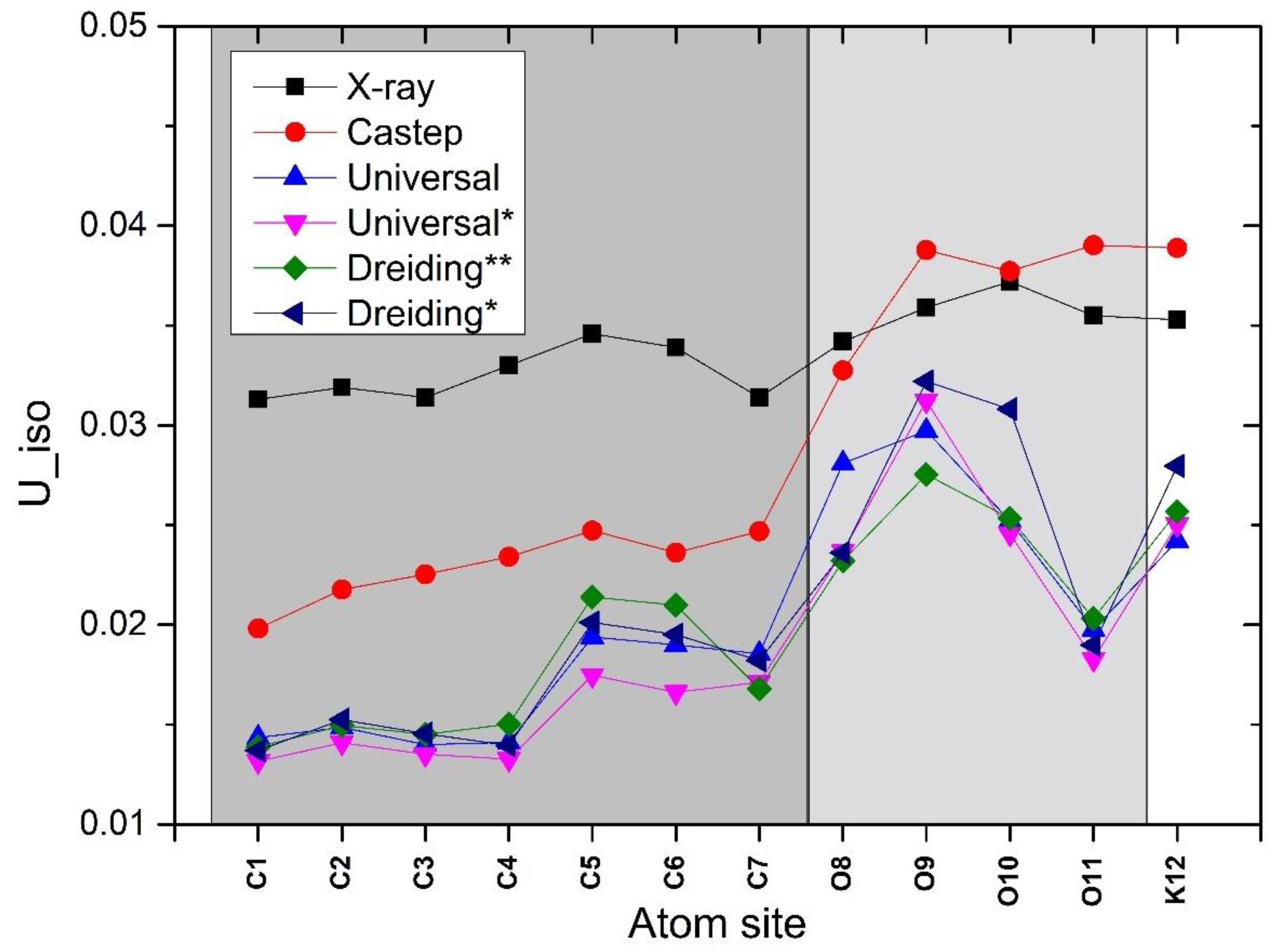
From our point of view, the data reported in Table 4 are definitely more interesting. They show the Uiso (isotropic displacement parameter), i.e., the mean-square displacement amplitude of an atom, averaged in all directions. It represents an isotropic parameter which simplifies the quantification of the libration amplitude of each atom in the structure as compared with the anisotropic one (adp or Uaniso), which is more useful for a graphical representation. In fact, being based on tensors, they emphasize the preferred orientation in a specific direction towards one or more atoms which perform their displacement. As a consequence, their analysis could be considered a prediction, at an atomic level, of preferential or forbidden movements of the atoms. These properties have evident consequences on the physical and mechanical properties of the substance, even at a macroscopical level.
These data are shown in Figure 5 where, in spite of different experimental and computational origins, similar trends are present. As there are no “real” Uiso values, neither can the experimental ones be considered “true”. In fact, these data are heavily affected by the scattering method used for their evaluation. Adps inferred by means of X-ray data are generally quite different from neutron ones and this difference relies on the different probe used [26,27,28]. However, the main approximation in the adp estimate is provided by a crude approximation—the so-called Independent Atom Model—used during data refinement, which forces a poorly realistic spherical model to simulate the electron density of atoms as if they were independent entities, without taking into account the changes in the electron density distribution due to chemical bonding and other structural features [29]. As hydrogen atoms contains one electron only, their scattering performance is poor. Thus, their Uiso are not reported in Figure (but in Table 4).
On one hand, the experimental Uiso are systematically bigger than the computed ones up to the oxygen atoms. On the other, confirming this trend, DFT-based data are regularly greater than the MM-based ones. Such a difference can be interpreted on the basis of a greater stiffness conferred by the FF parameters to the atoms in the unit cell.
The reported trends are clearly very similar no matter the experimental or computational method used to infer them. The Uiso magnitude is obtained by means of their Uaniso values and it is a trustful indicator of the atom ability to move, or better to librate, around their equilibrium position. As a consequence, the greater the magnitude of their Uiso values, the easier they can move although bound to other atoms although packed in a three-dimensional network. Interestingly, some carbon atoms, although belonging to the main backbone, are slightly more movable than others. This is true for C2, C5, C6, carbon atoms in spite of the different approaches used to calculate their adps.
A possible explanation can be provided invoking the fact that the aromatic ring side, to which C5 and C6 belong, is freer to move as there are no oxygen atoms overlooking that side. In fact, on the other side, the C2, which is slightly more movable than C1 and C3—i.e., its adp value is slightly bigger—lays nearby the oxygen atoms belonging to the carboxylic and hydroxylic groups, which are involved in classic hydrogen bonds. The C2 carbon atom is involved only in an intramolecular and nonconventional—thus weaker—H-bond, as described in Table 2.
The C5 carbon atom is constantly regarded as the most movable carbon atom in the structure, both in the experimental data and computed ones. To explain this behaviour, we consider the presence of a T-shaped interaction, reported in Table 3. These kinds of interactions [30] are extremely relevant in terms of the overall stabilization of the crystalline structure. On one hand, they provide an anchoring point along which the structure building block oscillates. On the other, they supply a preferential direction of movement which affects the adps magnitude.
In fact, the pi-stacking interactions provide a sort of symmetrical and equally distributed interactions among the different carbons of the involved rings. This is not true neither for the H-bonds which are focused on a specific group of atoms directly involved in the bond formation nor for the T-shaped interactions. Moreover, we are comparing the Uiso values, i.e., the isotropic ones, and therefore we can only compare their magnitude and not their deviation from a perfect sphere. This latter characteristic can instead show us the effect of the resultant of the forces acting on a specific atom and it is reported in Figure 6.
It is also important to bear in mind that the stacking interactions in the presence of hydrogen bonding are found to be stronger than in the absence of the hydrogen bonding [31]. Thus, in the presence of a large network of H-bonds and p-shaped interactions, the strongest ones—i.e., classic hydrogen bonds with a relevant electrostatic contribution and typical p-stacking interactions—contribute to the stiffening and stabilization of the crystalline building, while the weakest—i.e., the intramolecular C-H bond and the T-shaped interaction—represent only an attachment point which can undergo major fluctuations [32,33,34,35,36].
To strengthen this hypothesis, we overlaid the two most different structures in terms of Uaniso values. The result of the superimposition of the experimentally (X-ray)-inferred data and the MD (DFT-based) ones are reported in Figure 6.
The DFT-based simulation seems to overestimate, with respect to the X-ray data, the Uaniso. However, in spite of an almost completely isotropic distribution of the X-ray-inferred adps, although comparable in magnitude, the MD (DFT-based)-inferred ones show an evident oblate shape. This is particularly true for some atoms, the more movable ones. Interestingly, the direction towards which the oblate adps point is the 1/2 − X, −1/2 + Y, Z. This is shown in Figure 7 and corresponds to the already mentioned T-shaped interaction. There are, of course, minimal deviations from this principal direction due to the chemical environment and preferential interactions experienced by the atoms in the whole crystalline structure.

3. Materials and Methods

3.1. X-Ray Crystallography

PCA (257 mg, 1.67 mmol, Sigma Aldrich, Burlington, MA, USA, >98% purity, Merck Spa, Roma, Italy) was added, under a nitrogen atmosphere, to an anhydrous methanol solution (6 mL) containing potassium carbonate (230 mg, 1.67 mmol, Sigma Aldrich at 99% purity), and the resulting mixture was stirred for 1 h at room temperature, resulting in a pale yellow precipitate which was filtered and washed with cool methanol. A total of 100 mg of the so-obtained potassium salt was dissolved in 1 mL of water, and a few drops of methanol were added to the resulting solution; the slow and competitive evaporation of the two solvents at room temperature was allowed in order to obtain almost transparent and block-shaped single crystals suitable for X-ray diffraction.
Single-crystal X-ray diffraction data were collected on an Oxford Diffraction Supernova diffractometer with graphite-monochromated Cu Kα radiation (λ = 1.54184 Å) at 298 (2) K operated at 50 kV and 40 mA. Data collection and reduction were performed using the CrysAlisPro software package (Agilent (2014). CrysAlis PRO. Agilent Technologies Ltd., Yarnton, Oxfordshire, UK). Absorption corrections were applied using a multi-scan technique. The structure was solved by Superflip [37]. The hydrogen atoms belonging to the water molecule and the O–H moieties were located in a difference Fourier map and refined isotropically. The remaining hydrogen atoms bound to carbon were refined using a riding model with d(C–H) = 0.93 Å and Uiso = 1.2 Ueq(C). The figures were prepared using Mercury [38].
CCDC 2451846 contains the supplementary crystallographic data for this paper. These data can be obtained free of charge from The Cambridge Crystallographic Data Centre via http://www.ccdc.cam.ac.uk/data_request/cif.
This .cif is partially incomplete—i.e., misses the s.u. on hydrogen atoms. This is due to the fact that this is an old data collection (2016), and the file bearing all the relative information was lost. However, all the relevant data are present and the lack of s.u.s does not affect the reliability of the calculations and reasoning.
The computed .cifs obtained by means of the different in silico techniques are provided as Supplementary Materials.

3.2. Computational Details

3.2.1. Modelling

Crystal—The structural model of PCAK submitted to the MD simulations was generated using the X-ray structure (unit cell and atomic fractional coordinates) reported in the experimental .cif file. The positions of each atom were attained by considering their fractional parameters available in the .cif file for the single unit cell. The symmetry-related (space group name PBCA) images were allowed to develop, and then the structure was converted to P1, removing all the symmetry constrains. This allows the symmetry related atoms to move independently from one another during the MD simulation [16,21,39], using a robust and well-accepted procedure in which periodic boundary conditions (PBC) are applied. For the force-field-driven simulations, the P1 structure was replicated 3, 4 and 2 times along the a, b and c axes, respectively, to attain a supercell of 36.4545, 29.8544 and 31.2158 Å in length for each axes, respectively. The quantum-mechanical driven simulation used the single cell converted to P1 symmetry structure, as is (i.e., 12.1515, 7.4636 and 15.6079 Å).

3.2.2. Force Field Settings

To perform the molecular mechanics (MM) and MD calculations, the Forcite module of the BIOVIA Materials Studio 2025 package was used, with the implemented Universal [40] and Dreiding [14] force fields. Since the available parameters set file for the Dreiding FF did not have vdW parameters for the K+ ion, the file was augmented with the parameters obtained from either the Universal FF or from the literature [23]. The non-bond (NB) interactions settings adopted were an atom-based summation method for the vdW and Ewald summation for Coulomb. For the vdW interactions, a cut-off distance of 18.50 Å was used with a cubic-spline truncation, with width = 1.00 Å and buffer width = 0.50 Å, and long-range corrections. The Ewald summation parameters were accuracy = 0.00001 kcal mol−1, with a buffer width of 0.50 Å. The dielectric constant value ε was set to 1.0.

3.2.3. Charges

The atomic charges were assigned by using the charge equilibration (Qeq) method [41]. In this method the resulting charges on each atomic centre depend on the distance it has from the surrounding atoms. To avoid differences between equivalent atoms, we reset their values to the average. For example, the two equivalent oxygen atoms of the carboxylic group of PCAK attain charge values of −0.629 e and −0.482 e; they both were averaged to the mean value (−0.555 e), and so on for the other equivalent atoms.

3.2.4. Protocols

For either the Universal or Dreiding FFs molecular mechanics (MM) and Molecular Dynamics (MD)-implemented calculations, the Forcite module was used.
In all the necessary preliminary MM calculations, Forcite’s smart geometry optimization algorithm was used to a gradient of 0.1 kcal mol–1.
The MD simulations started from the MM energy-minimized structures, as attained from the modelling section. In all the runs, the constant number of particles, pressure and temperature (NPT) ensemble with PBC was used. In order to allow the cell to change both shape and volume, we used the Parrinello pressure control method [42], whereas a Berendsen’s thermostat with the default decay constant (0.01 ps) was used to control the temperature [43]. Pressure was set at 0.0, GPa, while the temperature was set to 298 K. The four collected runs, two with the potassium ion vdW parameters from the universal FF and two with these parameters from the literature [23], were made of transients of 1.0 ns, after a 0.1 ns equilibration period. The integration time step was 0.001 ps and the sampling every 1000 time-steps.

3.2.5. Quantum Mechanical Calculations

The quantum mechanical calculations were performed with CASTEP, a pseudopotentials plane-wave density functional theory (DFT) method, programme [44] available in BIOVIA Materials Studio 2025. The DFT level of the theory used was GGA: gradient-corrected functionals with PBE [45] exchange and correlation functional, together with the TS [46] dispersion correction (DFT-D correction). The SCF criterion was extended to a medium quality (cut_off_energy = 489.80 ev; elec_energy_tol = 0.0000002), with OTFG ultrasoft pseudopotentials and Koelling–Harmon relativistic treatment. Geometry optimization of the structure was executed with the LBFGS [47] method with tolerances set to: geom_energy_tol = 0.00001; geom_force_tol = 0.030; geom_stress_tol = 0.050; geom_disp_tol = 0.001; geom_max_iter = 100. The MD simulation was executed with the NPT ensemble using the Nose–Hover thermostat [48,49] at 298 K, and the Parrinello–Rahman barostat [50] with an external stress of 0.0001 GPa, integration time step of 1 fs, a thermal equilibration period of 1 ps, and a production run of 7 ps. The trajectory was saved at every step. In Figure S1 (in the Supplementary Materials) the result of the overall 8 ps equilibration on the cell axes parameters is displayed.

4. Conclusions

In this paper we described an efficient and robust method to predict the fine structural details of complex crystalline structures. We extended our forecasts to the adps prediction, with a very good agreement between the calculated data and the benchmark provided by the X-ray-inferred ones.
This information was used to provide basic information about the most valuable lines of strength present in the crystalline building, accounting, in our case, not only for the hydrogen network but also the π-staking and T-shaped interactions.
Moreover, the complex and controversial [11,12] coordination of the K+ ion is described.
This outcome was performed by means of the integration of the original data present in the Dreiding FF with the ones already present in the literature [23]. The use of MM-based MD simulations provides a less expensive computational path to achieve valuable structural information. In fact, they are in complete agreement with previously published data, thus confirming, once again, the goodness of our approach.

Supplementary Materials

The following supporting information can be downloaded at: https://www.mdpi.com/article/10.3390/inorganics13120385/s1, Figure S1: Cell parameters equilibration; Table S1: cell parameter deviation percentage.

Author Contributions

Conceptualization, F.P.; Methodology, G.M.L.; Software, G.M.L.; Validation, G.M.L.; Formal analysis, F.P.; Investigation, F.P.; Resources, F.P.; Data curation, G.M.L.; Writing—original draft, F.P.; Writing—review & editing, F.P.; Visualization, F.P. All authors have read and agreed to the published version of the manuscript.

Funding

This research received no external funding.

Institutional Review Board Statement

Not applicable.

Informed Consent Statement

Not applicable.

Data Availability Statement

The original contributions presented in this study are included in the article/Supplementary Materials. Further inquiries can be directed to the corresponding author.

Conflicts of Interest

The authors declare no conflict of interest.

References

  1. Stevenson, F. Humus Chemistry: Genesis, Composition, Reactions. Humus Chem. 1994, 72, 512. [Google Scholar]
  2. Tipping, E. Humic Substances in Soil, Sediment and Water: Geochemistry, Isolation and Characterization Edited by G. R. Aiken, D.M. McKnight, R.L. Wershaw and P. MacCarthy, Wiley, 1985. No. of Pages: 692. Price: £61.35 (Hardback). Geol. J. 1986, 21, 213–214. [Google Scholar] [CrossRef]
  3. Stevenson, F.J. Humic Substances in the Environment. Soil. Sci. Soc. Am. J. 1975, 39, vii+327. [Google Scholar] [CrossRef]
  4. Schnitzer, M.; Khan, S.U. Humic Substances in the Environment; References-Scientific Research Publishing; Marcel Dekker, Inc.: New York, NY, USA, 1972; Available online: https://www.scirp.org/reference/referencespapers?referenceid=1371146 (accessed on 7 October 2025).
  5. Vermeer, A.W.P.; McCulloch, J.K.; Van Riemsdijk, W.H.; Koopal, L.K. Metal Ion Adsorption to Complexes of Humic Acid and Metal Oxides: Deviations from the Additivity Rule. Environ. Sci. Technol. 1999, 33, 3892–3897. [Google Scholar] [CrossRef]
  6. Lubal, P.; Široký, D.; Fetsch, D.; Havel, J. The Acidobasic and Complexation Properties of Humic Acids: Study of Complexation of Czech Humic Acids with Metal Ions. Talanta 1998, 47, 401–412. [Google Scholar] [CrossRef]
  7. Tipping, E. Cation Binding by Humic Substances; Cambridge University Press: Cambridge, UK, 2002. [Google Scholar] [CrossRef]
  8. Abrahams, B.F.; Commons, C.J.; Hudson, T.A.; Arlt, R.S.; White, K.F.; Chang, M.; Jackowski, J.J.; Lee, M.; Lee, S.X.; Liu, H.D.; et al. Alkali Metal Salts of 4-Hydroxybenzoic Acid: A Structural and Educational Study. Acta Crystallogr. C Struct. Chem. 2021, 77, 340–353. [Google Scholar] [CrossRef] [PubMed]
  9. Abrahams, B.F.; Commons, C.J.; Hudson, T.A.; Arlt, R.S.; Ahl, R.; Carajias, E.D.; Chan, J.W.K.; Guo, Z.; Hill, R.E.; Mcginty, A.; et al. Complexes of 2,4,6-Trihydroxybenzoic Acid: Effects of Intramolecular Hydrogen Bonding on Ligand Geometry and Metal Binding Modes. Cryst. Struct. Commun. 2022, 78, 653–670. [Google Scholar] [CrossRef] [PubMed]
  10. Lombardo, G.M.; Portalone, G.; Colapietro, M.; Rescifina, A.; Punzo, F. From the X-Rays to a Reliable “Low Cost” Computational Structure of Caffeic Acid: DFT, MP2, HF and Integrated Molecular Dynamics–X-Ray Diffraction Approach to Condensed Phases. J. Mol. Struct. 2011, 994, 87–96. [Google Scholar] [CrossRef]
  11. Lombardo, G.M.; Rescifina, A.; Punzo, F. Functional Hybrid Co-Crystals of Humic Substances: A Growth Forecast. CrystEngComm 2014, 16, 5917–5923. [Google Scholar] [CrossRef]
  12. Lombardo, G.M.; Portalone, G.; Chiacchio, U.; Rescifina, A.; Punzo, F. Potassium Caffeate/Caffeic Acid Co-Crystal: The Rat Race between the Catecholic and Carboxylic Moieties in an Atypical Co-Crystal. Dalton Trans. 2012, 41, 14337–14344. [Google Scholar] [CrossRef]
  13. Dunitz, J.D. Crystal and Co-Crystal: A Second Opinion. CrystEngComm 2003, 5, 506. [Google Scholar] [CrossRef]
  14. Mayo, S.L.; Olafson, B.D.; Goddard, W.A. DREIDING: A Generic Force Field for Molecular Simulations. J. Phys. Chem. 1990, 94, 8897–8909. [Google Scholar] [CrossRef]
  15. Grassi, A.; Lombardo, G.M.; Punzo, F. Wrapping and Unwrapping an Indicaxanthin Molecule: A Computational Approach. Comput. Theor. Chem. 2020, 1191, 113028. [Google Scholar] [CrossRef]
  16. Valkonen, A.; Lombardo, G.M.; Rissanen, K.; Punzo, F.; Cametti, M. X-Ray Crystallographic and Computational Study on Uranyl-Salophen Complexes Bearing Nitro Groups. Dalton Trans. 2017, 46, 5240–5249. [Google Scholar] [CrossRef] [PubMed]
  17. Fraga, A.R.L.; Ferreira, F.F.; Lombardo, G.M.; Punzo, F. Experimental and Theoretical Characterization of N-(Diethylcarbamothioyl) Benzamide Triclinic Polymorph. J. Mol. Struct. 2013, 1047, 1–8. [Google Scholar] [CrossRef]
  18. Pistarà, V.; Lombardo, G.M.; Rescifina, A.; Bacchi, A.; D’Andrea, F.; Punzo, F. Experimental and in Silico Characterization of a Biologically Active Inosose. Struct. Chem. 2013, 24, 955–965. [Google Scholar] [CrossRef]
  19. Álvarez, J.L.G.; Amato, M.E.; Lombardo, G.M.; Carriedo, G.A.; Punzo, F. Self-Organization by Chiral Recognition Based on Ad Hoc Chiral Pockets in Cyclotriphosphazenes with Binaphthoxy and Biphenoxy Substituents: An X-Ray, NMR and Computational Study. Eur. J. Inorg. Chem. 2010, 2010, 4483–4491. [Google Scholar] [CrossRef]
  20. Lombardo, G.M.; Thompson, A.L.; Ballistreri, F.P.; Pappalardo, A.; Sfrazzetto, G.T.; Tomaselli, G.A.; Toscano, R.M.; Punzo, F. An Integrated X-Ray and Molecular Dynamics Study of Uranyl-Salen Structures and Properties. Dalton Trans. 2012, 41, 1951–1960. [Google Scholar] [CrossRef]
  21. Bastos, I.T.S.; Costa, F.N.; Silva, T.F.; Barreiro, E.J.; Lima, L.M.; Braz, D.; Lombardo, G.M.; Punzo, F.; Ferreira, F.F.; Barroso, R.C. A Combined Experimental and in Silico Characterization to Highlight Additional Structural Features and Properties of a Potentially New Drug. J. Mol. Struct. 2017, 1146, 735–743. [Google Scholar] [CrossRef]
  22. Kitaigorodsky, A. Molecular Crystals and Molecules, 1st ed.; Academic Press: New York, NY, USA, 1973. [Google Scholar]
  23. Thorat, A.; Chauhan, R.; Sartape, R.; Singh, M.R.; Shah, J.K. Effect of K+ Force Fields on Ionic Conductivity and Charge Dynamics of KOH in Ethylene Glycol. J. Phys. Chem. B 2024, 128, 3707–3719. [Google Scholar] [CrossRef]
  24. Robinson, K.; Gibbs, G.V.; Ribbe, P.H. Quadratic Elongation: A Quantitative Measure of Distortion in Coordination Polyhedra. Science (1979) 1971, 172, 567–570. [Google Scholar] [CrossRef]
  25. Jeffrey, G.A.; Maluszynska, H.; Mitra, J. Hydrogen Bonding in Nucleosides and Nucleotides. Int. J. Biol. Macromol. 1985, 7, 336–348. [Google Scholar] [CrossRef]
  26. Schlenker, M.; Guigay, J.P. Differences between Neutron and X-Ray Scattering. Int. Tables Crystallogr. 2006, B, 557–569. [Google Scholar] [CrossRef]
  27. Majumdar, A.; Müller, M.; Busch, S. Computation of X-Ray and Neutron Scattering Patterns to Benchmark Atomistic Simulations against Experiments. Int. J. Mol. Sci. 2024, 25, 1547. [Google Scholar] [CrossRef]
  28. Reich, V.; Majumdar, A.; Müller, M.; Busch, S. Comparison of Molecular Dynamics Simulations of Water with Neutron and X-Ray Scattering Experiments. EPJ Web Conf. 2022, 272, 01015. [Google Scholar] [CrossRef]
  29. Fabiola Sanjuan-Szklarz, W.; Woińska, M.; Domagala, S.; Dominiak, P.M.; Grabowsky, S.; Jayatilaka, D.; Gutmann, M.; Woźniak, K. On the Accuracy and Precision of X-Ray and Neutron Diffraction Results as a Function of Resolution and the Electron Density Model. IUCrJ 2020, 7, 920–933. [Google Scholar] [CrossRef] [PubMed]
  30. Grimme, S. Do Special Noncovalent π–π Stacking Interactions Really Exist? Angew. Chem. Int. Ed. 2008, 47, 3430–3434. [Google Scholar] [CrossRef] [PubMed]
  31. Ahmed, U.; Sundholm, D.; Johansson, M.P. The Effect of Hydrogen Bonding on the π Depletion and the π–π Stacking Interaction. Phys. Chem. Chem. Phys. 2024, 26, 27431–27438. [Google Scholar] [CrossRef]
  32. Martinez, C.R.; Iverson, B.L. Rethinking the Term “Pi-Stacking”. Chem. Sci. 2012, 3, 2191–2201. [Google Scholar] [CrossRef]
  33. Carter-Fenk, K.; Herbert, J.M. Reinterpreting π-Stacking. Phys. Chem. Chem. Phys. 2020, 22, 24870–24886. [Google Scholar] [CrossRef]
  34. Gao, X.C.; Hao, Q.; Wang, C.S. Improved Polarizable Dipole-Dipole Interaction Model for Hydrogen Bonding, Stacking, T-Shaped, and X-H···φ Interactions. J. Chem. Theory Comput. 2017, 13, 2730–2741. [Google Scholar] [CrossRef]
  35. Lessa, M.D.; Stoyanov, S.R.; de Carneiro, J.W.M.; da Costa, L.M. Density Functional Theory Investigation of the Contributions of π-π Stacking and Hydrogen Bonding with Water to the Supramolecular Aggregation Interactions of Model Asphaltene Heterocyclic Compounds. J. Mol. Model. 2024, 30, 145. [Google Scholar] [CrossRef]
  36. Han, T.; Pan, X.; Wang, Y.; Du, Y.; Xue, J. Hydrogen-Bond Stabilized π-π Stacking in the Solvent-Induced Dimerization of 4-Nitro-1-Naphthol: A Spectroscopic and Computational Investigation. J. Mol. Struct. 2024, 1312, 138505. [Google Scholar] [CrossRef]
  37. Palatinus, L.; Chapuis, G. SUPERFLIP-A Computer Program for the Solution of Crystal Structures by Charge Flipping in Arbitrary Dimensions. J. Appl. Crystallogr. 2007, 40, 786–790. [Google Scholar] [CrossRef]
  38. MacRae, C.F.; Sovago, I.; Cottrell, S.J.; Galek, P.T.A.; McCabe, P.; Pidcock, E.; Platings, M.; Shields, G.P.; Stevens, J.S.; Towler, M.; et al. Mercury 4.0: From Visualization to Analysis, Design and Prediction. J. Appl. Crystallogr. 2020, 53, 226–235. [Google Scholar] [CrossRef]
  39. Lombardo, G.M.; Punzo, F. False Asymmetry, Pseudosymmetry, Disorder, Polymorphism and Atomic Displacement Parameters. J. Mol. Struct. 2014, 1078, 158–164. [Google Scholar] [CrossRef]
  40. Rappé, A.K.; Casewit, C.J.; Colwell, K.S.; Goddard, W.A.; Skiff, W.M. UFF, a Full Periodic Table Force Field for Molecular Mechanics and Molecular Dynamics Simulations. J. Am. Chem. Soc. 1992, 114, 10024–10035. [Google Scholar] [CrossRef]
  41. Rappé, A.K.; Goddard, W.A. Charge Equilibration for Molecular Dynamics Simulations. J. Phys. Chem. 1991, 95, 3358–3363. [Google Scholar] [CrossRef]
  42. Parrinello, M.; Rahman, A. Strain Fluctuations and Elastic Constants. J. Chem. Phys. 1982, 76, 2662–2666. [Google Scholar] [CrossRef]
  43. Berendsen, H.J.C.; Postma, J.P.M.; Van Gunsteren, W.F.; Dinola, A.; Haak, J.R. Molecular Dynamics with Coupling to an External Bath. J. Chem. Phys. 1984, 81, 3684–3690. [Google Scholar] [CrossRef]
  44. Clark, S.J.; Segall, M.D.; Pickard, C.J.; Hasnip, P.J.; Probert, M.I.J.; Refson, K.; Payne, M.C. First Principles Methods Using CASTEP. Z. Krist.-Cryst. Mater. 2005, 220, 567–570. [Google Scholar] [CrossRef]
  45. Perdew, J.P.; Burke, K.; Ernzerhof, M. Generalized Gradient Approximation Made Simple. Phys. Rev. Lett. 1996, 77, 3865. [Google Scholar] [CrossRef]
  46. Tkatchenko, A.; Scheffler, M. Accurate Molecular van Der Waals Interactions from Ground-State Electron Density and Free-Atom Reference Data. Phys. Rev. Lett. 2009, 102, 073005. [Google Scholar] [CrossRef] [PubMed]
  47. Pfrommer, B.G.; Côté, M.; Louie, S.G.; Cohen, M.L. Relaxation of Crystals with the Quasi-Newton Method. J. Comput. Phys. 1997, 131, 233–240. [Google Scholar] [CrossRef]
  48. Nosé, S. A Molecular Dynamics Method for Simulations in the Canonical Ensemble. Mol. Phys. 1984, 52, 255–268. [Google Scholar] [CrossRef]
  49. Leimkuhler, B.; Noorizadeh, E.; Theil, F. A Gentle Stochastic Thermostat for Molecular Dynamics. J. Stat. Phys. 2009, 135, 261–277. [Google Scholar] [CrossRef]
  50. Parrinello, M.; Rahman, A. Polymorphic Transitions in Single Crystals: A New Molecular Dynamics Method. J. Appl. Phys. 1981, 52, 7182–7190. [Google Scholar] [CrossRef]
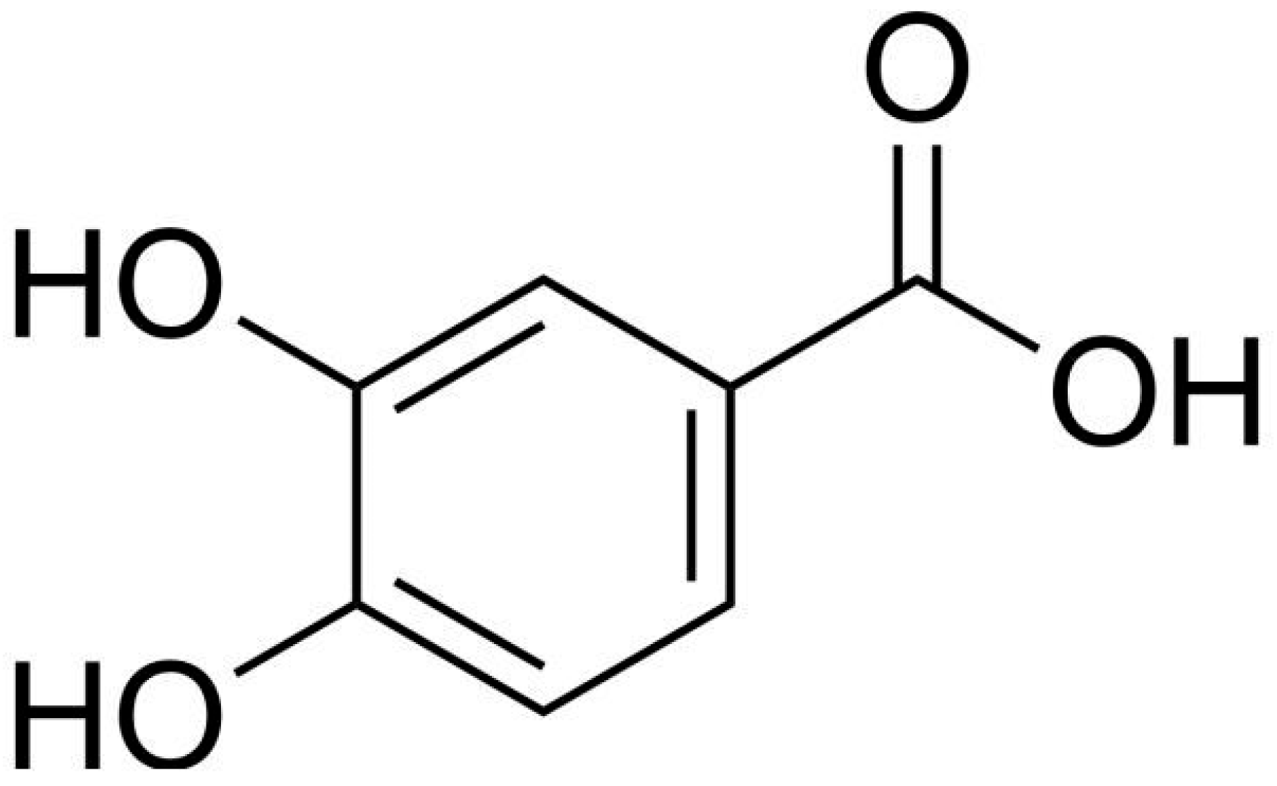
Figure 1. Chemical sketch of 3,4 dihydroxybenzoic acid (PCA).
Inorganics 13 00385 g001
Figure 2. Atomic numbering and potassium ion (in purple) coordination in PCA. Displacement ellipsoids are shown at 50% probability. H atoms are displayed as fixed-sized spheres.
Inorganics 13 00385 g002
Figure 3. (A) Unit cell of PCAK, along the b-axis, with Z = 8. Dashed lines evidence the K+ ion coordination interactions. (B) The typical (partially distorted) octahedral geometry of the K+ ion coordination is evidenced in purple.
Inorganics 13 00385 g003
Figure 4. (A) K+ ion (in purple) octahedral coordination as observed in the molecular building block. (B) The partially distorted octahedron is highlighted.
Inorganics 13 00385 g004
Figure 5. Uiso values for non-hydrogen atoms. * Refers to K+ parameters inferred by ref [23]. ** Refers to K+ parameters derived by the Universal FF (as they were missing in the original Dreiding FF).
Inorganics 13 00385 g005
Figure 6. X-ray vs. MD (DFT-based) adps superimposition. The X-ray model is reported with different colours while the MD one is in light yellow.
Inorganics 13 00385 g006
Figure 7. The most relevant T-shaped interaction between the C5—H51 and the corresponding aromatic ring on a neighbour molecule.
Inorganics 13 00385 g007
Table 1. The cell parameters derived by the different experimental and computational approaches. The * corresponds to the use of the K+ parameters inferred by ref [23], The ** to the use of the K+ parameters derived by the Universal FF.
X-RayCastepUniversalUniversal *Dreiding **Dreiding *
a12.1515(8)12.338511.941611.448712.077211.4048
b7.4636(5)7.35207.49647.52507.56557.5047
c15.6079(10)16.024216.937616.667916.259016.2707
α90.00090.00090.00090.00090.00090.000
β90.00090.00090.00090.00090.00090.000
γ90.00090.00090.00090.00090.00090.000
Table 2. Potential hydrogen bonds analysis. Bond lengths are given in Å. Donor–Acceptor distance is reported together with its standard uncertainty (s.u.). * Identifies bifurcated hydrogen bonds, i.e., the sum of three angles about H~360° [25].
Bond TypeD—H..AD–HH…AD…A
Inter-O10—H102..O8 a0.841.852.6594(2)
Intra-O11—H111..O100.812.412.7618(2)
Inter-* O11—H111..O8 b0.811.942.6946(2)
Inter-* O11—H111..O9 b0.812.543.0567(2)
Intra-C2—H21..O80.942.532.8405(2)
Symmetry codes: a 1 − x, 2 − y, 1 − z; b x, 3/2 − y, 1/2 + z.
Table 3. π-stacking and T-shaped noncovalent interactions. s.u. is reported in parentheses where its calculation is meaningful.
π-Stacking Interactions and T-Shaped Interactions
Ring-RingDistance (Å)Angle Between Planes (°)Slippage (Å)Crystallogr. Directions
Cg1–Cg13.4928(2)00.8551 − X, 1 − Y, 1 − Z
Cg1–Cg14.9373(3)81n.a.½ − X, 1/2 + Y, Z
X-H…Cg(π-Ring) Interactions (Details of the T-Shaped Ones)
X—H  Cg(J)Distance (Å)Angle of the Bond with the π Plane (°)Crystallogr. DirectionsX—H  Cg(J)
C5—H51 → Cg12.71571/2 − x, −1/2 + y, zC5—H51 → Cg1
Table 4. Uiso (isotropic displacement parameter) for non-Hydrogen atoms, with the corresponding atom numbering, calculate for every different experimental or in silico approach. The * corresponds to the use of the K+ parameters inferred by ref [23], the ** to the use of the K+ parameters derived by the Universal FF.
Uiso
X-RayCastepUniversalUniversal *Dreiding **Dreiding *
C10.03130.019810.014340.013160.013870.01370
C20.03190.021760.014860.014090.014950.01525
C30.03140.022530.013990.013510.014510.01454
C40.03300.023400.014110.013270.015020.01394
C50.03460.024720.019390.017470.021390.02011
C60.03390.023620.018990.016610.020980.01951
C70.03140.024680.018540.017120.016790.01821
080.03420.032760.028100.023670.023210.02360
090.03590.038780.029740.031240.027540.03221
0100.03720.037730.025200.024560.025330.03081
0110.03550.039030.019760.018280.020310.01898
K120.03530.038890.024210.025030.025670.02797
H210.03770.035000.026440.025020.025140.02675
H510.03790.042730.034220.030900.037900.03643
H610.04040.042320.031910.028420.037430.03417
H1110.05330.042160.029450.026490.025560.02592
H1020.05630.030170.046820.052080.038960.07369
Disclaimer/Publisher’s Note: The statements, opinions and data contained in all publications are solely those of the individual author(s) and contributor(s) and not of MDPI and/or the editor(s). MDPI and/or the editor(s) disclaim responsibility for any injury to people or property resulting from any ideas, methods, instructions or products referred to in the content.