Article

Calcium Phosphate Ceramic Powders Prepared from Mechanochemically Activated Precursors

by
Kostadinka Sezanova
1,
Yordanka Tuparova
1,
Pavletta Shestakova
2,
Pavel Markov
1,
Daniela Kovacheva
1 and
Diana Rabadjieva
1,*
1
Institute of General and Inorganic Chemistry, Bulgarian Academy of Sciences, Acad. G. Bonchev Str., bl. 11, 1113 Sofia, Bulgaria
2
Institute of Organic Chemistry with Centre of Phytochemistry, Bulgarian Academy of Sciences, Acad. G. Bonchev Str., bl. 9, 1113 Sofia, Bulgaria
*
Author to whom correspondence should be addressed.
Inorganics 2025, 13(10), 313; https://doi.org/10.3390/inorganics13100313
Submission received: 11 August 2025 / Accepted: 22 September 2025 / Published: 24 September 2025
(This article belongs to the Special Issue Featured Papers in Inorganic Materials 2025)

Abstract

The chemical and structural similarity of calcium orthophosphates to hard tissues in the human body makes them suitable as biomaterials for bone implants, cements, injection systems, etc., for bone regeneration and reconstruction. Tetracalcium phosphate (Ca4(PO4)2O, TTCP) is a promising component for such biomaterials due to its high calcium content and alkaline nature. The former makes it suitable for promoting mineralization, while the latter supports neutralization of the acidic environment, helping to prevent inflammation and improve the biocompatibility of the materials. However, it is the least used calcium orthophosphate due to the difficulties in its synthesis. This study examines the effect of high-energy mechanochemical activation on the phase evolution, particle morphology, and thermal behaviour of equimolar mixtures of Ca(OH)2 and CaHPO4, with the aim of optimizing precursor conditions for the synthesis of (TTCP)-rich ceramic materials. The results demonstrate that mechanochemical activation effectively induces structural disorder, promotes the formation of amorphous and nanocrystalline phases, and facilitates subsequent phase transitions upon calcination. The combined use of solid-state NMR, XRD, TEM, and thermal analysis provides a comprehensive understanding of the transformation pathways. Ultimately, 24 h of activation under the experimental conditions was identified as optimal for producing a precursor with a favorable phase composition for obtaining TTCP-rich ceramic materials after calcination at 1350 °C.

1. Introduction

Calcium orthophosphates are essential in obtaining bone-like biomaterials due to their chemical and structural compatibility with hard biological tissues, ability to support bone growth, and to be absorbed by the body [1]. If we assume that the facilitation of cell adhesion, proliferation, and differentiation, which ultimately leads to the formation of new bone, is more related to the morphology of these materials [2], then bioresorbability is an effect primarily of the solubility of the calcium-phosphate compound [3]. For example, hydroxyapatite (Ca5(PO4)3OH, HA), closely mimics the mineral component of natural bone and possesses highly biocompatible and osteoconductive properties, but it is the thermodynamically most stable calcium orthophosphate and has the lowest solubility [4]. Thus, monophasic HA is practically insoluble in the body fluids and does not actively participate in the process of bone remodeling [5]. On the other hand, the monophasic α- and β-tricalcium phosphates (Ca3(PO4)2, TCP) are more soluble, degrade faster, and in combination with HA balance bone growth and material resorption [6]. Therefore, it is important to produce multiphase calcium phosphate materials to mitigate the disadvantages of individual phases and enhance their advantages.
From a thermodynamic point of view, the most soluble calcium phosphate at physiological conditions (pH 7–8, and 37 °C) is tetracalcium phosphate (Ca4(PO4)2O, TTCP) [4]. The high calcium content makes it suitable for promoting mineralization, and the alkaline nature helps neutralize the acidic environment, which can be useful for preventing inflammation and improving biocompatibility [7,8,9]. However, it is the least used calcium orthophosphate due to difficulties in its synthesis.
Among synthetic biomaterials, calcium phosphate ceramic powders are widely used in orthopedic and maxillofacial surgery for the production of bone grafts and scaffolds and in dentistry as materials for tooth restoration, for remineralization treatment, and coatings for dental implants [10]. As the primary component, they are integrated in injectable calcium phosphate cements, drug delivery systems, and inorganic/organic composites [11,12].
Various methods have been developed for the production of calcium phosphate ceramic materials, such as additive manufacturing techniques (requiring a 3D printer), selective laser sintering, binder jetting technique, laser-assisted bioprinting, etc., which, however, require specific and expensive equipment [13]. The most common, relatively cheap and effective method is the sintering of precursors synthesized from solutions at ambient temperature or under hydrothermal conditions, by using mechanical activation, sonochemically assisted microwave radiation, etc. [14,15,16]. Although wet soft chemistry methods are the most common methods for obtaining sparingly soluble calcium orthophosphates, they have a number of disadvantages related to the large number of parameters that must be monitored and maintained within fixed limits to obtain a material with specified structural and morphological properties [17].
Mechanochemical high-energy milling is a fast and effective method for obtaining unified nanoscale materials and energetically activated precursors for the synthesis of high-temperature ceramic powders [18]. Reduction of particle size during the process, increasing the defects of the crystal lattices, developing specific surface area, etc., are prerequisites for lowering the temperatures for the synthesis of high-temperature calcium orthophosphates.
The aims of this work are: (i) systematic investigation of the effect of high-energy mechanochemical activation on the phase and thermal characteristics of equimolar mixture of DCPA (CaHPO4) and Ca(OH)2 to obtain precursors for the preparation of ceramic powders with dominant phase of TTCP, (ii) comparison of the effect of sintering mode on the phase composition of the selected powders and (iii) elucidation the phase transformation processes by using solid-state NMR techniques in combination with XRD and DTA-TG-MASS analyses.
The type and ratio between starting materials were chosen to obtain TTCP as the main phase after sintering. As a component of biomaterials for bone remineralization, TTCP is highly effective in forming HA in vivo, closely mimicking the natural bone mineral [19]. The most common method for obtaining TTCP is heating a mixture of calcium carbonate (CaCO3) and DCPA (CaHPO4) with a Ca/P ratio of 2 in the temperature range of 1450–1500 °C for 6–12 h [9]. Several methods have been proposed in the literature to reduce the synthesis temperature: (i) varying the composition of the starting substances. For example, from NH4H2PO4 and CaCO3 at 1350 °C [20] or from a coprecipitated mixture of nanoscale HA and CaCO3 by calcination above 1185 °C [21]; (ii) applying a series of grinding and sintering procedures, in which TTCP was obtained from NH4H2PO4 and Ca(CH3COO)2•H2O at 1230 °C, but with the presence of HA [22]. In addition, Romeo and Fanovich [23] investigated the solid-state reactions between CaCO3 and (NH4)2HPO4 through mechanochemical activation and heat treatment, finding that the smaller crystal size of the milled powders increased reactivity and significantly improved the purity of the final product. This identifies mechanochemical activation as a promising method for obtaining precursors for the synthesis of high-temperature calcium phosphate phases such as TTCP.
In only a few articles, the authors use CaO to obtain TTCP in combination with H3PO4 [24], followed by sintering at temperatures up to 1600 °C or with P2O5 [25] via the combustion method. Sargin et al. [20] also use CaO or Ca(OH)2 in their studies in combination with Ca3(PO4)2·3H2O and α-Ca2P2O7 and sintering in the temperature range 1000–1400 °C to obtain TTCP. Only Jeon et al. [26] reported the synthesis of TTCP from Ca(OH)2 and CaHPO4 by a semi-wet stirring method, in which the starting materials were mixed and stirred with ethanol, dried at 80 °C, and then calcined at 1500 °C. The authors obtained TTCP with 99% purity after 24 h of calcination.
However, there are no studies in the literature on the influence of high-energy activation of a Ca(OH)2 and DCPA mixture on the TTCP preparation temperatures, as well as on the use of solid-state NMR analysis for a more in-depth characterization of phase transformations both during high-energy activation and during high-temperature processing. Both reagents are widely available and relatively inexpensive. Furthermore, upon their heating, only H2O and adsorbed CO2 are released, which makes the system ecologically friendly. In systems involving CaCO3, the release of CO2 is dominant, and in systems with NH4H2PO4-ammonia is also released.

2. Results

2.1. Effect of High-Energy Activation on the Phase Composition and Particle Morphology of the Activated Mixtures

The evolution of phase composition during high-energy activation of a Ca(OH)2 and CaHPO4 (DCPA) mixture, investigated using X-ray powder diffraction (XRD), is shown in Figure 1, and quantitative phase analysis is summarized in Table 1.
The results reveal an amorphization of the initial substances and their progressive transformation into hydroxyapatite (HA) with increasing milling time. After 11 h of activation, the degree of sample crystallinity dropped sharply to 8% (Table 1), and the mean crystallite size of the HA was estimated at 19 nm, suggesting the formation of nanocrystalline apatite. We suggest that the amorphous component could be due to both amorphization of the starting crystalline compounds and the formation of amorphous calcium phosphate.
Prolonged activation to 24 h resulted in further conversion, with HA constituting 66% of the crystalline fraction. The crystallinity partially recovered to 18%, and the mean HA crystallite size remained around 20 nm. By 48 h activation, HA accounted for 94% of the crystalline content, and only minor amounts of the precursor phases remained. The degree of crystallinity and crystallite size of HA increased significantly, indicating growth of the apatite phase.
To gain a better understanding of the structural details of the samples, a solid-state NMR spectroscopy analysis was performed.
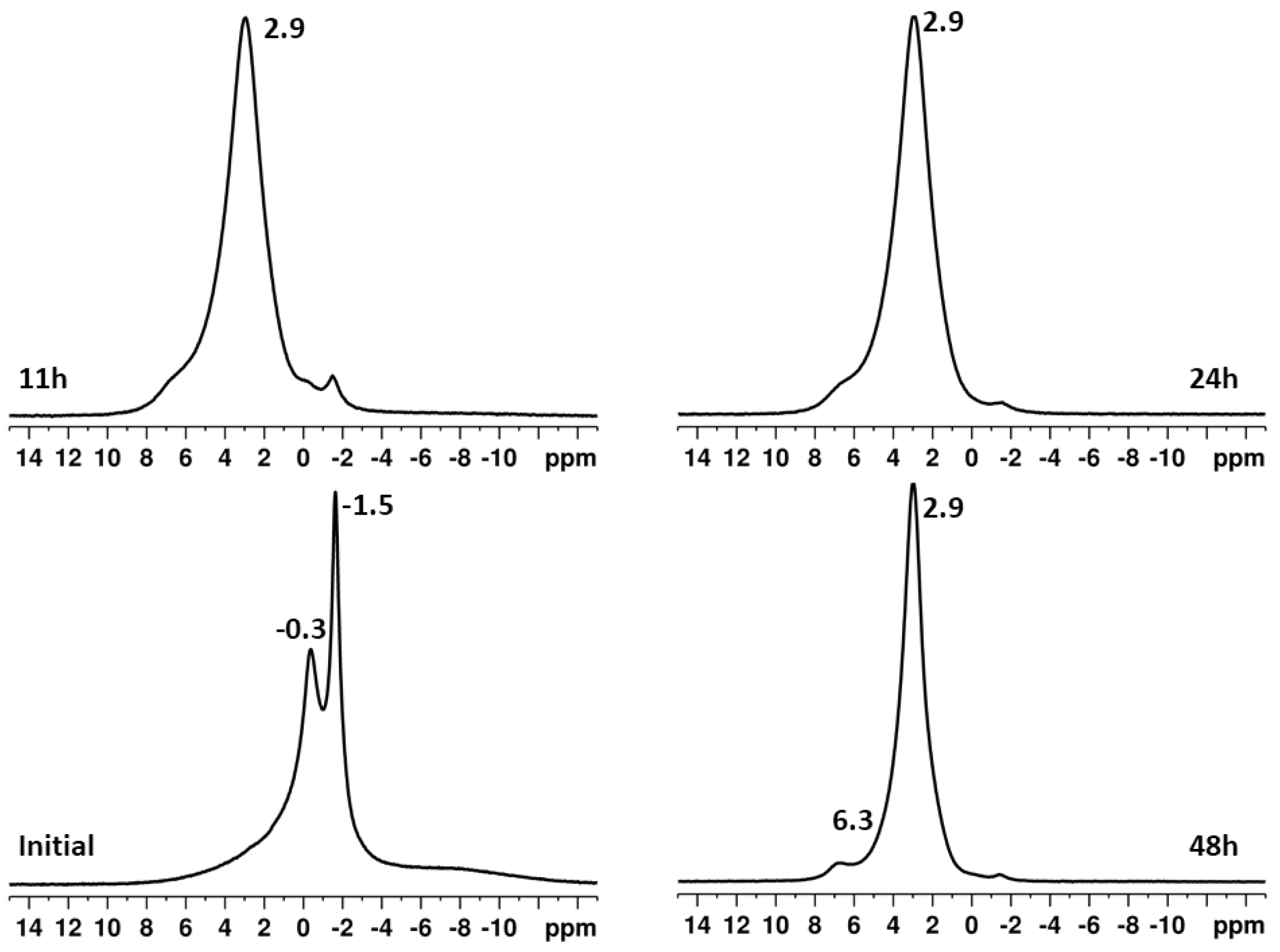
Figure 2 presents the direct excitation 31P spectra of the initial untreated material and of the samples obtained after high-energy activation times of 11, 24, and 48 h. The spectrum of the untreated sample shows two resonances at −0.3 and −1.5 ppm, which are characteristic of dicalcium phosphate anhydrous (DCPA) [27]. The direct excitation 31P NMR spectra (Figure 2) of all treated samples are dominated by an intense resonance around 2.9 ppm, characteristic of an amorphous calcium phosphate [28]. Additionally, two weak resonances at −0.3 and −1.5 ppm, indicating the presence of a small amount of DCPA, are observed, as well as a weak broad signal around 6.3 ppm. The latter could be attributed to the downfield resonance of β-TCP [29], while the other higher-field resonances of β-TCP are not visible due to overlapping with the broad signal at 2.9 ppm dominating the spectra. However, β-TCP is a high-temperature phase that usually forms upon heating calcium-deficient hydroxyapatite (HA) at temperatures above 800 °C. Osman et al. [30] showed that the chemical shift around 6.3 ppm more likely indicates the presence of surface-deprotonated phosphate groups, whose geometrical bonding distortion is probably similar to that of TCP and may explain the observed chemical shift. With increasing activation time, a narrowing of the signal for the amorphous phase is observed, which indicates ongoing crystallization processes in the sample and the formation of nanocrystalline HA (Figure S1a, Supplementary Material). In parallel to that, a decrease of DCPA resonances is also detected, showing its further transformation to apatite phase upon increasing activation time. In the 1H→31P cross-polarization magic-angle spinning (CPMAS) spectra (Figure S1b, Supplementary Material), the signals at −0.3 and −1.5 ppm are significantly enhanced compared to their intensity in the direct excitation spectra. This result confirms that the signals are associated with the presence of DCPA, which contains -HPO42− structures that allow for effective magnetization transfer. The enhancement of the signal at 2.9 ppm is due to the presence of H2O molecules in the amorphous phase.
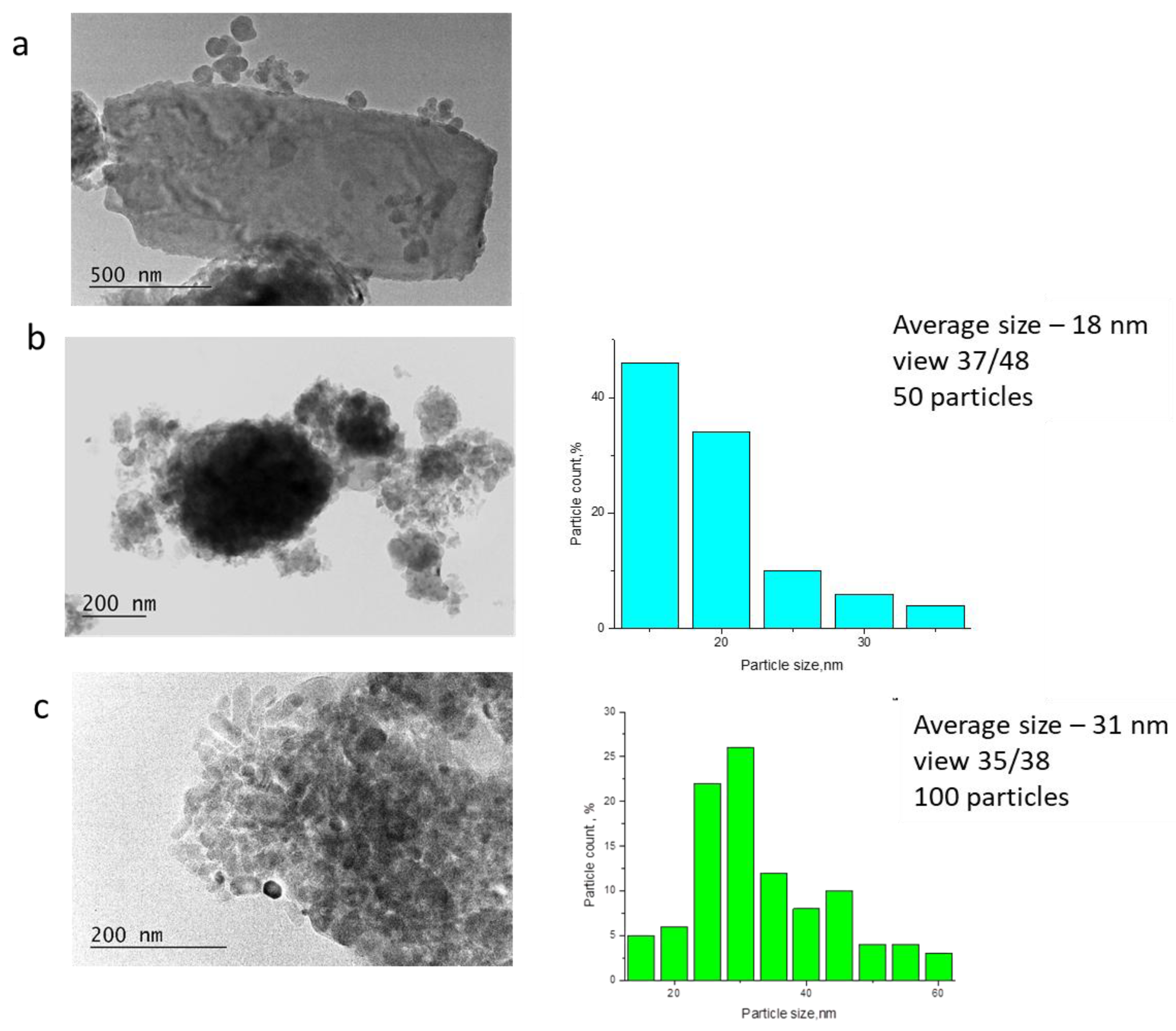
TEM analysis reveals that the time of high-energy activation influences the shape and size of the particles. In the starting mixture (Figure 3a), the large >1 μm in length) plate-like particles of DCPA [31] and the aggregated spherulitic particles (<100 nm) of Ca(OH)2 are clearly distinguished [32].
After 24 h of activation (Figure 3b), all particles in the mixture (Ca(OH)2, DCPA, and HA, see Figure S2 in Supplementary Material) are below 100 nm and exhibit irregular shapes. Particle size distribution shows that the particle sizes range between 15 and 30 nm, with the highest fraction of particles (46%) with a size of 15 nm, followed by those with a size of 20 nm (34%). After 48 h (Figure 3c), the morphology changes again. Ellipsoidal particles (a shape characteristic of poorly crystalline HA) appear, with clearly defined contours and larger sizes compared to those in the 24 h activated sample. The particle sizes are in the region of 15–60 nm with distribution maximum shifted to 30 nm.

2.2. Effect of High-Energy Activation on the Thermal Characteristics of the Mixtures

The thermal characteristics of high-energy activated samples were investigated by DTA-TG-MASS analysis in order to study the influence of activation time on the temperatures of phase transitions and thus to determine the regimes for subsequent sintering of the samples and preparation of ceramic powders.
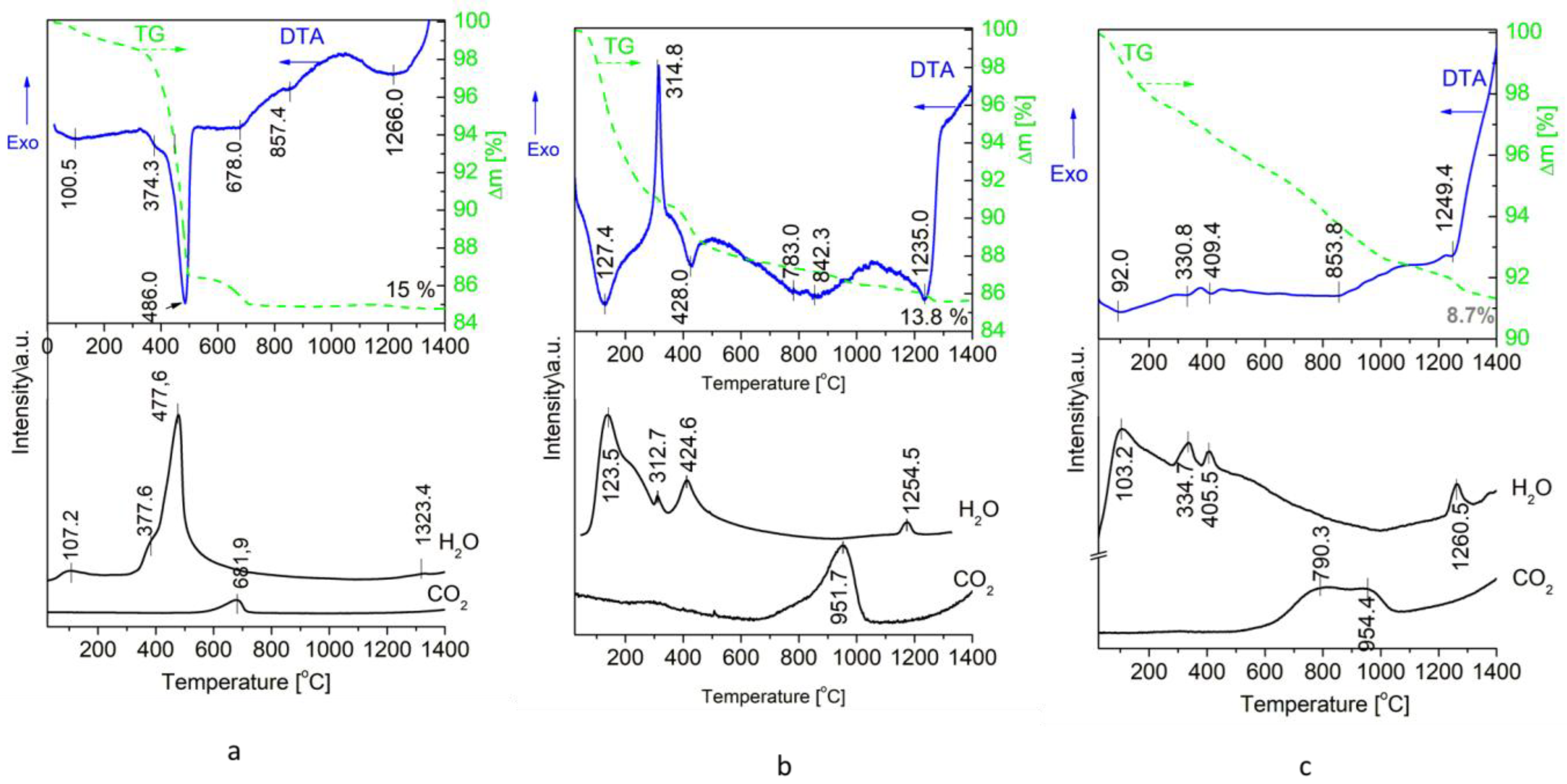
DTA-TG-MASS curves of an initial not activated mixture, and after activation times of 24 and 48 h, are presented in Figure 4.
The DTA-TG-MASS curves of an unactivated mixture are typical for the decomposition of the two initial substances in the temperature range 25–550 °C (Figure 4a). The first endo-effect in the DTA curve (Tmax = 100.5 °C), accompanied by a decrease in weight of 1.1% and release of H2O, can be attributed to the release of adsorbed water. The second strong endo-effect with Tmax at 486 and a shoulder at 374.3, accompanied by a weight decrease of 12.3%, is due to the decomposition of DCPA and Ca(OH)2, respectively [33,34].
The measured weight loss corresponds very well to the theoretically calculated one of 12.8%. The endo-effect at Tmax = 678 °C, accompanied by the release of CO2 and a weight loss of 1.4%, is due to the decomposition of CaCO3 impurities, coming probably from the initial Ca(OH)2. The endo-effect at Tmax = 857.4 °C is not accompanied by gas release and weight loss. It could be attributed either to some polymorphic transition or to the formation of a TCP by the reaction
Ca2P2O7 + CaO → Ca3(PO4)2
The temperature is too high for the polymorphic transition γ-Ca2P2O7 → β-Ca2P2O7, which, according to Hazaat et al. [35], occurs at 535 °C, while according to Mulongo-Masamba et al. [36] is at 730 °C.
The last endo-effect with Tmax = 1266 °C is the result of TTCP formation. The possible reaction is:
β-Ca2P2O7 + 2CaO → Ca4(PO4)2O
The release of a small amount of water, visible in the H2O release curve with Tmax at 1323.4 °C, is an indication of the possible occurrence of another type of process.
2Ca5(PO4)3OH + 2CaO → 3Ca4(PO4)2O +H2O
Evidence for this is the analogous peaks in the water curve in samples activated for 24 and 48 h, in which nanometric HA is dominant.
Significant differences are observed in the DTA-TG-MASS curves of high-energy activated samples (Figure 4b,c). In the DTA curve of the 24 h activated sample (Figure 4b), the first endo-effect at Tmax = 127.4 °C is much more intense and shifted to higher temperatures, compared to that in the initial unactivated mixture (Figure 4a). The weight loss is also significant (9%), which is an indication of the release of not only adsorbed but also chemically bound water. Such is characteristic of nanocrystalline hydroxyapatite [36]. The next peak is a very strong exothermic effect with Tmax= 314.8 °C, which can be attributed to the crystallization of amorphous Ca2P2O7 [35], obtained from the decomposition of residual DCPA. The following endo-effects are analogous to those described for the previous sample. The maximum temperature characterizing the formation of TTCP (1235.0 °C) is lower by 31 °C than that of an unactivated mixture, and the release of water at 1254.5 °C can be associated with the formation of TTCP by the reaction
2Ca5(PO4)3OH + 2CaO → 3Ca4(PO4)2O +H2O
In the DTA curve of the 48 h activated sample (Figure 4c), the strong exothermic effect is not detected. Since at 334.7 and 405.5 °C a negligible amount of water is released, we believe that minimal amounts of the starting substances remain, which decompose (DCPA—peak with Tmax at 330.8 °C and Ca(OH)2—peak with Tmax at 409.4 °C), but no crystallization process is registered because the amount of amorphous Ca2P2O7 is so small and also the decomposition and crystallization temperatures overlap. After that, several weak endothermic effects are observed. These effects can be attributed to the elementary processes in the thermal transformations of the calcium phosphate apatites [37]. The total weight loss is 8.7%, lower than that of the unactivated sample (Figure 4a), which reveals the dehydration and phase transformation processes during energy activation.
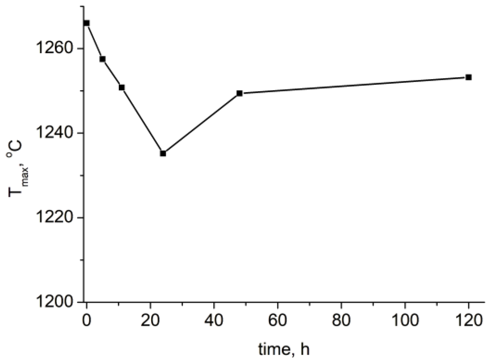
Additionally, DTA-TG-MASS analysis was performed on the samples activated for 5, 11, and 120 h (See Figure S3, Supplementary Material). The Tmax of the peak corresponding to TTCP formation versus activation time is plotted in Figure 5. It can be seen that up to 24 h of mechanochemical activation, the temperature decreases almost linearly, which is due to the dominant processes of fragmentation of the starting materials and formation of amorphous products. After 24 h, crystallization and agglomeration processes of the particles begin, leading to an increase in Tmax of the peak corresponding to TTCP formation.

2.3. Effect of High-Energy Activation on the Phase Composition After Calcination

The 24 h high-energy activated sample was selected for the calcination in two modes described in Section 4.3. The final annealing temperature is chosen to be 1350 °C, because in the DTA curves, this is the final temperature of the TTCP obtaining interval. The heating of the unactivated mixture was also done for comparison.
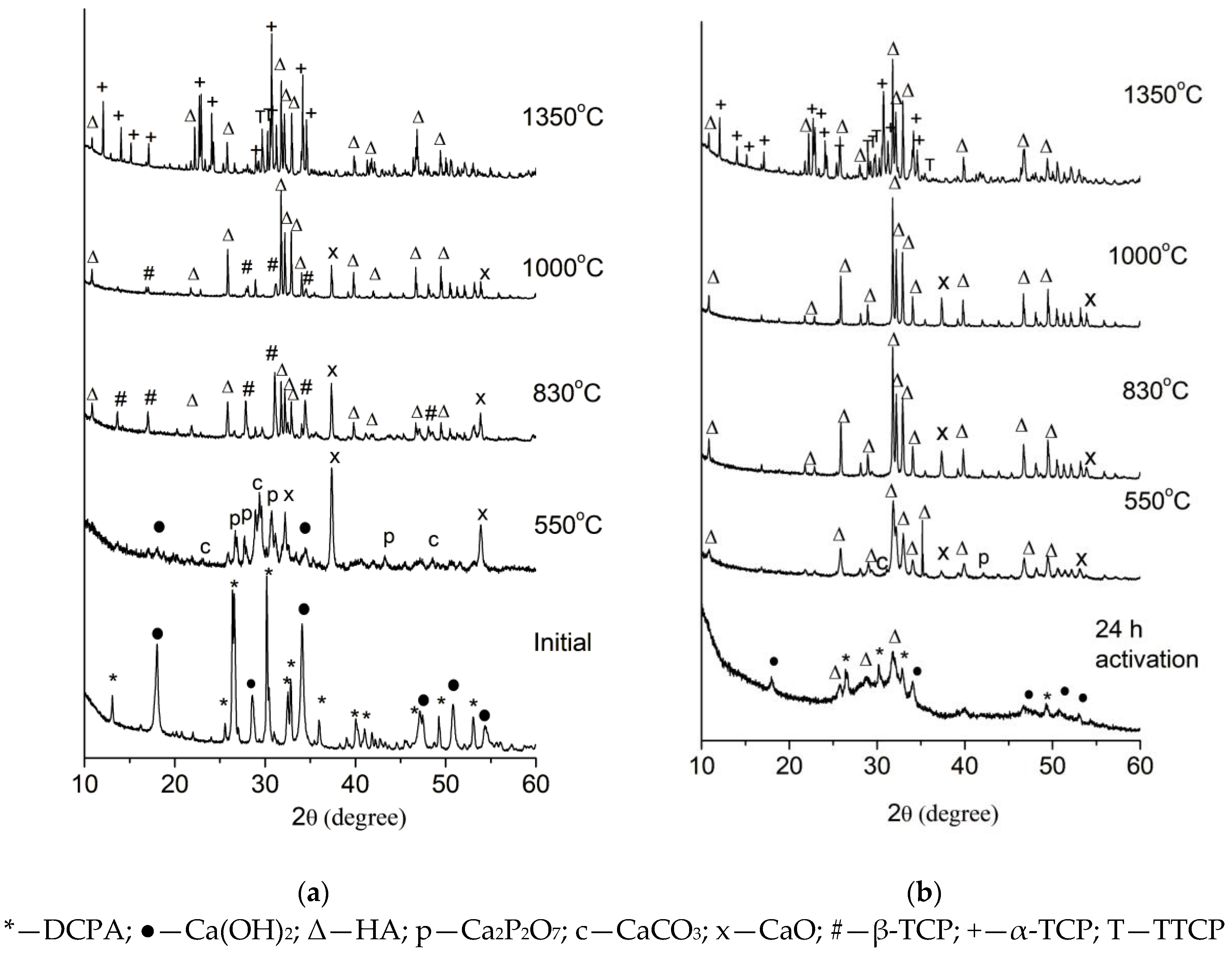
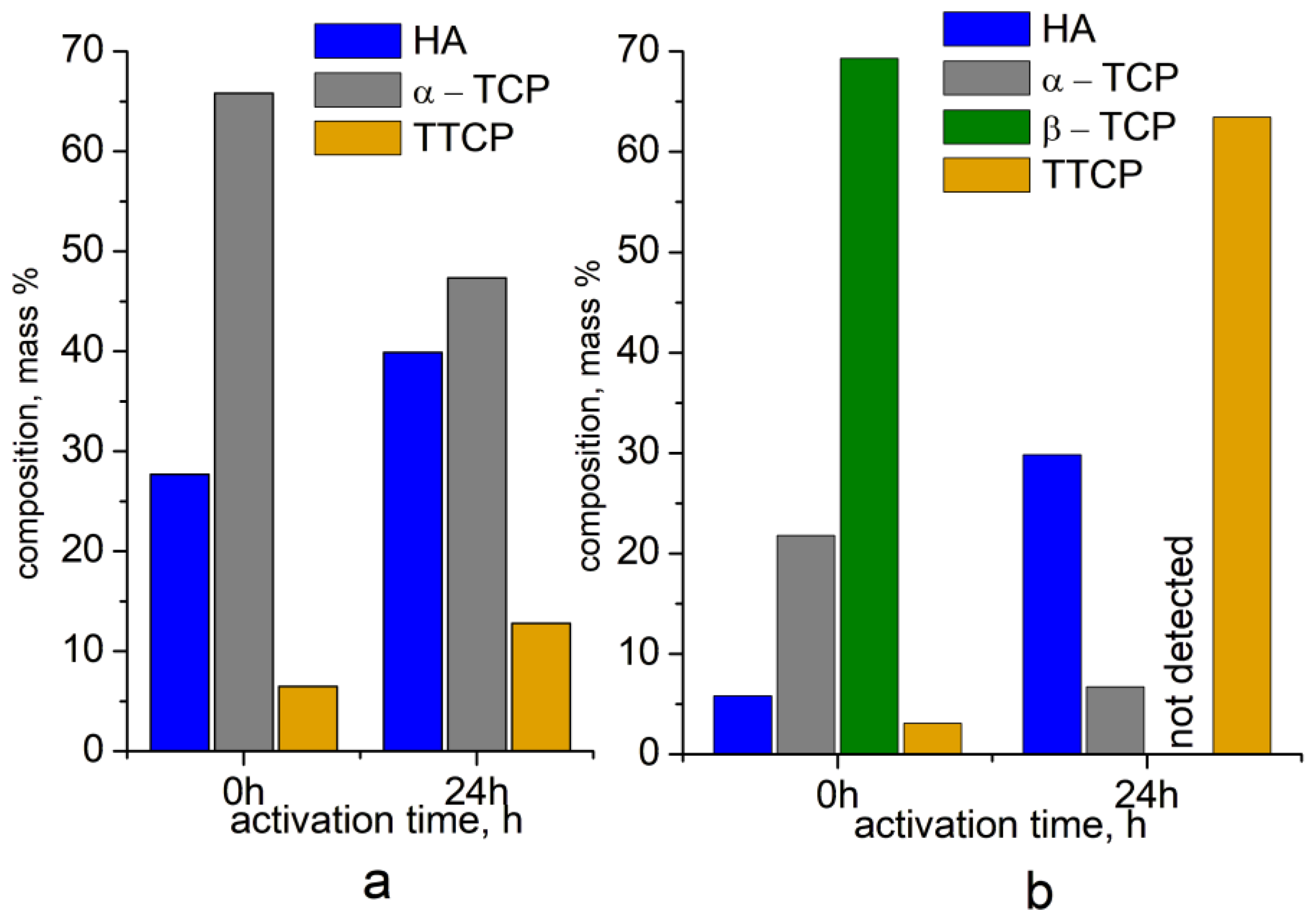
The results from XRD analysis (Figure 6a) show that during slow stepwise heating of the starting unactivated mixture at 550 °C, CaO and β-Ca2P2O7 are obtained, which are the result of the decomposition of the starting substances. Minor amounts of CaCO3 and HA were also identified. At 830–1000 °C, HA and β-TCP are obtained, and at 1350 °C—HA (28%), α-TCP (65%), and 5% TTCP (Figure 7a and Figure 8a, 0 h activation time).
In the 24 h activated sample (Figure 6b), HA is dominant up to 1000 °C, and its crystallinity increases with temperature (the intensities of the peaks in the X-ray powder patterns increase). No β-TCP was identified. At 1350 °C, HA, α-TCP, and TTCP were again identified (Figure 7b), but the ratios between the phases differed. The percentages of HA and α-TCP are close, and the amount of TTCP (13%) is higher than in the unactivated mixture (Figure 8a, 24 h activation time).
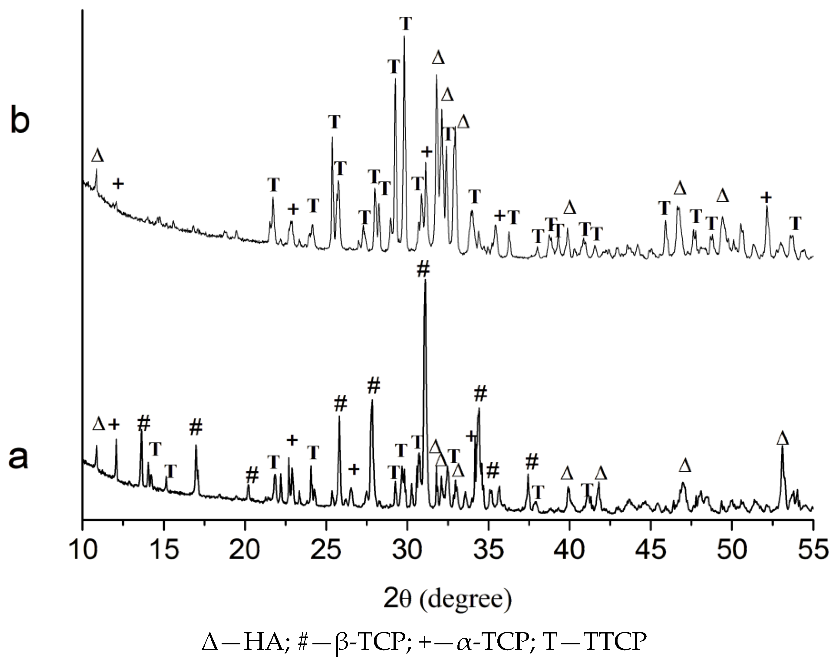
Rapid heating of an unactivated mixture to 1350 °C at a rate of 10 °C/min followed by rapid annealing resulted in a four-phase material (Figure 7a) consisting of HA, α- and β-TCP, and TTCP, with TCPs dominating over TTCP (Figure 8b, 0 h activation time). Three-phase materials (HA, α-TCP, and TTCP) were obtained from the 24 h mechanochemically activated precursor (Figure 7b), and the amount of TTCP reached 64% (Figure 8b, 24 h activation time).
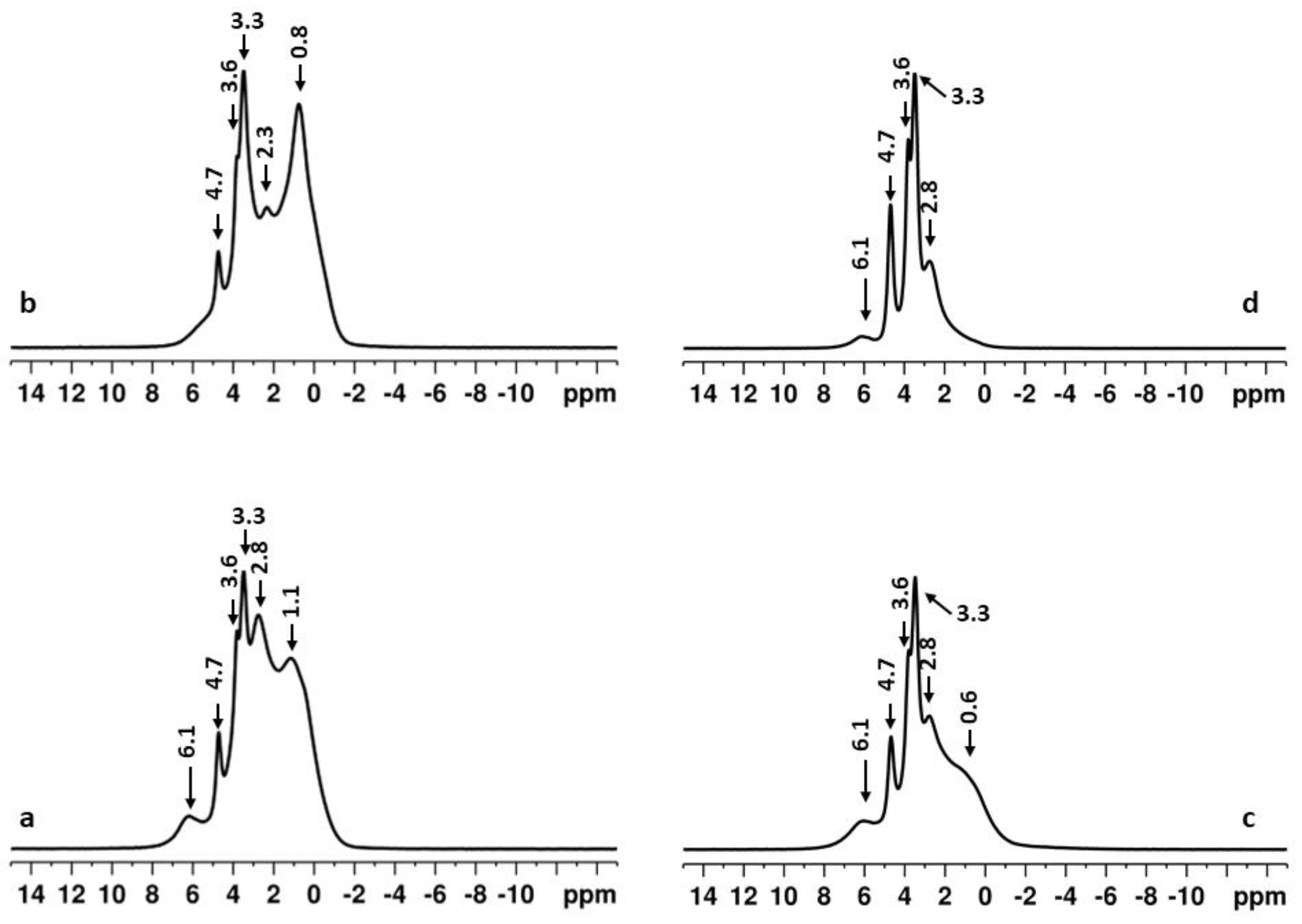
Solid-state NMR analysis proved the results. Figure 9 shows the direct excitation 31P spectra of the unactivated mixture and the 24 h activated sample, heated to 1350 °C under the two regimes.
In all samples, the presence of a mixture consisting of TTCP, HA, and disordered α- and β-TCP phases was detected. The deconvoluted spectra showing the characteristic signals of the different calcium phosphate phases present in the unactivated and activated samples heated step-wise to 1350 °C, are presented in Figure S4, Supplementary Material. The complex 31P spectral patterns resulting from this complex sample composition do not allow precise quantitative interpretation of the spectra due to the overlap of the resonances of the different calcium phosphate phases. Nevertheless, based on the changes in the relative intensities of the resonances of the characteristic peaks, we can conclude that the amount of HA and TCP components was higher in the unactivated mixture, as indicated by the high intensity of the broad resonances between −0.5 and 3 ppm (Figure 9a,b and Figure S4b, Supplementary Material). In the activated sample (Figure 9c,d and Figure S4a, Supplementary Material), the spectral pattern is dominated by the three sharp resonances between 3 ppm and 5 ppm characteristic of the TTCP, while the relative quantity of the HA and TCP components is much lower, particularly in the sample that was rapidly heated to 1350 °C (Figure 9d).
Since the solid-state NMR method is more sensitive than the XRD analysis for identifying calcium phosphates, NMR spectra were recorded on a 24 h activated sample heated at different temperatures according to the stepwise heating regime (Figure 10).
The spectrum of the initial sample after 24 h of high energy activation at 600 rpm without thermal treatment is also given for comparison. Heating the 24 h activated sample at 330 °C and 550 °C resulted in narrowing of the main resonance at around 2.9 ppm, indicating the partial transformation of the disordered apatite phase to nano crystalline HA (Figure 9). In parallel, the DCPA resonances and the signal at 6.5 ppm decrease in intensity, and they are no longer observed in the spectra of the samples calcined at temperatures higher than 330 °C. At these two temperatures, there are additional weak resonances in the region from −6 to −13 ppm showing the formation of a small amount of pyrophosphate phase (unidentified in X-ray powder pattern), most probably β-Ca2P2O7 [29]. These resonances disappear from the spectra of the samples calcined at higher temperatures. Further calcination up to 1000 °C resulted in a significant decrease of signal width due to the enhanced crystalline nature of the HA phase. At 1000 °C, the main resonance has two partially overlapping components that could be attributed to poorly crystalline HA and calcium-deficient hydroxyapatite (CDHA) (see the insert in the spectrum of the sample calcined at 1000 °C) [38]. Further increase of the temperature up to 1350 °C resulted in the formation of a new phase with the chemical shift signature of tetracalcium phosphate (TTCP) as identified by the relatively sharp resonances at 3.3, 3.6, and 4.5 ppm. The deconvolution of the overall spectral pattern (Figure S4a, Supplementary Material) shows that the sample is a mixture of several phases including TTCP, HA (2.8 ppm) as well as disordered α-TCP (relatively broad resonances at 0.6, 1.8, 3.5 ppm) [39] and β-TCP (relatively broad resonances at 0.1, 1.6, 2.8, 6.1 ppm) [40].

3. Discussion

The effect of high-energy mechanochemical activation on the phase and thermal characteristics of equimolar mixtures of DCPA (CaHPO4) and Ca(OH)2 is investigated in this work to find the optimal conditions for precursor preparation for obtaining ceramic powders with a dominant phase of TTCP. Combining methods of different sensitivity for characterizing solid phases allows us to shed light on the processes taking place.
The results show that high-energy mechanochemical activation under the experimental conditions promotes a decrease in the initial material crystallinity and the formation of nanocrystallized hydroxyapatite, whose crystallinity increases with activation time (Figure 1 and Figure 2 and Table 1). The NMR spectrum of a sample activated for 11 h also shows the formation of an amorphous apatite phase (Figure 2).
The formation of HA from DCPA can be explained by similarities in the structural elements that build up their crystal structures. Francis et al. [41] showed that the calcium ions located on the (010) and (110) planes in the CaHPO4•2H2O (DCPD) structure have a similar geometric arrangement as the calcium ions on the (010) planes in HA. These networks of calcium ions serve as templates for the growth of HA. The structures of DCPD and DCPA are similar, consisting of parallel to (010) plane distorted versions of the Ca—PO4 chains [42]. Therefore, we suggest that DCPA, like DCPD, can serve as a natural precursor for the formation of HA. Further, as a result of high-energy activation, simultaneously with the crushing of the starting substances, a process of their dehydration could also occur. The aqueous environment initiates the partial dissolution of DCPA and Ca(OH)2 and formation of the metastable amorphous calcium phosphate that transforms with time into more stable HA. Additionally, as a result of activation, defects are created in the crystal structure, leading to its destruction and energy activation of the particles, which is also a prerequisite for the implementation of phase transformations with time (Figure 1 and Figure 2).
DTA-TG-MASS analysis proved that Tmax of the effect characterizing the formation of TTCP is lowest in the sample activated 24 h (Figure 4 and Figure 5) under the experimental conditions, which makes it the most promising for further high-temperature treatment.
The calcination results showed that stepwise heating leads to the formation of HA and TCP regardless of whether the sample was mechanochemically activated or not.
We assume that slow heating and slow cooling allow the complete course of low-temperature reactions: (i) decomposition of the starting materials, (ii) interactions between the evolved gases and the existing mineral phases; (iii) interactions between the mineral phases themselves; and (iv) absorption of water from the air during slow cooling, and the occurrence of hydration processes. Holding at intermediate temperatures stabilizes the crystal structure of the newly formed compounds, and especially of HA, thereby increasing its thermal stability. All of this is the reason for the formation of minimal amounts of TTCP in the final product.
As a result of our studies and based on the summarized literature data, the following probable reactions can be assumed (Table 2).
Rapid heating and cooling of the 24 h mechanochemically activated mixture stimulated the formation of TTCP, whereas in the non-activated mixture, the dominant phase was β-TCP (Figure 6, Figure 7 and Figure 8). During rapid heating, low-temperature reactions occur only partially during the period of reaching the final temperature. A larger part of the starting substances react at 1350°, which stimulates the formation of the high-temperature phases—α-TCP and TTCP.
Although TTCP is the dominant phase in a sample activated for 24 h and rapidly heated to 1350 °C with a 3 h hold, these conditions are not sufficient to obtain a monophasic product. The hold time should likely be extended, or the calcination temperature should be increased. However, we believe that the material obtained by us is suitable as a component of calcium phosphate cements, since the available HA can act as a nucleating agent, and the presence of α-TCP will regulate the rate of dissolution of the solid phase in the liquid phase of the cements. Similar observations have been reported by Jeon et al. [26].

4. Materials and Methods

4.1. Materials

Ca(OH)2 (Merck, Darmstadt, Germany, p.a.) and CaHPO4 prepared by thermal dehydration of CaHPO4•2H2O (Sigma Aldrich, St. Louis, MO, USA) at 165 °C for 2 h were used. A high-temperature furnace, type VP 04/17 (LAC Ltd. Company, Stefanikova, Czech Republic) was used. No other phases except DCPA were detected by XRD analysis (Figure S5, Supplementary Materials).

4.2. High Energy Activation

The high-energy activation was carried out in a Fritsch 6 type planetary ball mill (FRITSCH GmbH - Milling and Sizing, Idar-Oberstein, Germany). A mixture of Ca(OH)2 and DCPA was prepared in a molar ratio of CaHPO4:Ca(OH)2 = 1:1 (Ca/P = 2:1). Activation is carried out in an agate vessel with a volume of 80 cm3. The prepared samples were treated for 5, 11, 24, 48, and 120 h at 600 rpm with agate balls (10 mm diameter) at a constant ratio of sample mass:mass of grinding balls = 1:3. Activation revolutions were selected based on preliminary exploratory experiments with activation at 300 and 450 rpm.
To verify the repeatability of the results, each experiment was performed twice, and each activated mixture was analysed with the appropriate methods described in Section 4.4.

4.3. Calcination of Activated Precursors

Selected samples from the high energy activation were treated in a high-temperature furnace, type VP 04/17 (LAC Ltd. Company, Stefanikova, Czech Republic), in the following operating modes:
  • Step heating at a rate of 10 °C/min to a given temperature (determined by DTA-TG-MASS analysis) and hold for 3 h, followed by slow cooling (The samples were left to cool overnight in the furnace itself after it was turned off).
  • Rapid heating at a rate of 10 °C/min to 1350 °C and holding for 3 h, followed by rapid annealing (The samples were removed from the furnace at a temperature of 1350°C and placed in a desiccator at room temperature).
Each heating was performed twice to verify the repeatability of the results, and each sample was analysed with the appropriate methods described in Section 4.4.

4.4. Characterisation

4.4.1. X-Ray Diffraction Analysis (XRD)

A Bruker D8 Advance diffractometer with CuKα radiation and a LynxEye detector (Bruker AXS Advanced X-ray Solutions GmbH, Billerica, MA, USA) was used to conduct powder X-ray diffraction. Data were gathered in the range of 10 to 90 2θ with a step of 0.03° 2θ and a counting rate of 57 s/step for the primary phase identification. The phase composition was identified using ICDD-PDF2 (2021) database.
The Bruker Topas v.4.2 program was used to refine the unit cell parameters, mean crystallite size, and quantitative phase composition of the samples using Rietveld quantification with starting models from the ICSD (cif files ## 202233, 14313, 97500, 410782, 20179, 51409, and 56306). For this purpose, powder diffraction patterns were collected at room temperature within the range of 5 to 120° 2θ with a step of 0.02° 2θ and 175 s/step counting time, sample rotation—15 rpm. The standard deviation of phase composition calculations is ±2%.

4.4.2. Differential Thermal Analysis Combined with Detection of Released Gases (DTA-TG-MASS)

The thermal characterization of the samples was performed on a LABSYSTM EVO (Setaram, Caluire, France) apparatus with a Pt/Pt-Rh thermocouple in a corundum crucible at a heating rate of 10 °C/min in the temperature range 25–1400 °C and in an atmosphere of synthetic air with a flow rate of 20 mL/s. The accuracy of temperature measurement was ±1 °C. A corundum crucible was used as a reference one. The weight of the samples was 29 ± 1 mg. The apparatus is equipped with a quadruple mass spectrometer (Pfeiffer vacuum OMNISTAR, GSD 301, Zürich, Switzerland), which serves for the analysis of the escaping gases. In the studied samples, the release of H2O (m = 18) and CO2 (m = 44) was determined.

4.4.3. Nuclear Magnetic Resonance (NMR) Studies

NMR spectra were recorded on a Bruker Avance HD III 600 NMR spectrometer (Bruker, Rheinstetten, Germany) operating at 599.90 MHz 1H frequency (242.84 MHz for 31P), using a 4 mm solid state iProbe CPMAS DR-H&F VTN. The samples were loaded in 4 mm zirconia rotors and spun at a magic angle spinning (MAS) rate of 10 kHz for all measurements. The quantitative direct excitation 31P NMR spectra were recorded with a onepulse sequence (Bruker Topspin library), 90° pulse length of 3.3 µs, 7 K time domain data points, spectrum width of 37 kHz, 32 scans, and a relaxation delay of 150 s. The spectra were processed with an exponential window function (line broadening factor 5) and zero filled to 16 K data points. The 1H-31P cross-polarization magic angle spinning (CP MAS) spectra were acquired with the following experimental parameters: 1H excitation pulse of 2.5 μs, 3.5 ms contact time, 5 s relaxation delay, and 256 scans were accumulated. 1H SPINAL-64 decoupling scheme was used during the acquisition of CP experiments. All 31P chemical shifts were referenced against the external solid reference NH4H2PO4 (δ 0.9 ppm). The DMfit software (release #20220502) was used for the deconvolution, simulation, and fitting of the experimental NMR data [48].

4.4.4. Transmission Electron Microscopy Analysis (TEM)

STEM JEOL JEM 2100 apparatus with an accelerating voltage of 200 kV, a maximum resolution of 0.23 nm between two points, and 0.14 nm in the lattice was used. The device is equipped with a GATAN CCD camera. The powder samples were suspended in ethyl alcohol and sonicated (Ultra Sonic Cleaner, KWAN SWO INSTRUMENTAL Co LTD, Taichung, Taiwan) with a frequency of 45 KHz for 6 min. The suspension is dropped onto a standard carbon-coated copper grid. ImageJ software (version 1.x) was used to calculate particle size distribution

5. Conclusions

High-energy mechanochemical activation significantly influences the phase composition, particle morphology, and thermal behaviour of equimolar mixtures of DCPA (CaHPO4) and Ca(OH)2, making it an effective method for preparing precursors for TTCP-rich ceramic materials.
XRD and solid-state NMR analyses reveal that high-energy milling for 11 to 48 h initiates the amorphization and transformation of the initial crystalline phases into amorphous calcium phosphate phase and nanocrystalline hydroxyapatite, which subsequently increases its crystallinity.
Thermal analysis demonstrates that mechanochemical activation reduces the temperatures at which phase transformations occur.
Calcination studies show that not only mechanochemical activation but also the calcination mode influence the final phase composition. Rapid heating of a 24 h activated precursor produces a material with TTCP as the dominant phase (64%), whereas the unactivated mixture or stepwise heating forms a multi-phase composition with lower TTCP content.
Under the experimental conditions, 24 h activation presents an effective balance between phase transformation, particle refinement, and process efficiency, making it a promising approach for the preparation of TTCP-rich ceramic powders, which are suitable components of calcium phosphate cements. The available HA can act as a nucleating agent, and the presence of α-TCP will regulate the rate of dissolution of the solid in the liquid phase of the cements.

Supplementary Materials

The following supporting information can be downloaded at: https://www.mdpi.com/article/10.3390/inorganics13100313/s1, Figure S1: Experimental HRTEM image of sample activated 24 h: (A) investigated areas; (B) Fourier filtered HRTEM images of CaHPO4, (112)—0.247 nm; (C) Fourier filtered HRTEM images of Apatite-(CaOH) hydroxyapatite, (001)—0.691 nm and (D) Experimental HRTEM image and Fourier filtered HRTEM image of Ca(OH)2, (002)—0.246 nm; Figure S2: (a) Overlaid and vertically scaled direct excitation 31P spectra of the samples activated for 11 h (black line), 24 h (blue line) and 48 h (red line), showing narrowing of the resonance linewidths with increasing activation time; (b) Overlaid 1H-31P CPMAS of the same samples with identical color code; Figure S3: DTA-TG-MASS curves of the samples obtained after (a) 5 h mechanochemically activation; (b) 11 h mechanochemically activation and (c) 120 h mechanochemically activation; Figure S4: Deconvoluted direct excitation 31P NMR spectra of: (a) activated sample (24 h at 600 rpm) heated step-wise to 1350 °C; (b) unactivated initial mixture heated step-wise to 1350 °C. The experimental spectra are given in black, while the simulated spectra are presented in red lines. The individual contributions of some characteristic lines of the different components obtained after the deconvolution of the spectra are given with colored lines: TTCP (blue), HA (light blue), α-TCP (magenta), and β-TCP (green); Figure S5: X-ray analysis of the initial DCPA.

Author Contributions

Conceptualization, D.R. and K.S.; sample preparation, Y.T. and K.S.; NMR analysis and discussion, P.S.; XRD analysis and calculation, D.K.; TEM analysis, P.M.; DTA-TG-MASS analysis and visualization, D.R. and Y.T.; writing—original draft preparation, D.R., P.S., K.S., D.K. and P.M.; writing—review and editing, D.R., P.S. and K.S. All authors have read and agreed to the published version of the manuscript.

Funding

This research received no external funding.

Institutional Review Board Statement

Not applicable.

Informed Consent Statement

Not applicable.

Data Availability Statement

The original contributions presented in this study are included in the article/Supplementary Material. Further inquiries can be directed to the corresponding author.

Acknowledgments

Research equipment of Distributed Research Infrastructure INFRAMAT, part of Bulgarian National Roadmap for Research Infrastructures, supported by Bulgarian Ministry of Education and Science was used in this investigation.

Conflicts of Interest

The authors declare no conflicts of interest.

Abbreviations

The following abbreviations are used in this manuscript:
HAHydroxyapatite
DCPADicalcium phosphate anhydrous
TCPThricalcium phosphate
TTCPTetracalcium phosphate
NMRNuclear Magnetic Resonance
XRDX-ray diffraction analysis
TEMTransmission electron microscopy
DTA-TG-MASSDifferential thermal analysis with thermogravimetry and mass spectrometry

References

  1. Dorozhkin, S.V. Calcium Orthophosphate (CaPO4)-Based Bioceramics: Preparation, Properties, and Applications. Coatings 2022, 12, 1380. [Google Scholar] [CrossRef]
  2. Rabel, K.; Kohal, R.J.; Steinberg, T.; Tomakidi, P.; Rolauffs, B.; Adolfsson, E.; Palmero, P.; Fürderer, T.; Altmann, B. Controlling osteoblast morphology and proliferation via surface micro-topographies of implant biomaterials. Sci. Rep. 2020, 10, 12810. [Google Scholar] [CrossRef]
  3. Wu, V.M.; Uskoković, V. Is there a relationship between solubility and resorbability of different calcium phosphate phases in vitro? Biochim. Biophys. Acta 2016, 1860, 2157–2168. [Google Scholar] [CrossRef] [PubMed]
  4. Chow, L.C.; Eanes, E.D. (Eds.) Octacalcium Phosphate; Karger Medical and Scientific Publishers: Basel, Switzerland, 2001. [Google Scholar]
  5. Jeong, J.; Kim, J.H.; Shim, J.H.; Hwang, N.S.; Heo, C.Y. Bioactive calcium phosphate materials and applications in bone regeneration. Biomat. Res. 2019, 23, 4. [Google Scholar] [CrossRef] [PubMed]
  6. Bohner, M.; Lemaitre, J. Can bioactivity be tested in vitro with SBF solution? Biomaterials 2009, 30, 2175–2179. [Google Scholar] [CrossRef]
  7. Ehara, A.; Ogata, K.; Imazato, S.; Ebisu, S.; Nakano, T.; Umakoshi, Y. Effects of α-TCP and TetCP on MC3T3-E1 proliferation, differentiation and mineralization. Biomaterials 2003, 24, 831–836. [Google Scholar] [CrossRef]
  8. Medvecky, L.; Stulajterova, R.; Giretova, M.; Mincik, J.; Vojtko, M.; Balko, J.; Briancin, J. Effect of tetracalcium phosphate/monetite toothpaste on dentin remineralisation and tubule occlusion in vitro. Dent. Mater. 2018, 34, 442–451. [Google Scholar] [CrossRef]
  9. Moseke, C.; Gbureck, U. Tetracalcium phosphate: Synthesis, properties and biomedical applications. Acta Biomater. 2010, 6, 3815–3823. [Google Scholar] [CrossRef]
  10. Samavedi, S.; Whittington, A.R.; Goldstein, A.S. Calcium phosphate ceramics in bone tissue engineering: A review of properties and their influence on cell behavior. Acta Biomater. 2013, 9, 8037–8045. [Google Scholar] [CrossRef]
  11. Xu, H.; Wang, P.; Wang, L.; Bao, C.; Chen, Q.; Weir, M.D.; Chow, L.C.; Zhao, L.; Zhou, X.; Reynolds, M.A. Calcium phosphate cements for bone engineering and their biological properties. Bone Res. 2017, 5, 17056. [Google Scholar] [CrossRef]
  12. Lukina, Y.; Safronova, T.; Smolentsev, D.; Toshev, O. Calcium Phosphate Cements as Carriers of Functional Substances for the Treatment of Bone Tissue. Materials 2023, 16, 4017. [Google Scholar] [CrossRef] [PubMed]
  13. Mahmoud, E.M.; Sayed, M.; Mansour, T.S.; Naga, S.M. Biodegradable ceramic materials for orthopedic and dentistry applications. Discov. Appl. Sci. 2025, 7, 990. [Google Scholar] [CrossRef]
  14. Balbuena, O.B.F.; Paiva, L.F.S.; Ribeiro, A.A.; Monteiro, M.M.; de Oliveira, M.V.; Pereira, L.C. Sintering parameters study of a biphasic calcium phosphate bioceramic synthesized by alcoholic sol-gel technique. Ceram. Int. 2021, 47, 32979–32987. [Google Scholar] [CrossRef]
  15. Safronova, T.V.; Putlyaev, V.I. Powder systems for calcium phosphate ceramics. Inorg. Mater. 2017, 53, 17–26. [Google Scholar] [CrossRef]
  16. Irfa’i, M.A.; Schmahl, W.W.; Pusparizkita, Y.M.; Muryanto, S.; Ismail, A.P.R.; Jamari, J.; Bayuseno, A.P. Hydrothermally synthesized-nanoscale carbonated hydroxyapatite with calcium carbonates derived from green mussel shell wastes. J. Mol. Struct. 2024, 1306, 137837. [Google Scholar] [CrossRef]
  17. Nikolenko, M.V.; Vasylenko, K.V.; Myrhorodska, V.D.; Kostyniuk, A.; Likozar, B. Synthesis of Calcium Orthophosphates by Chemical Precipitation in Aqueous Solutions: The Effect of the Acidity, Ca/P Molar Ratio, and Temperature on the Phase Composition and Solubility of Precipitates. Processes 2020, 8, 1009. [Google Scholar] [CrossRef]
  18. Ali, W.A.; Richards, S.E.; Alzard, R.H. Unlocking the potential of ball milling for nanomaterial Synthesis: An overview. J. Ind. Eng. Chem. 2025, 149, 63–93. [Google Scholar] [CrossRef]
  19. Chen, W.C.; Ju, C.P.; Tien, Y.C.; Lin, J.H. In vivo testing of nanoparticle-treated TTCP/DCPA-based ceramic surfaces. Acta Biomater. 2009, 5, 1767–1774. [Google Scholar] [CrossRef]
  20. Sargin, Y.; Kizilyalli, M.; Telli, C.; Güler, H. A new method for the solid-state synthesis of tetracalcium phosphate, a dental cement: X-ray powder diffraction and IR studies. J. Eur. Ceram. Soc. 1997, 17, 963–970. [Google Scholar] [CrossRef]
  21. Brosseau, C.; Liao, J.; Duan, X.; Li, Y.; Zheng, C.; Yang, Z.; Zhou, A.; Zou, D. Synthesis and Mechanism of Tetracalcium Phosphate from Nanocrystalline Precursor. J. Nanomater. 2014, 2014, 840102. [Google Scholar] [CrossRef]
  22. Jalota, S.; Tas, A.C.; Bhaduri, S.B. Synthesis of HA-seeded TTCP (CA4(PO4)2O) powders at 1230 °C from Ca(CH3COO)2.H2O and NH4H2PO4. J. Am. Ceram. Soc. 2005, 88, 3353–3360. [Google Scholar] [CrossRef]
  23. Romeo, H.E.; Fanovich, M.A. Synthesis of tetracalcium phosphate from mechanochemically activated reactants and assessment as a component of bone cements. J. Mater. Sci. Mater. Med. 2008, 19, 2751–2760. [Google Scholar] [CrossRef]
  24. Nizar, M.S.; Sofiyaningsih, N.; Manullang, R.J.; Wijayanti, R.B.; Rosmayanti, I.; Sumardan, D. Novel fast firing method to synthesize tetracalcium phosphate. AIP Conf. Proc. 2021, 2349, 020001. [Google Scholar] [CrossRef]
  25. Ayers, R.; Hannigan, N.; Vollmer, N.; Unuvar, C. Combustion synthesis of heterogeneous calcium phosphate bioceramics from calcium oxide and phosphate precursors. Int. J. Self-Propag. High-Temp. Synth. 2011, 20, 6–14. [Google Scholar] [CrossRef]
  26. Jeon, C.; Chun, S.; Lim, S.; Kim, S. Synthesis and Characterization of TTCP for Calcium Phosphate Bone Cement. Biomater. Res. 2011, 15, 1–6. [Google Scholar]
  27. Louati, B.; Hlel, F.; Guidara, K.; Gargouri, M. Analysis of the effects of thermal treatments on CaHPO4 by 31P NMR spectroscopy. J. Alloys Compd. 2005, 394, 13–18. [Google Scholar] [CrossRef]
  28. Edén, M. Structure and formation of amorphous calcium phosphate and its role as surface layer of nanocrystalline apatite: Implications for bone mineralization. Materialia 2021, 17, 101107. [Google Scholar] [CrossRef]
  29. Yu, Y.; Guo, H.; Pujari-Palmer, M.; Stevensson, B.; Grins, J.; Engqvist, H.; Eden, M. Advanced solid-state 1H/31P NMR characterization of pyrophosphate-doped calcium phosphate cements for biomedical applications: The structural role of pyrophosphate. Ceram. Int. 2019, 45, 20642–20655. [Google Scholar] [CrossRef]
  30. Ben Osman, M.; Diallo-Garcia, S.; Herledan, V.; Brouri, D.; Yoshioka, T.; Kubo, J.; Millot, Y.; Costentin, G. Discrimination of surface and bulk structure of crystalline hydroxyapatite nanoparticles by NMR. J. Phys. Chem. C 2015, 119, 23008–23020. [Google Scholar] [CrossRef]
  31. Lee, J.; Bae, J.-S.; Kim, Y.-I.; Yoo, K.-H.; Yoon, S.-Y. Synthesis, Characterization, and Biological Performances of Magnesium-Substituted Dicalcium Phosphate Anhydrous. Materials 2024, 17, 4605. [Google Scholar] [CrossRef] [PubMed]
  32. Pesce, C.; Luca, G.P.; Molinari, M.; Richardson, A. Effects of organic additives on calcium hydroxide crystallisation during lime slaking. Cem. Concr. Res. 2021, 139, 106254. [Google Scholar] [CrossRef]
  33. Duff, E.J. Orthophosphates. XIII,* Thermal Decomposition of Secondary Calcium Orthophosphate (CaHPO4) and Secondary Calcium Orthophosphate Dihydrate (CaHPO4•2H2O). J. Appl. Chem. Biotechnol. 1971, 21, 233–235. [Google Scholar] [CrossRef]
  34. Khachani, M.; El Hamidi, A.; Halim, M.; Arsalane, S. Non-isothermal kinetic and thermodynamic studies of the dehydroxylation process of synthetic calcium hydroxide Ca(OH)2. J. Mater. Environ. Sci. 2014, 5, 615–624. [Google Scholar]
  35. El Hazzat, M.; El Hamidi, A.; Halim, M.; Arsalane, S. Complex evolution of phase during the thermal investigation of brushite-type calcium phosphate CAHPO4•2H2O. Materialia 2021, 16, 101055. [Google Scholar] [CrossRef]
  36. Mulongo-Masamba, R.; El Kassri, T.; Khachani, M.; Arsalane, S.; Halim, M.; El Hamidi, A. Synthesis and thermal dehydroxylation kinetic of anhydrous calcium phosphate monetite CaHPO4. J. Therm. Anal. Calorim. 2016, 124, 171–180. [Google Scholar] [CrossRef]
  37. Mortier, A.; Lemaitre, J.; Rouxhet, P.G. Temmperature-Programmed Characterization of Synthetic Calcium-Deficient Phosphate Apatites. Thermochim. Acta 1989, 143, 265–282. [Google Scholar] [CrossRef]
  38. Miquel, J.L.; Facchini, L.; Legrand, A.P. Characterisation and Conversion Study into Natural Living Bone of Calcium Phosphate Bioceramics by Solid State NMR Spectroscopy. Clin. Mater. 1990, 5, 115. [Google Scholar] [CrossRef]
  39. Vogel, J.; Rüssel, C.; Günther, G.; Hartmann, P.; Vizethum, F.; Bergner, N. Characterization of plasma-sprayed hydroxyapatite by 31P-MAS-NMR and the effect of subsequent annealing. J. Mater. Sci. Mater. Med. 1996, 7, 495. [Google Scholar] [CrossRef]
  40. Guo, H.; Pujari-Palmer, M.; Yu, Y.; Stevensson, B.; Engqvist, H.; Edén, M. Quantitative phase analyses of biomedical pyrophosphate-bearing monetite and brushite cements by solid-state NMR and powder XRD. Ceram. Int. 2000, 46, 11000. [Google Scholar] [CrossRef]
  41. Francis, M.D.; Webb, N.C. Hydroxyapatite formation from a hydrated calcium monohydrogen phosphate precursor. Calc. Tis Res. 1970, 6, 335–342. [Google Scholar] [CrossRef]
  42. Dickens, B.; Bowen, J.S.; Brown, W.E. A refinement of the crystal structure of CaHPO4 (synthetic monetite). Acta Cryst. 1972, B28, 797–806. [Google Scholar] [CrossRef]
  43. Rodríguez-Lugo, V.; Angeles, C.; De la Isla, A.; Castano, V.M. Effect of bio-calcium oxide on the morphology of hydroxyapatite. Int. J. Basic Appl. Sci. 2015, 4, 395. [Google Scholar] [CrossRef]
  44. Jinlong, N.; Zhenxi, Z.; Dazong, J. Investigation of Phase Evolution During the Thermochemical Synthesis of Tricalcium Phosphate. J. Mater. Synth. Process. 2001, 9, 235–240. [Google Scholar] [CrossRef]
  45. Hui, P.; Meena, S.L.; Singh, G.; Agarawal, R.D.; Prakash, S. Synthesis of Hydroxyapatite Bio-Ceramic Powder by Hydrothermal Method. J. Miner. Mater. Charact. Eng. 2010, 9, 683–692. [Google Scholar] [CrossRef]
  46. Kivrak, N.; Taş, A.C. Synthesis of Calcium Hydroxyapatite-Tricalcium Phosphate (HA-TCP) Composite Bioceramic Powders and Their Sintering Behavior. J. Am. Ceram. Soc. 1998, 81, 2245–2252. [Google Scholar] [CrossRef]
  47. Böhme, N.; Hauke, K.; Dohrn, M.; Neuroth, M.; Geisler, T. High-temperature phase transformations of hydroxylapatite and the formation of silicocarnotite in the hydroxylapatite–quartz–lime system studied in situ and in operando by Raman spectroscopy. J. Mater. Sci. 2022, 57, 15239–15266. [Google Scholar] [CrossRef]
  48. Massiot, D.; Fayon, F.; Capron, M.; King, I.; Le Celve, S.; Alonson, B.; Durand, J.O.; Bujoli, B.; Gan, Z.H.; Hoatson, G. Modelling one- and two-dimensional solid-state NMR spectra. Magn. Reson. Chem. 2002, 40, 70–76. [Google Scholar] [CrossRef]
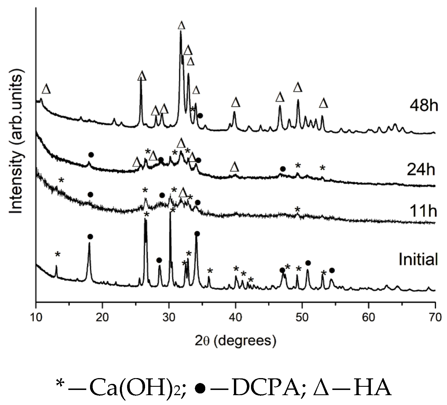
Figure 1. X-ray powder patterns of the initial mixture and samples after high-energy activation for 11, 24, and 48 h.
Inorganics 13 00313 g001
Figure 2. Direct excitation 31P spectra of the initial untreated material and of the samples obtained after high-energy activation times of 11, 24, and 48 h.
Inorganics 13 00313 g002
Figure 3. Images from transmission electron microscope and particle size distribution of the: (a) initial unactivated mixture; (b) 24 h activated mixture; and (c) 48 h activated mixture.
Inorganics 13 00313 g003
Figure 4. DTA-TG-MASS curves of: (a) initialunactivated mixture; (b) after 24 h high-energy activation; and (c) after 48 h high-energy activation.
Inorganics 13 00313 g004
Figure 5. Variation in Tmax of the peak corresponding to TTCP formation with increasing activation time.
Inorganics 13 00313 g005
Figure 6. X-ray powder patterns of initial unactivated mixture (a) and 24 h activated mixture (b) during the stepwise calcined samples at different temperatures.
Inorganics 13 00313 g006
Figure 7. X-ray powder patterns of initial unactivated mixture (a) and 24 h activated mixture (b) during rapid heating up to 1350 °C and rapid cooling.
Inorganics 13 00313 g007
Figure 8. Quantitative composition of the samples calcinated at 1350 °C, calculated from X-ray powder patterns: (a) after stepwise calcination; (b) after rapid heating and rapid annealing.
Inorganics 13 00313 g008
Figure 9. Direct excitation 31P NMR spectra of the unactivated mixture and the activated sample (24 h at 600 rpm), heated to 1350 °C under step-wise and direct regimes: (a) step-wise heated unactivated sample; (b) rapidly heated unactivated sample; (c) step-wise heated 24 h activated sample; (d) rapidly heated 24 h activated sample.
Inorganics 13 00313 g009
Figure 10. Direct excitation 31P NMR spectra of the samples obtained after 24 h of high energy activation at 600 rpm, followed by step-wise calcination at different temperatures. The spectrum of the initial sample after 24 h of high energy activation at 600 rpm without further thermal treatment is also given for comparison.
Inorganics 13 00313 g010
Table 1. Quantitative phase composition calculated from X-ray powder patterns.
SampleCa(OH)2 *
(%)
DCPA *
(%)
HA *
(%)
HA
Mean Size
(nm)
Degree of Crystallinity
Initial3466- 100%
11 h activation184537198%
24 h activation1123662018%
48 h activation429433100%
Note: * Phase composition is calculated from the crystalline component.
Table 2. Probable reactions occurring during step heating and slow cooling of the unactivated mixture and the sample activated for 24 h.
Temperature RegionUnactivated Mixture24 h Activated Sample
Up to 550 °C2CaHPO4 → β-Ca2P2O7 + H2O↑[33]Ca5(PO4)3OH(ncr)—stabilization of the structure, increase of crystallinity
Ca(OH)2 → CaO + H2O↑[34]2CaHPO4 → β-Ca2P2O7 + H2O↑[33]
2Ca(OH)2 + 3CaHPO4 → Ca5(PO4)3OH + 2H2O↑ [43]Ca(OH)2 → CaO + H2O↑[34]
CaO + CO2(atm) → CaCO3 2Ca(OH)2 + 3CaHPO4 → Ca5(PO4)3OH + 2H2O↑ [43]
CaO + CO2(atm) → CaCO3
550–830 °Cβ-Ca2P2O7 +CaO → 2β-Ca3(PO4)2[44]β-Ca2P2O7 +CaO → 2β-Ca3(PO4)2[44]
Ca5(PO4)3OH—structure stabilization Ca5(PO4)3OH—stabilization of the structure, increase of crystallinity
830–1000 °C3β-Ca3(PO4)2 + CaO + H2O = 2Ca5(PO4)3OH (during cooling)[45]Ca5(PO4)3OH—starting the transphormation in which non-stoichiometric HA is formed (NMR, this study)
Ca5(PO4)3OH—stabilization of the structure, increase of crystallinity
1000–1350 °C2Ca5(PO4)3OH → 3β-Ca3(PO4)2 + CaO + H2O[46]2Ca5(PO4)3OH → 3β-Ca3(PO4)2 + CaO + H2O[46]
β-Ca3(PO4)2 → α-Ca3(PO4)2[47]β-Ca3(PO4)2 → α-Ca3(PO4)2[47]
2Ca5(PO4)3OH + 2CaO → 3Ca4(PO4)2O +H2O[9]2Ca5(PO4)3OH + 2CaO → 3Ca4(PO4)2O +H2O[9]
Disclaimer/Publisher’s Note: The statements, opinions and data contained in all publications are solely those of the individual author(s) and contributor(s) and not of MDPI and/or the editor(s). MDPI and/or the editor(s) disclaim responsibility for any injury to people or property resulting from any ideas, methods, instructions or products referred to in the content.