Article

Dithiodipropionate and Fumarate Ni, Cu, and Zn Mixed Ligand Complexes

by
Ivana Loubalová
1,
Kamil Kotrle
1,
Peter Antal
1,
Lucie Hochvaldová
2,
Aleš Panáček
2,
Ivana Císařová
3,
Marcin Świątkowski
4 and
Pavel Kopel
1,*
1
Department of Inorganic Chemistry, Faculty of Science, Palacky University, 17. listopadu 12, CZ-779 00 Olomouc, Czech Republic
2
Department of Physical Chemistry, Faculty of Science, Palacky University, 17. listopadu 12, CZ-779 00 Olomouc, Czech Republic
3
Department of Inorganic Chemistry, Faculty of Science, Charles University, Hlavova 2030, CZ-128 00 Prague, Czech Republic
4
Institute of General and Ecological Chemistry, Lodz University of Technology, Zeromskiego 116, PL-90924 Lodz, Poland
*
Author to whom correspondence should be addressed.
Inorganics 2024, 12(10), 260; https://doi.org/10.3390/inorganics12100260
Submission received: 26 August 2024 / Revised: 25 September 2024 / Accepted: 26 September 2024 / Published: 28 September 2024
(This article belongs to the Section Coordination Chemistry)

Abstract

:
Three nickel, copper, and zinc complexes with dicarboxylic acids (3,3′-dithiodipropionic acid (H2dtdp) and fumaric acid (H2fu)) and N-donor ligands (1,10-phenanthroline (phen), N′–methyldipropylenetriamine (mdpta), and N,N,N′,N″,N″-pentamethyldiethylenetriamine (pmdien)) were synthesized. These complexes were characterized using elemental analysis, IR spectroscopy, and single-crystal X-ray diffraction. Interestingly, [Ni(dtdp)(phen)(H2O)3]∙0.5H2O (1) is a mononuclear complex, where the dtdp dianion employs only one carboxylate group for coordination to the central nickel atom. [(ClO4)(mdpta)Cu(μ-dtdp)Cu(mdpta)(H2O)](ClO4) (2) is a dinuclear copper complex with a dtdp bridge and different coordination on the copper center. [{Zn(pmdien)(H2O)}2(μ-fu)](ClO4)2 (3) is a symmetric dimer with a bridging fumarate ligand. These coordination compounds were tested for their antibacterial activities on Escherichia coli, Pseudomonas aeruginosa, Staphylococcus aureus, and Enterococcus faecalis bacteria strains. All the complexes show moderate activities on the mentioned strains.

1. Introduction

Carboxylic acids are versatile ligands capable of forming coordination compounds with metals through various coordination modes [1]. Carboxylate anions are exceptionally versatile in constructing diverse coordination compounds due to their remarkable ability to coordinate in up to fifteen different modes [2]. This flexibility stems from a carboxylate group’s capacity to form anywhere from one to five coordination bonds. When dicarboxylate anions are employed, the complexity and diversity of the resulting structures increase dramatically, facilitating the formation of dinuclear, trinuclear, or polynuclear complexes, as well as coordination polymers. This versatility makes them valuable for tuning structural motifs and, consequently, the properties of the resulting complexes. These complexes have potential applications in diverse fields, including optics, magnetism, catalysis, and bioactivity. Transition metal complexes with carboxylates are extensively studied not only for their magnetic properties, but also for their potential as drugs with anti-cancer, antibacterial, and other therapeutic effects [3,4,5,6,7]. A key advantage of carboxylic acids is their ability to function as bridging ligands, enabling the synthesis of dinuclear or polynuclear complexes [8,9]. Research indicates that polynuclear complexes often exhibit superior antibacterial properties compared to their mononuclear counterparts [10]. In light of this, we have explored the synthesis of several complexes containing carboxylic acids with the aim of developing new antibacterial agents.
Tricarboxylic and dicarboxylic acids in complexes are also of special interest due to their many possibilities of coordination to central atoms [11,12]. Our recently released review focused on copper, zinc, and nickel complexes with 2,2′-thiodioacetic, 3,3′-thiodipropionic, 3,3′-dithiodipropionic, and fumaric acid in combination with nitrogen donor ligands. Aside from structural findings, the review focuses on biological properties [12]. The carboxylate anions can be bonded monodentately to one or two metal centers or through a chelating mode. With two carboxylate groups, it is more complicated as chelating coordination or a bridging mode can occur and the complexes can be dinuclear, trinuclear, tetranuclear, or polymeric [13]. 2,2′-thiodioacetic (H2tda) and 3,3′-thiodipropionic acids (H2tdp) have a sulfur atom in the chain that can potentially be used for coordination to the central atom, and mostly mononuclear or dinuclear Cu and Zn complexes can be formed [14,15,16,17,18,19,20]. Interestingly, mononuclear or dinuclear nickel complexes can be prepared depending on the bidentate N-N donor ligands. Thus, mononuclear complexes [Ni(bpy)(tda)(H2O)]·4H2O and [Ni(tda)(1,3-pn)(H2O)]·H2O—bpy = 2,2′-bipyridine, 1,3-pn = 1,3-diaminopropane—were prepared, whereas dinuclear complexes are formed with [(en)Ni(μ-tda)2Ni(en)]·4H2O and [Ni2(μ-tda)2(1,2-pn)2], where ethylenediamine = en and 1,2-diaminopropane = 1,2-pn, respectively [21,22]. Interestingly, Cu thiodipropionate complexes with phen, imidazole, and benzimidazole were structurally characterized by Arici et al. [20], along with Zn thiodipropionate complexes [23,24,25], and do not involve thioether sulfur in coordination to the metal center. In comparison with tda and tdp complexes, there are plenty of structurally characterized Cu, Zn, and Ni fumarate complexes. Many of them are dimers or polymers depending on the method of preparation and nitrogen donor ligands [13,23,24,25,26,27,28].
Although mixed ligand complexes with the above-mentioned dianions of dicarboxylic acids have been synthesized and their molecular structures have even been solved, their biological study data are not often presented. Abbashadeh et al. presented the inhibition zones of [(phen)2Cu(μ-tda)Cu(phen)](ClO4)2 and [Cu(phen)(tda)] on the B. subtilis, S. aureus, E. faecalis, E. coli, K. pneumoniae, and P. aeruginosa bacterial strains [29]. Buchtelova et al. prepared and proved the antibacterial properties of [Cu2(pmdien)2(H2O)2(μ-tdp)](ClO4)2·H2O on E. coli, S. aureus, and MRSA [30]. Moreover, binding to DNA and ROS (reactive oxygen species) activity were studied in cancer cells. Similarly, the ROS, CT-DNA binding, and photo-induced cleavage activity of [{(phen)Cu}2(μ-dtdp)2]·2H2O were reported [31]. Paul et al. reported the structure, BSA and HSA interaction, and CT-DNA binding of the Schiff base complex {[Cu2(L)2(fu)]·(H2O)·(MeOH)}n, where HL = (E)-2-((1-hydroxybutan-2-ylimino)methyl)phenol [32].
In our previous paper, we prepared and structurally characterized [Cu2(μ-dtdp)(pmdien)2(H2O)2](ClO4)2 [22]. The antibacterial activity of E. faecalis, S. aureus, P. aeruginosa, and E. coli was checked. This paper is a continuation of our research in this area.

2. Results and Discussion

2.1. Characterization of Complexes

The mononuclear nickel complex (1) was prepared from nickel dithiodipropionate in a similar manner as in the preparation of nickel thioacetate [21]. The addition of phen to the precursor led to its complete dissolution, and from the blue solution, crystals were obtained. As was proved by an X-ray structure analysis, sulfur atoms, as well as the second carboxylate oxygen atoms, are not involved in coordination with nickel. Copper and zinc perchlorates were the sources of the central atoms for the rest of the complexes. To the solutions of perchlorates, nitrogen ligands were added, followed by the addition of the potassium salts of the dicarboxylate acids. Complexes 2 and 3 are binuclear with the expected dicarboxylate bridges between the central atoms. The structural formulae of the studied ligands are depicted in Scheme 1.
In the IR spectrum of dithiodipropionate nickel(II) (Figure S6), very strong peaks at 1395 and 1555 cm−1 can be assigned to νs(COO) and νas(COO) vibrations [13]. In complex 1, there are four peaks in the range that can be caused by COO- vibrations (1347 sh, 1403 vs. 1517 sh, and 1555 vs. 1575 sh) (Figure S5). The band at 1342 cm−1 belongs to the C-N deformation vibration of the aromatic ring of phenanthroline. Most of the C=C and C=N stretching and deformation vibration peaks of phenanthroline are hidden behind the very intense peaks of the carboxylic group. In complexes 2 and 3 (Figure S5), the bands corresponding to the N-H deformation vibrations are in the range of 1547 to 1583 cm−1, and those of the N–H stretching vibrations are in the region 3320–3260 cm−1. Very strong peaks observed in spectra of 2 and 3 in the 1062–1077 cm−1 range belong to ν4(ClO4) [11,13]. The splitting of the perchlorate peak in the spectrum of 3 observed maxima of 1096, 1044, and 1024 cm−1, which can be explained as a result of a reduction in the symmetry of the perchlorate anion to C3v or C2v [13].
In the visible region of the UV-Vis spectra of 1 (Figure S1), two absorption peaks are present at 608 and 734 nm, which can be assigned to 3A2g3T2g(F) and 3A2g1Eg transitions, respectively. The peak of the third spin-allowed d-d transition (3A2g3T1g(P)) bellow 400 nm is hidden behind the very intense peaks in the UV region [21]. The visible region of the spectrum of 2 exhibits one broad band at 626 nm, corresponding to d-d transitions that are routinely observed in many Cu(II) complexes with distorted square pyramidal geometry [14,15]. In the visible region of the UV-Vis spectra of 3, no absorption peak was observed due to the electron configuration of Zn(II).

2.2. Structural Analysis

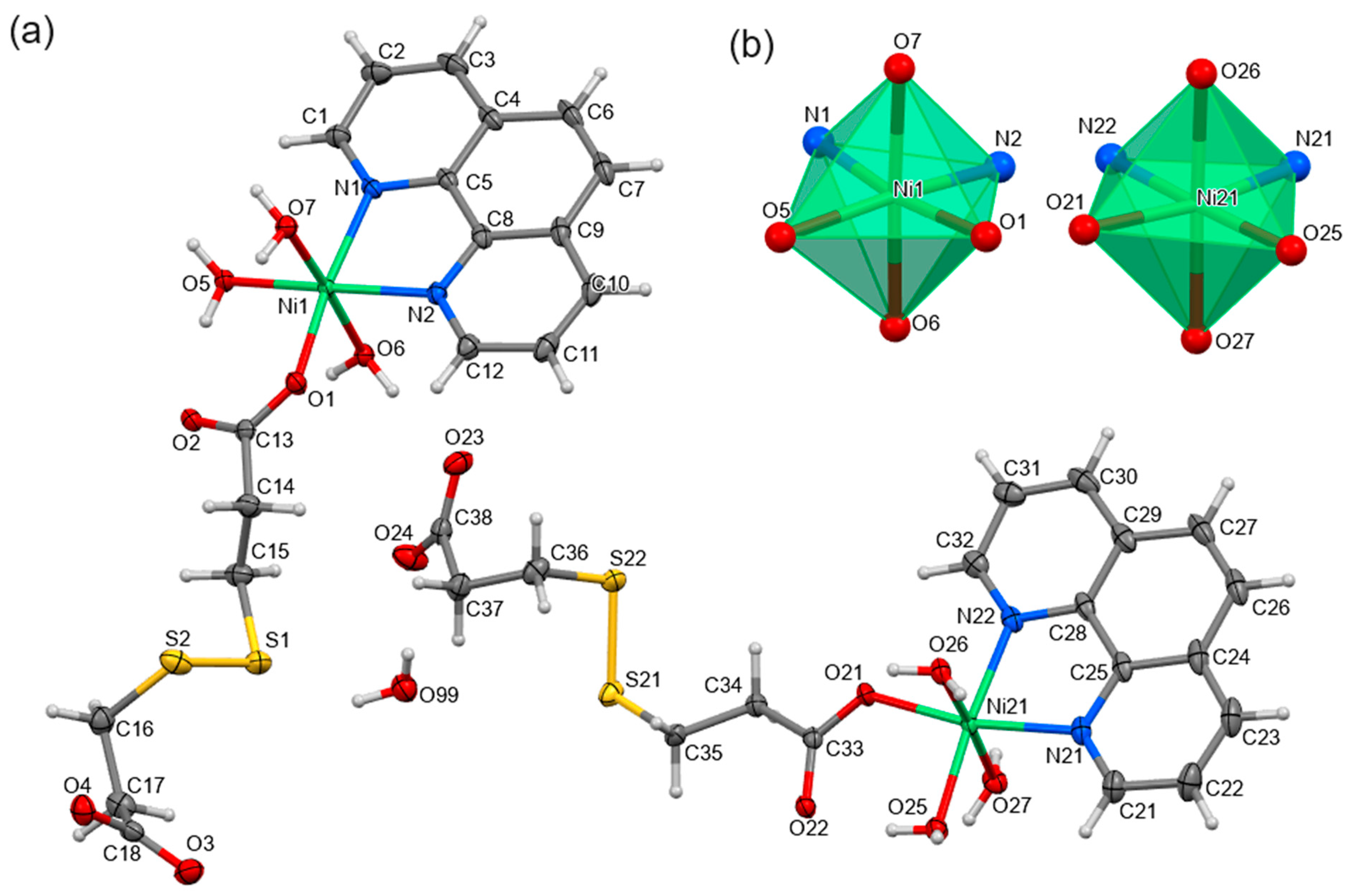
The crystal structures of three novel coordination compounds were determined by the single-crystal X-ray diffraction studies. Compound 1, [Ni(dtdp)(phen)(H2O)3]∙0.5H2O, is a mononuclear nickel complex containing two structurally independent coordination units in the asymmetric part, which are identical in composition and coordination bonds, along with one non-coordinating water molecule (Figure 1a). In both units, the nickel atom exhibits an octahedral coordination geometry (OC-6) with a coordination number of 6 (Figure 1b).
The inner coordination spheres of six-coordinated nickel atoms consist of one chelating 1,10-phenanthroline (phen), three terminal water molecules, and one 3,3′-dithiodipropionate (dtdp) anion coordinating through one oxygen atom. The second carboxylate group of dtdp is involved solely in hydrogen bonding. The geometries of the coordination polyhedra were determined using calculations performed in the SHAPE program [33], which is based on the Continuous Shape Measures (CShM) concept [34]. The CShM value SQ(P) represents the degree of similarity between a given structure (Q) and a reference geometry (P). It ranges from 0 to 100, where a value of 0 indicates perfect agreement with the reference geometry. Values close to 0 (typically below 1) suggest very good agreement, while increasing values represent progressively larger deviations from the ideal shape. While the theoretical upper limit for the SQ(P) value is 100, in reality, even highly distorted chemical structures seldom exhibit values exceeding 40. In the case of compound 1, both nickel atoms adopt a geometry very close to the ideal octahedron, as evidenced by SQ(OC-6) values lower than 1 (Table S1). The dtdp anion chains adopt the same trans–gauche–intermediate gauche/eclipsed–gauche–gauche conformation from the coordinated to the non-coordinated carboxylate group (Table S2). The supramolecular structure is stabilized by an extensive three-dimensional network of hydrogen bonds due to the large number of hydrogen bond donors and acceptors. The graph-set analysis is particularly useful in describing the hydrogen bonding patterns [35]. In this approach, hydrogen bonds are represented as edges connecting donor and acceptor atoms (nodes), allowing for a systematic classification of hydrogen bonding motifs. The general notation Gda(n) is used, where G denotes the graph pattern (e.g., R for ring, C for chain, etc.); a is the number of acceptors; d is the number of donors; and n represents the total number of atoms involved in the hydrogen bonding pattern. In the structure of 1, only O-H···O-type hydrogen bonds are present (Table S3), forming various cyclic motifs such as R22(8), R32(8), R43(10), R54(12), R63(12), and R64(14) (Figure S2). Additionally, an intramolecular hydrogen bond forming an S(6) motif is observed within the coordination units. Next to the hydrogen bonds, π-π stacking interactions (Figures S3 and S4) exist between phen molecules (Table S4), and they are propagated along the crystallographic [0 1 0] axis.
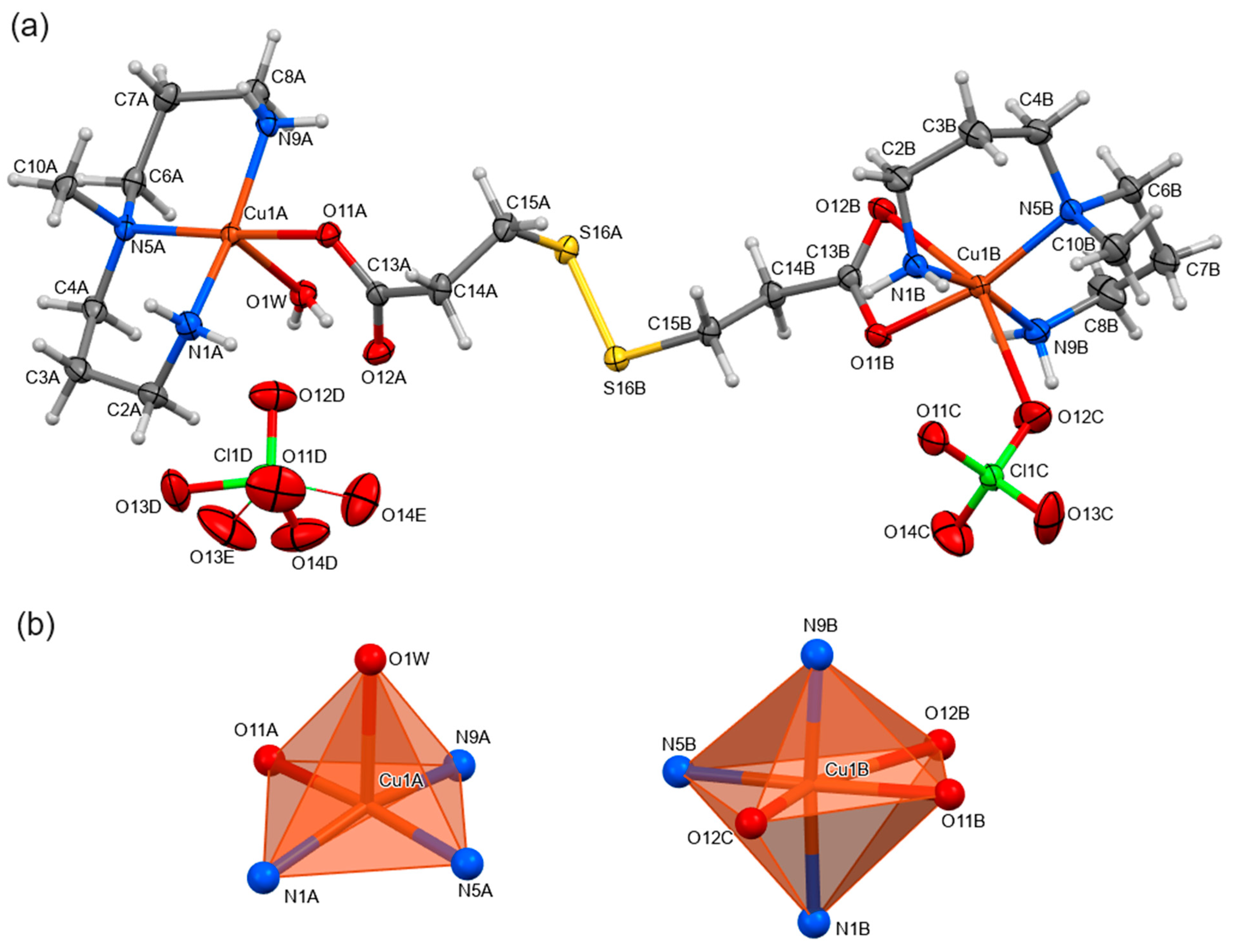
Compound 2, [(ClO4)(mdpta)Cu(μ-dtdp)Cu(mdpta)(H2O)](ClO4), is a dinuclear copper coordination complex composed of two distinct coordination units bridged by a dtdp anion (Figure 2a). The first central atom, Cu1a, possesses a coordination number of 5 and a square pyramidal (SPY-5) coordination polyhedron, while the second central atom, Cu1b, is six-coordinated with a distorted octahedral geometry—OC-6 (Figure 2b; Table S1). The SCu1b(OC-6) value is higher than the corresponding values for the nickel atoms of 1 (4.273 vs. 0460 and 0.440), which indicates larger deviations from an ideal octahedron.
Both copper atoms are chelated by tridentate N′–methyldipropylenetriamine (mdpta). Cu1a is further coordinated by one water molecule and one oxygen atom from the dtdp carboxylate group, with the second oxygen atom of this group forming an intramolecular hydrogen bond with the coordinating water molecule, creating an S(6) motif. Cu1b is coordinated by a monodentate perchlorate anion and a chelating carboxylate group from dtdp. An interesting feature of 2 is the competition between chlorate anions and water molecules for positions in the inner coordination sphere of the copper centers. Despite the presence of a water molecule in the inner coordination sphere of Cu1a leading to the formation of an intramolecular hydrogen bond that further stabilizes this arrangement, a perchlorate ion is found in the inner coordination sphere of Cu1b at the expense of a water molecule. This is accompanied by a change in the coordination mode of the dtdp carboxylate group to chelating, due to its inability to form an intramolecular hydrogen bond. The observed equilibrium between water and perchlorate ions suggests that the stability constants of both coordination units are similar, allowing for the formation of a dinuclear compound built from central atoms with different coordination spheres. The dtdp anion chain adopts a conformation analogous to that observed in 1 (Table S2). The dinuclear entity carries a single positive charge, which is balanced by a perchlorate anion in the outer coordination sphere. The supramolecular structure is stabilized by hydrogen bonds, with mdpta amine groups acting as donors, forming N-H···O bonds with carboxylate groups, perchlorate anions, and water molecules, as well as N-H···S bonds with dtdp sulfur atoms (Table S3). The coordinating water molecule also forms a hydrogen bond with the non-coordinating perchlorate anion. This diverse hydrogen bonding network results in cyclic motifs such as R22(12), R33(10), and R55(16) (Figure S3).
The phase purity of complexes 13 was confirmed via powder X-ray diffraction, employing Rietveld analysis to compare the measured data with the calculated pattern based on the crystal structure. Data refinement was conducted using parameters derived from method used by Bergmann et al. [36], with the microstrain (k2) and crystallite size (B1) parameters refined isotropically. The preferred orientation effects were addressed using the “SPHAR8” model. The fit demonstrates good agreement with the calculated powder pattern of complexes in all studied cases (Figure 3).
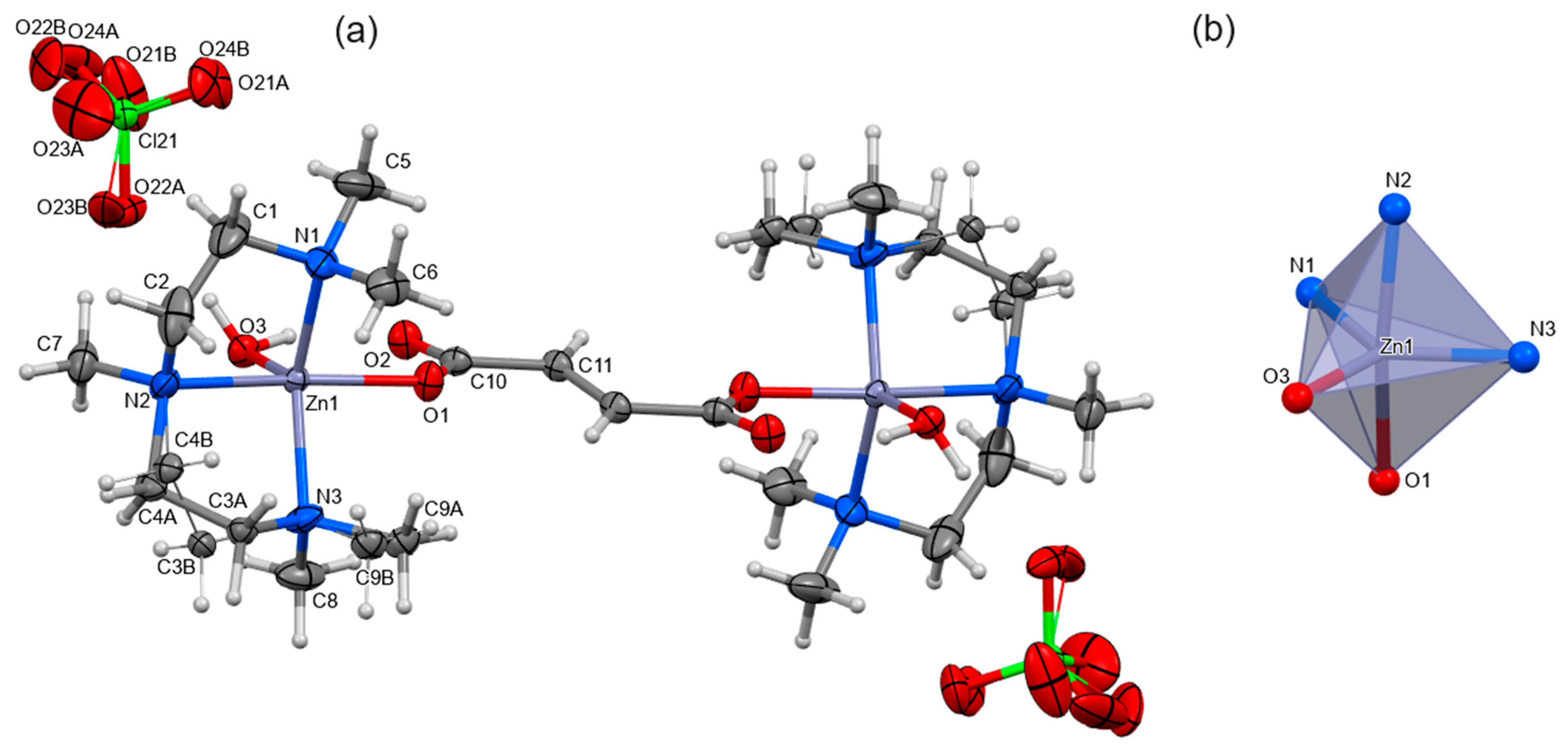
Compound 3, [{Zn(pmdien)(H2O)}2(μ-fu)](ClO4)2, is a symmetrical dinuclear zinc coordination complex with a fumarate (fu) anion serving as the bridging ligand (Figure 4a). It is isomorphous to the previously reported copper compound (CSD refcode: SIXMAO [13]). The fu chain adopts a trans conformation, and the symmetry of the dinuclear molecule is evidenced by an inversion center at the bisector of the fumarate anion (the special positions d of the P21/c space group). The coordination sphere of the five-coordinated zinc cation consists of a tridentate chelating N,N,N′,N″,N″-pentamethyldiethylenetriamine (pmdien), a water molecule, and a monodentate carboxylate group from the fu anion. The coordination polyhedron adopts a trigonal bipyramidal geometry (TBPY-5) (Figure 4b, Table S1).
An intramolecular hydrogen bond of the S(6) type is observed between the water molecule and the non-coordinated atom of the carboxylate group, similar to those in 1 and 2. The water molecule also forms a hydrogen bond with a non-coordinating perchlorate anion. The dinuclear entity carries a double positive charge, balanced by two perchlorate anions in the outer coordination sphere.

2.3. EPR Study of [(ClO4)(mdpta)Cu(μ-dtdp)Cu(mdpta)(H2O)](ClO4) (2)

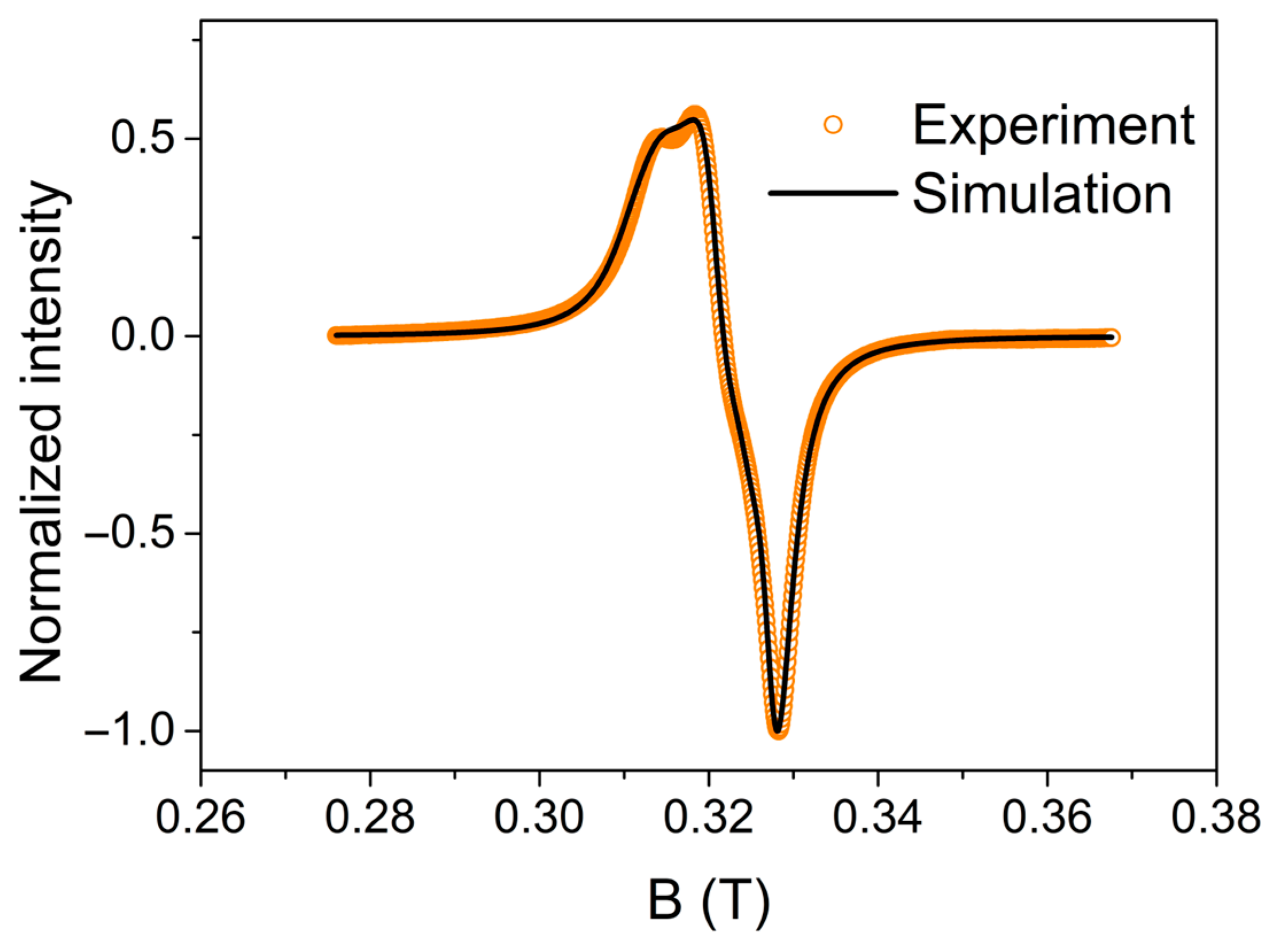
To complement the structural analysis, complex 2, containing Cu(II), was studied using EPR spectroscopy (Figure 5).
The molecular structure of [(ClO4)(mdpta)Cu(μ-dtdp)Cu(mdpta)(H2O)](ClO4) (2) contains two inequivalent Cu(II) centers, necessitating the use of two distinct sets of g-factors for fitting. The resulting parameters are g = 2.06374 ± 0.00004, g = 2.1711 ± 0.0003, gx = 2.0772 ± 0.0001, gy = 2.10756 ± 0.00006, and gz = 2.1520 ± 0.0002, with anisotropic linewidth ηx = 0.1428± 0.0005 GHz, ηy = 0.1493 ± 0.0004 GHz, and ηz = 0.254 ± 0.002 GHz. The first Cu(II) center exhibits a normal axial spectrum with g > g > ge (2.0023), indicating that the unpaired electron in the ground state resides in the d x 2 y 2 orbital, which is characteristic of an elongated octahedral or square pyramidal geometry [37]. The second Cu(II) center displays a rhombic spectrum with gz > gy > gx, also suggesting the presence of an unpaired electron in the d x 2 y 2 orbital in the ground state [38]. These findings are consistent with the shapes observed in the structural analysis, specifically an elongated octahedron and a tetragonal pyramid. Given the minimal distortion of the penta-coordinated square pyramidal site and the more pronounced deformation in the hexa-coordinated octahedral site, it is inferred that the axial g-factor g is associated with the penta-coordinated site, while the rhombic g-factor g′ corresponds to the hexa-coordinated site. The splitting of g′ is likely induced by the deformation of the angle between atoms O12b and O12c, which is 152° instead of 180°, as is expected in an ideal octahedron.

2.4. Antimicrobial Activity of the Complexes

The antibacterial activity of complexes 13 was evaluated against both Gram-positive (E. faecalis and S. aureus) and Gram-negative (P. aeruginosa and E. coli) bacteria. The minimum inhibitory concentration (MIC) values (Table 1) varied among the complexes and strains, with the complexes generally exhibiting a higher activity against Gram-positive bacteria. The Cu(II) complex (2) demonstrated the strongest antibacterial activity against all tested strains, with particularly high efficacy against S. aureus (0.78 g/L). Complex 2 is characterized by a dinuclear copper complex with distinct coordination geometries for the two copper atoms, which likely facilitates significant antimicrobial efficacy through potential synergistic interactions between the different coordination units. Dinuclear copper(II) complexes are known to possess enhanced antimicrobial properties compared to their mononuclear complexes [10], suggesting that the antimicrobial efficacy is highly dependent on the specific ligand environment and the overall structure of the complex. These findings underscore the critical role of the coordination environment and metal–ligand bonds in determining the biological activity of such complexes.
In our previous paper [22], we reported the MICs for three nickel and three copper complexes. Two of the nickel complexes, namely [Ni(tda)(phen)(H2O)]·3H2O and [Ni2(μ-tda)2(1,2-pn)2], were active against E. faecalis with an MIC of 8.44 g/L. The third complex, [Ni(tda)(1,3-pn)(H2O)]·H2O, showed the same MIC (8.44 g/L) for E. coli, E. faecalis, and S. Aureus. The copper complex [Cu2(μ-tdp)(pmdien)2(H2O)2](ClO4)2·H2O was active against E. faecalis and S. aureus only (MIC = 16.88 g/L). Much better results, i.e., lower MIC values, were obtained for [Cu2(μ-fu)(pmdien)2(H2O)2](ClO4)2 (E. coli—8.44, P. aeruginosa—2.11, E. faecalis—4.22, and S. Aureus—1.05) and [Cu2(μ-dtdp)(pmdien)2(H2O)2](ClO4)2 (E. coli—16.88, P. aeruginosa—1.05, E. faecalis—8.44, and S. Aureus—8.44). When we compare the MIC values with those presented in this paper, we can conclude that complex 1 with phen shows the same activity as [Ni(tda)(phen)(H2O)]·3H2O against E. faecalis, but 1 is active to all bacterial strains. Binuclear copper complex 2 is very similar to [Cu2(μ-tdp)(pmdien)2(H2O)2](ClO4)2·H2O, where tridentate ligand pmdien is exchanged to mdpta. Complex 2 shows a lower MIC for E. faecalis (3.13) and S. Aureus (0.78), whereas the pmdien complex shows a better parameter for P. aeruginosa (1.05).

3. Materials and Methods

3.1. Chemicals and Methods

3,3′-dithiodipropionic acid (H2dtdp), fumaric acid (H2fu), 1,10-phenanthroline (phen), N′–methyldipropylenetriamine (mdpta), and N,N,N′,N″,N″-pentamethyldiethylenetriamine (pmdien) were supplied by Sigma-Aldrich (St. Louis, MO, USA) in ACS purity. The C, H, N, and S analyses were carried out on an EA Thermo Scientific Flash 2000. IR spectra (400–4000 cm−1) were recorded with the help of an FT-IR spectrometer—Jasco FT/IR-4700 (Jasco, Easton, PA, USA)—using the ATR method. The diffuse-reflectance UV-Vis spectra (DR-UV/VIS) were obtained using a Cintra 3030 spectrometer (BGC Scientific Equipment, IL, USA) equipped with an integrated sphere assembly (barium sulfate as reference) in the range 200–800 nm. Electronic spectra were recorded at room temperature. The UV-VIS spectra of solutions of 13 (water:methanol = 1:1 v/v) were measured in 10 mm path length quartz cuvettes. The powder XRD patterns were measured using a Rigaku MiniFlex600 diffractometer (Rigaku, Austin, TX, USA) equipped with the Bragg–Brentano geometry and using Cu Kα radiation. EPR spectra were obtained with an Electron Paramagnetic Resonance Spectrometer (EPR) SPINSCAN X (LINEV systems), and measurements were carried out in the solid state with a polycrystalline powder sample. For the interpretation of data, programs Profex 5.2 (XPD) [39] and PHI 3.1.6 (EPR) [40] were used. For visualization, Mercury 2023.3.0 software was used [41].

3.2. Complex Preparation

3.2.1. Ni(dtdp)(H2O)

The dithiodipropionate nickel(II) compound was prepared similarly to the published method for nickel thiodiacetate [21]. To a solution of H2dtdp (4.2 g, 20 mmol) under stirring, a suspension of Ni(OH)2 (1.86 g, 20 mmol) in water (250 mL) was added. The suspension was heated at 80 °C for 8 h. After filtration, the solution was left to crystallize. The light-green product was filtered off and dried in an oven at 50 °C. Yield: 5.67 g (96.6%). Anal. Calc.: C, 25.3; H, 3.5; S, 22.5. Found: C, 24.8; H, 3.7; S 22.2%.
FT-IR (ATR, cm−1): 678 m, 839 w, 930 w, 1138 w, 1202 m, 1267 m, 1300 m, 1395 vs, 1555 vs, 1693 w, 3205 br.

3.2.2. [Ni(dtdp)(phen)(H2O)3]∙0.5H2O (1)

The phen (0.20 g, 1 mmol) was added to a stirred solution of [Ni(dtdp)(H2O)3] (0.32 g, 1 mmol) in water (50 mL), and was heated at 50 °C for 2 h. After cooling, the solution was filtered, and the filtrate was left for crystallization. Blue crystals (suitable for X-ray analyses) were collected on a frit funnel, washed with water, and dried in air. Yield: 0.15 g (46%). Anal. Calc.: C, 43.1; H, 4.7; N, 5.6; S, 12.7. Found: C, 43.4; H, 4.5; N, 5.4; S 12.5%.
FT-IR (ATR, cm−1): 424 w, 644 m, 672 m, 725 s, 770 w, 842 m, 1139 w, 1264 m, 1297 m, 1347 sh, 1403 vs, 1517 sh, 1555 vs, 1575 sh, 2917 sh, 3217 br.
DR-UV/VIS (λmax, nm): 300, 605, 740.

3.2.3. [(ClO4)(mdpta)Cu(μ-dtdp)Cu(mdpta)(H2O)](ClO4) (2)

The ligand mdpta (0.16 mL, 1 mmol) was added to a stirred solution of copper perchlorate hexahydrate (0.37 g, 1 mmol) in methanol (40 mL). The potassium salt of H2tdp was added. The salt was prepared by the reaction of H2dtdp (0.10 g) with KOH (0.05 g) in 5 mL of water. Potassium perchlorate was filtered off and the solution was left for crystallization. Dark blue crystals were obtained in a week. Yield: 0.36 g (48.0%). Anal. Calc.: C, 28.5; H, 5.7; N, 10.0; S, 7.6. Found: C, 28.2; H, 5.7; N, 9.9; S, 7.4%.
FT-IR (ATR, cm−1): 487 w, 514 w, 620 s, 766 w, 927 m, 988 sh, 1015 sh, 1063 vs, 1268 w, 1395 s, 1433 m, 1466 w, 1557 s, 1597 m, 2899 m, 2925 m, 2965 m, 3148 m, 3230 m, 3268 s, 3321 s, 3501 w.
DR-UV/VIS (λmax, nm): 280, 585.

3.2.4. [{Zn(pmdien)(H2O)}2(μ-fu)](ClO4)2 (3)

The ligand pmdien (0.2 mL, 1 mmol) was added to a stirred solution of zinc perchlorate hexahydrate (0.37 g, 1 mmol) in methanol (40 mL). The potassium salt of H2fu, prepared as described above, was added. The formed potassium perchlorate was filtered off and the solution was left to crystallize. White crystals were obtained in a week. Yield: 0.34 g (58%). Anal. Calc.: C, 32.1; H, 6.4; N, 10.2. Found: C, 31.5; H, 6.1; N, 9.6%.
FT-IR (ATR, cm−1): 438 w, 456 w, 489 w, 589 w, 623 s, 676 m, 760 w, 776 w, 800 m, 905 m, 932 s, 972 s, 1024 sh, 1044 vs, 1096 vs, 1248 w, 1309 w, 1368 sh, 1383 s, 1441 sh, 1471 s, 1525 s, 2823 w, 2885 m, 2983 w, 3133 w, 3399 s.
DR-UV/VIS (λmax, nm): 228.

3.3. Determination of Crystal Structures

The diffraction experiment for the crystal structure determination of 1 and 3 was performed on an XtaLAB Synergy Dualflex diffractometer equipped with a Pilatus 300K detector, while that of 2 was performed on a Bruker D8 VENTURE Kappa Duo with a PHOTONIII detector. The structure was solved by intrinsic methods (SHELXT) [42] and refined by full-matrix least squares based on F2 (SHELXL) [43]. The hydrogen atoms on carbon and nitrogen were fixed into idealized positions (riding model) and were assigned temperature factors of either Hiso(H) = 1.2 Ueq (pivot atom) or Hiso(H) = 1.5 Ueq (pivot atom for methyl moiety). The hydrogen atoms of the water molecule were found on a difference Fourier map and were refined under a rigid-body assumption with assigned temperature factors Hiso(H) = 1.2 Ueq (pivot atom). The outer coordination sphere chlorate anions in the structures of 2 and 3 were distorted over two positions with occupancy factors of 0.54:0.46 and 0.66:0.34, respectively. In 3, there are also three carbon atoms of pmdien (C3, C4, and C9), which are distorted over two positions (0.54:0.46). Details concerning crystal data and refinement are given in Table 2.
CCDC 2314973, 2369177, and 2369178 contain the supplementary crystallographic data for this paper. These data can be obtained free of charge at https://www.ccdc.cam.ac.uk/ [or from the Cambridge Crystallographic Data Centre (CCDC), 12 Union Road, Cambridge, CB2 1EZ, UK; fax: +44(0) 1223 336 033; email: [email protected]].

3.4. Determination of Antibacterial Activity

The standard dilution method was used to determine the antimicrobial activity and minimum inhibition concentration (MIC) of the complexes. For the purpose of antimicrobial testing, the water solution of each sample was used and diluted by the culture medium (Mueller–Hinton Broth, BD Difco, France) in a geometric progression from 2 to 1024 times in microtiter plates. The culture medium was inoculated with the tested bacteria at a concentration of 5 × 10⁵ CFU/mL and incubated for 24 h at 35+/−1 °C. After 18+/−2 h, the MIC was determined according to standard testing protocols (EUCAST) as the lowest concentration of the tested substance, inhibiting the visible growth of the tested bacterial strain. Escherichia coli CCM 3954, Pseudomonas aeruginosa CCM 3955, Staphylococcus aureus CCM 4223, and Enterococcus faecalis CCM 4224 were purchased from the Czech Collection of Microorganisms (Brno, Czech Republic) and were stored in cryotubes (ITEST plus, Czech Republic) at –80 °C.

4. Conclusions

Three novel complexes of dicarboxylic acids H2dptm and H2fu—mononuclear Ni(II) complex 1, asymmetric dinuclear Cu(II) complex 2, and symmetric Zn(II) complex 3—were prepared. Their structures were determined by X-ray structural analysis, and complexes were characterized by a wide array of methods, including CHNS analysis, FTIR spectroscopy, UV-Vis spectroscopy, and in the case of Cu(II) complex, also EPR spectroscopy and powder X-ray diffraction.
Biological experiments revealed that minimal inhibition concentrations among the studied complexes on all bacterial lines are the lowest for complex 2. It can be assumed that a role in its antibacterial properties has synergic effects between two non-symmetric Cu(II) centers. On the opposite side, the highest inhibition concentrations are for mononuclear complex 1.
The variability of the obtained structures displays the versatile nature of the used ligands. It can be noted that these results show that using suitable N-donor ligands in combination with dicarboxylic acid can lead to interesting dinuclear systems, which can be used for the further tuning of similar systems to potentially improve their bioactivity.

Supplementary Materials

The following supporting information can be downloaded at: https://www.mdpi.com/article/10.3390/inorganics12100260/s1, Figure S1. The UV-Vis spectra of complexes 1 (red; c = 1.6 mmol/dm3), 2 (green; c = 2.5 mmol/dm3), and 3 (blue; c = 2.5 mmol/dm3) measured in solution (water:methanol = 1:1 v/v); Table S1. Continuous Shape Measures values SQ(P), where Q is considered polyhedron and P is an ideal polyhedron, for the coordination polyhedra in the studied compounds (CN—coordination number). Table S2. Selected structural data of studied compounds. Table S3. Hydrogen bonds in studied compounds. Table S4. Stacking interactions in 1. Figure S2. Hydrogen bond ring motifs in the structure of 1. Figure S3. Part of molecular packing showing π-stacking interactions in the structure of 1. Figure S4. Hydrogen bond ring motifs in the structure of 2. Figure S5. FT-IR spectra of complexes 13. Figure S6. FT-IR spectrum of complex Ni(dtdp)(H2O). Figure S7. UV-VIS diffuse reflectance spectrum of complex 1. Figure S8. UV-VIS diffuse reflectance spectrum of complex 2. Figure S9. UV-VIS diffuse reflectance spectrum of complex 3.

Author Contributions

Conceptualization, P.K. and M.Ś.; validation, K.K. and P.A.; formal analysis, K.K.; investigation, P.A.; resources, A.P.; data curation, L.H.; writing—original draft preparation, I.L., K.K., P.A., L.H., I.C., M.Ś., and P.K.; writing—review and editing, P.K.; visualization, M.Ś. and I.C.; supervision, P.K.; project administration, P.K.; funding acquisition, A.P. and P.K. All authors have read and agreed to the published version of this manuscript.

Funding

This research was funded by IGA_ PrF_2024_020 and the institutional sources of the Department of Inorganic Chemistry, Palacky University Olomouc.

Data Availability Statement

Data are contained within the article and Supplementary Materials.

Conflicts of Interest

The authors declare no conflicts of interest.

References

  1. Li, S.; Song, J.; Ni, J.C.; Wang, Z.N.; Gao, X.; Shi, Z.; Bai, F.Y.; Xing, Y.H. Photoelectric properties and potential nitro derivatives sensing by a highly luminescent of Zn(II) and Cd(II) metal-organic frameworks assembled by the flexible hexapodal ligand, 1,3,5-triazine-2,4,6-triamine hexaacetic acid. RSC Adv. 2016, 6, 36000–36010. [Google Scholar] [CrossRef]
  2. Swiatkowski, M.; Kruszynski, R. Structurally diverse coordination compounds of zinc as effective precursors of zinc oxide nanoparticles with various morphologies. Appl. Organomet. Chem. 2019, 33, 18. [Google Scholar] [CrossRef]
  3. Svorec, J.; Polakovicova, P.; Moncol, J.; Kuchtanin, V.; Breza, M.; Soralova, S.; Padelkova, Z.; Mrozinski, J.; Lis, T.; Segla, P. Structural, magnetic and quantum-chemical study of dinuclear copper(II) thiophenecarboxylate and furancarboxylate complexes. Polyhedron 2014, 81, 216–226. [Google Scholar] [CrossRef]
  4. Kuchtanin, V.; Moncol, J.; Mrozinski, J.; Kalinska, B.; Padelková, Z.; Svorec, J.; Segl’a, P.; Melník, M. Study of copper(II) thiophenecarboxylate complexes with N-methylnicotinamide. Polyhedron 2013, 50, 546–555. [Google Scholar] [CrossRef]
  5. Shintoyo, S.; Murakami, K.; Fujinami, T.; Matsumoto, N.; Mochida, N.; Ishida, T.; Sunatsuki, Y.; Watanabe, M.; Tsuchimoto, M.; Mrozinski, J.; et al. Crystal Field Splitting of the Ground State of Terbium(III) and Dysprosium(III) Complexes with a Triimidazolyl Tripod Ligand and an Acetate Determined by Magnetic Analysis and Luminescence. Inorg. Chem. 2014, 53, 10359–10369. [Google Scholar] [CrossRef]
  6. Ashraf, J.; Riaz, M.A. Biological potential of copper complexes: A review. Turk. J. Chem. 2022, 46, 595–623. [Google Scholar] [CrossRef]
  7. Molinaro, C.; Martoriati, A.; Pelinski, L.; Cailliau, K. Copper Complexes as Anticancer Agents Targeting Topoisomerases I and II. Cancers 2020, 12, 2863. [Google Scholar] [CrossRef]
  8. Switlicka-Olszewska, A.; Machura, B.; Mrozinski, J.; Kalinska, B.; Kruszynski, R.; Penkala, M. Effect of N-donor ancillary ligands on structural and magnetic properties of oxalate copper(II) complexes. New J. Chem. 2014, 38, 1611–1626. [Google Scholar] [CrossRef]
  9. Switlicka-Olszewska, A.; Machura, B.; Mrozinski, J. Synthesis, magnetic behavior and structural characterization of novel one-dimensional copper(II) coordination polymer based on azide and oxalate bridges. Inorg. Chem. Commun. 2014, 43, 86–89. [Google Scholar] [CrossRef]
  10. Esfahani, M.H.; Behzad, M. Crystal structure and antibacterial properties of a new dinuclear copper complex based on an unsymmetrical NN′O type Schiff base ligand. J. Coord. Chem. 2020, 73, 154–163. [Google Scholar] [CrossRef]
  11. Kopel, P.; Kamenícek, J.; Petrícek, V.; Kurecka, A.; Kalinska, B.; Mrozinski, J. Syntheses and study on nickel and copper complexes with 1,3,5-benzenetricarboxylic acid.: Crystal and molecular structure of Cu3(mdpta)3(btc) (ClO4)4H2O. Polyhedron 2007, 26, 535–542. [Google Scholar] [CrossRef]
  12. Loubalová, I.; Kopel, P. Coordination Compounds of Cu, Zn, and Ni with Dicarboxylic Acids and N Donor Ligands, and Their Biological Activity: A Review. Molecules 2023, 28, 1445. [Google Scholar] [CrossRef] [PubMed]
  13. Mautner, F.A.; Vicente, R.; Louka, F.R.Y.; Massoud, S.S. Dinuclear fumarato- and terephthalato-bridged copper(II) complexes: Structural characterization and magnetic properties. Inorg. Chim. Acta 2008, 361, 1339–1348. [Google Scholar] [CrossRef]
  14. Alarcon-Payer, C.; Pivetta, T.; Choquesillo-Lazarte, D.; Gonzalez-Perez, J.M.; Crisponi, G.; Castineiras, A.; Niclos-Gutierrez, J. Thiodiacetato-copper(II) chelates with or without N-heterocyclic donor ligands: Molecular and/or crystal structures of Cu(tda) (n), Cu(tda)(Him)(2)(H2O) and Cu(tda)(5Mphen) center dot 2H(2)O (Him = imidazole, 5Mphen=5-methyl-1,10-phenanthroline). Inorg. Chim. Acta 2005, 358, 1918–1926. [Google Scholar] [CrossRef]
  15. Bonomo, R.P.; Rizzarelli, E.; Brescianipahor, N.; Nardin, G. Properties and X-ray crystal-structures of copper(ii) mixed complexes with thiodiacetate and 2,2′-bipyridyl or 2,2′-6′,2′-terpyridyl. J. Chem. Soc.-Dalton Trans. 1982, 681–685. [Google Scholar] [CrossRef]
  16. Neuman, N.I.; Perec, M.; Gonzalez, P.J.; Passeggi, M.C.G.; Rizzi, A.C.; Brondino, C.D. Single Crystal EPR Study of the Dinuclear Cu(II) Complex Cu(tda)(phen) (2)center dot H(2)tda (tda = Thiodiacetate, phen = Phenanthroline): Influence of Weak Interdimeric Magnetic Interactions. J. Phys. Chem. A 2010, 114, 13069–13075. [Google Scholar] [CrossRef]
  17. Kopel, P.; Travnicek, Z.; Marek, J.; Korabik, M.; Mrozinski, J. Syntheses and properties of binuclear copper(II) mixed-ligand complexes involving thiodiglycolic acid. The crystal structures of (phen)(2)Cu(mu-tdga)Cu(phen) (NO3)(2)center dot 5H(2)O and (H2O)(pmdien)Cu(mu-tdga)Cu(pmdien)(H2O) (ClO4)(2). Polyhedron 2003, 22, 411–418. [Google Scholar] [CrossRef]
  18. Khullar, S.; Mandal, S.K. Effect of Spacer Atoms in the Dicarboxylate Linkers on the Formation of Coordination Architectures-Molecular Rectangles vs 1D Coordination Polymers: Synthesis, Crystal Structures, Vapor/Gas Adsorption Studies, and Magnetic Properties. Cryst. Growth Des. 2014, 14, 6433–6444. [Google Scholar] [CrossRef]
  19. Baggio, R.; Perec, M.; Garland, M.T. Aqua(2,2′-bipyriayl-N,N′)(thiodiacetato-O,O′,S)zinc(II) tetrahydrate. Acta Crystallogr. Sect. C-Cryst. Struct. Commun. 1996, 52, 2457–2460. [Google Scholar] [CrossRef]
  20. Arici, M.; Yesilel, O.Z.; Sahin, O.; Tas, M. Dinuclear and polynuclear copper(II) complexes with 3,31-thiodipropionate and unprecedented coordination mode. Polyhedron 2014, 71, 62–68. [Google Scholar] [CrossRef]
  21. Kopel, P.; Travnicek, Z.; Marek, J.; Mrozinski, J. Syntheses and study on nickel(II) complexes with thiodiglycolic acid and nitrogen-donor ligands. X-ray structures of Ni(bpy)(tdga)(H2O)center dot 4H(2)O and (en)Ni(mu-tdga)(2)NI(en) center dot 4H(2)O (tdgaH(2)= thiodiglycolic acid). Polyhedron 2004, 23, 1573–1578. [Google Scholar] [CrossRef]
  22. Loubalová, I.; Zahradníková, E.; Masaryk, L.; Nemec, I.; Hochvaldová, L.; Panáček, A.; Kvítek, L.; Večeřová, R.; Swiatkowski, M.; Kopel, P. Antibacterial study on nickel and copper dicarboxylate complexes. Inorg. Chim. Acta 2023, 545, 121273. [Google Scholar] [CrossRef]
  23. Lazarou, K.N.; Terzis, A.; Perlepes, S.P.; Raptopoulou, C.P. Synthetic, structural and spectroscopic studies of complexes derived from the copper(II) perchlorate/fumaric acid/N,N′-chelates tertiary reaction systems. Polyhedron 2010, 29, 46–53. [Google Scholar] [CrossRef]
  24. Dalai, S.; Mukherjee, P.S.; Rogez, G.; Mallah, T.; Drew, M.G.B.; Chaudhuri, N.R. Synthesis, crystal structures, and magnetic properties of two new 1D copper(II) coordination polymers containing fumarate(-2) and chelating N,N′-donor as ligands. Eur. J. Inorg. Chem. 2002, 2002, 3292–3297. [Google Scholar] [CrossRef]
  25. Burrows, A.D.; Harrington, R.W.; Mahon, M.F.; Price, C.E. The influence of hydrogen bonding on the structure of zinc co-ordination polymers. J. Chem. Soc.-Dalton Trans. 2000, 3845–3854. [Google Scholar] [CrossRef]
  26. Zheng, Y.Q.; Lin, J.L.; Chen, B.Y. New catenary coordination polymers using fumarato ligand as bridging spacer: Crystal structures of Mn(phen)(2)(H2O)(2) L center dot 4H(2)O, Mn(phen)(H2O)(2)L and Zn(phen)L center dot H2L with-H2L fumaric acid. J. Mol. Struct. 2003, 646, 151–159. [Google Scholar] [CrossRef]
  27. Konar, S.; Zangrando, E.; Drew, M.G.B.; Ribas, J.; Chaudhuri, N.R. Synthesis, structural analysis, and magnetic behaviour of three fumarate bridged coordination polymers: Five-fold interpenetrated diamond-like net of Ni-II, sheets of Ni-II and Co-II. Dalton Trans. 2004, 260–266. [Google Scholar] [CrossRef]
  28. Ohmura, T.; Mori, W.; Hasegawa, M.; Takei, T.; Ikeda, T.; Hasegawa, E. Crystal structures and magnetic and gas-occlusion properties of microporous materials containing infinite chains of mononuclear metal (Cu(II), Zn(II), and Ni(II)) dicarboxylates unit. Bull. Chem. Soc. Jpn. 2003, 76, 1387–1395. [Google Scholar] [CrossRef]
  29. Abbaszadeh, A.; Safari, N.; Amani, V.; Notash, B.; Raei, F.; Eftekhar, F. Mononuclear and Dinuclear Copper(II) Complexes Containing N, O and S Donor Ligands: Synthesis, Characterization, Crystal Structure Determination and Antimicrobial Activity of Cu(phen)(tda) center dot 2H(2)O and (phen)(2)Cu(mu-tda)Cu(phen) (ClO4)(2 center dot)1.5H(2)O. Iran J. Chem. Chem. Eng.-Int. Engl. Ed. 2014, 33, 1–13. [Google Scholar]
  30. Buchtelova, H.; Skubalova, Z.; Strmiska, V.; Michalek, P.; Kociova, S.; Smerkova, K.; Kruszynski, R.; Bienko, A.; Kaj, M.; Lewinska, A.; et al. Synthesis and structural characterization of antimicrobial binuclear copper (II) coordination compounds bridged by hydroxy- and/or thiodipropionic acid. J. Inorg. Biochem. 2019, 191, 8–20. [Google Scholar] [CrossRef]
  31. Lahiri, D.; Bhowmick, T.; Pathak, S.; Shameema, O.; Patra, A.K.; Ramakumar, S.; Chakravarty, A.R. Anaerobic Photocleavage of DNA in Red Light by Dicopper(II) Complexes of 3,3′-Dithiodipropionic Acid. Inorg. Chem. 2009, 48, 339–349. [Google Scholar] [CrossRef] [PubMed]
  32. Paul, A.; Figuerola, A.; Bertolasi, V.; Manna, S.C. DNA/protein binding and magnetic properties of a 1D Cu(II) complex containing fumarate and tridentate Schiff base ligands. Polyhedron 2016, 119, 460–470. [Google Scholar] [CrossRef]
  33. Llunell, M.; Casanova, D.; Cirera, J.; Alemany, P.; Alvarez, S. SHAPE. Program for the Stereochemical Analysis of Molecular Fragments by Means of Continuous Shape Measures and Associated Tools; Universitat de Barcelona: Barcelona, Spain, 2013. [Google Scholar]
  34. Casanova, D.; Cirera, J.; Llunell, M.; Alemany, P.; Avnir, D.; Alvarez, S. Minimal Distortion Pathways in Polyhedral Rearrangements. J. Am. Chem. Soc. 2004, 126, 1755–1763. [Google Scholar] [CrossRef] [PubMed]
  35. Etter, M.C.; MacDonald, J.C. Graph-Set Analysis of Hydrogen-Bond Patterns in Organic Crystals. Acta Crystallogr. Sect. B 1990, 46, 256–262. [Google Scholar] [CrossRef] [PubMed]
  36. Taut, T.; Kleeberg, R.; Bergmann, J. The new seifert Rietveld program and its application to quantitative phase analysis. In Proceedings of the XVII Conference on Applied Crystallography, Wisla, Poland, 31 August–4 September 1997; pp. 87–92. [Google Scholar]
  37. Garribba, E.; Micera, G. The determination of the geometry of Cu(II) complexes—An EPR spectroscopy experiment. J. Chem. Educ. 2006, 83, 1229–1232. [Google Scholar] [CrossRef]
  38. Hathaway, B.J.; Billing, D.E. Electronic properties and stereochemistry of mono-nuclear complexes of copper(ii) ion. Coord. Chem. Rev. 1970, 5, 143–207. [Google Scholar] [CrossRef]
  39. Doebelin, N.; Kleeberg, R. Profex: A graphical user interface for the Rietveld refinement program BGMN. J. Appl. Crystallogr. 2015, 48, 1573–1580. [Google Scholar] [CrossRef]
  40. Chilton, N.F.; Anderson, R.P.; Turner, L.D.; Soncini, A.; Murray, K.S. PHI: A powerful new program for the analysis of anisotropic monomeric and exchange-coupled polynuclear d- and f-block complexes. J. Comput. Chem. 2013, 34, 1164–1175. [Google Scholar] [CrossRef]
  41. Macrae, C.F.; Edgington, P.R.; McCabe, P.; Pidcock, E.; Shields, G.P.; Taylor, R.; Towler, M.; van De Streek, J. Mercury: Visualization and analysis of crystal structures. J. Appl. Crystallogr. 2006, 39, 453–457. [Google Scholar] [CrossRef]
  42. Sheldrick, G.M. SHELXT-Integrated space-group and crystal-structure determination. Acta Crystallogr. Sect. A 2015, 71, 3–8. [Google Scholar] [CrossRef]
  43. Sheldrick, G.M. Crystal structure refinement with SHELXL. Acta Crystallogr. Sect. C-Struct. Chem. 2015, 71, 3–8. [Google Scholar] [CrossRef] [PubMed]
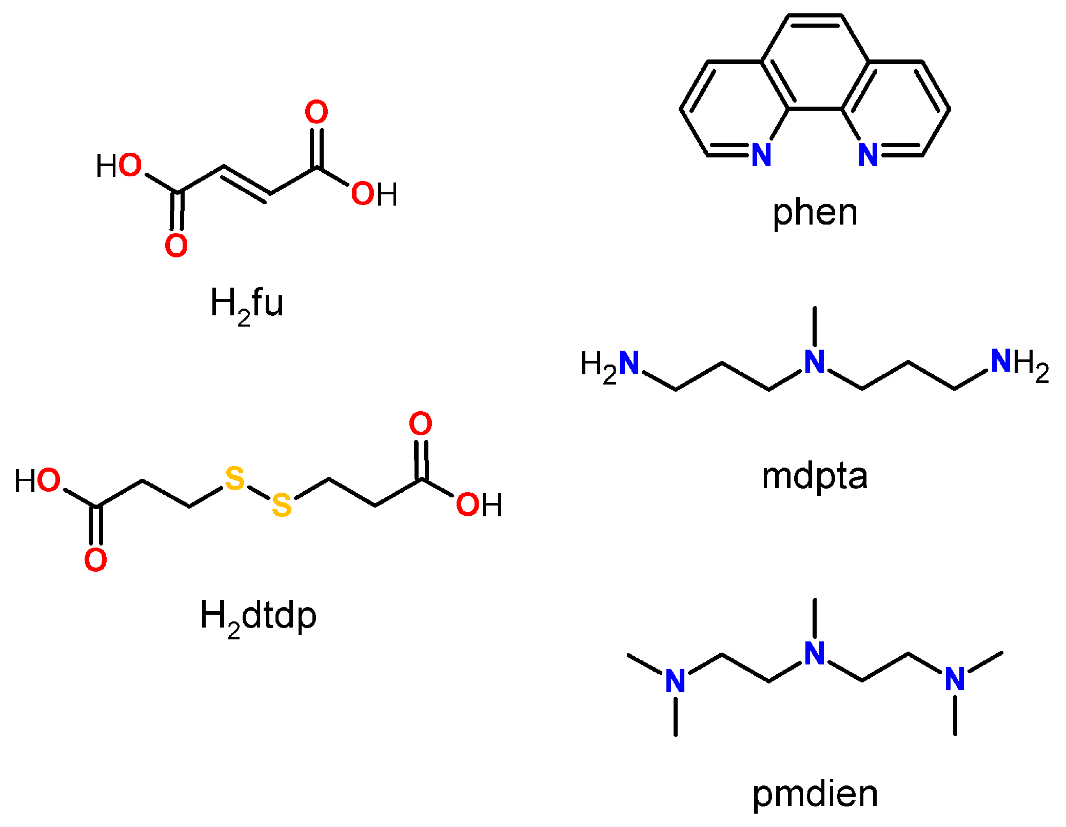
Scheme 1. Structural formulae of studied ligands.
Inorganics 12 00260 sch001
Figure 1. Molecular structure of 1, plotted with a 50% probability of displacement ellipsoids of nonhydrogen atoms and as spheres of arbitrary radii for hydrogen atoms (a). Coordination polyhedra of 1 (b).
Inorganics 12 00260 g001
Figure 2. Molecular structure of 2, plotted with a 50% probability of displacement ellipsoids of nonhydrogen atoms and as spheres of arbitrary radii for hydrogen atoms (a). The disordered atoms that belong to the second domain are indicated by thin bond lines. Coordination polyhedra of 2 (b).
Inorganics 12 00260 g002
Figure 3. Comparison of measured powder pattern with Rietveld fit and with pattern calculated from crystal structure for 13. Prepared sample seems to fit expected structure with no identifiable impurities.
Inorganics 12 00260 g003
Figure 4. Molecular structure of 3, plotted with a 50% probability of displacement ellipsoids of nonhydrogen atoms and as spheres of arbitrary radii for hydrogen atoms (a). The equivalent atoms (without labels) were generated according to the following transformation: −x + 1, −y + 1, −z. The disordered atoms belonging to the second domain are indicated by thin bond lines. Coordination polyhedron of 3 (b).
Inorganics 12 00260 g004
Figure 5. EPR spectra of 2 with experimental data (circles) and simulated data based on the parameters listed below (lines). Experiments were conducted at X-band frequency (9.46 GHz) and room temperature (302.08 K).
Inorganics 12 00260 g005
Table 1. Minimal inhibition concentration (MIC) [g/L] of complexes 13 for different bacterial strains.
MIC [g/L]
Complex E. coliP. aeruginosaE. faecalisS. aureus
125256.2512.5
26.2512.53.130.78
36.25>256.2512.5
Table 2. Detailed information about crystallographic experiments regarding studied complexes.
Compound (CCDC Number).1 (2369177)2 (2314973)3 (2369178)
Empirical formulaC36H46N4Ni2O15S4C20H48Cl2Cu2N6O13S2C11H26ClN3O7Zn
Formula weight1020.43842.74826.33
Temperature (K)100.3 (5)120 (2)100.0 (1)
Crystal systemorthorhombicmonoclinicmonoclinic
Space groupPbcaCcP21/c
a (Å)22.0565 (1)15.1199 (5)8.1853 (1)
b (Å)14.4105 (1)11.2259 (4)15.4006 (2)
c (Å)27.4138 (1)20.9787 (6)14.2517 (2)
α (°)909090
β (°)90105.828 (1)101.713 (2)
γ (°)909090
Volume (Å3)8713.34 (8)3425.80 (19)1759.14 (4)
Z842
ρcalc (g/cm3)1.5561.6341.560
μ (mm−1)3.4811.5861.584
F(000)42401752864
Crystal size (mm3)0.469 × 0.081 × 0.0510.166 × 0.116 × 0.1080.427 × 0.229 × 0.207
RadiationCu Kα (λ = 1.54184)Mo Kα (λ = 0.71073)Mo Kα (λ = 0.71073)
θ range for data collect. (°)3.224 to 78.8952.018 to 27.4822.645 to 31.581
Index ranges−28 ≤ h ≤ 27,−19 ≤ h ≤ 17,−10 ≤ h ≤ 11,
−18 ≤ k ≤ 16,−14 ≤ k ≤ 14,−22 ≤ k ≤ 20,
−34 ≤ l ≤ 33−27 ≤ l ≤ 27−19 ≤ l ≤ 20
Reflections collected/independent 201405/926028941/736959565/5458
Rint0.07340.03050.0305
Data/restraints/parameters9260/21/5927369/26/4275458/14/280
Goodness-of-fit on F21.1001.0561.071
Final R indexes [I > 2s(I)]R1 = 0.0408,R1 = 0.0174,R1 = 0.0297,
wR2 = 0.1077wR2 = 0.0426wR2 = 0.0764
R indexes (all data)R1 = 0.0426,R1 = 0.0179,R1 = 0.0366,
wR2 = 0.1088wR2 = 0.0429wR2 = 0.0809
Largest diff. peak and hole (e·Å−3)0.905/−0.7250.319/−0.2591.179/−0.440
Flack parameter-0.020 (3)-
Disclaimer/Publisher’s Note: The statements, opinions and data contained in all publications are solely those of the individual author(s) and contributor(s) and not of MDPI and/or the editor(s). MDPI and/or the editor(s) disclaim responsibility for any injury to people or property resulting from any ideas, methods, instructions or products referred to in the content.