


The Role of Complexes of Biogenic Metals in Living Organisms


Abstract
1. Introduction
2. Biologically Active Complex Compounds: Theoretical Overview
2.1. Mutual Selectivity and Affinity of Metals to Ligands
2.2. Inorganic Substances as Bioligands
2.3. Organic Substances as Bioligands
2.4. Main Factors for the Formation of Stable Metal Complexes
3. Biological Activity of Biogenic Metals and Their Coordination Compounds
3.1. Sodium and Potassium
3.2. Magnesium and Calcium
3.3. Manganese
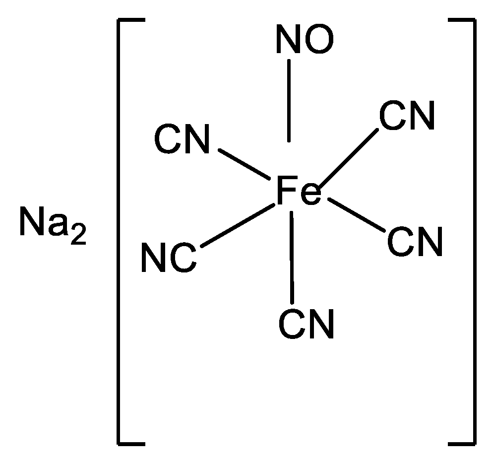
3.4. Iron
3.5. Cobalt
3.6. Copper
3.7. Zinc
3.8. Molybdenum
4. Conclusions
Funding
Acknowledgments
Conflicts of Interest
References
- Kostova, I.; Soni, R.K. Bioinorganic Chemistry; Shree Publishers & Distributors: New Delhi, India, 2011. [Google Scholar]
- Goswami, A.K.; Kostova, I. Medicinal and Biological Inorganic Chemistry; Walter de Gruyter GmbH & Co KG: Berlin, Germany, 2022. [Google Scholar]
- Franz, K.J.; Metzler-Nolte, N. Introduction: Metals in medicine. Chem. Rev. 2019, 119, 727–729. [Google Scholar] [CrossRef] [PubMed]
- Gasser, G. Metal complexes and medicine: A successful combination. Chimia 2015, 69, 442–446. [Google Scholar] [CrossRef] [PubMed]
- Mjos, K.D.; Orvig, C. Metallodrugs in medicinal inorganic chemistry. Chem. Rev. 2014, 114, 4540–4563. [Google Scholar] [CrossRef] [PubMed]
- David, S.S.; Meggers, E. Inorganic chemical biology: From small metal complexes in biological systems to metalloproteins. Curr. Opin. Chem. Biol. 2008, 12, 194–196. [Google Scholar] [CrossRef] [PubMed]
- Meggers, E. Exploring biologically relevant chemical space with metal complexes. Curr. Opin. Chem. Biol. 2007, 11, 287–292. [Google Scholar] [CrossRef]
- Meggers, E. From conventional to unusual enzyme inhibitor scaffolds: The quest for target specificity. Angew. Chem. Int. Ed. 2011, 50, 2442–2448. [Google Scholar] [CrossRef]
- Barry, N.P.E.; Sadler, P.J. Exploration of the medical periodic table: Towards new targets. Chem. Commun. 2013, 49, 5106–5131. [Google Scholar] [CrossRef]
- Barry, N.P.E.; Sadler, P.J. 100 years of metal coordination chemistry: From Alfred Werner to anticancer metallodrugs. Pure Appl. Chem. 2014, 86, 1897–1910. [Google Scholar] [CrossRef]
- Hartinger, C.G.; Dyson, P.J. Bioorganometallic chemistry—From teaching paradigms to medicinal applications. Chem. Soc. Rev. 2009, 38, 391–401. [Google Scholar] [CrossRef]
- López, R.; Díaz, N.; Suárez, D. Alkali and Alkaline-Earth Cations in Complexes with Small Bioorganic Ligands: Ab Initio Benchmark Calculations and Bond Energy Decomposition. Chem. Phys. Chem. 2020, 21, 99–112. [Google Scholar] [CrossRef]
- Hill, M.S.; Liptrot, D.J.; Weetman, C. Alkaline earths as main group reagents in molecular catalysis. Chem. Soc. Rev. 2016, 45, 972–988. [Google Scholar] [CrossRef]
- Westerhausen, M.; Koch, A.; Görls, H.; Krieck, S. Heavy Grignard reagents: Synthesis, physical and structural properties, chemical behavior and reactivity. Chem. Eur. J. 2017, 23, 1456–1483. [Google Scholar] [CrossRef]
- Krieck, S.; Westerhausen, M. Kudos and renaissance of s-block metal chemistry. Inorganics 2017, 5, 17. [Google Scholar] [CrossRef]
- Crea, F.; Stefano, C.D.; Foti, C.; Lando, G.; Milea, D.; Sammartano, S. Alkali metal ion complexes with phosphates, nucleotides, amino acids, and related ligands of biological relevance. Their properties in solution. Met. Ions Life Sci. 2016, 16, 133–166. [Google Scholar]
- Sigel, A.; Sigel, H.; Sigel, R.K. (Eds.) The Alkali Metal Ions: Their Role for Life; Springer International Publishing: New York, NY, USA, 2016; Volume 16. [Google Scholar]
- Dudev, T.; Lim, C. Competition among metal ions for protein binding sites: Determinants of metal ion selectivity in proteins. Chem. Rev. 2014, 114, 538–556. [Google Scholar] [CrossRef]
- Ussing, H.H.; Kruhoffer, P.; Thaysen, H.J.; Thorn, N.H. The Alkali Metal Ions in Biology: I. The Alkali Metal Ions in Isolated Systems and Tissues. II. The Alkali Metal Ions in the Organism; Springer Science & Business Media: Berlin/Heidelberg, Germany, 2013; Volume 13. [Google Scholar]
- Fromm, K.M. Chemistry of alkaline earth metals: It is not all ionic and definitely not boring! Coord. Chem. Rev. 2020, 408, 213193. [Google Scholar] [CrossRef]
- Kolev, S.K.; Petkov, P.S.; Rangelov, M.A.; Trifonov, D.V.; Milenov, T.I.; Vayssilov, G.N. Interaction of Na+, K+, Mg2+ and Ca2+ counter cations with RNA. Metallomics 2018, 10, 659–678. [Google Scholar] [CrossRef]
- Park, S.H.; Hwang, I.; McNaughton, D.A.; Kinross, A.J.; Howe, E.N.; He, Q.; Shin, I. Synthetic Na+/K+ exchangers promote apoptosis by disturbing cellular cation homeostasis. Chem 2021, 7, 3325–3339. [Google Scholar] [CrossRef]
- Kumari, J.; Rathore, M.S. Na+/K+-ATPase a primary membrane transporter: An overview and recent advances with special reference to algae. J. Membr. Biol. 2020, 253, 191–204. [Google Scholar] [CrossRef]
- Shahi, A.; Aslani, S.; Ataollahi, M.; Mahmoudi, M. The role of magnesium in different inflammatory diseases. Inflammopharmacology 2019, 27, 649–661. [Google Scholar] [CrossRef]
- Gelli, R.; Ridi, F.; Baglioni, P. The importance of being amorphous: Calcium and magnesium phosphates in the human body. Adv. Colloid. Interface Sci. 2019, 269, 219–235. [Google Scholar] [CrossRef] [PubMed]
- Martins, A.C.; Krum, B.N.; Queirós, L.; Tinkov, A.A.; Skalny, A.V.; Bowman, A.B.; Aschner, M. Manganese in the diet: Bioaccessibility, adequate intake, and neurotoxicological effects. J. Agricult. Food Chem. 2020, 68, 12893–12903. [Google Scholar] [CrossRef] [PubMed]
- Sinha, S.; Pereira-Reis, J.; Guerra, A.; Rivella, S.; Duarte, D. The role of iron in benign and malignant hematopoiesis. Antiox. Redox Signal. 2021, 35, 415–432. [Google Scholar] [CrossRef] [PubMed]
- Osman, D.; Cooke, A.; Young, T.R.; Deery, E.; Robinson, N.J.; Warren, M.J. The requirement for cobalt in vitamin B12: A paradigm for protein metalation. Biochim. Biophys. Acta (BBA)-Mol. Cell Res. 2021, 1868, 118896. [Google Scholar] [CrossRef]
- Altarelli, M.; Ben-Hamouda, N.; Schneider, A.; Berger, M.M. Copper deficiency: Causes, manifestations, and treatment. Nutr. Clin. Pract. 2019, 34, 504–513. [Google Scholar] [CrossRef]
- Maret, W. The redox biology of redox-inert zinc ions. Free Rad. Biol. Med. 2019, 134, 311–326. [Google Scholar] [CrossRef]
- Zhong, Q.; Kobe, B.; Kappler, U. Molybdenum enzymes and how they support virulence in pathogenic bacteria. Front. Microbiol. 2020, 11, 615860. [Google Scholar] [CrossRef]
- Adrogué, H.J.; Tucker, B.M.; Madias, N.E. Diagnosis and management of hyponatremia: A review. JAMA 2022, 328, 280–291. [Google Scholar] [CrossRef]
- Seay, N.W.; Lehrich, R.W.; Greenberg, A. Diagnosis and management of disorders of body tonicity—Hyponatremia and hypernatremia: Core curriculum 2020. Am. J. Kidney Dis. 2020, 75, 272–286. [Google Scholar] [CrossRef]
- Palmer, B.F.; Clegg, D.J. Physiology and pathophysiology of potassium homeostasis: Core curriculum 2019. Am. J. Kidney Dis. 2019, 74, 682–695. [Google Scholar] [CrossRef]
- Hunter, R.W.; Bailey, M.A. Hyperkalemia: Pathophysiology, risk factors and consequences. Nephrol. Dial. Transpl. 2019, 34 (Suppl. 3), iii2–iii11. [Google Scholar] [CrossRef]
- Chrysant, S.G.; Chrysant, G.S. Association of hypomagnesemia with cardiovascular diseases and hypertension. Int. J. Cardiol. Hypert. 2019, 1, 100005. [Google Scholar] [CrossRef]
- Van Laecke, S. Hypomagnesemia and hypermagnesemia. Acta Clin. Belg. 2019, 74, 41–47. [Google Scholar] [CrossRef]
- Pepe, J.; Colangelo, L.; Biamonte, F.; Sonato, C.; Danese, V.C.; Cecchetti, V.; Cipriani, C. Diagnosis and management of hypocalcemia. Endocrine 2020, 69, 485–495. [Google Scholar] [CrossRef]
- Motlaghzadeh, Y.; Bilezikian, J.P.; Sellmeyer, D.E. Rare causes of hypercalcemia: 2021 update. J. Clin. Endocrinol. Metabol. 2021, 106, 3113–3128. [Google Scholar] [CrossRef]
- Tuschl, K.; Mills, P.B.; Clayton, P.T. Disorders of Manganese Metabolism. In Physician’s Guide to the Diagnosis, Treatment, and Follow-Up of Inherited Metabolic Diseases; Springer: Cham, Switzerland, 2022; pp. 637–645. [Google Scholar]
- Tarnacka, B.; Jopowicz, A.; Maślińska, M. Copper, iron, and manganese toxicity in neuropsychiatric conditions. Int. J. Mol. Sci. 2021, 22, 7820. [Google Scholar] [CrossRef]
- Means, R.T. Iron deficiency and iron deficiency anemia: Implications and impact in pregnancy, fetal development, and early childhood parameters. Nutrients 2020, 12, 447. [Google Scholar] [CrossRef]
- Prabhu, A.; Cargill, T.; Roberts, N.; Ryan, J.D. Systematic review of the clinical outcomes of iron reduction in hereditary hemochromatosis. Hepatology 2020, 72, 1469–1482. [Google Scholar] [CrossRef]
- Nawaz, A.; Khattak, N.N.; Khan, M.S.; Nangyal, H.; Sabri, S.; Shakir, M. Deficiency of vitamin B12 and its relation with neurological disorders: A critical review. J. Basic Appl. Zool. 2020, 81, 1–9. [Google Scholar] [CrossRef]
- Crutsen, J.R.W.; Koper, M.C.; Jelsma, J.; Heymans, M.; Heyligers, I.C.; Grimm, B.; Schotanus, M.G.M. Prosthetic hip-associated cobalt toxicity: A systematic review of case series and case reports. EFORT Open Rev. 2022, 7, 188–199. [Google Scholar] [CrossRef]
- Prohaska, J.R. Impact of copper deficiency in humans. Ann. N. Y. Acad. Sci. 2014, 1314, 1–5. [Google Scholar] [CrossRef] [PubMed]
- Mulligan, C.; Bronstein, J.M. Wilson disease: An overview and approach to management. Neurolog. Clin. 2020, 38, 417–432. [Google Scholar] [CrossRef] [PubMed]
- Grüngreiff, K.; Gottstein, T.; Reinhold, D. Zinc deficiency—An independent risk factor in the pathogenesis of haemorrhagic stroke? Nutrients 2020, 12, 3548. [Google Scholar] [CrossRef] [PubMed]
- Mayr, S.J.; Mendel, R.R.; Schwarz, G. Molybdenum cofactor biology, evolution and deficiency. Biochim. Biophys. Acta (BBA)-Mol. Cell Res. 2021, 1868, 118883. [Google Scholar] [CrossRef] [PubMed]
- Albin, M.; Oskarsson, A. Molybdenum. In Handbook on the Toxicology of Metals; Academic Press: Cambridge, MA, USA, 2022; pp. 601–614. [Google Scholar]
- Zang, Y.; Li, L.K.; Zang, S.Q. Recent development on the alkaline earth MOFs (AEMOFs). Coord. Chem. Rev. 2021, 440, 213955. [Google Scholar] [CrossRef]
- Skipper, H.E.; May, C.V.; Rheingold, A.L.; Doerrer, L.H.; Kamenetska, M. Hard–Soft Chemistry Design Principles for Predictive Assembly of Single Molecule-Metal Junctions. J. Am. Chem. Soc. 2021, 143, 16439–16447. [Google Scholar] [CrossRef]
- Sun, Y.; Zhou, Q.; Zheng, J. Nephrotoxic metals of cadmium, lead, mercury and arsenic and the odds of kidney stones in adults: An exposure-response analysis of NHANES 2007–2016. Environ. Int. 2019, 132, 105115. [Google Scholar] [CrossRef]
- Kolodiazhnyi, O.I. Phosphorus compounds of natural origin: Prebiotic, stereochemistry, application. Symmetry 2021, 13, 889. [Google Scholar] [CrossRef]
- Saez, J.M.; Casillas García, V.; Polti, M.A.; Benimeli, C.S. Microemulsions as a novel tool for enhancing the bioremediation of xenobiotics. In Microbial Metabolism of Xenobiotic Compounds; Springer: Singapore, 2019; pp. 305–317. [Google Scholar]
- Czarny, R.S.; Ho, A.N.; Shing Ho, P. A Biological Take on Halogen Bonding and Other Non-Classical Non-Covalent Interactions. Chem. Record 2021, 21, 1240–1251. [Google Scholar] [CrossRef]
- Rodriguez-Navarro, C.; Cizer, Ö.; Kudłacz, K.; Ibañez-Velasco, A.; Ruiz-Agudo, C.; Elert, K.; Ruiz-Agudo, E. The multiple roles of carbonic anhydrase in calcium carbonate mineralization. Cryst. Eng. Comm. 2019, 21, 7407–7423. [Google Scholar] [CrossRef]
- Meggers, E. Targeting proteins with metal complexes. Chem. Commun. 2009, 2009, 1001–1010. [Google Scholar] [CrossRef]
- Gasser, G.; Ott, I.; Metzler-Nolte, N. Organometallic anticancer compounds. J. Med. Chem. 2011, 54, 3–25. [Google Scholar] [CrossRef]
- Gianferrara, T.; Bratsos, I.; Alessio, E. A categorization of metal anticancer compounds based on their mode of action. Dalton Trans. 2009, 2009, 7588–7598. [Google Scholar] [CrossRef]
- Graf, N.; Lippard, S.J. Redox activation of metal-based prodrugs as a strategy for drug delivery. Adv. Drug Deliv. Rev. 2012, 64, 993–1004. [Google Scholar] [CrossRef]
- Wang, X.; Wang, X.; Jin, S.; Muhammad, N.; Guo, Z. Stimuli-responsive therapeutic metallodrugs. Chem. Rev. 2019, 119, 1138–1192. [Google Scholar] [CrossRef]
- Stolpovskaya, E.V.; Trofimova, N.N.; Babkin, V.A. Evaluation of antioxidant activity of dihydroquercetin complexes with biogenic metal ions. Russ. J. Bioorg. Chem. 2017, 43, 742–746. [Google Scholar] [CrossRef]
- Guk, D.A.; Krasnovskaya, O.O.; Beloglazkina, E.K. Coordination compounds of biogenic metals as cytotoxic agents in cancer therapy. Russ. Chem. Rev. 2021, 90, 1566. [Google Scholar] [CrossRef]
- Perea-García, A.; Puig, S.; Peñarrubia, L. The role of post-transcriptional modulators of metalloproteins in response to metal deficiencies. J. Experim. Botany 2022, 73, 1735–1750. [Google Scholar] [CrossRef]
- Kircheva, N.; Dudev, T. Competition between abiogenic and biogenic metal cations in biological systems: Mechanisms of gallium’s anticancer and antibacterial effect. J. Inorg. Biochem. 2021, 214, 111309. [Google Scholar] [CrossRef]
- Rono, J.K.; Sun, D.; Yang, Z.M. Metallochaperones: A critical regulator of metal homeostasis and beyond. Gene 2022, 822, 146352. [Google Scholar] [CrossRef]
- Smethurst, D.G.; Shcherbik, N. Interchangeable utilization of metals: New perspectives on the impacts of metal ions employed in ancient and extant biomolecules. J. Biol. Chem. 2021, 297, 101374. [Google Scholar] [CrossRef] [PubMed]
- Zoroddu, M.A.; Aaseth, J.; Crisponi, G.; Medici, S.; Peana, M.; Nurchi, V.M. The essential metals for humans: A brief overview. J. Inorg. Biochem. 2019, 195, 120–129. [Google Scholar] [CrossRef] [PubMed]
- Zheng, X.; Cheng, W.; Ji, C.; Zhang, J.; Yin, M. Detection of metal ions in biological systems: A review. Rev. Anal. Chem. 2020, 39, 231–246. [Google Scholar] [CrossRef]
- Pickering, G.; Mazur, A.; Trousselard, M.; Bienkowski, P.; Yaltsewa, N.; Amessou, M.; Pouteau, E. Magnesium status and stress: The vicious circle concept revisited. Nutrients 2020, 12, 3672. [Google Scholar] [CrossRef]
- Xiao, D.; Zhang, J.; Zhang, C.; Barbieri, D.; Yuan, H.; Moroni, L.; Feng, G. The role of calcium phosphate surface structure in osteogenesis and the mechanisms involved. Acta Biomater. 2020, 106, 22–33. [Google Scholar] [CrossRef]
- Lingappa, U.F.; Monteverde, D.R.; Magyar, J.S.; Valentine, J.S.; Fischer, W.W. How manganese empowered life with dioxygen (and vice versa). Free Rad. Biol. Med. 2019, 140, 113–125. [Google Scholar] [CrossRef]
- Rutledge, H.L.; Tezcan, F.A. Electron transfer in nitrogenase. Chem. Rev. 2020, 120, 5158–5193. [Google Scholar] [CrossRef]
- Correnti, M.; Gammella, E.; Cairo, G.; Recalcati, S. Iron Mining for Erythropoiesis. Int. J. Mol. Sci. 2022, 23, 5341. [Google Scholar] [CrossRef]
- Temova Rakuša, Ž.; Roškar, R.; Hickey, N.; Geremia, S. Vitamin B12 in Foods, Food Supplements, and Medicines—A Review of Its Role and Properties with a Focus on Its Stability. Molecules 2022, 28, 240. [Google Scholar] [CrossRef]
- Randaccio, L.; Geremia, S.; Demitri, N.; Wuerges, J. Vitamin B12: Unique metalorganic compounds and the most complex vitamins. Molecules 2010, 15, 3228–3259. [Google Scholar] [CrossRef]
- Farver, O. Copper proteins. In Protein Electron Transfer; Garland Science: Abingdon, UK, 2020; pp. 161–188. [Google Scholar]
- Laity, J.H.; Lee, B.M.; Wright, P.E. Zinc finger proteins: New insights into structural and functional diversity. Curr. Opin. Struct. Biol. 2001, 11, 39–46. [Google Scholar] [CrossRef]
- Demtröder, L.; Narberhaus, F.; Masepohl, B. Coordinated regulation of nitrogen fixation and molybdate transport by molybdenum. Mol. Microbiol. 2019, 111, 17–30. [Google Scholar] [CrossRef]
- Tafesse, F.; Massoud, S.S.; Milburn, R.M. Amine ligand effects in hydroxoaquatetraaminecobalt (III) ion promoted hydrolysis of adenosine 5′-triphosphate. Inorg. Chem. 1993, 32, 1864–1865. [Google Scholar] [CrossRef]
- Debnath, M.; Chakraborty, S.; Kumar, Y.P.; Chaudhuri, R.; Jana, B.; Dash, J. Ionophore constructed from non-covalent assembly of a G-quadruplex and liponucleoside transports K+-ion across biological membranes. Nature Comm. 2020, 11, 1–12. [Google Scholar] [CrossRef]
- Ullah, F.; Khan, T.A.; Iltaf, J.; Anwar, S.; Khan, M.F.A.; Khan, M.R.; Mojzych, M. Heterocyclic Crown Ethers with Potential Biological and Pharmacological Properties: From Synthesis to Applications. Appl. Sci. 2022, 12, 1102. [Google Scholar] [CrossRef]
- Lei, J.; Nowbar, S.; Mariash, C.N.; Ingbar, D.H. Thyroid hormone stimulates Na-K-ATPase activity and its plasma membrane insertion in rat alveolar epithelial cells. Am. J. Physiol. Lung Cell Mol. Physiol. 2003, 285, L762–L772. [Google Scholar] [CrossRef]
- Wang, X.; Liu, J.; Drummond, C.A.; Shapiro, J.I. Sodium potassium adenosine triphosphatase (Na/K-ATPase) as a therapeutic target for uremic cardiomyopathy. Expert Opin. Ther. Targ. 2017, 21, 531–541. [Google Scholar] [CrossRef]
- Lichtstein, D.; Ilani, A.; Rosen, H.; Horesh, N.; Singh, S.V.; Buzaglo, N.; Hodes, A. Na+, K+-ATPase Signaling and Bipolar Disorder. Int. J. Mol. Sci. 2018, 19, 2314. [Google Scholar] [CrossRef]
- Amarelle, L.; Lecuona, E. The Antiviral Effects of Na,K-ATPase Inhibition: A Minireview. Int. J. Mol. Sci. 2018, 19, 2154. [Google Scholar] [CrossRef]
- Bartlett, D.E.; Miller, R.B.; Thiesfeldt, S.; Lakhani, H.V.; Shapiro, J.I.; Sodhi, K. The Role of Na/K-ATPase Signaling in Oxidative Stress Related to Aging: Implications in Obesity and Cardiovascular Disease. Int. J. Mol. Sci. 2018, 19, 2139. [Google Scholar] [CrossRef]
- Khajah, M.A.; Mathew, P.M.; Luqmani, Y.A. Na+/K+ ATPase activity promotes invasion of endocrine resistant breast cancer cells. PLoS ONE 2018, 13, e0193779. [Google Scholar] [CrossRef] [PubMed]
- Padan, E.; Landau, M. Sodium-proton (Na+/H+) antiporters: Properties and roles in health and disease. Met. Ions Life Sci. 2016, 16, 391–458. [Google Scholar] [PubMed]
- Orlowski, J.; Grinstein, S. Diversity of the mammalian sodium/proton exchanger SLC9 gene family. Pflugers Arch. Eur. J. Physiol. 2004, 447, 549–565. [Google Scholar] [CrossRef] [PubMed]
- Ueda, M.; Iguchi, T.; Masuda, T.; Komatsu, H.; Nambara, S.; Sakimura, S.; Hirata, H.; Uchi, R.; Eguchi, H.; Ito, S.; et al. Up-regulation of SLC9A9 Promotes Cancer Progression and Is Involved in Poor Prognosis in Colorectal Cancer. Anticanc. Res. 2017, 37, 2255–2263. [Google Scholar] [CrossRef]
- Hottinger, D.G.; Beebe, D.S.; Kozhimannil, T.; Prielipp, R.C.; Belani, K.G. Sodium nitroprusside in 2014: A clinical concepts review. J. Anaesth. Clin. Pharmacol. 2014, 30, 462–471. [Google Scholar]
- Speckyj, A.; Kosmopoulos, M.; Shekar, K.; Carlson, C.; Kalra, R.; Rees, J.; Aufderheide, T.P.; Bartos, J.A.; Yannopoulos, D. Sodium Nitroprusside–Enhanced Cardiopulmonary Resuscitation Improves Blood Flow by Pulmonary Vasodilation Leading to Higher Oxygen Requirements. JACC: Basic Translat. Sci. 2020, 5, 183–192. [Google Scholar]
- Crucitti, G.C.; Pescatori, L.; Messore, A.; Madia, V.N.; Pupo, G.; Saccoliti, F.; Di Santo, R. Discovery of N-aryl-naphthylamines as in vitro inhibitors of the interaction between HIV integrase and the cofactor LEDGF/p75. Eur. J. Med. Chem. 2015, 101, 288–294. [Google Scholar] [CrossRef]
- Rogolino, D.; Carcelli, M.; Sechi, M.; Neamati, N. Viral enzymes containing magnesium: Metal binding as a successful strategy in drug design. Coord. Chem. Rev. 2012, 256, 3063–3086. [Google Scholar] [CrossRef]
- Khan, N.; Chen, X.; Geiger, J.D. Role of divalent cations in HIV-1: Replication and pathogenicity. Viruses 2020, 12, 471. [Google Scholar] [CrossRef]
- Tarcsai, K.R.; Corolciuc, O.; Tordai, A.; Ongrádi, J. SARS-CoV-2 infection in HIV-infected patients: Potential role in the high mutational load of the Omicron variant emerging in South Africa. GeroSci. 2022, 44, 2337–2345. [Google Scholar] [CrossRef]
- Schwalfenberg, G.K.; Genuis, S.J. The Importance of Magnesium in Clinical Healthcare. Scientifica 2017, 2017, 1–14. [Google Scholar] [CrossRef]
- Glasdam, S.M.; Glasdam, S.; Peters, G.H. The Importance of Magnesium in the Human Body: A Systematic Literature Review. Adv. Clin. Chem. 2016, 73, 169–193. [Google Scholar]
- Case, D.R.; Zubieta, J.P.; Doyle, R. The Coordination Chemistry of Bio-Relevant Ligands and Their Magnesium Complexes. Molecules 2020, 25, 3172. [Google Scholar] [CrossRef]
- Aiello, D.; Carnamucio, F.; Cordaro, M.; Foti, C.; Napoli, A.; Giuffrè, O. Ca2+ Complexation with Relevant Bioligands in Aqueous Solution: A Speciation Study with Implications for Biological Fluids. Front. Chem. 2021, 9, 640219. [Google Scholar] [CrossRef]
- Jones, C.J.; Thornback, J.R. Medicinal Applications of Coordination Chemistry; Royal Society of Chemistry: London, UK, 2007. [Google Scholar]
- Lusty, J.R.; Peter, W.; Virtudes, M. CRC Handbook of Nucleobase Complexes: Transition Metal Complexes of Naturally Occurring Nucleobases and Their Derivatives; CRC Press: Boca Raton, FL, USA, 2017; Volume II. [Google Scholar]
- Chellan, P.; Sadler, P.J. The elements of life and medicines. Philos. Trans. A Math. Phys. Eng. Sci. 2015, 373, 20140182. [Google Scholar] [CrossRef]
- Zhang, P.; Sadler, P.J. Redox-active metal complexes for anticancer therapy. Eur. J. Inorg. Chem. 2017, 2017, 1541–1548. [Google Scholar] [CrossRef]
- Kaim, W.; Schwederski, B. Non-innocent ligands in bioinorganic chemistry—An overview. Coord. Chem. Rev. 2010, 254, 1580–1588. [Google Scholar] [CrossRef]
- Kaim, W.; Schwederski, B. Cooperation of metals with electroactive ligands of biochemical relevance: Beyond metalloporphyrins. Pure Appl. Chem. 2004, 76, 351–364. [Google Scholar] [CrossRef]
- Troughton, J.S.; Greenfield, M.T.; Greenwood, J.M.; Dumas, S.; Wiethoff, A.J.; Wang, J.; Spiller, M.; McMurry, T.J.; Caravan, P. Synthesis and evaluation of a high relaxivity manganese (II)-based MRI contrast agent. Inorg. Chem. 2004, 43, 6313–6323. [Google Scholar] [CrossRef]
- Miriyala, S.; Spasojevic, I.; Tovmasyan, A.; Salvemini, D.; Vujaskovic, Z.; St Clair, D.; Batinic-Haberle, I. Manganese superoxide dismutase, MnSOD and its mimics. Biochim. Biophys. Acta 2012, 1822, 794–814. [Google Scholar] [CrossRef]
- Singh, R.V.; Chaudhary, A. Biologically relevant tetra azamacrocyclic complexes of manganese: Synthetic, spectral, antimicrobial, antifertility and anti-inflammatory approach. J. Inorg. Biochem. 2004, 98, 1712–1721. [Google Scholar] [CrossRef] [PubMed]
- Doan, B.-T.; Meme, S.; Beloeil, J.-C. General Principles of MRI. In The Chemistry of Contrast Agents in Medical Magnetic Resonance Imaging, 2nd ed.; Merbach, A., Helm, L., Tóth, É., Eds.; John Wiley & Sons Ltd.: Chichester, UK, 2013; Volume 1, pp. 1–24. [Google Scholar]
- Pota, K.; Garda, Z.; Kálmán, F.K.; Barriada, J.L.; Esteban-Gómez, D.; Platas-Iglesias, C.; Tóth, I.; Brücher, E.; Tircsó, G. Taking the next Step toward Inert Mn2+ Complexes of Open-Chain Ligands: The Case of the Rigid PhDTA Ligand. New J. Chem. 2018, 42, 8001–8011. [Google Scholar] [CrossRef]
- Kálmán, F.K.; Tircsó, G. Kinetic Inertness of the Mn2+ Complexes Formed with AAZTA and Some Open-Chain EDTA Derivatives. Inorg. Chem. 2012, 51, 10065–10067. [Google Scholar] [CrossRef] [PubMed]
- Gale, E.M.; Atanasova, I.P.; Blasi, F.; Ay, I.; Caravan, P. A Manganese Alternative to Gadolinium for MRI Contrast. J. Am. Chem. Soc. 2015, 137, 15548–15557. [Google Scholar] [CrossRef]
- Garda, Z.; Molnár, E.; Kálmán, F.K.; Botár, R.; Nagy, V.; Baranyai, Z.; Brücher, E.; Kovács, Z.; Tóth, I.; Tircsó, G. Effect of the Nature of Donor Atoms on the Thermodynamic, Kinetic and Relaxation Properties of Mn(II) Complexes Formed With Some Trisubstituted 12- Membered Macrocyclic Ligands. Front. Chem. 2018, 6, 232. [Google Scholar] [CrossRef]
- Drahoš, B.; Kotek, J.; Hermann, P.; Lukeš, I.; Tóth, É. Mn2+ Complexes with Pyridine-Containing 15- Membered Macrocycles: Thermodynamic, Kinetic, Crystallographic, and 1H/17O Relaxation Studies. Inorg. Chem. 2010, 49, 3224–3238. [Google Scholar] [CrossRef]
- Drahoš, B.; Lukeš, I.; Tóth, É. Manganese(II) Complexes as Potential Contrast Agents for MRI. Eur. J. Inorg. Chem. 2012, 2012, 1975–1986. [Google Scholar] [CrossRef]
- Wani, W.A.; Baig, U.; Shreaz, S.; Shiekh, R.A.; Iqbal, P.F.; Jameel, E.; Hun, L.T. Recent advances in iron complexes as potential anticancer agents. New J. Chem. 2016, 40, 1063–1090. [Google Scholar] [CrossRef]
- Kalındemirtaş, F.D.; Kaya, B.; Bener, M.; Şahin, O.; Kuruca, S.E.; Demirci, T.B.; Ülküseven, B. Iron (III) complexes based on tetradentate thiosemicarbazones: Synthesis, characterization, radical scavenging activity and in vitro cytotoxicity on K562, P3HR1 and JURKAT cells. Appl. Organomet. Chem. 2021, 35, e6157. [Google Scholar] [CrossRef]
- Pierre, V.C.; Melchior, M.; Doble, D.M.J.; Raymond, K.N. Toward Optimized High-Relaxivity MRI Agents: Thermodynamic Selectivity of Hydroxypyridonate/Catecholate Ligands. Inorg. Chem. 2004, 43, 8520–8525. [Google Scholar] [CrossRef]
- Bukowski, M.R.; Zhu, S.; Koehntop, K.D.; Brennessel, W.W.; Que, L., Jr. Characterization of an FeIII-OOH species and its decomposition product in a bleomycin model system. J. Biol. Inorg. Chem. 2004, 9, 39–48. [Google Scholar] [CrossRef]
- Basu, U.; Roy, M.; Chakravarty, A.R. Recent advances in the chemistry of iron-based chemotherapeutic agents. Coord. Chem. Rev. 2020, 417, 213339. [Google Scholar] [CrossRef]
- Massoud, S.S.; Sigel, H. Metal Ion Coordinating Properties of Pyrimidine-Nucleoside 5′-monophosphates (CMP, UMP, TMP) and Simple Phosphate Monoesters Including D-Ribose 5′-monophosphate. Establishment of Relations Between Complex Stability and Phosphate Basicity. Inorg. Chem. 1988, 27, 1447–1453. [Google Scholar] [CrossRef]
- Sigel, H.; Massoud, S.S.; Tribolet, R. Comparison of Metal Ion Coordinating Properties of Tubercidin 5′-monophosphate (7-deaza-AMP) with those of Adenosine 5′-monophosphate (AMP) and 1,N6 -Ethanoadenosine 5′-monophosphate (ε-AMP). Definite Evidence for Metal Ion-Base Back-binding to N-7, and Extent of Macrochelate Formation in M(AMP) & M(ε-AMP). J. Am. Chem. Soc. 1988, 110, 6857–6865. [Google Scholar]
- Heffern, M.C.; Yamamoto, N.; Holbrook, R.J.; Eckermann, A.L.; Meade, T.J. Cobalt derivatives as promising therapeutic agents. Curr. Opin. Chem. Biol. 2013, 17, 189–196. [Google Scholar] [CrossRef]
- Bonaccorso, C.; Marzo, T.; La Mendola, D. Biological applications of thiocarbohydrazones and their metal complexes: A perspective review. Pharmaceuticals 2019, 13, 4. [Google Scholar] [CrossRef]
- Renfrew, A.K.; O’Neill, E.S.; Hambley, T.W.; New, E.J. Harnessing the properties of cobalt coordination complexes for biological application. Coord. Chem. Rev. 2018, 375, 221–233. [Google Scholar] [CrossRef]
- Munteanu, C.R.; Suntharalingam, K. Advances in cobalt complexes as anticancer agents. Dalton Trans. 2015, 44, 13796–13808. [Google Scholar] [CrossRef]
- Ambika, S.; Manojkumar, Y.; Arunachalam, S.; Gowdhami, B.; Meenakshi, K.K.; Solomon, R.V.; Sundararaman, M. Biomolecular interaction, anti-cancer and anti-angiogenic properties of cobalt(III) Schiff base complexes. Sci. Rep. 2019, 9, 1–14. [Google Scholar] [CrossRef]
- Kettenmann, S.D.; Louka, F.R.; Marine, E.; Fischer, R.C.; Mautner, F.A.; Kulak, N.; Massoud, S.S. Efficient Artificial Nucleases for Mediating DNA Cleavage Based on Tuning the Steric Effect in the Pyridyl Derivatives of Tripod Tetraamine-Cobalt (II) Complexes. Eur. J. Inorg. Chem. 2018, 20–21, 2322–2338. [Google Scholar] [CrossRef]
- Massoud, S.S.; Perkins, R.S.; Louka, F.R.; Xu, W.; Le Roux, A.; Dutercq, Q.; Terenzi, H. Efficient hydrolytic cleavage of plasmid DNA by chloro-cobalt (II) complexes based on sterically hindered pyridyl tripod tetraamine ligands: Synthesis, crystal structure and DNA cleavage. Dalton Trans. 2014, 43, 10086–10103. [Google Scholar] [CrossRef] [PubMed]
- Begum, W.; Rai, S.; Banerjee, S.; Bhattacharjee, S.; Mondal, M.H.; Bhattarai, A.; Saha, B. A comprehensive review on the sources, essentiality and toxicological profile of nickel. RSC Adv. 2022, 12, 9139–9153. [Google Scholar] [CrossRef] [PubMed]
- Doniz Kettenmann, S.; Nossol, Y.; Louka, F.R.; Legrande, J.R.; Marine, E.; Fischer, R.C.; Massoud, S.S. Copper (II) complexes with tetradentate piperazine-based ligands: DNA cleavage and cytotoxicity. Inorganics 2021, 9, 12. [Google Scholar] [CrossRef]
- Xu, W.; Craft, J.A.; Fontenot, P.R.; Barens, M.; Knierim, K.D.; Albering, J.H.; Massoud, S.S. Effect of the central metal ion on the cleavage of DNA by [M (TPA) Cl] ClO4 complexes (M= CoII, CuII and ZnII, TPA= tris (2-pyridylmethyl) amine): An efficient artificial nuclease for DNA cleavage. Inorg. Chim. Acta 2011, 373, 159–166. [Google Scholar] [CrossRef]
- Massoud, S.S.; Perkins, R.S.; Knierim, K.D.; Comiskey, S.P.; Otero, K.H.; Michel, C.L.; Xu, W. Effect of the chelate ring size on the cleavage activity of DNA by copper (II) complexes containing pyridyl groups. Inorg. Chim. Acta 2013, 399, 177–184. [Google Scholar] [CrossRef]
- Massoud, S.S.; Louka, F.R.; Salem, N.M.; Fischer, R.C.; Torvisco, A.; Mautner, F.A.; Trávníček, Z. Dinuclear doubly bridged phenoxido copper (II) complexes as efficient anticancer agents. Eur. J. Med. Chem. 2023, 246, 114992. [Google Scholar] [CrossRef]
- Massoud, S.S.; Louka, F.R.; Dial, M.T.; Malek, A.J.; Fischer, R.C.; Mautner, F.A.; Trávníček, Z. Identification of potent anticancer copper (ii) complexes containing tripodal bis [2-ethyl-di (3, 5-dialkyl-1 H-pyrazol-1-yl)] amine moiety. Dalton Trans. 2021, 50, 11521–11534. [Google Scholar] [CrossRef]
- Massoud, S.S.; Louka, F.R.; Tusa, A.F.; Bordelon, N.E.; Fischer, R.C.; Mautner, F.A.; Trávníček, Z. Copper (II) complexes based on tripodal pyridyl amine derivatives as efficient anticancer agents. New J. Chem. 2019, 43, 6186–6196. [Google Scholar] [CrossRef]
- Psomas, G.; Tarushi, A.; Efthimiadou, E.K.; Sanakis, Y.; Raptopoulou, C.P.; Katsaros, N. Synthesis, structure and biological activity of copper(II) complexes with oxolinic acid. J. Inorg. Biochem. 2006, 100, 1764–1773. [Google Scholar] [CrossRef]
- Gokhale, N.H.; Padhye, S.S.; Padhye, S.B.; Anson, C.E.; Powell, A.K. Copper complexes of carboxamidrazone derivatives as anticancer agents. 3. Synthesis, characterization and crystal structure of [Cu(appc)Cl2], (appc=N1 -(2-acetylpyridine)pyridine-2- carboxamidrazone). Inorg. Chim. Acta 2001, 319, 90–94. [Google Scholar] [CrossRef]
- Efthimiadou, E.K.; Katsarou, M.E.; Karaliota, A.; Psomas, G. Copper(II) complexes with sparfloxacin and nitrogen-donor heterocyclic ligands: Structure–activity relationship. J. Inorg. Biochem. 2008, 102, 910–920. [Google Scholar] [CrossRef]
- Khan, G.; Merajver, S. Copper chelation in cancer therapy using tetrathiomolybdate: An evolving paradigm. Expert Opin. Investig. Drugs 2009, 4, 541–548. [Google Scholar] [CrossRef]
- Boswell, C.A.; Sun, X.; Niu, W.; Weisman, G.R.; Wong, E.H.; Rheingold, A.L.; Anderson, C.J. Comparative in vivo stability of copper-64-labeled cross-bridged and conventional tetra azamacrocyclic complexes. J. Med. Chem. 2004, 47, 1465–1474. [Google Scholar] [CrossRef]
- Sun, X.; Kim, J.; Martell, A.E.; Welch, M.J.; Anderson, C.J. In vivo evaluation of copper-64-labeled monooxo-tetraazamacrocyclic ligands. Nucl. Med. Biol. 2004, 31, 1051–1059. [Google Scholar] [CrossRef]
- Yang, Y.; Joshi, M.; Takahashi, Y.H.; Ning, Z.; Qu, Q.; Brunzelle, J.S.; Couture, J.F. A non-canonical monovalent zinc finger stabilizes the integration of Cfp1 into the H3K4 methyltransferase complex COMPASS. Nucl. Acids Res. 2020, 48, 421–431. [Google Scholar] [CrossRef]
- Pellei, M.; Del Bello, F.; Porchia, M.; Santini, C. Zinc coordination complexes as anticancer agents. Coord. Chem. Rev. 2021, 445, 214088. [Google Scholar] [CrossRef]
- Odularu, A.T.; Ajibade, P.A.; Mbese, J.Z. Impact of molybdenum compounds as anticancer agents. Bioinorg. Chem. Appl. 2019, 2019, 6416198. [Google Scholar] [CrossRef]
- Li, Y.; Fang, M.; Xu, Z.; Li, X. Tetrathiomolybdate as an old drug in a new use: As a chemotherapeutic sensitizer for non-small cell lung cancer. J. Inorg. Biochem. 2022, 2022, 111865. [Google Scholar] [CrossRef]


| Ions | Na+ and K+ | Mg2+ and Ca2+ | Zn2+ | Fe, Cu, Co, Mn, and Mo (in the Form of Mn+) |
|---|---|---|---|---|
| Function | Charge carriers | Solid structures | Acids and catalysts | Catalysts for oxidation–reduction reactions |
| Characteristics | Mobile | Moderately mobile | Static and inert | Static, inert |
| Stability in biological systems | Weak stability complexes | Moderate stability complexes | Stable complexes | Stable complexes |
| Preferred donor atoms | Coordination with donor O atoms | Coordination with donor O atoms | Coordination with donor N atoms | Coordination with donor N and S atoms |
| Biometal | Biological System/Bioligand |
|---|---|
| Na | Extracellular cation; buffer systems, osmosis, Na+/K+- pump; activator of Na-specific ATP-ase [22] |
| K | Membrane pumps Na+/K+ ATPase; activator of pyruvate phosphokinase and K-specific ATPase [23] |
| Mg | Chlorophyll; activator of phosphotransferase, phosphohydrase [24] |
| Ca | Ca2+-ATPase membrane pump; calmodulin transduces Ca2+ signals in cells; calcitonin, aspartates, glutamates [25] |
| Mn | Activator of enzymes - pyruvate carboxylase, arginase, cholinesterase, phosphoglucomutase, peroxidase, aminophenol oxidase; glutamine synthetase; Mn superoxide dismutase in mitochondria [26] |
| Fe Fe (heme) Fe (non-heme) | Hematopoietic processes and electron transfer [27]. Hemoglobin; cytochromes; catalase; peroxidase; tryptophan, dioxygenase. Pyrocatechase, ferredoxins, hemerythrin, transferrin, aconitase. |
| Co (B12 coenzyme) Co (non-corrin) | Vitamin B12 (cyanocobalamin); glutamate mutase, dioldehydrase, methionine synthetase [28]. Dipeptidase, ribonucleotide reductase. |
| Cu | Processes of hematopoiesis, respiration, angiogenesis and neuromodulation; Cu-containing metalloproteins and metalloenzymes (about 1% of total proteome): cytochromes, Cu(histidine)2, tyrosinase, amino oxidase, laccase, peroxidase, ascorbate oxidase, ceruloplasmin, superoxide dismutase, plastocyanin, and methionine synthetase [29]. |
| Zn | Processes of reproduction; the enzymes carbonic anhydrase, Zn(gluconate)2, carboxypeptidase, and alcohol dehydrogenase [30]. |
| Mo | Enzymes oxidases: aldehyde, sulfite, molybdopterin, a xanthine oxidases in purine metabolism [31]. |
| Metal | Specific Function | Deficiency Signs | Excess/Toxic Effect and Antidotes |
|---|---|---|---|
| Sodium | Membrane pumps Na+/K+ ATPase | Hyponatremia [32] | Hypernatremia [33] |
| Potassium | Membrane pumps Na+/K+ ATPase | Hypokalemia [34] | Hyperkalemia [35] |
| Magnesium | MAGT1, magnesium transporter 1 | Hypomagnesemia; cardiac arrest [36] | Hypermagnesemia is reversed by intravenous injection of a corresponding amount of Ca2+; hypotension [37] |
| Calcium | Ca2+-sensor protein troponin; Ca2+-ATPase membrane pump; calmodulin transduces Ca2+ signals in cells | Hypocalcemia; demineralization of bone [38] | Hypercalcemia; magnesium deficiency;hyperparathyroidism or malignancy [39] |
| Manganese | Carbohydrate metabolism; glutamine synthetase; Mn superoxide dismutase in mitochondria; pyruvate carboxylase, etc. | Growth depression; bone and cartilage deformities; membrane abnormalities connective tissue defects [40] | Psychiatric neurodegenerative disorder—manganism [41]; ROS production; interferes with iron metabolism |
| Iron | Hemoglobin (Fe2+); catalase and peroxidase (Fe2+ → Fe3+); cytochrome c (Fe2+ → Fe3+); non-heme Fe proteins ca1% of human proteome | Iron deficiency anemia; general weakness [42] | Hemochromatosis; cirrhosis of the liver; blockage of blood vessels; antidote—desferrioxamine [43] |
| Cobalt | Constituent of vitamin B12; hematopoietic processes are involved in the synthesis of hemoglobin uptake and carrier proteins for vitamin B12 | Anemia; anorexia; growth retardation; need for vitamin B12 [44] | Hypothyroidism; overproduction of erythrocytes; interferes with the synthesis of hemoglobin; inhibits the consumption of O2 in heart mitochondria [45] |
| Copper | Cu- containing metalloproteins and oxidative metalloenzymes involved in heme synthesis; Cu proteins ca 1% of human proteome | Anemia; ataxia; defective melamine production and keratinization, circulatory disorders, bone defects [46] | Excess of Cu―liver necrosis in Wilson’s disease; hypertension; rheumatoid arthritis; antidote—cysteine; D-penicillamine [47] |
| Zinc | The enzyme carbonic anhydrase, endocrine glands, the processes of reproduction; Zn2+ proteins ca 10% of human proteome | Deficiencies in the development of the skeleton, sexual development; anorexia; growth reduction; depression of immune response a pronounced need for vitamin A [48] | Relatively non-toxic except at high doses; excess is quickly removed and does not harm; antidote—D-penicillamine |
| Molybdenum | Oxidases: aldehyde; sulfite; xanthine; molybdopterin; purine metabolism | Growth depression; defective keratinization; need for specific enzymes [49] | Excess disturbs purine metabolism—endemic gout; urate deposits; osteoporosis; anemia [50] |
| Type | Acid | Base |
|---|---|---|
| Hard | Acceptors with high positive charges, low polarizability, land ow LUMO energy; difficult to reduce | Donors with low polarizability, high electronegativity, and low HOMO energy; difficult to oxidize |
| Soft | acceptors; with lower positive charges, high polarizability, and high LUMO energy; easily reduced ones. | donors with high polarizability, low electronegativity, and high HOMO energy; easily oxidized ones. |
| Type | Acid | Base |
|---|---|---|
| Hard | Na+, K+, Mg2+, and Ca2+, Mn2+ | F−, Cl−, OH−, H2O, ROH, CH3CO2−, NH3, ClO4−, CO32−, PO43−, SO42−, and NO3− |
| Intermediate | Fe2+, Co2+, Cu2+, and Zn2+ | aniline, pyridine, N3−, Br−, NO2−, and SO32− |
| Soft | Cu+, Ag+, Cd2+, Hg+, and Hg2+ | I−, H−, CO, CN−, R3P, R2S, RSH, SCN−, S2O32−, alkenes, and arenes |
| Biogenic Metal Ion | Typical Coordination Number and Geometry | Preferred Donor Atoms and Bioligands | Biological Functions |
|---|---|---|---|
| Sodium, Na+ Potassium, K+ | 6, octahedral 6-8, flexible | O-Ether, hydroxyl, carboxylate O-Ether, hydroxyl, carboxylate | Charge carrier, osmotic balance, nerve impulses, and muscle contractions [69]. Charge carrier, osmotic balance, nerve impulses, and muscle contractions [70]. |
| Magnesium, Mg2+ Calcium, Ca2+ | 6, octahedral 6-8, flexible | O-Carboxylate, phosphate O-Carboxylate, carbonyl, phosphate | Structural function in hydrolases, isomerases, phosphate transfer, and trigger reactions [71]. Structure, charge carrier, phosphate transfer, and trigger reactions [72]. |
| Manganese, Mn2+ (d5) Manganese, Mn3+ (d4) | 6, octahedral 6, tetragonal | O-Carboxylate, phosphate, N-imidazole O-Carboxylate, phosphate, hydroxide | Structural function in oxidases, and photosynthesis. Structural function in oxidases, and photosynthesis [73]. |
| Iron, Fe2+ (d6) Iron, Fe2+ (d6) Iron, Fe3+ (d5) Iron, Fe3+ (d5) Iron, Fe2+ (d6) | 4, tetrahedral 6, octahedral 4, tetrahedral 6, octahedral 6, octahedral | S-Thiolate O-Carboxylate, alkoxide, oxide, phenolate S-Thiolate O-Carboxylate, alkoxide, oxide, phenolate N-Imidazole, porphyrin | Electron transfer, nitrogen fixation in nitrogenases, and electron transfer in oxidases. Electron transfer, nitrogen fixation in nitrogenases, and electron transfer in oxidases [74]. Dioxygen transport in hemoglobin and myoglobin [75]. |
| Cobalt, Co2+ (d7) Cobalt, Co3+ (d6) Cobalt, Co2+ (d7) Cobalt, Co+ (d8) | 4, tetrahedral 6, octahedral 6, octahedral 6, octahedral, missing ligand | S-Thiolate, thioether, N-imidazole O-Carboxylate, N-imidazole O-Carboxylate, N-imidazole O-Carboxylate, N-imidazole | Enzyme catalysis: Alkyl group transfer, oxidases. Enzyme catalysis: Alkyl group transfer in Vitamin B12 (cyanocobalamin). Enzyme catalysis: Alkyl group transfer in Vitamin B12r [76]. Enzyme catalysis: Alkyl group transfer in vitamin B12s [77]. |
| Copper, Cu+ (d10) Copper, Cu2+ (d9) Copper, Cu2+ (d9) | 4, tetrahedral 5, square pyramid 6, tetragonal 4, square planar | S-Thiolate, thioether, N-imidazole O-Carboxylate N-Imidazole O-Carboxylate, N-imidazole | Electron transfer in Type I blue copper proteins [78]. Type II copper oxidases, hydroxylases Type III copper hydroxylases, dioxygen transport in hemocyanin. Enzyme catalysis: Type II copper in oxidases. |
| Zinc, Zn2+ (d10) Zinc, Zn2+ (d10) | 4, tetrahedral 5, square pyramid | O-Carboxylate, carbonyl, S-thiolate, N-imidazole O-Carboxylate, carbonyl, N-imidazole | Structural function in zinc fingers [79]; gene regulation, anhydrases, and dehydrogenases. Structural function in hydrolases, peptidases. |
| Molybdenum, Mo4+ (d2) | 6, octahedral | O-Oxide, carboxylate, phenolate, S-sulfide, thiolate | Enzyme catalysis, nitrogen fixation in nitrogenases [80], and oxo transfer in oxidases. |
Disclaimer/Publisher’s Note: The statements, opinions and data contained in all publications are solely those of the individual author(s) and contributor(s) and not of MDPI and/or the editor(s). MDPI and/or the editor(s) disclaim responsibility for any injury to people or property resulting from any ideas, methods, instructions or products referred to in the content. |
© 2023 by the author. Licensee MDPI, Basel, Switzerland. This article is an open access article distributed under the terms and conditions of the Creative Commons Attribution (CC BY) license (https://creativecommons.org/licenses/by/4.0/).
Share and Cite
Kostova, I. The Role of Complexes of Biogenic Metals in Living Organisms. Inorganics 2023, 11, 56. https://doi.org/10.3390/inorganics11020056
Kostova I. The Role of Complexes of Biogenic Metals in Living Organisms. Inorganics. 2023; 11(2):56. https://doi.org/10.3390/inorganics11020056
Chicago/Turabian StyleKostova, Irena. 2023. "The Role of Complexes of Biogenic Metals in Living Organisms" Inorganics 11, no. 2: 56. https://doi.org/10.3390/inorganics11020056
APA StyleKostova, I. (2023). The Role of Complexes of Biogenic Metals in Living Organisms. Inorganics, 11(2), 56. https://doi.org/10.3390/inorganics11020056