Abstract
The τ-phase Ba1.3Ca0.7SiO4 alkaline earth silicate powders were synthesized using the solution combustion technique. For this purpose, metal nitrate–urea mixtures were used as an oxidant and a fuel. Urea’s main function was to help lower the nominal combustion temperature (~550 °C) of the mixtures through exothermic reactions, leading to a relatively mild post-annealing temperature (~750 °C). If the urea concentration increased, the interconnected silicate particle size decreased with nanoscale crystallite (average, 33 ± 3 nm), affecting optical properties. Finally, the photoluminescence spectra suggested that the light emission was through trap sites, because the emitted blue and green lights (2.6 and 2.3 eV, respectively) were smaller than the bandgap (~3.2 eV) of the Ba1.3Ca0.7SiO4 semiconductor.
1. Introduction
Silicate ceramic materials have received significant attention as a host matrix due to their several advantages, such as simple synthesis, durable crystal structure, thermal and chemical stability, environmental friendliness, and visible light transparency [1,2,3]. Silicates can have both amorphous and crystalline structures, and can be grouped as low, medium, and high melting point solids [1,2,3,4]. Therefore, it has been a topic of interest to study silicate materials by choosing an appropriate processing condition linked to the structure–property relationship. For example, Ca2MgSi2O7, Sr2MgSi2O7, and Sr3MgSi2O8 were synthesized through a solid-state reaction at a sintering temperature of 1370 °C [5,6]. However, at this temperature, the reaction could be incomplete, suggesting the necessity of a higher temperature (1450 °C) with slow kinetics [7,8]. In addition, Sr3SiO5, Ca3SiO5, and Mg3SiO5 silicates were synthesized using a sol-gel method at 950 °C [6,9,10,11,12].
In the case of solution combustion synthesis, it was reported that very fine, homogenous, and crystalline samples could be obtained through the exothermic reactions between metal nitrate and fuel materials [11,12,13,14,15,16,17,18,19,20,21]. For example, the europium-doped (Ca2-xSrx)MgSi2O7 (x = 0, 0.5, 1.5, and 2), Sr3MgSi2O8, and Sr2MgSi2O7 were synthesized by pre-heating at 500 °C and subsequent post-annealing at 900 °C [22,23]. This combustion process has advantages, such as simplicity, homogeneity, purity, short duration, and ease of scale-up, which should be suitable for versatile applications, such as luminescent diodes, fuel cells, energy conversion and storage devices [19,24,25,26]. Importantly, lower fusion temperatures open up opportunities for possibly more energy-efficient production processes. In addition, combustion synthesis allows the ceramic powders to have agglomerated particles with ~0.5–5 μm diameters in their morphologies [27,28,29]. In this study, to tune the microstructure of the powder samples, the molar ratios of oxidizers and fuel were chosen to achieve a certain combustion temperature. This is because the heat generated from the exothermic reactions should be dependent on the fuel amounts and species [30,31,32,33,34,35,36,37,38]. The fuel species in combustion synthesis could be urea, glycine, citric acid, hydrazine, or carbohydrates, and their mixtures, in which a fuel has a partial solubility in an aqueous medium, for example, urea 1.08 g/mL in water [31,32,33,34,35,36,37,38]. In this paper, starting with the aforementioned silicate examples, we introduce our specific case, namely, Ba1.3Ca0.7SiO4. Note that this complex silicate was first synthesized using the classical solid-state reaction in 1986 [39]. Here, we report the effect of urea (i.e., a type of fuel with high solubility in water and an appropriate decomposition temperature, ~350 °C) [40,41] concentration on the thermal, structural, morphological, and photoluminescent properties of τ-phase Ba1.3Ca0.7SiO4 semiconductors for lighting and light-emitting diode (LED) devices [42,43,44]. It was found that the urea concentration affected the microstructural morphologies and properties of the silicate semiconductor, an optical material. Importantly, the hexagonal Ba1.3Ca0.7SiO4 compound is known to exhibit only a single τ-phase without a phase transformation, indicating that it is a very stable material and worthy of a fundamental study [39].
2. Results and Discussion
Figure 1 shows the thermal behavior of as-synthesized samples after burning at 550 °C for ~5–10 min, according to the literature reports [11,12,13,14,15,16,17,19]. Here, Figure 1a–c denote the metal nitrate:urea (i.e., oxidant:fuel) ratios 1:0.75, 1:1.15, and 1:2.15, respectively, which correspond to the low, medium, and high concentrations of urea as a model composition, respectively [45]. As shown in Figure 1, there were two drastic decompositions at ~25–100 °C and ~450–700 °C. The former was related to the desorption of residual small molecules (e.g., H2O) due to the hygroscopic nature of the samples, whereas the latter originated in the further decomposition of the precursor and fuel materials (i.e., because of an incomplete combustion of samples at 550 °C). Here, it was noticeable that with increasing urea concentration, the differential thermal analysis (DTA) peak decreased from 669 °C (metal nitrate:urea = 1:0.75) to 648 °C (metal nitrate:urea = 1:1.15), and 631 °C (metal nitrate:urea = 1:2.15). Note that urea has two hetero atoms (O and N) with lone pair electrons acting as a Lewis base. Hence, urea can undergo coordination bonding with the metal cations (Ca2+ and Ba2+). Therefore, the DTA peaks observed in the range of 631–669 °C might have been related to the decomposition of the nitrate clathrates (adducts). However, the corresponding decomposition peaks at lower temperatures (<100 °C) were 91 °C, 94 °C, and 85 °C. On the other hand, when the molar ratio of metal nitrate:urea was 1:2.15, the corresponding peak was significantly downshifted to 85 °C. Furthermore, it was interesting to see an additional peak at 203 °C when the molar ratio of metal nitrate:urea was 1:2.15 (i.e., a presence of incomplete decomposition during the fast exothermal reaction). Importantly, based on the thermogravimetric analysis (TGA) data in Figure 1, it was found that the post-annealing temperature should be greater than 700 °C for Ba1.3Ca0.7SiO4 silicate powders. Thus, in this study, we chose 750 °C as a post-annealing temperature for complete decomposition of precursors such as Ba(NO3)2, Ca(NO3)2, Si(OC2H5)4, and CH4N2O. Note that the final Ba1.3Ca0.7SiO4 silicate powders were expected to be synthesized according to the below reaction (1), for which the precursor mixtures should have undergone the two-stage processing at 550 °C and 750 °C (at post-annealing) in the presence of the initial urea fuel (nCH4NO2, n = 0.75–2.15) [18,19,20,21,22,23,24,25,26,46,47]. Here, the byproducts could be 4NO2, O2, 2.8H2O, 2(C2H5)O, etc., which were expected to further decompose into nitrogen, carbon, and hydrogen (i.e., a production of large amounts of gases, as reported regarding the solution combustion processes) [11,12,13,14,15,16,17,47].
1.3Ba(NO3)2 → 1.3BaO + 2.6NO2 + 0.65O2
0.7Ca(NO3)2·4H2O → 0.7CaO + 1.4NO2 + 0.35O2 + 2.8H2O
Si(OC2H5)4 → SiO2 + 2(C2H5)O
1.3BaO + 0.7CaO + SiO2 + [byproducts] ↑ → Ba1.3 Ca0.7SiO4
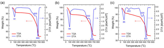
Figure 1.
Thermogravimetric analysis (TGA) and differential thermal analysis (DTA) curves for the Ba1.3Ca0.7SiO4 samples after burning at 550 °C, at a heating rate of 10 °C/min under a N2 flow of 80 cm3/min: (a) metal nitrate:urea = 1:0.75, (b) metal nitrate:urea = 1:1.15, and (c) metal nitrate:urea = 1:2.15.
Figure 2 shows the Fourier transform infrared (FT-IR) spectra for the samples after burning at 550 °C; Figure 2a–c correspond to metal nitrate:urea = 1:0.75, 1:1.15, and 1:2.15 samples, respectively. As shown in Figure 2, O-H stretching (from adsorbed water) was observed at 3648 cm−1, whereas N-H stretching and N-O stretching were displayed at 2207 cm−1 and 1434 cm−1, respectively. Furthermore, Si-O stretching, Si-O-Si bending, Ba-O stretching, and Ca-O stretching were exhibited at 970 cm−1, 835 cm−1, 725 cm−1, and 673 cm−1, respectively. Interestingly, when the urea concentration was relatively high (see Figure 2c), small peaks were additionally observed, displaying the presence of trivial decomposed molecules due to the rapid and intensive exothermic reactions in this sample. Recall also the additional DTA peak at 203 °C in Figure 1c.
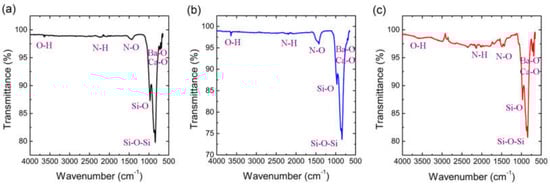
Figure 2.
FTIR spectra of Ba1.3Ca0.7SiO4 samples burned at 550 °C: (a) metal nitrate:urea = 1:0.75, (b) metal nitrate:urea = 1:1.15, and (c) metal nitrate:urea = 1:2.15.
As shown in Figure 1 and Figure 2, the combustion reaction at 550 °C was insufficient for a complete reaction, indicating further annealing was needed, at higher than 700 °C. Hence, we chose the post-annealing temperature of 750 °C for 2 h. We then characterized the X-ray diffraction (XRD) patterns of the Ba1.3Ca0.7SiO4 samples as a function of the molar ratio of metal nitrate:urea (see Figure 3). The first observation was that all the diffraction peaks were consistent with that of τ-phase hexagonal Ba1.3Ca0.7SiO4 (JCPDS, No. 36–1449) [39]. Here, the crystallite size (t) of Ba1.3Ca0.7SiO4 samples could be estimated through the Scherrer equation,
where is the wavelength of x-ray, and is the full-width at half maximum (FWHM) at the diffraction angle, . The results are summarized in Table 1 and Figure 4. As shown in Figure 4, the crystallite size initially decreased with increasing urea concentration up to nitrate:urea = 1:1.15, and then leveled off. However, a similar crystallite size (~31.2–31.0 nm) does not necessarily mean an indistinguishable property, which demanded further characterization of the samples. Furthermore, the experimental XRD data were shifted to the left compared with the JCPDS #:36–1499 prediction. For example, the d-spacing at the (102) crystallographic plane was 0.290 nm (experiment at 2 θ = 30.8°) and 0.287 nm (JCPDS at 2 θ = 31.1°), respectively, based on Bragg’s law . Hence, this comparison indicated that the interplanar distance should have been partially expanded due to drastic exothermic reactions when the solution combustion synthesis was employed.
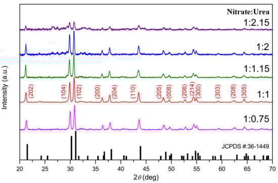
Figure 3.
XRD spectra of τ-phase Ba1.3Ca0.7SiO4 powder samples as a function of the molar ratio of metal nitrate:urea (after post-annealing at 750 °C for 2 h).

Table 1.
Crystallite size (t) of τ-phase Ba1.3Ca0.7SiO4 silicate powders at θ = 15.42 °, i.e., (101) crystallographic plane. Here, β denotes a full width at half maximum (FWHM).
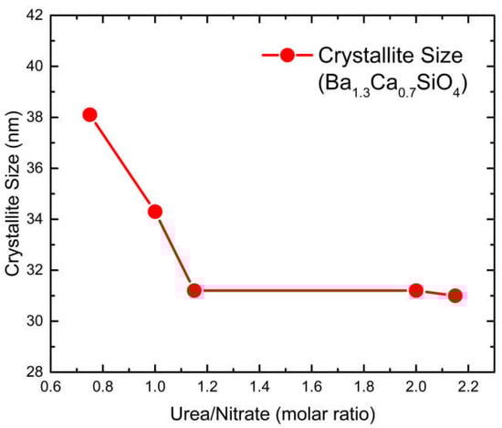
Figure 4.
Crystallite size of τ-phase Ba1.3Ca0.7SiO4 powder samples as a function of the molar ratio of metal nitrate:urea.
Figure 5 shows (a) the excitation and (b) emission spectra of the nanocrystalline Ba1.3Ca0.7SiO4 samples as a function of the metal nitrate:urea molar ratio (after post-annealing at 750 °C for 2 h). First, note that the τ-phase Ba1.3Ca0.7SiO4 samples had an optical bandgap of ca. 3.2 eV, corresponding to ~390 nm [41]. Hence, the excitation peaks at 316 nm, 340 nm, and 393 nm in Figure 5a were related to the electronic structure of Ba1.3Ca0.7SiO4. However, the emission lights at 487 nm (~2.6 eV) and 537 nm (~2.3 eV) had smaller energies than the bandgap (~3.2 eV) of the silicate samples did, suggesting that the emissions did not originate from a band-to-band transition, but from a band-to-trap (or trap-to-trap or trap-to-band) transition. Here, hole-/electron-traps indicated a defect site in the silicate samples.
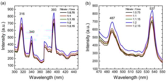
Figure 5.
Photoluminescence properties of (a) excitation and (b) emission of τ-phase Ba1.3Ca0.7SiO4 powder samples.
Therefore, based on the aforementioned photoluminescence (PL) results, a PL mechanism was suggested, as shown in Figure 6. When the wide bandgap Ba1.3Ca0.7SiO4 silicate was excited through photon absorption, the electrons in the conduction band (CB) could recombine with holes in the hole trap site for light emission. Similarly, the electron trap-to-valence band (VB) and trap-to-trap transition could be possible reasons for PL emission.
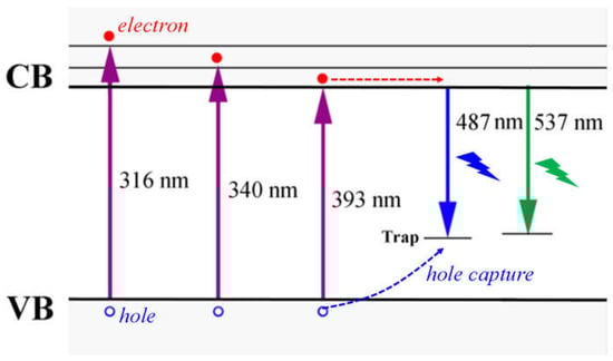
Figure 6.
Photoluminescence (PL) process in τ-phase Ba1.3Ca0.7SiO4 powder samples, suggesting the recombination should be related with the trap sites. Note that here, only hole traps were drawn for simplicity.
Figure 7 shows the scanning electron microscopy (SEM) images for τ-phase Ba1.3Ca0.7SiO4 samples when the molar ratios of metal nitrate:urea were (a) 1:0.75, (b) 1:1.15, and (c) 1:2.15. As shown in Figure 7, when the urea amount was low, the microstructural morphology appeared rod-like. However, when the urea amount was medium, the morphology appeared plate-like. Finally, when the urea amount was high, the morphology changed drastically, exhibiting inter-connected granular particles with some pores (due to a rapid escape of volatile gas molecules). Note that the average crystallite sizes of (b) and (c) were similar (i.e., (b) 31.2 nm and (c) 31.0 nm, respectively, as shown in Figure 3), because the crystallite size was partially polycrystals. This indicated that although the microscale particle size was different, the small crystallite domains could be similar, as in (b) and (c) (recall Figure 4 and Table 1). Finally, Figure 8 displays both the energy-dispersive X-ray spectroscopy (EDX) spectra (a, b, and c) and elemental mapping (d, e, and f) for the τ-phase Ba1.3Ca0.7SiO4 samples, confirming that the silicate samples were composed of Ba, Ca, Si, and O atoms. However, the compound was synthesized via a small-scale explosive reaction (i.e., solution combustion synthesis). Hence, the versatile surface defects may have affected some of the EDX results; however, a trace of aluminum was also observed as an impurity due to the alumina crucible used for the combustion synthesis process.
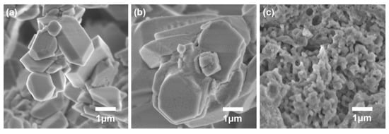
Figure 7.
The SEM images for τ-phase Ba1.3Ca0.7SiO4 samples when the molar ratios of metal nitrate:urea were (a) 1:0.75, (b) 1:1.15, and (c) 1:2.15.
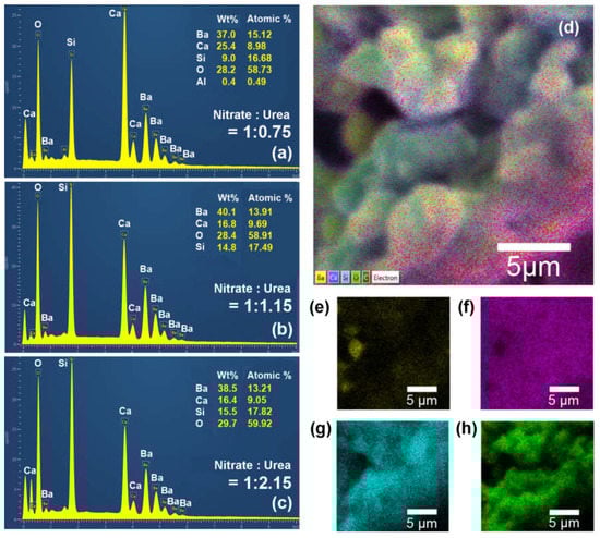
Figure 8.
The EDX spectra for τ-phase Ba1.3Ca0.7SiO4 powder samples when the molar ratios of metal nitrate:urea were (a) 1:0.75, (b) 1:1.15, and (c) 1:2.15. The EDX elemental mapping of τ-phase Ba1.3Ca0.7SiO4: (d) a layered sample, (e) barium, (f) calcium, (g) silicon, and (h) oxygen.
3. Materials and Methods
The τ-phase Ba1.3Ca0.7SiO4 silicate powders were synthesized as a function of urea concentration using a solution/gel combustion method. The precursor materials were Ba(NO3)2 (99.9%; molar mass = 261.35 g/mol), Ca(NO3)2·4H2O (99.9%; molar mass = 236.15 g/mol), Si(OC2H5)4 (99.99%; molar mass = 208.33 g/mol), and CH4N2O (99.9%; molar mass = 60.06 g/mol), which were obtained from Sigma-Aldrich and used as received without further purification. All precursors with the molar ratios of metal nitrate:urea in the range of 1:0.75–1:2.15 were mixed in 10 mL deionized water. Specifically, the stoichiometric amounts of 1.000 g of Ba(NO3)2, 0.487 g of Ca(NO3)2·4H2O, 0.614 g of Si(OC2H5)4, and 0.265 g (to 0.766 g) of CH4N2O were dissolved into water and continuously stirred for 30 min. Next, the solution was heated at 80 °C until a transparent solution formed. Finally, the precursor mixture was transferred to an alumina crucible and quickly inserted into a pre-heated 8 L muffle furnace heated at 550 °C for ~5–10 min. The mixture then self-ignited with a white flame, leading to a highly porous foamy solid product. Finally, the powder samples were post-annealed at 750 °C for 2 h, resulting in τ-phase Ba1.3Ca0.7SiO4 alkaline earth silicate powders.
Thermogravimetric analysis (TGA) and differential thermal analysis (DTA) were carried out using a TGA/DTA thermal analyzer (NETZSCH STA 449 C, Selb, Germany) with N2 flow at the heating rate of 10 °C/min. The functional groups of materials were analyzed using a Fourier transform infrared (FT-IR) spectrophotometer (IRAffinity-1S, Shimadzu). X-ray diffraction (XRD) patterns for the samples were identified using a D8 advanced Bruker diffractometer with a detector (12 mm × 16 mm) suitable for CuKα (λ = 1.54060 Å) irradiation, operating at an applied voltage of 45 kV with a current intensity of 40 mA, during which XRD data were collected in the range of 2 θ = 20–70°. Note that the accuracy and reproducibility of XRD are 0.005° and 0.0002°, respectively. The microstructural morphologies of the powder samples were analyzed using a PHI700 nanoprobe and Shimadzu model ZU SSX–550 super scanning electron microscopy (SEM) coupled with energy-dispersive X-ray spectroscopy (EDX) (Oxford x-Max N). EDX was used to determine the elemental analysis and chemical composition of the samples. The photoluminescence (PL) spectra of synthesized samples were recorded on a Hitachi F-7000 phosphorescence spectrometer. Note that all the characterizations were carried out at ~23 °C.
4. Conclusions
The Ba1.3Ca0.7SiO4 semiconductors were prepared through combustion synthesis, for which urea was employed as a fuel with the molar ratios of metal nitrate:urea = 1:0.75, 1:1.15, and 1:2.15. First, the TGA results indicated that when the urea ratio increased from 1:0.75 to 1:1.15 and 1:2.15, the main decomposition temperature decreased from 669 °C to 648 °C and 631 °C, respectively, indicating urea’s role as a fuel during the exothermic reactions. Second, the FT-IR spectra demonstrated the presence of the main functional groups, such as Si-O, N-H, Ba-O, and Ca-O. Third, the XRD patterns indicated that the silicate Ba1.3Ca0.7SiO4 had a τ-phase hexagonal structure with an average crystallite size of ~33 ± 3 nm. When the molar ratio of metal nitrate:urea was greater than 1:1.15, the crystallite size levelled off at ~31 nm. Fourth, the PL spectra showed that the blue/green light emission was processed through trap sites because the recombination energy (~2.3–2.6 eV) was smaller than the bandgap (~3.2 eV) of silicate samples. Fifth, according to SEM results, the aggregated particle size decreased with increasing urea concentration. Sixth, EDX spectra confirmed the elemental components (Ba, Ca, Si, and O) of the Ba1.3Ca0.7SiO4 silicate powders. Finally, it was proven that Ba1.3Ca0.7SiO4 silicate powders could be synthesized at a post-annealing temperature of 750 °C after burning the samples at 550 °C.
Author Contributions
Conceptualization, F.B.D. and J.Y.K.; methodology, D.R.G., F.B.D., M.K.H. and J.Y.K.; formal analysis, D.R.G., F.B.D., M.K.H. and J.Y.K.; investigation, D.R.G.; resources, F.B.D.; data curation, D.R.G. and M.K.H.; writing—original draft preparation, D.R.G.; writing—review and editing, J.Y.K.; supervision, F.B.D. and J.Y.K.; funding acquisition, D.R.G. and F.B.D. All authors have read and agreed to the published version of the manuscript.
Funding
This research received no external funding. The APC was funded by JYK.
Data Availability Statement
The datasets used and/or analyzed during the current study are available from the corresponding author upon reasonable request.
Acknowledgments
We acknowledge financial support from the Ministry of Education in Ethiopia and the National Research Foundation of South Africa.
Conflicts of Interest
The authors declare no competing interests.
References
- Mondal, K.; Kumari, P.; Manam, J. Influence of doping and annealing temperature on the structural and optical properties of Mg2SiO4: Eu3+ synthesized by combustion method. Curr. Appl. Phys. 2016, 16, 707–719. [Google Scholar] [CrossRef]
- Naik, R.; Prashantha, S.; Nagabhushana, H.; Sharma, S.; Nagabhushana, B.; Nagaswarupa, H.; Premkumar, H. Low temperature synthesis and photoluminescence properties of red emitting Mg2SiO4: Eu3+ nanophosphor for near UV light emitting diodes. Sens. Actuators B Chem. 2014, 195, 140–149. [Google Scholar] [CrossRef]
- Hiramatsu, H.; Yusa, H.; Igarashi, R.; Ohishi, Y.; Kamiya, T.; Hosono, H. An exceptionally narrow band-gap (∼4 eV) silicate predicted in the cubic perovskite structure: BaSiO3. Inorg. Chem. 2017, 56, 10535–10542. [Google Scholar] [CrossRef] [PubMed]
- ALOthman, Z.A. A Review: Fundamental Aspects of Silicate Mesoporous Materials. Materials 2012, 5, 2874–2902. [Google Scholar] [CrossRef]
- Wu, C.; Chang, J.; Ni, S.; Wang, J. In vitro bioactivity of akermanite ceramics. J. Biomed. Mater. Res. Part A 2006, 76, 73–80. [Google Scholar] [CrossRef]
- Alvani, A.S.; Moztarzadeh, F.; Sarabi, A. Effects of dopant concentrations on phosphorescence properties of Eu/Dy-doped Sr3MgSi2O8. J. Lumin. 2005, 114, 131–136. [Google Scholar] [CrossRef]
- Pan, W.; Ning, G.-L.; Wang, J.-H.; Lin, Y. A novel synthesis of alkaline earth silicate phosphor Sr3MgSi2O8: Eu2+,Dy3+. Chin. J. Chem. 2007, 25, 605–608. [Google Scholar] [CrossRef]
- Pritts, I.M.; Daugherty, K.E. The effect of stabilizing agents on the hydration rate of β-C2S. Cem. Concr. Res. 1976, 6, 783–795. [Google Scholar] [CrossRef]
- Nettleship, I.; Shull, J.L.; Kriven, W.M. Chemical preparation and phase stability of Ca2SiO4 and Sr2SiO4 powders. J. Eur. Ceram. Soc. 1993, 11, 291–298. [Google Scholar] [CrossRef]
- Singh, D.; Sheoran, S.; Bhagwan, S.; Kadyan, S. Optical characteristics of sol-gel derived M3SiO5: Eu3+ (M= Sr, Ca and Mg) nanophosphors for display device technology. Cogent Phys. 2016, 3, 1262573. [Google Scholar] [CrossRef]
- Varma, A.; Mukasyan, A.S.; Rogachev, A.S.; Manukyan, K.V. Solution combustion synthesis of nanoscale materials. Chem. Rev. 2016, 116, 14493–14586. [Google Scholar] [CrossRef]
- Patil, K.C.; Aruna, S.; Mimani, T. Combustion synthesis: An update. Curr. Opin. Solid State Mater. Sci. 2002, 6, 507–512. [Google Scholar] [CrossRef]
- Wen, W.; Wu, J.-M. Nanomaterials via solution combustion synthesis: A step nearer to controllability. RSC Adv. 2014, 4, 58090–58100. [Google Scholar] [CrossRef]
- Siddique, F.; Gonzalez-Cortes, S.; Mirzaei, A.; Xiao, T.; Rafiq, M.A.; Zhang, X. Solution combustion synthesis: The relevant metrics for producing advanced and nanostructured photocatalysts. Nanoscale 2022, 14, 11806–11868. [Google Scholar] [CrossRef]
- Deganello, F.; Tyagi, A.K. Solution combustion synthesis, energy and environment: Best parameters for better materials. Prog. Cryst. Growth Charact. Mater. 2018, 64, 23–61. [Google Scholar] [CrossRef]
- Novitskaya, E.; Kelly, J.P.; Bhaduri, S.; Graeve, O.A. A review of solution combustion synthesis: An analysis of parameters controlling powder characteristics. Int. Mater. Rev. 2020, 66, 188–214. [Google Scholar] [CrossRef]
- Aruna, S.T.; Mukasyan, A.S. Combustion synthesis and nanomaterials. Curr. Opin. Solid State Mater. Sci. 2008, 12, 44–50. [Google Scholar] [CrossRef]
- Kingsley, J.J.; Manickam, N.; Patil, K.C. Combustion synthesis and properties of fine particle fluorescent aluminous oxides. Bull. Mater. Sci. 1990, 13, 179–189. [Google Scholar] [CrossRef]
- Kingsley, J.; Patil, K. A novel combustion process for the synthesis of fine particle α-alumina and related oxide materials. Mater. Lett. 1988, 6, 427–432. [Google Scholar] [CrossRef]
- Qiu, Z.; Zhou, Y.; Lü, M.; Zhang, A.; Ma, Q. Combustion synthesis of long-persistent luminescent MAl2O4: Eu2+, R3+ (M = Sr, Ba, Ca, R = Dy, Nd and La) nanoparticles and luminescence mechanism research. Acta Mater. 2007, 55, 2615–2620. [Google Scholar] [CrossRef]
- Raza, M.A.; Rahman, I.Z.; Beloshapkin, S. Synthesis of nanoparticles of La0.75Sr0.25Cr0.5Mn0.5O3−δ (LSCM) perovskite by solution combustion method for solid oxide fuel cell application. J. Alloys Compd. 2009, 485, 593–597. [Google Scholar] [CrossRef]
- Bhatkar, V.B.; Bhatkar, N.V. Combustion synthesis and photoluminescence study of silicate biomaterials. Bull. Mater. Sci. 2011, 34, 1281–1284. [Google Scholar] [CrossRef]
- Talwar, G.; Joshi, C.; Moharil, S.; Dhopte, S.; Muthal, P.; Kondawar, V. Combustion synthesis of Sr3MgSi2O8: Eu2+ and Sr2MgSi2O7: Eu2+ phosphors. J. Lumin. 2009, 129, 1239–1241. [Google Scholar] [CrossRef]
- Thoda, O.; Xanthopoulou, G.; Vekinis, G.; Chroneos, A. Review of recent studies on solution combustion synthesis of nanostructured catalysts. Adv. Eng. Mater. 2018, 20, 1800047. [Google Scholar] [CrossRef]
- Carlos, E.; Martins, R.; Fortunato, E.; Branquinho, R. Solution combustion synthesis: Towards a sustainable approach for metal oxides. Chem. Eur. J. 2020, 26, 9099–9125. [Google Scholar] [CrossRef]
- Branquinho, R.; Santa, A.; Carlos, E.; Salgueiro, D.; Barquinha, P.; Martins, R.; Fortunato, E. Developments in Combustion Technology; Kyprianidis, K.G., Skvaril, J., Eds.; InTechOpen: Rijeka, Croatia, 2016; pp. 397–417. [Google Scholar] [CrossRef]
- Kelkar, S.A.; Shaikh, P.A.; Pachfule, P.; Ogale, S.B. Nanostructured Cd2SnO4 as an energy harvesting photoanode for solar water splitting. Energy Environ. Sci. 2012, 5, 5681–5685. [Google Scholar] [CrossRef]
- Wen, W.; Wu, J.-M.; Tu, J.-P. A novel solution combustion synthesis of cobalt oxide nanoparticles as negative-electrode ma-terials for lithium ion batteries. J. Alloys Compd. 2012, 513, 592–596. [Google Scholar] [CrossRef]
- Ekambaram, S.; Patil, K.C. Synthesis and properties of rare earth doped lamp phosphors. Bull. Mater. Sci. 1995, 18, 921–930. [Google Scholar] [CrossRef]
- Amosov, A.P.; Novikov, V.A.; Kachkin, E.M.; Kryukov, N.A.; Titov, A.A.; Sosnin, I.M.; Merson, D.L. The Solution Combustion Synthesis of ZnO Powder for the Photodegradation of Phenol. Ceramics 2022, 5, 928–946. [Google Scholar] [CrossRef]
- Mokkelbost, T.; Kaus, I.; Grande, T.; Einarsrud, M.-A. Combustion synthesis and characterization of nanocrystalline CeO2-based powders. Chem. Mater. 2004, 16, 5489–5494. [Google Scholar] [CrossRef]
- Jiang, Y.; Yang, S.; Hua, Z.; Huang, H. Sol-gel auto combustion synthesis of metals and metal alloys. Angew. Chem. Int. Ed. 2009, 48, 8529–8531. [Google Scholar] [CrossRef]
- Bera, P.; Hegde, M. Characterization and catalytic properties of combustion synthesized Au/CeO2 catalyst. Catal. Lett. 2002, 79, 75–81. [Google Scholar] [CrossRef]
- Ianoş, R.; Tăculescu, A.; Păcurariu, C.; Lazău, I. Solution combustion synthesis and characterization of magnetite, Fe3O4, Nanopowders. J. Am. Ceram. Soc. 2012, 95, 2236–2240. [Google Scholar] [CrossRef]
- Voskanyan, A.A.; Chan, K. Solution combustion synthesis using furfuryl alcohol as fuel and a combustible solvent. J. Exp. Nanosci. 2015, 10, 466–475. [Google Scholar] [CrossRef]
- Valefi, M.; Falamaki, C.; Ebadzadeh, T.; Hashjin, M.S. New insights of the glycine-nitrate process for the synthesis of nano-crystalline 8YSZ. J. Am. Ceram. Soc. 2007, 90, 2008–2014. [Google Scholar] [CrossRef]
- Thompson, J.G.; Withers, R.L.; Hyde, B.G. Further consideration of phases in the system Ba2SiO4-Ca2SiO4. J. Am. Ceram. Soc. 1987, 70, C-383–C-386. [Google Scholar] [CrossRef]
- Ianoş, R.; Muntean, E.; Lazău, R.; Băbuță, R.; Moacă, E.-A.; Păcurariu, C.; Dabici, A.; Hulka, I. One-step synthesis of near-infrared reflective brown pigments based on iron-doped lanthanum aluminate, LaAl1-xFexO3. Dye. Pigment. 2018, 152, 105–111. [Google Scholar] [CrossRef]
- Matkovic, B.; Popović, S.; Grzeta, B.; Halle, R. Phases in the system Ba2SiO4-Ca2SiO4. J. Am. Ceram. Soc. 1986, 69, 132–134. [Google Scholar] [CrossRef]
- Yim, S.D.; Kim, S.J.; Baik, A.J.H.; Nam, I.-S.; Mok, Y.S.; Lee, J.-H.; Cho, B.K.; Oh, S.H. Decomposition of urea into NH3 for the SCR process. Ind. Eng. Chem. Res. 2004, 43, 4856–4863. [Google Scholar] [CrossRef]
- Wang, D.; Dong, N.; Hui, S.; Niu, Y. Analysis of urea pyrolysis products in 132.5–190 °C. Energy Procedia 2019, 158, 2170–2175. [Google Scholar] [CrossRef]
- Golja, D.R.; Dejene, F.B.; Kim, J.Y. Trivalent rare-earth-codoped silicate phosphor materials (Ba1.3Ca0.7−x−y SiO4: xDy3+/yEu3+) for solid-state lighting. J. Phys. Condens. Matter 2022, 34, 294006. [Google Scholar] [CrossRef] [PubMed]
- Golja, D.R.; Dejene, F.B.; Kim, J.Y. Single-phase silicate phosphors (Ba1.3Ca0.7−xSiO4:xDy3+) doped with dysprosium for white solid-state lighting. Adv. Condens. Matter Phys. 2022, 2022, 4317275. [Google Scholar] [CrossRef]
- Golja, D.R.; Woldemariam, M.M.; Dejene, F.B.; Kim, J.Y. Photoluminescence processes in τ-phase Ba1.3Ca0.7-x-ySiO4:xDy3+/yTb3+ phosphors for solid-state lighting. R. Soc. Open Sci. 2022, 9, 220101. [Google Scholar] [CrossRef] [PubMed]
- Mukasyan, A.S.; Costello, C.; Sherlock, K.P.; Lafarga, D.; Varma, A. Perovskite Membranes by Aqueous Combustion Synthesis: Synthesis and Properties. Sep. Purif. Technol. 2001, 25, 117–126. [Google Scholar] [CrossRef]
- Wynne, A.M. The thermal decomposition of urea: An undergraduate thermal analysis experiment. J. Chem. Educ. 1987, 64, 180–182. [Google Scholar] [CrossRef]
- Fang, H.L.; DaCosta, H.F. Urea thermolysis and NOx reduction with and without SCR catalysts. Appl. Catal. B Environ. 2003, 46, 17–34. [Google Scholar] [CrossRef]
Disclaimer/Publisher’s Note: The statements, opinions and data contained in all publications are solely those of the individual author(s) and contributor(s) and not of MDPI and/or the editor(s). MDPI and/or the editor(s) disclaim responsibility for any injury to people or property resulting from any ideas, methods, instructions or products referred to in the content. |
© 2023 by the authors. Licensee MDPI, Basel, Switzerland. This article is an open access article distributed under the terms and conditions of the Creative Commons Attribution (CC BY) license (https://creativecommons.org/licenses/by/4.0/).