Article

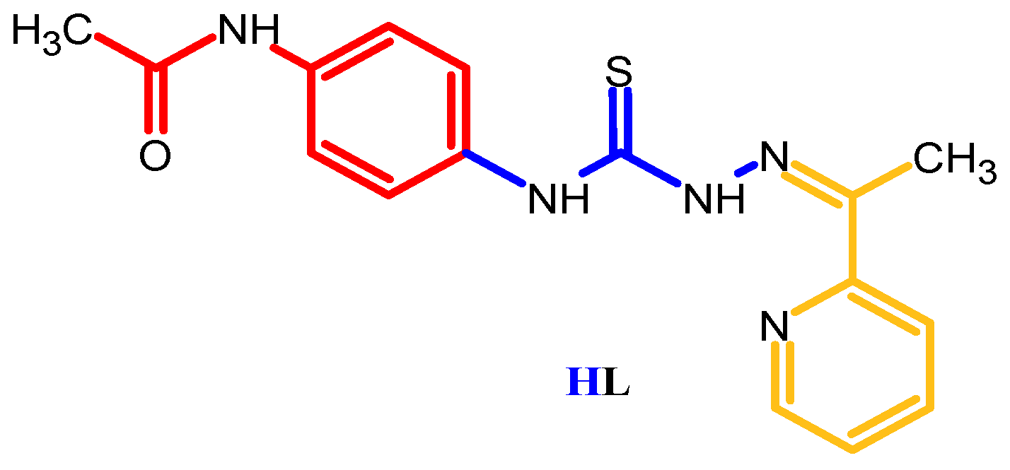
Synthesis, Characterization, and Biological Properties of the Copper(II) Complexes with Novel Ligand: N-[4-({2-[1-(pyridin-2-yl)ethylidene]hydrazinecarbothioyl}amino)phenyl]acetamide

1
Laboratory of Advanced Materials in Biopharmaceutics and Technics, Moldova State University, 60 Mateevici Street, MD-2009 Chisinau, Moldova
2
Biological Invasions Research Center, Institute of Zoology, Moldova State University, 1 Academiei Street, MD-2028 Chisinau, Moldova
3
Laboratory of Physical Methods of Solid State Investigation “Tadeusz Malinowski”, Institute of Applied Physics, MD-2028 Chisinau, Moldova
4
Laboratoire de Chimie de Coordination UPR 8241, Centre National de la Recherche Scientifique, 31400 Toulouse, France
5
Department of Dentistry, University of Medicine and Pharmacy “Nicolae Testemitanu”, 165 Stefan cel Mare si Sfant Bd., MD-2004 Chisinau, Moldova
*
Authors to whom correspondence should be addressed.
Inorganics 2023, 11(10), 408; https://doi.org/10.3390/inorganics11100408
Submission received: 19 September 2023 / Revised: 4 October 2023 / Accepted: 9 October 2023 / Published: 16 October 2023
(This article belongs to the Special Issue 10th Anniversary of Inorganics: Bioinorganic Chemistry)

Abstract

:
For the first time, a thiosemicarbazone-type ligand containing a paracetamol structural unit was synthesized. Five new coordination compounds based on copper(II) salts: [Cu(L)CH3COO] (1), [{Cu(L)Cl}2]·H2O (2), [Cu(L)H2O·DMF]NO3 (3), [Cu(L)Br] (4), [Cu(L)H2O]ClO4 (5), were obtained, where HL is N-[4-({2-[1-(pyridin-2-yl)ethylidene]hydrazinecarbothioyl}amino)phenyl]acetamide. The new HL was characterized by NMR, FTIR, spectroscopy, and X-ray crystallography. All copper(II) coordination compounds were characterized by elemental analysis, FTIR, EPR spectroscopy, and molar electrical conductivity. Furthermore, single crystal X-ray diffraction analysis elucidated the structures of thiosemicarbazone HL as well as complexes 1–3. All compounds were tested for antimicrobial, antifungal, and antioxidant activities, and their toxicity to Daphnia magna was studied. Biological evaluation has revealed that most of the synthesized compounds demonstrate promising antibacterial, antifungal, and antioxidant activities. In many cases, their antibacterial/antifungal activity is comparable to that of certain drugs used in medicine for these purposes, and in some cases, even surpasses them. HL and complexes 2–5 exhibit antioxidant activity that surpasses that of Trolox. Furthermore, HL and complex 2 display virtually no toxicity to D. magna.

1. Introduction

The study of new materials in the field of coordination chemistry is increasing day by day, leading to the discovery of substances with advanced biological properties compared to the drugs currently used in medicinal practice. A class of organic compounds called thiosemicarbazones is expected to be the most promising due to a wide range of biological activities, including antituberculosis [1,2,3,4], antineoplastic activity [2], anticancer [5], antioxidant [6], antiviral [7], antimicrobial [8,9], antifungal [1,10,11,12], anticonvulsant [13,14,15], antiproliferative activity [16], anticancer activity [17]. Interest in this family of compounds has grown significantly over the years, from the first report in 1940 to the thousands of papers published by 2022.
Coordination compounds based on 3d metal ions have d orbitals partially coupled with electrons and exhibit different variable oxidation states, playing an important role in redox processes. They hold promise in the development of pharmaceutical agents [18].
The identification of potential new antibactericidal/antifungicidal drugs with increased efficiency based on coordinative combinations is in vogue, their stake would be low side effects; and overcoming the resistance achieved by current drugs [19,20,21,22,23,24].
Thiosemicarbazones have attracted the attention of researchers [18,25] for four key aspects in the field of coordination chemistry: high coordination tendency; formation of coordinative complexes with increased stability; high selectivity capacity; and ability to form macrocycles.
Thiosemicarbazones possess a dual aspect based on biological [26,27] and computational principles [28]. From a biological standpoint, thiosemicarbazones exhibit various pharmacological properties. From a theoretical aspect, they are ideal targets for computational studies due to their donor-acceptor capabilities in the development of therapeutic agents [29].
One important biological property of thiosemicarbazones is their ability to inhibit ribonucleotide reductase (RR) synthesis [30]. Substitution in the para position is a decisive factor in the antifungal potential of 2-acetylpyridine thiosemicarbazones [31].
The strategy of choosing the thiosemicarbazone skeleton was based on two positions, namely: position one—the thiosemicarbazone fragment must contain a fragment that is easily metabolized by the human body and does not show toxic effects. The second position is that the part responsible for the selective biological effect (pyridin-2-yl) is included in the thiosemicarbazone phial casing. Thus, the hypothesis from those elucidated could be reproduced in the figure below (Figure 1):
The aim of the present investigation is the synthesis, characterization, and study of antibacterial, antifungal, and antioxidant activities of Paracetamol (4-Aminoacetanilide) with a thiosemicarbazone fragment (HL) and Cu(II) coordination compounds with HL: [Cu(L)CH3COO] (1), [{Cu(L)Cl}2]·H2O (2), [Cu(L)H2O·DMF]NO3 (3), [Cu(L)Br] (4), [Cu(L)H2O]ClO4 (5).

2. Results and Discussion

In this work, a new thiosemicarbazone based on 4-aminoacetanilide was synthesized, functionalized according to the organic synthesis procedure: synthesis of the isothiocyanate group and hydrazone following the nucleophilic addition reaction with the formation of the HL ligand (Figure 2).
The thiosemicarbazone HL was characterized by FT-IR, 1H NMR, and 13C NMR spectroscopy. Its structure was determined using X-ray diffraction analysis. Complexes 1–5 were synthesized by the interaction of ethanolic solution of N-[4-({2-[1-(pyridin-2-yl)ethylidene]hydrazinecarbothioyl}amino)phenyl]acetamide and copper(II) salts (1–5) in a 1:1 molar ratio. The composition of the thiosemicarbazone HL and complexes 1–5 was confirmed using elemental analysis data.
In the NMR spectra (Figures S1 and S2), two tautomeric forms (Figure 3) were determined in the case of thiosemicarbazone HL. The thiol tautomeric form is identified by the characteristic peak of the SH group at the chemical shift of 14.54 ppm in the hydrogen spectrum (1H-NMR) corresponding to data from the specialized literature [32,33]. The ionic tautomeric form of the carbon spectrum (13C-NMR) is found at the chemical shift 177.82 ppm, which is consistent with the literature data [16,34,35].

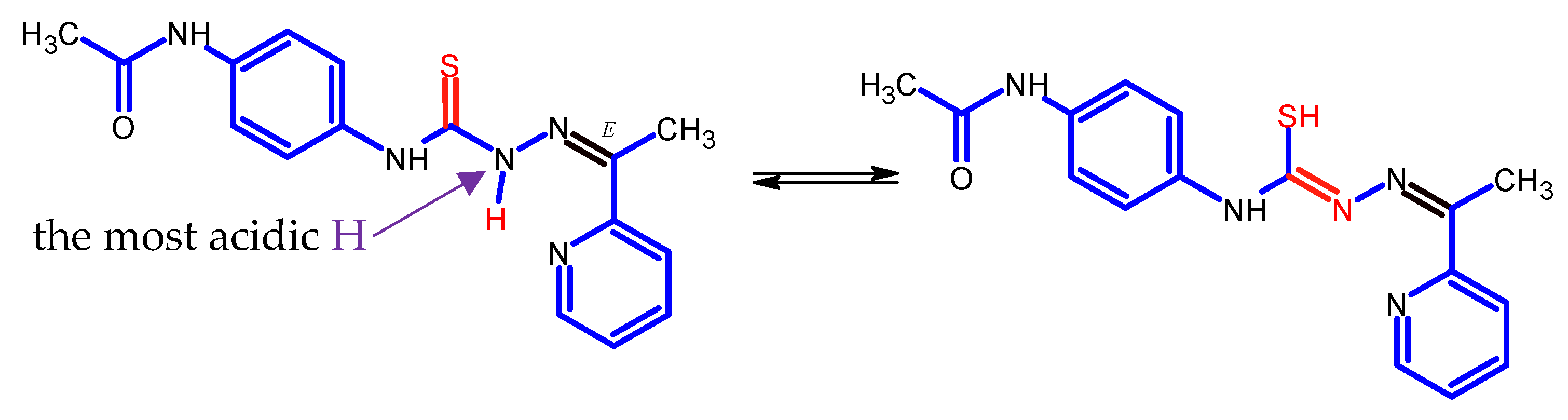
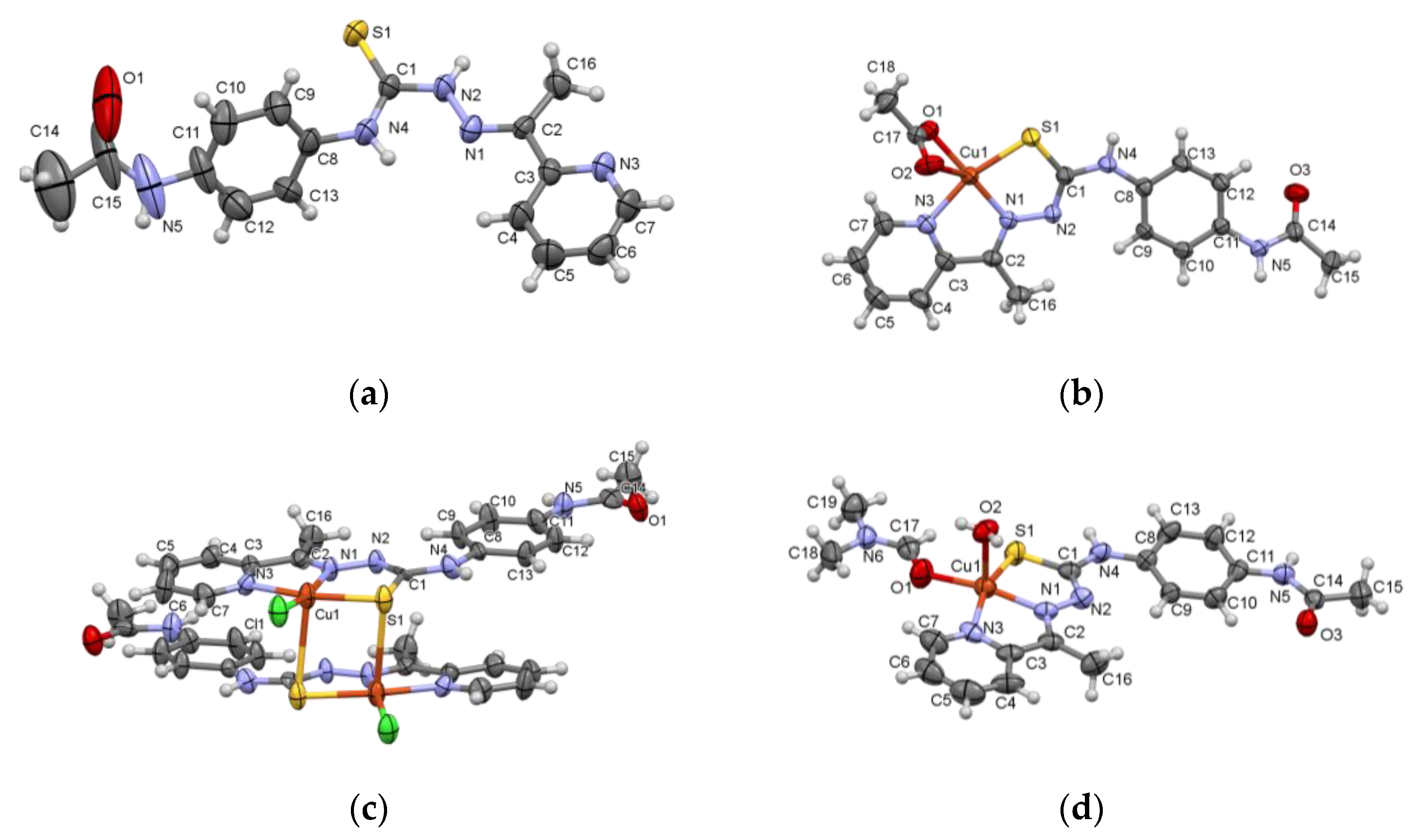
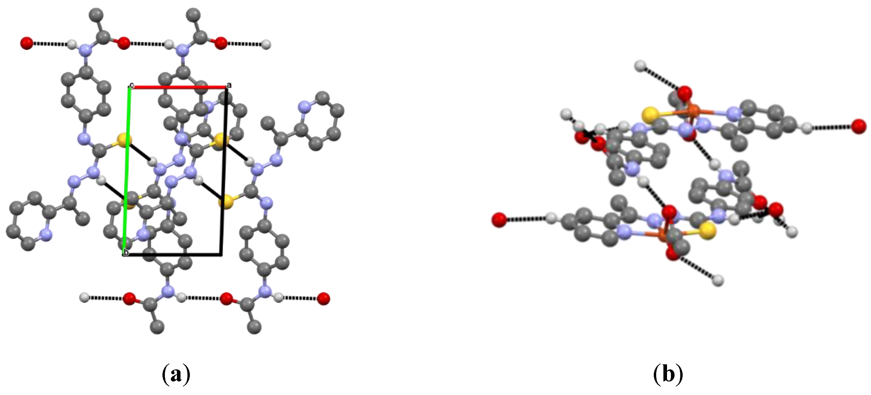
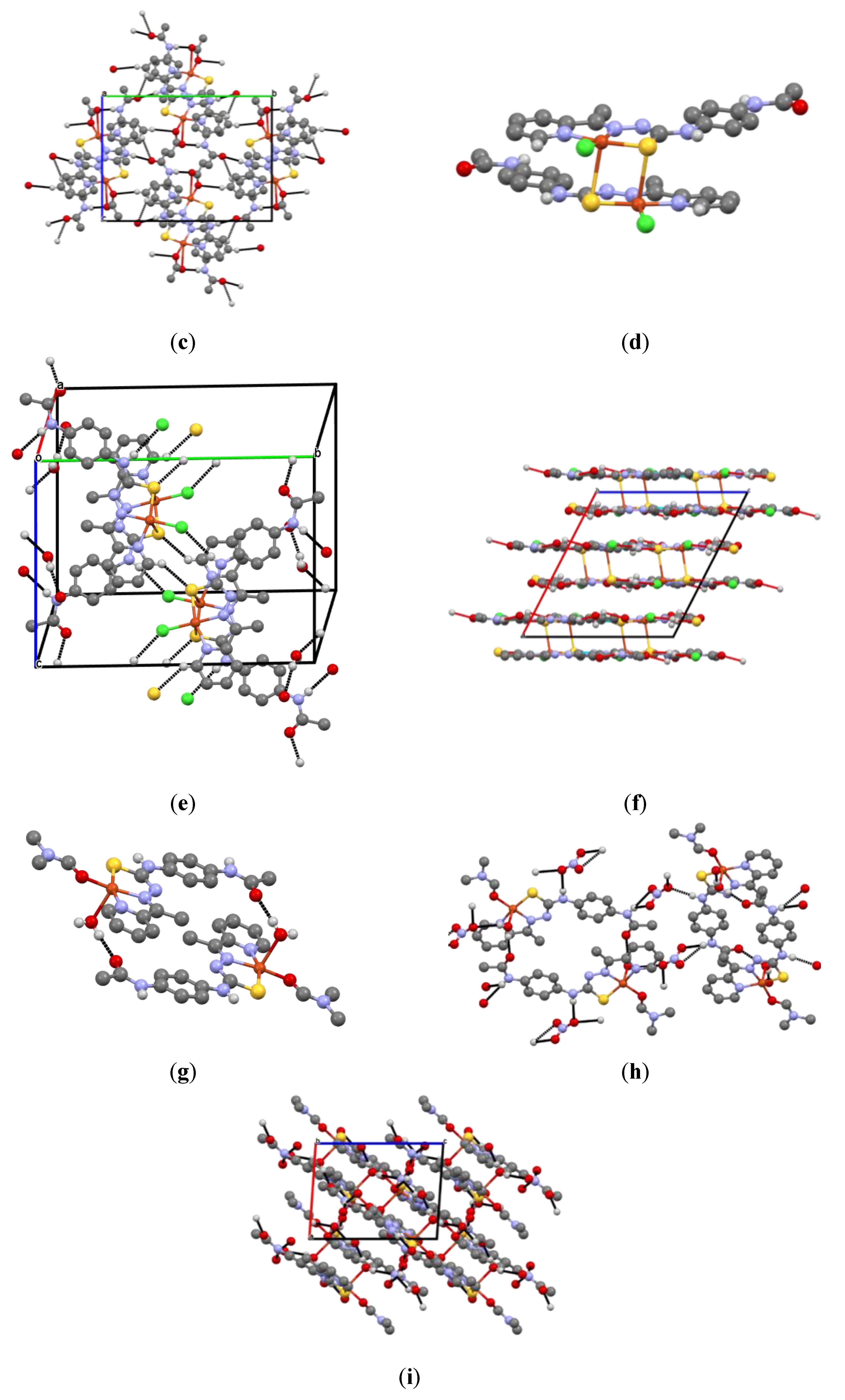
2.1. Structural Characterization of Ligand HL and Coordination Compounds 13

The X-ray structures of HL and coordination compounds 1–3 are presented in Figure 4 and Figure 5, and Table 1, Table 2 and Table 3. In the molecule of HL (Figure 4a) the substituents at the N(2)–C(1) bond are in the E position. The A(S(1)-N(1)-N(2)-N(4)-C(1)-C(2)) core is practically planar within 0.1 Å and the dihedral angle between the given core and pyridine ring is equal to 10.4°. However, the HL is nonplanar because the dihedral angle between the best plane of A and the benzene ring is 58.2°. In the crystal, the ligand HL forms the dimers via N(2)-H... S(1) hydrogen bonds (HB), which are liked by N(5)-H... O(2) HB into the layers along the a-axis (Table 3, Figure 5a).
In complexes 1–3, the ligand HL acts as a mononegative tridentate around the metallic ions through the SNN set of donor atoms. The studied complexes are five-coordinated in a distorted square–pyramidal coordination geometry. Deprotonation of N(2) atom in 1–3 has led to the decrease of N(2)-C(1) bond distance if compared with that in HL. The bond lengths S(1)-C(1) in these complexes are increased due to the coordination of sulfur atoms to central metals and the maximal changing of this bond is observed in 2 which is equal to 1.757(8) Å (Table 2). However, the composition of the coordination polyhedron of the central atom in these compounds is different. Its basal plane includes three donor atoms of the L, oxygen atoms O(1) of coordinated CH3COO and DMF molecules in 1, 3, and chlorine ion in 2 (Figure 4b–d, Table 2). The deviations of these atoms from their mean plane are within 0.1 Å, while the Cu atoms deviate from these planes by 0.01, 0.13, and 0.17 Å toward the apexes of the pyramids. The apexes of the metal’s coordination pyramids in 1, and 3 are occupied by oxygen atoms O(2) of CH3COO and water molecules with distances of 2.755 (3) and 2.338 (4) Å, respectively. In 2, the complexes form the centrosymmetric dimers, and the apex of the coordination pyramid is occupied by the sulfur atom of the adjacent complex with a distance of 2.982(3) Å (Figure 5d). In the crystal of 1, the complexes are joined by N5-H... O2 hydrogen bonds in centrosymmetric dimers, which are further linked by C5-H... O1, N4-H... O3, and C13-H... O3 HB, forming the 3D hydrogen bonding networks (Figure 5b,c). In 2, dimers linked by water molecules as well as N(4)-H... Cl(1) and C(7)-H... S1 HB form the layers along a- axis (Figure 5d–f). The water molecules in crystal 3 form the centrosymmetric dimers, which are joined by the NO3-group in the layers along the same direction (Figure 5g–i, Table 3). In both 2 and 3, van der Waals interaction occurs between the layers.
Molar conductivity was determined in an ethanol-water (3:1) solution in the range of 36–106 µS/cm with the ratio of electrolyte 1:1 or non-electrolyte being determined. Coordinative combinations with anions CH3COO, Cl, NO3, Br, ClO4 form the internal or external sphere, which upon solvolysis can be in equilibrium with the undissociated form. The activity of molar conductivity date in case coordination combinations 1, 2, and 4 are non-electrolytes. For compounds 3 and 5, the acidic residue is found in the outer sphere.
In the IR spectrum (Figure S3) of thiosemicarbazone HL, the presence of groups was determined to be: C=S (thionic), C=N(azomethenic), 1,4-disubstituted Aryl, N-H(hydrazinic). The coordination combinations show bands in the IR spectra characteristic of the coordination of nitrogen, sulfur, and pyridinic nitrogen donor atoms with the central copper(II) atom. In the IR spectrum of complex 1, the acetate ion is coordinated bidentate, which generates two bands with high intensity at 1603–1563 cm−1 characteristic νa(COO) and νs(COO). In the case of the nitrate ion from complex 3 in the IR spectrum, a band at 1309 and 1251 cm−1 characteristic of ionic form is presented (Figures S3–S8) [36].
The shift of the bands from higher to lower wave numbers in the case of the azomethine group from the free ligand 1611 cm−1 to 1584–1531 cm−1 in the case of coordination combinations 1–5 indicates the coordination at the central atom.
The EPR signature of complexes 1–5 were measured in DMSO at 200 µM (Figure S9), complexes 1 and 3–5 show similar signatures while that of 5 is the one which is the best resolved while an additional broad signal centered near g=2 contributes to the overall spectra of 1, 3 and 4. Two different fingerprints corresponding to two mononuclear Cu(II) species (called I and II) can be observed and distinguished by their EPR parameters. Species I has the following parameters: A// = 180 G, g// = 2.20, and species II has: A// = 170 G, g// = 2.15. The ratio between these two species depends on the complex under study. This may indicate that these four complexes evolve and form the same two mononuclear EPR-sensitive species in solution but in different proportions. Complex 2 exhibits a different fingerprint with an ill-resolved pattern in line with the binuclear species mainly kept in solution.

2.2. Biological Activity

2.2.1. Antimicrobial and Antifungal Activity

The literature indicates that thiosemicarbazones and their copper(II) complexes often demonstrate antimicrobial and antifungal properties [10]. Therefore, the antimicrobial and antifungal characteristics of the synthesized compounds were examined. The ligand HL and its complexes 1–5 were assessed against Gram-positive bacteria (Staphylococcus aureus ATCC 25923), Gram-negative bacteria (Escherichia coli ATCC 25922), and fungal strains (Candida albicans ATCC 10231). The MIC (minimum inhibitory concentration, μg mL−1), MBC (minimum bactericidal concentrations, μg mL−1), and MFC (minimum fungicidal concentrations, μg mL−1) values for the compounds against bacteria and fungi are shown in Table 4. Nitrofurazone [37] and miconazole [38] were used as standard drugs for comparing antimicrobial and antifungal activities, respectively.
The data obtained highlight that copper complexes exhibit the highest level of antimicrobial activity. Among the tested complexes, the most significant activity is observed against S. aureus, surpassing the activity of the proligand HL. In the case of complexes 2–5, their activity is even higher than that of nitrofurazone. While HL and complexes 1, 3, and 5 show no activity against E. coli, complexes 2 and 4 demonstrate moderate activity. Conversely, complexes 1, 3, and 5 do not display any activity against the fungal strain C. albicans. HL and complexes 4 exhibit moderate activity, whereas complex 2 demonstrates stronger activity than miconazole.

2.2.2. Antioxidant Activity

Free radicals play a significant role in various detrimental biological processes, including protein denaturation and lipid peroxidation, contributing to the development of numerous human diseases [39,40]. Hence, investigating the antioxidant potential of the synthesized compounds becomes crucial to determine whether they can mitigate the levels of free radicals and provide protection against oxidative stress in the human body. The antioxidant activity against the 2,2-azinobis-(3-ethylbenzothiazoline-6-sulphonate) radical cation (ABTS•+) was evaluated for the compounds under examination: HL and copper(II) complexes 1–5. The obtained results, represented as semi-maximal inhibitory concentrations (IC50), are presented in Table 5.
The ligand HL and complexes 2–5 exhibit remarkable antioxidant activity, surpassing the activity of Trolox, a standard antioxidant utilized in medical applications. The tested ligand HL demonstrates an activity that is four times higher than that of Trolox. Complexes showed antioxidant activity against ABTS•+ with IC50 of 10.1–47.4 µM. Among the copper(II) complexes, the antioxidant potency follows this sequence: [Cu(L)H2O]ClO4 ≥ [Cu(L)H2O·DMF]NO3 ≥ [{Cu(L)Cl}2]·H2O ≥ [Cu(L)Br] ≥ [Cu(L)CH3COO].

2.2.3. Acute In Vivo Toxicity of the Tested Compounds Assessed through Immobilization of the Crustacean Daphnia magna

To determine the toxicity of the tested compounds, the immobilization test on the crustacean Daphnia magna was conducted following a European Standardized Methodology. International organizations for animal protection recommend conducting in vivo toxicity research on Daphnia magna. In this context, as an alternative method used in this study, the complete replacement of animal toxicity testing with tests on invertebrate organisms was employed. This paper includes relevant information regarding the results of an experiment aimed at assessing the toxicity of the tested compounds through acute toxicity bioassays on an aquatic organism species from the arthropod subphylum, such as the crustaceans represented by D. magna. This organism is frequently used in laboratory experiments due to its structure, transparency, and ability to survive under a coverslip, making it easily observable under a microscope [40].
The test allowed for the evaluation of the acute toxicity of the tested compounds on D. magna at 24 h, expressed as the median lethal concentration (LC50), which was calculated using the dose–response relationship determined by the least squares fitting method with the assistance of GraphPad software. All data are presented as means ± standard deviation (SD). Thus, the LC50 values were determined, and the assessment of the effects on aquatic organisms was conducted. The LC50 of the tested compounds were used as quantitative indicators of their toxicity and for the comparative evaluation of the obtained results.
Microscopic analysis of the control D. magna organisms that were not exposed to chemical compounds did not reveal any pathological changes. The effect of the compounds at the median lethal concentration on D. magna was determined through microscopic examination, indicating slight movements in over 50% of these invertebrate organisms. Additionally, it was observed that a significant portion of the D. magna remained immobile, especially at high concentrations of the chemical compound, as they exhibited a total cytotoxic effect. Upon examination, it was noted that the limbs and bodies of D. magna were deformed, and their contents were mixed with the growth media (Table 6).
As shown in Table 6, HL and the complex 2 dimers with LC50 ≥ 100 μM have practically no impact on D. magna, whereas complexes 1, 3–5 exhibit toxicity with LC50 values ranging from 1.0 to 65.4 μM after 24 h of exposure.

3. Materials and Methods

All the reagents used were chemically pure 3d metal salts Cu(CH3COO)2·H2O, CuCl2·2H2O, CuBr2, Cu(NO3)2·3H2O, Cu(ClO4)2·6H2O (Merck, Darmstadt, Germany) were used as supplied 2-Acetylpyridine was used as received (Sigma-Aldrich, Munich, Germany). The solvents were purified and dried according to standard procedures [41].

3.1. Synthesis

3.1.1. Synthesis of N-[4-({2-[1-(pyridin-2-yl)ethylidene]hydrazinecarbothioyl}amino)-phenyl]acetamide (HL)

The mixture consisting of 0.192 g (1 mmol) N-(4-isothiocyanatophenyl)acetamide, 0.135 g (1 mmol) 2-[1-hydrazinylideneethyl]pyridine, and 5 mL of THF is stirred at 50 °C for 3 h. It is distilled the solvent, then the solid is recrystallized from ethanol. The following are obtained: 0.301 g (92%).
HL (aciform pale yellow crystals), m.p. = 188–190 °C; Rf = 0.59 (ethyl acetate-benzene 2:1). Elemental analysis for C16H17N5OS, calc. (%): C, 58.7; N, 21.4; Found, (%): C, 58.6; N, 21.3.
1H-NMR (DMSO-d6) δ (ppm), 400 MHz: 2.05, s, 3H(CH3CO); 2.51, s, 3H (CH3-C=N); 7.41; 7.42; 7.56; 7.58, m, 4H (phenyl); 8.52; 8.54; 8.59; 8.60, m, 4H (pyridine); 10.00, s, 1H {HNC(S)}; 10.14, s, 1H {HNC(O)}; 10.60, s, 1H (NH-N=) the full spectrum in Figure S1.
13C-NMR (DMSO-d6) δ (ppm) 100 MHz: 12.9, (CH3-C=N); 24.4, (CH3CO); 119.1; 126.1; 132.6; 134.5; 137.4; 144.8 (phenyl); 148.9, (azomethine) 121.7; 127.1; 136.9; 149.5; 154.9, (pyridine); 168.7, (C=O); 177.8, (C=S) the full spectrum is shown in Figure S2.
Selected FT-IR data, ν (cm−1): 3252 (N-H, amide II); 3196(N4-H); 3043(C-H, aryl/py); 2996(C-H, CH3 from py); 2967 (C-H of CH3 amide II); 1657 (C=O, amide II); 1611 (N-H); 1578 (C=N, azomethine); 1513 (C=C, aryl); 1486 (C=C, aryl); 1463, 1403(C=C, from py); 1366(C-N); 1299 (C=S) stretching; 1041(N-N); 841(C=S); 830 (1.4-sub,); 620 (py in plan), the full spectrum is shown in Figure S3.

3.1.2. Synthesis of Copper(II) Complexes (15)

[Cu(L)CH3COO] (1)

Copper(II) acetate Cu(CH3COO)2·H2O (0.1996 g, 1 mmol) was added to a hot (50–55 °C) ethanolic solution (10 mL) of N-[4-({2-[1-(pyridin-2-yl)ethylidene]hydrazinecarbothioyl}amino)phenyl]acetamide HL (0.3274 g, 1 mmol). The mixture was stirred for 1 h under reflux. By cooling to room temperature, the green precipitate was obtained. It was filtered out, washed with cold ethanol, and dried in vacuo. Green solid. Yield: 92%; m.p. = >300 °C; FW: 448.98 g/mol; Anal. Calc. for C18H19CuN5O3S: C, 48.15; H, 4.27; Cu, 14.15; N, 15.60; S, 7.14; Found: C, 48.21; H, 4.46; Cu, 14.01; N, 15.46; S, 7.39. Selected FT-IR data, ν (cm−1): ν(NH amide II) 3154, ν(CH Aryl) 3060, ν(C=Npy) 1509, ν(C–S) 675, the full spectrum is shown in Figure S4. Molar electrical conductivity (EtOH): 36 µS/cm.

[{Cu(L)Cl}2]·H2O (2)

The coordination compound 2 was synthesized similarly to compound 1 using CuCl2·H2O (0.1705 g; 1 mmol) and HL (0.3274 g; 1 mmol). Brown solid. Yield: 89%; m.p.= >300 °C; FW: 868.81 g/mol; Anal. Calc. for C32H32Cl2Cu2N10O2S2: C, 45.17; H, 3.79; Cu, 14.94; N, 16.46; S, 7.54; Found: C, 45.33; H, 3.81; Cu, 14.82; N, 16.52; S, 7.37. Selected FT-IR data, ν (cm−1): ν(NH) 3375–3214, ν(CH Aryl) 3070, ν(C=Npy) 1556, ν(C–S) 670, the full spectrum is shown in Figure S5. Molar electrical conductivity (EtOH): 42 µS/cm.

[Cu(L)H2O·DMF]NO3 (3)

The coordination compound 3 was synthesized similarly to compound 1 using Cu(NO3)2·3H2O (0.2416 g; 1 mmol) and HL (0.3274 g; 1 mmol). Green solid. Yield: 81%; m.p.= >300 °C; FW: 543.06 g/mol; Anal. Calc. for C19H25CuN7O6S: C, 42.02; H, 4.64; Cu, 11.70; N, 18.05; S, 5.90; Found: C, 42.20; H, 4.51; Cu, 11.86; N, 18.16; S, 5.98. Selected FT-IR data, ν (cm−1): ν(NH) 3268, ν(CH Aryl) 3062, ν(C=Npy) 1508, ν(C–S) 689, the full spectrum is shown in Figure S6. Molar electrical conductivity (EtOH): 102 µS/cm.

[Cu(L)Br] (4)

The coordination compound 4 was synthesized similarly to compound 1 using copper(II) bromide CuBr2 (0.2234 g, 1 mmol) and HL (0.3274 g; 1 mmol). Green solid. Yield: 92%; m.p.= >300 °C; FW: 469.84 g/mol; Anal. Calc. for C16H16BrCuN5OS: C, 40.90; H, 3.43; Cu, 13.52; N, 14.91; S, 6.82; Found: C, 40.41; H, 3.57; Cu, 13.49; N, 14.82; S, 6.78. Selected FT-IR data, ν (cm−1): ν(NH) 3318, ν(C=O) 1667, ν(C=N py) 1555, ν(C–S) 671, the full spectrum is shown in Figure S7. Molar electrical conductivity (EtOH): 47 µS/cm.

[Cu(L)H2O]ClO4 (5)

The coordination compound 5 was synthesized similarly to compound 1 using Cu(ClO4)2·6H2O (0.3705 g; 1 mmol) and HL (0.3274 g; 1 mmol). Green solid. Yield: 78%; m.p. = >300 °C; FW: 507.41 g/mol; Anal. Calc. for C16H18ClCuN5O6S: C, 37.87; H, 3.58; Cu, 12.52; N, 13.80; S, 6.32; Found: C, 37.98; H, 3.36; Cu, 12.68; N, 13.66; S, 6.21. Main FT-IR peaks (cm−1):ν(NH) 3331, ν(CH Aryl) 3071, ν(C=Npy) 1567, ν(C–S) 673, the full spectrum is shown in Figure S8. Molar electrical conductivity (EtOH): 106 µS/cm.

3.2. FT-IR Spectroscopy

FTIR spectra were recorded at room temperature using the BRUKER ALPHA spectrometer, in the wavelength range 4000–400 cm−1, in the scientific research laboratory “Advanced Materials in Biopharmaceutics and Technics” of the State University of Moldova, Republic of Moldova. The spectral results were interpreted using the OPUS version 7.5 program.

3.3. NMR Spectroscopy

Nuclear Magnetic Resonance (NMR) 1H, 13C, NMR spectra were recorded at room temperature using the BRUKER DRX-400 spectrometer (Institute of Chemistry, State University of Moldova, Republic of Moldova). Chemical shifts are measured in ppm relative to tetramethylsilane (TMS), as solvents were used: DMSO-d6. The obtained results were processed using the MestReNova v 14.1.2 program.

3.4. Molar Conductivity

Most dissolved substances in water/protic organic solvents dissociate into ions that conduct electricity. Conductometric analysis was performed on the ADWA AD8000 (pH/mV/EC/TDS and Temperature Meter). Calibration of the electrode (AD 76309) was performed with standard solutions of 1430.0 µS/cm and 12,880.0 µS/cm. Samples were solubilized in H2O/DMF/DMSO/EtOH or mixtures of the listed solvents. The concentration of the investigated samples was 1·10−3M [8,42].

3.5. Melting Point

The substance sample, dry and finely pulverized beforehand by drying the crystals on a watch glass, is introduced into a capillary with a diameter of about 1 mm welded at one end. The height of the substance layer in the capillary should be 4–6 mm. The substance is introduced into the capillary by repeated “knocks” on a hard surface. Recorded the melting point on the Stuart® SMP10 Apparatus, in the range of ambient temperature to 300 °C with a resolution of 1 °C.

3.6. Thin Layer Chromatographic

Thin-layer chromatography, also called partition chromatography, is based on the differences between the partition coefficients of the substances being separated. The analysis was performed using chromatographic plates (Macherey-Nagel, 0.2 mm Silica gel 60 with fluorescent indicator UV254) [43].

3.7. X-ray Crystallography

Single-crystal X-ray diffraction measurements for ligand HL and coordination compounds 2–4 were carried out on an XCalibur E charge-coupled device (CCD) diffractometer equipped with a CCD area detector and a graphite monochromator utilizing MoKα radiation at room temperature. Final unit cell dimensions were obtained and refined on an entire data set. All calculations necessary to solve the structures and to refine the proposed model were carried out with the SHELXS97 and SHELXL2015 program packages [44,45]. The nonhydrogen atoms were treated anisotropically (full-matrix least squares method on F2). The H atoms were placed in calculated positions and were treated using riding model approximations with Uiso(H) = 1.2Ueq(C) and Uiso(H) = 1.5Ueq(O). The disordered water molecules were found in compound HL. The X-ray data and the details of the refinement of studied compounds are summarized in Table 1, the selected bond lengths and angles as well as hydrogen bond parameters are given in Table 2 and Table 3. The geometric parameters were calculated by the PLATON program and Mercury software was used for visualization of structures. The hydrogen atoms that were not involved in the hydrogen bonding were omitted from the generation of the packing diagrams.

3.8. EPR Study

EPR data were acquired using a Bruker Elexsys E 500 spectrometer, operating at a microwave frequency of approximately 9.47 GHz. Spectra were recorded with a microwave power of 10 mW over a sweep width of 200 mT, centered at 320 mT, and a modulation amplitude of 0.4 mT. These experiments were conducted at a temperature of 110 K using a liquid nitrogen cryostat.
To prepare the samples, approximately 10 μmol of the respective compound was dissolved in 1 mL of DMSO, resulting in a 10 mM stock solution. Solutions of 0.1 mM, 0.2 mM, and 0.5 mM were prepared by diluting the stock solution with DMSO. For low-concentration measurements, multiple scans (4 to 6) were averaged [46].

3.9. Antibacterial and Antifungal Activity

Standard strains of Staphylococcus aureus (ATCC 25923), Escherichia coli (ATCC 25922), and Candida albicans (ATCC 10231) were used to determine antibacterial and antifungal activities of the proligand HL and copper(II) coordination compounds 1–5. The antibacterial and antifungal activity of the synthesized compounds was evaluated using the microdilution broth test, which allowed us to determine the minimum inhibitory concentration (MIC), minimum bactericidal concentration (MBC), and minimum fungicidal concentration (MFC). We followed established reference methods: the Third Edition, 2002 of the “Reference Method for Broth Dilution Antifungal Susceptibility Testing of Yeasts” (Clinical and Laboratory Standards Institute document M27-A3) for fungi and the 9th edition, 2012 of the “Methods for Dilution Antimicrobial Susceptibility Tests for Bacteria that Grow Aerobically” (Clinical and Laboratory Standards Institute) for bacteria.
For MIC assays, a stock solution of each tested compound (10 mg mL−1) was prepared in dimethyl sulfoxide (DMSO). This stock solution was then diluted in Muller Hinton Broth (MHB) for bacteria and liquid RPMI (Roswell Park Memorial Institute) 1640 medium with L-glutamine and 0.165 M MOPS buffer (without sodium bicarbonate) for fungi. Subsequent dilutions were made using 2% of peptonate bullion. Plates were covered and incubated on a shaker at 37 °C for 24 h (bacteria), and 48 h (Candida spp.). MICs were visually assessed after the respective incubation period, and the lowest sample concentration with no (or minimal) growth was recorded.
To determine the minimum bactericidal concentrations (MBC), 10 μL aliquots from wells with no microorganism growth were plated on Mueller–Hinton Agar (for bacteria) or Sabouraud Dextrose Agar (for fungi) and incubated at 37 °C for 24 h (bacteria), 48 h (Candida spp.). The lowest concentration that resulted in no growth after subculturing was considered the MBC or MFC. Furacillinum served as the standard antibacterial drug, while nystatin was used as the standard antifungal drug. All experiments were conducted in triplicate.

3.10. Antioxidant Activity

The antioxidant activity of the synthesized compounds was assessed using the ABTS·+ method as described by Re et al. [47] with some modifications. The ABTS·+ radical cations were generated by mixing a 7 mM solution of ABTS (2,20-azino-bis(3-ethylbenzothiazoline-6-sulphonic acid)) (Sigma-Aldrich, USA) with a 2.45 mM solution of potassium persulfate (K2S2O8) (Sigma-Aldrich, USA) at 25 °C in the dark for 12–20 h. The resulting solution was then further diluted with acetate-buffered saline (0.02 M, pH 6.5) to achieve an absorbance of 0.7 ± 0.1 at 734 nm.
To prepare the samples for testing, the synthesized compounds were dissolved in DMSO to create solutions with concentrations of 1, 10, and 100 μM. Subsequently, 20 μL of each solution was transferred to a 96-well microtiter plate, followed by the addition of 180 μL of the ABTS·+ working solution, which was then thoroughly mixed. The decrease in absorbance at 734 nm was measured precisely after a 30 min incubation at 25 °C. All measurements were performed in triplicate, with DMSO serving as the negative control. Blank samples were also run using solvent without ABTS·+.
The percent inhibition (I, %) of free radical cations ABTS·+ was calculated using the following Formula (1):
I % = A b s c o n t r o l A b s s a m p l e A b s c o n t r o l 100 %
where Abs734 nm (control) represents the absorbance of the control solution, and Abs734 nm (sample) denotes the absorbance in the presence of sample solutions or standards for positive controls. The IC50 values were determined using the Hill equation.

3.11. Toxicity

The toxicity assessment of the tested compounds was conducted using Daphnia magna (Straus, 1820). The Daphnia magna used in this study were obtained from a parthenogenetic culture [12,47,48,49,50]. The experimental design adhered to ISO 6341: 2012 guidelines.
Daphnia magna were nourished with Chlorella vulgaris, a unicellular algae cultivated using aseptic techniques to prevent contamination by bacteria, algae, or protozoa. Chlorella vulgaris was grown in Prat’s growth medium, which consisted of KNO3 (1 μM), MgSO4·7H2O (40 μM), K2HPO4·3H2O (400 μM), and FeCl3·6H2O (3.6 μM) in distilled water (pH adjusted to 7.0, autoclaved, and stored at 5 °C).
D. magna were maintained in aerated aqueous straw infusion growth media supplemented with CaCl2 (11.76 g/L), NaHCO3 (2.59 g/L), KCl (0.23 g/L), and MgSO4·7H2O (4.93 g/L) to maintain a pH of approximately 7.5 ± 0.2 and ensure dissolved oxygen levels of ≥6.0 mg/L.
Juveniles were selected based on size and acclimated to fresh medium for 24 h. The D. magna were cultured in Costar® 24-well clear sterile multiple well plates, covered with lids to prevent contamination and evaporation while allowing gaseous exchange. Each well contained 10 daphnids in 1000 μL of each dilution of the tested compounds.
The bioassay was conducted with concentrations ranging from 0.1 to 100 μM (0.1, 1, 10, and 100 μM) to determine the LC50 for each compound. Stock solutions were diluted with aqueous straw infusion growth media to achieve the required concentrations. The final test solutions contained up to 0.1% DMSO and had a final volume of 1 mL. A 0.1% DMSO solution in aerated medium (pH~7.5 ± 0.2; O2 ≥ 6.0 mg/L) served as the negative control.
Throughout the experiment, juvenile daphnids were incubated at 22 ± 2 °C under a 16 h light/dark cycle (500–1000 lx). Mobility (viability) of the test organisms was assessed after the 24 h exposure. The experiment was conducted in triplicate.
Daphnids were considered immobilized if they did not swim during the 15 s period following gentle agitation of the test and control solutions, even if they could still move their antennae. The percentage of viability (V, %) of Daphnia magna was calculated using the Formula (2):
V % = N s a m p l e N c o n t r o l 100 %
where N represents the number of viable Daphnia magna. LC50 values, which represent the median lethal concentration that kills 50% of the juvenile daphnids, were determined using the least squares fit method based on the dose-response equation.

4. Conclusions

The new thiosemicarbazone skeleton functionalized with a medicinal fragment such as paracetamol was used as a ligand, resulting in the formation of the thiosemicarbazone HL. Five coordination compounds based on copper(II) salts were synthesized. HL coordinates with the central ion via the azomethine nitrogen atom, the pyridinic nitrogen, and the thionic sulfur atom. Most of the coordination compounds (1, 3–5) are exclusively monomers, while compound 2 forms a dimer through the sulfur atom of the adjacent molecule.
The structures of HL and complexes 1–3 have been determined using single-crystal X-ray diffraction analysis. The HL ligand is in a non-deprotonated form, and it is deprotonated in the case of compounds 1–3. The copper atom in 1–3 is five-coordinated in a distorted square–pyramidal coordination geometry. In the crystal, these compounds form centrosymmetric dimers where the monomers are held together by a bridge sulfur atom (in complex 2) and hydrogen bonds.
Two mononuclear Cu(II) species 1, 3–5 can be observed in solution of DMSO by EPR studies.
All compounds were tested for antimicrobial, antifungal, and antioxidant activities, and their toxicity to Daphnia magna was studied. Biological evaluation has revealed that most of the synthesized compounds demonstrate promising antibacterial, antifungal, and antiradical activities. In many cases, their antibacterial/antifungal activity is comparable to that of certain drugs used in medicine for these purposes, and in some cases, even surpasses them. HL and complexes 2–5 exhibit antioxidant activity that surpasses that of Trolox which is used in medical practice. Furthermore, HL and complexes 2, and 5 display virtually no toxicity to D. magna.

Supplementary Materials

The following supporting information can be downloaded at: https://www.mdpi.com/article/10.3390/inorganics11100408/s1, Figure S1. 1H-NMR spectrum of thiosemicarbazone HL; Figure S2. 13C-NMR spectrum of thiosemicarbazone HL; Figure S3. FT-IR spectrum of HL; Figure S4. FT-IR spectrum of the coordination compound [Cu(L)CH3COO] (1); Figure S5. FT-IR spectrum of the coordination compound ([Cu(L)Cl])2·H2O (2); Figure S6. FT-IR spectrum of the coordination compound [Cu(L)(H2O)(DMF)]NO3 (3); Figure S7. FT-IR spectrum of the coordination compound [Cu(L)Br] (4); Figure S8. FT-IR spectrum of the coordination compound [Cu(L)(H2O)]ClO4 (5); Figure S9. RES spectrum of the coordination compound (1–5).

Author Contributions

Conceptualization, A.G.; methodology, R.R. and O.G.; validation, R.R., O.G., V.T. and A.G.; formal analysis, O.G. and R.R.; investigation, O.G., C.H. and D.I.; resources, C.H., D.I. and A.G.; data curation, A.G.; writing—original draft preparation, V.T. and Y.C.; writing—review and editing, R.R., O.G. and A.G.; visualization, R.R., C.H. and Y.C.; supervision, A.G.; project administration, A.G. All authors have read and agreed to the published version of the manuscript.

Funding

This work was fulfilled with the financial support of the National Agency for Research and Development. Projects 20.80009.5007.10; 20.80009.7007.12 and 20.80009.5007.15.

Data Availability Statement

Data are contained within the article or supplementary material.

Acknowledgments

The authors thank Greta Balan and Olga Burduniuc for their help in performing antibacterial and antifungal testing.

Conflicts of Interest

The authors declare no conflict of interest.

References

  1. He, Z.; Qiao, H.; Yang, F.; Zhou, W.; Gong, Y.; Zhang, X.; Wang, H.; Zhao, B.; Ma, L.; Liu, H.-M.; et al. Novel Thiosemicarbazone Derivatives Containing Indole Fragment as Potent and Selective Anticancer Agent. Eur. J. Med. Chem. 2019, 184, 111764. [Google Scholar] [CrossRef] [PubMed]
  2. Nishida, C.R.; de Montellano, P.R.O. Bioactivation of Antituberculosis Thioamide and Thiourea Prodrugs by Bacterial and Mammalian Flavin Monooxygenases. Chem. Biol. Interact. 2011, 192, 21–25. [Google Scholar] [CrossRef] [PubMed]
  3. Peter, R.D.; Van Helden, P.D. Antituberculosis Chemotherapy; Bolliger, C.T., Ed.; Karger Publishers: Basel, Switzerland, 2011; ISBN 978-3-8055-9627-5. [Google Scholar]
  4. Souza, M.R.P.; Coelho, N.P.; Baldin, V.P.; Scodro, R.B.L.; Cardoso, R.F.; da Silva, C.C.; Vandresen, F. Synthesis of Novel (-)-Camphene-Based Thiosemicarbazones and Evaluation of Anti-Mycobacterium Tuberculosis Activity. Nat. Prod. Res. 2019, 33, 3372–3377. [Google Scholar] [CrossRef]
  5. Jiang, X.; Fielding, L.A.; Davis, H.; Carroll, W.; Lisic, E.C.; Deweese, J.E. Inhibition of Topoisomerases by Metal Thiosemicarbazone Complexes. Int. J. Mol. Sci. 2023, 24, 12010. [Google Scholar] [CrossRef]
  6. Prathima, B.; Rao, Y.S.; Reddy, S.A.; Reddy, Y.P.; Reddy, A.V. Copper (II) and nickel (II) complexes of benzyloxybenzaldehyde-4- phenyl-3-thiosemicarbazone: Synthesis, characterization and biological activity. Spectrochim. Acta Part A Mol. Biomol. Spectrosc. 2010, 77, 248–252. [Google Scholar] [CrossRef]
  7. Sahoo, J.; Sahoo, C.R.; Nandini Sarangi, P.K.; Prusty, S.K.; Padhy, R.N.; Paidesetty, S.K. Molecules with Versatile Biological Activities Bearing Antipyrinyl Nucleus as Pharmacophore. Eur. J. Med. Chem. 2020, 186, 111911. [Google Scholar] [CrossRef]
  8. Gaber, A.; Refat, M.S.; Belal, A.A.M.; El-Deen, I.M.; Hassan, N.; Zakaria, R.; Alhomrani, M.; Alamri, A.S.; Alsanie, W.F.; Saied, E.M. New Mononuclear and Binuclear Cu(II), Co(II), Ni(II), and Zn(II) Thiosemicarbazone Complexes with Potential Biological Activity: Antimicrobial and Molecular Docking Study. Molecules 2021, 26, 2288. [Google Scholar] [CrossRef] [PubMed]
  9. Al-Mutairi, A.A.; Al-Alshaikh, M.A.; Al-Omary, F.A.M.; Hassan, H.M.; El-Mahdy, A.M.; El-Emam, A.A. Synthesis, Antimicrobial, and Anti-Proliferative Activities of Novel 4-(Adamantan-1-yl)-1-arylidene-3-thiosemicarbazides, 4-Arylmethyl N′-(Adamantan-1-yl)piperidine-1-carbothioimidates, and Related Derivatives. Molecules 2019, 24, 4308. [Google Scholar] [CrossRef]
  10. Fathy, A.; Ibrahim, A.B.M.; Abd Elkhalik, S.; Meurer, F.; Bodensteiner, M.; Abbas, S.M. Thiosemicarbazones and Derived Antimony Complexes: Synthesis, Structural Analysis, and In Vitro Evaluation against Bacterial, Fungal, and Cancer Cells. Inorganics 2022, 10, 172. [Google Scholar] [CrossRef]
  11. Opletalova, V.; Dolezel, J.; Kunes, J.; Buchta, V.; Vejsova, M.; Kucerova-Chlupacova, M. Synthesis and Antifungal Screening of 2-{[1-(5-Alkyl/arylalkylpyrazin-2-yl)ethylidene]hydrazono}-1,3-thiazolidin-4-ones. Molecules 2016, 21, 1592. [Google Scholar] [CrossRef]
  12. Khan, A.A.; Alanazi, A.M.; Alsaif, N.; Algrain, N.; Wani, T.A.; Bhat, M.A. Enhanced Efficacy of Thiosemicarbazone Derivative-Encapsulated Fibrin Liposomes against Candidiasis in Murine Model. Pharmaceutics 2021, 13, 333. [Google Scholar] [CrossRef] [PubMed]
  13. Kshirsagar, A.; Toraskar, M.P.; Kulkarni, V.M.; Dhanashire, S.; Kadam, V. Microwave Assisted Synthesis of Potential Anti Infective and Anticonvulsant Thiosemicarbazones. Int. J. Chem. Tech. Res. 2009, 1, 696–701. [Google Scholar]
  14. Tripathi, L.; Kumar, P.; Singh, R.; Stables, J.P. Design, Synthesis and Anticonvulsant Evaluation of Novel N-(4-Substituted Phenyl)-2-[4-(Substituted) Benzylidene]-Hydrazinecarbothio Amides. Eur. J. Med. Chem. 2012, 47, 153–166. [Google Scholar] [CrossRef]
  15. Tripathi, L.; Kumar, P. Augmentation of GABAergic Neurotransmission by Novel N-(Substituted)-2-[4- (Substituted)Benzylidene]Hydrazinecarbothioamides—A Potential Anticonvulsant Approach. Eur. J. Med. Chem. 2013, 64, 477–487. [Google Scholar] [CrossRef] [PubMed]
  16. Wittmann, C.; Gruene, T.; Prado-Roller, A.; Aranđelović, S.; Reynisson, J.; Arion, V.B. Latonduine-1-Amino-Hydantoin Hybrid, Triazole-Fused Latonduine Schiff Bases and Their Metal Complexes: Synthesis, X-ray and Electron Diffraction, Molecular Docking Studies and Antiproliferative Activity. Inorganics 2023, 11, 30. [Google Scholar] [CrossRef]
  17. Milunović, M.N.M.; Palamarciuc, O.; Sirbu, A.; Shova, S.; Dumitrescu, D.; Dvoranová, D.; Rapta, P.; Petrasheuskaya, T.V.; Enyedy, E.A.; Spengler, G.; et al. Insight into the Anticancer Activity of Copper(II) 5-Methylenetrimethylammonium-Thiosemicarbazonates and Their Interaction with Organic Cation Transporters. Biomolecules 2020, 10, 1213. [Google Scholar] [CrossRef]
  18. Aly, A.A.; Abdallah, E.M.; Ahmed, S.A.; Rabee, M.M.; Bräse, S. Transition Metal Complexes of Thiosemicarbazides, Thiocarbohydrazides, and Their Corresponding Carbazones with Cu(I), Cu(II), Co(II), Ni(II), Pd(II), and Ag(I)—A Review. Molecules 2023, 28, 1808. [Google Scholar] [CrossRef]
  19. Mjos, K.D.; Orvig, C. Metallodrugs in Medicinal Inorganic Chemistry. Chem. Rev. 2014, 114, 4540–4563. [Google Scholar] [CrossRef]
  20. Johnstone, T.C.; Suntharalingam, K.; Lippard, S.J. The Next Generation of Platinum Drugs: Targeted Pt(II) Agents, Nanoparticle Delivery, and Pt(IV) Prodrugs. Chem. Rev. 2016, 116, 3436–3486. [Google Scholar] [CrossRef]
  21. Wenzel, M.; Casini, A. Mass Spectrometry as a Powerful Tool to Study Therapeutic Metallodrugs Speciation Mechanisms: Current Frontiers and Perspectives. Coord. Chem. Rev. 2017, 352, 432–460. [Google Scholar] [CrossRef]
  22. Laws, K.; Bineva-Todd, G.; Eskandari, A.; Lu, C.; O’Reilly, N.; Suntharalingam, K. A Copper(II) Phenanthroline Metallopeptide That Targets and Disrupts Mitochondrial Function in Breast Cancer Stem Cells. Angew. Chemie Int. Ed. 2018, 57, 287–291. [Google Scholar] [CrossRef] [PubMed]
  23. Deka, B.; Sarkar, T.; Banerjee, S.; Kumar, A.; Mukherjee, S.; Deka, S.; Saikia, K.K.; Hussain, A. Novel Mitochondria Targeted Copper(<scp>ii</scp>) Complexes of Ferrocenyl Terpyridine and Anticancer Active 8-Hydroxyquinolines Showing Remarkable Cytotoxicity, DNA and Protein Binding Affinity. Dalt. Trans. 2017, 46, 396–409. [Google Scholar] [CrossRef]
  24. Zhang, J.; Fang, R.; Li, Y.; Jin, J.; Yang, F.; Chen, J. Encapsulation of Au(III) Complex Using Lactoferrin Nanoparticles to Combat Glioma. Mol. Pharm. 2023, 20, 3632–3644. [Google Scholar] [CrossRef]
  25. Zhang, Z.; Zhang, J.; Yang, T.; Li, S.; Xu, G.; Liang, H.; Yang, F. Developing an Anticancer Platinum(II) Compound Based on the Uniqueness of Human Serum Albumin. J. Med. Chem. 2023, 66, 5669–5684. [Google Scholar] [CrossRef] [PubMed]
  26. Richardson, D.R.; Gholam Azad, M.; Afroz, R.; Richardson, V.; Dharmasivam, M. Thiosemicarbazones reprogram pancreatic cancer bidirectional oncogenic signaling between cancer cells and stellate cells to suppress desmoplasia. Future Med. Chem. 2022, 14, 1005–1017. [Google Scholar] [CrossRef] [PubMed]
  27. Laamari, Y.; Bimoussa, A.; Fawzi, M.; Oubella, A.; Rohand, T.; Van Meervelt, L.; Ait Itto, M.Y.; Morjani, H.; Auhmani, A. Synthesis, crystal structure and evaluation of anticancer activities of some novel heterocyclic compounds based on thymol. J. Mol. Struct. 2023, 1278, 134906. [Google Scholar] [CrossRef]
  28. Balakrishnan, N.; Haribabu, J.; Dharmasivam, M.; Jayadharini, J.P. Dhanabalan Anandakrishnan, Srividya Swaminathan, Nattamai Bhuvanesh, Cesar Echeverria, and Ramasamy Karvembu. Organometallics 2023, 42, 259–275. [Google Scholar] [CrossRef]
  29. Bullo, S.; Jawaria, R.; Faiz, I.; Shafiq, I.; Khalid, M.; Asghar, M.A.; Baby, R.; Orfali, R.; Perveen, S. Efficient Synthesis, Spectroscopic Characterization, and Nonlinear Optical Properties of Novel Salicylaldehyde-Based Thiosemicarbazones: Experimental and Theoretical Studies. ACS Omega 2023, 8, 13982–13992. [Google Scholar] [CrossRef]
  30. Colleen Moore, E.; Sartorelli, A.C. Inhibition of Ribonucleotide Reductase by α-(N)-Heterocyclic Carboxaldehyde Thiosemicarbazones. Pharmacol. Ther. 1984, 24, 439–447. [Google Scholar] [CrossRef]
  31. Duan, X.; Xie, Z.; Ma, L.; Jin, X.; Zhang, M.; Xu, Y.; Liu, Y.; Lou, H.; Chang, W. Selective Metal Chelation by a Thiosemicarbazone Derivative Interferes with Mitochondrial Respiration and Ribosome. Microbial Spectr. 2022, 10, e0195121. [Google Scholar] [CrossRef]
  32. Rosu, T.; Negoiu, M.; Pasculescu, S.; Pahontu, E.; Poirier, D.; Gulea, A. Metal-Based Biologically Active Agents: Synthesis, Characterization, Antibacterial and Antileukemia Activity Evaluation of Cu(II), V(IV) and Ni(II) Complexes with Antipyrine-Derived Compounds. Eur. J. Med. Chem. 2010, 45, 774–781. [Google Scholar] [CrossRef] [PubMed]
  33. Kuznetcova, I.; Bacher, F.; Alfadul, S.M.; Tham, M.J.R.; Ang, W.H.; Babak, M.V.; Rapta, P.; Arion, V.B. Elucidation of Structure-Activity Relationships in Indolobenzazepine-Derived Ligands and Their Copper(II) Complexes: The Role of Key Structural Components and Insight into the Mechanism of Action. Inorg. Chem. 2022, 61, 10167–10181. [Google Scholar] [CrossRef] [PubMed]
  34. Stepanenko, I.; Babak, M.V.; Spengler, G.; Hammerstad, M.; Popovic-bijelic, A.; Shova, S.; Büchel, G.; Darvasiova, D.; Rapta, P.; Arion, V. Coumarin-based Triapine Derivatives and Their Copper(Ii) Complexes: Synthesis, Cytotoxicity and Mr2 Rnr Inhibition Activity. Biomolecules 2021, 11, 862. [Google Scholar] [CrossRef] [PubMed]
  35. Fuior, A.; Cebotari, D.; Haouas, M.; Marrot, J.; Minguez Espallargas, G.; Guérineau, V.; Touboul, D.; Rusnac, R.V.; Gulea, A.; Floquet, S. Synthesis, Structures, and Solution Studies of a New Class of [Mo2O2S2]-Based Thiosemicarbazone Coordination Complexes. ACS Omega 2022, 7, 16547–16560. [Google Scholar] [CrossRef]
  36. Kawaguchi, K.; Fujimori, R.; Tang, J.; Ishiwata, T. FTIR Spectroscopy of NO3: Perturbation Analysis of the Ν3+ν4 State. J. Phys. Chem. A 2013, 117, 13732–13742. [Google Scholar] [CrossRef]
  37. Shahjahan, M.; Shalaby, A. Determination of nitrofurazone in some pharmaceutical preparations. Int. J. Pharm. 1998, 168, 169–172. [Google Scholar] [CrossRef]
  38. Hsin, Y.K.; Thangarajoo, T.; Choudhury, H.; Pandey, M.; Meng, L.W. Bapi Gorain, Stimuli-Responsive in situ Spray Gel of Miconazole Nitrate for Vaginal Candidiasis. J. Pharm. Sci. 2023, 112, 562–572. [Google Scholar] [CrossRef]
  39. Ulchina, I.; Graur, V.; Tsapkov, V.; Chumakov, Y.; Garbuz, O.; Burduniuc, O.; Ceban, E.; Gulea, A. Introducing N-Heteroaromatic Bases into Copper(II) Thiosemicarbazon Complexes: A Way to Change their Biological Activity. ChemistryOpen 2022, 11, e202200208. [Google Scholar] [CrossRef]
  40. Garbuz, O.; Gudumac, V.; Toderas, I.; Gulea, A. Antioxidant Properties of Synthetic Compounds and Natural Products. Action Mechanisms; Monograph; CEP USM: Chișinău, Moldova, 2023; ISBN 978-9975-62-516-6. [Google Scholar]
  41. Perrin, D.D.; Armarego, W.L.; Perrin, D.R. Purification of Laboratory Chemicals, 4th ed.; Butterworth-Heinemann, Pergamon Press: Oxford, UK, 1966. [Google Scholar]
  42. Joseph, M.; Sreekanth, A.; Suni, V.; Kurup, M.R.P. Spectral Characterization of Iron(III) Complexes of 2-Benzoylpyridine N(4)-Substituted Thiosemicarbazones. Spectrochim. Acta—Part A Mol. Biomol. Spectrosc. 2006, 64, 637–641. [Google Scholar] [CrossRef]
  43. Touchstone, J.C. Thin-Layer Chromatographic Procedures for Lipid Separation. J. Chromatogr. B Biomed. Sci. Appl. 1995, 671, 169–195. [Google Scholar] [CrossRef]
  44. Sheldrick, G.M. A short history of SHELX. Acta Crystallogr. Sect. A Found. Crystallogr. 2008, 64, 112–122. [Google Scholar] [CrossRef] [PubMed]
  45. Sheldrick, G.M. Crystal structure refinement with SHELXL. Acta Crystallogr. Sect. C Struct. Chem. 2015, 71, 3–8. [Google Scholar] [CrossRef] [PubMed]
  46. Graur, V.; Chumakov, Y.; Garbuz, O.; Hureau, C.; Tsapkov, V.; Gulea, A. Synthesis, Structure, and Biologic Activity of Some Copper, Nickel, Cobalt, and Zinc Complexes with 2-Formylpyridine N4-Allylthiosemicarbazone. Bioinorg. Chem. Appl. 2022, 2022, 2705332. [Google Scholar] [CrossRef]
  47. Re, R.; Pellegrini, N.; Proteggente, A.; Pannala, A.; Yang, M.; Rice-Evans, C. Antioxidant activity applying an improved ABTS radical cation decolorization assay. Free Radic. Biol. Med. 1999, 26, 1231–1237. [Google Scholar] [CrossRef]
  48. Gulea, A.; Toderas, I.; Garbuz, O.; Ulchina, I.; Graur, V.; Railean, N. Biological Evaluation of a Series of Amine-Containing Mixed-Ligand Copper(II) Coordination Compounds with 2-(2-hydroxybenzylidene)-N-(prop-2-en-1-yl)hydrazinecarbothioamide. Microsc. Microanal. 2022, 28, 1696–1702. [Google Scholar] [CrossRef] [PubMed]
  49. Li, Q.; Zhao, Q.; Guo, J.; Li, X.; Song, J. Transcriptomic Analysis of Diethylstilbestrol in Daphnia Magna: Energy Metabolism and Growth Inhibition. Toxics 2023, 11, 197. [Google Scholar] [CrossRef] [PubMed]
  50. Li, S.; Cui, Y.; Wen, M.; Ji, G. Toxic Effects of Methylene Blue on the Growth, Reproduction and Physiology of Daphnia magna. Toxics 2023, 11, 594. [Google Scholar] [CrossRef]
Figure 1. Structural thiosemicarbazone formula of HL.
Inorganics 11 00408 g001
Figure 2. Scheme of synthesis of thiosemicarbazone HL.
Inorganics 11 00408 g002
Figure 3. Tautomeric forms of HL.
Inorganics 11 00408 g003
Figure 4. The molecular structures and atom-numbering of HL (a), [Cu(L)CH3COO] in 1 (b), [{Cu(L)Cl}2]·H2O in 2 (c), [Cu(L)H2O·DMF]NO3 in 3 (d).
Inorganics 11 00408 g004
Figure 5. The crystal packing fragments of (a) HL, (c) 1, (f) 2, (i) 3, dimers in (b) 1, (d) 2, (g) 3, layers in (e) 2, and (h) 3.
Inorganics 11 00408 g005aInorganics 11 00408 g005b
Table 1. Crystal and structure refinement data for HL and 1–3.
Identification CodeHL[Cu(L)CH3COO] (1)
CCDC22132142213213
Empirical formulaC16H17N5OSC18H19N5O3SCu
Formula weight327.40448.98
Temperature/K293(2)293(2)
Crystal systemtriclinicmonoclinic
Space groupP-1P21/c
a/Å5.6303(7)8.5347(4)
b/Å10.2646(10)17.6158(5)
c/Å14.941(2)13.5270(5)
α/°108.622(11)90
β/°92.330(11)105.779(5)
γ/°91.118(9)90
Volume/Å3817.11(18)1957.08(13)
Z24
ρcalcg/cm31.3311.524
μ/mm−10.2101.252
F(000)344.0924.0
Crystal size/mm30.55 × 0.7 × 0.060.21 × 0.18 × 0.15
RadiationMoKα (λ = 0.71073)MoKα (λ = 0.71073)
2Θ range for data collection/°5.94 to 50.0766.26 to 50.084
Index ranges−6 ≤ h ≤ 5, −12 ≤ k ≤ 9,
17 ≤ l ≤ 17
−10 ≤ h ≤ 10, −20 ≤ k ≤ 15,
−16 ≤ l ≤ 11
Reflections collected50617038
Independent reflections2881 [Rint = 0.0413,
Rsigma = 0.1093]
3429 [Rint = 0.0363,
Rsigma = 0.0613]
Data/restraints/parameters2881/6/2133429/0/256
Goodness-of-fit on F21.0351.013
Final R indexes [I ≥2σ (I)]R1 = 0.0821, wR2 = 0.1964R1 = 0.0445, wR2 = 0.0872
Final R indexes [all data]R1 = 0.1530, wR2 = 0.2358R1 = 0.0709, wR2 = 0.0975
Largest diff. peak/hole/e Å−30.62/−0.370.33/−0.30
Identification code[{Cu(L)Cl}2]·H2O (2)[Cu(L)H2O·DMF]NO3 (3)
CCDC 2213215 2213216
Empirical formulaC16H18N5O2SClCuC19H25N7O6SCu
Formula weight443.40543.07
Temperature/K293(2)N/A
Crystal systemmonoclinicmonoclinic
Space groupC2/cP21/n
a/Å15.166(3)8.1545(7)
b/Å18.8615(15)26.686(2)
c/Å14.090(2)10.8802(9)
α/°9090
β/°117.14(2)93.856(8)
γ/°9090
Volume/Å33586.6(12)2362.3(3)
Z84
ρcalcg/cm31.6421.5268
μ/mm−11.5051.063
F(000)1816.01126.3
Crystal size/mm30.32 × 0.06 × 0.010.43 × 0.18 × 0.05
RadiationMoKα (λ = 0.71073)Mo Kα (λ = 0.71073)
2Θ range for data collection/°6.174 to 50.55.92 to 50.5
Index ranges−18 ≤ h ≤ 11, −22 ≤ k ≤ 14, −9 ≤ l ≤ 16−11 ≤ h ≤ 8, −37 ≤ k ≤ 37,
−15 ≤ l ≤ 15
Reflections collected34849282
Independent reflections2313 [Rint = 0.0852,
Rsigma = 0.1628]
4091 [Rint = 0.0634,
Rsigma = 0.1693]
Data/restraints/parameters2313/0/2404091/0/314
Goodness-of-fit on F20.8921.011
Final R indexes [I ≥ 2σ (I)]R1 = 0.0713, wR2 = 0.1092R1 = 0.0683, wR2 = 0.1090
Final R indexes [all data]R1 = 0.1564, wR2 = 0.1432R1 = 0.1396, wR2 = 0.1442
Largest diff. peak/hole/e Å−30.45/−0.401.37/−0.82
Table 2. Selected bond lengths (Å) and angles (deg) in fragments of thiosemicarbazones in HL and 1–3.
Bondd, Å
HL1123
Cu1-S1 2.2452(11)2.257(3)2.2458(18)
Cu1-O1(Cl1) 1.942(3)2.273(3)1.986(4)
Cu1-N1 1.948(3)2.005(8)1.972(4)
Cu1-N3 2.014(3)1.981(8)2.013(5)
Cu1-O2(S1′) 2.755(3)2.982(3)2.338(4)
S1-C11.672(5)1.747(3)1.757(8)1.744(6)
N1-N21.367(5)1.372(4)1.370(11)1.376(7)
N1-C21.288(7)1.301(4)1.286(13)1.288(9)
N2-C11.354(7)1.322(4)1.301(11)1.311(7)
N3-C31.339(7)1.362(4)1.343(12)1.369(8)
N4-C11.339(7)1.343(4)1.378(12)1.351(7)
C2-C31.475(6)1.462(5)1.487(15)1.466(11)
Angle ω°
S1-Cu1-O1(Cl1) 97.24(7)98.16(10)98.65(13)
S1-Cu1-N1 84.71(8)83.4(2)84.43(15)
S1-Cu1-N3 164.46(8)163.2(2)162.20(16)
S1-Cu1-O2(S1′) 100.77(6)96.07(11)103.02(13)
O1(Cl1)-Cu1-N1 174.04(11)168.1(3)170.24(18)
O1(Cl1)-Cu1-N3 97.82(10)98.2(2)94.5(2)
N1-Cu1-N3 80.68(11)79.8(3)80.7(2)
O1(Cl1)-Cu1-O2(S1′) 52.17(10)91.31(10)88.55(16)
N1-Cu1- O2(S1′) 133.09(10)100.3(3)99.82(16)
N3-Cu1- O2(S1′) 85.55(9)87.3(3)89.22(19)
Cu1-S1-C1 94.78(11)95.5(3)94.86(19)
Cu1-N3-C3 112.2(2)114.5(7)112.3(5)
Cu1-N1-N2 123.58(19)124.1(6)122.9(3)
Cu1-N1-C2 118.1(2)117.6(7)117.3(4)
N2-N1-C2118.3(4)118.3(3)118.3(8)119.8(5)
N1-N2-C1119.1(4)111.2(2)111.6(6)111.3(4)
S1-C1-N2119.9(3)125.4(2)125.3(7)126.0(4)
N1-C2-C3115.1(4)113.5(3)114.1(8)114.5(6)
N3-C3-C2116.5(4)115.4(3)114.0(9)115.0(6)
Table 3. Hydrogen bond distances (Å) and angles (deg) in HL and 1–3.
D–H⋅⋅⋅Ad(H⋅⋅⋅A)d(D⋅⋅⋅A)∠(DHA)Symmetry
Transformation for
Acceptor
HL
N2-H... S12.863.714(4)174.02-x,1-y,2-z
N5-H... O21.852.63(2)150.0x,-1 + y,z
1
N4-H... O32.142.937(4)153.0x,3/2-y,-1/2 + z
N5-H... O22.02.854(4177.01-x,1-y,-z
C5-H... O12.573.477(4)164.0x,3/2-y,-1/2 + z
C13-H... O32.543.211(4)130.02-x,-1/2 + y,-1/2-z
2
O2-H... O12.152.878(11)144.0x. y, z
N4-H... Cl12.813.666(8)171.0x,2-y,1/2 + z
N5-H... O22.263.105(13)166/0x,1-y,-1/2 + z
C7-H... S12.723.459(10)137/0x,2-y,-1/2 + z
3
O2-H... O31.8042.678(6)174.02-x,1-y,2-z
O2-H... O52.333.088(7)165.0x, y, z
O2-H... S12.863.214(5)110.01-x,1-y,1-z
N4-H... O52.132.985(7)174.01-x,1-y,1-z
N5-H... O42.283.133(9)170.03/2-x,-1/2 + y,3/2-z
N5-H... O62.523.227(9)140.53/2-x,-1/2 + y,3/2-z
Table 4. Minimal inhibitory, and bactericidal/fungicidal concentrations (μg mL−1) of HL, and copper(II) complexes 1–5 in relation to test microbes and fungi.
CompoundStaphylococcus aureus
(ATCC 25923)
Escherichia coli
(ATCC 25922)
Candida albicans
(ATCC 10231)
MICMBCMICMBCMICMFC
HL≥500≥500≥500≥50031.3190.9
(1) [Cu(L)CH3COO]15.631.3≥500≥500≥500≥500
(2) [{Cu(L)Cl}2]·H2O3.97.8250.050015.673.5
(3) [Cu(L)H2O·DMF]NO33.97.8≥500≥500≥500≥500
(4) [Cu(L)Br]3.93.962.5120.631.366.5
(5) [Cu(L)H2O]ClO43.97.8≥500≥500≥500≥500
Nitrofurazone4.79.44.74.7--
Miconazole----16.076.9
Note: MIC—minimum inhibitory concentration; MBC—minimum bactericidal concentration; MFC—minimum fungicidal concentration; «-»—data not available.
Table 5. Antioxidant activity of the tested compounds ligand HL and copper(II) complexes 1–5 against cation radicals ABTS•+.
CompoundIC50, µM
HL8.5 ± 1.5
(1) [Cu(L)CH3COO]47.4 ± 1.9
(2) [{Cu(L)Cl}2]·H2O24.3 ± 1.3
(3) [Cu(L)H2O·DMF]NO323.3 ± 0.9
(4) [Cu(L)Br]32.4 ± 1.6
(5) [Cu(L)H2O]ClO410.1 ± 0.3
Trolox33.3 ± 0.2
Table 6. Results of the Daphnia magna bioassay for toxicity indicator determination of the tested compounds ligand HL and copper(II) complexes 1–5.
CompoundLC50 (µM)
HL≥100
(1) [Cu(L)CH3COO]3.5  ±  2.8
(2) [{Cu(L)Cl}2]·H2O≥100
(3) [Cu(L)H2O·DMF]NO38.9 ± 1.3
(4) [Cu(L)Br]1.0  ±  0.1
(5) [Cu(L)H2O]ClO465.4  ±  11.8
Disclaimer/Publisher’s Note: The statements, opinions and data contained in all publications are solely those of the individual author(s) and contributor(s) and not of MDPI and/or the editor(s). MDPI and/or the editor(s) disclaim responsibility for any injury to people or property resulting from any ideas, methods, instructions or products referred to in the content.

