Communication

Plasmonic Surface Lattice Resonances in Suspended Symmetric Double-Layer Gratings

1
Department of Biomedical Engineering, Dalian University of Technology, Dalian 116024, China
2
Department of Electrical and Electronic Engineering, Southern University of Science and Technology, Shenzhen 518055, China
3
School of Optoelectronic Engineering and Instrumentation Science, Dalian University of Technology, Dalian 116024, China
*
Author to whom correspondence should be addressed.
Photonics 2022, 9(12), 890; https://doi.org/10.3390/photonics9120890
Submission received: 4 November 2022 / Revised: 17 November 2022 / Accepted: 21 November 2022 / Published: 22 November 2022
(This article belongs to the Special Issue Light Control and Particle Manipulation)

Abstract

:
Surface lattice resonances (SLRs) with high-quality factors supported by metal nanoparticle arrays are useful for plasmonic nanolasers, biochemical sensors, and surface-enhanced Raman spectroscopy. Most nanoparticle arrays are fabricated on a substrate, and the refractive index mismatch between the substrate and superstrate suppresses the performance of SLRs. In this work, we propose unique SLRs excited in suspended, self-aligned symmetric double-layer gratings with index-matched environment. The self-aligned double-layer gratings are fabricated using a single-step electron beam lithography and exhibit a Fano-like spectra resulting from interference between out-of-plane plasmonic resonances and diffraction modes. By changing the incident angle and refractive index of the surrounding medium, the SLRs can be tuned from visible to near-infrared regions with a high-quality factor of 120.

1. Introduction

By arranging metal nanoparticles in defined lattices, scattering light from one nanoparticle can be coupled between neighboring particles as collective plasmons, leading to the formation of surface lattice resonances (SLRs) [1,2,3,4,5]. At the lattice resonance, diffractive coupling in the nanoparticle arrays suppresses the radiative loss. With suppressed radiative loss, SLRs supported by metal nanoparticle arrays exhibit dramatically stronger optical properties compared with the localized surface plasmon resonances (LSPRs) that are induced by a single or small cluster of nanoparticles [6,7,8], such as ultranarrow spectral linewidths, large local field enhancements, high-quality factors, and wide spectral range tunability. These attractive properties of SLRs enable a variety of exciting applications, ranging from plasmon lasing [9,10,11] to nonlinear optical phenomena generation [12,13,14], ultrasensitive biochemical sensing [15,16,17,18,19], and light wavefront modulators [20,21]. The finite-difference time-domain (FDTD) method is widely utilized in the computation of near- and far-field scattering characteristics of nanoparticles, and the spectral element method, viewed as a new version of finite element method, is also employed extensively for capturing resonances due to its high accuracy [22,23,24].
To date, the characteristics of SLRs have been extensively investigated by changing various geometries, illumination conditions, and dielectric environments. It has been confirmed that the strong asymmetry between the substrate and superstrate inhibits radiative coupling between nanoparticles under normal incidence [6,25,26]. As a result, a symmetric dielectric environment is more favorable for exciting SLRs. In light of oblique incidence, charge oscillations can be excited perpendicular to the plane of the nanoparticle arrays with certain height. Therefore, SLRs can be observed in asymmetric index environments as well, but the quality is relatively low [1,27,28,29,30]. Currently, the approaches to fabricate nanoparticle arrays, or the so-called nanostructures, include electron beam lithography (EBL) [31,32,33,34,35,36], conformal imprint lithography [37,38], focused ion beam milling [39,40,41], soft lithography [42], and laser-assisted fabrication [43,44]. To obtain high-quality SLRs, one can add index-matched superstrate or embed nanoparticles into the dielectric substrate to achieve the refractive index match between the upper medium and the substrate [6,45,46,47]. However, these index-matching schemes inhibit the implementation of applications, such as biological sensors, which commonly work in fluidic environments. Therefore, new approaches are desirable for obtaining nanoparticle arrays with tunable index-matched environment.
In this work, we demonstrate that suspended double-layer gold gratings can support SLRs with tunability over a wide spectral range. We experimentally fabricate the suspended, self-aligned double-layer gratings with only a single-step EBL. The suspended gratings can be further loaded in a homogeneous environment with the same refractive index on the top and bottom sides. The double-layer gratings maintain the polarization-dependent characteristics. In addition, the SLRs of the suspended double-layer gratings exhibit a narrow Fano-like line shape that results from the interference between out-of-plane plasmons and diffraction modes. By varying the incident angles, we can maintain SLRs in the range of 500–1000 nm with a high quality factor. Moreover, the SLRs can be tuned by changing the refractive index of the surrounding environment, and the quality factors can reach 120 under oblique incidence. Such SLRs-based suspended double-layer gratings could potentially be useful for sensing applications.

2. Fabrication of Suspended Double-Layer Gratings

Figure 1 illustrates the fabrication process of the suspended symmetric double-layer gratings. We employed the commercial Si3N4 membrane with 50 nm thickness to support double-layer gratings. Firstly, each side of the Si3N4 membrane was spin-coated with a layer of resist after hydrophilic treatment by ultraviolet ozone cleaning. The bottom and top surfaces were coated with 120 nm AR-P 6200.05 resist (5% solids content) and 400 nm AR-P 6200.13 resist (13% solids content), respectively. The optimal exposure doses for AR-P 6200.05 and AR-P 6200.13 were 150 and 350 µC/cm2, respectively. After spin-coating, EBL with an exposure dose of 350 µC/cm2 was carried out for resist writing with an acceleration voltage of 80 kV and a beam current of 2 nA. When the electron beam passes through the Si3N4 membrane from the top to the bottom, its energy is attenuated, and the bottom resist can then be exposed with the exact dose [35]. Notably, with single-step EBL fabrication, the top and bottom patterns are perfectly self-aligned, avoiding additional alignment processes. After pattern development, a metal layer was subsequently deposited on both sides. After a lift-off process, the suspended symmetric double-layer gratings can then be successfully achieved.
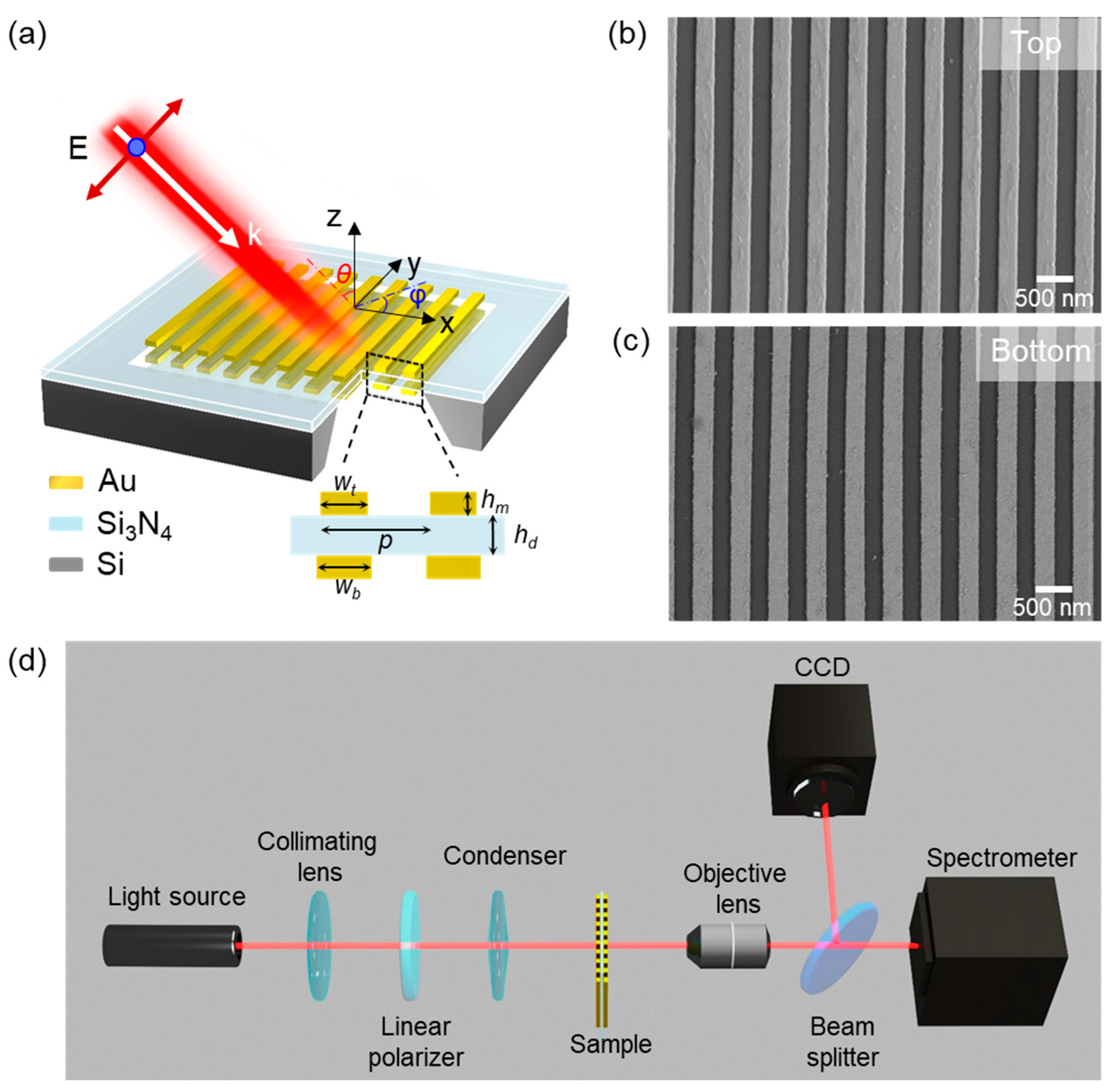
Figure 2a shows the proposed suspended symmetric double-layer gold gratings spaced by a Si3N4 membrane. The widths of the designed top and bottom gratings are Wt = 250 nm and Wb = 250 nm. The grating period is P = 500 nm. The thicknesses of both the top and bottom gratings are hm = 30 nm. The thickness of the Si3N4 membrane is hd = 50 nm. The total thickness of the structure is 110 nm, which is much smaller than the wavelength of our interest. Figure 2b,c shows the top- and bottom-view scanning electron microscope (SEM) images of the fabricated sample. The widths of the fabricated gratings on the top and bottom surfaces of the Si3N4 membrane are Wt = 242 nm and Wb = 256 nm, respectively, resulting in a fabrication error of only ~3%. The slight difference in width is well under the fabrication tolerance and has negligible impact on the transmission spectra. As a result, the suspended double-layer gratings are considered as a symmetric structure. Figure 2d shows the experimental setup for measurements of the transmission spectra.

3. Results and Discussion

The transmission spectra of the suspended double-layer gratings were experimentally measured using a microspectrophotometer system (CRAIC 20/30PV) equipped with a broadband light source (70W Xenon lamp, Olympus). Linearly polarized light is produced by inserting a broadband linear polarizer into the microspectrophotometer system. The transmitted light was collected by a 100× objective lens (NA = 0.8). Figure 3a shows the measured transmission spectra under transverse-magnetic (TM) and transverse-electric (TE) polarized light at normal incidence. Figure 3b illustrates the polarization-dependent transmission as a function of the polarization angle with a step size of 15°. The polar plot shows an obvious linearly polarized transmission, with the minimum and maximum transmission at 0° (TM) and 90° (TE) at both 612 and 758 nm. The experimental spectra are in good agreement with the simulated spectra (see Figure 3c,d) based on the FDTD method (ANSYS Lumerical Software). In the simulation, we extracted the dielectric function of gold from the handbook of Johnson and Christy [48]. The wavelength-dependent refractive index of Si3N4 was measured by the ellipsometer. In the 2D model, the boundary conditions were periodic along the x-axis and a perfectly matched layer along the z-axis. A uniform mesh size of 2 nm in both x- and z-directions is applied for the region of structures. The time step Δt = 2 × 10⁻¹⁸ s is used to satisfy the Courant stability [49]. Note that the slight discrepancies between experimental and simulated results are possibly caused by the surface roughness of the gold nanostructures and the approximations in the 2D model.
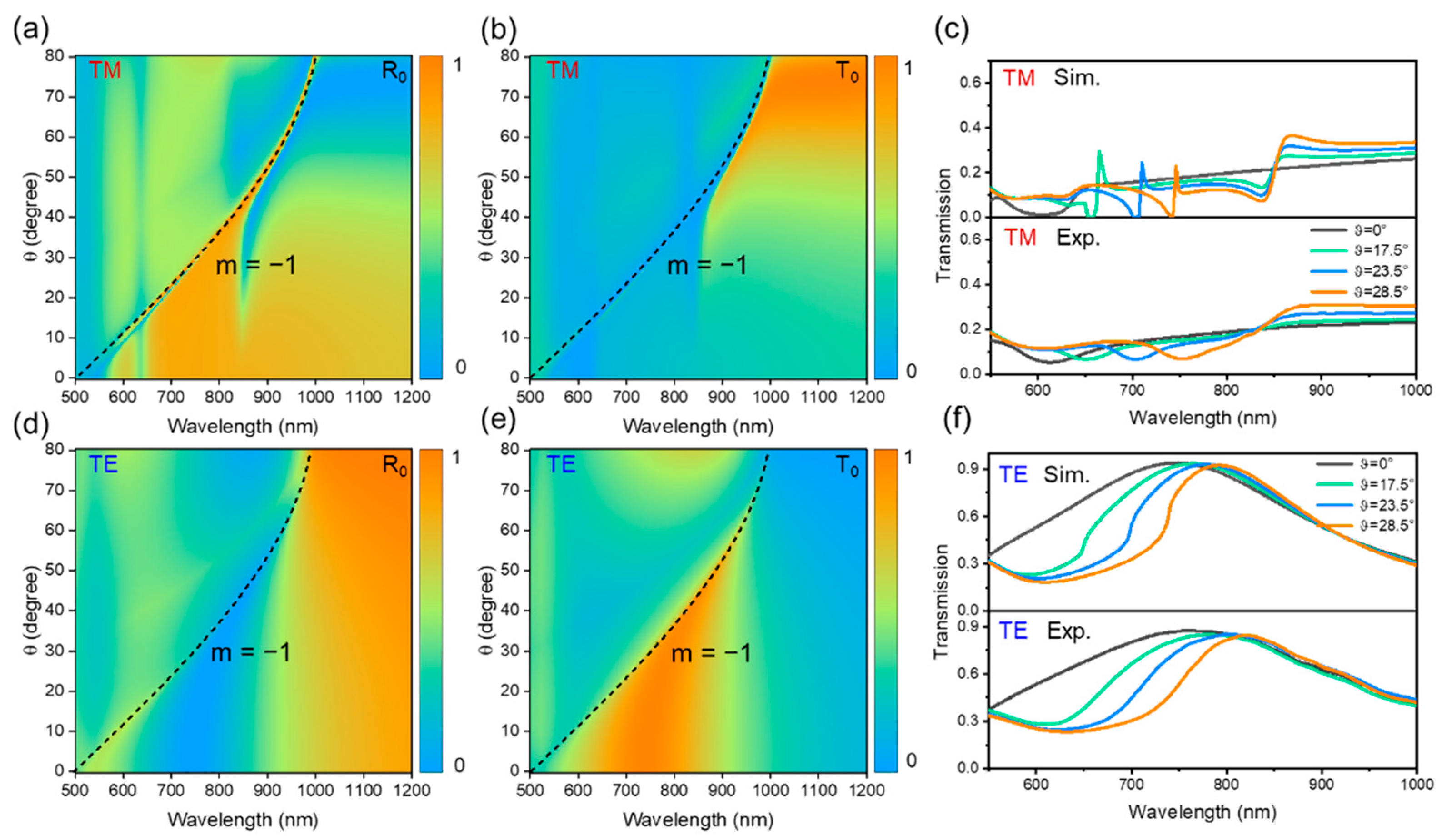
Similar to metal nanoparticle arrays, our proposed double-layer gratings can also induce SLRs. As illustrated in Figure 4, we calculated the 0th-order reflection and transmission spectra as a function of the incident angle for both TM and TE polarizations with an azimuthal angle of φ = 0° in air. It can be clearly seen that the strongly coupled double-layer gratings demonstrate a narrow Fano-like dispersive feature under TM-polarized light incidence. The Fano-like resonance will red-shift when the incident angle θ increases, while under TE-polarized light incidence, the narrow resonance disappears, and the broad in-plane resonance red-shifts and becomes narrow with the increasing θ. For both TM- and TE-polarized light, the resonance wavelengths match perfectly with the Rayleigh anomaly (RA) wavelength of the diffraction order of m = −1, which is represented by the black dotted line. The RA wavelength λRA,m can be obtained from the equation [3]:
λ R A , m = P ( ± n 0 sin θ ) / m   ,
where n0 is the refractive index of the surrounding environment and P is the period of the gratings. As known, the position of out-of-plane SLRs closely follows the RA wavelengths, while the position of LSPRs is almost independent of the incident angle [50,51,52]. It is worth mentioning that the angle-dependent wavelengths of the Fano-like resonances closely follow the RA wavelength in our experiment, which confirms the resonances to be SLRs. By varying the incident angle, SLRs can be further tuned due to the change in out-of-plane electric field components. More importantly, the position of SLRs is highly dependent on the incident angle. When the incident angle increases, the SLRs will red-shift.
In Figure 4c,f, we experimentally measured the transmission spectra for TM- and TE-polarized light incidence with θ = 0°, 17.5°, 23.5°, and 28.5°, respectively. These transmission spectra for TM polarization have roughly the same shape with the simulated ones, while the measured transmission spectra are a good match with the simulated spectra for TE polarization, confirming the detection of plasmonic resonances. Observation of SLRs requires a large area of nanostructures to ensure useful interference of light scattering and a collimated incident light with good coherence [1,46,53]. The narrow SLRs may not be easily detected, since the detecting area is only 5 μm × 5 μm using a 100× objective lens. Moreover, the microscope system could not provide the collimated incident light emitted by the Xenon lamp in our experiment. The incident angle was also limited by the short work distance of the used high-NA objective lens in our experiments.
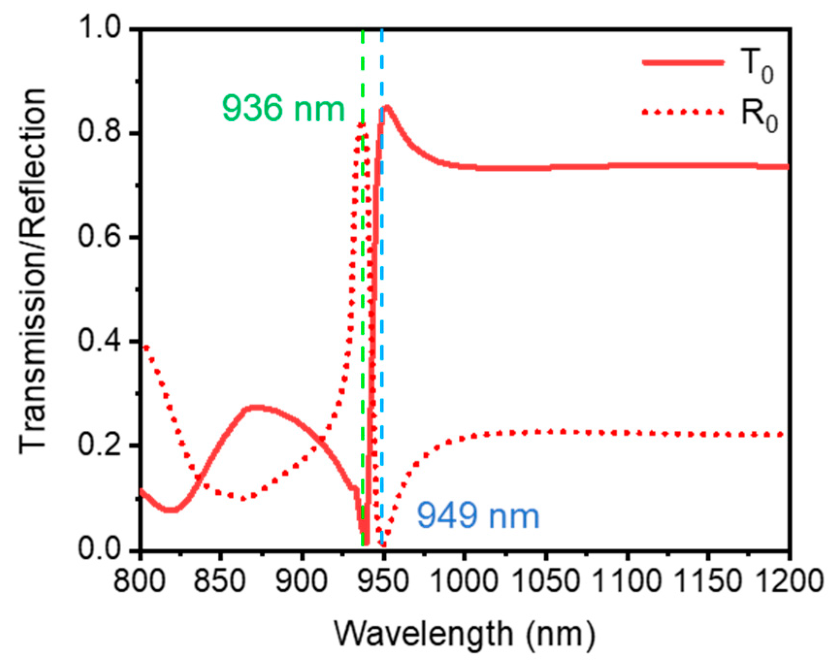
In order to gain physical insights into SLRs, we calculated the electric field enhancement, charge distribution, and surface current under TM incidence with θ = 60° by the FDTD method. The plane wave in our simulation was set with the broadband fixed angle source technique (BFAST) when the gratings were under oblique incidence. The reflection and transmission spectra are shown in Figure 5. The reflection spectrum shows a narrow peak at ~936 nm, with the full width at half maximum (FWHM) of 13 nm, and the transmission spectrum changes dramatically as well at this wavelength. We further calculated the near-field electric field distributions and electric field vectors at the wavelengths of 936 and 949 nm, respectively. The incident electric field amplitude |E0| is set to 1. Figure 6a,b depicts that only the top grating has strong electric field intensity at 936 nm. The corresponding electric field vectors and charge distributions plotted in Figure 6c demonstrate that an in-plane dipole emerges in the top grating. At 949 nm, it can be clearly seen that the strongly coupled double-layer gratings can trap the incident light within the dielectric Si3N4 layer. The surrounding-enhanced field is induced dominantly by the out-of-plane component Ez, resulting from the hybrid out-of-plane quadrupolar oscillations presented in Figure 6f. The electric field intensity enhancement is much higher than that of the incident light (|E0|2) by orders of magnitude due to the strong quadrupolar interactions. Therefore, the near-field optical picture reveals that the Fano-like resonance is induced by the coupling between the subradiant out-of-plane resonances and RA.
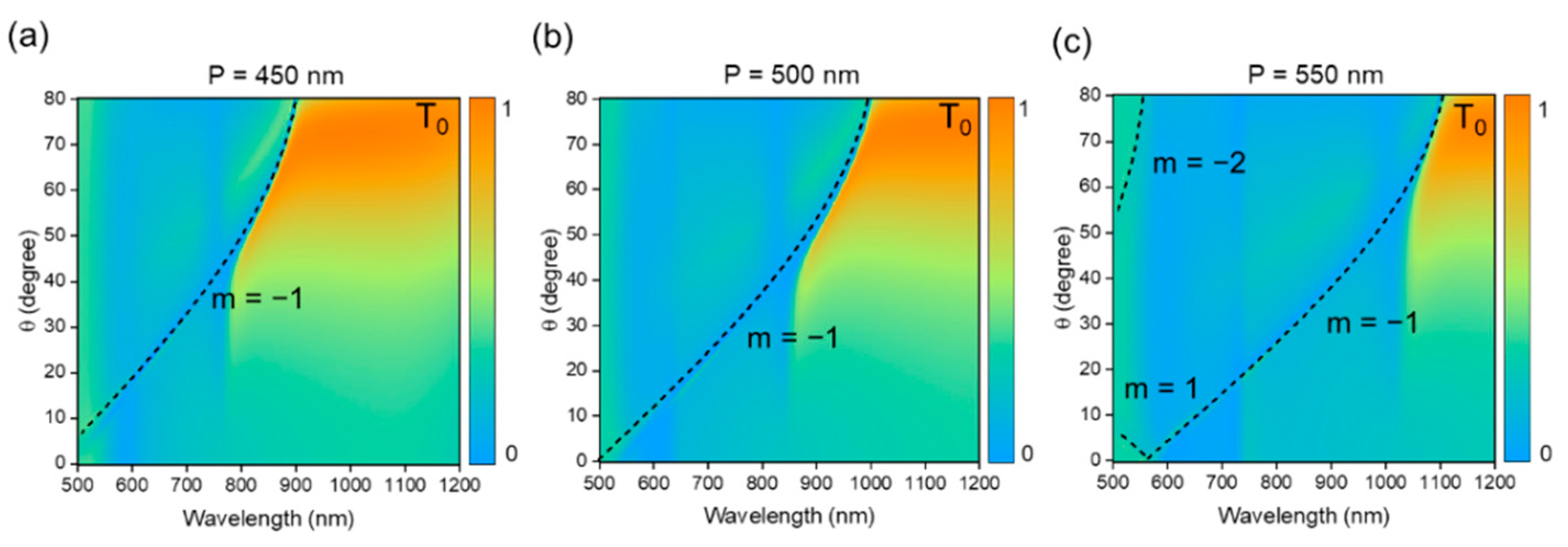
Furthermore, to reveal interference between plasmons and diffractive modes, we investigated the effect of the variation of the grating period while keeping the other structural parameters fixed. Figure 7 shows the transmission spectra as a function of the incident angle under TM-polarized light. The diffraction lines were labeled with the order.
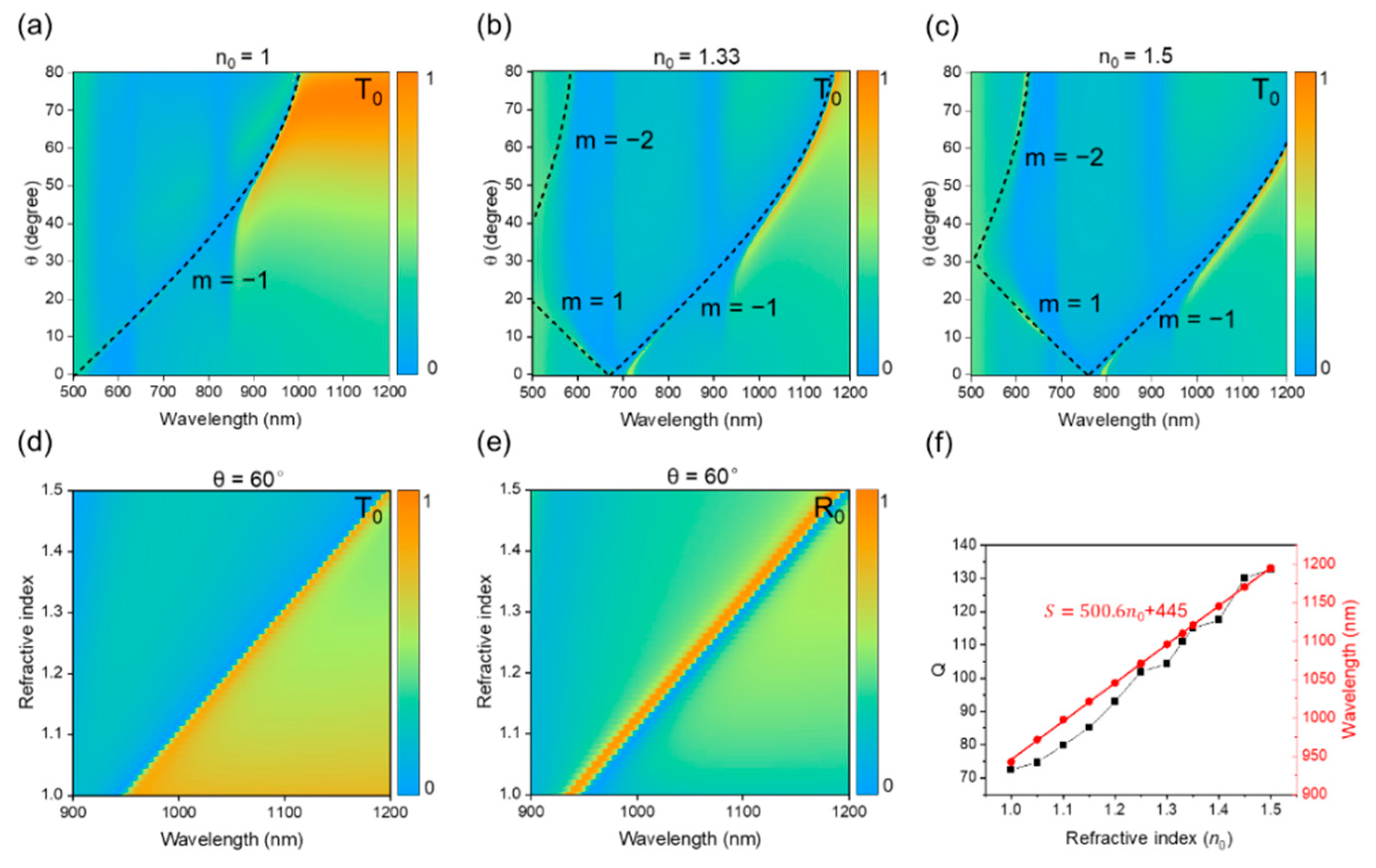
Number m. According to Equation (1) and simulated results, the calculated m = −1 diffraction mode red-shifts as the period increases. SLRs could not be induced under normal incidence, since the incident light does not have out-of-plane electric field component Ez to excite the out-of-plane oscillations. At the period of 550 nm, the m = −2 and m = 1 diffraction modes were located closer to the wavelength of interest. Equation (1) shows that the refractive index of the surrounding environment affects the diffraction modes as well. As known, to support SLRs with a high Q factor in most nanoparticle arrays, the superstrate has to be carefully chosen to have a refractive index matched with the substrate material, so as to ensure a symmetric cladding index. In our suspended double-layer gratings, it is convenient to obtain the homogeneity of the environment by immersing the double-layer gratings in refractive index-matching liquids. We therefore changed the refractive index of the surrounding environment from 1.0 to 1.5 in the simulation. As seen in Figure 8a–c, the wavelength of SLRs had red-shifts when increasing the refractive index. In Figure 8d,e, at θ = 60°, the calculated FWHMs for n0 = 1.0, 1.33, and 1.5 are 13, 10, and 10 nm, respectively. The Q factor increases from 72 for n0 = 1.0 to 111 for n0 = 1.33 and reaches 120 for n0 = 1.5. The proposed SLRs with high Q factor are appealing for sensing applications. Figure 8f shows the sensitivities, defined as S = dλ/dn0 and S = 500.6 nm/RIU. The figure of merit (FOM) reaches 38.5 for the refractive index sensing. It is worth noting that the surface plasmon resonances will result in photothermal heating. In the future, for practical sensing with high accuracy in measurements, the temperature-dependent refractive index should be taken into account [54,55].

4. Conclusions

In conclusion, we proposed and experimentally demonstrated optical performances of the suspended symmetric double-layer gratings in the near-infrared regime. The fabricated double-layer gratings exhibit perfect polarization characteristics under normal incidence. More importantly, we realized SLRs from the suspended symmetric double-layer gratings that can be dynamically tuned by varying the incident angle and the refractive index of surrounding environments. Both experimental and simulation results reveal that the SLRs closely follow the diffraction edges and exhibit a Fano-like spectra resulting from interference between out-of-plane plasmonic resonances and diffraction modes. The out-of-plane SLRs at incident angle θ = 60° for n0 = 1.5 showed a high quality factor of Q = 120. With these unique characteristics, the suspended symmetric double-layer gratings are promising for many potential applications, including high-performance photoluminescent devices, refractive index sensors, and nonlinear optical devices.

Author Contributions

Conceptualization, Y.J.L.; methodology, M.C. and J.W.; software, M.C. and J.L.; validation, M.C. and J.L.; formal analysis, M.C. and J.W.; investigation, Y.L., W.C., D.K. and D.L.; resources, Y.J.L.; data curation, M.C.; writing—original draft preparation, M.C. and J.L.; writing—review and editing, Y.J.L. and T.C.; visualization, M.C.; supervision, Y.J.L. and T.C.; project administration, Y.J.L.; funding acquisition, Y.J.L. All authors have read and agreed to the published version of the manuscript.

Funding

This research was funded by the National Natural Science Foundation of China (Grant No. 62075093 and 62211530039), Guangdong Innovative and Entrepreneurial Research Team Program (Grant No. 2017ZT07C071), and Natural Science Foundation of Guangdong Province (Grant No. 2019A1515110864).

Institutional Review Board Statement

Not applicable.

Informed Consent Statement

Not applicable.

Data Availability Statement

Data underlying the results presented in this paper are not publicly available at this time but may be obtained from the authors upon reasonable request.

Acknowledgments

The authors would like to thank the assistance of SUSTech Core Research Facilities for support with fabrication.

Conflicts of Interest

The authors declare no conflict of interest.

References

  1. Kravets, V.G.; Kabashin, A.V.; Barnes, W.L.; Grigorenko, A.N. Plasmonic surface lattice resonances: A review of properties and applications. Chem. Rev. 2018, 118, 5912–5951. [Google Scholar] [CrossRef] [PubMed]
  2. Wang, W.; Ramezani, M.; Väkeväinen, A.I.; Törmä, P.; Rivas, J.G.; Odom, T.W. The rich photonic world of plasmonic nanoparticle arrays. Mater. Today 2018, 21, 303–314. [Google Scholar] [CrossRef]
  3. Kravets, V.G.; Schedin, F.; Grigorenko, A.N. Extremely narrow plasmon resonances based on diffraction coupling of localized plasmons in arrays of metallic nanoparticles. Phys. Rev. Lett. 2008, 101, 087403. [Google Scholar] [CrossRef] [PubMed]
  4. Liu, J.; He, H.; Xiao, D.; Yin, S.; Ji, W.; Jiang, S.; Luo, D.; Wang, B.; Liu, Y. Recent advances of plasmonic nanoparticles and their applications. Materials 2018, 11, 1833. [Google Scholar] [CrossRef] [PubMed] [Green Version]
  5. Goerlitzer, E.S.A.; Mohammadi, R.; Nechayev, S.; Volk, K.; Rey, M.; Banzer, P.; Karg, M.; Vogel, N. Chiral surface lattice resonances. Adv. Mater. 2020, 32, 202001330. [Google Scholar] [CrossRef] [Green Version]
  6. Auguié, B.; Barnes, W.L. Collective resonances in gold nanoparticle arrays. Phys. Rev. Lett. 2008, 101, 143902. [Google Scholar] [CrossRef] [PubMed] [Green Version]
  7. Ross, M.B.; Mirkin, C.A.; Schatz, G.C. Optical properties of one-, two-, and three-dimensional arrays of plasmonic nanostructures. J. Phys. Chem. C 2016, 120, 816–830. [Google Scholar] [CrossRef]
  8. Wang, D.; Guan, J.; Hu, J.; Bourgeois, M.R.; Odom, T.W. Manipulating light-matter interactions in plasmonic nanoparticle lattices. Acc. Chem. Res. 2019, 52, 2997–3007. [Google Scholar] [CrossRef]
  9. Hakala, T.K.; Rekola, H.T.; Väkeväinen, A.I.; Martikainen, J.P.; Nečada, M.; Moilanen, A.J.; Törmä, P. Lasing in dark and bright modes of a finite-sized plasmonic lattice. Nat. Commun. 2017, 8, 13687. [Google Scholar] [CrossRef] [Green Version]
  10. Wang, D.; Yang, A.; Wang, W.; Hua, Y.; Schaller, R.D.; Schatz, G.C.; Odom, T.W. Band-edge engineering for controlled multi-modal nanolasing in plasmonic superlattices. Nat. Nanotechnol. 2017, 12, 889–894. [Google Scholar] [CrossRef]
  11. Chu, A.; He, H.; Yin, Z.; Peng, R.; Yang, H.; Gao, X.; Luo, D.; Chen, R.; Xing, G.; Liu, Y.J. Plasmonically enhanced upconversion luminescence via holographically formed silver nanogratings. ACS Appl. Mater. Interfaces 2020, 12, 1292–1298. [Google Scholar] [CrossRef]
  12. Czaplicki, R.; Kiviniemi, A.; Laukkanen, J.; Lehtolahti, J.; Kuittinen, M.; Kauranen, M. Surface lattice resonances in second-harmonic generation from metasurfaces. Opt. Lett. 2016, 41, 2684–2687. [Google Scholar] [CrossRef]
  13. Michaeli, L.; Keren-Zur, S.; Avayu, O.; Suchowski, H.; Ellenbogen, T. Nonlinear surface lattice resonance in plasmonic nanoparticle arrays. Phys. Rev. Lett. 2017, 118, 243904. [Google Scholar] [CrossRef] [PubMed]
  14. Hooper, D.C.; Kuppe, C.; Wang, D.; Wang, W.; Guan, J.; Odom, T.W.; Valev, V.K. Second harmonic spectroscopy of surface lattice resonances. Nano Lett. 2019, 19, 165–172. [Google Scholar] [CrossRef] [PubMed]
  15. Gutha, R.R.; Sadeghi, S.M.; Sharp, C.; Wing, W.J. Biological sensing using hybridization phase of plasmonic resonances with photonic lattice modes in arrays of gold nanoantennas. Nanotechnology 2017, 28, 355504. [Google Scholar] [CrossRef]
  16. Danilov, A.; Tselikov, G.; Wu, F.; Kravets, V.G.; Ozerov, I.; Bedu, F.; Grigorenko, A.N.; Kabashin, A.V. Ultra-narrow surface lattice resonances in plasmonic metamaterial arrays for biosensing applications. Biosens. Bioelectron. 2018, 104, 102–112. [Google Scholar] [CrossRef] [Green Version]
  17. Gutha, R.R.; Sadeghi, S.M.; Wing, W.J. Ultrahigh refractive index sensitivity and tunable polarization switching via infrared plasmonic lattice modes. Appl. Phys. Lett. 2017, 110, 153103. [Google Scholar] [CrossRef]
  18. Li, G.; Shen, Y.; Xiao, G.; Jin, C. Double-layered metal grating for high-performance refractive index sensing. Opt. Express 2015, 23, 8995–9003. [Google Scholar] [CrossRef]
  19. Wang, Z.; Liu, J.; Wang, J.; Ma, Z.; Kong, D.; Jiang, S.; Luo, D.; Liu, Y.J. Graphene oxide-coated metal–insulator–metal SERS substrates for trace melamine detection. Nanomaterials 2022, 12, 1202. [Google Scholar] [CrossRef]
  20. Li, J.; Ma, Y.; Gu, Y.; Khoo, I.C.; Gong, Q. Large spectral tunability of narrow geometric resonances of periodic arrays of metallic nanoparticles in a nematic liquid crystal. Appl. Phys. Lett. 2011, 98, 213101. [Google Scholar] [CrossRef]
  21. Thackray, B.D.; Thomas, P.A.; Auton, G.H.; Rodriguez, F.J.; Marshall, O.P.; Kravets, V.G.; Grigorenko, A.N. Super-narrow, extremely high quality collective plasmon resonances at telecom wavelengths and their application in a hybrid graphene-plasmonic modulator. Nano Lett. 2015, 15, 3519–3523. [Google Scholar] [CrossRef]
  22. Lee, U. Spectral Element Method in Structural Dynamics; John Wiley & Sons Asia: Singapore, 2009. [Google Scholar]
  23. Mahariq, I.; Kurt, H. On- and off-optical-resonance dynamics of dielectric microcylinders under plane wave illumination. J. Opt. Soc. Am. B 2015, 32, 1022–1030. [Google Scholar] [CrossRef]
  24. Mahariq, I.; Giden, I.H.; Alboon, S.; Aly, W.H.F.; Youssef, A.; Kurt, H. Investigation and analysis of acoustojets by spectral element method. Mathematics 2022, 10, 3145. [Google Scholar] [CrossRef]
  25. Reshef, O.; Saad-Bin-Alam, M.; Huttunen, M.J.; Carlow, G.; Sullivan, B.T.; Ménard, J.M.; Dolgaleva, K.; Boyd, R.W. Multiresonant high-Q plasmonic metasurfaces. Nano Lett. 2019, 19, 6429–6434. [Google Scholar] [CrossRef] [PubMed] [Green Version]
  26. Sadeghi, S.M.; Wing, W.J.; Campbell, Q. Tunable plasmonic-lattice mode sensors with ultrahigh sensitivities and figure-of-merits. J. Appl. Phys. 2016, 119, 244503. [Google Scholar] [CrossRef]
  27. Yang, X.; Xiao, G.; Lu, Y.; Li, G. Narrow plasmonic surface lattice resonances with preference to asymmetric dielectric environment. Opt. Express 2019, 27, 25384. [Google Scholar] [CrossRef] [PubMed]
  28. Khlopin, D.; Laux, F.; Wardley, W.P.; Martin, J.; Wurtz, G.A.; Plain, J.; Bonod, N.; Zayats, A.V.; Dickson, W.; Gérard, D. Lattice modes and plasmonic linewidth engineering in gold and aluminum nanoparticle arrays. J. Opt. Soc. Am. B 2017, 34, 691–700. [Google Scholar] [CrossRef] [Green Version]
  29. Vecchi, G.; Giannini, V.; Gómez Rivas, J. Surface modes in plasmonic crystals induced by diffractive coupling of nanoantennas. Phys. Rev. B-Condens. Matter Mater. Phys. 2009, 80, 201401. [Google Scholar] [CrossRef] [Green Version]
  30. Li, G.H.Y.; Li, G. Necessary conditions for out-of-plane lattice plasmons in nanoparticle arrays. J. Opt. Soc. Am. B 2019, 36, 805–810. [Google Scholar] [CrossRef]
  31. Auguié, B.; Barnes, W.L. Diffractive coupling in gold nanoparticle arrays and the effect of disorder. Opt. Lett. 2009, 34, 401–403. [Google Scholar] [CrossRef]
  32. Kravets, V.G.; Schedin, F.; Kabashin, A.V.; Grigorenko, A.N. Sensitivity of collective plasmon modes of gold nanoresonators to local environment. Opt. Lett. 2010, 35, 956–958. [Google Scholar] [CrossRef] [Green Version]
  33. Si, G.; Zhao, Y.; Lv, J.; Lu, M.; Wang, F.; Liu, H.; Xiang, N.; Huang, T.J.; Danner, A.J.; Teng, J.; et al. Reflective plasmonic color filters based on lithographically patterned silver nanorod arrays. Nanoscale 2013, 5, 6243–6248. [Google Scholar] [CrossRef] [PubMed] [Green Version]
  34. Leong, E.S.P.; Deng, J.; Khoo, E.H.; Wu, S.; Phua, W.K.; Liu, Y.J. Fabrication of suspended, three-dimensional chiral plasmonic nanostructures with single-step electron-beam lithography. RSC Adv. 2015, 5, 96366–96371. [Google Scholar] [CrossRef]
  35. Cen, M.; Wang, J.; Liu, J.; He, H.; Li, K.; Cai, W.; Cao, T.; Liu, Y.J. Ultrathin suspended chiral metasurfaces for enantiodiscrimination. Adv. Mater. 2022, 34, 2203956. [Google Scholar] [CrossRef] [PubMed]
  36. He, H.; Cen, M.; Wang, J.; Xu, Y.; Liu, J.; Cai, W.; Kong, D.; Li, K.; Luo, D.; Cao, T.; et al. Plasmonic chiral metasurface-induced upconverted circularly polarized luminescence from achiral upconversion nanoparticles. ACS Appl. Mater. Interfaces 2022, in press. [Google Scholar] [CrossRef]
  37. Rodriguez, S.R.K.; Rivas, J.G. Surface lattice resonances strongly coupled to rhodamine 6G excitons: Tuning the plasmon-exciton-polariton mass and composition. Opt. Express 2013, 21, 27411–27421. [Google Scholar] [CrossRef] [PubMed] [Green Version]
  38. Leong, E.S.P.; Wu, S.; Zhang, N.; Loh, W.W.; Khoo, E.H.; Si, G.Y.; Dai, H.T.; Liu, Y.J. Optical properties of ultrafine line and space polymeric nanogratings coated with metal and metal-dielectric-metal thin films. Nanotechnology 2014, 25, 055203. [Google Scholar] [CrossRef]
  39. Liu, Y.J.; Si, G.Y.; Leong, E.S.P.; Xiang, N.; Danner, A.J.; Teng, J.H. Light-driven plasmonic color filters by overlaying photoresponsive liquid crystals on gold annular aperture arrays. Adv. Mater. 2012, 24, 131–135. [Google Scholar] [CrossRef]
  40. Si, G.; Zhao, Y.; Lv, J.; Wang, F.; Liu, H.; Teng, J.; Liu, Y.J. Direct and accurate patterning of plasmonic nanostructures with ultrasmall gaps. Nanoscale 2013, 5, 4309–4313. [Google Scholar] [CrossRef]
  41. Lv, J.; Khoo, E.H.; Leong, E.S.P.; Hu, L.; Jiang, X.; Li, Y.; Luo, D.; Si, G.; Liu, Y.J. Maskless fabrication of slanted annular aperture arrays. Nanotechnology 2017, 28, 225302. [Google Scholar] [CrossRef]
  42. Cattoni, A.; Ghenuche, P.; Haghiri-Gosnet, A.M.; Decanini, D.; Chen, J.; Pelouard, J.L.; Collin, S. λ3/1000 plasmonic nanocavities for biosensing fabricated by soft UV nanoimprint lithography. Nano Lett. 2011, 11, 3557–3563. [Google Scholar] [CrossRef]
  43. Kuznetsov, A.I.; Evlyukhin, A.B.; Goņalves, M.R.; Reinhardt, C.; Koroleva, A.; Arnedillo, M.L.; Kiyan, R.; Marti, O.; Chichkov, B.N. Laser fabrication of large-scale nanoparticle arrays for sensing applications. ACS Nano 2011, 5, 4843–4849. [Google Scholar] [CrossRef]
  44. Yin, Z.; Shi, Y.; Cen, M.; Cao, T.; Xu, C.; Luo, D.; Li, G.; Liu, Y.J. Vectorial holography-mediated growth of plasmonic metasurfaces. Sci. Bull. 2021, 66, 1518–1524. [Google Scholar] [CrossRef]
  45. Auguié, B.; Bendaña, X.M.; Barnes, W.L.; García De Abajo, F.J. Diffractive arrays of gold nanoparticles near an interface: Critical role of the substrate. Phys. Rev. B-Condens. Matter Mater. Phys. 2010, 82, 155447. [Google Scholar] [CrossRef] [Green Version]
  46. Bin-Alam, M.S.; Reshef, O.; Mamchur, Y.; Alam, M.Z.; Carlow, G.; Upham, J.; Sullivan, B.T.; Ménard, J.M.; Huttunen, M.J.; Boyd, R.W.; et al. Ultra-high-Q resonances in plasmonic metasurfaces. Nat. Commun. 2021, 12, 974. [Google Scholar] [CrossRef]
  47. Humphrey, A.D.; Barnes, W.L. Plasmonic surface lattice resonances on arrays of different lattice symmetry. Phys. Rev. B-Condens. Matter Mater. Phys. 2014, 90, 075404. [Google Scholar] [CrossRef] [Green Version]
  48. Johnson, P.B.; Christy, R.W. Optical Constant of the Nobel Metals. Phys. Rev. B 1972, 6, 4370–4379. [Google Scholar] [CrossRef]
  49. Zheng, F.; Chen, Z.; Zhang, J. A finite-difference time-domain method without the courant stability conditions. IEEE Microw. Guid. Wave Lett. 1999, 9, 441–443. [Google Scholar] [CrossRef] [Green Version]
  50. Zhou, W.; Odom, T.W. Tunable subradiant lattice plasmons by out-of-plane dipolar interactions. Nat. Nanotechnol. 2011, 6, 423–427. [Google Scholar] [CrossRef] [PubMed]
  51. Hua, Y.; Fumani, A.K.; Odom, T.W. Tunable lattice plasmon resonances in 1D nanogratings. ACS Photonics 2019, 6, 322–326. [Google Scholar] [CrossRef]
  52. Fang, X.; Xiong, L.; Shi, J.; Li, G. High-Q quadrupolar plasmonic lattice resonances in horizontal metal–insulator–metal gratings. Opt. Lett. 2021, 46, 1546–1549. [Google Scholar] [CrossRef] [PubMed]
  53. Sung, J.; Hicks, E.M.; Van Duyne, R.P.; Spears, K. Nanoparticle spectroscopy: Plasmon coupling in finite-sized two-dimensional arrays of cylindrical silver nanoparticles. J. Phys. Chem. C 2008, 112, 4091–4096. [Google Scholar] [CrossRef]
  54. Nisar, M.S.; Kang, S.; Zhao, X. Photothermal effect in plasmonic nanotip for LSPR sensing. Sensors 2020, 20, 671. [Google Scholar] [CrossRef] [Green Version]
  55. Baffou, G.; Quidant, R.; García De Abajo, F.J. Nanoscale control of optical heating in complex plasmonic systems. ACS Nano 2010, 4, 709–716. [Google Scholar] [CrossRef] [PubMed]
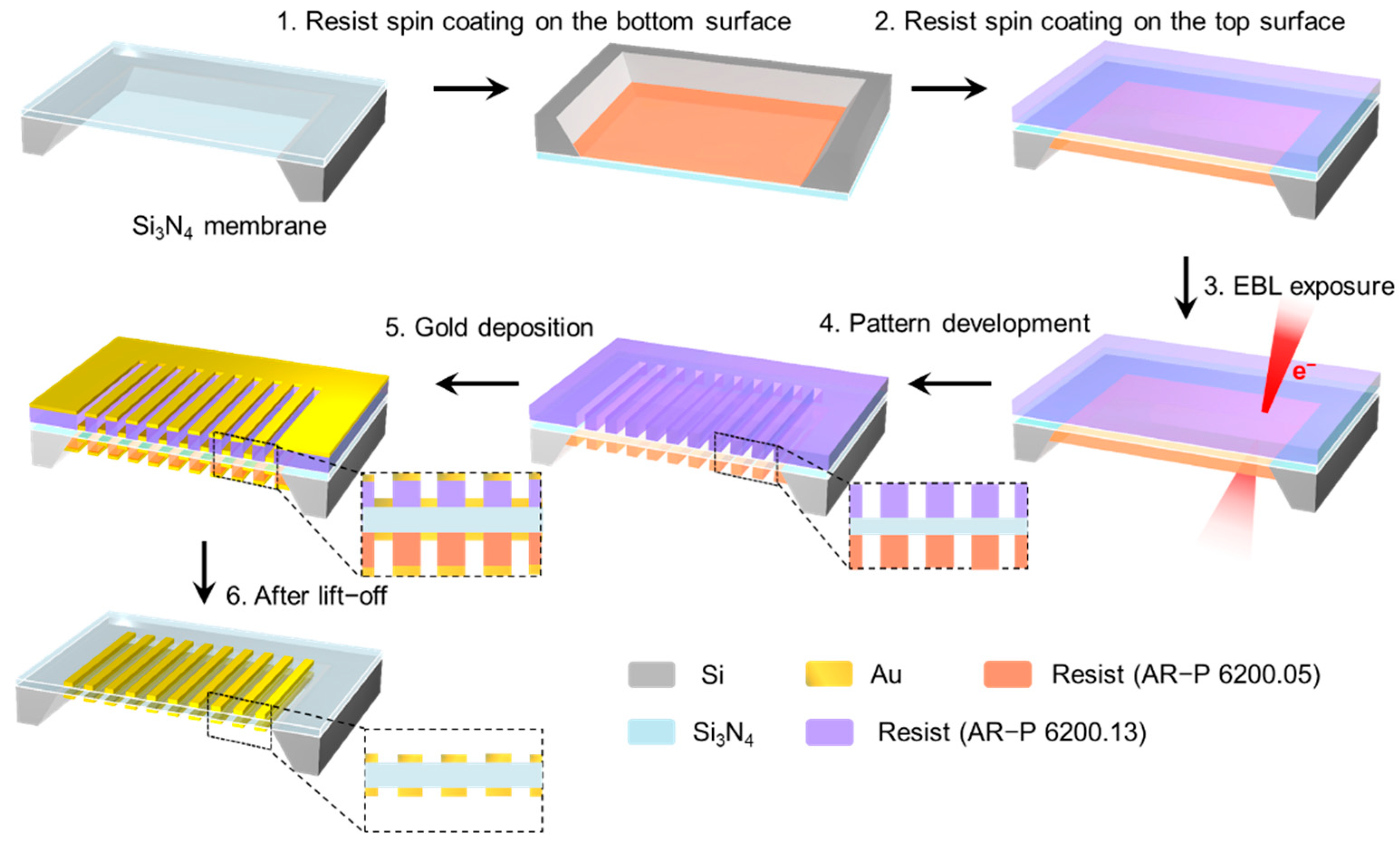
Figure 1. Schematic fabrication process of suspended symmetric double-layer gratings.
Photonics 09 00890 g001
Figure 2. (a) Schematic configuration of the suspended double-layer gratings in air with all structural parameters. Top- (b) and bottom-view (c) SEM images of the fabricated double-layer gratings. Scale bar: 500 nm. (d) The microspectrophotometer system for the transmission spectra measurement.
Photonics 09 00890 g002
Figure 3. Measured (a) and simulated (c) transmission spectra of the suspended double-layer gold gratings under TM- and TE-polarized light incidence. Polar plots of experimentally measured (b) and simulated (d) transmission at 612 and 758 nm in the experiment and at 609 and 733 nm in the simulation, respectively.
Photonics 09 00890 g003
Figure 4. Angle-dependent 0th-order reflection (a) and transmission (b) spectra of the suspended double-layer gratings under TM-polarized incidence. The black dashed lines indicate RA wavelengths of the diffraction order of m = −1. (c) Simulated and measured transmission spectra with θ = 0°, 17.5°, 23.5°, and 28.5°, respectively. (df) The corresponding results under TE-polarized incidence.
Photonics 09 00890 g004
Figure 5. Simulated transmission and reflection spectra of gold double-layer gratings under TM-polarized light incidence with the incident angle of θ = 60°.
Photonics 09 00890 g005
Figure 6. (ac) Calculated electric field intensity distributions of |Ez|2 (a), |Ex|2 (b), and |E|2 (c) at the wavelengths of 936 nm. Electric field vectors (green arrows) are mapped in (c). The double-layer grating structure is outlined by white lines. (df) The corresponding electric field intensity distributions at the wavelengths of 949 nm. Symbols “+” and “−” in (c,f) indicate positive and negative charge distributions.
Photonics 09 00890 g006
Figure 7. Angle-dependent 0th-order transmission spectra for the suspended double-layer gratings under TM-polarized incidence with the periods of 450 (a), 500 (b), and 550 nm (c). The black dashed lines indicate different diffraction modes.
Photonics 09 00890 g007
Figure 8. Simulated angle-dependent 0th-order transmission spectra for the suspended double-layer gratings under TM-polarized incidence with the refractive index n0 of 1 (a), 1.33 (b), and 1.5 (c). The black dashed lines indicate different diffraction modes. The 0th-order transmission (d) and reflection (e) spectra as functions of refractive index with θ = 60°. (f) The quality factor and sensitivity of the suspended symmetric double-layer gratings with θ = 60°.
Photonics 09 00890 g008
Publisher’s Note: MDPI stays neutral with regard to jurisdictional claims in published maps and institutional affiliations.