Abstract
The vacuum chamber is an important part of microparticle optical levitation technology. The traditional vacuum chamber has a large volume and many peripheral components, which cannot meet the requirements of miniaturization and on-chip optical levitation technology. Therefore, this study proposes a novel microparticle vacuum chamber based on the micro-electro-mechanical system (MEMS) process. This MEMS microparticle vacuum chamber adopts a “glass-silicon-glass” three-layer vacuum bonding process, with a volume of only 15 mm × 12 mm × 1.2 mm, including particle chamber, cantilever resonator chamber, and getter chamber, which can encapsulate microparticles in a tiny vacuum environment and realize optical levitation of microparticles. At the same time, the air pressure in the micro vacuum chamber is monitored by the cantilever resonator, which can provide a miniaturized microparticle chamber with a more accurate vacuum environment for microparticle optical levitation. The research of this paper has significance for promoting the development of miniaturized optical levitation technology.
1. Introduction
Since the birth of optical levitation technology in the 1970s, this new means of levitation technology has been widely used in many frontier research fields such as micro-molecule capture, targeted therapy, quantum precision measurement, and dark matter detection [1,2,3,4,5] with its high precision, high resolution, and nondestructive contact properties. However, the current common optical levitation systems are generally conventional optical devices consisting of optical elements, desktop lasers, and large-volume mechanical vacuum systems, especially for mechanical vacuum chambers, which can achieve high vacuums (10−4 torr) [6,7,8,9,10]. In particular, in the field of optical rotation [11], an environment of high vacuum level is often required to reduce air damping for increasing the rotational speed of optical rotating particles. At present, most mechanical vacuum systems mainly include vacuum vessels, connecting pipes, vacuum valves, vacuum pumps, vacuum gauges, and so on [12]. The overall volume is about one cubic meter. Although the mechanical vacuum chamber can achieve a high vacuum environment, it is not convenient to be used in compact optical levitation fields due to its large volume and weight. Miniaturization and on-chip optical levitation are important trends in the development of optical levitation, but the traditional vacuum system cannot meet the requirements. However, so far, there are few research works and papers reported on the miniaturized particle vacuum system to the best of our knowledge, so a miniaturized vacuum system is urgently needed.
Inspired by the atomic vapor cell in the nuclear magnetic resonance gyroscope [13,14,15], MEMS technology has become the best way to prepare small-volume vacuum chambers and replace large-volume mechanical vacuum chambers due to its advantages of high efficiency, good repeatability, and small size of processing devices [16,17,18,19]. However, in order to realize the vacuum level detection of MEMS small-volume vacuum chambers, most of the existing vacuum detection methods for MEMS wafer-level chambers use sensitive structures embedded in the chamber to calibrate the vacuum level. Almost all of them need to convert the electrical signal output with the help of outer lead bonding [20,21,22], which will significantly increase the design and processing difficulty of MEMS microparticle chambers.
To solve the above problems, in this paper, the authors propose a vacuum detection technique with an electromagnetic drive without outer lead bonding based on the realization of the MEMS microparticle chamber structure design. This design simplifies the processing difficulty of the microparticle chamber by using an external electromagnet to drive a resonator prepared from iron material embedded inside the chamber, which can determine the vacuum level of the packaged MEMS microparticle chamber. The vacuum level of the packaged MEMS microparticle chamber was determined with the help of the resonant characteristics of the resonator, and then the optical levitation and rotation of one micron-sized particle were realized with the help of this vacuum environment.
The results show that the microparticle chamber can not only maintain a certain vacuum level, but also have a built-in pressure measurement function, and the volume is small and easy to integrate (the overall volume is on the cubic millimeter level). Compared with the large-volume high-intensity mechanical vacuum chamber, this millimeter-scale microparticle chamber can be better adapted to the miniaturization and on-chip optical levitation system, which presents a new research idea for the integration of optical levitation systems.
2. Overall Structural Design of the Microparticle Chamber
2.1. Structure Design
Since optical levitation technology mainly uses focused lasers to trap microparticles stored in a vacuum chamber, it is necessary to ensure good light transmission on the upper and lower surfaces of the designed microparticle chamber. Since silicon wafer is the most basic processing material in the field of integrated circuits and MEMS, it is an ideal structural material with high melting point and stable mechanical properties, so the main structure of the microparticle chamber adopts the sandwich structure of “glass-silicon wafer-glass”. The current MEMS wafer-level vacuum detection technology includes the use of an internally mounted tuning fork quartz crystal as a sensor for vacuum measurement [23], the use of a micro-bridge structure of Pirani gauge as a sensitive unit for vacuum measurement [24,25,26], and so on. However, all of these methods require an external circuit structure to output the measured electrical signal, which includes the design of the circuit structure, lead processing, package preparation, etc., making the structure of the microparticle vacuum chamber more complex, increasing the size and the probability of air leakage from the seal.
Hence, to overcome these problems, the authors choose a three-chamber structure: Chamber I contains a resonator with a cantilevered beam structure. The resonant motion of the cantilever resonator is driven by an external AC field, and the resonant frequencies of the cantilever resonator under different vacuum environments before bonding are calibrated by a laser vibrometer to determine the vacuum level inside the MEMS vacuum chamber after bonding. Chamber II contains a small piece of getter. Getter is often used in vacuum electronic devices (especially in the MEMS devices) to create a good working environment and maintain a certain vacuum level. It is made up of various elements, such as Ti, Zr, Ta, Th, etc. When the getter is activated at a high temperature (350 °C for 20 min), it can absorb the residual gas (H2, H2O) in the particle chamber and maintain the vacuum. Chamber III contains the microparticles for levitation. Nevertheless, the traditional MEMS vacuum devices need enough cleanliness to ensure the bonding effect; the structure is also the biggest difference between microparticle chamber and traditional MEMS vacuum devices. After filling in the particles, the vacuum pumping process will pump some of the particles to the surface of the silicon wafer, increasing the probability of failure of the bond process. Hence, a structure with a glass sheet with a hole and a piece of tinfoil attached to it is used to cover the surface of Chamber III to prevent the particles from escaping during the vacuum pumping process. In order to ensure the same vacuum level inside the two chambers, the three chambers are connected by two micro-channels, which can also prevent the particles excited by the piezoelectric sheet from spreading into Chambers I and II, as shown in Figure 1.
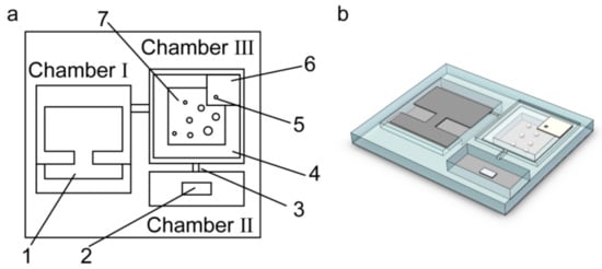
Figure 1.
Structure diagram of the MEMS particle chamber. (a) Top view of the MEMS particle chamber (1: Resonator, 2: Getter, 3: Micro-channels, 4: Small glass sheet with one hole, 5: Micropore, 6: Tinfoil; 7: Vaterite particles); (b) Three-dimensional stereogram of the MEMS particle chamber.
2.2. Design and Vibration Modal Simulation of Cantilever Resonator
To avoid designing additional circuit systems, the iron cantilever resonator is selected as the resonator in the MEMS chamber, and the external electromagnet is used as the driving unit to calibrate the vacuum level in the vacuum chamber. The cantilever resonator parameters in millimeter are shown in Figure 2, and the thickness of iron material is chosen as 0.1 mm.
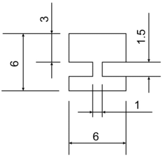
Figure 2.
Structure and corresponding dimensions of cantilever resonator.
In order to guide the determination of its resonant frequencies in the experimental tests, the inherent frequencies of the cantilever resonator were first analyzed by using finite element analysis. The simulation results are shown in Figure 3. The characteristic frequencies of the first four modes of the cantilever resonator are 4153.8 Hz, 6269.7 Hz, 16,041 Hz, and 25,167 Hz. Since the driving method uses the electromagnetic field driven by the AC variation directly below the MEMS microparticle chamber, mode one has the largest contact area with the gas inside the chamber and is the most sensitive to the vacuum variation inside the chamber. In the subsequent experimental tests, only the vibration near the characteristic frequency in mode one is focused on. The simulated characteristic frequencies and quality factors of the resonant beam are shown in Table 1.
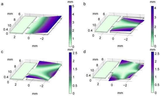
Figure 3.
Working modal simulation of the cantilever resonator (Characteristic frequency (a–d): 4153.8 Hz, 6269.7 Hz, 16,041 Hz, 25,167 Hz).

Table 1.
Simulated characteristic frequencies and quality factors of cantilever resonator.
In order to theoretically guide the calibration of the MEMS microparticle chamber vacuum test, it is necessary to analyze the influence mechanism of damping on the resonant beam. The cantilever resonator will be affected by multiple influences including air damping loss, thermoelastic loss, support loss, surface defect loss, and other environmental losses.
In this paper, the air squeeze-film damping is used as the main factor for the cantilever resonator to sense the air pressure, as shown in Figure 4. Hence, firstly, the influence of the squeeze-film damping on the resonant frequency of the cantilever resonator is analyzed. After that, a brief analysis of the thermoelastic damping existing in the system will be carried out.
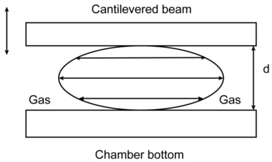
Figure 4.
Squeeze-film damping model. The distance between cantilevered beam and chamber bottom is d.
2.2.1. Squeeze-Film Damping
For the designed MEMS microparticle chamber, the two most important parameters to evaluate the gas thinness are the aerodynamic viscosity μ at the air pressure and Knudsen number Kn, which is the ratio of the molecular free range to the characteristic size of the resonator, i.e.,
where λ is the molecular free range at a given air pressure, λ0 is the molecular free range at atmospheric pressure, P0 is the standard atmospheric pressure, P is the given air pressure, and d is the distance between the cantilever resonator and the bottom of the chamber. According to the calculated value of Kn, the gas flow characteristics within the MEMS microparticle chamber can be initially judged: when Kn < 0.001, it is the continuous flow region; when 0.001 < Kn < 0.1, it is the slip flow region; when 0.1 < Kn < 10, it is the excessive flow region; when Kn > 10, it is the molecular flow region. While in the microscopic domain, the aerodynamic viscosity μ is generally replaced by the effective viscosity factor μeff, which is related to Kn and the choice of different mathematical treatment models, as shown in Table 2 [27].

Table 2.
Effective viscosity coefficient.
According to the resonant beam structure designed in this paper, it can be calculated that when the air pressure is located at 10–10,000 Pa, Kn is between 0.0013 and 1.286; at this time the gas flow is between the slipstream and over-flow regions, and the viscosity effect produced by the gas molecules is obviously affected by the pressure, so the effective viscosity coefficient must be used as a substitute. The effective viscosity coefficient of the N-S equation model is used in this section.
When the cantilever resonator makes resonant motion, the gas between the cantilever resonator and the bottom of the chamber will be sucked and discharged with its periodic motion, and this process will be affected by the resonant motion of the cantilever resonator due to the change of gas molecules caused by the change of gap, because some of the gas is not compensated or dispersed in time. Equation (2) is the pressure film damping formula for the rectangular structure,
where W is the width of the rectangular beam, L is the length of the rectangular beam, h is the thickness of the rectangular beam, and β is the scale factor that varies with the width–length ratio of the rectangular beam, since the width–length ratio of the rectangular beam designed in this paper is 0.5, β ≈ 0.7 [27], and the aerodynamic viscosity μ is 17.9 × 10⁻⁶ Pa·s at 20 °C.
2.2.2. Thermoelastic Damping
In addition, when the cantilever resonator is excited, the length of the middle plane does not change. On the left and right sides of the middle plane, the material will have different temperature gradients due to compression and tension. This internal temperature conduction will lead to thermoelastic damping loss. Most thermoelastic damping models are based on Zener’s classical thermoelastic damping theory [28]. The following expression of the thermoelastic damping coefficient of the micro cantilever beam is given without deduction [27],
where E is the modulus of elasticity of the material, α is the coefficient of thermal expansion of material, Cp is the heat capacity per unit volume under fixed pressure or stress, and ξ is a dimensionless coefficient,
where ω0 is the undamped intrinsic angular frequency and χ is the thermal diffusivity of the material.
Since the damping terms other than squeeze-film damping are not affected by the air pressure, in order to simplify the calculation, the remaining damping terms are considered as a constant in the subsequent simulation calculation.
2.2.3. Vibration Modal Simulation of Cantilever Resonator
According to the damped forced vibration equation, it is known that,
Simplification leads to,
where x is the vibration displacement, c is the damping coefficient (sum of all damping coefficients), δ is the damping rate, δ = c/(2m), the damping ratio ζ = , k is the equivalent stiffness of the system, F is the amplitude value of external driving force, ω is driving frequency, and t is driving time.
In this system, since the mass and surface area of the end rectangular structure are much larger than that of the thin support, the cantilever structure is simplified to a single cantilever resonator with constant cross-section in this section. The cantilever resonator first-order inherent frequency ω0 is shown in Equation (7) [29],
where k1 = 1.76, xf is the distributed load force per unit length and I is the moment of inertia of the cantilever resonator.
The above variables and corresponding parameters are shown in Table 3.

Table 3.
Calculated parameters of squeeze-film damping.
From that, it can be calculated that,
Equivalent stiffness k is,
Since the damping ratio ζ is always less than 1 when the air pressure is in the range of 10–10,000 Pa, the system is an underdamped system with damped intrinsic angular frequency ωd and intrinsic frequency fd as,
The relationship between the variation of the intrinsic frequency and quality factor with air pressure is thus established, and its curve distribution is shown in Figure 5.
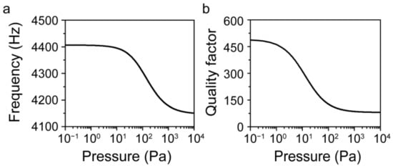
Figure 5.
Resonant frequency and quality factor variation curve with air pressure. (a) Variation of resonant frequency with air pressure; (b) Variation of quality factor with air pressure.
From the calculation results, it can be seen that when the air pressure is lower than 10 Pa, the resonant frequency of the system has stabilized, which indicates that the change of air pressure will have little effect on the resonant frequency of the cantilever resonator at this time. When the air pressure gradually decreases, the air damping of the system will also gradually decrease, but due to the presence of other damping components such as thermoelastic damping, the quality factor will also tend to a fixed value. Since the measurement of the quality factor is indirect, its data accuracy is not as direct as that of the resonant frequency, so in the subsequent experiments, this paper mainly uses the resonant frequency curve to calibrate and test the air pressure.
3. Preparation of the Microparticle Chamber
In order to prevent the particles from being projected onto the upper glass surface and not being trapped during the piezoelectric wafer excitation process, the silicon wafers selected must be of sufficient thickness. When the focusing laser passes through the lower surface and upper surface of the glass sheet, there is absorption and refraction phenomena of light. In order to ensure the transmittance and optical path invariance, the thickness of the glass sheet should be as thin as possible while meeting the anode bonding strength. The cantilever resonator is embedded inside Chamber I, and its thickness should not exceed half of the wafer thickness and facilitate external electromagnetic field driving. Combining the above considerations, a 4-inch diameter double-throw silicon wafer with a thickness of 1 mm, two pieces of glass wafer with a thickness of 100 μm, and a pure iron foil with a thickness of 100 μm are used.
In MEMS processing, lithography and dry and wet etching processes are commonly used for batch removal of material and, thus, obtaining correct grinding patterns on the silicon wafers, which are selective, reproducible, and accurate. However, the process time often depends on the chemical reaction rate and is not easy to shape quickly. Due to fact that the designed microparticle chamber is large and the roughness of the chamber sidewalls has no effect on the stiffness of the optical trap, the UV nanosecond dicing technique was chosen for the chamber forming process. To ensure a highly gas-tight fit between the glass and the wafer, a high-temperature anodic bonding process is used for the encapsulation of the chamber.
The flow chart of MEMS microparticle vacuum chamber preparation is shown in Figure 6.
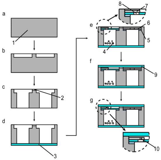
Figure 6.
Preparation process of the MEMS microparticle vacuum chamber (1: Silicon wafer, 2: Micro-channels, 3: Lower glass sheet, 4: Vaterite particles; 5: High temperature adhesive; 6: Cantilever resonator; 7: Tinfoil; 8: Small glass sheet with one hole; 9: Upper glass sheet; 10: Micropore in the tinfoil). (a–g) refer to each processing step. See the text for its specific description.
- (a)
- Cleaning. Using acetone and HF solution to remove small particles of organic and inorganic materials from the silicon wafer surface.
- (b)
- First dicing. Using a laser dicing machine to pre-process the three chamber structures. (This Figure only shows the structure of Chamber I and Chamber III)
- (c)
- Second dicing. Processing of the micro-channels connecting three chambers.
- (d)
- First anodic bonding. The bonding temperature is usually set at 340 °C and the voltage is set at 800 V.
- (e)
- Particle-filling, getter-filling, and adhesion of small glass pieces, cantilever resonator to the silicon wafer. The adhesive is a high temperature adhesive with a heat resistance of 400 °C. The vacuum pumping process will allow particles in Chamber III to be pumped to the surface of the silicon wafer during the second anode bonding process, and then reduce the bonding vacuum level. Therefore, a small piece of tinfoil is bonded to the top of the small glass sheet with one hole to maximize the bonding effect.
- (f)
- Second anodic bonding.
- (g)
- Using the laser to release one micro-hole in the tinfoil. To ensure the air pressure connectivity of the three chambers and to accurately characterize the vacuum level, a laser is used to release a micro-hole in the tinfoil. The released micro-hole must be directly above the hole in the perforated glass sheet. It is important to note that the power and focus of the laser must be strictly controlled to ensure that the upper glass layer is not damaged.
After completing the entire process, each chamber in the wafer is scribed and the individual microparticle chambers cut down are shown in Figure 7. Since it is not possible to use immersion cleaning before step (f), there are a few impurities on the surface that affect the bonding, resulting in a small amount of air bubbles around the bonded chambers, which caused the actual encapsulation pressure of the chambers to be higher than the set bonding air pressure.
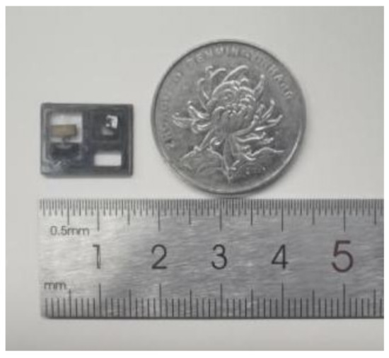
Figure 7.
Image of the MEMS microparticle chamber.
4. Test of Microparticle Chamber Vacuum Level
4.1. Principle of Vacuum Level Test
As described in the previous section, the frequency detection method is used to test and calibrate the vacuum inside the microparticle chamber. Firstly, a laser vibration measurement system (Polytec, IT6322A) is used for the calibration experiment of the vacuum level inside the MEMS microparticle chamber. The method of laser vibration measurement is shown in Figure 8.
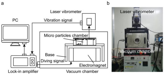
Figure 8.
Method of laser vibration measurement. (a) Schematic diagram of principle; (b) Image of the laser vibration system.
The driving signal generated by the lock-in amplifier makes the electromagnet generate an AC magnetic field, and the cantilever resonator vibrates under the action of magnetic force. The laser vibrometer transmits the vibration signal back to the phase-locked amplifier and displays it on the PC. In order to facilitate the testing of the resonant frequency and amplitude of the cantilever resonator under different air pressure conditions, before step (f), the upper glass sheet is tightly attached to the processed silicon wafer so as to maximize the simulation of the gas-tight environment after bonding. Then, the resonant peak is calibrated by adjusting the vacuum chamber air pressure.
After step (g), the resonant frequency and amplitude of the cantilever resonator are still tested with the laser vibration measurement system to detect the vacuum level of the chamber after bonding.
4.2. Test and Calibration of the Vacuum Level
To characterize the consistency of the vacuum level of multiple microparticle chambers processed on the same wafer, the vacuum level of four vacuum chambers on the same silicon wafer were tested. After being tested by the phase-locked amplifier and the laser vibrometer, the resonance spectrum of Chamber I before the getter is activated is shown in Figure 9. The resonant frequency of the cantilever resonator is maintained in the range of 4555 Hz to 4561 Hz. The resonant frequency and quality value of the cantilever resonator are different from the simulated results due to the uneven adhesion of the high-temperature adhesive and the deformation and warping caused by the laser thermal effect when cutting the cantilever resonator structure, as well as the influence of uneven density distribution of materials.
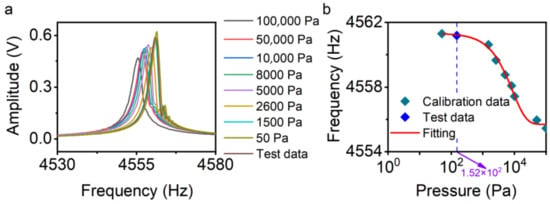
Figure 9.
Variation of resonant frequency of cantilever with air pressure of microparticle chamber in Chamber I. The test data are obtained through experiments after step (g), and the remaining calibration data are obtained through experiments before step (f). (a) Resonance spectrum; (b) Calibrating plot.
After testing, the air pressure in the four microparticle vacuum chambers is shown in Table 4.

Table 4.
Test pressure of four microparticle vacuum chambers on the same wafer before getter is activated.
The resonant frequency and the sensitivity to the vacuum level of the cantilever resonator structure within the four chambers are not consistent due to the different surface roughnesses of the iron, laser spot calibration positions, bonding positions, and thicknesses of the high temperature adhesive, but the test air pressure is maintained at about 150 Pa. After activation of getter, the minimum air pressure in the particles chamber can reach about 10 Pa.
In order to facilitate the subsequent optical levitation and optical rotation experiments, a batch of microparticle chambers with a vacuum degree of 3 kPa is also prepared. Figure 10 shows the resonant spectrum of the microparticle chamber when the air pressure in the chamber is controlled at 3 kPa.
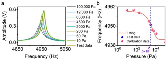
Figure 10.
Variation of resonant frequency of cantilever with air pressure of the microparticle chamber prepared for optical levitation test. (a) Resonance spectrum; (b) Calibrating plot.
To evaluate the accuracy of the calculation, a bonded micro-chamber is selected and a hole is drilled on its glass surface. The resonant frequency of the cantilever resonator in this chamber before drilling a hole is 4950.9 Hz, and the pressure corresponding to the calibration curve in Figure 10b is 3.025 kPa. After drilling a hole, by placing it in the vacuum chamber under the calculated vacuum level and retesting the resonant frequency, the test results show that the resonant frequency is 4951.2 Hz. The pressure corresponding to the calibration curve in Figure 10b is 2.873 kPa. Limited by the accuracy of the vacuum chamber and vacuum gauge, there is a certain deviation between these two results. However, the test results are in good agreement with the calculated results within the error range.
5. Test of Laser-Induced Rotating Single Particle
In the previous section, a vacuum chamber containing microparticles was prepared by using the MEMS process, and its vacuum level was calibrated. In this section, the vacuum environment of the microparticle vacuum chamber is further evaluated by building a laser-induced rotating system and combining it with the vacuum chamber to achieve rotational manipulation of a single vaterite particle.
5.1. Setup of the Optical Systems
A laser-induced rotating system is built as shown in Figure 11. The laser with an output wavelength of 1064 nm is collimated and passed sequentially through a half-wave plate (to adjust the output power) and a quarter-wave plate (to adjust the polarization of the laser), after which it is focused using a microscope objective with a numerical aperture (NA) of 1.25 and a magnification of 100×. A CCD camera combined with dichroic filters and white light illumination was used for observation. To avoid laser damage to the CCD lens, an infrared cutoff filter was used to block the laser. The beam-splitting light is focused to the balanced light detector after passing a polarization beam-splitting cube, and the electrical signal is received by the data acquisition card and transmitted to the computer.

Figure 11.
The experimental setup used for the trapping and rotation of a single vaterite particle in the microparticle vacuum chamber. (a) Schematic of the setup (L1–9, Lens; λ/2, Half-wave plate; λ/4, Quarter-wave plate; PBS1–2, Polarizing beam splitter; MO1–2, Microscope objective; VC, Vacuum chamber; DF, Dichroic filters; F, Filters; BD, Beam dump; M1–2, Mirror; NDF1–2, Neutral-density filters); (b) Image of the setup.
5.2. Laser-Induced Rotating and Detecting of Single Vaterite Particle
The laser power is set to 70 mW, and the vibration effect of the ceramic piezoelectric sheet is used to force the particles to overcome their own gravity and the van der Waals force on the glass surface and be excited into the air. When the combined force on the particle is zero, it can be stably trapped in the optical trap. By adjusting the output laser to a circularly polarized state, the rotation of the vaterite particle can be achieved.
However, when the vacuum level is high, the thermal motion of the particles will be more intense, which puts forward higher requirements on the stiffness of the optical trap and increases the difficulty of trapping. For this reason, the internal air pressure of the vacuum chamber is controlled at 3 kPa to ensure the efficiency of particle trapping.
Figure 12 shows the trapping process of a single vaterite particle with a diameter of 3.5 μm.
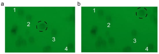
Figure 12.
Trapping process of single vaterite particle. (a) Before trapping; (b) After trapping (Particles 1–4 are reference particles).
The rotational signal of the particle in the atmosphere collected with the balanced light detector is shown in the Figure 13. It is not difficult to find that its rotation speed is about 26 Hz.
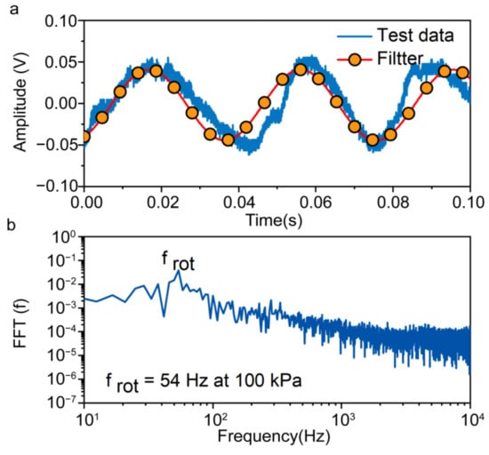
Figure 13.
Test of rotation signal in the atmosphere. (a) Time domain signals; (b) Frequency domain signals.
Using this data as a reference, the rotational signal of one particle in vacuum is obtained by trapping the particle in the microparticle vacuum chamber. The rotation speed of the particle can be achieved at 162 Hz under a vacuum environment of 3 kPa, as shown in Figure 14, which further confirms the vacuum environment of the microparticle chamber.
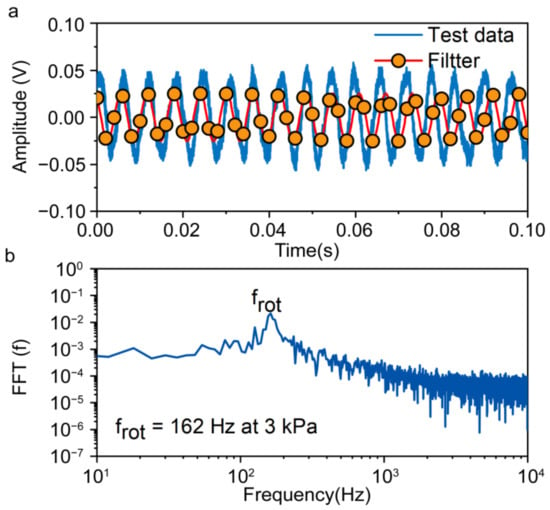
Figure 14.
Test of rotation signal under a vacuum environment of 3 kPa. (a) Time domain signals; (b) Frequency domain signals.
6. Discussion
This MEMS microparticle chamber achieves a vacuum environment, but there is still a big gap between its vacuum level and that of a large-volume mechanical vacuum chamber. This is due to the inability to use immersion cleaning after filling the particles and resonator before the bonding process, which inevitably leads to some small molecular particles being adhered to the wafer surface during air pressure calibration and other operations, which reduces the bonding effect to some extent. In addition, the random nature of the trapping process will also interfere with the excitation trapping of microparticles within the chamber, as the optical characteristics of different particles vary. Further work could focus on optimizing the chamber preparation process and further increasing the vacuum level. The increase in vacuum level will also increase the thermal motion of the particles, making them more difficult to trap, so an attempt could be made to select a group of particles with similar morphology and optical characteristics before the filling process to increase the success rate of particle trapping.
7. Conclusions
A MEMS microparticle chamber with built-in vacuum measurement with a size of 15 mm × 12 mm × 1.2 mm is presented in this study. Differently from the traditional MEMS packaging devices, the microparticle chamber is filled with particles, which increases the difficulty of the bonding process and affects the improvement of vacuum level. For this reason, this paper proposes a structure with a glass sheet with a hole and a piece of tinfoil attached to it. It solves the problem of particle contamination to a certain extent. The test calibration of the vacuum level of the MEMS microparticle chamber is achieved by using an electromagnetic excitation and a laser vibrometer system without outer lead bonding. This approach avoids the design process for the circuit and complex sensitive cell of traditional MEMS vacuum chambers, while reducing the difficulty of vacuum testing.
The results show that the microparticle chamber has a built-in vacuum level measurement function and can achieve a certain degree of vacuum packaging effect. This method of preparing and testing MEMS microparticle chambers can provide a certain vacuum environment with small volume that is easy to manufacture in bulk for multidisciplinary fields, including optical levitation and laser-induced rotation technology.
Author Contributions
Conceptualization, Y.W. and D.X.; methodology, J.P.; software, J.P.; validation, Y.W., D.X. and K.Z.; formal analysis, J.P.; investigation, J.P.; resources, Y.W.; data curation, J.P.; writing—original draft preparation, J.P.; writing—review and editing, J.P.; visualization, J.P.; supervision, K.Z.; project administration, Y.W.; funding acquisition, Y.W. All authors have read and agreed to the published version of the manuscript.
Funding
This research was funded by the National Natural Science Foundation of China, grant No. 51975579.
Institutional Review Board Statement
Not applicable.
Informed Consent Statement
Not applicable.
Data Availability Statement
The data that support the presented results are available from the corresponding author upon reasonable request.
Conflicts of Interest
The authors declare no conflict of interest.
References
- D’este, E.; Baj, G.; Beuzer, P.; Ferrari, E.; Pinato, G.; Tongiorgi, E.; Cojoc, D. Use of optical tweezers technology for long-term, focal stimulation of specific subcellular neuronal compartments. Integr. Biol. 2011, 3, 568–577. [Google Scholar] [CrossRef] [PubMed]
- Zhang, Y.; Wu, X.; Min, C.; Zhu, S.; Urbach, H.P.; Yuan, X. Engineered tumor cell apoptosis monitoring method based on dynamic laser tweezers. BioMed Res. Int. 2014, 2014, 279408. [Google Scholar] [CrossRef] [PubMed]
- Monteiro, F.; Afek, G.; Carney, D.; Krnjaic, G.; Wang, J.; Moore, D.C. Search for Composite Dark Matter with Optically Levitated Sensors. Phys. Rev. Lett. 2020, 125, 181102. [Google Scholar] [CrossRef] [PubMed]
- Gonzalez-Ballestero, C.; Aspelmeyer, M.; Novotny, L.; Quidant, R.; Romero-Isart, O. Levitodynamics: Levitation and control of microscopic objects in vacuum. Science 2021, 374, eabg3027. [Google Scholar] [CrossRef]
- Delic, U.; Reisenbauer, M.; Dare, K.; Grass, D.; Vuletic, V.; Kiesel, N.; Aspelmeyer, M. Cooling of a levitated nanoparticle to the motional quantum ground state. Science 2020, 367, 892–895. [Google Scholar] [CrossRef] [PubMed]
- Rider, A.D.; Blakemore, C.P.; Gratta, G.; Moore, D.C. Single-beam dielectric-microsphere trapping with optical heterodyne detection. Phys. Rev. A 2018, 97, 013842. [Google Scholar] [CrossRef]
- Monteiro, F.; Ghosh, S.; Fine, A.G.; Moore, D.C. Optical levitation of 10-ng spheres with nano-g acceleration sensitivity. Phys. Rev. A 2017, 96, 063841. [Google Scholar] [CrossRef]
- Arita, Y.; Mazilu, M.; Dholakia, K. Laser-induced rotation and cooling of a trapped microgyroscope in vacuum. Nat. Commun. 2013, 4, 2374. [Google Scholar] [CrossRef] [PubMed]
- Arita, Y.; Simpson, S.H.; Zemánek, P.; Dholakia, K. Coherent oscillations of a levitated birefringent microsphere in vacuum driven by nonconservative rotation-translation coupling. Sci. Adv. 2020, 6, eaaz9858. [Google Scholar] [CrossRef] [PubMed]
- Xie, S.; Sharma, A.; Romodina, M.; Joly, N.Y.; Russell, P.S.J. Tumbling and anomalous alignment of optically levitated anisotropic microparticles in chiral hollow-core photonic crystal fiber. Sci. Adv. 2021, 7, eabf6053. [Google Scholar] [CrossRef] [PubMed]
- Bruce, G.D.; Rodriguez-Sevilla, P.; Dholakia, K. Initiating revolutions for optical manipulation: The origins and applications of rotational dynamics of trapped particles. Adv. Phys. X 2021, 6, 1838322. [Google Scholar] [CrossRef]
- Kim, K.P.; Lee, K.S.; Yang, H.L.; Kim, M.K.; Kim, H.K.; Kim, G.H.; Kim, K.M.; Kim, H.T.; Kim, K.H.; Park, M.K.; et al. Overview of the KSTAR vacuum pumping system. Fusion Eng. Des. 2009, 84, 1038–1042. [Google Scholar] [CrossRef]
- Hasegawa, M.; Chutani, R.K.; Gorecki, C.; Boudot, R.; Dziuban, P.; Giordano, V.; Clatot, S.; Mauri, L. Microfabrication of cesium vapor cells with buffer gas for MEMS atomic clocks. Sens. Actuat. A Phys. 2011, 167, 594–601. [Google Scholar] [CrossRef]
- Perez, M.A.; Nguyen, U.; Knappe, S.; Donley, E.A.; Kitching, J.; Shkel, A.M. Rubidium vapor cell with integrated Bragg reflectors for compact atomic MEMS. Sens. Actuat. A Phys. 2009, 154, 295–303. [Google Scholar] [CrossRef]
- Liew, L.-A.; Moreland, J.; Gerginov, V. Wafer-level filling of microfabricated atomic vapor cells based on thin-film deposition and photolysis of cesium azide. Appl. Phys. Lett. 2007, 90, 114106. [Google Scholar] [CrossRef]
- Noor, R.M.; Kulachenkov, N.; Asadian, M.H.; Shkel, A.M. Study on Mems Glassblown Cells for NMR Sensors. In Proceedings of the 2019 IEEE International Symposium on Inertial Sensors and Systems (INERTIAL), Naples, FL, USA, 1 April 2019. [Google Scholar]
- Noor, R.M.; Shkel, A.M. MEMS Components for NMR Atomic Sensors. J. Microelectromech. Syst. 2018, 27, 1148–1159. [Google Scholar]
- Leedy, K.D.; Strawser, R.E.; Cortez, R.; Ebel, J.L. Thin-Film Packaged RF MEMS Switches. J. Microelectromech. Syst. 2007, 16, 304–309. [Google Scholar] [CrossRef]
- Stark, B.H.; Najafi, K. A low-temperature thin-film electroplated metal vacuum package. J. Microelectromech. Syst. 2004, 13, 147–157. [Google Scholar] [CrossRef]
- Kaajakari, V.; Kiihamäki, J.; Oja, A.; Pietikäinen, S.; Kokkala, V.; Kuisma, H. Stability of wafer level vacuum packaged single-crystal silicon resonators. Sens. Actuat. A Phys. 2006, 130–131, 42–47. [Google Scholar] [CrossRef][Green Version]
- Junseok, C.; Stark, B.H.; Najafi, K. A micromachined Pirani gauge with dual heat sinks. IEEE Trans. Adv. Packag. 2005, 28, 619–625. [Google Scholar] [CrossRef]
- Wei, D.; Fu, J.; Liu, R.; Hou, Y.; Liu, C.; Wang, W.; Chen, D. Highly Sensitive Diode-Based Micro-Pirani Vacuum Sensor with Low Power Consumption. Sensors 2019, 19, 188. [Google Scholar] [CrossRef] [PubMed]
- Gan, Z.; Huang, D.; Wang, X.; Lin, D.; Liu, S. Getter free vacuum packaging for MEMS. Sens. Actuat. A Phys. 2009, 149, 159–164. [Google Scholar] [CrossRef]
- Wang, X.; Liu, C.; Zhang, Z.; Liu, S.; Luo, X. A micro-machined Pirani gauge for vacuum measurement of ultra-small sized vacuum packaging. Sens. Actuat. A Phys. 2010, 161, 108–113. [Google Scholar] [CrossRef]
- Topalli, E.S.; Topalli, K.; Alper, S.E.; Serin, T.; Akin, T. Pirani Vacuum Gauges Using Silicon-on-Glass and Dissolved-Wafer Processes for the Characterization of MEMS Vacuum Packaging. IEEE Sens. J. 2009, 9, 263–270. [Google Scholar] [CrossRef]
- Mironov, A.E.; Yu, N.; Park, S.; Tuggle, M.; Gragg, J.; Kucera, C.; Hawkins, T.; Ballato, J.; Eden, J.G.; Dragic, P. All optical fiber thermal vacuum gauge. J. Phys. Photonics 2020, 2, 014006. [Google Scholar] [CrossRef]
- Meng, G.; Zhang, W. Micro-Electro-Mechanical-System Dynamics, 1st ed.; Science Press: Beijing, China, 2008; pp. 156–187. [Google Scholar]
- Zener, C. Elasticity and Anelasticity of Metals, 1st ed.; University Chicago Press: Chicago, IL, USA, 1948; pp. 175–180. [Google Scholar]
- Liu, C.; Huang, Q. Foundations of MEMS, 2nd ed.; China Machine Press: Beijing, China, 2011; pp. 133–154. [Google Scholar]
Publisher’s Note: MDPI stays neutral with regard to jurisdictional claims in published maps and institutional affiliations. |
© 2022 by the authors. Licensee MDPI, Basel, Switzerland. This article is an open access article distributed under the terms and conditions of the Creative Commons Attribution (CC BY) license (https://creativecommons.org/licenses/by/4.0/).