Article

Liquid–Liquid Interfacial Self-Assembly of Au-Ag Nanoparticles for High-Performance SERS Detection of Thiram in Environmental Water Samples


1
College of Optical and Electronic Technology, China Jiliang University, Hangzhou 310018, China
2
Key Laboratory of Microbiological Metrology, Measurement & Bio-Product Quality Security, State Administration for Market Regulation, College of Life Sciences, China Jiliang University, Hangzhou 310018, China
3
Stem Cell Translation Laboratory, Shanxi Bethune Hospital, Shanxi Academy of Medical Sciences, Tongji Shanxi Hospital, Third Hospital of Shanxi Medical University, Taiyuan 030032, China
*
Authors to whom correspondence should be addressed.
Photonics 2026, 13(5), 507; https://doi.org/10.3390/photonics13050507
Submission received: 20 April 2026 / Revised: 15 May 2026 / Accepted: 19 May 2026 / Published: 21 May 2026
(This article belongs to the Special Issue Novel Developments in Optoelectronic Materials and Devices)

Abstract

Gold and silver nanoparticles have attracted extensive attention in SERS detection due to their excellent plasmonic properties. In this study, a high-performance SERS substrate was successfully prepared by a liquid–liquid self-assembly strategy. Driven by the Marangoni effect, Au-Ag nanoparticles spontaneously form a uniform and dense monolayer structure on the silicon wafer, constructing an efficient plasmon “hotspot” region, which significantly improves the detection sensitivity of the substrate. The performance of the SERS substrate was systematically evaluated using CV and Me B as Raman probe molecules. The results show that the substrate exhibits an excellent enhancement effect and good SERS sensitivity for both probe molecules. The characteristic vibration peak can be clearly identified, and the detection limit (LOD) of crystal violet is 6.76 × 10⁻¹¹ M. The substrate was applied to detect thiram residues in lake water with a LOD of 1.084 × 10⁻⁷ M, achieving highly sensitive detection. This study shows that Au-Ag nanoparticles deposited on silicon wafers by liquid–liquid self-assembly strategy can be used as a high-performance SERS substrate. It can be used for rapid and sensitive detection of thiram pesticide residues in water, and provides an efficient and feasible analysis tool for water environment safety monitoring.

1. Introduction

In agricultural production, pests, diseases, and weeds are the main threats to the healthy growth of plants. As an effective control measure, pesticides are widely used to control or eliminate pests and weeds, and to regulate the growth of plants and insects. Thiram is a dithiocarbamate fungicide, which is commonly used in seed treatment and foliar spraying. After application, it can enter surface water through rainfall runoff, soil leaching and atmospheric deposition. Moreover, the substance has a stimulating effect on the respiratory tract and skin, and drinking after long-term exposure may cause allergic reactions, which is a dangerous chemical (UN 3077) [1,2]. Although pesticides are crucial for ensuring the yield and quality of crops, their excessive use or improper application has brought severe environmental and health problems [3]. Some pesticides leave residues in soil, water sources, and plants. These residues may accumulate in agricultural products and affect human health through respiration and skin contact [4]. Among them, pesticide residues in water bodies (such as lake water) directly threaten drinking water safety and aquatic ecosystems. Studies have even shown that mixed exposure to multiple pesticides can significantly increase the risk of cancer in humans and cause potential damage to human organs such as the liver [2].
Currently, traditional pesticide detection methods, such as gas chromatography (GC) and gas chromatography-mass spectrometry (GC-MS) [5], offer accurate detection. However, their complex sample pretreatment processes, expensive instruments, and professional operational requirements limit their practicality for large-scale on-site detection, failing to meet the needs for rapid screening of pesticide residues in water environments. Therefore, developing a rapid, sensitive, and low-cost pesticide residue detection technology is of great significance for ensuring food safety, protecting water environments, and safeguarding human health.
Surface-enhanced Raman scattering (SERS) is an advanced analytical technique that significantly improves the detection sensitivity of molecular vibrational modes by tremendously enhancing the Raman scattering signals through the electromagnetic field generated on metal surfaces or metal nanoparticles [6]. This technology can provide high-resolution fingerprint vibrational information for pesticide residues, helping researchers identify and quantify various pesticide molecules. It features advantages such as high sensitivity, rapid detection speed, low sample consumption, and non-destructive detection [7,8,9]. It overcomes the defects of weak scattering signals and large background interference in traditional Raman spectroscopy, enabling the rapid qualitative and quantitative detection of trace substances [10]. With the continuous development of SERS technology, it has been widely applied in the field of rapid pesticide residue detection, particularly showing great potential in ensuring food safety and environmental monitoring [11].
The increasing application of SERS technology in pesticide residue detection proves its importance as an efficient and sensitive alternative detection method. Kang et al. [12] prepared a porous Au-based SERS substrate via calcination for the detection of environmental pollutants; Zhang et al. [13] fabricated a SERS substrate based on Au trihedral nanostructures for the sensitive detection of fentanyl; Xu et al. [14] synthesized multi-spiked Au nanostructures as a SERS substrate to detect tetracycline in complex matrices. However, SERS substrates often suffer from inconsistent nanogap spacing and uneven hotspot distribution, making it difficult for their detection sensitivity and signal reproducibility to meet the demands of trace pesticide residue detection [15]. Therefore, there is an urgent need to develop suitable methods to prepare SERS substrates with high-density hotspots and excellent uniformity.
Interfacial self-assembly methods require relatively simple synthesis equipment. SERS substrates prepared by this method possess ordered periodic structures and uniform, abundant hotspots [15], which can effectively solve the problem of uneven hotspot distribution in traditional substrates. Liquid–liquid self-assembly, an important branch of liquid-phase self-assembly, utilizes the incompatibility between the dispersed phase and the continuous phase in an emulsion system. By adjusting conditions such as temperature, pH, and concentration, the dispersed phase spontaneously forms an ordered structure within the continuous phase [16]. Although pure silver nanoparticles offer superior SERS cross-sections compared to gold, they are highly susceptible to oxidation under ambient conditions, severely limiting their practical application. To address this issue, Liu et al. constructed a three-dimensional Ag NPs-TOCNF/PAAM hydrogel SERS platform for thiram detection, where the hydrogel encapsulation effectively protected the Ag NPs from oxidation and improved substrate stability. However, despite its excellent enhancement performance, the fabrication process of this substrate is relatively complex, and the uniformity of the hydrogel structure is difficult to precisely control, which may compromise signal reproducibility [17]. On the other hand, Wang et al. developed a SERS substrate based on colloidal gold nanoparticles (AuNPs) for label-free thiram detection. However, the introduction of aggregating agents led to uncontrollable aggregation of the nanoparticles, resulting in poor signal stability and reproducibility. Moreover, the enhancement factor of Au-based systems is much lower than that of Ag-based counterparts, leading to limited signal intensity, a relatively high limit of detection, and room for further improvement in sensitivity [18]. It has been successfully used to prepare SERS substrates such as monolayer metal nanoparticle films, significantly improving the detection sensitivity and stability of the substrates [19,20,21].
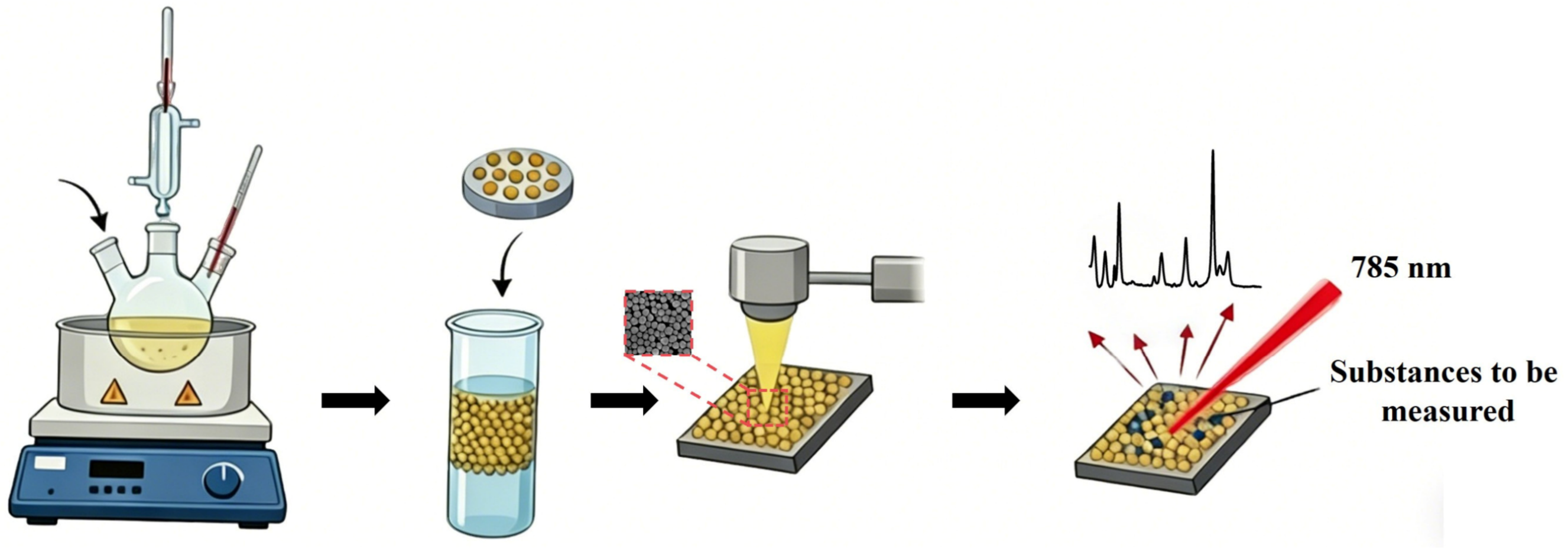
Based on the above background, this study employed a liquid–liquid self-assembly strategy to deposit Au-Ag nanoparticles onto a silicon wafer, successfully fabricating a high-performance SERS substrate. Subsequently, Au-Ag NPs were used to detect crystal violet, methylene blue, and thiram. Furthermore, the substrate was immersed in lake water to sample and detect thiram (Figure 1).

2. Materials and Methods

2.1. Synthesis of Gold-Silver Nanoparticles (Au-Ag NPs)

Au-Ag NPs were synthesized using a one-pot citrate reduction method. First, 77.5 mL of ultrapure water was heated to boiling in a constant-temperature oil bath. Then, 1 mL of 0.01 M AgNO3 (Shanghai Lingfeng Chemical Reagent Co., Ltd., Shanghai, China) and 1 mL of 0.01 M HAuCl4 (Sinopharm Chemical Reagent Co., Ltd., Shanghai, China) were added to the reaction flask. After heating and stirring for about 10 min, 8 mL of 1% sodium citrate (Thermo Fisher Scientific, Waltham, MA, USA) was rapidly added to the solution. The mixture was continuously heated and stirred for 15 min, during which the solution gradually turned orange-red. The mixture was then removed from the heating plate and allowed to cool naturally to room temperature to obtain Au-Ag NPs. The prepared Au-Ag NPs were centrifuged at 3500 r/min for 15 min, and the supernatant was collected to remove large and irregularly agglomerated nanoparticles. Ultrapure water was then added to dilute it to the original volume, followed by ultrasonication for 10 min. The solution was further centrifuged at 6500 r/min for 15 min; the precipitate was retained, diluted to the original volume, and sonicated again for 10 min. The collected Au-Ag NPs were stored sealed in the dark at 4 °C for future use.

2.2. Interfacial Self-Assembly of Au-Ag NPs

The liquid–liquid interfacial self-assembly of the nanoparticles was achieved using a simplified method reported in the literature [22]. In a 5 mL centrifuge tube, 800 µL of dichloromethane and 100 µL of the Au-Ag NPs suspension were sequentially added, followed by the addition of deionized water to bring the total volume to 3 mL. The mixture was vigorously vortexed for 1 min and left to stand for 30 s. Then, 400 µL of n-hexane was slowly added. At this time, a monolayer film of Au-Ag NPs with a bright golden luster spontaneously formed at the interface between the water phase and n-hexane. The upper n-hexane phase was removed, and a silicon wafer was vertically immersed below the monolayer film and slowly withdrawn. The nanoparticle monolayer was uniformly attached to the surface of the silicon wafer. After natural drying, it was used for subsequent characterization and experiments.

2.3. SERS Performance Testing of Au-Ag NPs

Using crystal violet (CV) and methylene blue (MeB) as probe molecules, a series of standard solutions with a concentration range of 10−6 to 10−10 M was prepared. The Au-Ag NPs silicon wafer samples were immersed in 100 µL of CV and MeB solutions of different concentrations, incubated overnight, taken out, and dried at room temperature before SERS detection. Raman spectra were collected using an XploRA PLUS confocal Raman spectrometer (HORIBA France SAS, Longjumeau, France) with the following experimental conditions: 785 nm laser, 50× objective lens, 600 g/mm grating, and an acquisition time of 10 s for all Raman spectra.
For the detection of thiram, the thiram solution was diluted with an aqueous methanol solution (volume ratio 1:1). The Au-Ag NPs substrate was fully immersed in thiram solutions of different concentrations, then dried and measured under the same experimental conditions described above.
To detect thiram residues in water bodies (lake water), lake water was first collected (from the university’s artificial lake) and used to dilute thiram into solutions of various concentrations. The Au-Ag NPs silicon wafer samples were immersed in these solutions, dried, and then tested.

2.4. Instruments

Scanning electron microscopy (SEM) images were obtained on ZEISS Gemini 360 (Carl Zeiss Microscopy GmbH, Jena, Germany). Transmission electron microscopy (TEM) images and energy dispersive spectrometer (EDS) were measured by JEOL JEM-F200 (JEOL Ltd., Tokyo, Japan). UV–vis absorption spectra were collected by a UV–vis spectrophotometer (TU–1901, PERSEE, Beijing, China). Raman spectra were acquired using a HORIBA XploRA PLUS confocal Raman microscope (HORIBA France SAS, Longjumeau, France) equipped with a 50× objective (NA = 0.5). A 785 nm laser was used as the excitation source with a laser power at the sample of approximately 1.9 mW and a spot diameter of about 1.92 μm. All measurements were performed with a holographic grating of 600 grooves/mm, providing a spectral resolution of approximately 8 cm−1. The SERS spectra were collected with an accumulation time of 10 s. For spectral preprocessing, baseline correction was carried out using the baseline calibration module in the instrument software.

2.5. Enhancement Factor (EF)

The core mechanism of SERS technology originates from the localized surface plasmon resonance (LSPR) [23] generated by noble metal (e.g., gold, silver) nanostructures under incident light excitation. The extremely strong localized electromagnetic field induced by this resonance can amplify the Raman scattering signals of molecules adsorbed on or near the metal surface by orders of magnitude, with conventional enhancement efficiencies reaching 106 or even higher. In SERS research, EF serves as a key evaluation index [24] and is widely used to quantitatively characterize the SERS activity of different nanostructures and substrate materials.
The EF is defined as the intensity ratio of the SERS signal to the normal Raman signal after normalizing the number of molecules. It intuitively reflects the synergistic electromagnetic and chemical enhancement effects of the nanostructures on the Raman signal. The calculation formula is as follows:
EF = I S E R S / C S E R S I R S / C R S ,
where CSERS and CRS represent the concentration of CV on the target SERS substrate and the reference substrate (bare Si substrate), respectively. ISERS and IRS denote the corresponding Raman intensities.

3. Results and Discussion

3.1. Characterization of Au-Ag NPs

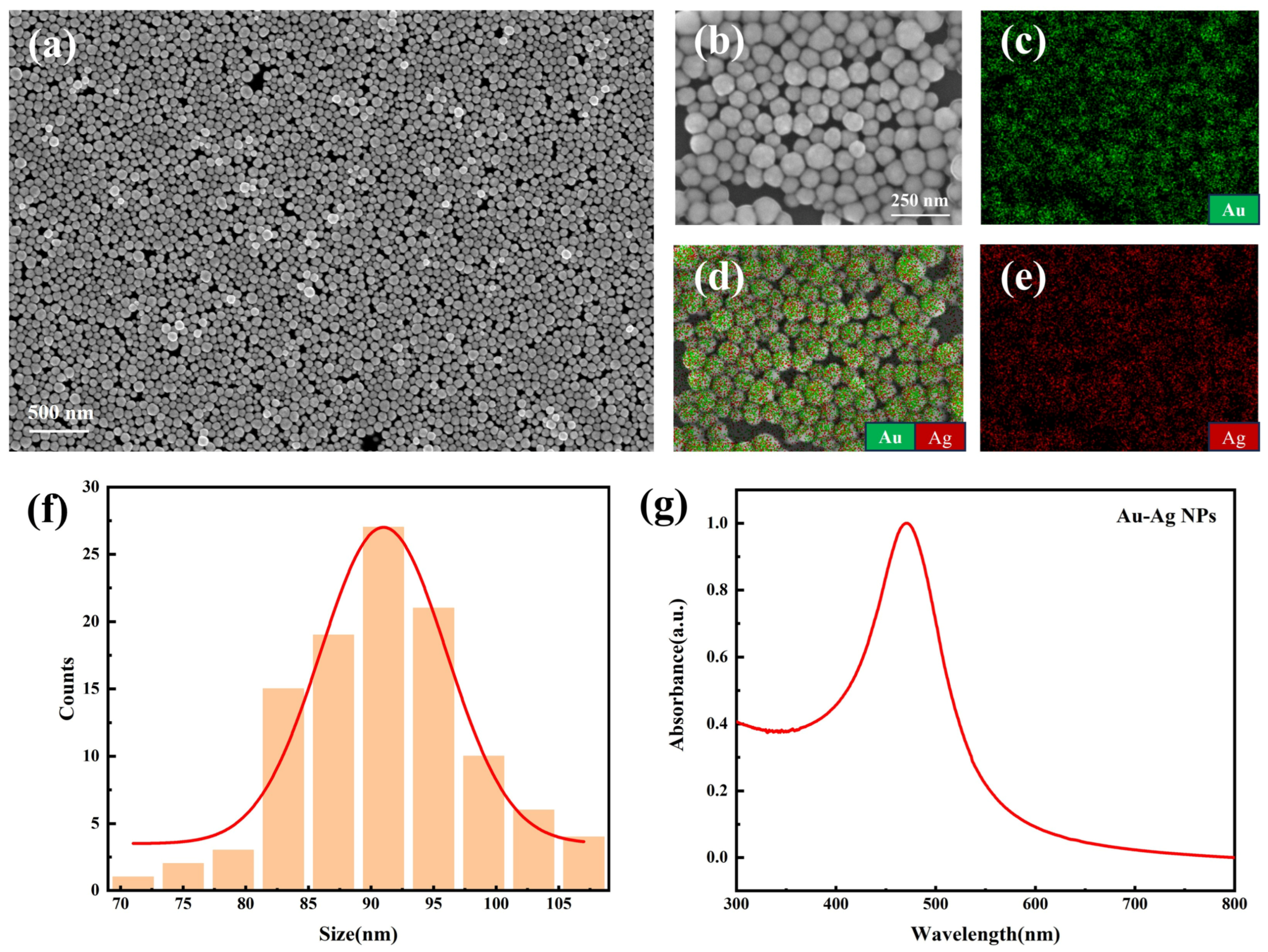
In this experiment, Au-Ag bimetallic nanoparticles (Au-Ag NPs) were successfully prepared using the liquid-phase sodium citrate reduction method. Figure 2 shows the scanning electron microscopy (SEM) images of the Au-Ag NPs. The morphological characterization results reveal that the Au-Ag NPs exhibit a regular spherical structure with uniform particle size and good dispersibility, without obvious agglomeration or irregularly shaped particles. Furthermore, energy dispersive spectrometer (EDS) elemental mapping analysis showed that Au and Ag are continuously and uniformly distributed within the field of view, and their signals highly overlap spatially. This confirms that Au and Ag have achieved uniform alloying, with no phenomenon of elemental segregation or core–shell separation. The UV-Vis absorption spectrum (Figure 2) shows that the synthesized Au-Ag NPs exhibit a single, sharp localized surface plasmon resonance (LSPR) characteristic absorption peak at approximately 470 nm, with no obvious impurity peaks or broadened tails, indicating uniform nanoparticle size and consistent optical properties. These results are highly consistent with the morphological uniformity observed by SEM, indicating the successful preparation of Au-Ag NPs.
The liquid–liquid interface (e.g., oil-water interface) provides a two-dimensional assembly platform with the lowest free energy for nanoparticles. Driven by the Marangoni effect, capillary forces, or interfacial tension gradients, nanoparticles can spontaneously arrange into dense and ordered single or multiple thin films at the interface [22]. After transferring to a solid substrate such as a silicon wafer, a uniform SERS-active layer at the centimeter scale or even larger can be obtained, greatly reducing the problem of uneven “hotspot” distribution and ensuring high signal reproducibility both within and between batches. During the self-assembly process, the gaps between nanoparticles can be controlled within the sub-nanometer to several-nanometer range, forming a large number of uniform electromagnetic field enhancement “hotspots”. Compared to random deposition or spin-coating methods, liquid–liquid self-assembly can achieve a high degree of consistency in particle spacing. As shown in the SEM image of the Au-Ag NPs self-assembled on the silicon wafer, it is clearly visible that the gold-silver nanoparticles form a large-area dense monolayer arrangement on the silicon wafer surface. The particle sizes are uniform, the average size is about 90.95 ± 6.77 nm, and the inter-particle gaps are consistent (about 2–5 nm), presenting a typical hexagonal or disordered close-packed structure. These uniform sub-nanometer gaps constitute a large number of evenly distributed SERS “hotspots”, which are conducive to generating strong and highly reproducible electromagnetic field enhancement effects, laying a structural foundation for the subsequent highly sensitive detection of trace pesticide molecules.

3.2. SERS Properties of Au-Ag NPs

Among plasmonic metals, Au and Ag possess highly unique and superior properties [25,26]. Au is the most stable oxidation-resistant noble metal, while Ag has excellent plasmonic properties. The bulk damping of Ag is negligible, and based on their SERS intensity ratios, the average scattering cross-section of Ag binding sites is much higher than that of Au [27,28]. However, Ag-based SERS platforms are unstable due to oxidation when exposed to environmental conditions, which limits their SERS applicability [29]. This is especially true when Ag NPs do not contain surfactants. Because both Au and Ag have a face-centered cubic (FCC) structure with similar atomic radii, the synergistic effect of these two metals can improve their stability and SERS sensitivity [30,31,32].
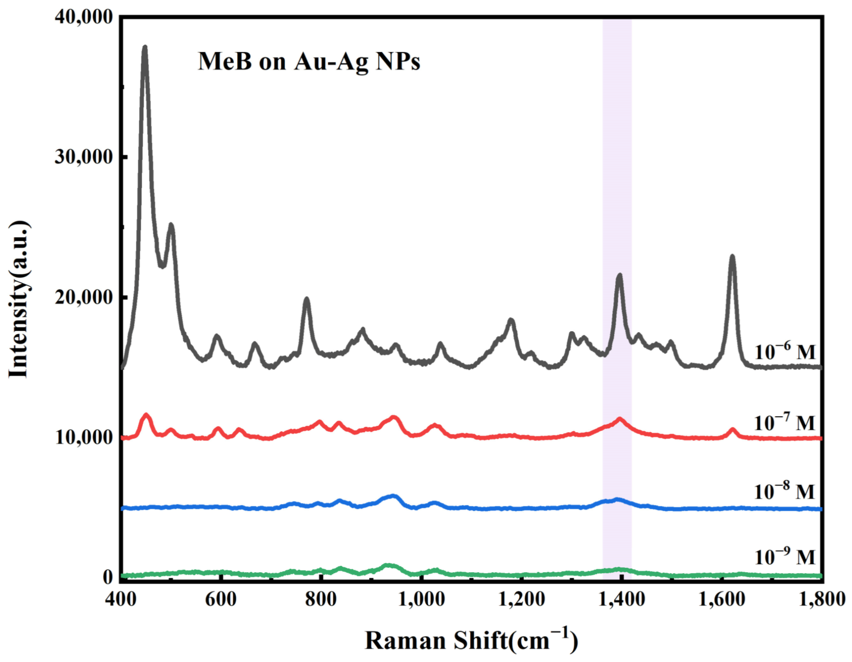
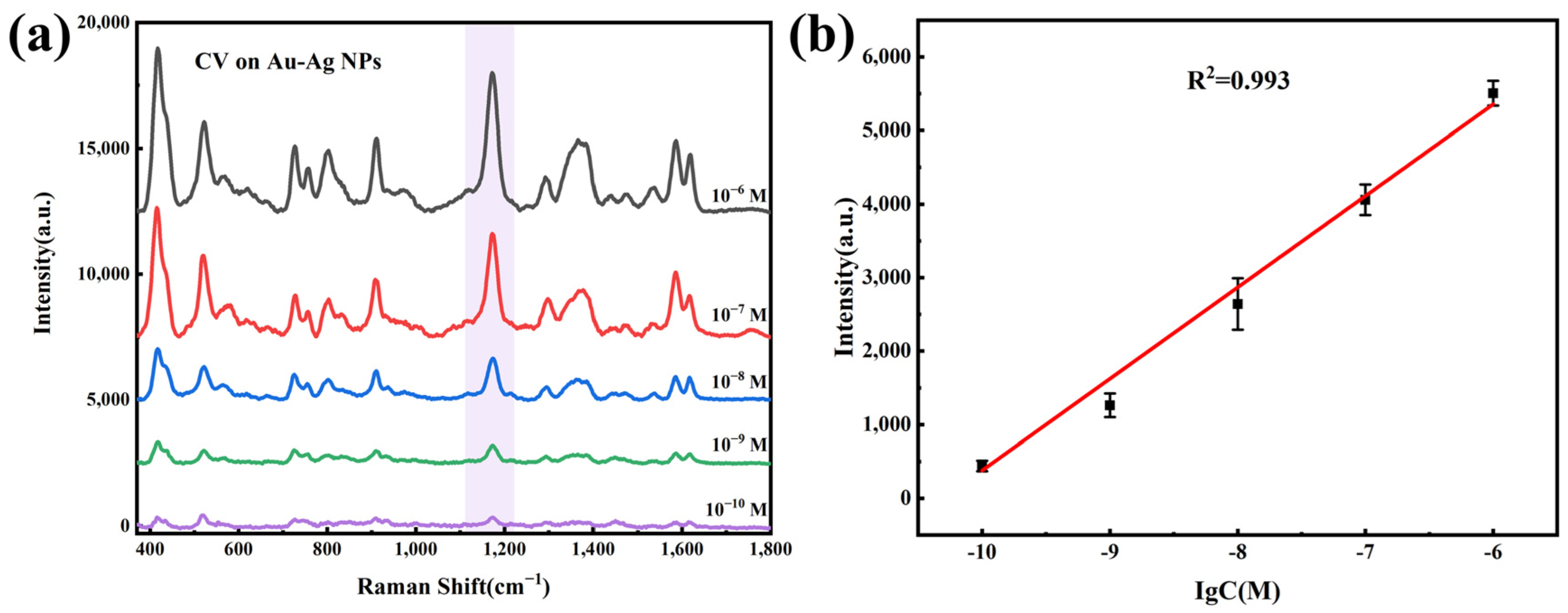
CV and MeB are two common Raman reporter molecules [33] used to preliminarily evaluate the SERS performance of Au-Ag NPs. Figure 3 shows the Raman spectra of different concentrations of MeB on the Au-Ag NPs substrate. Significantly enhanced characteristic peaks are observed at 1396 and 1623 cm−1, corresponding to the longitudinal and transverse stretching modes of the benzene ring, respectively. Additional characteristic peaks are present at 887 cm−1 (out-of-plane bending of C-H bonds) and 1302 cm−1 (stretching mode of C=C bonds) [34,35]. CV dye molecules were similarly used to illustrate the SERS effect of Au-Ag NPs. As shown in Figure 4a, the Raman spectrum of CV molecules on the substrate shows typical characteristic peaks, including the strongest peak at 1179 cm−1, corresponding to the in-plane vibration of the C-H ring; peaks at 733, 761, and 807 cm−1 (out-of-plane vibration of the C-H ring); 918 cm−1 (ring skeleton vibration); 1373 cm−1 (N-phenyl stretching); and 1297, 1585, and 1618 cm−1 (C-C ring stretching) [36].
To better characterize the performance of the developed SERS substrate, the limit of detection (LOD) was expressed as the sample blank value plus three standard deviations (s.d.). The limit of quantitation (LOQ) was expressed as the sample blank value plus ten s.d. [37].
LOD(y) = mean of blank + 3 × s.d. of blank
LOQ(y) = mean of blank + 10 × s.d. of blank
The LOD and LOQ for CV were calculated to be 6.76 × 10⁻¹¹ M and 1.02 × 10⁻¹⁰ M, respectively.
Subsequently, we collected the Raman characteristic peak intensity of CV molecules at 1179 cm−1 and plotted a linear fit of the peak intensity against the logarithm of the concentration. As shown by the linear fitting results in Figure 4b, the peak intensity (at 1179 cm−1) for the detection of CV using Au-Ag NPs exhibits a good linear relationship with the logarithm of the concentration, further indicating that Au-Ag NPs, as a SERS substrate, can be used for quantitative analytical detection, which is a major advantage of SERS detection technology.
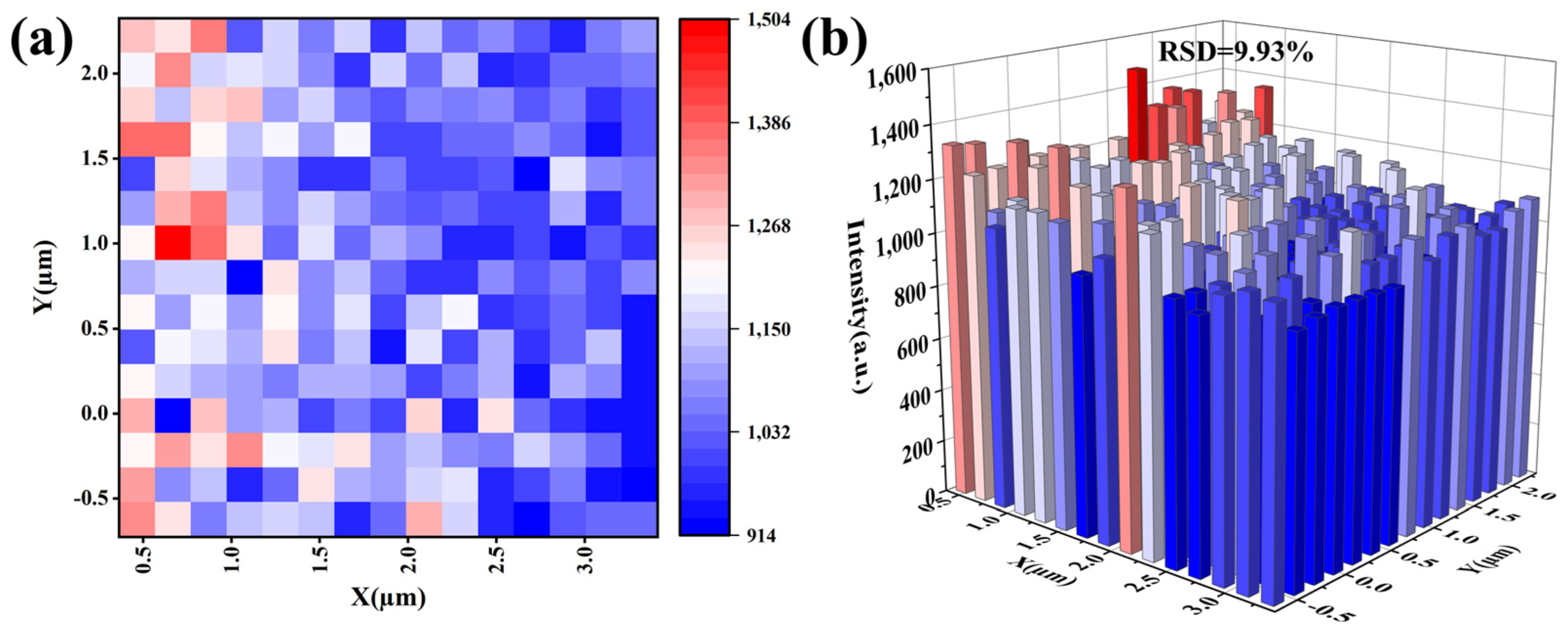
In the evaluation of spatial uniformity, SERS detection was performed on 225 points within a 3.5 µm × 3.5 µm area on the Au-Ag NPs substrate. As shown in Figure 5a,b, the spatial uniformity of this region was assessed by the relative standard deviation (RSD). The result showed an RSD of 9.93%, indicating that the Au-Ag NPs possess excellent spatial uniformity.
Next, we calculated the SERS Enhancement Factor (EF) for CV on the Au-Ag NPs substrate. When CSERS was 1 × 10¹⁰ M, the measured ISERS was approximately 439.6 counts/s; when CRS was 1 × 10³ M, the obtained IRS was approximately 357 counts/s. The calculated enhancement factor EF is about 1.23 × 10⁷. This result further proves that the Au-Ag NPs have an excellent enhancement effect on CV.

3.3. SERS Detection of Thiram

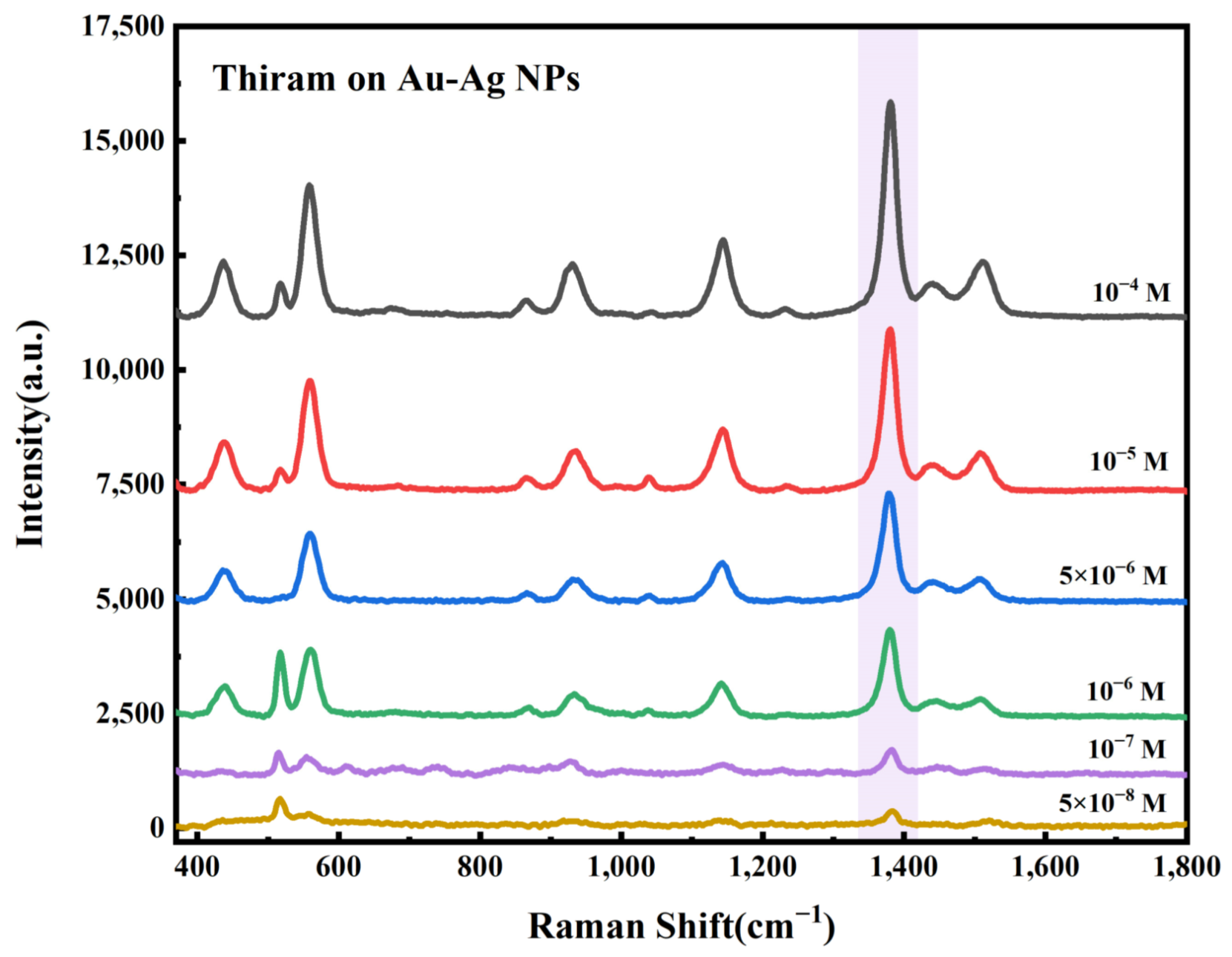
Au-Ag NPs demonstrated their SERS analytical sensitivity by being immersed in solutions of target substances. To explore the capability of Au-Ag NPs in pesticide residue detection, the common pesticide thiram was used as the target molecule. Figure 6 shows the SERS spectra of different concentrations of thiram measured on the Au-Ag NPs substrate. The mode at 549 cm−1 is assigned to the S-S stretching mode. The vibrational mode around 1371 cm−1 is attributed to CH3 deformation and C-N stretching. The modes located at 1141 cm−1 and 1502 cm−1 are assigned to C-N stretching and CH3 rocking modes, respectively [38]. By measuring the Raman spectra of thiram at various concentrations (5.0 × 10⁻⁸ M~1.0 × 10⁻⁴ M) in methanol solutions, the lowest limit of detection for the Au-Ag NPs was evaluated. The results indicated that even at a concentration of 5.42 × 10⁻⁸ M, the characteristic peaks of thiram could still be detected.
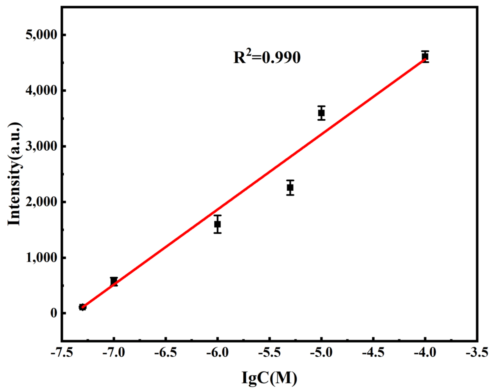
The linear relationship between the thiram concentration and the mode intensity of the characteristic peak, as well as spatial uniformity, was used to evaluate the SERS performance of the Au-Ag NPs substrate. Similar to the CV detection, a linear fit was performed using lgCthiram and the intensity of the thiram marker band located at 1371 cm−1. As shown in Figure 7, by plotting the SERS intensity against the logarithm of the thiram concentration, a good linear correlation (R2 = 0.990) was established.
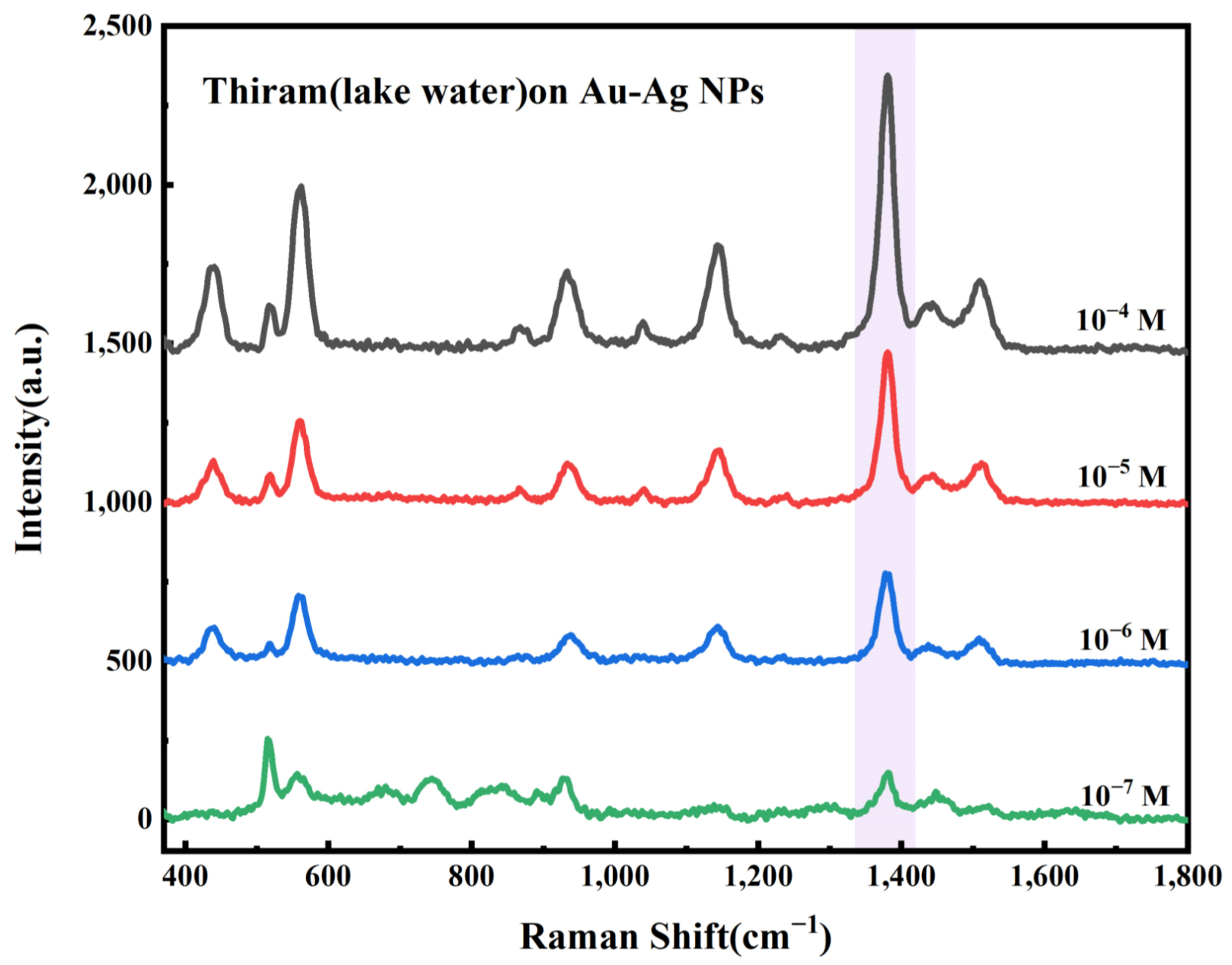
Further experiments investigated whether the prepared Au-Ag NPs substrate could be used for the detection of pesticide residues in water bodies. In brief, the Au-Ag NPs substrate was immersed in water (lake water from the university’s artificial lake) containing pesticide residues, dried, and then subjected to SERS detection. The concentrations of thiram in the lake water were 1.0 × 10⁻⁴ M, 1.0 × 10⁻⁵ M, 1.0 × 10⁻⁶ M, and 1.0 × 10⁻⁷ M, respectively. The experimental results, as shown in Figure 8, demonstrate that using a simple immersion sampling method, the Au-Ag NPs still achieved a detection limit of 1.084 × 10⁻⁷ M. After unit conversion, the detection limit was 13.0 μg/L, which was close to the maximum residue limit (10 μg/L) of a single pesticide in EU regulations. Therefore, the Au-Ag NPs substrate is expected to be used for sensitive SERS detection of pesticide residues in water.

4. Discussion

In this study, a high-performance Surface-Enhanced Raman Scattering (SERS) substrate based on Au-Ag nanoparticles (Au-Ag NPs) was successfully fabricated via a liquid–liquid interfacial self-assembly strategy. The substrate exhibits excellent SERS activity and achieves ultra-low detection concentrations for dye molecules (CV is 6.76 × 10⁻¹¹ M) and trace pesticide residues (1.084 × 10⁻⁷ M). These findings strongly support our initial hypothesis that combining the synergistic plasmonic properties of bimetallic alloys with the ordered arrangement of interfacial self-assembly can significantly enhance both the sensitivity and reproducibility of SERS detection.
Our results offer a compelling solution to a long-standing dilemma in SERS substrate design: the trade-off between signal intensity and substrate stability. By synthesizing Au-Ag bimetallic alloys, this study successfully leveraged the Face-Centered Cubic (FCC) structural compatibility of both metals. The EDS and UV-Vis characterizations confirm a uniform alloying without phase segregation. This synergistic interaction not only preserves the exceptional electromagnetic enhancement of Ag but also inherits the chemical stability of Au, yielding a robust platform for reliable sensing.
Furthermore, the implementation of the liquid–liquid interfacial self-assembly method effectively addressed the pervasive issue of uneven “hotspot” distribution found in many traditional SERS substrates (e.g., random deposition or spin-coating). Driven by the Marangoni effect, the Au-Ag NPs spontaneously formed a highly ordered, dense monolayer with uniform sub-nanometer to few-nanometer gaps (2–5 nm). These precise interparticle gaps generated dense, uniformly distributed electromagnetic “hotspots” via strong localized surface plasmon resonance (LSPR). This structural superiority is quantitatively reflected in our results: the substrate achieved a remarkable Enhancement Factor (EF) of 1.23 × 10⁷ and an excellent spatial uniformity with a Relative Standard Deviation (RSD) of 9.93%. The strong linear correlation observed for both CV and Thiram further underscores its reliability for precise quantitative analysis.
From a practical perspective, the implications of this study are highly significant for environmental monitoring and food safety. Traditional pesticide detection techniques, such as Gas Chromatography-Mass Spectrometry (GC-MS), offer high accuracy but are hindered by complex sample pretreatment, high costs, and an inability to perform rapid on-site screening. In contrast, our Au-Ag NPs SERS substrate successfully detected thiram in real complex water matrices (lake water) with a minimum detection concentration of 1.084 × 10⁻⁷ M. This demonstrates the substrate’s strong anti-interference capability against environmental matrices, proving it to be a rapid, cost-effective, and highly sensitive alternative for the routine screening of pesticide residues in agricultural and environmental water bodies.
While the current findings demonstrate significant potential for water quality monitoring, future research should explore the multiplexed detection capabilities of this substrate to identify complex mixtures of multiple pesticides simultaneously. Additionally, transferring the self-assembled Au-Ag NPs monolayer onto flexible and adhesive substrates (such as polymer films or filter paper) could expand its application to flexible swabbing, enabling direct, non-destructive extraction and detection of pesticide residues on the irregular surfaces of fruits and vegetables. Finally, integrating these high-performance substrates with miniaturized, portable Raman spectrometers will be a crucial next step toward realizing true on-site, point-of-need screening for environmental protection and agricultural safety.

5. Conclusions

In summary, this study successfully constructed a high-performance surface-enhanced Raman scattering (SERS) substrate composed of gold-silver nanoparticles (Au-Ag NPs) on a silicon surface, relying on liquid–liquid interface self-assembly technology. Benefiting from the ordered assembly driven by the Marangoni effect, the substrate forms a dense nanoparticle monolayer with uniform sub-nanometer gaps, constructing high-density and highly uniform plasmonic “hotspots”. Combining the chemical stability of gold with the strong plasmonic enhancement effect of silver, it significantly improves SERS detection sensitivity and signal reproducibility. Performance characterizations show that the substrate achieves a limit of detection of 6.76 × 10⁻¹¹ M for CV and also exhibits excellent enhancement effects for MeB. Applied to the detection of thiram residues in actual water bodies such as lake water, it achieves highly sensitive quantitative analysis at 1.084 × 10⁻⁷ M, effectively resisting water matrix interference and fulfilling on-site rapid detection requirements. The findings demonstrate that the liquid–liquid self-assembled Au-Ag NPs silicon-based SERS substrate holds outstanding application value in monitoring pesticide residues in water environments, offering new ideas and experimental support for the controllable preparation of high-performance SERS substrates and their expansion in environmental analysis fields.

Author Contributions

Conceptualization, J.L. (Jiali Liu), L.J. and J.H.; methodology, J.L. (Jiali Liu) and Y.F.; validation, J.L. (Jiali Liu), J.L. (Jiafan Liu) and L.J.; formal analysis, J.L. (Jiali Liu), J.L. (Jiafan Liu) and L.J.; investigation, J.L. (Jiali Liu) and L.Y.; resources, J.L. (Jiafan Liu) and Z.M.; data curation, J.L. (Jiali Liu); writing—original draft preparation, J.L. (Jiali Liu) and J.L. (Jiafan Liu); writing—review and editing, J.L. (Jiafan Liu) and J.H.; visualization, J.L. (Jiafan Liu) and J.H.; supervision, J.L. (Jiafan Liu) and J.H. All authors have read and agreed to the published version of the manuscript.

Funding

This work was supported by the National Key R&D Program of China (Grant No. 2024YFF0619402), the Fundamental Research Program of Shanxi Province (Grant No. 202203021221236), Shanxi Bethune Hospital Research Program (Grant No. 2023RC12 and 2023GZRZ01) and the National Undergraduate Innovation and Entrepreneurship Training Program (Grant No. 202310356024).

Data Availability Statement

The raw date supporting the conclusions of this article will be made available by the authors on request.

Conflicts of Interest

The authors declare no conflict of interest.

References

  1. Arias-Estévez, M.; López-Periago, E.; Martínez-Carballo, E.; Simal-Gándara, J.; Mejuto, J.-C.; García-Río, L. The mobility and degradation of pesticides in soils and the pollution of groundwater resources. Agric. Ecosyst. Environ. 2008, 123, 247–260. [Google Scholar] [CrossRef]
  2. Xu, M.; Gao, Y.; Han, X.; Zhao, B. Detection of pesticide residues in food using surface-enhanced Raman spectroscopy: A review. J. Agric. Food Chem. 2017, 65, 6719–6726. [Google Scholar] [CrossRef]
  3. Hassannejad, S.; Lotfi, R.; Ghafarbi, S.P.; Oukarroum, A.; Abbasi, A.; Kalaji, H.M.; Rastogi, A. Early identification of herbicide modes of action by the use of chlorophyll fluorescence measurements. Plants 2020, 9, 529. [Google Scholar] [CrossRef] [PubMed]
  4. Reynoso, E.C.; Torres, E.; Bettazzi, F.; Palchetti, I. Trends and perspectives in immunosensors for determination of currently-used pesticides: The case of glyphosate, organophosphates, and neonicotinoids. Biosensors 2019, 9, 20. [Google Scholar] [CrossRef]
  5. Xu, X.; Wang, S.; Hou, S.; Li, Z.; Lu, Z.; Liang, S. A multi-residue method for the determination of 77 pesticides in red ginseng using quechers and gas chromatography/tandem mass spectrometry (GC-MS/MS). Agronomy 2022, 12, 2479. [Google Scholar] [CrossRef]
  6. Liu, H.; Liu, X.; Mo, L.; Chen, C.; Zhong, H.; Guo, Z.; Liu, Z. Progress in the development and application of transitional technology of surface-enhanced Raman spectroscopy. Colloid Interface Sci. Commun. 2021, 43, 100443. [Google Scholar] [CrossRef]
  7. Ding, Y.; Hao, B.; Zhang, N.; Lv, H.; Zhao, B.; Tian, Y. Rapid determination of thiram and atrazine pesticide residues in fruit and aqueous system based on surface-enhanced Raman scattering. Spectrochim. Acta Part A Mol. Biomol. Spectrosc. 2023, 285, 121873. [Google Scholar] [CrossRef] [PubMed]
  8. Fateixa, S.; Raposo, M.; Nogueira, H.; Trindade, T. A general strategy to prepare SERS active filter membranes for extraction and detection of pesticides in water. Talanta 2018, 182, 558–566. [Google Scholar] [CrossRef]
  9. Yu, H.; Guo, D.; Zhang, H.; Jia, X.; Han, L.; Xiao, W. Facile fabrication of flexible AuNPs@CDA SERS substrate for enrichment and detection of thiram pesticide in water. Spectrochim. Acta Part A Mol. Biomol. Spectrosc. 2023, 285, 121930. [Google Scholar] [CrossRef]
  10. Sun, M.; Wan, S.; Liu, Y.; Jia, Y.; Xu, H. Chemical mechanism of surface-enhanced resonance Raman scattering via charge transfer in pyridine–Ag2 complex. J. Raman Spectrosc. 2008, 39, 402–408. [Google Scholar] [CrossRef]
  11. Bhavya, M.; Shenoy, B.M.; Bhol, P.; Swain, S.; Saxena, M.; John, N.S.; Hegde, G.; Samal, A.K. Femtomolar detection of thiram via SERS using silver nanocubes as an efficient substrate. Environ. Sci. Nano 2020, 7, 3999–4009. [Google Scholar] [CrossRef]
  12. Kang, S.; Wang, W.; Rahman, A.; Nam, W.; Zhou, W.; Vikesland, P.J. Highly porous gold supraparticles as surface-enhanced Raman spectroscopy (SERS) substrates for sensitive detection of environmental contaminants. RSC Adv. 2022, 12, 32803–32812. [Google Scholar] [CrossRef]
  13. Zhang, M.; Pan, J.; Xu, X.; Fu, G.; Zhang, L.; Sun, P.; Yan, X.; Liu, F.; Wang, C.; Liu, X.; et al. Gold-trisoctahedra-coated capillary-based SERS platform for microsampling and sensitive detection of trace fentanyl. Anal. Chem. 2022, 94, 4850–4858. [Google Scholar] [CrossRef]
  14. Xu, X.; Li, T.; Liu, Y.; Zhou, L.; Li, Y.; Luo, Y.; Xu, Y.; Zhao, L.; Song, W.; Jiang, D.; et al. Engineering Assembly of Plasmonic Virus-Like Gold SERS Nanoprobe Guided by Intelligent Dual-Machine Nanodevice for High-Performance Analysis of Tetracycline. Small 2024, 20, 2309502. [Google Scholar] [CrossRef]
  15. Ding, W.; Xia, Y.; Song, H.; Li, T.; Yang, D.; Dong, A. Macroscopic superlattice membranes self-assembled from gold nanobipyramids with precisely tunable tip arrangements for SERS. Angew. Chem. Int. Ed. 2024, 63, e202401945. [Google Scholar] [CrossRef]
  16. Yang, L.; Liu, Y.; Ren, X.; Jia, R.; Si, L.; Bao, J.; Shi, Y.; Sun, J.; Zhong, Y.; Duan, P.-C.; et al. Microemulsion-assisted self-assembly of indium porphyrin photosensitizers with enhanced photodynamic therapy. ACS Nano 2024, 18, 3161–3172. [Google Scholar] [CrossRef]
  17. Liu, R.; Ren, J.; Li, J.; Wang, H.; Zhang, B.; Lu, Y.; Chen, X.; Liu, Y.; You, R. Three-dimensional Ag NPs-cellulose fiber/polyacrylamide hydrogels as a novel SERS platform for the efficient determination of thiram in fruits and juice. Colloids Surf. A-Physicochem. Eng. Asp. 2023, 677, 132377. [Google Scholar] [CrossRef]
  18. Wang, K.; Yue, Z.; Fang, X.; Lin, H.; Wang, L.; Cao, L.; Sui, J.; Ju, L. SERS detection of thiram using polyacrylamide hydrogel-enclosed gold nanoparticle aggregates. Sci. Total Environ. 2023, 856, 159108. [Google Scholar] [CrossRef] [PubMed]
  19. Fang, G.; Lin, X.; Wu, J.; Xu, W.; Hasi, W.; Dong, B. Porous nanoframe based plasmonic structure with high-density hotspots for the quantitative detection of gaseous benzaldehyde. Small 2025, 21, 2408670. [Google Scholar] [CrossRef]
  20. Lin, S.; Dong, M.; Li, C.; Lin, X.; Cong, Y.; Xu, W.; Bao, Z.; Dong, B. Machine learning-assisted ultrasensitive SERS immunoassays across wide concentration ranges toward clinical ovarian cancer diagnosis. Adv. Funct. Mater. 2025, 35, e09813. [Google Scholar] [CrossRef]
  21. Wang, X.; Hasi, W.; Lin, S.; Han, S.; Fang, G.; Lin, X. Plasmonic nanocavities with metallic spacer layers for ultrasensitive detection of Cr6+ ions. ACS Photonics 2025, 12, 3754–3762. [Google Scholar] [CrossRef]
  22. Konrad, M.P.; Doherty, A.P.; Bell, S.E. Stable and uniform SERS signals from self-assembled two-dimensional interfacial arrays of optically coupled Ag nanoparticles. Anal. Chem. 2013, 85, 6783–6789. [Google Scholar] [CrossRef]
  23. Jeon, T.Y.; Kim, D.J.; Park, S.-G.; Kim, S.-H.; Kim, D.-H. Nanostructured plasmonic substrates for use as SERS sensors. Nano Converg. 2016, 3, 18. [Google Scholar] [CrossRef] [PubMed]
  24. Ayhan, M.E. A single-step fabrication of Ag nanoparticles and CVD graphene hybrid nanostructure as SERS substrate. Microelectron. Eng. 2020, 233, 111421. [Google Scholar] [CrossRef]
  25. Andolina, C.M.; Bon, M.; Passerone, D.; Saidi, W.A. Robust, multi-length-scale, machine learning potential for Ag–Au bimetallic alloys from clusters to bulk materials. J. Phys. Chem. C 2021, 125, 17438–17447. [Google Scholar] [CrossRef]
  26. Sulaiman, D.; Alwan, A.M.; Hamoudi, W.K. Enhanced pesticides’ limit of detection using bimetallic alloys nanoparticles. J. Mater. Sci. Mater. Electron. 2021, 32, 18689–18698. [Google Scholar] [CrossRef]
  27. Klar, T.; Perner, M.; Grosse, S.; Von Plessen, G.; Spirkl, W.; Feldmann, J. Surface-plasmon resonances in single metallic nanoparticles. Phys. Rev. Lett. 1998, 80, 4249. [Google Scholar] [CrossRef]
  28. Mohammadpour, M.; Jamshidi, Z. Effect of chemical nature of the surface on the mechanism and selection rules of charge-transfer surface-enhanced Raman scattering. J. Phys. Chem. C 2017, 121, 2858–2871. [Google Scholar] [CrossRef]
  29. Han, Y.; Lupitskyy, R.; Chou, T.-M.; Stafford, C.M.; Du, H.; Sukhishvili, S. Effect of oxidation on surface-enhanced Raman scattering activity of silver nanoparticles: A quantitative correlation. Anal. Chem. 2011, 83, 5873–5880. [Google Scholar] [CrossRef] [PubMed]
  30. Tyson, C.; Bzowski, A.; Kristof, P.; Kuhn, M.; Sammynaiken, R.; Sham, T. Charge redistribution in Au-Ag alloys from a local perspective. Phys. Rev. B 1992, 45, 8924. [Google Scholar] [CrossRef]
  31. Zhang, Q.; Cobley, C.M.; Zeng, J.; Wen, L.-P.; Chen, J.; Xia, Y. Dissolving Ag from Au−Ag alloy nanoboxes with H2O2: A method for both tailoring the optical properties and measuring the H2O2 concentration. J. Phys. Chem. C 2010, 114, 6396–6400. [Google Scholar] [CrossRef]
  32. Zhu, X.; Zhuo, X.; Li, Q.; Yang, Z.; Wang, J. Gold nanobipyramid-supported silver nanostructures with narrow plasmon linewidths and improved chemical stability. Adv. Funct. Mater. 2016, 26, 341–352. [Google Scholar] [CrossRef]
  33. Yang, G.; Fang, X.; Jia, Q.; Gu, H.; Li, Y.; Han, C.; Qu, L.-L. Fabrication of paper-based SERS substrates by spraying silver and gold nanoparticles for SERS determination of malachite green, methylene blue, and crystal violet in fish. Microchim. Acta 2020, 187, 310. [Google Scholar] [CrossRef]
  34. Awada, C. Plasmonic Enhanced SERS in Ag/TiO2 Nanostructured Film: An Experimental and Theoretical Study. Micromachines 2022, 13, 1595. [Google Scholar] [CrossRef] [PubMed]
  35. Peng, Y.; Cai, P.; Yang, L.; Liu, Y.; Zhu, L.; Zhang, Q.; Liu, J.; Huang, Z.; Yang, Y. Theoretical and experimental studies of Ti3C2 MXene for surface-enhanced Raman spectroscopy-based sensing. ACS Omega 2020, 5, 26486–26496. [Google Scholar] [CrossRef]
  36. Chen, X.; Nguyen, T.H.; Gu, L.; Lin, M. Use of standing gold nanorods for detection of malachite green and crystal violet in fish by SERS. J. Food Sci. 2017, 82, 1640–1646. [Google Scholar] [CrossRef]
  37. Zhang, B.; Yang, J.; Jiang, Q.; Zheng, R.; Qiu, L.; Chen, J.; You, R.; Yan, Z.; Lu, Y. AuNPs/C-CNF/PVA hydrogel SERS sensor for comprehensive detection of antitumor drug separation and enrichment. Chem. Eng. J. 2025, 516, 164282. [Google Scholar] [CrossRef]
  38. Xiao, L.; Feng, S.; Hua, M.Z.; Lu, X. Rapid determination of thiram on apple using a flexible bacterial cellulose-based SERS substrate. Talanta 2023, 254, 124128. [Google Scholar] [CrossRef] [PubMed]
Figure 1. SERS detection of dye molecules and pesticides using Au-Ag NPs.
Photonics 13 00507 g001
Figure 2. (a) SEM image of Au-Ag NPs; (be) SEM and EDS spectra of Au-Ag NPs; (f) Particle size distribution histogram of Au-Ag NPs; (g) UV-Vis absorption spectrum of Au-Ag NPs.
Photonics 13 00507 g002
Figure 3. SERS spectra of the dye molecule MeB at a concentration gradient on Au-Ag NPs.
Photonics 13 00507 g003
Figure 4. (a) SERS spectra of the dye molecule CV at a concentration gradient on Au-Ag NPs, (b) Linear relationship between the Raman characteristic peak intensity at 1179 cm1 and the logarithm of the CV detection concentration.
Photonics 13 00507 g004
Figure 5. (a) SERS intensity map of CV at 1179 cm1; (b) Histogram of SERS intensities for CV at 1179 cm1.
Photonics 13 00507 g005
Figure 6. SERS spectra of Thiram at different concentrations measured using the Au-Ag NPs substrate.
Photonics 13 00507 g006
Figure 7. Linear relationship between the Raman peak intensity at 1371 cm−1 and the logarithm of Thiram concentration.
Photonics 13 00507 g007
Figure 8. SERS spectra of thiram at different concentrations in lake water measured on the Au-Ag NPs substrate.
Photonics 13 00507 g008
Disclaimer/Publisher’s Note: The statements, opinions and data contained in all publications are solely those of the individual author(s) and contributor(s) and not of MDPI and/or the editor(s). MDPI and/or the editor(s) disclaim responsibility for any injury to people or property resulting from any ideas, methods, instructions or products referred to in the content.