Article

Analysis and Suppression of Pump Beam Alignment Error in SERF Co-Magnetometer

1
School of Instrumentation and Optoelectronic Engineering, Beihang University, Beijing 100191, China
2
Hefei National Laboratory, Hefei 230088, China
*
Authors to whom correspondence should be addressed.
Photonics 2025, 12(6), 550; https://doi.org/10.3390/photonics12060550
Submission received: 12 April 2025 / Revised: 5 May 2025 / Accepted: 22 May 2025 / Published: 29 May 2025
(This article belongs to the Special Issue Quantum Enhanced Devices and Instruments for Sensing Applications)

Abstract

:
The beam angle error of the pump light in a K-Rb-21Ne spin-exchange relaxation-free atomic co-magnetometer (SERFCM) significantly degrades the efficiency of optical pumping and the system’s ability to suppress magnetic field noise. In this work, a system response model that incorporates the pump beam alignment error (PBAE) is established. The influence of PBAE on the scale factor, bandwidth, and magnetic noise response of the inertial output is analyzed. Theoretical results show that PBAE increases the internal magnetic field gradient, reduces the efficiency of nuclear spin hyperpolarization, and increases the nuclear spin relaxation rate, ultimately degrading the system’s scale factor, bandwidth, and magnetic noise suppression capability. Experimental results demonstrate that, compared to the original SERFCM with PBAE, aligning the pump laser using the proposed method improves the polarization strength of nuclear spins by approximately 10% and enhances magnetic noise suppression by 40%.

1. Introduction

Quantum precision measurement instruments based on polarized atoms have demonstrated ultra-high sensitivity to magnetic fields and inertial rotation [1,2]. This enables a broad range of applications, from inertial navigation [3,4] and biomagnetic detection [5] to fundamental physics research, including tests of Lorentz and CPT symmetries and the search for spin-dependent interactions [6,7]. Among these, the SERFCM operating in a self-compensating mode has attracted increasing attention due to its high precision, compactness, and dual sensitivity to both magnetic fields and rotation. The Romalis team first validated its ultra-high sensitivity, but its ultimate sensitivity has yet to be achieved [2,8]. Further enhancement of sensitivity and precision requires overcoming key challenges such as polarization efficiency of the atomic ensemble and magnetic field bias errors, which are strongly affected by factors like spin relaxation rates and magnetic field gradients.
In practical operation, a common defect is the misalignment between the pump beam and the main magnetic field or the atomic cell, caused by the pump beam angle error (PBAE). Such misalignment reduces the polarization efficiency of the atomic ensemble and introduces polarization gradients, thereby degrading overall system performance. Chann et al. were the first to investigate this effect and established a corresponding mathematical model of optical pumping, revealing that misalignment between the pump light and the main magnetic field decreases hyperpolarization efficiency [9]. Building upon this model, Wu et al. further explored the effect of tilted pump beams on the optical pumping rate in atomic magnetometers [10]. In addition, misalignment between the pump laser and the atomic cell also affects the spatial distribution of atomic polarization within the system, leading to increased magnetic field gradients and further performance degradation [11,12]. However, studies specifically addressing misalignment errors in the SERFCM remain limited. Existing work primarily focuses on the suppression of non-orthogonality between the pump and probe beams [13], as well as between components of actively compensated three-dimensional magnetic fields [14], and the evaluation and mitigation of misalignment between the pump beam and either the magnetic field or the cell [15]. However, few studies have addressed the evaluation and suppression of PBAE as it enters the cell, or its spatial misalignment relative to the cell.
This paper investigates the impact of PBAE on the performance of an SERFCM and proposes a pump laser alignment method based on the atomic response to suppress the PBAE. The structure of the paper is as follows: Section 2 introduces the experimental setup of the SERFCM; Section 3 derives the inertial signal output equation and the magnetic noise response equation of the SERFCM considering the influence of PBAE; Section 4 experimentally measures the nuclear spin polarization strength and longitudinal relaxation rate of 21Ne atoms under different PBAE conditions and evaluates the system’s magnetic noise suppression performance under the same conditions. Section 5 summarizes our conclusions.

2. Theoretical Principles

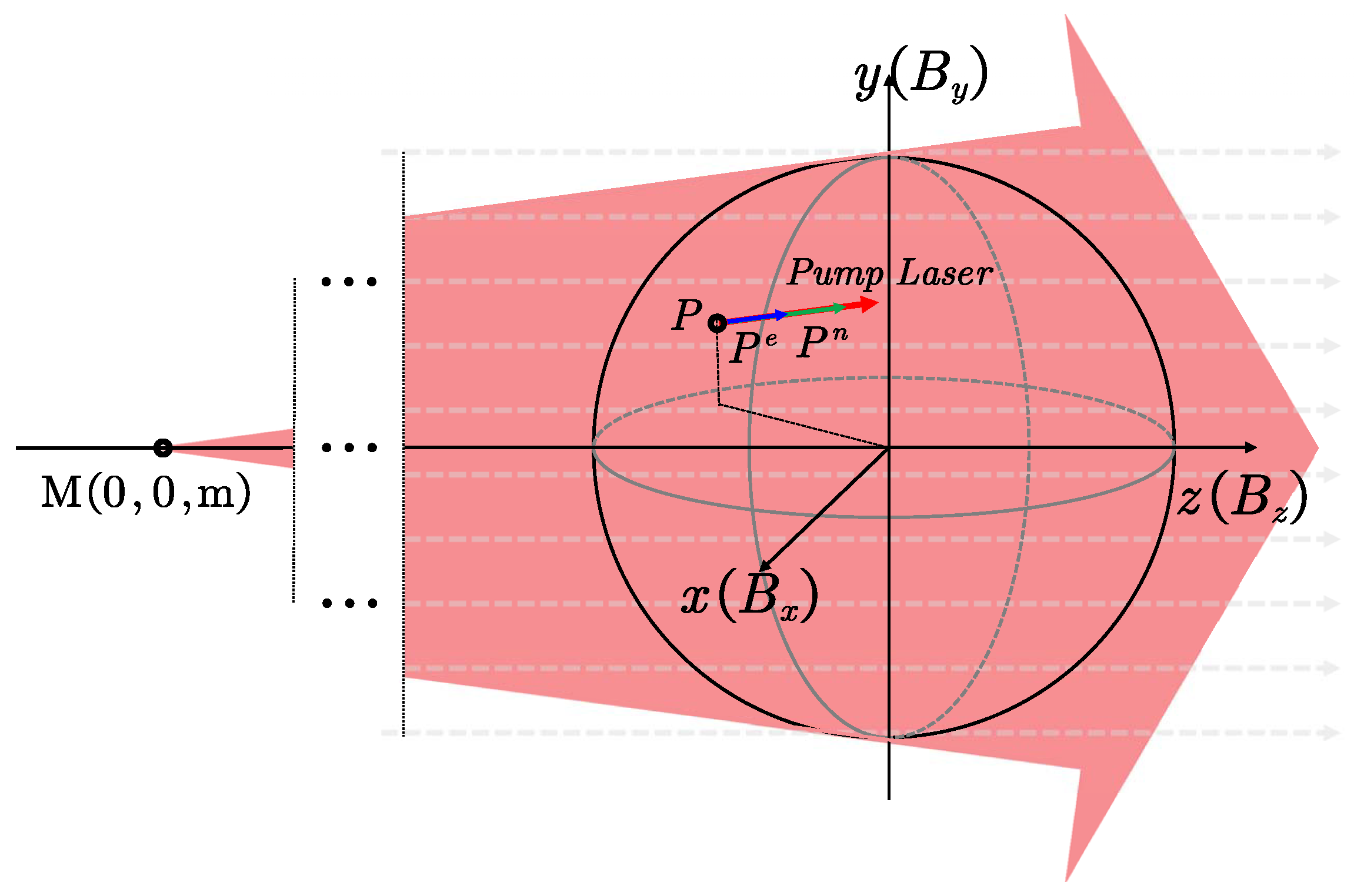
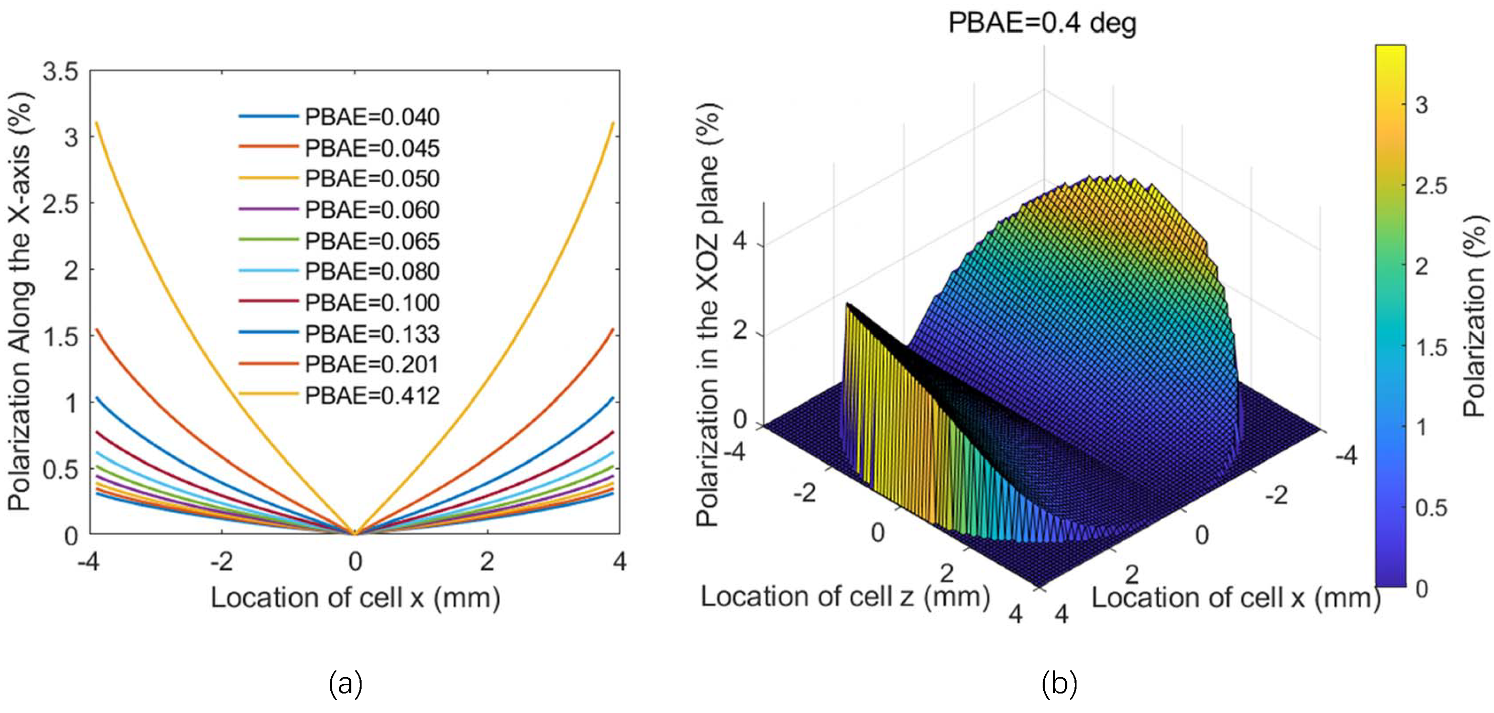
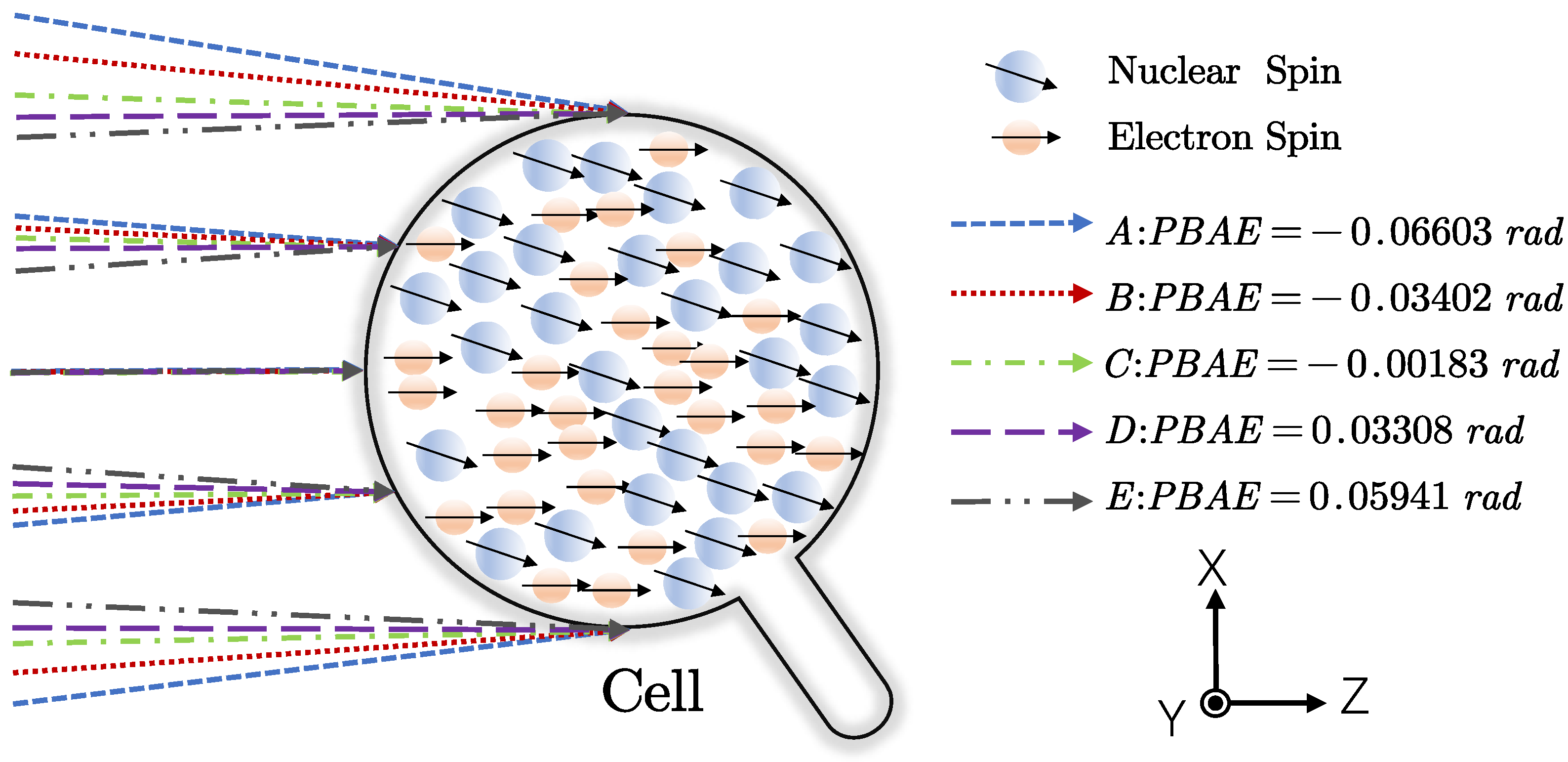
2.1. Mathematical Model

In an SERFCM with K-Rb-21Ne atoms as the sensitive core, a circularly polarized pump light at the wavelength of the potassium D1 line is used to directly pump potassium atoms. The polarized K atoms then polarize Rb atoms through rapid K-Rb spin-exchange collisions, and they can be regarded as one spin species [16,17]. The coupling dynamics equations for the entire ensemble can be represented by simplified Bloch equations [8,18]:
P e t = γ e Q P e B + λ M n P n + L × P e Ω × P e + R P s p + R m s m + R s e e n P n R T r e , R T r e , R L o e P e Q P e P n t = γ n B + λ M e P e × P n Ω × P n + R s e n e P e R T r n , R T r n , R L o n P n
where P e = [ P x e , P y e , P z e ] T denotes the electron spin polarization of the equivalent alkali-metal atoms in K-Rb-21Ne, and P n = [ P x n , P y n , P z n ] T is the polarization of the nuclear spin of the noble gas 21Ne atom. γ e and γ n are the electron and nuclear gyromagnetic ratios, respectively. And Q ( P e ) = ( Q A D r + Q B ) / ( 1 + D r ) is the hybrid slowing-down factor of the hybrid alkali-metal atoms, where D r is the density ratio of atoms A and B [19]. Taking different isotopes into account, the density of atom A ( I = 3 / 2 ) is n A = n K + n 87 R b and the density of atom B ( I = 5 / 2 ) is n B = n 85 R b . For I = 3 / 2 and I = 5 / 2 , Q A / B are as follows [20]:
Q A = Q 3 / 2 P e = 2 3 + P e 2 1 + P e 2 Q B = Q 5 / 2 P e = 2 19 + 26 P e 2 + 3 P e 4 3 + 10 P e 2 + 3 P e 4
L represents the total light-shift of the two alkali-metal atoms and comprises two components: the longitudinal component resulting from the pump beam and the transverse component induced by the probe beam. λ is the geometrical factor containing the enhancement factor. M e and M n give the total magnetization density of electron and nucleus, respectively. For a uniformly magnetized sphere, the magnetic field B e = [ B x e , B y e , B z e ] T generated by electron spin polarization and the magnetic field B n = [ B x n , B y n , B z n ] T generated by nuclear polarization can be represented as the products λ M e P e and λ M n P n , respectively. B is the external magnetic field and Ω is the angular velocity of rotation. R p s p and R m s m describe the optical pumping effects of the pump and probe beams on the atoms, respectively, where R p and R m represent the pump and probe rates. s p and s m explicitly account for the degree of circular polarization and orientation of the pump and probe beams with | s | = 1 for circularly polarized light. R s e e n is the spin-exchange rate from nuclear to electron spin; R s e n e is the spin-exchange rate from electron to nuclear spin. R T r e and R L o e are the transverse and longitudinal relaxation rates of electron spin, respectively, and are given by the following expressions:
R L o e = R s d e + R W e + R s e e n + R p + R L o e R T r e = R s e e + R s d e + R W e + R s e e n + R p + R T r e
where R s e e and R s d e are the spin-exchange collision relaxation rate and spin destruction collision relaxation rate of electron spin. R W e is the wall relaxation rate. R L o e and R T r e are the longitudinal and transverse magnetic field gradient relaxation rates, respectively. The relaxation rates caused by magnetic field gradients can be expressed as follows [21,22]:
R L o e = D e | B x | 2 + | B y | 2 B z e 2 R T r e = 8 γ e 2 R a d 4 | B z | 2 175 D e
where D e gives the diffusion constant of the electron. R a d is the cell radius. B x , B y , and B z are the magnetic field gradients along the three coordinate axes. R L o n and R T r n are the longitudinal and transverse relaxation rates of nuclear spin, respectively, and are given by the following expressions:
R L o n = R s d n + R W n + R s e e n + R d d + R L o n R T r n = R s d n + R W n + R s e e n + R d d + R T r n
where R s d n denotes the spin-destruction rate and R W n represents the relaxation rate caused by collision with the wall. R d d is the dipole–dipole relaxation rate. R L o n and R T r n are the relaxation rates caused by the magnetic field gradient. Similar to electron spin relaxation, the nuclear spin relaxation rate induced by magnetic field gradients can be expressed as follows:
R L o n = D n | B x | 2 + | B y | 2 B z n 2 R T r n = D n 8 γ n 2 R a d 4 | B z | 2 175 D n
where D n is the diffusion constant of the nucleus.
In previous studies, when solving the aforementioned Bloch equations, it was commonly assumed that the propagation directions of the pump and probe beams were orthogonal and precisely aligned with the three axes of the active magnetic compensation coils. However, in practice, the large spot size of the pump beam requiring beam expansion inevitably introduces PBAE. These errors result in a misalignment angle between the pump beam and the cell as well as the magnetic field.
In the modeling process, the PBAE should be taken into account. First, it is assumed that the orthogonality among the coordinate axes of the three-dimensional compensation coils can be well ensured through precision machining. Then, based on the coordinate axes of the 3D flexible coils, a magnetic field coordinate system (x–y–z) is established, as shown in Figure 1. Additionally, assuming a pump beam angular error exists, the beam converges at point M (0, 0, m) on the z-axis. The acute angle between the tangent to the cell contour at point M and the z-axis is defined as the PBAE, measured in radians (rad). s p at point P (x,y,z) inside the cell is then given by
s p = s p x , s p y , s p z s p x = x x 2 + y 2 + z m 2 s p y = y x 2 + y 2 + z m 2 s p z = z m x 2 + y 2 + z m 2
The pumping rate at point P inside the cell is therefore given by R p = R p x , R p y , R p z = R p × s p , from which it can be seen that when a PBAE is present, both R p x and R p x are non-zero, resulting in pumping rates along the transverse x and y directions. Similar to the z-axis, the transverse pumping rates will induce corresponding polarization and polarization gradients in the transverse directions. As atoms absorb the pump light, their intensity undergoes significant attenuation along the propagation direction. Consequently, the optical pumping rate should be treated as a function of the propagation distance L r p . The attenuation along the propagation direction can be described by the following differential equation [23]:
R p L r p = R e W R p 0 R e exp R p 0 R e n σ ν L r p
where R e is the total electron relaxation rate, n is the atomic number density, and σ ( ν ) represents the absorption cross-section, which accounts for the detuning from resonance. L r p is the propagation distance of the pump light within the cell:
L r p = B + B 2 4 A C 2 A A = x 2 + y 2 + ( m z ) 2 B = 2 ( x 2 + y 2 ) + 2 z ( m z ) C = x 2 + y 2 + z 2 16
Once the system stabilizes, the influence of transverse external magnetic fields and angular velocity inputs can be neglected. As a result, the electron spin polarization and nuclear spin polarization along the pump beam direction exhibit negligible variation and can be approximated as remaining at their respective steady-state values [24]. Therefore, the steady-state polarizations of the alkali-metal electron spin and noble gas nuclear spin can be expressed as follows:
P e R p R L o e , P n P e R s e n e R L o n
where R p z is
R p z = x 2 + y 2 + z 2 16 R p s p z d x d y d z
Equations (7) and (8) show that, due to the PBAE, transverse optical pumping occurs, which directly affects the system output. Additionally, the pump rate exhibits a transverse gradient as the pump light propagates through the cell. This transverse gradient in pump rate further leads to a gradient in spin polarization. Since polarized atoms generate magnetic fields, this polarization gradient induces a magnetic field gradient. According to Equations (3) and (5), the polarization gradient increases the longitudinal relaxation rate of both electron and nuclear spin. Moreover, as shown in Equation (5), the presence of PBAE reduces the proportion of s p z , resulting in a decrease in longitudinal polarization efficiency. According to Equations (7)–(10), a simple simulation is performed, and the results are shown in the Figure 2 The simulation results indicate that as the PBAE decreases, the transverse polarization gradient is reduced, while the longitudinal polarization strength is enhanced.
Once the system stabilizes, the transverse components of electron and nuclear spin, induced by inertial and magnetic field inputs, can be linearized as a linear system. The frequency response of the system to the input signal can be determined using the state equation of a linear system. The state equation of the system can be written as follows:
X ˙ = AX + MU
where X = [ P x e , P y e , P x n , P y n ] T is the state vector. B x and B y and Ω x and Ω y are system input variables, and the system input vector U can be written as U = [ Ω x , Ω y , B x , B y ] T . Then, the matrix A can be expressed as
A = R T r e Q γ e Q λ M e P z e R s e e n Q γ e Q λ M n P z e γ e Q λ M e P z e R T r e Q γ e Q λ M n P z e R s e e n Q R s e n e γ n λ M e P z n R T r n γ n λ M n P z n γ n λ M e P z n R s e n e γ n λ M n P z n R T r n
The matrix M can be written as
M = 0 P z e 0 P z e γ e Q 0 P z e γ e Q 1 Q 0 P z e 0 P z e γ e Q 0 P z e γ e Q 0 0 1 Q 0 P z n 0 P z n γ n 0 0 0 0 P z n 0 P z n γ n 0 0 0 0 0
Then, the transfer function matrix of the system is
G s = s I A 1 M = s I A M d e t ( s I A )
Figure 3 illustrates a classic single-axis SERFCM. Considering only the detection direction of the probe beam, the inertial measurement of the system corresponds to Ω y , and the primary source of error is the magnetic field B x along this direction. By solving Equation (15), the system’s response to the magnetic field G B x ( s ) corresponds to the element in the first row and third column of the matrix G ( s ) , and its expression is given as follows:
G B x s = K B x T B x 1 s + 1 T B x 2 s + 1 w 1 2 w 2 2 s 2 + 2 ζ 1 w 1 s + w 1 2 s 2 + 2 ζ 2 w 2 s + w 2 2 K B x = P z e B z n R T r e R T r n γ e γ n γ e B z e R T r n + γ n B z n R T r e 2 + R T r e 2 R T r n 2 T B x 1 = 1 R T r n T B x 2 = B z e γ e + Q P e B z n γ n R T r e B z n γ n + B z e R T r n γ e ζ 1 = R T r n 2 R T r n 2 + B z n γ n 2 ζ 2 = R T r e 2 R T r e 2 + B z e γ e 2 w 1 = R T r n 2 + B z n γ n 2 w 2 = R T r e Q P e 2 + B z e Q P e 2
Since the system exhibits a linear response to rotation only in the low-frequency range, we primarily consider its response in this regime [7]. The expression for the transfer function G Ω y ( s ) considering low frequencies is
G Ω y s = K Ω y w 1 s + w 1
where K Ω y is
K Ω y = P z e B z n 2 R T r e γ e γ n γ e B z e R T r n + γ n B z n R T r e 2 + R T r e 2 R T r n 2
It can be seen from the above equation that PBAE affects both the spin polarization and relaxation rate, thereby directly influencing the scale factor of the system’s inertial output. In addition, the bandwidth reflects the applicable frequency range of the system and serves as a key performance metric for the SERFCM. Let s = j w , where w is the angular frequency in radians per second. This substitution places the Laplace variable on the imaginary axis for steady-state frequency response analysis. The frequency w 1 is defined as the point at which the magnitude response satisfies 20 l o g | W Ω y ( j w ) | = 20 l o g | K Ω y | 3 d B . Therefore, the bandwidth can be simplified to w 1 [25]. Due to the presence of PBAE at w 1 , the nuclear spin polarization is reduced, leading to a decrease in the system bandwidth.
For inertial measurement systems, the magnetic field signal serves as an error source, and the system’s response to the magnetic field reflects its ability to suppress magnetic noise. The ratio of the system’s response to the magnetic field to its inertial response is used to evaluate the magnetic noise suppression capability, which can be expressed as follows:
A B M S = K B x s K Ω y s = R T r n B z n
As shown in Equation (19), the presence of PBAE also degrades the system’s ability to suppress magnetic noise.
In summary, PBAE leads to transverse magnetic field gradients and reduces longitudinal polarization efficiency, thereby decreasing the system’s inertial measurement bandwidth and its ability to suppress magnetic noise. Therefore, suppressing PBAE is crucial.

2.2. In Situ Suppression Method for PBAE

The angular misalignment of the pump beam induces transverse optical pumping, generating transverse polarization and reducing polarization efficiency along the z-direction. To suppress the misalignment error of the pump laser, this study proposes a new alignment method based on atomic spin precession signals. When the pump laser power is sufficiently low, the electron spins can be partially polarized, while the nuclear spins remain unpolarized. Based on the polarized electron spins [26], a dual-beam atomic magnetometer can be implemented, with the pump and probe beams being circularly and linearly polarized, respectively. Using the zero-field calibration method of atomic magnetometers [27], the residual magnetic field inside the magnetic shielding cylinder can be measured. Once the residual magnetic field is compensated to zero and prior to nuclear spin polarization, the setup effectively functions as a single-beam atomic magnetometer [28]. The corresponding equation can then be simplified as
P e t = γ e Q P e B × P e + R P s p R T r e , R T r e , R L o e P e Q P e
Similar to the previous section, the response of the magnetometer signal to the magnetic field can be derived as follows:
G B x s = B z P z e γ e 2 R T r e + Q P e 2 + B z 2 γ e 2
From the preceding analysis, it is evident that collimation errors in the pump light lead to a reduction in the electron spin polarization rate along the z-direction and an increase in the relaxation rate. As shown in Equation (21), both the electron spin polarization rate and the relaxation rate directly affect the magnetometer signal’s response to the magnetic field. Therefore, the magnetometer signal can be used as a reference for suppressing pump light collimation errors. Based on Equations (11) and (21), a simulation is performed to evaluate how the magnetometer’s response to magnetic fields varies with pump light collimation errors, as shown in Figure 4.

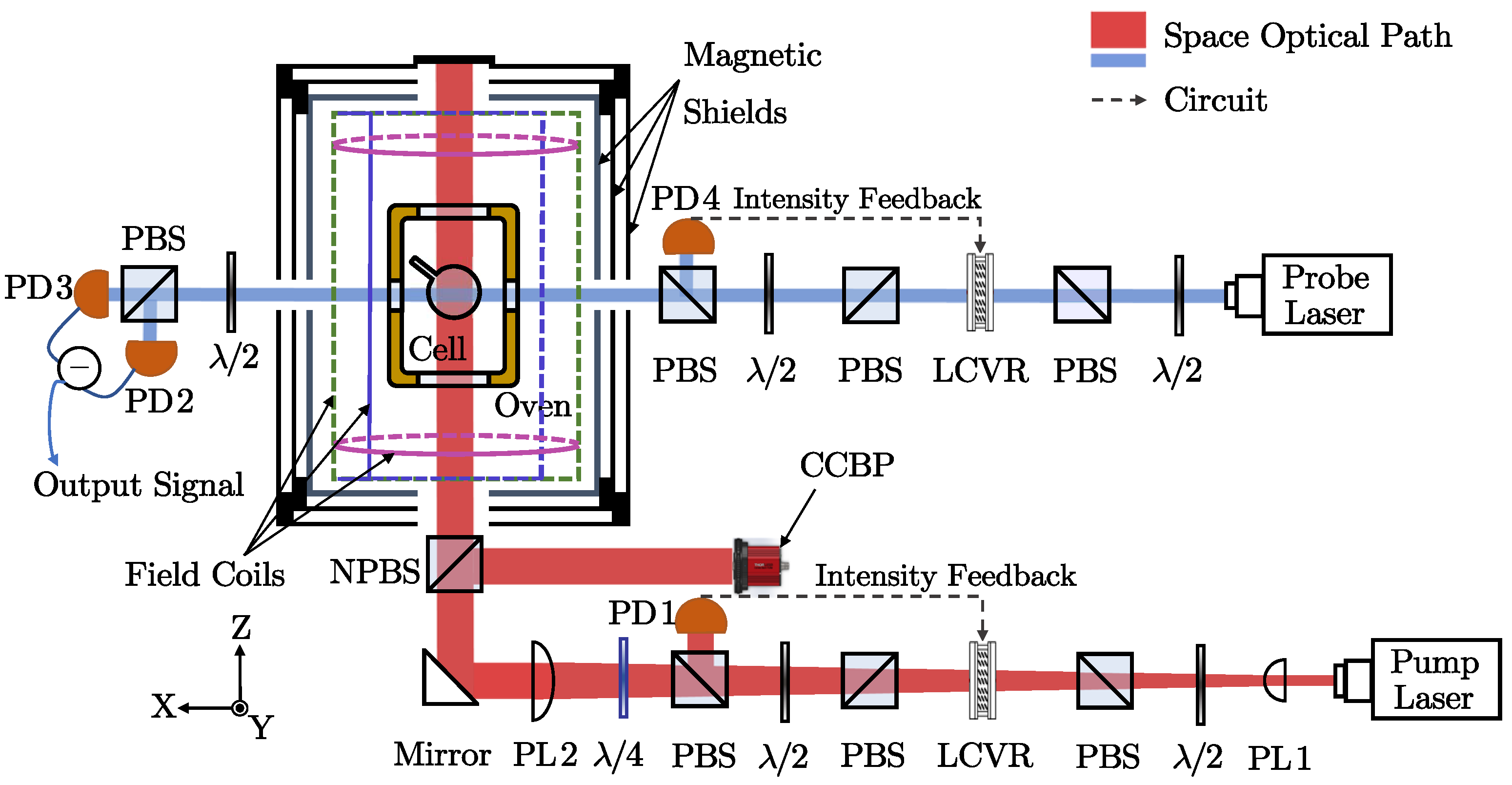
3. Experimental Setup of SERFCM

The schematic diagram of the experimental setup for the SERFCM is shown in Figure 3. The sensitive core of the SERFCM is a spherical cell with a 10 mm outer diameter and a thickness of 1 mm, made of GE180 aluminosilicate glass. The cell contains a mixture droplet of K-Rb in natural abundance with a density ratio of about 1:112 at 185 °C. And the 0.68 atm cell is filled with 500 Torr of 21Ne(70% isotope enriched) gas and 18 Torr of N2. Nitrogen is used to suppress relaxation rate caused by the radiation trapping. Specifically, the 2 mm wide stem brought by the inflation unit needs to be tilted to avoid the propagation direction of the pump light and probe light.
The cell contains both potassium and rubidium atoms. Potassium atoms are polarized by pump light via the 770 nm pump laser. And through rapid spin-exchange collisions, the high-density alkali-metal atoms are polarized, aiming to achieve a more uniform polarization distribution using hybrid optical pumping techniques [8]. Then, under the combined action of two polarized alkali-metal atoms, the 21Ne atoms are hyperpolarized.
A heating film made of constantan serves as a non-magnetic heat source, while an oven made of boron nitride ceramic material conducts heat and supports the cell. The PID algorithm and a 99 kHz AC modulation signal are employed for temperature control and non-magnetic heating. Two layers of permalloy magnetic shielding cylinders are used to shield external magnetic fields, with an additional layer of ferrite magnetic shielding cylinders inside to further reduce magnetic noise. Remaining magnetic fields are compensated with a three-dimensional magnetic field coil to maintain system operation at the compensation point.
The pump beam is generated by a distributed Bragg reflector (DBR) semiconductor laser with a central wavelength of 770.108 nm and a linewidth below 300 kHz. A pair of plano-convex lenses expands the pump beam into an 8 mm Gaussian profile. The pump power is maintained at 20 mW by a long-term intensity stabilization system (LISS) to ensure power stability over time. A zero-order quarter-wave plate converts the linearly polarized light into circularly polarized light, which is used to polarize the potassium atoms as the resulting pump beam illuminates the cell. The probe beam is generated by a distributed feedback (DFB) semiconductor laser with a central wavelength of 795.309 nm. Similarly, the probe beam power is stabilized at 1 mW by the LISS. A PBS placed after the LISS re-polarizes the beam into linearly polarized light for detecting the projection component of polarized rubidium atoms along the x-axis. After passing through the cell, the probe beam is collected by a balanced polarimeter, which extracts the differential signal using a pair of photodiodes.

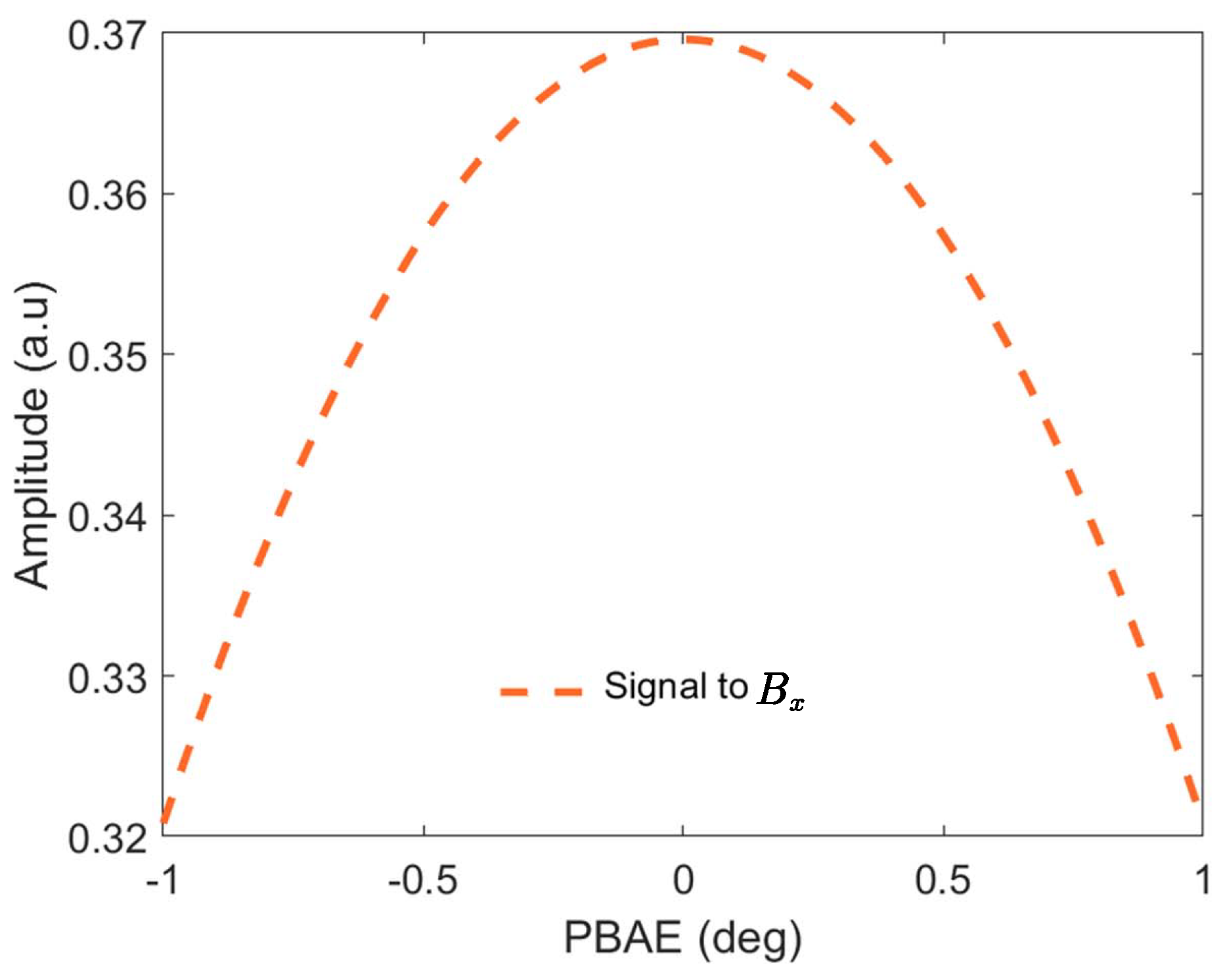
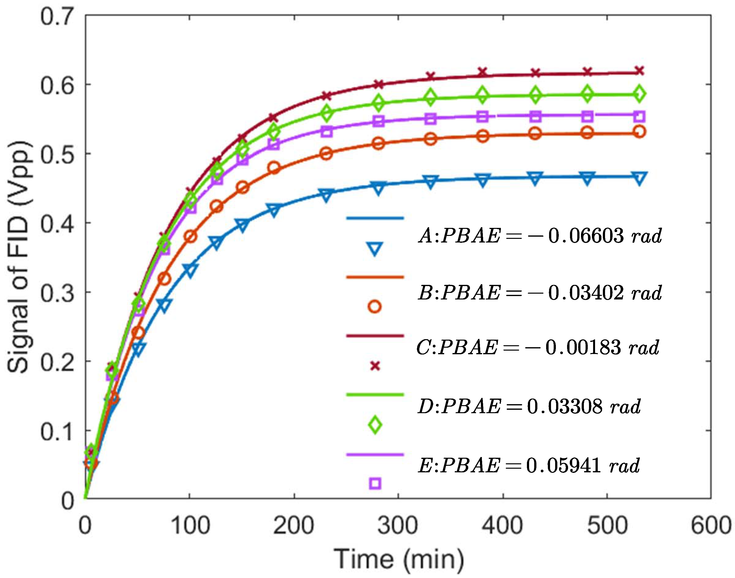
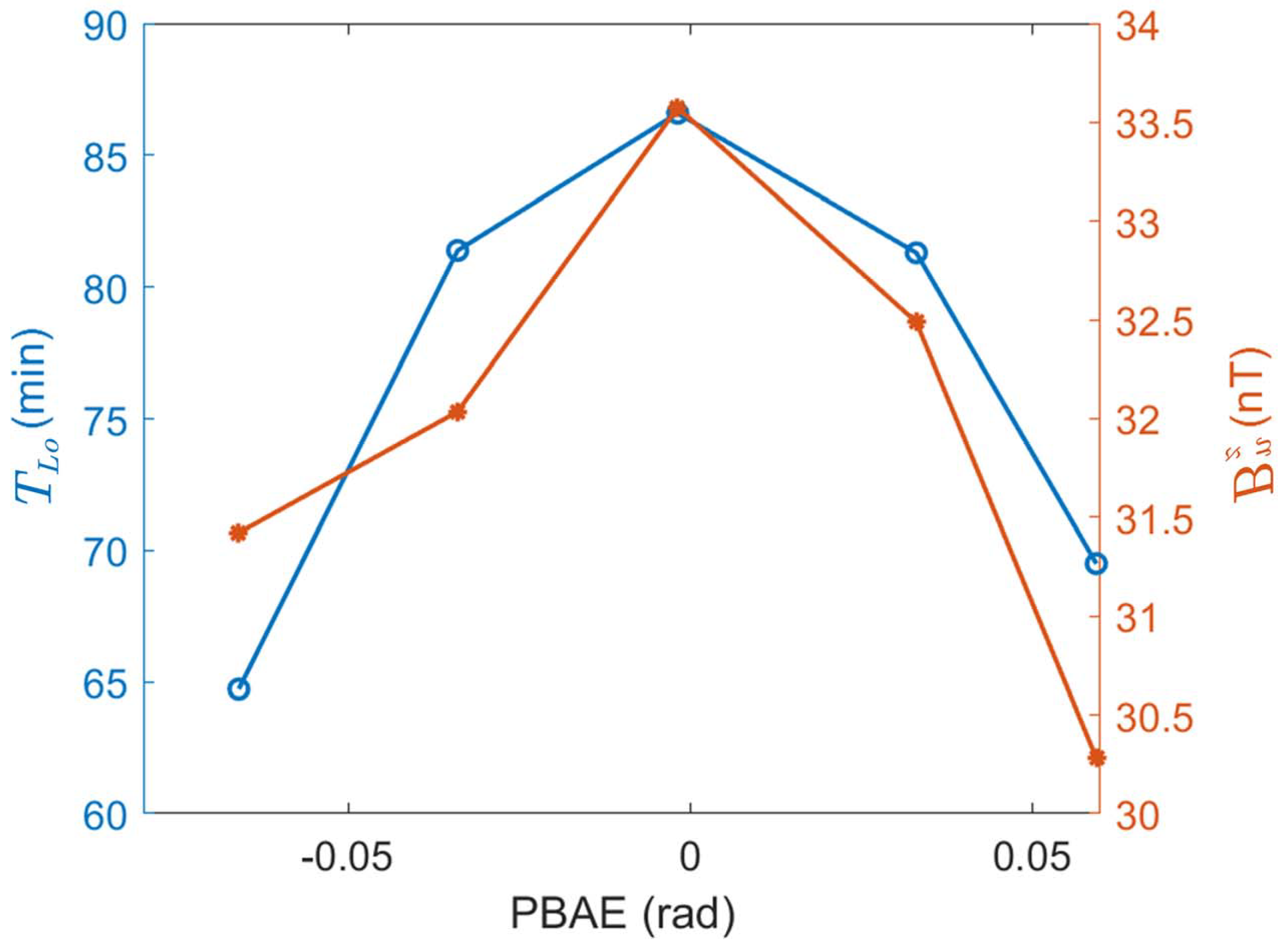
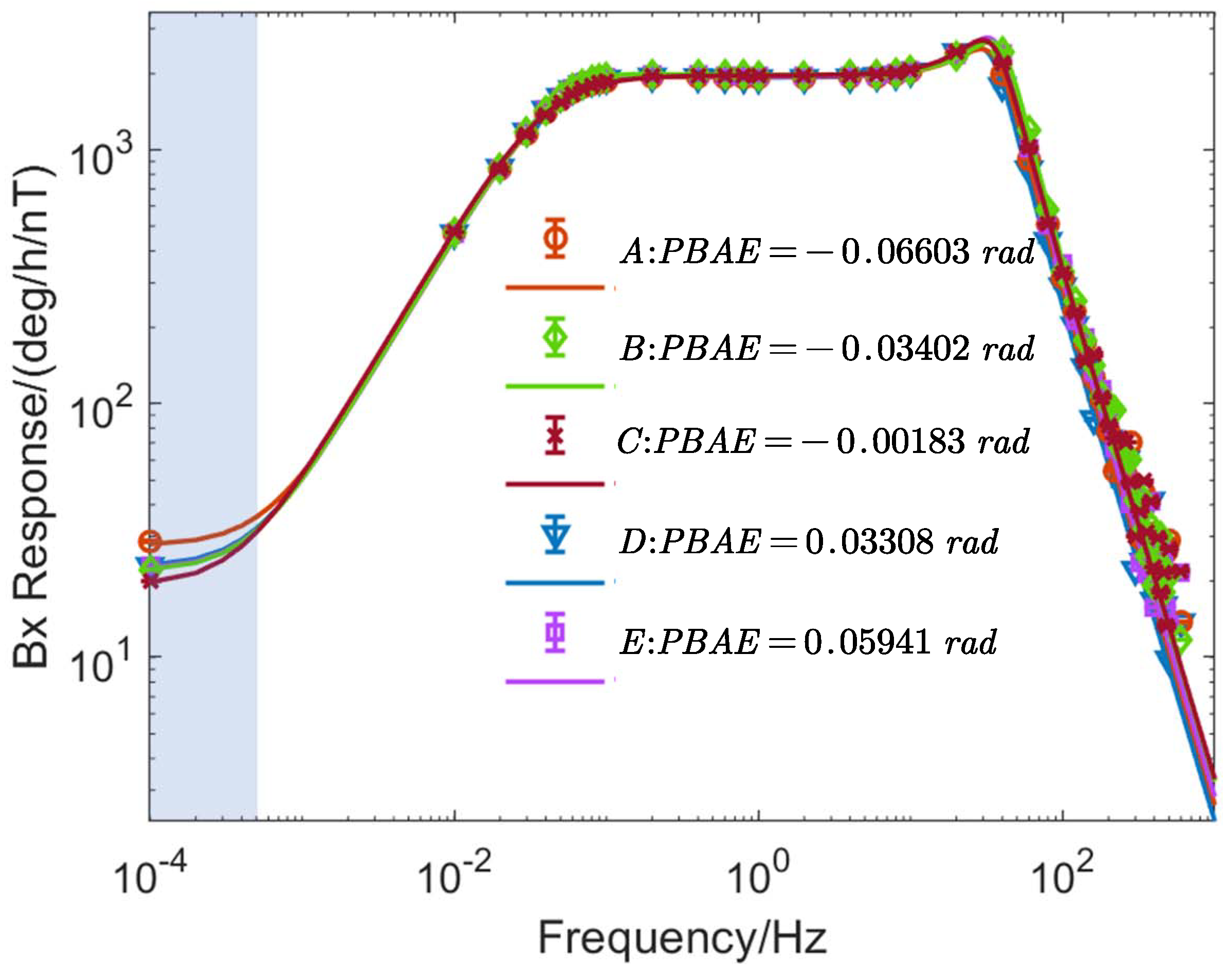
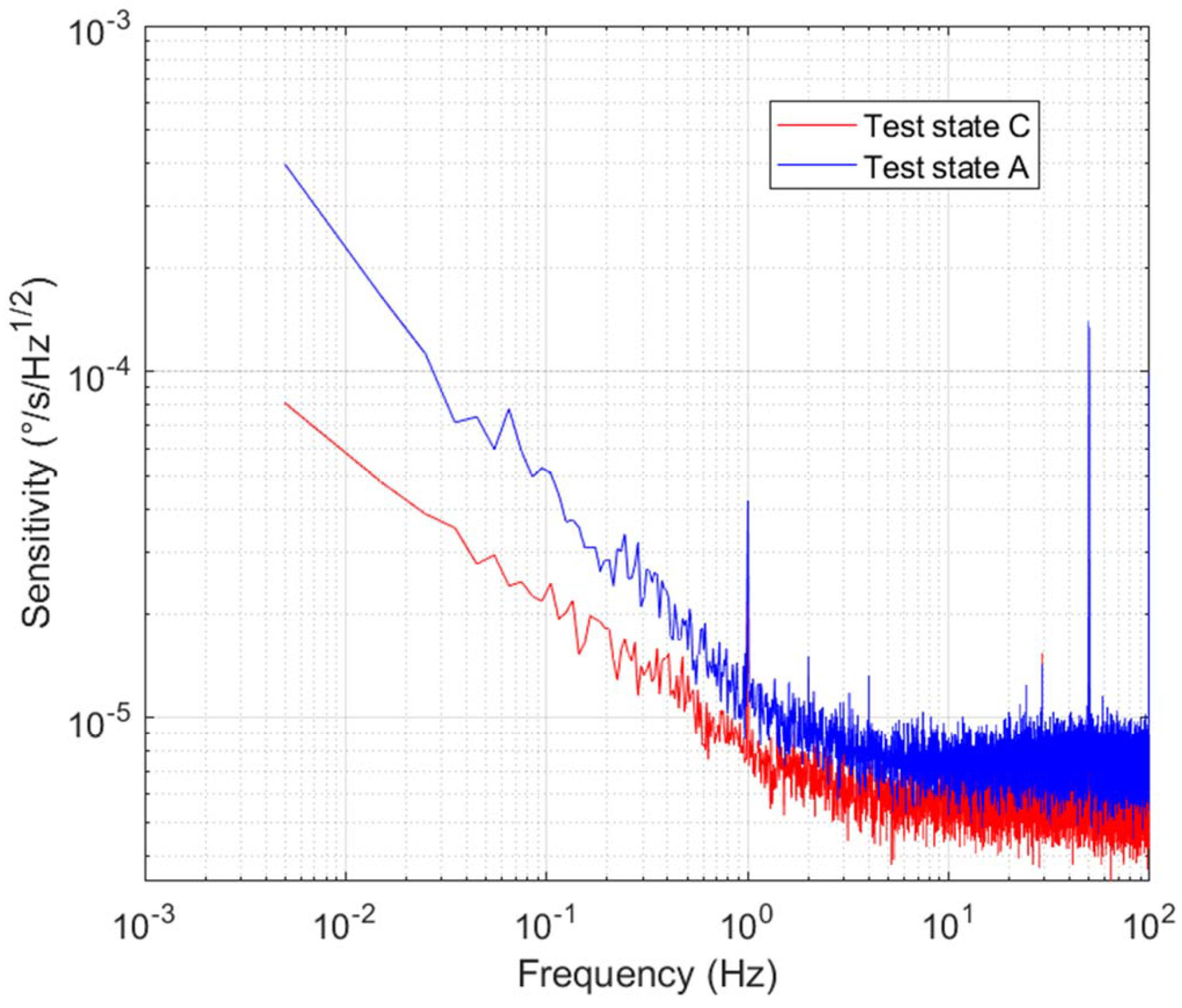
4. Experimental Results and Discussion

First, to begin the experiment, the cell is heated to 185 °C. A probe beam ( 794.2 nm , 659 μ W ) is introduced, followed by activation of the pump laser to initiate optical pumping. The experimental tests in this section validate the feasibility of feedback control of the electron spin polarization based on optical rotation measurements and assess its impact on the performance of the SERFCM. To investigate the impact of the PBAE on the performance of the SERFCM and to validate the theoretical analysis presented in Section 3, the direction of the pump laser is adjusted to five test configurations, as illustrated in Figure 5, by repositioning the plano-convex lenses PL1 and PL2 in Figure 3.
Among the five experimental configurations, four operational states (A, B, D, and E) are defined based on the relative position of the pump laser within the optical aperture. Meanwhile, the pump beam is reflected by the NPBS and directed into the CCBP, which is used to measure the beam parameters before it enters the cell. These measurements are then used to evaluate the PBAE. Following the dual-beam magnetometer alignment method proposed in Section 3, a transverse AC magnetic field with an amplitude of 10 nT and a frequency of 100 Hz is applied along the x-axis. Through precise adjustment of the pump beam angle using a six-degree-of-freedom translation stage, the configuration yielding the maximum amplitude of the dual-beam magnetometer response was identified as test state C.
As described in Section 3, PBAE increases the relaxation rate induced by magnetic field gradients, thereby degrading the system’s ability to suppress magnetic noise. Consequently, the influence of the pump beam angle error on the system’s magnetic noise suppression can be evaluated by measuring the relaxation rate under different PBAE conditions. During rotation measurements, it is difficult to directly obtain the nuclear spin relaxation rate. Instead, it can be inferred by measuring the relaxation time. Conveniently, during the initialization phase of nuclear spin hyperpolarization, the longitudinal relaxation time of nuclear spins can be measured via nuclear magnetic resonance-free induction decay (NMR-FID) signals associated with the nuclear spin hyperpolarization process [29]. Initially, a 1200 nT magnetic field is applied along the z-axis, and the pump light is turned on. The amplitude of the FID signal is then measured. The initial amplitude of the FID signal is proportional to the polarization of the nuclear spins. The initial amplitude of the FID signal is measured sequentially over time until it stabilizes and no longer changes. The following equation can be used to fit the initial amplitudes at all time points to determine the longitudinal relaxation time:
S LFID t = A 1 e t / T L o
where A represents the FID amplitude after the 21Ne atoms have been fully hyperpolarized, and t is the time elapsed since the start of the pumping process. T L o denotes the longitudinal relaxation time of the 21Ne atoms, which is the inverse of the longitudinal relaxation rate R L o n .
The test results of the nuclear spin hyperpolarization process under the five test conditions mentioned above are shown in Figure 6, which presents the measured initial amplitudes of the FID signals under different conditions. As shown, when the pump laser is in the well-aligned state without PBAE (Test State C), the initial amplitudes of the FID signals at different time points are consistently higher than those under misaligned conditions (i.e., Test States A, B, D, and E). This provides preliminary validation of the effectiveness of the proposed pump beam alignment method.
After measuring the FID signal of the hyperpolarized nuclear spin and stabilizing the system, the total magnetic field of the atomic ensemble is obtained by performing multiple magnetic field compensations using the three-axis active magnetic compensation coils. The electron spin polarization under operating conditions is then measured using magnetic resonance [19], from which the electron magnetic field is calculated. By subtracting the electron magnetic field from the total magnetic field, the nuclear spin magnetic field is derived, and the nuclear spin polarization is subsequently determined.
Figure 7 shows the measured longitudinal relaxation time and the magnetic field generated by nuclear spin polarization of 21Ne under the five PBAE conditions. The results indicate that as the PBAE decreases, the magnetic field generated by the nuclear spin polarization of 21Ne increases, and the longitudinal relaxation time becomes longer, meaning the relaxation rate decreases. Figure 7 also shows that, compared to the adjusted state, in state A (PBAE = −0.06603 rad), the nuclear spin polarization is reduced by 10%, and the longitudinal relaxation rate increases by 26.3%.
As analyzed earlier, magnetic noise is the main source of error in the system signal, and the PBAE also affects the system’s ability to suppress magnetic noise. Therefore, the system’s frequency response to the magnetic field B x in the x-direction is tested under five different PBAE conditions. Sinusoidal signals with an amplitude of 3.4 nT and varying frequencies are sequentially applied along the x-axis, and the corresponding response amplitudes of the SERFCM signal are measured and recorded. To better evaluate the signal-to-noise ratio, the results are normalized by the response of the inertial signal at zero frequency. The experimental data are then fitted using the equations presented in Section 2.1. The resulting amplitude–frequency responses of the system to B x are shown in Figure 8. The good agreement between the fitted curves and the experimental data confirms the accuracy of the model.
For rotation measurement instruments, the low-frequency range is particularly critical. As shown in the blue-shaded region of Figure 8, when the PBAE is 0.06603 rad, the system’s ability to suppress magnetic fields in the low-frequency range is reduced by 40%. As the PBAE decreases, the amplitude of the response to the magnetic field B x also decreases, indicating improved magnetic noise suppression. Moreover, according to Equation (19), the reduction in low-frequency response amplitude supports the accuracy of the three measured parameters shown in Figure 7.
The sensitivity of the SERFCM signal is evaluated through the sensitivity curves, with the experimental results shown in Figure 9. As observed, after correcting the PBAE of the pump light, the overall sensitivity curve of the SERFCM decreases, particularly showing significant suppression in the low-frequency range. This indicates that the SERFCM’s sensitivity and long-term stability in rotation rate measurements improved to some extent.
In summary, the experimental results demonstrate that the proposed model is accurate and that tuning the pump beam alignment can effectively enhance the magnetic noise suppression capability of the SERFCM system.

5. Conclusions

In conclusion, this paper systematically investigate the influence of PBAE on the performance of an SERFCM. We demonstrate how PBAE affects the scale factor, bandwidth, and magnetic noise suppression capability of the SERFCM. Theoretical analysis indicates that PBAE reduces the inertial measurement scale factor and bandwidth of the system, increases the longitudinal relaxation rate of nuclear spin, and weakens the system’s ability to suppress magnetic noise. To mitigate PBAE, we propose an alignment method based on atomic response. Experimental validation is then carried out, and the results show strong agreement with the theoretical analysis. Specifically, under a PBAE of −0.06603 rad, the nuclear spin polarization strength decreases by 10% and the longitudinal relaxation rate increases by 26.3%. In addition, frequency response measurements reveal that PBAE significantly deteriorates the system’s ability to suppress low-frequency magnetic noise. Moreover, the in situ suppression method proposed in this work also shows potential for application in other atomic sensors, such as optical pumping magnetometers (OPMs) and nuclear magnetic resonance gyroscopes (NMRGs).

Author Contributions

Conceptualization, Q.Y. and W.F.; methodology, Q.Y.; software, Z.W.; validation, Q.Y., H.P., and W.F.; formal analysis, H.P.; investigation, W.F.; resources, Q.Y.; data curation, Q.Y.; writing—original draft preparation, Q.Y.; writing—review and editing, X.H. and W.F.; visualization, X.H.; supervision, W.Q. and W.F.; project administration, Q.Y.; funding acquisition, W.Q., Z.W., and W.F. All authors have read and agreed to the published version of the manuscript.

Funding

This work was supported by the National Natural Science Foundation of China (Grant Nos. 62103026, 61673041); the Beijing Natural Science Foundation (Grant No. 3252013, 4244095); and the Innovation Program for Quantum Science and Technology (2021ZD0300400 and 2021ZD0300402).

Institutional Review Board Statement

Not applicable.

Informed Consent Statement

Not applicable.

Data Availability Statement

The data presented in this study are available upon reasonable request from the corresponding author.

Conflicts of Interest

The authors declare no conflicts of interest.

References

  1. Smiciklas, M.; Brown, J.; Cheuk, L.; Smullin, S.; Romalis, M. New Test of Local Lorentz Invariance Using a 21Ne-Rb-K Comagnetometer. Phys. Rev. Lett. 2011, 107, 171604. [Google Scholar] [CrossRef]
  2. Kornack, T.; Ghosh, R.; Romalis, M. Nuclear spin gyroscope based on an atomic comagnetometer. Phys. Rev. Lett. 2005, 95, 230801. [Google Scholar] [CrossRef]
  3. Pei, H.; Pang, H.; Quan, W.; Fan, W.; Yuan, L.; Zhang, K.; Fang, C. Pulsed optical pumping in electron spin vapor. Measurement 2024, 231, 114619. [Google Scholar] [CrossRef]
  4. Li, F.; Pang, H.; Wang, Z.; Fan, W.; Zhang, M.; Liu, Z.; Li, J.; Qin, B.; Zhou, X.; Lei, X.; et al. In situ real-time measurement for electron spin polarization in atomic spin gyroscopes. iScience 2025, 28, 111757. [Google Scholar] [CrossRef]
  5. Zheng, W.; Su, S.; Zhang, G.; Bi, X.; Lin, Q. Vector magnetocardiography measurement with a compact elliptically polarized laser-pumped magnetometer. Biomed. Opt. Express 2020, 11, 649–659. [Google Scholar] [CrossRef]
  6. Flambaum, V.; Romalis, M. Limits on lorentz invariance violation from coulomb interactions in nuclei and atoms. Phys. Rev. Lett. 2017, 118, 142501. [Google Scholar]
  7. Wei, K.; Zhao, T.; Fang, X.; Xu, Z.; Liu, C.; Cao, Q.; Wickenbrock, A.; Hu, Y.; Ji, W.; Fang, J.; et al. Ultrasensitive atomic comagnetometer with enhanced nuclear spin coherence. Phys. Rev. Lett. 2023, 130, 063201. [Google Scholar] [CrossRef]
  8. Kornack, T.; Romalis, M. Dynamics of two overlapping spin ensembles interacting by spin exchange. Phys. Rev. Lett. 2002, 89, 253002. [Google Scholar] [CrossRef]
  9. Chann, B.; Babcock, E.; Anderson, L.; Walker, T. Skew light propagation in optically thick optical pumping cells. Phys. Rev. A 2002, 66, 033406. [Google Scholar] [CrossRef]
  10. Wu, Z.; Jia, Y.; Chai, Z.; Liu, Z. Influence of skew light propagation on electron spin polarization in an atomic magnetometer. In Proceedings of the First Optics Frontier Conference, SPIE, Hangzhou, China, 24–26 April 2021; Volume 11850, pp. 36–41. [Google Scholar]
  11. Zhao, J.; Liu, G.; Lu, J.; Ding, M.; Ma, Y.; Ji, J.; Yang, K.; Ma, D.; Xing, B.; Zhang, N.; et al. Improvement of spin polarization spatial uniformity in optically pumped atomic magnetometers based on counter-propagating pump beams and atomic diffusion. Meas. Sci. Technol. 2020, 32, 035902. [Google Scholar] [CrossRef]
  12. Jia, Y.; Liu, Z.; Zhou, B.; Liang, X.; Wu, W.; Peng, J.; Ding, M.; Zhai, Y.; Fang, J. Pump beam influence on spin polarization homogeneity in the nuclear magnetic resonance gyroscope. J. Phys. Appl. Phys. 2019, 52, 355001. [Google Scholar] [CrossRef]
  13. Kornack, T.W. A Test of CPT and Lorentz Symmetry Using a Potassium-Helium-3 co-Magnetometer. Ph.D. Thesis, Princeton University, Princeton, NJ, USA, 2005. [Google Scholar]
  14. Xing, L.; Quan, W.; Fan, W.; Zhang, W.; Fu, Y.; Song, T. The method for measuring the non-orthogonal angle of the magnetic field coils of a K-Rb-21 Ne co-magnetometer. IEEE Access 2019, 7, 63892–63899. [Google Scholar] [CrossRef]
  15. Huang, J.; Fan, W.; Wang, Z.; Yuan, L.; Zhang, K.; Pei, H.; Pang, H.; Quan, W. Analysis and suppression of the misalignment error for the pumping laser in the atomic comagnetometer. Opt. Express 2022, 30, 6374–6387. [Google Scholar] [CrossRef]
  16. Chen, Y.; Quan, W.; Duan, L.; Lu, Y.; Jiang, L.; Fang, J. Spin-exchange collision mixing of the K and Rb ac Stark shifts. Phys. Rev. A 2016, 94, 052705. [Google Scholar] [CrossRef]
  17. Wang, X.; Chen, Y.; Quan, W.; Fan, W.; Fang, J. A rapid spin exchange tightly bound alkali metal hybrid optical pumping system. J. Phys. At. Mol. Opt. Phys. 2018, 51, 045001. [Google Scholar] [CrossRef]
  18. Babcock, E.; Nelson, I.; Kadlecek, S.; Driehuys, B.; Anderson, L.; Hersman, F.; Walker, T. Hybrid Spin-Exchange Optical Pumping of 3He. Phys. Rev. Lett. 2003, 91, 123003. [Google Scholar] [CrossRef] [PubMed]
  19. Wei, K.; Zhao, T.; Fang, X.; Xu, Z.; Zhai, Y.; Quan, W. Accurate polarimetry of hybrid K-Rb and 21Ne atoms based on spin-exchange interactions. IEEE Sens. J. 2020, 21, 5879–5885. [Google Scholar] [CrossRef]
  20. Savukov, I.; Romalis, M. Effects of spin-exchange collisions in a high-density alkali-metal vapor in low magnetic fields. Phys. Rev. A 2005, 71, 023405. [Google Scholar] [CrossRef]
  21. Cates, G.; Schaefer, S.; Happer, W. Relaxation of spins due to field inhomogeneities in gaseous samples at low magnetic fields and low pressures. Phys. Rev. A 1988, 37, 2877. [Google Scholar] [CrossRef]
  22. Fang, X.; Wei, K.; Zhai, Y.; Zhao, T.; Chen, X.; Zhou, M.; Liu, Y.; Ma, D.; Xiao, Z. Analysis of effects of magnetic field gradient on atomic spin polarization and relaxation in optically pumped atomic magnetometers. Opt. Express 2022, 30, 3926–3940. [Google Scholar] [CrossRef]
  23. Zhao, T.; Zhai, Y.; Liu, C.; Xie, H.; Cao, Q.; Fang, X. Spin polarization characteristics of hybrid optically pumped comagnetometers with different density ratios. Opt. Express 2022, 30, 28067–28078. [Google Scholar] [CrossRef]
  24. Fang, J.; Wan, S.; Yuan, H. Dynamics of an all-optical atomic spin gyroscope. Appl. Opt. 2013, 52, 7220–7227. [Google Scholar] [CrossRef] [PubMed]
  25. Pang, H.; Liu, F.; Fan, W.; Wu, Z.; Yuan, Q.; Quan, W. Comprehensive analysis of the effects of magnetic field gradient on the performance of the SERF co-magnetometer. Opt. Express 2023, 31, 5215–5228. [Google Scholar] [CrossRef] [PubMed]
  26. Fang, J.; Wan, S.; Chen, Y.; Li, R. Light-shift measurement and suppression in atomic spin gyroscope. Appl. Opt. 2012, 51, 7714–7717. [Google Scholar] [CrossRef]
  27. Wyllie, R.; Kauer, M.; Smetana, G.; Wakai, R.; Walker, T. Magnetocardiography with a modular spin-exchange relaxation-free atomic magnetometer array. Phys. Med. Biol. 2012, 57, 2619. [Google Scholar] [CrossRef]
  28. Savukov, I.; Kim, Y.; Shah, V.; Boshier, M. High-sensitivity operation of single-beam optically pumped magnetometer in a kHz frequency range. Meas. Sci. Technol. 2017, 28, 035104. [Google Scholar] [CrossRef]
  29. Eklund, E.J. Microgyroscope Based on Spin-Polarized Nuclei; University of California: Irvine, CA, USA, 2008. [Google Scholar]
Figure 1. PBAE diagram of the magnetic field coordinate system. The coordinate system is established with the center of the alkali-metal cell as the origin. The blue arrow represents P e , the green arrow represents P n , and the red arrow indicates the direction of the pump beam. Red represents the beam of light.
Photonics 12 00550 g001
Figure 2. Simulation of the effect of PBAE on x-axis polarization in the XOZ plane. (a) shows the polarization distribution along the x-axis under different PBAE values, while (b) presents a 3D illustration of the x-axis polarization distribution in the XOZ plane when PBAE is 0.4 deg. To more clearly present the data, a colorbar has been added to the (b).
Photonics 12 00550 g002
Figure 3. Schematic of SERF co-magnetometer. PD: photodetector; PBS: polarizing beam splitter; λ / 2 : half waveplate; λ / 4 : quarter waveplate; GT: Glan–Taylor calcite polarizers; NPBS: non-polarizing beam splitter; PL: planoconvex lens; LCVR: liquid crystal variable retarder; CCBPs: CMOS Camera Beam Profilers. Coordinate system is magnetic field coordinate system. The red beam represents the pump light, and the blue beam represents the probe light.
Photonics 12 00550 g003
Figure 4. Illustration of the effect of PBAE on the magnetometer’s response to B x .
Photonics 12 00550 g004
Figure 5. Schematic diagram of five PBAE states of the pump light in the magnetic field coordinate system. Each color represents a distinct PBAE state, with dashed arrows indicating the corresponding propagation directions of the pump light.
Photonics 12 00550 g005
Figure 6. Schematic diagram of the relaxation time tests.
Photonics 12 00550 g006
Figure 7. Schematic diagram of influence of PBAE on longitudinal relaxation time and nuclear spin polarization magnetic field.
Photonics 12 00550 g007
Figure 8. Schematic diagram of B x response measurements for different PBAEs. Error bars represent standard deviations.
Photonics 12 00550 g008
Figure 9. Sensitivity curves under uncorrected and corrected PBAE conditions.
Photonics 12 00550 g009
Disclaimer/Publisher’s Note: The statements, opinions and data contained in all publications are solely those of the individual author(s) and contributor(s) and not of MDPI and/or the editor(s). MDPI and/or the editor(s) disclaim responsibility for any injury to people or property resulting from any ideas, methods, instructions or products referred to in the content.

