Review

Retinal Laser Therapy Mechanisms, Innovations, and Clinical Applications

1
Department of Ophthalmology and Visual Sciences, University of Michigan, Ann Arbor, MI 48105, USA
2
Department of Ophthalmology, The First Affiliated Hospital of Nanjing Medical University, Nanjing 210029, China
3
Department of Ophthalmology, Wilmer Eye Institute, Johns Hopkins University, Baltimore, MD 21287, USA
4
Department of Biomedical Engineering, Johns Hopkins University, Baltimore, MD 21287, USA
*
Author to whom correspondence should be addressed.
Photonics 2025, 12(11), 1043; https://doi.org/10.3390/photonics12111043
Submission received: 9 September 2025 / Revised: 17 October 2025 / Accepted: 20 October 2025 / Published: 22 October 2025
(This article belongs to the Special Issue Novel Techniques and Applications of Ophthalmic Optics)

Abstract

Retinal laser therapy has been a mainstay for treating proliferative diabetic retinopathy, retinal vascular disease, and retinal breaks since 1961. However, conventional millisecond photocoagulation can cause permanent scarring and procedure discomfort, motivating the development of damage-sparing approaches that preserve the neurosensory retina. Clinically, panretinal photocoagulation remains effective for proliferative disease but trades off peripheral visual field and night vision. This review synthesizes development, mechanisms, and clinical evidence for laser modalities, including short-pulse selective retinal therapy (SRT), subthreshold diode micropulse (SDM), and pattern-scanning photocoagulation. We conducted a targeted narrative search of PubMed/MEDLINE, Embase, Web of Science, and trial registries (1960–September 2025), supplemented by reference list screening. We prioritized randomized/prospective studies, large cohorts, systematic reviews, mechanistic modeling, and relevant preclinical work. Pulse duration is the primary determinant of laser–tissue interaction. In the microsecond regime, SRT yields retinal pigment epithelium (RPE)-selective photodisruption via microcavitation and uses real-time optoacoustic or OCT feedback. SDM 100–300 µs delivers nondamaging thermal stress with low duty cycles and titration-based dosing. Pattern-scanning platforms improve throughput and tolerance yet remain destructive photocoagulation. Feedback-controlled SRT shows anatomic/functional benefit in chronic central serous chorioretinopathy and feasibility in diabetic macular edema. SDM can match threshold macular laser for selected DME and may reduce anti-VEGF injection burden. Sub-nanosecond “rejuvenation” lasers show no overall benefit in intermediate AMD and may be harmful in specific phenotypes. Advances in delivery, dosimetry, and closed-loop feedback aim to minimize collateral damage while retaining therapeutic effect. Key gaps include head-to-head trials (SRT vs. PDT/SDM), standardized feedback thresholds across pigmentation and devices, and long-term macular safety to guide broader clinical adoption.

1. Introduction

Lasers are used in almost all fields of medicine, particularly ophthalmology and retina, due to the optical transparency of the eye. Lasers are capable of treating different retinal conditions, including retinal tears, PDR, DME, and central and branch retinal vein occlusions. It has also shown positive effects in central serous chorioretinopathy, choroidal neovascularization, and tumors, with proven efficacy in multiple large prospective clinical trials [1].
Beyond clinical outcomes, retinal laser therapy rests on a well-developed physical framework for laser matter interactions in tissue. Canonical models describe how wavelength, pulse duration, and confinement regimes govern energy deposition, heat flow, and stress from thermal denaturation at millisecond scales to thermomechanical microcavitation and plasma-seeded events at microsecond–nanosecond scales [2,3]. Rate–equation and energy balance treatments of water (a tissue surrogate) quantify free electron generation, breakdown thresholds, and bubble/shock dynamics under 532 nm nanosecond pulses, providing pulse–duration-dependent predictions that complement ophthalmic data [4]. Clinically, condition-focused reviews such as retinal arterial macroaneurysm (RAM) synthesize how these physics considerations map to threshold vs. subthreshold dosing and selective tissue targeting, bridging laboratory principles to laser choices at the slit lamp [5]. We highlight RAM because its management spans threshold and subthreshold strategies, making it a concise example of how pulse duration and energy delivery translate to practical dosing decisions. This context motivates our physics-based approach to short-pulse SRT (µs), contrasted with thermal micropulse (100–300 µs) and conventional photocoagulation (ms), throughout the review.

1.1. History

Max Planck (1900) and Albert Einstein (1917) laid the groundwork for Theodore H. Maiman’s first ruby laser in 1960 [6]. Initially dubbed “a solution looking for a problem,” lasers quickly found ophthalmic use. Meyer-Schwickerath’s 1945 eclipse observation led to the development of carbon/xenon arc photocoagulators for sealing retinal breaks, although control was difficult [7]. Early retinal applications of the ruby laser soon appeared [8,9,10]. Subsequent decades saw krypton, Nd:YAG, and diode platforms broaden wavelengths and delivery while retaining a thermal photocoagulation mechanism [11].

1.2. Principles of Laser

Lasers amplify light to generate a collimated, high-intensity beam at a discrete wavelength. A laser system comprises an active (gain) medium inside an optical cavity and an excitation source (Figure 1). The cavity’s two opposing mirrors recirculate photons through the lasing material; a partially reflective output coupler emits a fraction of the intracavity beam. The lasing medium may be gas, liquid, or solid [12]. Excitation produces a beam characterized by monochromaticity, spatial coherence, and low divergence [13]. These properties, unlike ordinary light sources, underpin ophthalmic applications in which tightly focused energy is used for diagnostic and therapeutic procedures.

1.3. Laser–Tissue Interactions

Laser–tissue interactions are determined primarily by the wavelength, pulse duration, spot size, and irradiance of the laser, as well as optical tissue properties, including the absorption coefficient. There are three main interactions as follows:
  • Photochemical interactions: In photodynamic therapy (PDT), exogenous photosensitizers preferentially accumulate in target tissues and are then activated by light to drive non-thermal photochemistry [15]. Therapeutic photochemical interactions used in PDT are typically performed at very low irradiances (<1 W/cm2) and with long exposure ranging from seconds to tens of minutes [16].
  • Photothermal interactions: Absorbed light is converted to heat. The temperature rise and exposure time determine outcomes ranging from coagulation/necrosis to carbonization, melting, or vaporization. Conventional retinal photocoagulation operates in this thermal regime [17].
    Clinically important retinal laser effects include sublethal, adaptive thermal hormesis in viable retinal pigment epithelium (RPE). Brief, low-dose heating activates heat-shock responses (e.g., HSP70) and downstream cytoprotective signaling that modulate permeability and retinal homeostasis without coagulative necrosis. The intended endpoint of nondamaging paradigms (e.g., subthreshold/endpoint management approaches) is to treat large areas at high density while explicitly sparing tissue destruction [18,19].
  • Photomechanical interactions: With short, high-peak-power pulses, energy is deposited faster than it can dissipate, producing thermoelastic pressure transients, cavitation/microbubbles, and shock waves that enable photodisruption and scalpel-like tissue breakup. This regime underlies microcavitation-based selectivity, distinct from thermal micropulse (100–300 µs), which operates in a photothermal regime (see ‘Subthreshold diode micropulse (SDM)’ section) [2].
In the following section, we translate these interaction regimes into the major clinical retinal laser strategies, dosing frameworks, and endpoints used in practice.

1.4. Retinal Laser Therapy

Retinal laser therapy has improved significantly since its advent over 50 years ago. Retinal photocoagulation usually includes the following:
  • Peripheral scatter laser (e.g., panretinal photocoagulation, or PRP) to treat proliferative diabetic retinopathy, proliferative sickle cell retinopathy, and retinal venous occlusive diseases with associated neovascularization.
  • Macular focal or grid laser photocoagulation to treat diabetic macular edema or macular edema from branch retinal vein occlusion.
  • Laser therapy of focal chorioretinal lesions, including extrafoveal choroidal neovascularization and retinal and choroidal tumors.
  • Laser to create adhesions for retinal tears, holes, lattice degeneration, and retinal detachment.
While conventional photocoagulation produces permanent chorioretinal scars, the therapeutic benefit is not derived from tissue destruction per se but from biologic responses in surviving cells. Consequently, retinal damage is best categorized as an adverse effect rather than a therapeutic mechanism [20].
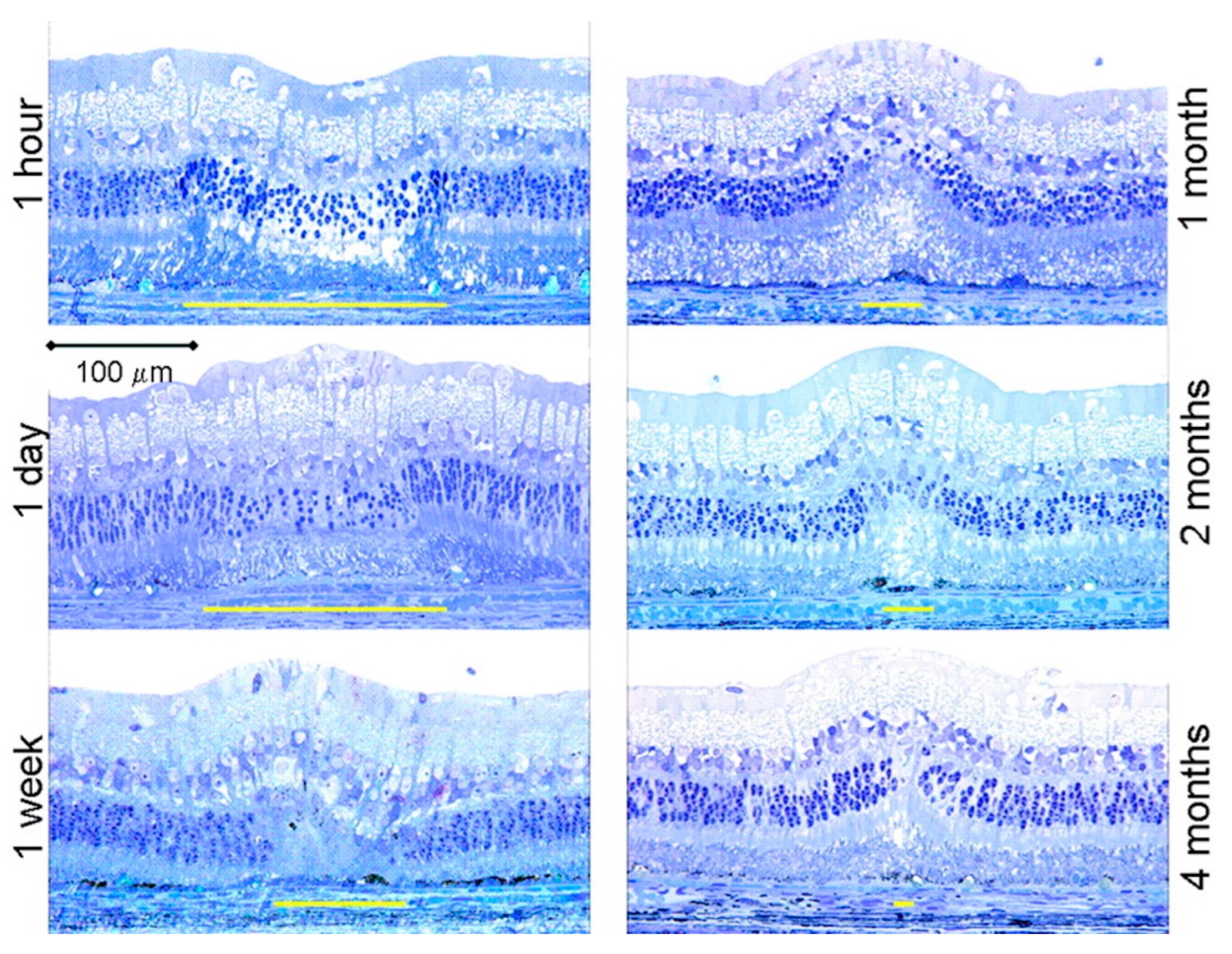
After sublethal exposures, limited wound remodeling (e.g., lateral migration/“sliding” of adjacent RPE and photoreceptors) can partially fill small defects in models (Figure 2) [21] and selected human observations. However, this does not re-establish normal retinal architecture or function. Clinically, photocoagulation scars enlarge over time (“atrophic creep”) and are associated with persistent visual field loss [21,22,23,24].
In damaging photocoagulation (e.g., conventional PRP), proposed benefits include relief of retinal hypoxia via partial reduction in photoreceptor oxygen demand and downstream VEGF modulation [25,26]. A post-laser inflammatory cascade overlaps with pathways seen in neurodegeneration [27]. The expression of cytokines such as heat shock protein (Hsp) and transforming growth factor-β (TGF-β) was shown to be up-regulated in the inflammatory response in retinal photocoagulation. These cytokines can also antagonize the vascular permeability effects of VEGF [19,28]. Accordingly, we reference oxygen demand reduction only for ablative PRP and avoid implying it as a mechanism for SDM.
While PRP is effective, it carries important tradeoffs—discomfort, permanent scarring, procedure time, and reduced peripheral/color/night vision [29,30]. To achieve better outcomes and minimize the side effects of conventional retinal laser therapy, new lasing mediums, wavelengths, pulse durations, and laser delivery systems have been developed.
For example, a modern method of retinal photocoagulation termed Pattern Scanning Laser Photocoagulation (PASCAL®) (Iridex Corporation, Mountain View, CA, USA) was developed to apply spots in a defined pattern to reduce treatment time, improve patient comfort, and improve accuracy of treatment [31]. However, pattern-scanning platforms remain destructive PRP tools and are not macula-safe. Durable neovascular regression can be less than with traditional argon PRP if the total treated area is reduced, so efficacy depends on matching area/density rather than a fixed spot count [12,32]. Selective Retinal Therapy (SRT) refers to significantly shorter pulse duration lasers in the microsecond domain. This results in RPE-selective retinal laser therapy, wherein selective therapy of RPE cells can be achieved while the surrounding neurosensory retinal temperature remains sublethal [33]. Another approach is Subthreshold Diode Micropulse Laser Photocoagulation (SDM), where non-visible retinal treatment is designed to reduce collateral tissue damage [34]. Spatial and temporal modulation of laser beams has also been reported [35]. These novel methods will be described in more detail subsequently in this manuscript.

1.5. Effect of Pulse Duration

Pulse duration is a primary determinant of laser–tissue interaction, significantly influencing both spatial precision and safety. Studies demonstrate that shorter pulse durations produce lesions with reduced width and axial depth. These lesions are less sensitive to power variations, thereby enhancing treatment accuracy and control [36]. In contrast, longer pulses (tens to hundreds of milliseconds) allow thermal heat to diffuse beyond the target zone, raising the risk of collateral damage to the neurosensory retina and choroid. This underscores why short pulses are safer in critical retinal regions.
In the microsecond and below regime, rapid heating around RPE melanosomes precipitates thermomechanical microcavitation and selective RPE disruption. As pulse duration lengthens toward tens of microseconds and above, effects become increasingly thermal (protein denaturation) with greater lateral heat diffusion [37,38]. Experimental and modeling studies place the mechanistic crossover around ~50 µs, with microbubble-driven RPE injury dominating at shorter pulses and thermal denaturation at longer exposures [39]. This mechanistic distinction is not semantic; it dictates what physiological signals exist to detect a “just-at-threshold” treatment in real time and which feedback modalities are appropriate during surgery.
Selective Retina Therapy (SRT) operates intentionally in the microsecond regime (typically ~1.7–5 µs) to leverage thermomechanical microcavitation confined to the RPE [40]. Clinical/bench implementations use pulse trains from frequency-doubled, Q-switched sources (e.g., 527–532 nm) with repetition rates around 100 Hz, delivering multiple microsecond pulses per spot to achieve selective RPE effects while sparing the neurosensory retina [41]. Because SRT lesions are subvisible ophthalmoscopically, SRT systems can incorporate real-time optoacoustic (photoacoustic) dosimetry to detect microbubble events and lock treatment at the selective threshold. OCT-based strategies are also described [40,42]. These feedback requirements and parameter choices flow directly from the thermomechanical mechanism that defines SRT.
By contrast, repetitive short continuous-wave “micropulse” delivery (often used at 100–300 µs micropulse widths and 5–15% duty cycles within a 100–200 ms envelope) seeks sublethal thermal effects with interpulse cooling rather than microcavitation [43]. Because the mechanism is thermal and intentionally subvisible, there is no microbubble signal to monitor. Instead, clinicians rely on titration and thermal dose surrogates [44,45]. A widely used framework (Endpoint Management) maps energy to biological response: RPE heat-shock responses (e.g., HSP70) at ~25% of a visible-threshold dose and onset of tissue damage around ~40%, defining a nondamaging “therapeutic window” for high-density macular treatment [46]. These features (pulse width in the hundreds of microseconds, low duty cycle, and a subvisible endpoint) are hallmarks of micropulse and indicate a sublethal photothermal mechanism, distinct from microsecond SRT, which achieves RPE selectivity via microcavitation.

2. Conventional Retinal Laser Therapy

Conventional photocoagulation is used for diabetic retinopathy, retinal vein occlusions, sickle cell retinopathy, retinal tears/detachment, and selected cases of age-related macular degeneration. Delivery options include slit-lamp systems (most commonly used) [47], laser indirect ophthalmoscopy for a wide field or media opacity [48], and intraoperative endolaser [49]. All of these involve connecting a laser light source through a fiber optic cable. Typical parameters are 100–200 ms pulses, 100–500 µm spots, and 100–750 mW power [50]. The laser parameters, including pulse duration, spot size, wavelength, and laser power, all have decisive effects on the final retinal burns produced. They are titrated to produce detectable gray–white lesions as a desired endpoint of treatment because of the thermal coagulation at this setting.
The Diabetic Retinopathy Study (DRS) established that PRP reduces severe vision loss by ~60% at 2 years in high-risk PDR [51]. The DRS also demonstrated that the argon laser achieved similar efficacy with fewer adverse effects than the xenon arc and became the predominant light source. Bulky water-cooled argon units were later replaced by air-cooled, frequency-doubled Nd:YAG 532 nm systems. DRS-style PRP typically uses 100 ms, 200–500 µm, 200–300 mW, delivering ~1500–5000 burns over 1–4 sessions. The procedure is time-consuming and often uncomfortable, and conventional PRP carries trade-offs, including permanent scarring and reduced peripheral, color, and night vision. It has also been associated with protective effects in retinal and choroidal neovascular situations in DR [52].
The Early Treatment Diabetic Retinopathy Study (ETDRS) validated conventional laser for diabetic retinopathy and showed that focal treatment for CSME reduced the incidence of vision loss by ~50% at 3 years [50,53]. Benefits extend to patients with severe NPDR eligible for focal/grid photocoagulation. Although mechanisms are not fully defined, proposed effects include occlusion of leaking microaneurysms and RPE-mediated cytokine responses that promote fluid resorption [54]. Trials indicate that the indirect (non-focal) grid alone has a limited effect on edema [55], and the modified ETDRS grid outperforms the mild macular grid (MMG) [56]. Furthermore, DRCR.net Protocol B found focal/grid superior to 1 mg or 4 mg intravitreal triamcinolone in efficacy and safety [57]. Clinically, focal/grid laser remains an effective, low-burden option for anti-VEGF refractory CSME [58]. Compared with PRP, macular focal/grid is generally less painful and faster, but visible scars may enlarge, and complications can include CNV, subretinal fibrosis, and visual field loss.
Laser photocoagulation has been used for retinal tears and prophylaxis of detachment since early xenon-arc applications [59]. Treatment decisions consider symptoms, tear type, traction, and risk factors. Versus cryotherapy, laser is typically less uncomfortable, feasible under topical anesthesia, and associated with lower risks of PVR and RPE-cell dispersion while maintaining high efficacy [60]. The goal is to achieve a firm chorioretinal adhesion surrounding the lesion to block the spread of subretinal fluid [61]. In Stickler syndrome, conventional single-spot, encircling indirect retinopexy reduces the lifetime risk of rhegmatogenous detachment by 55%, and the AAO recognizes prophylactic laser use in such high-risk settings [62].
Laser photocoagulation has been demonstrated to be a safe and effective treatment for symptomatic flap tears and other high-risk retinal breaks (Figure 3) [63]. Treatment is typically delivered at the slit lamp using wide-angle contact lenses—commonly a three-mirror lens (~150° field)—with three confluent rows of burns encircling the lesion [64]. Tears at the equator or posterior are readily treated via slit-lamp delivery. In contrast, the laser indirect ophthalmoscope (LIO) aligns the aiming beam within the indirect view to enable treatment of far-peripheral tears up to the ora serrata. LIO is generally favored for peripheral pathology and avoided for posterior tears because of reduced control over eye movement [48].

3. Selective Retinal Therapy

SRT is designed to selectively disrupt retinal pigment epithelium (RPE) cells while sparing photoreceptors and the inner retina, and is particularly valuable in macular disease, where RPE dysfunction rather than photoreceptor ablation is the desired target [33,66,67]. During microsecond scale exposures, deposited energy remains thermally confined around melanosomes, preventing broad coagulation. RPE melanosomes absorb strongly in the green. The melanosome absorption coefficient is in the 102–103 cm−1 range (including values reported near ~2.2 × 103 cm−1 at 532 nm), supporting selective heating of pigmented RPE [68].

3.1. Mechanism: Cavitation Dominates in the Lower-Microsecond Range

With ~1–5 µs pulses, heating occurs under stress confinement (heat rises faster than it can spread). Rapid expansion around melanosomes forms tiny intracellular microbubbles that mechanically disrupt the RPE while minimizing collateral thermal damage to the adjacent retina [37,38]. Multiple preclinical studies delineate a mechanistic crossover near ~50 µs. Below this, cavitation-mediated RPE injury dominates. Above it, protein denaturation with lateral heat diffusion contributes increasingly to tissue response [39]. After SRT, neighboring viable RPE cells migrate and proliferate to restore the monolayer, which aligns with the therapy’s regenerative intent [67].

3.2. Implementation and Real-Time Dosimetry

Pulsed SRT implementations generally use frequency-doubled, Q-switched sources (527–532 nm) to deliver trains of ~1.7–5 µs pulses at ~100 Hz to each spot, producing subvisible selective RPE effects without ophthalmoscopic endpoints [41]. Because SRT endpoints are intentionally subvisible, systems can employ optoacoustic (photoacoustic) feedback. The device can detect the onset of the first microbubble event in a pulse train and halt immediately, locking in a just-threshold dose tailored to each spot [40,42]. Recent algorithmic work further improves microbubble detection robustness across variable fundus conditions, enhancing selectivity and reproducibility in both preclinical and clinical settings [42]. OCT-based monitoring has also matured from time-resolved ultra-high resolution OCT capable of detecting dynamic RPE changes during SRT to real-time OCT feedback-controlled SRT that executes accurate, uniform grids while avoiding overtreatment [69,70].

3.3. Dosing, Tissue Variability, and Modeling

Thresholds vary with ocular pigmentation, melanosome loading, local perfusion, and other heatsink effects. Closed loop dosimetry (optoacoustic or OCT) reduces inter- and intra-eye variability by determining the effective threshold per spot in real time [40,42]. Beyond classical heat transfer theory, recent models incorporate melanosome geometry/heterogeneity and even nonlinear absorption of melanin at 532 nm to refine threshold predictions for short-pulse RPE targeting [71,72]. The strong 532 nm photoacoustic response of ocular melanin independently supports the reliability of optoacoustic dosimetry for SRT at green wavelengths [73].

3.4. Distinguishing SRT from Subthreshold Micropulse (SDM)

SRT operates with ~1–5 µs pulse trains that intentionally trigger cavitation and RPE cell death at threshold and can use intrinsic physical feedback (optoacoustic and, increasingly, OCT) to stop at the first microbubble [37,40,70]. However, the safety window is narrow. Microsecond dosing can extend damage beyond RPE into photoreceptor outer segments when energy overshoots or feedback is inadequate, as shown by damage threshold and cavitation mechanism studies across the lower µs regime. In contrast, subthreshold micropulse (SDM) uses 100–300 µs pulses at 5–15% duty cycle within ~100–200 ms envelopes, producing sublethal photothermal stress with inter-pulse cooling, no cavitation, and no inherent microbubble signal [74,75]. SDM dosing therefore relies on titration and biological surrogates (e.g., HSP70 induction near ~25% of visible threshold and damage onset near ~40%) to remain in a non-damaging thermal window [18]. Importantly, modeling and measurements show that when average power and exposure duration are matched, micropulse and continuous-wave yield very similar thermal outcomes, reinforcing that SDM is thermal modulation rather than cavitation-based [18].

3.5. Historical Development

Foundational preclinical work with ~5 µs, 514 nm, 500 Hz pulse trains established RPE-selective effects with intact photoreceptors and RPE repopulation on histology, launching the concept of selective macular laser [33]. Early clinical pilots using a 1.7 µs Nd:YLF platform reported no visual-field loss by microperimetry after SRT, consistent with subvisible RPE-selective dosing [76]. A scanning-beam SRT approach later achieved microsecond dwell per RPE location with a fast CW beam (e.g., PASCAL), confirming selective RPE effects and subsequent RPE repair in rabbits [77,78].

3.6. Clinical Evidence and Safety

Prospective and real-world cohorts using feedback-controlled SRT consistently report subretinal-fluid resolution with BCVA gains and no visible scarring, aligning with the subvisible, RPE-selective design [79,80]. A randomized controlled trial further demonstrated the safety and efficacy of real-time feedback-controlled SRT for chronic CSCR, strengthening causal inference for benefit [79,81]. Systematic evaluations of CSCR therapies position SRT among effective options, while acknowledging half-dose/fluence PDT as a benchmark and highlighting SRT’s utility where PDT is unavailable [82].
A prospective two-center phase II study of SRT for clinically significant DME reported anatomical/functional improvement with optoacoustic thresholding, and no adverse effects typical of coagulative burns [76,83]. Additional prospective series in DME corroborate feasibility and safety, with RPE selectivity confirmed by multimodal imaging and controlled dosimetry [84].
While drusen reduction after RPE-selective lasers has been reported, 2RT® (sub-nanosecond “rejuvenation” laser) is mechanistically distinct and showed mixed outcomes. We will analyze this alongside PDT and other non-conventional lasers in Section 5 [85,86].
Across animal and human SRT studies, treatments are typically subvisible with no immediate ophthalmoscopic burn, no scotomas on microperimetry, and RPE restoration after targeted disruption [33,70,76]. Because the therapeutic endpoint is invisible, optoacoustic or OCT-based closed-loop control can limit variability and avoid overtreatment [42,69].

3.7. Cautions and Limitations

Despite the subvisible endpoint, short-pulse RPE-disruptive paradigms (SRT; sub-nanosecond 2RT) have not demonstrated benefit in intermediate AMD (iAMD) and may worsen high-risk phenotypes. The LEAD randomized trial (292 eyes, 36 months) showed no overall benefit, and eyes with reticular pseudodrusen (RPD) had accelerated progression to late AMD (post hoc interaction; harmful direction in the RPD subgroup) [85]. An editorial advised against treating iAMD with laser outside clinical trials [87]. Importantly, that caution arose from evidence with sub-nanosecond “2RT/SNL” and should not be generalized to other laser modalities with different mechanisms and dosing. In particular, fixed-parameter 810 nm SDM (sublethal photothermal; non-titrated, panmacular dosing) has a separate evidence base, with large real-world comparative cohorts reporting reduced conversion from dry to neovascular AMD under periodic SDM (“Vision Protection Therapy”). However, these data are observational rather than randomized [88,89]. A 60 month observational extension of LEAD likewise found no overall benefit [85].

4. Innovations in Retinal Laser Therapy

The associated side effects of conventional retinal laser therapy with 100–200 ms pulse duration led specialists to seek improvements. In recent years, new technology has been developed to modify conventional retinal laser therapy to minimize retinal damage without compromising the therapeutic effect. Most of the innovations have focused on modulations of the pulse duration and spot size.

4.1. Pattern Scanning Laser

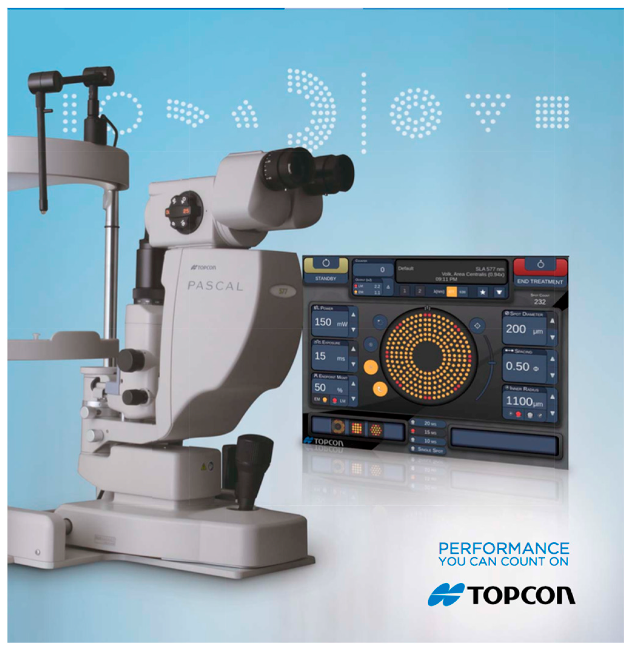
Introduced in 2005, PASCAL (pattern scanning laser) is a semi-automated retinal photocoagulation system that delivers patterns of 4–56 burns in <1 s using 10–30 ms pulses [31]. A 514 nm argon source is delivered through a standard slit-lamp. Beam positioning uses two-axis galvanometric mirrors, foot-pedal triggering, and a touchscreen GUI for parameter selection (Figure 4). Long-duration (>100 ms) burns extend beyond RPE/photoreceptors to inner nuclear, ganglion, and nerve fiber layers [36]. Shorter-pulse millisecond burns show less inner-retinal damage on histology [90]. They are associated with a smaller reduction in visual field sensitivity [91].
Pattern scanning short-pulse systems (e.g., PASCAL) are destructive photocoagulation platforms, not damage-sparing therapy. They are therefore not suitable for foveal treatment (PRP specifically avoids the macula). When traditional PRP settings are used, PASCAL-PRP can be less effective than standard argon PRP for durable neovascular regression in treatment-naïve high-risk PDR if equivalent treatment areas are not utilized [32]. Biophysically, shorter pulses (≈10–20 ms) confine thermal spread, creating smaller perilesional zones than 100 ms exposures. This can thereby reduce collateral tissue engagement. Comparable efficacy requires consideration to ensure that an equivalent treatment area is performed [92].
Short exposures allow delivery of multiple spots within a single fixation and are associated with reduced discomfort [93,94]. For destructive millisecond photocoagulation (e.g., multispot pattern-scanning PRP), completing many burns within a single fixation aids tolerance and accurate placement. By contrast, this consideration is not safety-critical for nondamaging SDM (810 nm, ~5% duty; non-visible endpoint, panmacular coverage), where fixation primarily affects efficiency rather than safety [20,95,96]. The benefit is attributed to more confined heating at the RPE–photoreceptor interface, sparing the inner retina and choroid [93,97]. PASCAL’s well-aligned arrays (4–56 spots, varied shapes/angles) support accurate placement, shorter treatment times, and better tolerance/acceptance [98,99]. A 2021 systematic review of 13 RCTs/4 CCTs (1961 eyes) ranked multispot pain scores below single-spot by ≈2.7 VAS units, translating to 40% fewer patients needing peri-procedural analgesia [94].
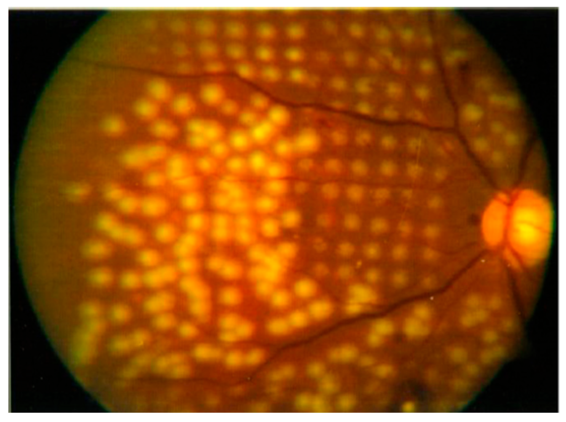
In practice, PASCAL PRP commonly uses 200 µm/20 ms spots from the arcades and 500 µm toward the far periphery with 3 × 3 to 7 × 7 patterns (Figure 5). A randomized clinical trial comparing multispot 532 nm pattern-scan with single-spot 532 nm GLX found similar regression of retinopathy, less collateral damage, and shorter, less painful sessions for multispot [100]. Another study reported reduced efficacy vs. traditional argon in high-risk PDR when an equivalent number of spots was applied [32]. However, subsequent analysis showed the treated retinal area was significantly smaller in the PASCAL arm, which may explain the higher recurrence of neovascularization [101].
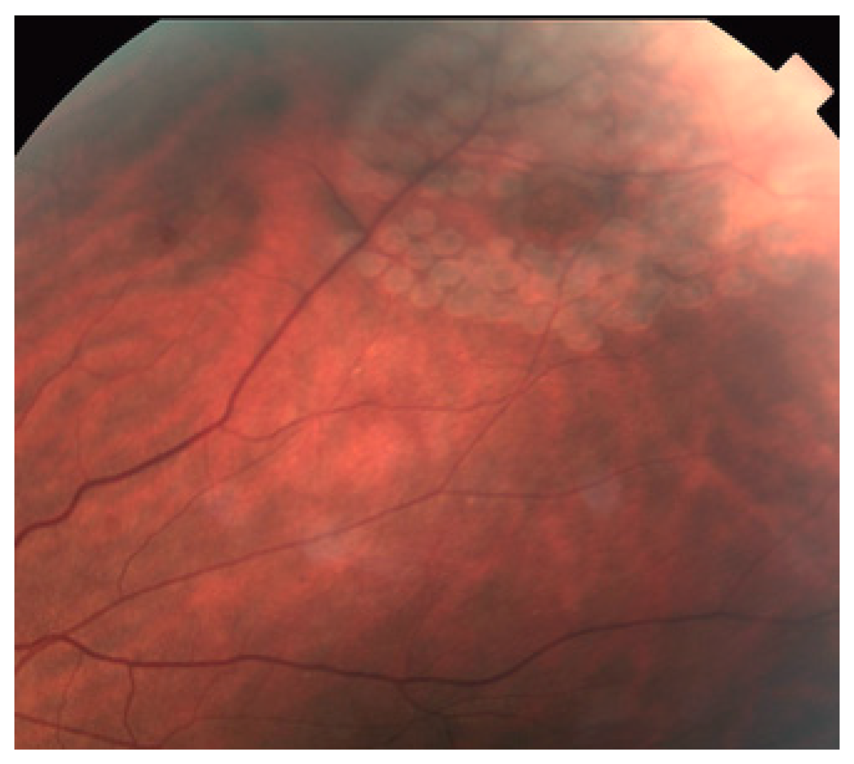
PASCAL has been used for macular edema in diabetic retinopathy and branch retinal vein occlusion, employing ring/arc patterns with a central foveal exclusion zone to prevent treatment within a preset distance of the FAZ. “Subthreshold” focal-grid applications have been reported with the aim of reducing postoperative scar enlargement. However, laser scars progressively enlarge over time (“atrophic creep”), and thus the only reliable way to prevent scar enlargement is to avoid creating a scar in the first place through favoring nondamaging paradigms when clinically appropriate [20,23,103]. Because subvisible burns are not ophthalmoscopically apparent, patterned delivery can assist lesion placement in this setting. A 2012 retrospective series found short-pulse PASCAL parameters for macular edema produced clinical and visual outcomes comparable to conventional argon settings. The report also compared retinopexy using a multi-spot arc pattern (Figure 6) [104]. More recent studies likewise report efficacy and safety for DME with PASCAL comparable (and in some cases favorable) to conventional methods [95]. The current PASCAL interface allows selection of spot patterns to surround retinal tears, accommodating small and large lesions with varied geometries.

4.2. Subthreshold Diode Micropulse Laser

Another novel laser modality, termed subthreshold diode micropulse (SDM) photocoagulation, was developed to minimize collateral tissue damage, especially for the treatment of the macula. In this review, SDM refers specifically to near-infrared 810 nm diode micropulse delivered at 5% duty cycle with a nondamaging endpoint (no visible burn), typically applied in confluent, high-density patterns over dysfunctional retina (often panmacular) and without titration to a visible test burn [20,96,105]. SDM was first pioneered in the late 1990s and designed to selectively affect RPE with minimal effects on the neurosensory retina [102]. A near-infrared diode laser (810 nm) operating with bursts of submillisecond pulses was used [106]. In this review, ‘subthreshold’ refers to dosing designed to avoid an ophthalmoscopically visible intraretinal burn at the time of treatment. The exact mechanism of SDM is still not completely understood. Human retinal thermodynamics do indicate association with sublethal thermal induction of heat-shock proteins in the distressed RPE cells, which is also supported by in vitro RPE experiments demonstrating HSP70 mRNA peaks after exposure [107,108]. Based on the hypothesis that the RPE is ultimately responsible for modulating the exudative response after heat stimulation by a nearby laser burn, therapeutic effects of SDM may be achieved by altering the metabolic activity of the RPE, resulting in the release of cytokines that regulate angiogenesis and vascular leakage [109,110]. These therapeutic effects may thus be produced with milder retinal irradiances, causing lower temperature rises associated with less or no significant retinal damage [111]. Proteomic profiling showed SDM induces a long-term normalization of specific retinal neuro-inflammatory pathways [112].
In this technique, a micropulse laser is delivered as a train of microsecond laser pulses separated by variable quiet intervals achieved by adjustment of the duty cycle of the laser, allowing the tissue to return to baseline temperature between pulses. A duty cycle is the fraction of a period when a laser has been emitted in which the laser is “on”. This “on” time is the duration of each micropulse (typically 100 μs to 300 μs), and the “off” time (1700 μs to 1900 μs) is the interval between micropulses [113,114]. When a low duty cycle is used, the “on” time is short, which limits the time for thermal dissipation between each pulse, thereby reducing collateral damage [75]. So as to achieve the beneficial effects of the laser, repeated pulses are added. Benefits from damaging macular laser modalities (threshold/suprathreshold) are mediated indirectly by surviving cells at lesion margins (RPE/photoreceptors) and post-injury remodeling [109,110,111]. By contrast, SDM produces sublethal photothermal stimulation and not coagulation, with RPE heat shock responses and cytokine modulation in directly treated but viable cells [107,108], consistent with nondamaging “therapeutic window” concepts in subthreshold dosing. A recent meta-analysis of eight RCTs reported superior BCVA and CMT reductions for subthreshold micropulse laser at visible wavelengths (predominantly 577 nm) using ~15% duty cycles compared with conventional laser, but the same analysis did not show significant gains at 5% duty cycle in those 577 nm protocols [115]. These findings do not directly apply to 810 nm SDM (5% duty cycle, panmacular dosing).
Low-intensity/high-density micropulse laser allows for complete and confluent coverage of the entire diseased retina, such as the areas of macular thickening constituting diabetic macular edema (DME) or ischemic retina with proliferative diabetic retinopathy (PDR) [116]. For SDM, this translates clinically into confluent, panmacular coverage for many macular indications to maximize the nondamaging biological effect while avoiding collateral injury [96,117]. Various kinds of commercial micropulse lasers are available at wavelengths of 532 nm, 577 nm, 586 nm, 660 nm, and 810 nm. The poor absorption of these wavelengths by the yellow xanthophyll pigment of the macula may also allow for safer treatment administration closer to the center of the fovea [118]. These have also been shown to produce significant improvement in visual field mean deviation on automated perimetry [119]. A meta-analysis in 2024 also concluded that adding SDM to anti-VEGF therapy reduced the mean injection burden by 1.85 intravitreal injections over 12 months without an increase in any adverse effects [120].
The most widely used application of SDM laser is for the treatment of clinically significant macular edema (CSME) [121]. A three-year follow-up period case series of 25 eyes demonstrated that SDM photocoagulation had a beneficial, long-term effect on visual acuity improvement and resolution of CSME [74]. There are several randomized clinical trials comparing the results of the ETDRS or modified ETDRS focal laser protocol for conventional argon laser photocoagulation to SDM photocoagulation in eyes with DME. SDM was demonstrated as equal or superior to modified-ETDRS laser photocoagulation with less damage to RPE confirmed by microperimetry and fundus autofluorescence [122,123]. The multicenter, double masked DIAMONDS RCT (n = 266) found 577 nm subthreshold micropulse laser (SML) (Iridex IQ 577® in MicroPulse® mode (Iridex Corporation, Mountain View, CA, USA)) clinically equivalent to standard threshold macular laser for center-involving DME at 24 months [124]. DIAMONDS used a titration-based protocol (visible threshold test spot in CW mode, then 5% duty cycle 200 ms SML at 4× threshold) and therefore evaluated 577 nm SML rather than 810 nm SDM. Accordingly, statements about visible burns or collateral coagulative effects should not be attributed to SDM. They apply to threshold/suprathreshold photocoagulation or to micropulse regimens outside the nondamaging window (often using titration frameworks distinct from SDM).
Preliminary studies of SDM for macular edema secondary to branch retinal vein occlusions and subretinal fluid from central serous chorioretinopathy have also found that SDM has the ability to achieve similar clinical effects as conventional laser therapy [121]. Unlike those diseases involving dysfunction of RPE, SDM was also shown to be an alternative to conventional PRP for the treatment of PDR. In a retrospective study of 99 eyes with severe non-proliferative retinopathy or any degree of PDR that were treated with subthreshold 810 nm micropulse PRP, the results indicated that SDM is comparable to conventional PRP [125]. Importantly, SDM-PRP has been reported to induce neovascular regression and stabilize proliferative disease without ophthalmoscopically visible retinal burns (i.e., damage-free PRP) [125]. These damage-free outcomes indicate that reduced photoreceptor oxygen consumption via ablative retinal destruction is not required for therapeutic benefit. Rather, efficacy is consistent with sublethal RPE stress-response activation targeted in modern nondamaging laser [20]. However, these studies have limitations. To confirm the therapeutic effects and safety of SDM, further large, prospective, multicenter, randomized, controlled clinical trials are still needed. Large multicenter RCTs are now underway to validate these findings over longer follow-up and to standardize dosing algorithms [126]. SDM also has drawbacks, including that it can take a longer time to achieve the desired clinical result than conventional lasers.

4.3. Recent Advancements in Retinal Laser Therapy

Additional recent innovations in retinal laser therapy include endpoint management and image-guided, navigated laser; closed-loop, temperature-controlled photocoagulation; teleguided (remote) navigated laser; and even robot-assisted delivery platforms.

4.3.1. Endpoint Management

One technique to modify laser therapy to precisely control laser energy relative to titration level is called “Endpoint Management” [127]. EpM determines a visible–threshold test burn (“mild retinal whitening”) at a reference site and then delivers treatment at a fixed fraction (typically ~30%) of that threshold power and pulse duration [128]. A commercially available model-based titration algorithm has been developed for both the 532 nm and 577 nm PASCAL lasers, which allows for predictable laser dosimetry ranging from nondamaging to intense coagulative tissue effects. Endpoint management begins with titrating laser power to a barely visible burn. This energy level is defined as a 100% nominal energy level, and other pulse energies can be selected as a percentage of that energy by clinicians to be delivered to the treatment locations. This provides an approach to reproducible subvisible retinal laser therapy. Endpoint management with sub-visible therapy has also shown anatomical and functional gains in retinitis pigmentosa cystoid macular edema with the goal of minimizing tissue effect. However, pigmentary RPE changes have been reported after 577 nm EpM in chronic CSCR (5/11 eyes, 45.4%) [128,129].
With short-pulse exposures (10–20 ms), the safe therapeutic window narrows, so a fixed fraction of a visible test burn may overshoot in some eyes and undershoot in others. The safe therapeutic window tightens as pulse duration decreases [35]. Inter- and intra-ocular variation in melanin (RPE/choroid) further alters local absorption, meaning the visible threshold at a test site does not guarantee the same thermal dose elsewhere [130].
Accordingly, EpM should be presented as a titration strategy rather than a guarantee of nondamaging treatment. Reported outcomes and safety depend strongly on pulse duration, wavelength (e.g., 577 nm), and local pigmentation, and real-world series have documented RPE changes under EpM protocols.

4.3.2. Navigated Laser (NAVILAS)

Another commercially available system is the “Navigated laser (NAVILAS)”. This system uses a novel fundus imaging and laser treatment device that allows various imaging modalities, including infrared, color, and fluorescein angiography, along with integrated laser treatment of the retina [131]. The acquisition of retinal images can be used to create a detailed treatment plan, and then the laser is delivered automatically to the target area of the retina according to the treatment, which allows for better guidance of laser delivery and high precision and reproducibility of theoretically <60–110 μm.

4.3.3. OCT Monitoring with Automatic Dosimetry

In addition, some more sophisticated laser systems are under development and might become available clinically in the coming years. These include non-invasive and real-time monitoring of retinal temperature with optoacoustic or optical coherence tomography (OCT) imaging to achieve real-time automatic dosimetry for more reproducible, uniform photocoagulation lesions [132]. In SRT, optoacoustic (OA) feedback detects microbubble formation at melanosomes, which is used as a surrogate for RPE cell disintegration, i.e., it confirms a destructive RPE endpoint rather than a nondamaging effect [40,42]. OA monitoring requires an ultrasonic transducer integrated into a contact lens and signal-processing algorithms to detect the first microbubble event, adding system complexity [40,42,73]. OCT-based feedback has been demonstrated in real time but largely in bench/ex vivo preparations, and temperature-controlled/model-predictive control implementations remain at a translational engineering stage [69,70,132,133]. Accordingly, these closed-loop autodosimetry platforms, particularly OCT-based feedback and temperature-controlled/MPC approaches, remain investigational with limited clinical adoption to date, with further work needed to standardize feedback thresholds across devices and pigmentation. This automatic feedback system can halt the laser emission when the desired temperature is attained. Thus, the treatment time and risk of overtreatment can be minimized, especially for sub-threshold treatment of retinal diseases. A pilot study in porcine eyes showed that a model-predictive, optoacoustic closed-loop controller kept peak retinal temperature within ±2 °C of the 45 °C set point. It also reduced lesion variability by 40%, confirming tighter dosimetry [133]. Modifications have also been described using optical coherence tomography (OCT) to monitor and analyze tissue effects introduced by SRT. Time-resolved ultra-high resolution OCT also shows promising results in guiding SRT in an automatic feedback mode [70]. A 2023 ex vivo study demonstrated real-time OCT feedback-controlled selective-retina-therapy, reporting accurate RPE photodisruption over a 15 × 5 lesion grid without overtreatment [69]. Because the OA-verified endpoint is RPE injury, SRT/OA is generally not applied to the foveal center in clinical protocols. Studies target extrafoveal leakage in CSCR and similar indications [134,135]. For foveal disease, nondamaging subthreshold diode micropulse (SDM, 810 nm; 5% DC) is reported safe for transfoveal use without visible burns or RPE injury [105].

4.3.4. Sub-Nanosecond Rejuvenation Laser (2RT)

Another new innovation in retinal laser therapy is the sub-nanosecond “rejuvenation” laser (2RT®). The 2-pulse retinal rejuvenation laser delivers 400 ps, 532 nm pulses in the photomechanical regime, where energy deposition at melanosomes induces microbubble formation and RPE photodisruption at threshold. Direct HSP-mediated sublethal stimulation is not expected at these ultrashort pulse durations, and any HSP signal would be secondary to wound healing rather than a nondamaging thermal response [37,38,39,43]. The pivotal LEAD randomized trial (292 eyes, 36 months) found no overall benefit, yet a prespecified subgroup analysis revealed a striking disease-modifying signal. In eyes without reticular pseudodrusen, 2RT® cut progression to late AMD by 68% (adjusted HR 0.32; 95% CI 0.16–0.65; p = 0.002) [85]. Importantly, eyes with reticular pseudodrusen (RPD) progressed faster under 2RT (harmful subgroup interaction). Therefore, 2RT is contraindicated in iAMD with RPD. Subsequent cohort work confirmed a structural correlation. SNL-treated eyes showed a significant reduction in drusen area and number after 6 months (p < 0.001) compared with matched controls, without visual-acuity loss [86]. A phase III confirmatory study (LASER-iAMD, NCT05192913; 740 eyes) is now underway. If validated, sub-nanosecond laser therapy could become the first laser-based intervention to delay the progression of intermediate AMD. On the offhand, a 60 month observational extension of LEAD likewise showed no overall benefit of SNL/2RT [85,87].

4.3.5. Remote/Teleguided Photocoagulation

Real-time photocoagulation can now be delivered entirely off-site. In a 2025 proof-of-concept, surgeons in Europe and the USA planned and executed NAVILAS 577 s treatments over a secure VPN link. Three eyes (PDR, focal DME, BRVO) received 267 ± 41 burns, and every spot landed within the 60-µm accuracy window. No technical problems or adverse events occurred [136]. These early demonstrations suggest that teleguided platforms could extend subspecialty retinal laser to sites without on-site surgeons. A separate 5-site Chinese study used a 5G tele-ophthalmology platform to perform PRP and focal/grid laser 1200 km from the operating surgeon. All nine eyes were treated without any perceptible transmission delay while delivering a mean 913 ± 243 PRP spots per session [137]. Together, these early trials show that teleguided navigated laser can maintain spot-placement accuracy and workflow efficiency, opening the door to remote expert care for patients in underserved regions.

5. Discussion

Across the spectrum of retinal laser therapies, clinical benefit hinges on how much energy the RPE absorbs and on how tightly that energy is confined in space and time. These determinants map directly to safety, durability, and endpoints such as scarring or subvisible effects [2,3]. The discussion below organizes current options by mechanism, dosimetry/feedback, and best-supported indications, and then positions SRT relative to alternatives.
Three regimes dominate retinal laser–tissue interaction: photothermal coagulation, nondamaging thermal stimulation, and photomechanical cavitation [2,17]. Coagulative millisecond exposures diffuse heat well beyond the RPE, leaving visible burns and durable scars useful for PRP in PDR, but they can result in decreased peripheral field of vision and night vision [29,30]. Nondamaging thermal paradigms (e.g., Endpoint Management) target sublethal RPE stress with Arrhenius-based titration near the HSP70 window, relying on modeled thermal dose instead of visible endpoints [19,43,127]. SRT intentionally crosses into photomechanical cavitation with 1–5 µs pulses, creating intracellular microbubbles at melanosomes and RPE selective disruption. This non-visible endpoint can be coupled to optoacoustic (microbubble) or OCT monitoring for closed-loop dosimetry [37,40,70].
Short-pulse green light concentrates energy in RPE melanosomes (μa in the 102–103 cm−1 range at 532 nm), enabling RPE-selective heating that transitions to microbubble-mediated disruption in the lower-µs regime [37,66,68]. Mechanistically, RPE cell death at threshold is intended, and the adjacent viable RPE repopulates the treated area [33,67]. Operationally, SRT can have intrinsic physical feedback, which includes optoacoustic detectors that stop delivery on the first microbubble, or OCT monitoring structural dynamics [40,42,69].
In chronic CSCR, a randomized trial showed real-time feedback-controlled SRT improves fluid and function with a favorable safety profile, and prospective real-world data at 24 months corroborate sustained benefit [80,81]. Systematic assessments place half-dose PDT as the gold standard but recognize SRT as reasonable where PDT is unavailable or when extrafoveal leakage permits [82]. In DME, phase II and subsequent series report anatomic/functional gains with optoacoustic thresholding and no coagulative sequelae, supporting feasibility and safety while larger trials are pursued [33,83,84].
Micropulse (SDM) operates in 100–300 µs bursts at 5–15% duty within 100–200 ms envelopes, with sublethal thermal goals, no cavitation, and no intrinsic microbubble signal [74,75,114]. Modeling and measurements show CW ≈ micropulse for matched average power/irradiation time, underscoring that SDM tunes thermal dose rather than invoking SRT-like photomechanical effects [18,138].
For PDR, PRP remains a durable, low-maintenance option with five-year outcomes non-inferior to anti-VEGF in Protocol S, albeit with more visual-field loss but far fewer injections [124,139,140,141]. Pattern scanning (PASCAL) and navigated platforms can improve throughput, comfort, and accuracy. Randomized controlled data show less pain and shorter sessions, and navigated systems deliver higher spot accuracy in focal laser and PRP [94,100,142,143,144]. These remain first-line therapies for indications where visible coagulation is intended (e.g., retinopexy, classic PRP) [43,127,145].
Endpoint management formalizes titration where a barely visible lesion is set to 100%, then subvisible therapy is delivered at modeled thermal doses (e.g., ≈30%) to sit below damage yet within HSP response [19,127]. SDM applies a similar nondamaging logic using micropulses. The DIAMONDS RCT showed 577 nm SDM was non-inferior to threshold macular laser for center-involving DME < 400 µm, and cost-effectiveness analyses favor SDM [124,146]. Meta-analyses suggest adding SDM to anti-VEGF can reduce injection burden without compromising vision or CMT when protocols are standardized [115,147]. Together, EpM and SDM represent thermal-window strategies that modulate biology without killing RPE, complementing but not duplicating SRT.
Because pigmentation and ocular transmission vary, open-loop power/time settings can over or under treat patients even in experienced hands. Emerging controllers using optoacoustic temperature sensing with model-predictive control (MPC) hold peak retinal temperature within tight bounds and reduce lesion variability [132,133]. These systems formalize thermal dose delivery and conceptually parallel SRT’s reliance on intrinsic feedback, though the feedback signal (temperature vs. microbubble/OCT) reflects the different mechanisms [133].
In DME, anti-VEGF remains first-line while SDM (and EpM) are options for CST < 400 µm or injection-burden reduction, per DIAMONDS and meta-analyses [115,124]. SRT is feasible/safe with optoacoustic thresholding and may suit selected, extrafoveal cases [83,84]. In retinopexy for retinal tears, conventional photocoagulation remains preferred for creating visible adhesive chorioretinal scars, where photocoagulation is the goal [61,63]. In intermediate AMD, an iAMD without reticular pseudodrusen subgroup signaled a potential effect of 2RT® while confirmatory phase III work is ongoing [85,86].
In this particular area, the key knowledge gaps include head-to-head comparisons of SRT vs. PDT in CSCR, SRT vs. SDM/EpM in DME, and standardization of feedback thresholds (microbubble, OCT, or temperature) across devices and pigmentation levels. Furthermore, decision analytic work should compare lifetime field loss and injection burden (anti-VEGF) versus durable photothermal endpoints (PRP) and subvisible regimens, incorporating patient reported outcomes and costs over multiyear horizons [124]. Finally, navigated/teleguided platforms may help with equitable access for PRP and focal/grid laser where expertise is scarce, but require governance and training frameworks before scale-up [136,137]. Future implementation studies should address reliability, training, reimbursement, and equitable access to support broader adoption.

6. Conclusions and Outlook

Since the development of lasers over 65 years ago, retinal laser photocoagulation has become a vital therapeutic method in treating numerous retinal disorders. Recent innovations and modifications to conventional laser photocoagulation have focused on maintaining or improving the clinical efficacy while reducing the numerous side effects, including scotoma formation, choroidal neovascularization, subretinal fibrosis, retinal scarring, and painful treatments. Laser therapy has thus consequently evolved toward laser modalities that have refined traditional laser parameters to minimize side effects and collateral damage while retaining the therapeutic effects. Pattern scanning laser (PASCAL) can make the therapy procedure quicker and less painful for patients. Selective retinal therapy (SRT) can selectively treat RPE cells while sparing the neurosensory retina to reduce damage and avoid scotoma. Subthreshold micropulse laser is used to produce therapy without inducing detectable intraretinal damage. These novel laser treatments have made retinal photocoagulation more refined and effective while reducing collateral tissue damage. However, with the developments in pharmacologic therapies such as intravitreal injection of anti-vascular endothelial growth factor (anti-VEGF) agents and corticosteroids, research is also now investigating new approaches, including combinations of laser and pharmacologic therapy. Scientifically rigorous studies have shown promising results with regard to the efficacy of intravitreal steroids and anti-VEGF therapies, but the repeated and long-term injections can be a considerable burden on the patients, physicians, family members, and the healthcare system while carrying rare but devastating ocular and systemic risks [148]. In the future, we will increasingly consider combination therapy of laser with pharmacologic agents. However, concurrent therapy with laser and steroids has been shown to not be simply additive but can affect the outcomes of one another [149]. Laser therapy still remains the gold-standard therapy in various retinal conditions. We expect that continued improvements in laser therapy will continue to ensure that lasers continue to play a critical role in our care of patients for many years to come.

Author Contributions

Conceptualization, Y.M.P.; methodology, X.X., L.M. and Y.M.P.; validation, X.X., L.M. and Y.M.P.; formal analysis, X.X., L.M. and Y.M.P.; investigation, X.X., L.M. and Y.M.P.; resources, Y.M.P.; data curation, X.X., L.M. and Y.M.P.; writing—original draft preparation, X.X.; writing—review and editing, X.X., L.M. and Y.M.P.; visualization, Y.M.P.; supervision, Y.M.P.; project administration, Y.M.P.; funding acquisition, Y.M.P. All authors have read and agreed to the published version of the manuscript.

Funding

This work was funded by grants from the National Eye Institute (YMP: 1R01EY033000 and 1R01EY034325), as well as the Foundation Fighting Blindness Non-Rodent Large Animal Award RC-CMM-0824-0899-JHU (YMP), Fight for Sight-International Retinal Research Foundation (YMP: FFSGIA16002), the Alcon Research Institute Young Investigator Grant (YMP), and unrestricted departmental funding from Research to Prevent Blindness. Additional support came from the Jonas Friedenwald Professorship in Ophthalmology (YMP). The funding organizations had no role in the design or conduct of this research or the decision to submit the manuscript for publication.

Institutional Review Board Statement

Not applicable.

Informed Consent Statement

Not applicable.

Data Availability Statement

It is a review article, so no new data was utilized or created.

Conflicts of Interest

Y.M.P. has served as a consultant to Iridex Corporation. The other authors declare no conflicts of interest. The funding organizations had no role in the design or conduct of this research.

Abbreviations

The following abbreviations are used in this manuscript:
AAOAmerican Academy of Ophthalmology
AMDAge-related macular degeneration
BCVABest-corrected visual acuity
BRVOBranch retinal vein occlusion
CCTControlled clinical trial
CIConfidence interval
CMECystoid macular edema
CMTCentral macular thickness
CNVChoroidal neovascularization
CSCRCentral serous chorioretinopathy
CSMEClinically significant macular edema
CSTCentral subfield thickness
DMEDiabetic macular edema
DRSDiabetic Retinopathy Study
DRDiabetic retinopathy
DRCR.netDiabetic Retinopathy Clinical Research Network
EPMEndpoint Management
ETDRSEarly Treatment Diabetic Retinopathy Study
FAZFoveal avascular zone
HSPHeat shock protein
iAMDIntermediate age-related macular degeneration
ILMInner limiting membrane
IVIIntravitreal injection
LIOLaser indirect ophthalmoscope
MDMean deviation (visual field index)
MMGMild macular grid
NPDRNon-proliferative diabetic retinopathy
OCTOptical coherence tomography
PASCALPAttern SCAn Laser
PDTPhotodynamic therapy
PDRProliferative diabetic retinopathy
PRPPan-retinal photocoagulation
RCTRandomized controlled trial
RDRetinal detachment
RPERetinal pigment epithelium
RPRetinitis pigmentosa
SDMSubthreshold diode micropulse (laser)
SMLSubthreshold micropulse laser
SMLPSubthreshold micropulse laser photocoagulation
SNLSub-nanosecond laser
SRFSubretinal fluid
SRTSelective retinal therapy
VFVisual field
VEGFVascular endothelial growth factor
VPNVirtual private network
WF-SMLWide field subthreshold micropulse laser

References

  1. Munnerlyn, C.R. Lasers in ophthalmology: Past, present and future. J. Mod. Opt. 2003, 50, 2351–2360. [Google Scholar] [CrossRef]
  2. Vogel, A.; Venugopalan, V. Mechanisms of pulsed laser ablation of biological tissues. Chem. Rev. 2003, 103, 577–644. [Google Scholar] [CrossRef]
  3. Birngruber, R.; Hillenkamp, F.; Gabel, V.P. Theoretical investigations of laser thermal retinal injury. Health Phys. 1985, 48, 781–796. [Google Scholar] [CrossRef]
  4. Delibasic, H.; Petrovic, V.; Petrovic, I. Laser Breakdown in Water Induced by λ = 532 nm Nanosecond Pulses: Analytical Calculation of the Number Density of Free Electrons. J. Phys. Soc. Jpn. 2020, 89, 114501. [Google Scholar] [CrossRef]
  5. Bianco, L.; Gawęcki, M.; Antropoli, A.; Arrigo, A.; Bandello, F.; Battaglia Parodi, M. Laser Treatment for Retinal Arterial Macroaneurysm. Photonics 2022, 9, 851. [Google Scholar] [CrossRef]
  6. Hecht, J. A short history of laser development. Appl. Opt. 2010, 49, F99–F122. [Google Scholar] [CrossRef]
  7. Meyer-Schwickerath, G.R. The history of photocoagulation. Aust. N. Z. J. Ophthalmol. 1989, 17, 427–434. [Google Scholar] [CrossRef]
  8. Bridges, W.B. Laser Oscillation in Singly Ionized Argon in the Visible Spectrum. Appl. Phys. Lett. 1964, 4, 128–130. [Google Scholar] [CrossRef]
  9. Beetham, W.P.; Aiello, L.M.; Balodimos, M.C.; Koncz, L. Ruby-laser photocoagulation of early diabetic neovascular retinopathy: Preliminary report of a long-term controlled study. Trans. Am. Ophthalmol. Soc. 1969, 67, 39–67. [Google Scholar] [CrossRef] [PubMed]
  10. L’Esperance, F.A. An ophthalmic argon laser photocoagulation system: Design, construction, and laboratory investigations. Trans. Am. Ophthalmol. Soc. 1968, 66, 827–904. [Google Scholar] [PubMed]
  11. Palanker, D.V.; Blumenkranz, M.S.; Marmor, M.F. Fifty years of ophthalmic laser therapy. Arch. Ophthalmol. 2011, 129, 1613–1619. [Google Scholar] [CrossRef] [PubMed]
  12. Palanker, D.V. Ophthalmic Laser Therapy: Mechanisms and Applications. Master’s Thesis, University of New South Wales, Sydney, Australia, 2014. [Google Scholar]
  13. Yun, S.H.; Adelman, R.A. Recent developments in laser treatment of diabetic retinopathy. Middle East. Afr. J. Ophthalmol. 2015, 22, 157–163. [Google Scholar] [CrossRef] [PubMed]
  14. Blumenkranz, M.S. The Evolution of Laser Therapy in Ophthalmology: A Perspective on the Interactions Between Photons, Patients, Physicians, and Physicists: The LXX Edward Jackson Memorial Lecture. Am. J. Ophthalmol. 2014, 158, 12–25.E1. [Google Scholar] [CrossRef]
  15. Ion, R.M. Photodynamic therapy (PDT): A photochemical concept with medical applications. Rev. Roum. Chim. 2007, 52, 1093–1102. [Google Scholar]
  16. Woodburn, K.W.; Engelman, C.J.; Blumenkranz, M.S. Photodynamic therapy for choroidal neovascularization: A review. Retina 2002, 22, 391–405. [Google Scholar] [CrossRef]
  17. Knappe, V.; Frank, F.; Rohde, E. Principles of lasers and biophotonic effects. Photomed. Laser Surg. 2004, 22, 411–417. [Google Scholar] [CrossRef]
  18. Wang, J.; Quan, Y.; Dalal, R.; Palanker, D. Comparison of Continuous-Wave and Micropulse Modulation in Retinal Laser Therapy. Investig. Ophthalmol. Vis. Sci. 2017, 58, 4722–4732. [Google Scholar] [CrossRef]
  19. Sramek, C.; Mackanos, M.; Spitler, R.; Leung, L.; Nomoto, H.; Contag, C.H. Non-damaging retinal phototherapy: Dynamic range of heat shock protein expression. Investig. Ophthalmol. Vis. Sci. 2011, 52, 1780–1787. [Google Scholar] [CrossRef]
  20. Keunen, J.E.E.; Battaglia-Parodi, M.; Vujosevic, S.; Luttrull, J.K. International Retinal Laser Society Guidelines for Subthreshold Laser Treatment. Trans. Vis. Sci. Technol. 2020, 9, 15. [Google Scholar] [CrossRef]
  21. Paulus, Y.M.; Jain, A.; Gariano, R.F.; Stanzel, B.V.; Marmor, M.; Blumenkranz, M.S. Healing of retinal photocoagulation lesions. Investig. Ophthalmol. Vis. Sci. 2008, 49, 5540–5545. [Google Scholar] [CrossRef] [PubMed]
  22. Koinzer, S.; Saeger, M.; Hesse, C.; Portz, L.; Kleemann, S.; Schlott, K. Correlation with OCT and histology of photocoagulation lesions in patients and rabbits. Acta Ophthalmol. 2013, 91, e603–e611. [Google Scholar] [CrossRef] [PubMed]
  23. Maeshima, K.; Utsugi-Sutoh, N.; Otani, T.; Kishi, S. Progressive enlargement of scattered photocoagulation scars in diabetic retinopathy. Retina 2004, 24, 507–511. [Google Scholar] [CrossRef]
  24. Subash, M.; Comyn, O.; Samy, A.; Qatarneh, D.; Antonakis, S.; Mehat, M.; Tee, J.; Mansour, T.; Xing, W.; Bunce, C.; et al. The Effect of Multispot Laser Panretinal Photocoagulation on Retinal Sensitivity and Driving Eligibility in Patients with Diabetic Retinopathy. JAMA Ophthalmol. 2016, 134, 666–672. [Google Scholar] [CrossRef]
  25. Stefánsson, E. The therapeutic effects of retinal laser treatment and vitrectomy. A theory based on oxygen and vascular physiology. Acta Ophthalmol. Scand. 2001, 79, 435–440. [Google Scholar] [CrossRef] [PubMed]
  26. Ascaso, F.J.; Huerva, V.; Grzybowski, A. The role of inflammation in the pathogenesis of macular edema secondary to retinal vascular diseases. Mediat. Inflamm. 2014, 2014, 432685. [Google Scholar] [CrossRef] [PubMed]
  27. Paulus, Y.M.; Kuo, C.; Morohoshi, K.; Nugent, A.; Zheng, L.L.; Nomoto, H.; Blumenkranz, M.S.; Palanker, D.; Ono, S.J. Serum inflammatory markers after rupture retinal laser injury in mice. Ophthalmic Surg. Lasers Imaging Retin. 2015, 46, 362–368. [Google Scholar] [CrossRef]
  28. Matsumoto, M.; Yoshimura, N.; Honda, Y. Increased production of transforming growth factorbeta 2 from cultured human retinal pigment epithelial cells by photocoagulation. Investig. Ophthalmol. Vis. Sci. 1994, 35, 4245–4252. [Google Scholar]
  29. Prskavec, F.H.; Fulmek, R.; Klemen, C.; Stelzer, N. Changes in the visual field and dark adaptation following panretinal photocoagulation in diabetic retinopathy. Klin. Monbl. Augenheilkd. 1986, 189, 385–387. [Google Scholar] [CrossRef]
  30. Russell, P.W.; Sekuler, R.; Fetkenhour, C. Visual Function After Pan-Retinal Photocoagulation: A Survey. Diabetes Care 1985, 8, 57–63. [Google Scholar] [CrossRef]
  31. Blumenkranz, M.S.; Yellachich, D.; Andersen, D.E.; Wiltberger, M.W.; Mordaunt, D.; Marcellino, G.R. Semiautomated patterned scanning laser for retinal photocoagulation. Retina 2006, 26, 370–376. [Google Scholar] [CrossRef]
  32. Chappelow, A.V.; Tan, K.; Waheed, N.K.; Kaiser, P.K. Panretinal Photocoagulation for Proliferative Diabetic Retinopathy: Pattern Scan Laser Versus Argon Laser. Am. J. Ophthalmol. 2012, 153, 137–142.e2. [Google Scholar] [CrossRef] [PubMed]
  33. Roider, J.; Michaud, N.A.; Flotte, T.J.; Birngruber, R. Response of the retinal pigment epithelium to selective photocoagulation. Arch. Ophthalmol. 1992, 110, 1786–1792. [Google Scholar] [CrossRef]
  34. Sinclair, S.H.; Mainster, M.A. Seminars in ophthalmology. Semin. Ophthalmol. 1999, 14, 197–199. [Google Scholar] [CrossRef]
  35. Sramek, C.; Paulus, Y.; Nomoto, H.; Huie, P.; Brown, J.; Palanker, D. Dynamics of retinal photocoagulation and rupture. J. Biomed. Opt. 2009, 14, 034007. [Google Scholar] [CrossRef] [PubMed]
  36. Jain, A.; Blumenkranz, M.S.; Paulus, Y.; Wiltberger, M.W.; Andersen, D.E.; Huie, P.; Palanker, D. Effect of pulse duration on size and character of the lesion in retinal photocoagulation. Arch. Ophthalmol. 2008, 126, 78–85. [Google Scholar] [CrossRef]
  37. Schuele, G.; Rumohr, M.; Huettmann, G.; Brinkmann, R. RPE damage thresholds and mechanisms for laser exposure in the microsecond-to-millisecond time regimen. Investig. Ophthalmol. Vis. Sci. 2005, 46, 714–719. [Google Scholar] [CrossRef]
  38. Lee, H.; Alt, C.; Pitsillides, C.M.; Lin, C.P. Optical detection of intracellular cavitation during selective laser targeting of the retinal pigment epithelium: Dependence of cell death mechanism on pulse duration. J. Biomed. Opt. 2007, 12, 064034. [Google Scholar] [CrossRef]
  39. Seifert, E.; Sonntag, S.R.; Kleingarn, P.; Theisen-Kunde, D.; Grisanti, S.; Birngruber, R.; Miura, Y.; Brinkmann, R. Investigations on Retinal Pigment Epithelial Damage at Laser Irradiation in the Lower Microsecond Time Regime. Investig. Ophthalmol. Vis. Sci. 2021, 62, 32. [Google Scholar] [CrossRef]
  40. Schuele, G.; Elsner, H.; Framme, C.; Roider, J.; Birngruber, R.; Brinkmann, R. Optoacoustic real-time dosimetry for selective retina treatment. J. Biomed. Opt. 2005, 10, 064022. [Google Scholar] [CrossRef] [PubMed]
  41. Považay, B.; Brinkmann, R.; Stoller, M.; Kessler, R. Selective Retina Therapy. In High Resolution Imaging in Microscopy and Ophthalmology: New Frontiers in Biomedical Optics; Bille, J.F., Ed.; Springer: Cham, Switzerland, 2019; pp. 237–259. [Google Scholar]
  42. Seifert, E.; Tode, J.; Pielen, A.; Theisen-Kunde, D.; Framme, C.; Roider, J.; Miura, Y.; Birngruber, R.; Brinkmann, R. Algorithms for optoacoustically controlled selective retina therapy (SRT). Photoacoustics 2022, 25, 100316. [Google Scholar] [CrossRef]
  43. Lavinsky, D.; Wang, J.; Huie, P.; Dalal, R.; Lee, S.J.; Lee, D.Y.; Palanker, D. Nondamaging Retinal Laser Therapy: Rationale and Applications to the Macula. Investig. Ophthalmol. Vis. Sci. 2016, 57, 2488–2500. [Google Scholar] [CrossRef]
  44. Scholz, P.; Altay, L.; Fauser, S. A Review of Subthreshold Micropulse Laser for Treatment of Macular Disorders. Adv. Ther. 2017, 34, 1528–1555. [Google Scholar] [CrossRef]
  45. Chhablani, J.; Alshareef, R.; Kim, D.T.; Narayanan, R.; Goud, A.; Mathai, A. Comparison of different settings for yellow subthreshold laser treatment in diabetic macular edema. BMC Ophthalmol. 2018, 18, 168. [Google Scholar] [CrossRef]
  46. Wang, J.; Lavinsky, D.; Huie, P.; Dalal, R.; Lee, D.Y.; Lee, S.J.; Palanker, D.V. Determining the therapeutic window of non-damaging retinal laser therapy by protein expression. Investig. Ophthalmol. Vis. Sci. 2016, 57, 5845. [Google Scholar]
  47. Little, H.L.; Zweng, H.C.; Peabody, R.R. Argon laser slit-lamp retinal photocoagulation. Trans. Am. Acad. Ophthalmol. Otolaryngol. 1970, 74, 85–97. [Google Scholar]
  48. Law, N.M.; Fan, R.F. Clinical experience with the laser indirect ophthalmoscope. Ann. Acad. Med. Singap. 1991, 20, 750–754. [Google Scholar]
  49. Smiddy, W.E. Diode endolaser photocoagulation. Arch. Ophthalmol. 1992, 110, 1172–1174. [Google Scholar] [CrossRef] [PubMed]
  50. Early Treatment Diabetic Retinopathy Study Research Group. Treatment Techniques and Clinical Guidelines for Photocoagulation of Diabetic Macular Edema: Early Treatment Diabetic Retinopathy Study Report Number 2. Ophthalmology 1987, 94, 761–774. [Google Scholar] [CrossRef] [PubMed]
  51. The Diabetic Retinopathy Study Research Group. Photocoagulation Treatment of Proliferative Diabetic Retinopathy: Clinical Application of Diabetic Retinopathy Study (DRS) Findings. Ophthalmology 1981, 88, 583–600. [Google Scholar] [CrossRef]
  52. Huang, T.; Li, X.; Xie, J.; Zhang, L.; Zhang, G.; Zhang, A.; Chen, X.; Cui, Y.; Meng, Q. Long-Term Retinal Neurovascular and Choroidal Changes After Panretinal Photocoagulation in Diabetic Retinopathy. Front. Med. 2021, 8, 752538. [Google Scholar] [CrossRef] [PubMed]
  53. Early Treatment Diabetic Retinopathy Study Research Group. Early Photocoagulation for Diabetic Retinopathy. Ophthalmology 1991, 98, 766–785. [Google Scholar] [CrossRef]
  54. Ogata, N.; Ando, A.; Uyama, M.; Matsumura, M. Expression of cytokines and transcription factors in photocoagulated human retinal pigment epithelial cells. Graefes Arch. Clin. Exp. Ophthalmol. 2001, 239, 87–95. [Google Scholar] [CrossRef]
  55. The Diabetic Retinopathy Study Research Group. Photocoagulation for Diabetic Macular Edema: Early Treatment Diabetic Retinopathy Study report number 1. Arch. Ophthalmol. 1985, 103, 1796–1806. [Google Scholar] [CrossRef]
  56. Writing Committee for the Diabetic Retinopathy Clinical Research Network; Fong, D.S.; Strauber, S.F.; Aiello, L.P.; Beck, R.W.; Callanan, D.G.; Danis, R.P.; Davis, M.D.; Feman, S.S.; Ferris, F.; et al. Comparison of the modified Early Treatment Diabetic Retinopathy Study and mild macular grid laser photocoagulation strategies for diabetic macular edema. Arch. Ophthalmol. 2007, 125, 469–480. [Google Scholar] [CrossRef]
  57. Diabetic Retinopathy Clinical Research Network (DRCR.net). A Randomized Trial Comparing Intravitreal Triamcinolone Acetonide and Focal/Grid Photocoagulation for Diabetic Macular Edema. Ophthalmology 2008, 115, 1447–1459. [Google Scholar] [CrossRef] [PubMed]
  58. Nozaki, M.; Ando, R.; Kimura, T.; Kato, F.; Yasukawa, T. The Role of Laser Photocoagulation in Treating Diabetic Macular Edema in the Era of Intravitreal Drug Administration: A Descriptive Review. Medicina 2023, 59, 1319. [Google Scholar] [CrossRef] [PubMed]
  59. Colyear, B.H.; Pischel, D.K. Preventive treatment of retinal detachment by means of light coagulation. Trans. Pac. Coast. Otoophthalmol. Soc. Annu. Meet. 1960, 41, 193–217. [Google Scholar] [PubMed]
  60. Glaser, B.M.; Vidaurri-Leal, J.; Michels, R.G.; Campochiaro, P.A. Cryotherapy during Surgery for Giant Retinal Tears and Intravitreal Dispersion of Viable Retinal Pigment Epithelial Cells. Ophthalmology 1993, 100, 466–470. [Google Scholar] [CrossRef]
  61. Brod, R.D.; Flynn, H.W.; Lightman, D.A. Asymptomatic rhegmatogenous retinal detachments. Arch. Ophthalmol. 1995, 113, 1030–1032. [Google Scholar] [CrossRef]
  62. Morris, R.E.; Kuhn, F.; Richardson, C. Preventing Retinal Detachment: The Encircling Laser Retinopexy Technique. Clin. Ophthalmol. 2023, 17, 1505–1513. [Google Scholar] [CrossRef]
  63. Pollak, A.; Oliver, M. Argon laser photocoagulation of symptomatic flap tears and retinal breaks of fellow eyes. Br. J. Ophthalmol. 1981, 65, 469–472. [Google Scholar] [CrossRef]
  64. Mainster, M.A.; Crossman, J.L.; Erickson, P.J.; Heacock, G.L. Retinal laser lenses: Magnification, spot size, and field of view. Br. J. Ophthalmol. 1990, 74, 177–179. [Google Scholar] [CrossRef]
  65. Silva, R.A.; Blumenkranz, M.S. Prophylaxis for Retinal Detachments. Available online: https://www.aao.org/education/current-insight/prophylaxis-retinal-detachments (accessed on 15 September 2025).
  66. Gabel, V.; Birngruber, R.; Hillenkamp, F. Visible and near infrared light absorption in pigment epithelium and choroid. In Proceedings of the 23rd Consilium Ophthalmologicum, Kyoto) Excerpta Medic, Kyoto, Japan, 14–20 May 1978; pp. 658–662. [Google Scholar]
  67. Richert, E.; Papenkort, J.; von der Burchard, C.; Klettner, A.; Arnold, P.; Lucius, R.; Brinkmann, R.; Framme, C.; Roider, J.; Tode, J. Selective retina therapy and thermal stimulation of the retina: Different regenerative properties—Implications for AMD therapy. BMC Ophthalmol. 2021, 21, 412. [Google Scholar] [CrossRef]
  68. Jacques, S.L.; McAuliffe, D.J. The melanosome: Threshold temperature for explosive vaporization and internal absorption coefficient during pulsed laser irradiation. Photochem. Photobiol. 1991, 53, 769–775. [Google Scholar] [CrossRef]
  69. Burri, C.; Salzmann, S.; Wandel, J.; Hoffmann, L.; Považay, B.; Meier, C.; Frenz, M. Real-time OCT feedback-controlled RPE photodisruption in ex vivo porcine eyes using 8 microsecond laser pulses. Biomed. Opt. Express 2023, 14, 6328–6349. [Google Scholar] [CrossRef]
  70. Steiner, P.; Ebneter, A.; Berger, L.E.; Zinkernagel, M.; Považay, B.; Meier, C.; Kowal, J.H.; Framme, C.; Brinkmann, R.; Wolf, S.; et al. Time-Resolved Ultra–High Resolution Optical Coherence Tomography for Real-Time Monitoring of Selective Retina Therapy. Investig. Ophthalmol. Vis. Sci. 2015, 56, 6654–6662. [Google Scholar] [CrossRef] [PubMed]
  71. Song, W.; Zhang, L.; Ness, S.; Yi, J. Wavelength-dependent optical properties of melanosomes in retinal pigmented epithelium and their changes with melanin bleaching: A numerical study. Biomed. Opt. Express 2017, 8, 3966–3980. [Google Scholar] [CrossRef] [PubMed]
  72. Shimojo, Y.; Nishimura, T.; Ozawa, T.; Tsuruta, D.; Awazu, K. Nonlinear absorption-based analysis of energy deposition in melanosomes for 532-nm short-pulsed laser skin treatment. Lasers Surg. Med. 2023, 55, 305–315. [Google Scholar] [CrossRef]
  73. Liu, W.; Zhang, H.F. Photoacoustic imaging of the eye: A mini review. Photoacoustics 2016, 4, 112–123. [Google Scholar] [CrossRef]
  74. Sivaprasad, S.; Sandhu, R.; Tandon, A.; Sayed-Ahmed, K.; Mchugh, D.A. Subthreshold micropulse diode laser photocoagulation for clinically significant diabetic macular oedema: A three-year follow up. Clin. Exp. Ophthalmol. 2007, 35, 640–644. [Google Scholar] [CrossRef] [PubMed]
  75. Brader, H.S.; Young, L. Subthreshold Diode Micropulse Laser: A Review. Semin. Ophthalmol. 2016, 31, 30–39. [Google Scholar] [CrossRef] [PubMed]
  76. Roider, J.; Brinkmann, R.; Wirbelauer, C.; Laqua, H.; Birngruber, R. Subthreshold (retinal pigment epithelium) photocoagulation in macular diseases: A pilot study. Br. J. Ophthalmol. 2000, 84, 40–47. [Google Scholar] [CrossRef]
  77. Framme, C.; Alt, C.; Schnell, S.; Sherwood, M.; Brinkmann, R.; Lin, C.P. Selective targeting of the retinal pigment epithelium in rabbit eyes with a scanning laser beam. Investig. Ophthalmol. Vis. Sci. 2007, 48, 1782–1792. [Google Scholar] [CrossRef] [PubMed]
  78. Paulus, Y.M.; Jain, A.; Nomoto, H.; Sramek, C.; Gariano, R.F.; Andersen, D.; Schuele, G.; Leung, L.S.; Leng, T.; Palanker, D. Selective retinal therapy with microsecond exposures using a continuous line scanning laser. Retina 2011, 31, 380–388. [Google Scholar] [CrossRef] [PubMed]
  79. Jeon, S.H.; Kim, M.; Roh, Y. Use of a Fundus Image-Based Titration Strategy for Selective Retina Therapy for Central Serous Chorioretinopathy. J. Clin. Med. 2024, 13, 5230. [Google Scholar] [CrossRef]
  80. Yoon, C.K.; Yu, H.G. Selective retina therapy with real-time feedback-controlled technology in central serous chorioretinopathy: A 24-month follow-up real-world prospective study. BMJ Open Ophthalmol. 2024, 9, e001517. [Google Scholar] [CrossRef]
  81. Lee, J.; Kim, M.; Jeon, S.; Lee, S.; Roh, Y. The Effect of Selective Retina Therapy with Automatic Real-Time Feedback-Controlled Dosimetry for Chronic Central Serous Chorioretinopathy: A Randomized, Open-Label, Controlled Clinical Trial. J. Clin. Med. 2021, 10, 4295. [Google Scholar] [CrossRef]
  82. Getahun, H.; Apte, R.S. Therapeutic interventions for chronic central serous chorioretinopathy: A comprehensive assessment of systematic reviews. Int. J. Retin. Vitr. 2025, 11, 34. [Google Scholar] [CrossRef]
  83. Roider, J.; Liew, S.H.M.; Klatt, C.; Elsner, H.; Poerksen, E.; Hillenkamp, J.; Brinkmann, R.; Birngruber, R. Selective retina therapy (SRT) for clinically significant diabetic macular edema. Graefes Arch. Clin. Exp. Ophthalmol. 2010, 248, 1263–1272. [Google Scholar] [CrossRef]
  84. Park, Y.G.; Kim, J.R.; Kang, S.; Seifert, E.; Theisen-Kunde, D.; Brinkmann, R.; Roh, Y. Safety and efficacy of selective retina therapy (SRT) for the treatment of diabetic macular edema in Korean patients. Graefes Arch. Clin. Exp. Ophthalmol. 2016, 254, 1703–1713. [Google Scholar] [CrossRef]
  85. Guymer, R.H.; Chen, F.K.; Hodgson, L.A.B.; Caruso, E.; Harper, C.A.; Wickremashinghe, S.S.; Cohn, A.C.; Sivarajah, P.; Tindill, N.; Luu, C.D.; et al. Subthreshold Nanosecond Laser in Age-Related Macular Degeneration: Observational Extension Study of the LEAD Clinical Trial. Ophthalmol. Retin. 2021, 5, 1196–1203. [Google Scholar] [CrossRef]
  86. Chichan, H.; Maus, M.; Heindl, L.M. Subthreshold Nanosecond Laser, from Trials to Real-Life Clinical Practice: A Cohort Study. Clin. Ophthalmol. 2021, 15, 1887–1895. [Google Scholar] [CrossRef] [PubMed]
  87. Rosenfeld, P.J.; Feuer, W.J. Warning: Do Not Treat Intermediate AMD with Laser Therapy. Ophthalmology 2019, 126, 839–840. [Google Scholar] [CrossRef]
  88. Luttrull, J.K.; Gray, G.; Bhavan, S.V. Vision protection therapy for prevention of neovascular age-related macular degeneration. Sci. Rep. 2023, 13, 16710. [Google Scholar] [CrossRef] [PubMed]
  89. Luttrull, J.K.; Gray, G. Real World Data Comparison of Standard Care vs SDM Laser Vision Protection Therapy for Prevention of Neovascular AMD. Clin. Ophthalmol. 2022, 16, 1555–1568. [Google Scholar] [CrossRef] [PubMed]
  90. Paulus, Y.M.; Kaur, K.; Egbert, P.R.; Blumenkranz, M.S.; Moshfeghi, D.M. Human histopathology of PASCAL laser burns. Eye 2013, 27, 995–996. [Google Scholar] [CrossRef]
  91. Hassanpoor, N.; Ahoor, M.; Latifi, A.; Niyousha, M. Conventional and Pattern Scanning Pan-Retinal Photocoagulation Laser in Diabetic Patients’ Visual Field. J. Lasers Med. Sci. 2022, 13, e40. [Google Scholar] [CrossRef]
  92. Palanker, D.; Lavinsky, D.; Blumenkranz, M.S.; Marcellino, G. The impact of pulse duration and burn grade on size of retinal photocoagulation lesion: Implications for pattern density. Retina 2011, 31, 1664–1669. [Google Scholar] [CrossRef]
  93. Muqit, M.; Marcellino, G.R.; Gray, J.; Mclauchlan, R.; Henson, D.B.; Young, L.B. Pain responses of Pascal 20 ms multi-spot and 100 ms single-spot panretinal photocoagulation. Br. J. Ophthalmol. 2010, 2, 1493–1498. [Google Scholar] [CrossRef]
  94. Azarcon, C.P.; Artiaga, J.C.M. Comparison of Pain Scores Among Patients Undergoing Conventional and Novel Panretinal Photocoagulation for Diabetic Retinopathy: A Systematic Review. Clin. Ophthalmol. 2021, 15, 953–971. [Google Scholar] [CrossRef]
  95. Koca, S.; Kılıç, D. Long-term longitudinal retinal changes after conventional and pattern scan laser panretinal photocoagulation in diabetic retinopathy. Photodiagnosis Photodyn. Ther. 2023, 44, 103845. [Google Scholar] [CrossRef] [PubMed]
  96. Kozak, I.; Luttrull, J.K. Modern retinal laser therapy. Saudi J. Ophthalmol. 2015, 29, 137–146. [Google Scholar] [CrossRef] [PubMed]
  97. Zhuo, Y.; Bhuckory, M.; Li, H.; Hattori, J.; Pham-Howard, D.; Veysset, D.; Ling, T.; Palanker, D. Retinal thermal deformations measured with phase-sensitive optical coherence tomography in vivo. Light Sci. Appl. 2025, 14, 151. [Google Scholar] [CrossRef]
  98. Amoroso, F.; Pedinielli, A.; Astroz, P.; Semoun, O.; Capuano, V.; Miere, A.; Souied, E.H. Comparison of pain experience and time required for pre-planned navigated peripheral laser versus conventional multispot laser in the treatment of diabetic retinopathy. Acta Diabetol. 2020, 57, 535–541. [Google Scholar] [CrossRef]
  99. Inan, S.; Polat, O.; Yıgıt, S.; Inan, U.U. PASCAL laser platform produces less pain responses compared to conventional laser system during the panretinal photocoagulation: A randomized clinical trial. Afr. Health Sci. 2018, 18, 1010–1017. [Google Scholar] [CrossRef]
  100. Nagpal, M.; Marlecha, S.; Nagpal, K. Comparison of laser photocoagulation for diabetic retinopathy using 532-nm standard laser versus multispot pattern scan laser. Retina 2010, 30, 452–458. [Google Scholar] [CrossRef]
  101. Palanker, D.; Blumenkranz, M.S. Panretinal photocoagulation for proliferative diabetic retinopathy. Am. J. Ophthalmol. 2012, 153, 780–781. [Google Scholar] [CrossRef]
  102. Paulus, Y.M.; Palanker, D.; Blumenkranz, M.S. Short-pulse Laser Treatment: Redefining Retinal Therapy. Retin. Physician 2010, 7, 56–59. [Google Scholar]
  103. Muqit, M.; Gray, J.; Marcellino, G.R.; Henson, D.B.; Young, L.B.; Patton, N. Barely Visible 10-Millisecond Pascal Laser Photocoagulation for Diabetic Macular Edema: Observations of Clinical Effect and Burn Localization. Am. J. Ophthalmol. 2010, 149, 979–986.e2. [Google Scholar] [CrossRef]
  104. Muqit, M.; Sanghvi, C.; Mclauchlan, R.; Delgado, C.; Young, L.B.; Charles, S.J. Study Study of clinical applications and safety for Pascal® laser photocoagulation in retinal vascular disorders. Acta Ophthalmol. 2012, 90, 155–161. [Google Scholar] [CrossRef]
  105. Luttrull, J.K.; Sinclair, S.H. Safety of transfoveal subthreshold diode micropulse laser for fovea-involving diabetic macular edema in eyes with good visual acuity. Retina 2014, 34, 2010–2020. [Google Scholar] [CrossRef]
  106. Pankratov, M.M. Pulsed delivery of laser energy in experimental thermal retinal photocoagulation. In Proceedings of the SPIE, Laser-Tissue Interaction, Los Angeles, CA, USA, 1 June 1990; Volume 1202, pp. 1202–1209. [Google Scholar]
  107. Chang, D.B.; Luttrull, J.K. Comparison of Subthreshold 577 and 810 nm Micropulse Laser Effects on Heat-Shock Protein Activation Kinetics: Implications for Treatment Efficacy and Safety. Transl. Vis. Sci. Technol. 2020, 9, 23. [Google Scholar] [CrossRef] [PubMed]
  108. Inagaki, K.; Shuo, T.; Katakura, K.; Ebihara, N.; Murakami, A.; Ohkoshi, K. Sublethal Photothermal Stimulation with a Micropulse Laser Induces Heat Shock Protein Expression in ARPE-19 Cells. J. Ophthalmol. 2015, 2015, 729792. [Google Scholar] [CrossRef] [PubMed]
  109. Flaxel, C.; Bradle, J.; Acott, T.; Samples, J.R. Retinal pigment epithelium produces matrix metalloproteinases after laser treatment. Retina 2007, 27, 629–634. [Google Scholar] [CrossRef] [PubMed]
  110. Hattenbach, L.O.; Beck, K.F.; Pfeilschifter, J.; Koch, F.; Ohrloff, C.; Schacke, W. Pigment-epitheliumderived factor is upregulated in photocoagulated human retinal pigment epithelial cells. Ophthalmic Res. 2005, 37, 341–346. [Google Scholar] [CrossRef] [PubMed]
  111. Mainster, M.A. Decreasing Retinal Photocoagulation Damage: Principles and Techniques. Semin. Ophthalmol. 1999, 14, 200–209. [Google Scholar] [CrossRef]
  112. Frizziero, L.; Calciati, A.; Midena, G.; Torresin, T.; Parrozzani, R.; Pilotto, E.; Midena, E. Subthreshold Micropulse Laser Modulates Retinal Neuroinflammatory Biomarkers in Diabetic Macular Edema. J. Clin. Med. 2021, 10, 3134. [Google Scholar] [CrossRef]
  113. Sivaprasad, S.; Elagouz, M.; Mchugh, D.; Shona, O.; Dorin, G. Micropulsed Diode Laser Therapy: Evolution and Clinical Applications. Surv. Ophthalmol. 2010, 55, 516–530. [Google Scholar] [CrossRef]
  114. Kiire, C.; Sivaprasad, S.; Chong, V. Subthreshold micropulse laser therapy for retinal disorders. Retina Today 2011, 1, 67–70. [Google Scholar]
  115. Hu, X.; Cao, L.; Gao, Y.; Luan, J.; Xu, X. Comparative Efficacy of Subthreshold Micropulse Laser Photocoagulation versus Conventional Laser Photocoagulation for Diabetic Macular Edema: A Meta-Analysis. Ophthalmic Res. 2023, 66, 611–619. [Google Scholar] [CrossRef]
  116. Su, D.; Hubschman, J. A Review of Subthreshold Micropulse Laser and Recent Advances in Retinal Laser Technology. Ophthalmol. Ther. 2017, 6, 1–6. [Google Scholar] [CrossRef]
  117. Luttrull, J.K. Improved retinal and visual function following panmacular subthreshold diode micropulse laser for retinitis pigmentosa. Eye 2018, 32, 1099–1110. [Google Scholar] [CrossRef]
  118. Bressler, S.B.; Almukhtar, T.; Aiello, L.P.; Bressler, N.M.; Ferris, F.L.; Glassman, A.R. Green or yellow laser treatment for diabetic macular edema: Exploratory assessment within the Diabetic Retinopathy Clinical Research Network. Retina 2013, 33, 2080–2088. [Google Scholar] [CrossRef] [PubMed]
  119. Luttrull, J.K.; Bhavan, S.V. Visual Field Improvement by Standardized Automated Perimetry Following Panmacular Subthreshold Diode Micropulse Laser (SDM) in Open-Angle Glaucoma and Other Optic Atrophies. Diagnostics 2025, 15, 912. [Google Scholar] [CrossRef] [PubMed]
  120. Xu, D.; Zhu, T.; Huang, L.; Wang, X.; Chen, M. Clinical efficacy of subthreshold micropulse laser combined with anti-VEGF drugs in the treatment of diabetic macular edema: A meta-analysis. Medicine 2024, 103, e34583. [Google Scholar] [CrossRef]
  121. Luttrull, J.K.; Dorin, G. Subthreshold diode micropulse laser photocoagulation (SDM) as invisible retinal phototherapy for diabetic macular edema: A review. Curr. Diabetes Rev. 2012, 8, 274–284. [Google Scholar] [CrossRef]
  122. Figueira, J.; Khan, J.; Nunes, S.; Sivaprasad, S.; Rosa, A.; De Abreu, J.F. Prospective randomised controlled trial comparing sub-threshold micropulse diode laser photocoagulation and conventional green laser for clinically significant diabetic macular oedema. Br. J. Ophthalmol. 2009, 93, 1341–1344. [Google Scholar] [CrossRef]
  123. Lavinsky, D.; Cardillo, J.A.; Melo, L.; Dare, A.; Farah, M.E.; Belfort, R. Randomized clinical trial evaluating mETDRS versus normal or high-density micropulse photocoagulation for diabetic macular edema. Investig. Ophthalmol. Vis. Sci. 2011, 52, 4314–4323. [Google Scholar] [CrossRef] [PubMed]
  124. Lois, N.; Campbell, C.; Waugh, N.; Azuara-Blanco, A.; Maredza, M.; Mistry, H.; McAuley, D.; Acharya, N.; Aslam, T.M.; Bailey, C.; et al. Diabetic Macular Edema and Diode Subthreshold Micropulse Laser: A Randomized Double-Masked Noninferiority Clinical Trial. Ophthalmology 2023, 130, 14–27. [Google Scholar] [CrossRef]
  125. Luttrull, J.K.; Musch, D.C.; Spink, C.A. Subthreshold diode micropulse panretinal photocoagulation for proliferative diabetic retinopathy. Eye 2008, 22, 607–612. [Google Scholar] [CrossRef]
  126. Li, Z.; Lu, T.; Zhou, L.; Huang, C.; Zhao, H.; Liang, J.; Li, C.; Cong, Q.; Lan, Y.; Jin, C. Retinal and Choroidal Alterations in Diabetic Retinopathy Treatment using Subthreshold Panretinal Photocoagulation with Endpoint Management Algorithm: A Secondary Analysis of a Randomized Clinical Trial. Ophthalmol. Ther. 2023, 12, 1867–1880. [Google Scholar] [CrossRef]
  127. Lavinsky, D.; Sramek, C.; Wang, J.; Huie, P.; Dalal, R.; Mandel, Y.; Palanker, D. Subvisible Retinal Laser Therapy: Titration Algorithm and Tissue Response. Retina 2014, 34, 87–97. [Google Scholar] [CrossRef]
  128. Önen, M.; Zor, K.R.; Doğan, L.; Küçük, E.; Yıldırım Biçer, G.; Özer, Ö. The results of subthreshold 577 nm yellow laser application using pascal laser system in patients with chronic central serous chorioretinopathy. Lasers Med. Sci. 2025, 40, 262. [Google Scholar] [CrossRef] [PubMed]
  129. Kaya, S.Ç.; Çitirik, M.; Yilmaz, M.; Şensoy, E. Short-term efficacy and safety of nondamaging retinal laser therapy for retinitis pigmentosa-associated cystoid macular edema. Turk. J. Med. Sci. 2025, 55, 652–657. [Google Scholar] [CrossRef]
  130. Veysset, D.; Zhuo, Y.; Hattori, J.; Bhuckory, M.B.; Pandiyan, V.P.; Sabesan, R.; Palanker, D.V. Retinal absorption measurements for laser therapy through interferometric imaging of the thermal expansion. Investig. Ophthalmol. Vis. Sci. 2022, 63, 1062-F0309. [Google Scholar]
  131. Kernt, M.; Cheuteu, R.; Vounotrypidis, E.; Haritoglou, C.; Kampik, A.; Ulbig, M.W. Focal and panretinal photocoagulation with a navigated laser (NAVILAS). Acta Ophthalmol. 2011, 89, e662–e664. [Google Scholar] [CrossRef] [PubMed]
  132. Schlott, K.; Koinzer, S.; Ptaszynski, L.; Bever, M.; Baade, A.; Roider, J. Automatic temperature controlled retinal photocoagulation. J. Biomed. Opt. 2012, 17, 061223. [Google Scholar] [CrossRef]
  133. Kleyman, V.; Eggert, S.; Schmidt, C.; Schaller, M.; Worthmann, K.; Brinkmann, R.; Müller, M.A. Model Predictive Temperature Control for Retinal Laser Treatments. Transl. Vis. Sci. Technol. 2024, 13, 28. [Google Scholar] [CrossRef]
  134. Oh, J.; Yoon, C.K.; Kim, B.H.; Yu, H.G. Evaluation of the Safety and Efficacy of Selective Retina Therapy Laser Treatment in Patients with Central Serous Chorioretinopathy. Korean J. Ophthalmol. 2021, 35, 51–63. [Google Scholar] [CrossRef]
  135. Park, Y.G.; Kang, S.; Kim, M.; Yoo, N.; Roh, Y.J. Selective retina therapy with automatic real-time feedback-controlled dosimetry for chronic central serous chorioretinopathy in Korean patients. Graefes Arch. Clin. Exp. Ophthalmol. 2017, 255, 1375–1383. [Google Scholar] [CrossRef]
  136. Cusumano, A.; Ross, R.; Falsini, B.; Lombardo, M. Teleguided photocoagulation treatments across continents with a remotely programmed laser for retinal diseases. Ther. Adv. Ophthalmol. 2025, 17, 2515–8414. [Google Scholar] [CrossRef]
  137. Chen, H.; Pan, X.; Yang, J.; Fan, J.; Qin, M.; Sun, H.; Liu, J.; Li, N.; Ting, D.S.W.; Chen, Y. Application of 5G Technology to Conduct Real-Time Teleretinal Laser Photocoagulation for the Treatment of Diabetic Retinopathy. JAMA Ophthalmol. 2021, 139, 975–982. [Google Scholar] [CrossRef]
  138. Miura, Y.; Inagaki, K.; Hutfilz, A.; Seifert, E.; Schmarbeck, B.; Murakami, A.; Ohkoshi, K.; Brinkmann, R. Temperature Increase and Damage Extent at Retinal Pigment Epithelium Compared between Continuous Wave and Micropulse Laser Application. Life 2022, 12, 1313. [Google Scholar] [CrossRef]
  139. Gross, J.G.; Glassman, A.R.; Liu, D.; Sun, J.K.; Antoszyk, A.N.; Baker, C.W.; Bressler, N.M.; Elman, M.J.; Ferris, F.L., III; Gardner, T.W.; et al. Five-Year Outcomes of Panretinal Photocoagulation vs Intravitreous Ranibizumab for Proliferative Diabetic Retinopathy: A Randomized Clinical Trial. JAMA Ophthalmol. 2018, 136, 1138–1148. [Google Scholar] [CrossRef]
  140. Beaulieu, W.T.; Bressler, N.M.; Melia, M.; Owsley, C.; Mein, C.E.; Gross, J.G.; Jampol, L.M.; Glassman, A.R. Panretinal Photocoagulation Versus Ranibizumab for Proliferative Diabetic Retinopathy: Patient-Centered Outcomes from a Randomized Clinical Trial. Am. J. Ophthalmol. 2016, 170, 206–213. [Google Scholar] [CrossRef] [PubMed]
  141. Sun, J.K.; Glassman, A.R.; Beaulieu, W.T.; Stockdale, C.R.; Bressler, N.M.; Flaxel, C.; Gross, J.G.; Shami, M.; Jampol, L.M. Rationale and Application of the Protocol S Anti–Vascular Endothelial Growth Factor Algorithm for Proliferative Diabetic Retinopathy. Ophthalmology 2019, 126, 87–95. [Google Scholar] [CrossRef] [PubMed]
  142. Chhablani, J.; Mathai, A.; Rani, P.; Gupta, V.; Arevalo, J.F.; Kozak, I. Comparison of Conventional Pattern and Novel Navigated Panretinal Photocoagulation in Proliferative Diabetic Retinopathy. Investig. Ophthalmol. Vis. Sci. 2014, 55, 3432–3438. [Google Scholar] [CrossRef]
  143. Polat, O.; Inan, S.; Baysal, Z.; Yigit, S.; Inan, U.U. Comparison of navigated laser and conventional single-spot laser system for induced pain during panretinal photocoagulation. Lasers Med. Sci. 2020, 35, 687–693. [Google Scholar] [CrossRef]
  144. Kozak, I.; Oster, S.F.; Cortes, M.A.; Dowell, D.; Hartmann, K.; Kim, J.S.; Freeman, W.R. Clinical Evaluation and Treatment Accuracy in Diabetic Macular Edema Using Navigated Laser Photocoagulator NAVILAS. Ophthalmology 2011, 118, 1119–1124. [Google Scholar] [CrossRef]
  145. Palanker, D. The Scientific Rationale for Non-Damaging Retinal Laser Therapy. Insert to Retina Today 2015, Nov/Dec, 2–4. Available online: https://assets.bmctoday.net/retinatoday/pdfs/1215_insert3.pdf (accessed on 20 October 2025).
  146. Mistry, H.; Maredza, M.; Campbell, C.; Lois, N. Subthreshold micropulse laser versus standard laser for the treatment of central-involving diabetic macular oedema with central retinal thickness of <400µ: A cost-effectiveness analysis from the DIAMONDS trial. BMJ Open 2023, 13, e067684. [Google Scholar] [CrossRef] [PubMed]
  147. Hosoya, H.; Ueta, T.; Hirasawa, K.; Toyama, T.; Shiraya, T. Subthreshold micropulse laser combined with anti-vascular endothelial growth factor therapy for diabetic macular edema: A systematic review and meta-analysis. Graefes Arch. Clin. Exp. Ophthalmol. 2024, 262, 3073–3083. [Google Scholar] [CrossRef] [PubMed]
  148. Kim, L.A.; D’Amore, P.A. A brief history of anti-VEGF for the treatment of ocular angiogenesis. Am. J. Pathol. 2012, 181, 376–379. [Google Scholar] [CrossRef] [PubMed]
  149. Nomoto, H.; Lavinsky, D.; Paulus, Y.M.; Leung, L.; Dalal, R.; Blumenkranz, M.S. Effect of intravitreal triamcinolone acetonide on healing of retinal photocoagulation lesions. Retina 2013, 33, 63–70. [Google Scholar] [CrossRef] [PubMed]
Figure 1. Basic components of a typical laser system with an optical pump, lasing medium, and optical resonator cavity with 2 parallel mirrors, one of which is semi-transparent to allow for an output laser beam. Adapted from Blumenkranz et al. [14].
Photonics 12 01043 g001
Figure 2. Histology shows retinal filling in of the damaged area in rabbits over four months, produced at short (7 ms) pulse duration. The yellow bar indicates the extent of damage at the RPE-photoreceptor junction. Adapted from Paulus et al. [21].
Photonics 12 01043 g002
Figure 3. Color fundus photographs of a patient presented with a posterior retinal tear and subclinical detachment and an avulsed vessel treated with laser retinopexy. Adapted from Silva et al. [65].
Photonics 12 01043 g003
Figure 4. The PASCAL® Synthesis™ photocoagulator (Topcon Medical Laser Systems, Inc. (TMLS), Livermore, CA, USA) and a touchscreen graphic user interface with various adjustable setting options, including power and pulse duration. This figure is from TOPCON PASCAL® synthesis photocoagulator brochure.
Photonics 12 01043 g004
Figure 5. Fundus photograph comparing conventional laser (lower left laser spots in the image) with patterned scanning laser (upper right laser spots in the image, PASCAL, Topcon, Santa Clara, CA, USA) demonstrating more uniformly spaced, smaller, and less intense laser spots with PASCAL. Adapted from Paulus et al. [102].
Photonics 12 01043 g005
Figure 6. Color fundus photograph of patient with superior retinal break. Post-treatment with PASCAL® 20 ms laser retinopexy. Adapted from Muqit et al. [104].
Photonics 12 01043 g006
Disclaimer/Publisher’s Note: The statements, opinions and data contained in all publications are solely those of the individual author(s) and contributor(s) and not of MDPI and/or the editor(s). MDPI and/or the editor(s) disclaim responsibility for any injury to people or property resulting from any ideas, methods, instructions or products referred to in the content.