Article

Myconoside Affects the Viability of Polarized Epithelial MDCKII Cell Line by Interacting with the Plasma Membrane and the Apical Junctional Complexes

by
Aneliya Kostadinova
1,
Galya Staneva
1,*,
Tanya Topouzova-Hristova
2,
Daniela Moyankova
3,
Vesela Yordanova
1,
Ralitsa Veleva
1,2,
Biliana Nikolova
1,
Albena Momchilova
1,
Dimitar Djilianov
3 and
Rusina Hazarosova
1
1
Institute of Biophysics and Biomedical Engineering, Bulgarian Academy of Sciences, Acad. G. Bonchev Street, Bl. 21, 1113 Sofia, Bulgaria
2
Faculty of Biology, Sofia University ‘St. Kliment Ohridski’, 8 Dragan Tzankov Blvd., 1164 Sofia, Bulgaria
3
Agrobioinstitute, Agricultural Academy, 8 Dragan Tzankov Blvd., 1164 Sofia, Bulgaria
*
Author to whom correspondence should be addressed.
Separations 2022, 9(9), 239; https://doi.org/10.3390/separations9090239
Submission received: 7 July 2022 / Revised: 17 August 2022 / Accepted: 25 August 2022 / Published: 2 September 2022
(This article belongs to the Section Analysis of Natural Products and Pharmaceuticals)

Abstract

:
The phenyl glycoside myconoside, extracted from Balkan endemic Haberlea rhodopensis, has a positive effect on human health, but the exact molecular mechanism of its action is still unknown. The cell membrane and its associated junctional complex are the first targets of exogenous compound action. We aimed to study the effect of myconoside on membrane organization and cytoskeleton components involved in the maintenance of cell polarity in the MDCKII cell line. By fluorescent spectroscopy and microscopy, we found that at low concentrations, myconoside increases the cell viability by enhancing membrane lipid order and adherent junctions. The opposite effect is observed in high myconoside doses. We hypothesized that the cell morphological and physicochemical changes of the analyzed cell compartments are directly related to cell viability and cell apical-basal polarity. Our finding contributes to a better understanding of the beneficial application of phytochemical myconoside in pharmacology and medicine.

Graphical Abstract

1. Introduction

Along with being a major source of food, plants have always been used for medical purposes during humankind’s evolution. Accumulating knowledge and experience about various phytochemicals, people have generated plant extracts with antioxidants and other beneficial properties. The well-documented involvement of the antioxidant mechanisms in plant responses to various kinds of stress reasonably focused the interest on species that tolerate unfavorable environmental conditions.
Only a very limited number of plant species, known as resurrection plants, possess extreme vegetative desiccation tolerance. Apart from being interesting models of stress tolerance, they are becoming sources of extracts and/or biologically active compounds that can be used in the prevention and treatment of various diseases [1].
Haberlea rhodopensis is a Balkan endemite which is extensively studied for its extreme desiccation tolerance. On the other hand, H. rhodopensis is a well-known source for producing extracts with various applications [2,3]. Studies in both directions (for stress resistance and source of extracts) revealed that one of the main identified biologically active molecules is myconoside [4]. Various extracts or isolated myconoside were reported to show antioxidant, anti-ageing, cytoprotective and radioprotective effects, among others [2,5,6,7].
There are many alleged molecular mechanisms of action of the plant bioactive compounds on cell signaling. However, it should be pointed out that in order to perform their functions in a specific location of the living cell, the phytochemicals must first interact with the cell plasma membrane. Myconoside is a caffeoyl phenylethanoid glycoside enriched in hydroxyl (OH) groups and acting as a donor and acceptor of hydrogen bonds. The availability of OH groups and the hydrogen bond formation could be considered a prerequisite for the high myconoside affinity towards lipid headgroups [8].
The theory of plasma membrane lipid heterogeneity has focused research interest for several decades. This hypothesis states that a large number and variety of lipids can form domains with specific functions. Such structures are raft-like domains, enriched in sphingomyelin and cholesterol, existing in a more ordered phase state (liquid-ordered phase—Lo) than the surrounding lipids in the liquid-disordered phase state (Ld phase). Raft-like membrane domains can be viewed as platforms for the segregation of signaling proteins involved in numerous cellular processes [9]. Studies show that maintaining membrane heterogeneity is important for cell viability [10]. There is a hypothesis that the structuring and maintenance of rafts are performed by the actin cytoskeleton [11]. Zonula occludens protein 1 (ZO-1 protein)—a member of occludin protein family and an intermediator between lipid rafts and actin cytoskeleton—is also likely to be involved in these processes. At the same time, microdomains take part in cell adhesion. Therefore, it can be suggested that the three above-mentioned structures act synergistically to perform certain cellular functions [12].
The aim of this study was to test the influence of myconoside on the membrane organization and junctional epithelial phenotype, which interfere with cell viability in polarized non-cancer MDCKII cell line. MDCKII cells are known to be used as a model for polarized epithelial monolayers to study changes in cell contacts, plasmalemma polarization and epithelial barrier functions. In our recent study, we demonstrated the effect of myconoside on membrane lipid order, F-actin and ZO-1 rearrangement in relation to cell viability of a non-polarized cancer A549 cell line [13]. Comparative analysis of the obtained results between two cell lines, a polarized non-cancer cell line (MDCKII) and a non-polarized cancer one (A549), are presented in the discussion.

2. Materials and Methods

2.1. Myconoside Extraction

Myconoside was extracted, purified and identified as described in [14]. In short, freeze-dried plant material was boiled at 100 °C for 10 min. The extract was homogenized and filtrated. The filtrate was centrifuged at 40,000× g at 20 °C for 10 min, and the supernatant was filtrated through filter paper. Two rounds of separations were performed, followed by additional purifications. Freeze-dried resulting fractions were monitored with analytical high-performance anion exchange chromatography with pulsed amperometric detection (HPAEC-PAD). NMR spectrometry confirmed the identity of the purified compound to previous data. Myconoside was stored under −200 °C until use [14]. For cell experiments, myconoside was dissolved in dimethyl sulfoxide (DMSO) and re-suspended in the cell culturing medium as the final concentration of DMSO did not exceed 0.3% v/v.

2.2. Cell Culture

Madin–Darby Canine Kidney II (MDCKII) cells were grown in supplemented Dulbecco’s Modified Eagle Medium (DMEM) in flasks (25 cm2) at standard conditions as described in [13].

2.3. Cell Viability Assay

Cell viability was measured using the conventional crystal violet assay [15] by calculating percent cell viability compared to the untreated cells. We used this method to test the cell viability induced by myconoside because it has been demonstrated that compounds with antioxidant activity are able to alter the succinate dehydrogenase activity, leading to false results of enlarged enzymatic activity [16,17]. In our previous results, we found that an extract of H. rhodopensis containing myconoside is able to “increase the cell proliferation” by 30% for 24 h, which is not consistent with the regular cell cycle and clearly demonstrates impairment of the MTT protocol by the natural compounds [18].

2.4. Fluorescent Staining of F-Actin and Zonula Occludens (ZO-1)

Fluorescent staining of F-actin and ZO-1 was performed as described in [13]. For indirect immunofluorescence experiments, fluorescein-conjugated rabbit anti-rat IgG (Boehringer Mannheim Biochemicals, Germany) was used. More details of the protocol are given in [13].

2.5. Fluorescent Cell Staining for Spectroscopy and Microscopy Experiments

Fluorescent markers 6-Dodecanoyl-2-dimethyl-aminonaphthalene (Laurdan) and di-4-ANEPPDHQ were purchased from Sigma Aldrich (Saint Louis, MS, USA) and Thermo Fisher Scientific (Waltham, MA, USA).
Solubilized in DMSO fluorescent probes Laurdan and di-4-ANEPPDHQ (stock concentration of 5 mM) were used to study plasma membrane lipid order. Di-4-ANEPPDHQ GP (generalized polarization) images and the histograms of the GP values of membrane lipid order in MDCKII cells were obtained by using protocols for labeling of cells with fluorescent probe [19,20]. The calculations of GP were founded on the equation GP = (I 500–580 − I 620–750)/(I 500–580 + I 620–750). Di-4-ANEPPDHQ staining was illustrated by a fluorescent Leica TCS SPE microscope (Germany). Laurdan GP values were determined by using this equation: GP = (I 440 − I 490)/(I 440 + I 490), where I440 and I490 refer to the average emission intensities at those wavelengths [21]. A highly sensitive fluorescence spectrophotometer Jasco 8300 was used for a measurement of the membrane lipid order.

3. Results

3.1. Cell Viability

Cell viability refers to the number of healthy cells in a sample. Cell viability measurement is important for determining the cell’s physiological state. Environmental factors such as bioactive compounds affect cell viability. Since the effect of myconoside on non-cancer polarized epithelial MDCKII cells has not yet been reported in the literature, we analyzed the effect of this plant-derived compound on cell viability. Cells were treated with myconoside concentrations from 5 to 30 µg/mL for 24 h in order to determine how this phytochemical influences cell viability and to assess the appropriate concentrations for the subsequent experiments (Figure 1). At 5 µg/mL myconoside, a statistically significant increase in cell viability was observed. At concentrations up to 20 µg/mL, myconoside reduced MDCKII cell viability to about 80%. No difference in cell viability was detected at myconoside concentrations above 20 µg/mL. Based on these results, we used 5 and 20 µg/mL myconoside concentrations, at which two opposite phenomena of cell viability were observed.

3.2. Actin Filament Cytoskeleton

To further elucidate the mechanism of myconoside interaction with MDCKII cells and in particular to explain the observed opposite effects on cell viability, we turned our attention on the role of the cell membrane and its associated junctional complex, actin cytoskeleton and zonula occludens proteins. Because cell morphology is implicated in a variety of cellular functions, we first investigated the myconoside-induced changes in the actin filament structure, the latter representing a structural unit of the cytoskeleton. The actin cytoskeleton is very important for cell viability and determines the shape and motility of cells. The morphological changes of F-actin after myconoside treatment, the distribution and the shape of F-actin, were visualized by using a fluorescent marker (Figure 2). The red color in Figure 2 matches to F-actin staining and the blue color to the nucleus. The fluorescent images of the control MDCKII cell line showed a typical polygonal cell shape and stress fiber network composed of bundles of actin filaments, crossing the cytoplasm (Figure 2a, follow the arrows). Because MDCKII cells formed tight junctions, the F-actin fluorescence localized at the junctions appeared smooth, bright and tight (Figure 2a, arrows). The addition of 5 µg/mL myconoside caused a more clearly observable compact F-actin network (an increase in the number and thickness of actin stress fibers) (Figure 2b, follow the arrows). Furthermore, a process of cell division was observed (Figure 2b, see the asterisks). A total of 20 µg/mL myconoside induced remodeling of the actin structure. The rearrangement of the actin structure is associated with the cluster of actin granular aggregates mainly in both the plasma membrane and cytoplasm (Figure 2c, arrows). In addition to a diffuse F-actin network, the high myconoside concentration caused slight rounding of the cell shape, a reduction in cell number and disruption of cell–cell contacts (Figure 2c).

3.3. Zonula Occludens (ZO-1) Protein

By using tight junction-associated protein zonula occludens-1 (ZO-1), the actin filaments interact with the cell membrane. In addition to regulating cell growth and differentiation, ZO-1 plays a role in maintaining the polarity of epithelial cells [22]. In this study, we investigated the influence of myconoside on the ZO-1 organization in MDCKII cells (Figure 3). In nontreated MDCKII cells, ZO-1 is presented by a discontinuous punctate line, located entirely at the plasma membrane in the zone of the junctional complex (Figure 3a, see the arrows). The addition of 5 µg/mL myconoside stimulates dotted-like linear ZO-1 organization (“string-like” junctions) (Figure 3b, see the asterisks). At the treatment with 20 μg/mL myconoside, a change in ZO-1 from a “string-like” to a granular structure was observed (Figure 3c). Moreover, the cell size and shape were also affected by myconoside (Figure 3c).

3.4. Plasma Membrane Lipid Order

It is well known that the plasma membrane is highly heterogeneous in terms of lipid composition. Thus, domains with different degrees of lipid lateral order are formed. Such types of domains, including specific proteins, are called lipid rafts. They play a critical role in cell signal transduction pathways. The impact of myconoside on the lipid structural order was analyzed by means of Laurdan and di-4-ANEPPDHQ fluorescence spectroscopy and microscopy. Fluorescent membrane probes Laurdan and di-4-ANEPPDHQ work on the same principle, i.e., they are sensitive to changes in the polarity of the environment in the lipid bilayer [23,24]. However, there are some specific differences between the two fluorescent dyes. While Laurdan is a sensitive indicator mainly of the membrane lipid order, di-4-ANEPPDHQ is a detector for the cholesterol-rich membrane regions [25].

3.4.1. Visualization of Lipid Packing by di-4-ANEPPDHQ Fluorescence Microscopy

To visualize the lipid order of every pixel from the confocal fluorescence image, di-4-ANEPPDHQ GP was used. In turn, it demonstrates the distribution of the lipid order both in plasma membranes and the intracellular membranes. Pseudo-colored GP images of di-4-ANEPPDHQ stained MDCKII cells and their corresponding GP histograms are presented in Figure 4A,B. Di-4-ANEPPDHQ shows green emission (500–580 nm) and the red one (620–750 nm), indicating a higher and lower lipid order of lipid membrane. The determination of lipid order catching both ratiometric green (Figure 4A, first column) and red channel (Figure 4A, second column) at the same time corresponds to GP values (Figure 4A, fourth column). The third column demonstrates a merge of the two channels (Figure 4A). In pseudo-colored GP images, red and blue-green colors correspond to higher and lower membrane lipid order (Figure 4A, fourth column). GP values (+1) correspond to a higher lipid order (red color), whereas GP values (−1)—to a lower one (blue-green color). Normalized histograms resulted from the fluorescence images of the first and second channels and indicate how many pixels a given GP value has (Figure 4B). The gray points curve corresponds to the untreated MDCKII cells, and the red curve indicates a higher degree of lipid order when cells are treated with 5 µg/mL myconoside, whereas the blue curve marks the lower lipid order at 20 µg/mL myconoside treatment (Figure 4B).

3.4.2. Membrane Order Measurements by Laurdan Fluorescence Spectroscopy

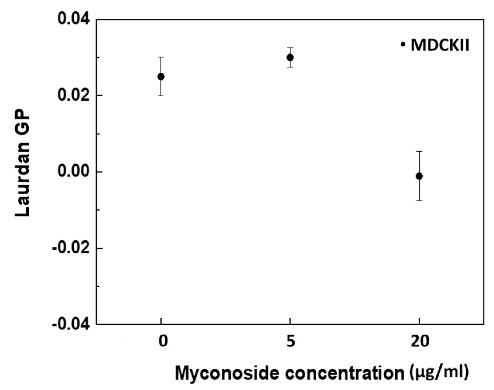
Laurdan fluorescence spectroscopy, involving a volumetric cuvette technique for averaging GP values from a large number of cells, was used. Thus, statistically significant results for untreated and myconoside-treated cells were obtained. Laurdan GP showed changes in membrane lipid order in MDCKII cells, induced by both myconoside concentrations (5 and 20 µg/mL) at 37 °C (Figure 5). Similar to di-4-ANEPPDHQ, Laurdan GP values can theoretically vary from +1 (being most ordered) to −1 (being least ordered). At control MDCKII cells, Laurdan GP showed values of about 0.025. When 5 µg/mL myconoside was added to control cells, an increase in the GP values (0.030) was observed. Higher myconoside concentrations (20 µg/mL) were able to reduce the lipid order (−0.002).

4. Discussion

Secondary plant metabolites are essential biological substances which underlie the medical properties of plant extracts and thus determine their benefits to human health. Secondary metabolites are not directly related to plant growth and development, but their production and accumulation are realized in response to various environmental factors (unfavorable conditions) as a protective mechanism for plants. Therefore, along with the understanding of the benefits of plant food, the secondary metabolites found in the plants are increasingly receiving research attention. Such a type of metabolite is H. rhodopensis myconoside, involved in the protection of plants against desiccation and supporting their recovery [1]. Myconoside is a major factor in desiccation tolerance in H. rhodopensis. The protection of plants from invading bacteria, fungi and parasites is achieved through the cytotoxic properties of their natural bioactive compounds. In this context, both quinones and polyphenols, which at high doses exhibit toxic properties, at low doses exhibit positive hormetic features [18,26,27]. As a derivative of quercetin, the 3,7-dihydroxy-2-[4-(2-chloro-1,4-naphthoquinone-3-yloxy)-3-hydroxyphenyl]-5-hydroxychromen-4-one (CHNQ) exhibits high cytotoxicity leading to the generation of oxidants in human neonatal B-HNF-3 fibroblasts. Synthetic precursors of CHNQ, quercetin and 2-chloro-3-hydroxy-[1,4]naphthoquinone, do not exhibit such strong effects, as only CHNQ at a low concentration partially protects cells against oxidative stress [27]. Curcumin [28], Ginkgo biloba [29], ginseng [30] and green tea [31] have similar hormetic-based chemo- and neuroprotective properties. Therefore, they are widely used as dietary supplements. Myconoside, similarly to these natural compounds, demonstrates favorable hormetic responses at a low dose treatment (5 µg/mL) that merits to be further studied in different cell lines, healthy and in disease, for the search of new potential agents in protection and healing of pathologies, related to the aging. To our knowledge, the present results are the first report that states the hormetic-like response of natural phenyl glycoside molecules such as myconoside in MDCKII cell line. It is noteworthy that we did not observe a similar hormetic-like response of myconoside at a low dose treatment of a cancer A549 cell line published in our previous study [13]. Instead, the cytotoxic effect was observed at a progressive increase in myconoside concentration. Certain cytotoxic agents at low doses are known to induce hormesis and thus stimulate cancer cell proliferation [32,33]. Comparing the responses of myconoside on both cell lines studied by our scientific group, non-polarized A549 [13] and polarized MDCKII cell line, a selective cytotoxic effect was observed for A549 at high myconoside concentrations as well as the lack of one in the MDCKII cell line. The pleiotropic effect of phytochemicals on human health is well documented, but the exact molecular mechanism of their cellular intake and induction in various cellular signaling processes is still unclear, in particular the cell-specific differences. The first step in the cellular intake of phytochemicals is the interaction with the plasma membrane. The cell plasma membrane is highly heterogeneous in terms of lipid and protein composition [34,35,36,37]. Thus, domains (clusters) with different lipid lateral structural organization are formed. MDCKII cells are used as a model for polarized epithelial monolayers [38] to study changes in cell contacts, plasmalemma polarization and epithelial barrier functions. Epithelial cells, unlike other cell types, have morphologically different membrane structures and functions. Apical–basal plasma membrane polarity is very important for epithelial cell organization and function. The formation and maintenance of epithelial cell polarity are performed by protein complexes, cytoskeletal components and membrane lipids [39,40]. The membrane–cytoskeleton interactions play a vital role in cellular polarization. Apical and basal plasma membrane surfaces of the polarized epithelial cell have different lipid–protein composition. Sphingolipid–cholesterol microdomains (lipid rafts) represent domains that accumulate predominantly in the apical plasma membrane and serve as platforms for the segregation of certain signaling proteins [9]. Many studies have proven the important role of cholesterol-rich membrane domains in the regulation of membrane–cytoskeleton properties and processes. Moreover, the apical part of the cell membrane plays an essential role in the absorption of nutrients. The organization and maintenance of membrane heterogeneity are extremely important for cell viability [41].
In this study, we demonstrated the effect of myconoside on the structural organization of MDCKII cell membranes. The direct myconoside–lipids, myconoside–lipid domain and indirect myconoside–cytoskeleton interactions were established in relation to the cell viability. The control nontreated MDCKII cells showed positive Laurdan GP values, which corresponds to a quite high degree of plasma membrane lipid order. Moreover, di-4-ANEPPDHQ fluorescence GP images, as well as the color bar corresponding to GP values, and their histograms indicated a pattern which is a characteristic of ordered membranes. This high degree of lipid order is an intrinsic characteristic of the normal healthy cells. The membrane lipid order is governed by the cholesterol content and the saturated to unsaturated fatty acid ratio of plasma membrane lipids. Cholesterol is a key component of the animal eukaryotic plasma membrane playing a major role in the physiochemical behavior and functions of membrane lipids determining the cell membrane rigidity and proteins by forming membrane platforms for the protein attachments [42,43]. The cholesterol content depends on both the cell type [44] and the cell health [45]. The cholesterol level in most mammalian plasma membranes is quite high, about 30 mol% [46]. However, in cancer cells, the cholesterol content is lower. Due to its low cholesterol content, the cancer cells exhibit lower membrane lipid order and high plasticity, which is important for their penetration into blood vessels. Indeed, in our previous studies, we reported di-4-ANEPPDHQ GP value of −0.250 for A549 cancer cell line [13] versus −0.050 for MDCKII cells in this study. A similar trend is observed when plasma membrane lipid order was probed by Laurdan: the GP value for untreated A549 cells was around −0.015, which implies lipid membranes in a liquid-disordered phase state (Ld) [13]. MDCKII cells showed a Laurdan GP value of +0.025, indicating that non-cancer cells had a higher lipid order than cancer ones [13], which in turn is probably due to higher quantity cell–cell contacts.
Myconoside treatment influences lipid order by interaction with the plasma membrane of MDCKII cells. Myconoside hydroxyl groups probably interact with the polar phospholipid head-group of the membrane. Through this interaction, myconoside changes the native plasma membrane organization. At the low concentration (5 µg/mL), myconoside increases the plasma membrane order. We suggest that myconoside acts as a filling compound by decreasing the number of water molecules surrounding the fluorescent dye or by decreasing water dipolar relaxations. Opposite to this, the high myconoside concentration (20 µg/mL) is able to decrease membrane lipid order, acting as a separating lipid molecules compound allowing a larger number of water molecules to penetrate more deeply into the membrane. A similar trend in lipid order as a function of myconoside concentration was observed for the cancer A549 cell line where cell viability reaches IC50 and myconoside cytotoxicity was observed [13]. In the MDCKII cell line, however, higher myconoside concentration induces a slight decrease in cell viability as the IC50 is not reached.
The staining of certain proteins with crystal violet dye is a method to detect cell viability [47]. The higher viability corresponds to a larger number of stained proteins. The increased cell viability by the addition of 5 µg/mL myconoside can be interpreted as follows. In addition to protein sorting and cell polarity [48], sphingolipid–cholesterol microdomains take part in cell adhesion [49]. In the plasma membrane of MDCKII cells, myconoside acts as a filling compound, inducing the formation of more ordered domains, in particular raft-like domains. These domains play a role in the segregation of certain proteins, including adhesive proteins. This assumption can explain the reinforced dotted linear ZO-1 organization as the myconoside increases membrane rigidity and exhibits a stabilizing and strengthening effect on cell junctions. In addition, the higher myconoside concentrations impaired to some extent the ZO-1 structure. Despite these significant changes, cell viability does not reach the IC50, implying that myconoside does not induce a cytotoxic effect.
It is well established that lipid rafts interact with the actin cytoskeleton [12]. However, the actin cytoskeleton is indirectly associated with raft domains through the tight junction proteins. ZO-1 belongs to the family of occludin proteins and plays a role as a tight junction-1 peripheral protein, situated on the surface of the cytoplasmic membrane [50,51]. By using model membranes, it has been demonstrated that actin filaments take part in the formation of ordered lipid domains [52]. In model lipid bilayers, it has been shown that dynamic membrane-bound F-actin can serve as a membrane domain switch that contributes to the membrane organization changes on the temporal and spatial scale during cell signaling response [52,53]. In this study, we observed concerted changes between the plasma membrane organization and the cytoskeleton induced by myconoside as an exogenously added molecule, primarily affecting lipid rafts. In our previous work, we demonstrated that myconoside stabilizes and promotes the formation of raft-like domains at low concentrations (2.5–12.5 µg/mL) in biomimetic membranes, whereas higher myconoside concentrations (15–40 µg/mL) act as a raft-like domain disrupting molecules [13]. The increased membrane lipid order induced by lower myconoside concentration in MDCKII cells is positively related to a more compact F-actin network (an increase in the number and thickness of actin stress fibers) and more pronounced ZO-1 visualized cell-cell contacts. Moreover, lower myconoside concentration was able to stabilize the specific size, shape and density of MDCKII cells. A similar effect of F-actin and tight-junction proteins on lipid raft formation was suggested by other authors [53,54,55]. The last statement is in accordance with the increased cell plasma membrane lipid order observed in this study as well as the higher raft fraction detected in the presence of low myconoside concentration in biomimetic systems, described here above [13]. On the other hand, membrane microdomains participate in adhesion and motility. Therefore, the lipid rafts and the actin cytoskeleton act cooperatively via various cell adhesion molecules. In this study, we have shown that myconoside is able to influence the membrane lipid order and the raft-like domain formation as well as the F-protein network and ZO-1 distribution. All these three players govern membrane structure, organization and functions. It could be suggested that myconoside first changes the membrane lipid order. That, in turn, changes the physicochemical properties of the raft domains [13]. Such changes in the lipid organization of the plasma membrane have the ability to provoke conformational changes in proteins by influencing certain cell signaling processes. Low concentrations of myconoside enhance the raft fraction and form denser contacts between the cells, leading to increased cell viability. Oppositely, high myconoside concentrations disrupt the raft domains, thus making the distribution of F-actin and ZO-1 more diffuse which possibly impairs cell contacts in the treated MDCKII cells. The effect of high concentrations of myconoside on cancer A549 cells is much higher because myconoside (20 µg/mL) is able to decrease the cell viability by 50%, exhibiting a cytotoxic effect [13]. The same myconoside concentration, however, decreases only by 20% MDCKII cell viability. A biphasic mode of interaction of myconoside with the cell plasma membranes for both cell lines (MDCKII and A549 [13]) was observed with myconoside concentration increase. First, myconoside acts as a filler molecule at low concentrations, leading to an increase in membrane lipid order, and second, as a spacing molecule at high concentrations, leading to lipid order decrease. Low myconoside concentrations are able to enhance membrane lipid order and tighten cell junctions correlated to an increase in the cell proliferation observed for MDCKII cells. A statistically significant increase in cell proliferation was not observed for A549 cells treated with low myconoside concentrations [13]. By using Laurdan GP values we calculated the difference between the relative effects of both myconoside concentrations (5 and 20 µg/mL) for each cell line: −1.28 for MDCKII, and, +2.40 for A549 [13] cells. Such larger difference in lipid order change for A549 together with disassembling of ZO-1 proteins from string-like to granular appearance could be functionally connected to the observed cytotoxicity in A549 versus missing one in the MDCKII cell line. All these results suggest that the higher membrane lipid order and tight junctions, essential for the maintenance of cellular polarity, as it is for the MDCKII cell line, make the membrane more resistant to myconoside cytotoxic effects.

5. Conclusions

Many dietary phytochemicals have beneficial effects on human health by supporting the prevention of different diseases. In the present study, we evaluated the influence of the phenolic glycoside myconoside on MDCKII cells. The combination of spectroscopy and microscopy studies demonstrated that myconoside interacts with the cellular plasma membrane and apical junctional complexes by altering their structural organization. Low myconoside concentration exhibited a stimulating effect on MDCKII viability by increasing plasma membrane lipid packing and stabilizing apical junctional complexes. Based on the reported results, we suggest a potential mechanism of action of myconoside on cell functioning related to the myconoside-induced hormetic effect. Thus, our findings lead to a better understanding of the possibilities for the application of potentially beneficial phytochemicals such as myconoside in the fields of pharmacology and medicine.

Author Contributions

Conceptualization, R.H., G.S. and D.D.; Methodology, A.K., V.Y. and R.V.; Software, G.S.; Validation, A.K., T.T.-H. and B.N.; Analysis, R.V. and T.T.-H.; Investigation, A.K., D.M. and T.T.-H.; Data Curation, A.K., V.Y. and D.M.; Writing—Original Draft Preparation, A.K., R.H. and D.D.; Writing—Review and Editing, G.S. and A.M.; Visualization, A.K., R.V. and G.S.; Supervision, G.S. and A.M.; Funding Acquisition, G.S., A.M. and B.N. All authors have read and agreed to the published version of the manuscript.

Funding

This study was financially supported by the National Science Fund of Bulgaria, Grant KP-06-N58/6-2021 (all lipid order experiments). The authors thank the Bulgarian Ministry of Education and Science for support: Scientific Infrastructure on Cell Technologies in Biomedicine (SICTB) DO1-154/28/08/2018 (cell culture technologies) and D01-392/2020 “National Center for Biomedical Photonics”, part of Bulgarian National Roadmap for Scientific Infrastructures 2020–2027 (Imaging platform). The authors also acknowledge COST Action 17 121: Correlated Multimodal Imaging in Life Sciences-COMULIS by grant KP-06-COST/10/2020 (fluorescent probes for ZO-1 and F-actin cell experiments).

Institutional Review Board Statement

Not applicable.

Informed Consent Statement

Not applicable.

Data Availability Statement

Data are available from the corresponding author upon reasonable request and are deposited on the institution website of the corresponding author (http://biomed.bas.bg/bg/).

Conflicts of Interest

The authors declare no conflict of interest.

Ethics Approval

Not Necessary. The cell lines used in this study were purchased from the American Type Culture Collection—ATCC (Manassas, VA, USA) and were used as recommended by the provider.

References

  1. Gechev, T.S.; Hille, J.; Woerdenbag, H.J.; Benina, M.; Mehterov, N.; Toneva, V.; Fernie, A.R.; Mueller-Roeber, B. Natural products from resurrection plants: Potential for medical applications. Biotechnol. Adv. 2014, 32, 1091–1101. [Google Scholar] [CrossRef]
  2. Georgiev, Y.N.; Ognyanov, M.H.; Denev, P.N. The ancient Thracian endemic plant Haberlea rhodopensis Friv. and related species: A review. J. Ethnopharmacol. 2020, 249, 112359. [Google Scholar] [CrossRef]
  3. Todorova, R.; Atanasov, A. Haberlea rhodopensis: Pharmaceutical and medical potential as a food additive. Nat. Prod. Res. 2015, 30, 1–23. [Google Scholar] [CrossRef]
  4. Dell’Acqua, G.; Schweikert, K. Skin benefits of a myconoside-rich extract from resurrection plant Haberlea rhodopensis. Int. J. Cosmet. Sci. 2012, 34, 132–139. [Google Scholar] [CrossRef]
  5. Kondeva-Burdina, M.; Zheleva-Dimitrova, D.; Nedialkov, P.; Girreser, U.; Mitcheva, M. Cytoprotective and antioxidant effects of phenolic compounds from Haberlea rhodopensis Friv. (Gesneriaceae). Pharmacogn. Mag. 2013, 9, 294–301. [Google Scholar] [CrossRef]
  6. Mihaylova, D.; Bahchevanska, S.; Toneva, V. Examination of the antioxidant activity of Haberlea Rhodopensis leaf extracts and their phenolic constituents. J. Food Biochem. 2013, 37, 255–261. [Google Scholar] [CrossRef]
  7. Popov, B.; Georgieva, S.; Gadjeva, V. Modulatory effects of total extract of Haberlea rhodopensis against the cyclophosphamide induced genotoxicity in rabbit lymphocytes in vivo. Trakia J. Sci. 2011, 9, 51–57. [Google Scholar]
  8. Sirk, T.W.; Brown, E.F.; Sum, A.K.; Friedman, M. Molecular dynamics study on the biophysical interactions of seven green tea catechins with lipid bilayers of cell membranes. J. Agric. Food Chem. 2008, 56, 7750–7758. [Google Scholar] [CrossRef] [PubMed]
  9. Simons, K.; Ikonen, E. Functional rafts in cell membranes. Nature 1997, 387, 569–572. [Google Scholar] [CrossRef]
  10. Yamazaki, S.; Iwama, A.; Takayanagi, S.; Morita, Y.; Eto, K.; Ema, H.; Nakauchi, H. Cytokine signals modulated via lipid rafts mimic niche signals and induce hibernation in hematopoietic stem cells. EMBO J. 2006, 25, 3515–3523. [Google Scholar] [CrossRef]
  11. Chichili, G.R.; Rodgers, W. Cytoskeleton-membrane interactions in membrane raft structure. Cell. Mol. Life Sci. 2009, 66, 2319–2328. [Google Scholar] [CrossRef] [PubMed] [Green Version]
  12. Head, B.P.; Patel, H.H.; Insel, P.A. Interaction of membrane/lipid rafts with the cytoskeleton: Impact on signaling and function: Membrane/lipid rafts, mediators of cytoskeletal arrangement and cell signaling. Biochim. Biophys. Acta 2014, 1838, 532–545. [Google Scholar] [CrossRef] [PubMed]
  13. Kostadinova, A.; Hazarosova, R.; Topouzova-Hristova, T.; Moyankova, D.; Yordanova, V.; Veleva, R.; Nikolova, B.; Momchilova, A.; Djilianov, D.; Staneva, G. Myconoside interacts with the plasma membranes and the actin cytoskeleton and provokes cytotoxicity in human lung adenocarcinoma A549 cells. J. Bioenerg. Biomembr. 2022, 54, 31–43. [Google Scholar] [CrossRef] [PubMed]
  14. Moyankova, D.; Mladenov, P.; Berkov, S.; Peshev, D.; Georgieva, D.; Djilianov, D. Metabolic profiling of the resurrection plant Haberlea rhodopensis during desiccation and recovery. Physiol. Plant. 2014, 152, 675–687. [Google Scholar] [CrossRef]
  15. Saotome, K.; Morita, H.; Umeda, M. Cytotoxicity test with simplified crystal violet staining method using microtitre plates and its application to injection drugs. Toxicol Vitro 1989, 3, 317–321. [Google Scholar] [CrossRef]
  16. Wang, P.; Henning, S.M.; Heber, D. Limitations of MTT and MTS-based assays for measurement of antiproliferative activity of green tea polyphenols. PLoS ONE 2010, 5, e10202. [Google Scholar] [CrossRef]
  17. Maioli, E.; Torricelli, C.; Fortino, V.; Carlucci, F.; Tommassini, V.; Pacini, A. Critical Appraisal of the MTT Assay in the Presence of Rottlerin and Uncouplers. Biol. Proced. Online 2009, 11, 229. [Google Scholar] [CrossRef]
  18. Kostadinova, A.; Doumanov, J.; Moyankova, D.; Ivanov, S.; Mladenova, K.; Djilianov, D.; Topouzova-Hristova, T. Haberlea rhodopensis extracts affect cell periphery of keratinocytes. Comptes Rendus L’académie Bulg. Sci. 2016, 69, 439. [Google Scholar]
  19. Owen, D.M.; Rentero, C.; Magenau, A.; Abu-Siniyeh, A.; Gaus, K. Quantitative imaging of membrane lipid order in cells and organisms. Nat. Protoc. 2011, 7, 24–35. [Google Scholar] [CrossRef]
  20. Butler, C.E.; Wheeler, G.; Graham, J.; Tyler, K.M. Visual Discrimination of Membrane Domains in Live Cells by Widefield Microscopy. In Fluorescent Methods to Study Biological Membranes; Mély, Y., Duportail, G., Eds.; Springer: Berlin/Heidelberg, Germany, 2013; pp. 163–184. [Google Scholar]
  21. Parasassi, T.; De Stasio, G.; Ravagnan, G.; Rusch, R.M.; Gratton, E. Quantitation of lipid phases in phospholipid vesicles by the generalized polarization of Laurdan fluorescence. Biophys. J. 1991, 60, 179–189. [Google Scholar] [CrossRef]
  22. Miyoshi, J.; Takai, Y. Molecular perspective on tight-junction assembly and epithelial polarity. Adv. Drug Deliv. Rev. 2005, 57, 815–855. [Google Scholar] [CrossRef] [PubMed]
  23. Jin, L.; Millard, A.C.; Wuskell, J.P.; Clark, H.A.; Loew, L.M. Cholesterol-enriched lipid domains can be visualized by di-4-ANEPPDHQ with linear and nonlinear optics. Biophys. J. 2005, 89, L04–L06. [Google Scholar] [CrossRef] [PubMed] [Green Version]
  24. Parasassi, T.; Conti, F.; Gratton, E. Time-resolved fluorescence emission spectra of Laurdan in phospholipid vesicles by multifrequency phase and modulation fluorometry. Cell. Mol. Biol. 1986, 1, 103–108. [Google Scholar]
  25. Amaro, M.; Reina, F.; Hof, M.; Eggeling, C.; Sezgin, E. Laurdan and di-4-ANEPPDHQ probe different properties of the membrane. J. Phys. Appl. Phys. 2017, 50, 134004. [Google Scholar] [CrossRef] [PubMed]
  26. Dilberger, B.; Weppler, S.; Eckert, G.P. Phenolic acid metabolites of polyphenols act as inductors for hormesis in C. elegans. Mech. Ageing Dev. 2021, 198, 111518. [Google Scholar] [CrossRef]
  27. Milackova, I.; Rackova, L.; Majekova, M.; Mrvova, N.; Stefek, M. Protection or cytotoxicity mediated by a novel quinonoid-polyphenol compound? Gen. Physiol. Biophys. 2015, 34, 51–64. [Google Scholar] [CrossRef] [PubMed]
  28. Calabrese, E.J.; Dhawan, G.; Kapoor, R.; Mattson, M.P.; Rattan, S.I. Curcumin and hormesis with particular emphasis on neural cells. Food Chem. Toxicol. 2019, 129, 399–404. [Google Scholar] [CrossRef]
  29. Calabrese, E.J.; Calabrese, V.; Tsatsakis, A.; Giordano, J.J. Hormesis and Ginkgo biloba (GB): Numerous biological effects of GB are mediated via hormesis. Aging Res. Rev. 2020, 64, 101019. [Google Scholar] [CrossRef]
  30. Calabrese, E. Hormesis and Ginseng: Ginseng Mixtures and Individual Constituents Commonly Display Hormesis Dose Responses, Especially for Neuroprotective Effects. Molecules 2020, 25, 2719. [Google Scholar] [CrossRef]
  31. Calabrese, E.J.; Tsatsakis, A.; Agathokleous, E.; Giordano, J.; Calabrese, V. Does Green Tea Induce Hormesis? Dose Response 2020, 18, 1559325820936170. [Google Scholar] [CrossRef]
  32. Calabrese, E.J. Cancer biology and hormesis: Human tumor cell lines commonly display hermetic (biphasic) dose responses. Crit. Rev. Toxicol. 2005, 35, 463–582. [Google Scholar] [CrossRef] [PubMed]
  33. Aoishi, Y.; Yoshimasu, T.; Oura, S.; Kawago, M.; Hirai, Y.; Miyasaka, M.; Ohashi, T.; Nishimura, Y. Quantitative Evaluation of Hormesis in Breast Cancer Using Histoculture Drug Response Assay. Dose Response 2019, 17, 1559325819896183. [Google Scholar] [CrossRef] [PubMed]
  34. Karnovsky, M.J.; Kleinfeld, A.M.; Hoover, R.L.; Dawidowicz, E.A.; McIntyre, D.E.; Salzman, E.A.; Klausner, R.D. Lipid domains in membranes. Ann. N. Y. Acad. Sci. 1982, 401, 61–75. [Google Scholar] [CrossRef]
  35. Pakiet, A.; Kobiela, J. Changes in lipids composition and metabolism in colorectal cancer: A review. Lipids Health Dis. 2019, 18, 29. [Google Scholar] [CrossRef] [PubMed]
  36. Pike, L.J. Lipid rafts: Bringing order to chaos. J. Lipid Res. 2003, 44, 655–667. [Google Scholar] [CrossRef]
  37. Pike, L.J. The challenge of lipid rafts. J. Lipid Res. 2009, 50, S323–S328. [Google Scholar] [CrossRef]
  38. Cereijido, M.; Robbins, E.S.; Dolan, W.J.; Rotunno, C.A.; Sabatini, D.D. Polarized monolayers formed by epithelial cells on a permeable and translucent support. J. Cell Biol. 1978, 77, 853–880. [Google Scholar] [CrossRef]
  39. Roignot, J.; Peng, X.; Mostov, K. Polarity in mammalian epithelial morphogenesis. Cold Spring Harb. Perspect. Biol. 2013, 5, a013789. [Google Scholar] [CrossRef]
  40. Ikenouchi, J. Roles of membrane lipids in the organization of epithelial cells: Old and new problems. Tissue Barriers 2018, 6, 1–8. [Google Scholar] [CrossRef]
  41. Yang, B.; Oo, T.N.; Rizzo, V. Lipid rafts mediate H2O2 prosurvival effects in cultured endothelial cells. FASEB J. 2006, 20, 1501–1503. [Google Scholar] [CrossRef]
  42. Yeagle, P. Biology of Cholesterol; Philip, L., Yeagle, P., Eds.; CRC Press: Boca Raton, FL, USA, 1988. [Google Scholar]
  43. Song, Y.; Kenworthy, A.K.; Sanders, C.R. Cholesterol as a co-solvent and a ligand for membrane proteins. Protein Sci. 2014, 23, 1–22. [Google Scholar] [CrossRef] [PubMed]
  44. Hoejholt, K.L.; Mužić, T. Calcium electroporation and electrochemotherapy for cancer treatment: Importance of cell membrane composition investigated by lipidomics, calorimetry and in vitro efficacy. Sci. Rep. 2019, 9, 4758. [Google Scholar] [CrossRef] [PubMed]
  45. Szlasa, W.; Zendran, I.; Zalesińska, A.; Tarek, M. Lipid composition of the cancer cell membrane. J. Bioenerg. Biomembr. 2020, 52, 321–342. [Google Scholar] [CrossRef] [PubMed]
  46. Craig, M.; Yarrarapu, S.N.S.; Dimri, M. Biochemistry, Cholesterol; StatPearls Publishing: Tampa, FL, USA, 2022. [Google Scholar]
  47. Feoktistova, M.; Geserick, P.; Leverkus, M. Crystal Violet Assay for Determining Viability of Cultured Cells. Cold Spring Harb. Protoc. 2016, 4, 343–346. [Google Scholar] [CrossRef]
  48. Bagnat, M.; Simons, K. Lipid rafts in protein sorting and cell polarity in budding yeast Saccharomyces cerevisiae. Biol. Chem. 2002, 383, 1475–1480. [Google Scholar] [CrossRef]
  49. Murai, T. The role of lipid rafts in cancer cell adhesion and migration. Int. J. Cell Biol. 2012, 2012, 763283. [Google Scholar] [CrossRef]
  50. Stevenson, B.R.; Siliciano, J.D.; Mooseker, M.S.; Goodenough, D.A. Identification of ZO-1: A high molecular weight polypeptide associated with the tight junction (zonula occludens) in a variety of epithelia. J. Cell Biol. 1986, 103, 755–766. [Google Scholar] [CrossRef]
  51. Fanning, A.S.; Jameson, B.J.; Jesaitis, L.A.; Anderson, J.M. The tight junction protein ZO-1 establishes a link between the transmembrane protein occludin and the actin cytoskeleton. J. Biol. Chem. 1998, 273, 29745–29753. [Google Scholar] [CrossRef]
  52. Liu, A.P.; Fletcher, D.A. Actin polymerization serves as a membrane domain switch in model lipid bilayers. Biophys. J. 2006, 91, 4064–4070. [Google Scholar] [CrossRef]
  53. Head, B.P.; Patel, H.H.; Roth, D.M.; Murray, F.; Swaney, J.S.; Niesman, I.R.; Farquhar, M.G.; Insel, P.A. Microtubules and actin microfilaments regulate lipid raft/caveolae localization of adenylyl cyclase signaling components. J. Biol. Chem. 2006, 281, 26391–26399. [Google Scholar] [CrossRef]
  54. Forstner, M.B.; Yee, C.K.; Parikh, A.N.; Groves, J.T. Lipid lateral mobility and membrane phase structure modulation by protein binding. J. Am. Chem. Soc. 2006, 128, 15221–15227. [Google Scholar] [CrossRef] [PubMed]
  55. Viola, A.; Gupta, N. Tether and trap: Regulation of membrane-raft dynamics by actin-binding proteins. Nat. Rev. Immunol. 2007, 7, 889–896. [Google Scholar] [CrossRef] [PubMed]
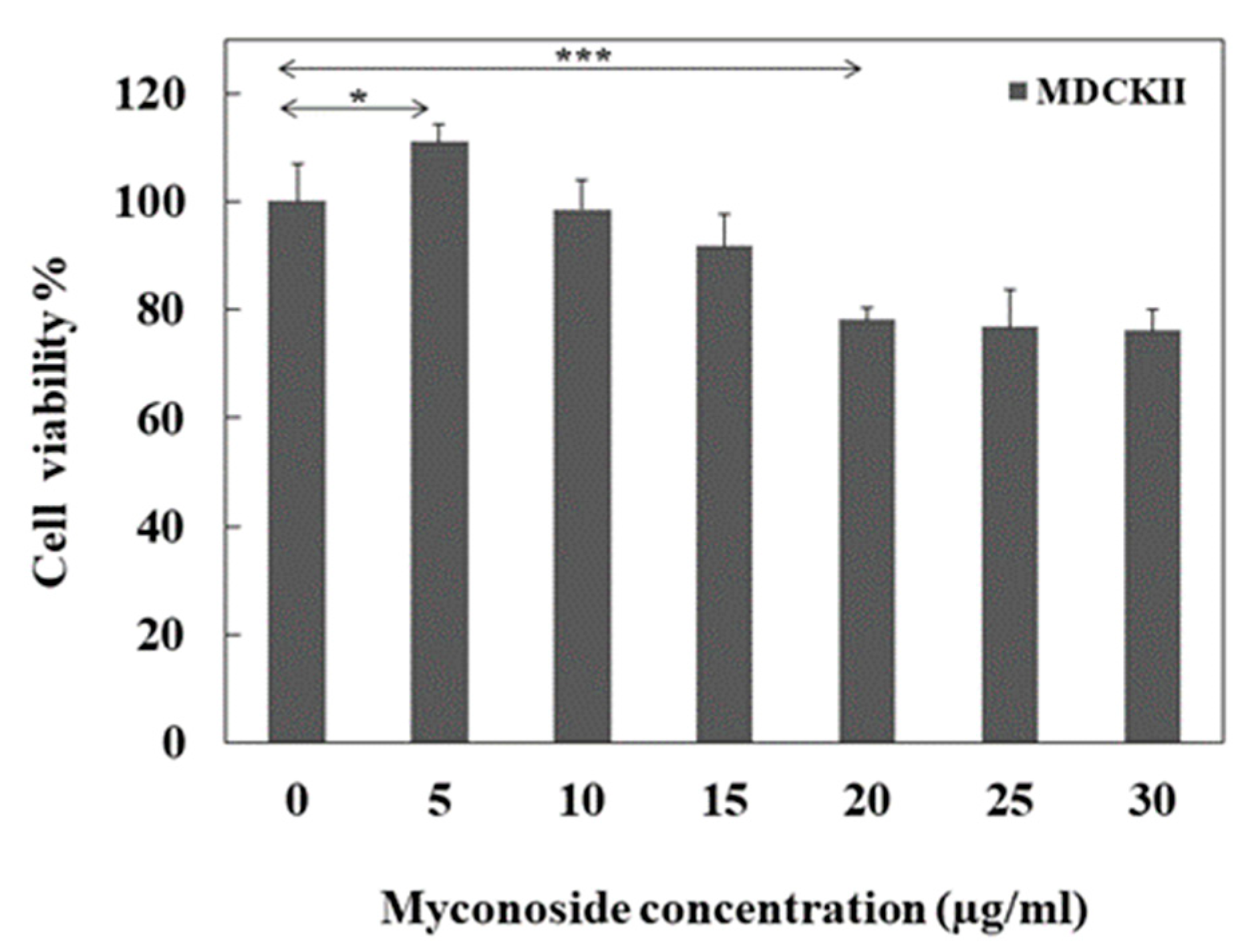
Figure 1. Cell viability after 24 h treatment of MDCKII cell line with myconoside. Data are presented as % viability compared to the untreated cells and as means ± SEM of 3 independent experiments. * p < 0.05 and *** p < 0.001 are presented as follows *, ***.
Separations 09 00239 g001
Figure 2. F-actin staining (red color) of untreated and treated with 5 and 20 µg/mL myconoside MDCKII cells. The blue color matches to the cellular nucleus. Untreated MDCKII cells (a) and myconoside-treated ones (b,c). The arrows show the F-actin fibers and the asterisks—cell division. A total of 5 µg/mL myconoside stabilizes the F-actin network (b), while 20 µg/mL induces a granular pattern of F-actin ((c), arrows). Scale bar: 50 µm.
Separations 09 00239 g002
Figure 3. ZO-1 staining of untreated MDCKII cell line (a). MDCKII cells treated with 5 µg/mL (b) and 20 µg/mL (c) myconoside. Arrows and asterisks indicate the typical localization and distribution of ZO-1 proteins (a,b). A total of 5 µg/mL myconoside strengthens linear cellular contacts (b), whereas 20 µg/mL myconoside disrupts linear ZO-1 organization (c). Scale bar: 10 µm.
Separations 09 00239 g003
Figure 4. Visualization of MDCKII cell membrane lipid packing by confocal fluorescence microscopy. (A) Fluorescence GP images of untreated and myconoside-treated MDCKII cells stained with di-4-ANEPPDHQ. Two channels (first and second columns) have green and red colors, respectively. Merged channels are exhibited in the third column. GP of the cells indicating the range of the calculated GP values (fourth column). The GP values vary from −1 (blue color, low lipid order) to +1 (red color, high lipid order). (B) Histogram of the GP values acquired from GP images of untreated and myconoside-treated MDCKII cells stained with di-4-ANEPPDHQ. The gray curve corresponds to untreated cells, and the red and blue ones to myconoside-treated cells with 5 µg/mL and 20 µg/mL, respectively.
Separations 09 00239 g004
Figure 5. Fluorescence spectroscopy study of plasma membrane lipid order of untreated and treated with myconoside MDCKII cells. Laurdan GP values as a function of myconoside concentration. Error bars correspond to SDs (n = 3). GP values of all treated with myconoside MDCKII cells are significantly different from the untreated ones at p < 0.05.
Separations 09 00239 g005
Publisher’s Note: MDPI stays neutral with regard to jurisdictional claims in published maps and institutional affiliations.