Article

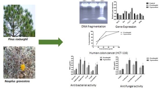
In Vitro Anti-Colorectal Cancer and Anti-Microbial Effects of Pinus roxburghii and Nauplius graveolens Extracts Modulated by Apoptotic Gene Expression

1
Nutrition and Food Science Department, Food Industries and Nutrition Research Institute, National Research Centre, Cairo 12622, Egypt
2
Pharmacognosy Department, Pharmaceutical and Drug Industries Research Institute, National Research Centre, Cairo 12622, Egypt
3
Hormones Department, Medical Research and Clinical Studies Institute, National Research Centre, Cairo 12622, Egypt
4
Biochemistry Department, Faculty of Agriculture, Cairo University, Giza 12613, Egypt
5
Natural Products Chemistry Laboratory, Biotechnology Research Institute, Chonnam National University, 77 Yongbong-ro, Buk-gu, Gwangju 500-757, Republic of Korea
6
Department of Food Technology, Food Industries and Nutrition Research Institute, National Research Centre, Cairo 12622, Egypt
*
Authors to whom correspondence should be addressed.
Separations 2022, 9(12), 393; https://doi.org/10.3390/separations9120393
Submission received: 3 November 2022 / Revised: 18 November 2022 / Accepted: 21 November 2022 / Published: 24 November 2022
(This article belongs to the Section Analysis of Food and Beverages)

Abstract

:
The use of phytochemicals is gaining increasing attention for treating cancer morbidity with minimal burden side effects. This study evaluated the cytotoxicity and antimicrobial activities of Pinus roxburghii branch (P. roxburghii) and Nauplius graveolens (N. graveolens) extracts in vitro. Cell viability was estimated using MTT assay. DNA fragmentation was determined to detect apoptotic pathway initiation. Mechanistically, the apoptotic pathway was tracked by estimating the relative mRNA expression levels of the Bcl-2, Bax, Cas3, NF-κB, and PI3k genes by qRT-PCR. P. roxburghii exhibited moderate antioxidant activity, while N. graveolens possessed highly significant (p < 0.05) scavenging activity against DPPH and ABTS assays. HPLC analysis demonstrated that catechin and chlorogenic acid were the predominant polyphenolic compounds in P. roxburghii and N. graveolens, respectively. The P. roxburghii and N. graveolens extracts inhibited the viability of HCT-116 cells with IC50 values of 30.6 µg mL−1 and 26.5 µg mL−1, respectively. DNA fragmentation analysis showed that the proposed extracts induced apoptosis in HCT-116 cells. Moreover, the IC50 doses of the selected extracts significantly (p < 0.05) upregulated Bax and cleaved Cas-3, and downregulated Bcl-2, NF-κB, and PI3k genes versus the GAPDH gene as a housekeeping gene in comparison to the control group. The Bax/Bcl-2 ratio was raised upon treatment. The mentioned extracts exhibited antimicrobial action against all tested bacteria and fungi. The highest antibacterial effect was recorded against E. coli, with inhibition zones of 12.0 and 11.2 mm for P. roxburghii and N. graveolens, respectively. On the other hand, the highest antifungal action was registered for Penicillium verrucosum and A. niger, with inhibition zones of 9.8 and 9.2 mm for the tested extracts, respectively. In conclusion, the outcomes of this study indicate that P. roxburghii and N. graveolens extracts could potentially be used as anticancer, antibacterial, and antifungal agents.

Graphical Abstract

1. Introduction

Cancer is one of the major causes of morbidity and mortality worldwide. Among noncommunicable diseases, cancer is the second most significant cause of death after cardiovascular disease [1,2]. Colorectal cancer (CRC) is the third leading reason of cancer in both males and females worldwide. In 2020, CRC accounted for 1.9 million cases globally and 935,000 deaths [3]. Colorectal cancers in the proximal colon represents approximately 41% of all colorectal cancers, with incidences of approximately 22% and 28% in the distal colon and the rectum, respectively [4]. Different lifestyle factors are associated with CRC risk, including high alcohol consumption, high-fat, low-fiber diets, obesity, smoking, lack of physical exercise, aging, and family history [5,6]. Various approaches have been applied in cancer treatment, such as immunotherapy, chemotherapy, surgery, and radiotherapy. Unfortunately, the toxicity of these treatments can damage both healthy and cancer cells, causing serious side effects, such as vomiting, nausea, gastrointestinal disorders, low white blood cell count, and hair loss [7,8]. This is the purpose of searching for new, natural substances with potential cytotoxic impacts on cancer cells and fewer side effects on healthy cells. Cancer chemoprevention can be described using natural dietary or artificial factors that may reverse or prohibit carcinogenic progression. The concept of chemoprevention is gaining increasing attention because of its promising role in reducing cancer morbidity with minimal burden side effects [9]. Many medicinal plants are considered the best reservoirs for novel diverse bioactive compounds as potent anticancer agents [10,11].
Pinus roxburghii follows the Pinaceae family and is recognized as chir pine. A Pinus roxburghii grown in Egypt’s El-Orman Botanical Garden [12]. P. roxburghii is of particular interest for its many medicinal and pharmacological properties, such as anticancer, antioxidant, antimicrobial, antidyslipidemic, and anti-inflammatory effects) [13,14,15,16]. Nauplius graveolens (Forssk.) Wiklund (N. graveolens) belongs to the Asteraceae family called as Tafss [17], and was collected for this study from Egypt’s south Sinai desert, namely in Wadi Feiran. Nauplius graveolens has many biological impacts, such as antitumor, anti-inflammatory, anticancer, antimicrobial, and antioxidant activities) [18,19,20].
Many published studies have documented that the expression of the Bcl-2, Bax, Cas3, NF-κB, and PI3k genes plays a crucial role in cancer cell suppression, cell cycle arrest, and cell apoptosis [21]. Bcl-2 and Bax are two key genes of the Bcl-2 family and represent the role regulators of the mitochondrial apoptotic pathway) [22]. B-cell lymphoma 2 (Bcl-2) is an anti-apoptotic gene that plays a vital role in boosting or suppressing the intrinsic apoptotic pathway triggered by mitochondrial dysfunction [23]. Bax protein is a pro-apoptotic gene that elevates cell death by altering mitochondrial membrane permeabilization when exposed to different cellular stresses. Caspase-3 is a member of the family of cysteine proteases that play roles in the implementation and completion of apoptotic processes [24,25]. Nuclear factor kappa-B (NF-κB) is a transcription factor that controls the expression of several genes responsible for cell growth regulation and proliferation, survival, apoptosis, and immune and stress responses [26,27]. Phosphoinositide kinases (PIKs) are lipid kinases that phosphorylate the inositol ring of PIKs, thus functioning as signal transducers. PI3K plays a vital role in cell survival, the cell cycle, programmed cell death, DNA repair, and angiogenesis [28]. This research study aimed to explore the effectiveness of P. roxburghii and N. graveolens extracts as potent anticancer agents, particularly against human colon cancer, as well as their antioxidant, antibacterial, and antifungal activities against mycotoxigenic fungi and pathogenic bacteria.

2. Materials and Methods

2.1. Preparation of Extracts

The maceration procedure with 90% aqueous methanol was used to extract the P. roxburghii and N. graveolens plants. P. roxburghii was provided by Orman Botanical Garden, Ministry of Agriculture, Giza, Egypt (Authentication number: 2151). The branch was stripped of its foliar leaves before being cut into small pieces, dried at 40 °C, then ground into a fine powder. A total of 75 g of powder was extracted using 450 mL of methanol. After that, the sample was filtered and evaporated using a rotary evaporator (Büchi, Flawil, Switzerland). The obtained residue was freeze-dried and kept in sterilized containers at −20 °C. N. graveolens was gathered from the desert of Wadi Feiran, South Sinai, Egypt (Authentication number: 3187). The entire N. graveolens plant was treated similarly to P. roxburghii [29]. According to Boulos’ (2002) [30] taxonomic categorization, the tested plants were determined by the pharmacognosy department of the National Research Centre, Egypt.

2.2. Determination of Phytochemicals

2.2.1. Total Phenolic Content

The total phenolic content (TPC) of the extracts was estimated using the Folin-Ciocalteu reagent, as stated by the technique of Zaky et al. (2019) [31]. 20 μL of the extract was prepared with 1.58 mL of DW, before Folin-Ciocalteu reagent (100 μL) was added. Three minutes later, 300 μL of 20% sodium carbonate was combined. The mixture was placed for 30 min in the dark. The absorbance was controlled at 765 nm (Cary 60 spectrophotometer, Agilent Technologies, Santa Clara, California, USA). The reads were expressed as μg of gallic acid (GAE)/mg extract.

2.2.2. Total Flavonoid Content

P. roxburghii and N. graveolens extracts were assessed for total flavonoid content according to the aluminum chloride colorimetric assay) [32]. The values are presented as µg catechin (CE) per mg of the dried extract.

2.2.3. Total Carotenoids

The total carotenoid content was estimated as outlined by Moore et al. (2005) [33]. The absorbance was detected at 470 nm against a blank using a spectrophotometer. The findings are expressed as β-carotene equivalent (βCE) µg/g extract using a β-carotene standard curve.

2.3. Determination of Antioxidant Activity

2.3.1. DPPH Activity

The DPPH ability was checked using the procedure described by Zaky et al. [34]. The standard curve was designed using Trolox, and the values are presented as µg Trolox equivalents (TE)/g extract. The inhibition of free radicals was assessed according to the equation:
Inhibition (%) = [(A control − A sample)/A control] × 100
where:
  • A control: Negative control absorbance.
  • A sample: The tested compound absorbance.

2.3.2. ABTS Radical Scavenging Activity

The ABTS test was estimated as documented by Zaky et al. [35]. The percent inhibition of ABTS was detected according to the following formula:
%ABTS = [(Abs0 − Abs1)/Abs0] × 100
where:
  • Abs0: Control sample absorbance
  • Abs1: Extract sample absorbance

2.4. Assessment of Polyphenolic Compounds by HPLC

HPLC analysis was performed according to Zaky et al. [36] using an Agilent 1260 series. The separation was done employing an Eclipse C18 column (4.6 mm × 250 mm i.d., 5 μm). An Agilent Zorbox SB-C18 column (250 × 4.6 mm i.d., 5 μm) was utilized at a column temperature of 30 °C. The filtered sample (20 μL) was injected, and the mobile phase was composed of two solvents, 0.1% formic acid (A) and methanol (B), with a flow rate of 1.0 mL/min. The gradient was set as follows: 0 min 25% B; 20 min 25% B; 30 min 35% B; 40 min 100% B; 42 min 100% B; and 50 min 25% B. The peaks of the chromatogram were measured at 280 nm. Based on a standard curve, the concentrations of each compound were determined and expressed as μg/g DW of extract.

2.5. Cell Culture

The human colon cancer cell line (HCT-116) and human skin normal cell line (BJ-1) were kindly donated by Professor Linder, Karolinska Center, Department of Oncology and Pathology, Karolinska Institute and Hospital, Stockholm, Sweden. The assay was carried out in a laminar flow class II biosafety cabinet. The cells were preserved in RPMI culture medium supplemented with a 1% antimycotic/antibiotic mixture (25 μg/mL amphotericin B, 10.000 U/mL potassium penicillin, and 10.000 μg/mL streptomycin sulfate) and 1% L-glutamine and mixed with 10% heat-inactivated fetal bovine serum (Gibco, Life Technologies Limited, Paisley, UK). Culturing and subculturing were performed according to standard techniques [37]. Dimethyl sulfoxide (DMSO) and doxorubicin were used as negative and positive controls, respectively.

2.5.1. Cell Viability Assay

This assay was performed according to the method of Mosmann (1983) [38]. In this method, the yellow-colored MTT (3-(4,5-dimethylthiazol-2-yl)-2,5-diphenyl tetrazolium bromide) was reduced to purple formazan. After culturing the cells for 10 days, cells at a concentration of 10 × 103 cells/well were seeded in freshly prepared medium in 96-well microtiter plates for 24 h at 37 °C under 5% CO2 in a CO2 incubator (Sheldon, TC2323, Cornelius, OR, USA). Cells were incubated solely (negative control) or with the addition of P. roxburghii and N. graveolens methanolic extracts at a final concentration of 100 μg/mL. After 72 h of incubation, the medium was emptied with 40 μL MTT (2.5 mg/mL) into each well. The plates were incubated for 4 h under 5% CO2 at 37 °C. Then, an aliquot of 200 μL of 10% sodium dodecyl sulfate (SDS) was added. The absorbance was detected at 595 nm using a microplate multiwell reader and a reference wavelength of 690 nm. The percentage of changes in cell viability was calculated as follows:
Percentage cytotoxicity: (1 − (av(x)/(av (NC)) × 100
where X is the treated sample absorbance, av is the average of the control absorbance, and NC is the negative control absorbance.

2.5.2. Determination of IC50 Values

Serial dilutions of P. roxburghii and N. graveolens extracts ranged between 12.5, 25, 75, and 100 µg mL−1 for the assay of cytotoxicity dose-response studies on human colon cancer and normal human cell lines. The IC50 values were assessed for each extract using probit analysis and the SPSS 19.0 program (SPSS Ltd., Surrey, UK).

2.5.3. Selectivity Index (SI)

The selectivity index (SI) defines the cytotoxic selectivity of the extracts for cancer cells versus normal cells [39] according to the equation:
SI = IC50 of plant extract in a normal cell line/IC50 of the same plant extract in a cancer cell line

2.6. DNA Extraction

The human colon cancer cell line (HCT-116) was inoculated at a density of 105 cells per well in a 6-well plate. At the end of the 24 h incubation, media were removed and replaced with fresh media containing the respective IC50 dosages of 30.6 μg mL−1 of the extract P. roxburghii and 26.5 μg mL−1 of the extract N. graveolens followed by incubation for 24 h. At the termination of the 24 h incubation period, the cells were trypsinized, collected, and washed with PBS buffer. Following the manufacturer’s guidelines, DNA was isolated from the cell pellet by a DNeasy Blood and Tissue kit (Qiagen, Hilden, Germany)

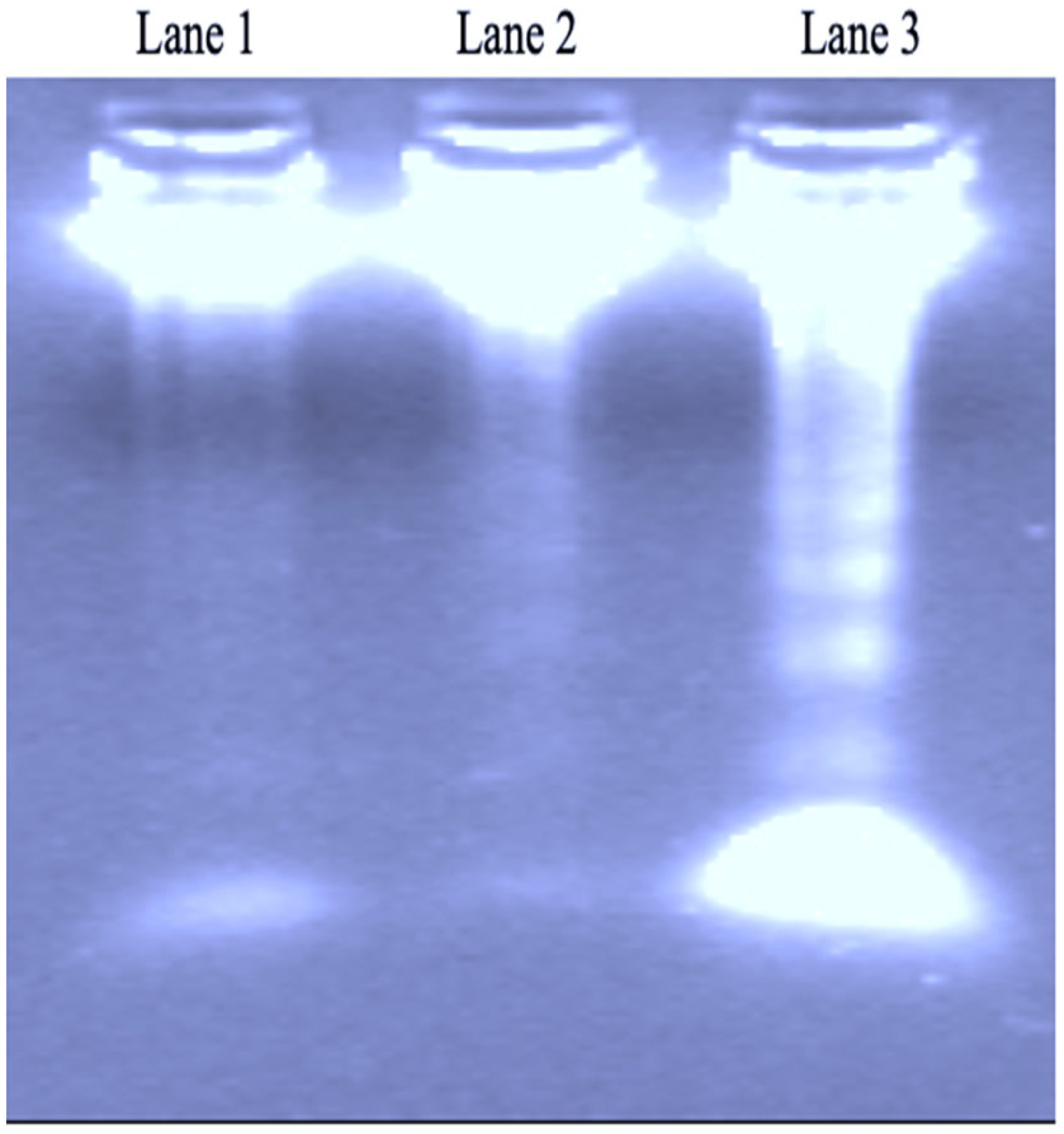
DNA Fragmentation Analysis

The concentration and purity of the DNA were evaluated by a Nanodrop UV spectrophotometer (Thermo Fisher Scientific, Waltham, Massachusetts, USA). Aliquots containing 100 ng of cellular DNA were subjected to agarose gel electrophoresis (1.5% agarose in TAE buffer pH 8.2) and stained with 0.5 μg mL−1 ethidium bromide. The DNA bands were checked under UV transilluminators.

2.7. RNA Extraction and cDNA Synthesis

RNA was isolated from HCT-116 cells employing the RNeasy mini kit as instructed by the manufacturer (Qiagen, Hilden, Germany). Total RNA concentration and purity were evaluated using a UV spectrophotometer. A cDNA synthesis kit (Intron Biotechnology, Seongnam-si, Korea) was used in accordance with the manufacturer’s guidelines to synthesize cDNA.

2.8. Quantitative Real-Time PCR

The genetic expression levels for Bcl-2, Bax, Cas3, PI3K and NF-κB, and GAPDH were utilized as housekeeping genes and tested using optimized protocols of real-time PCR as recently published by Gad et al. (2022) [40]. The specific primers were developed via NCBI BLAST prior to being procured from Thermo Fisher Scientific (Waltham, MA, USA) (https://blast.ncbi.nlm.nih.gov/Blast.cgi, accessed on 5 March 2021) and their sequences are listed in Table 1.

2.9. Antimicrobial Activity

2.9.1. Tested Microorganisms

The inhibitory impact of extracts was detected against six pathogenic bacterial strains. The bacterial strains consisted of gram-positive bacteria (Staphylococcus aureus ATCC 13565, Bacillus cereus EMCC 1080, and Staphylococcus sciuri 2–6) and gram-negative bacteria (Salmonella typhi ATCC 25566, Pseudomonas aeruginosa NRRL B-272, and Escherichia coli 0157:H7 ATCC 51659). The strains were obtained from MARSIN, Microbiology Department, Faculty of Agriculture, Ain Shams University, Egypt. Bacterial cultures were precultured on nutrient agar slant at 37 °C for 24 h and kept at 4 °C. Antifungal activity was determined using four fungal strains: A. niger IMI288550; Aspergillus ochraceus ITAL 14; Penicillium verrucosum BFE 500; and Fusarium proliferatum MPVP 328. Fungal strains were obtained from the Applied Mycology Department, Cranfield University, UK. The selected cultures were pregrown on potato dextrose agar (PDA) slants at 25 °C for 120 h and left in a refrigerator at 4 °C until further use.

2.9.2. Disc Diffusion Technique

Nutrient agar slants of each bacterial species were incubated for 24 h, and then 5 mL of tryptic soy broth (TSB) was used for inoculation of microorganisms on test tubes. The liquid cultures were incubated at 37 °C for 2–6 h until the culture turbidity reached 0.5 McFarland.
The antibacterial activity of P. roxburghii and N. graveolens extracts was examined against bacterial strains using the Kirby-Bauer disc diffusion method [41]. DMSO and tetracycline (500 µg mL−1) were used as negative and positive controls, respectively. The plates were incubated for 24 h at 37 °C. The antibacterial activity of the extracts was assessed by detecting the development of the growth clear zone (inhibition zone) around the extract discs at the end of the incubation period. The inhibition zone diameters were measured in millimeters, including the diameter of the extract paper disc. Fungal spore inoculum was prepared by suspending freshly prepared fungal discs (5 days growth) in 0.01% Tween 80 test tubes until the spore suspension density reached 0.5 McFarland corresponding to 2 × 10⁸ CFU mL−1. DMSO and nystatin (as a fungicide = 1000 Unit mL−1) were used as negative and positive controls, respectively. The cultured plates were incubated for 24–48 h at 25 °C. After 48 h, the antifungal activity of the extracts was detected by measuring the inhibition zone diameter (mm) [42]. Treatments were detected in triplicate, and the averages of the experimental results were calculated.

2.9.3. Determination of Minimum Inhibitory Concentration (MIC)

The MIC assay was evaluated by the tube dilution method [43,44]. Twenty-four-hour cultures of the bacterial strains were suspended in 10 mL of TSB until they reached 0.5 McFarland (equivalent to CFU 108 per mL). Ten concentrations of P. roxburghii and N. graveolens extracts were prepared in DMSO from 5.0 mg mL−1 to 10.0 µg mL−1. Aliquots of 100 µL of each bacterial culture suspension were inoculated in each tube and incubated for 24 h at 37 °C. The turbidity of each tube culture was detected by UV spectrophotometry at 600 nm. The minimum concentration of the extracts that inhibited bacterial growth was taken as the MIC value specific for each strain.
The MIC against fungi was assessed by the technique mentioned above [45,46]. Different concentrations of P. roxburghii and N. graveolens extracts were mixed in 0.5 mL of 0.1% Tween 80 (Merck, Darmstadt, Germany), homogenized with 9.5 mL warm, presolidified PDA medium, and then poured into 6 cm sterilized Petri dishes. Aliquots of 3 µL of fungal spore suspension (108 CFU mL−1, equivalent to 0.5 McFarland standard) were transferred to the center of each plate, and the cultured plates were incubated at 25 °C for 24–48 h, after which the fungal mycelial growth was checked and the MIC was recorded.

2.10. Statistical Analyses

Statistical analysis was carried out employing GraphPad Prism 8.0 software (San Diego, CA, USA). Except where otherwise stated, the findings are stated as the mean ± SEM of three independent experiments. The data were assessed by one-way ANOVA and displayed as the means ± SDs using SPSS 19.0 (SPSS Ltd., Surrey, UK). Individual comparisons were determined by Duncan’s multiple range test (DMRT). Differences were considered significant at p < 0.05.

3. Results and Discussion

3.1. Total Phenolic Content and Antioxidant Activity

Table 2 shows the total phenolic, total flavonoid, and total carotenoid contents in the P. roxburghii and N. graveolens extracts. The N. graveolens extract exhibited a high amount of total phenolics (73.86 µg GAE/g) compared to P. roxburghii (32.70 µg GAE/g). The flavonoid contents of the P. roxburghii and N. graveolens extracts were valued at 11.67 and 41.55 catechins eq µg/g, respectively, and the total carotenoid values were 0.24 and 0.11 µg/g, respectively. The antioxidant activities of the P. roxburghii and N. graveolens extracts were determined by the DPPH and ABTS methods, as shown in Table 2. The N. graveolens extract showed high antioxidant activity, with values of 37.81 and 25.81 μg TE/g compared to P. roxburghii, which had values of 17.66 and 20.95 μg TE/g estimated by DPPH and ABTS, respectively. The current study shows that P. roxburghii and N. graveolens extracts contained high amounts of total phenolics, flavonoids, and carotenoids. It is widely known that the generation of free radicals within the body involves cellular oxidative damage and triggers cancer pathways and other diseases [47]. These results were compatible with data attained by Sharma et al. (2016) [48], who found that P. roxburghii bark contains various compounds, like flavonoids, beta-carotene lycopene, phenolics, and tannins. Another study confirmed by Rawat et al. (2009) [49] observed that P. roxburghii bark is a good source of flavonoids and phenolics. Additionally, Haddouchi et al. (2016) [50] revealed that N. graveolens extract contained a high content of total phenolics. Ramdane et al. (2017) [18] revealed that A. graveolens contained polyphenolics, flavonoids, and tannins. The antioxidant actions of the tested extracts were determined by DPPH and ABTS assays. These extracts exhibited high antioxidant potential against DPPH and ABTS assays. These findings agree with a study demonstrating that P. roxburghii bark possessed significant antioxidant activity against DPPH) [48]. Likewise, Puri et al. (2011) [14] indicated that P. roxburghii needles have significant scavenging against (ABTS) radical cations. These outcomes are also consistent with data obtained by Ramdane et al. (2017) [18], who demonstrated that A. graveolens extract has antioxidant potential by the DPPH assay. Similarly, Alilou et al. (2014) [51] found a strong activity of the essential oil of A. graveolens with the DPPH.

3.2. Identification of Bioactive Compounds Using HPLC

Phenolic compound profiles of P. roxburghii and N. graveolens extracts were identified using HPLC (Table 3). Catechin was the predominant phenolic compound in P. roxburghii extracts. Meanwhile, chlorogenic acid was the most abundant phenolic compound in N. graveolens, which has important antioxidant and cardioprotective properties. Various polyphenol components were also recognized in the studied extracts, namely caffeic, kaempferol, syringic, p-coumaric vanillic, and p-hydroxybenzoic acids. P. roxburghii was devoid of apigenin-7-glucoside, rosmarinic acid, cinnamic acid and rutin but rich in gallic acid, protocatechuic and ferulic acids. It was observed that N. graveolens had the highest amount of rosmarinic and cinnamic acids, apigenin-7-glucoside and rutin. Rawat et al. (2009) [49] found that various polyphenolic compounds separated from branches of P. roxburghii included protocatechuic acid, caffeic acid, kaempferol, catechin, and gallocatechin. Additionally, Ramdane et al. (2017) [18] noticed that the main phenolic components in A. graveolens extract were chlorogenic acids and coumaric.

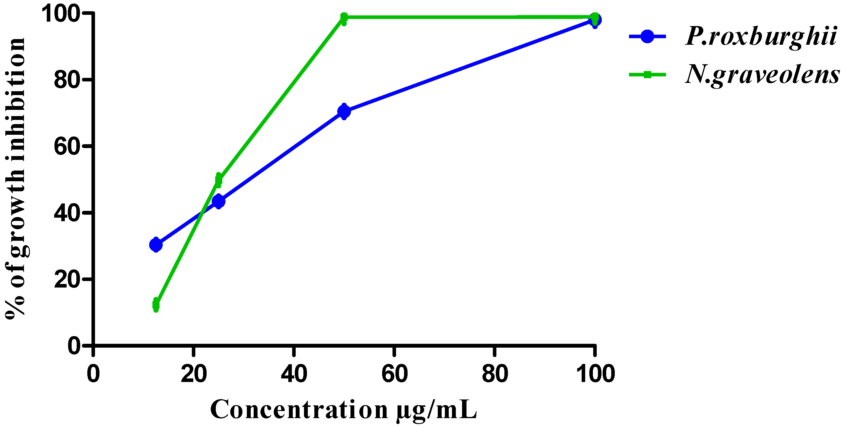
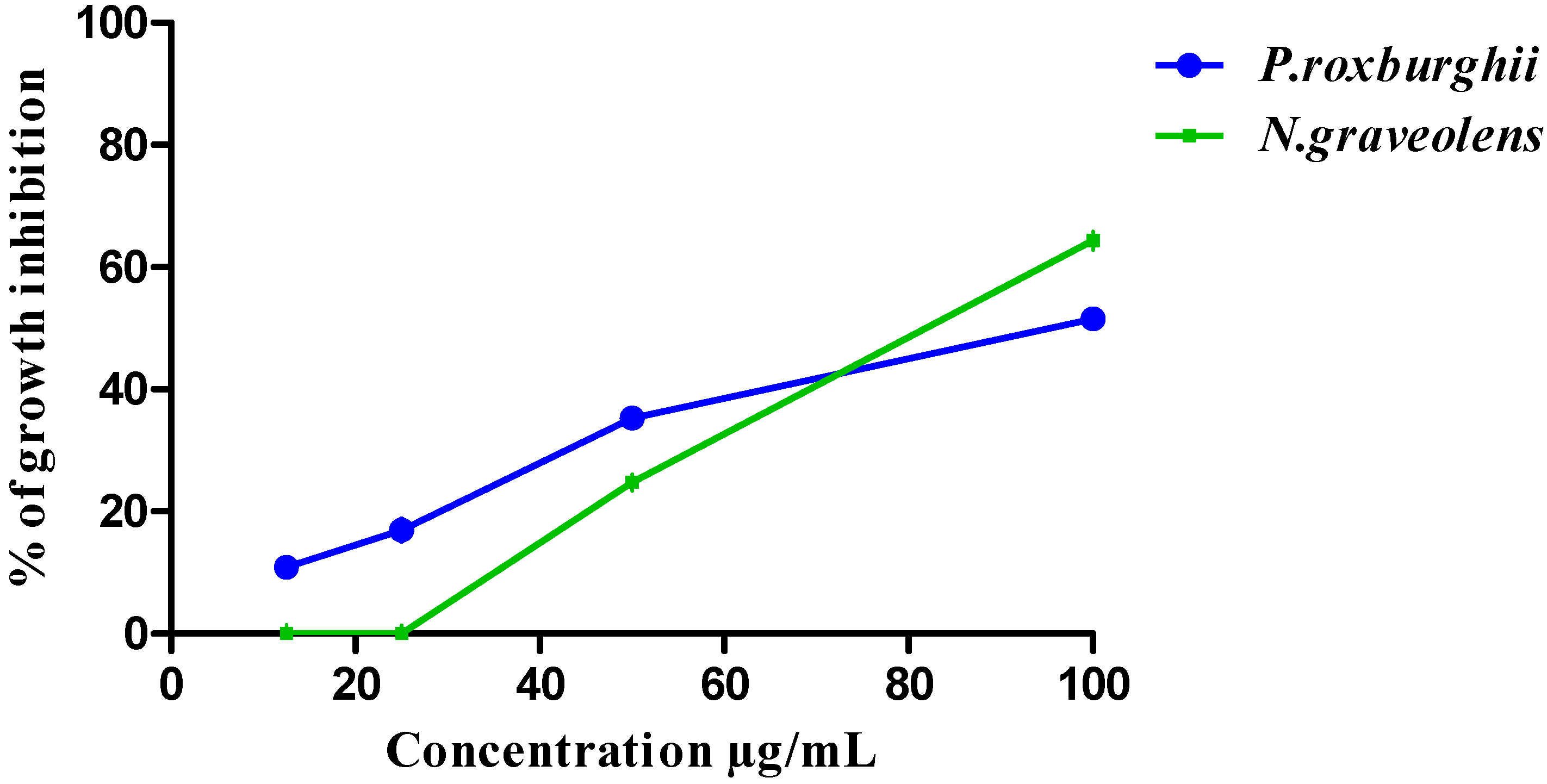
3.3. HCT-116 and BJ-1 Cytotoxicity of P. roxburghii and N. graveolens Extracts

The cytotoxicity results of P. roxburghii and N. graveolens extracts against HCT-116 cells at different concentrations are summarized in Figure 1. The extracts exhibited highly significant inhibition against the HCT-116 cell line at levels of 100 and 50 μg mL−1. In contrast, the concentrations of 25 and 12.5 μg mL−1 revealed low significant antiproliferative activity against human colon cancer. The IC50 values for both extracts were 30.6 and 26.5 μg mL−1, respectively.
The P. roxburghii and N. graveolens extracts were further examined for cytotoxicity on BJ-1-cell lines at various concentrations to estimate their SI values, as illustrated in Figure 2. The IC50 values for P. roxburghii and N. graveolens extracts against BJ-1 cells were 91.7 and 84.3 μg mL−1, respectively. The selectivity index was estimated according to the ratio of the IC50 value of the extracts from the BJ-1 normal human cell line to colon human cancer cells HCT-116. Based on the results, the P. roxburghii and N. graveolens extracts were highly active, with a wide selectivity index for colon cancer cells SI = 2.99 and 3.1, respectively. In accordance with the National Cancer Institute (NCI, USA), a plant screening program, crude extracts can be identified as strongly cytotoxic if the IC50 is <30–40 μg mL−1 [52]. Depending on these criteria, these extracts displayed possibility as new anticancer drugs. The SI generally indicates the safety of a crude extract employed for anticancer treatment. SI values > 2 are considered highly selective [53]. Based on the results, the P. roxburghii and N. graveolens extracts were highly selective, with selectivity indices for colon cancer cells of 2.99 and 3.1, respectively. Likewise, the study of Al-Rashidi et al. (2011) [54] clarified that plant extracts could be considered anticancer agents with low toxicity toward normal cells and high toxicity against cancer cells. The impact of the mentioned extracts as cytostatic agents is in line with Kulshrestha and Khan (2019) [55], who found that different extracts and compounds of P. roxburghii essential oil show promising anticancer activity against a human lung cancer cell line (A549). Additionally, Sajid et al. (2018) [56] confirmed the anticancer activity of P. roxburghii essential oil against colon, leukemia, pancreatic, head, neck, and lung cancer cell lines through cell proliferation suppression and induction of apoptosis in cancer cells. These findings are strongly supported by Achoub et al. (2019) [57], who observed that A. graveolens extract significantly inhibited the growth of HCT-116 and DLD1 cells. Moreover, Tayeh et al. (2018) [58] revealed that the crude extract of A. graveolens and its fraction inhibited the growth of mammalian lymphoma cells (BS-24-1).

3.4. DNA Fragmentation Activity

The DNA fragmentation activity of P. roxburghii and N. graveolens extracts was investigated, as shown in Figure 3. DNA fragmentation was observed after treatment of HCT-116 cells with IC50 doses of both extracts. The results revealed that the tested extracts inhibited the growth of HCT-116 cells through apoptosis. It was found that N. graveolens was more efficient in inhibiting cancer cell growth and inducing apoptosis than P. roxburghii. Generally, a DNA fragmentation assay is used to detect whether the action of the extract is associated with cell apoptosis [59]. The outcomes of the current study elucidated that treatment with the IC50 doses of P. roxburghii and N. graveolens extracts induced apoptosis in HCT-116 cells. These findings are consistent with a previous study by Kumari et al. (2019) [60], reporting that P. roxburghii needles could induce DNA fragmentation leading to apoptosis. Tayeh et al. (2018) [58] emphasized the activity of A. graveolens fractions in promoting apoptosis and found a DNA ladder with fragments of 180 bp because of cell death. Gad et al., 2022 [40] observed that supplementation with one-tenth of the LD50 of extracts suppressed the tumor growth rate in a BALB/c mouse model.

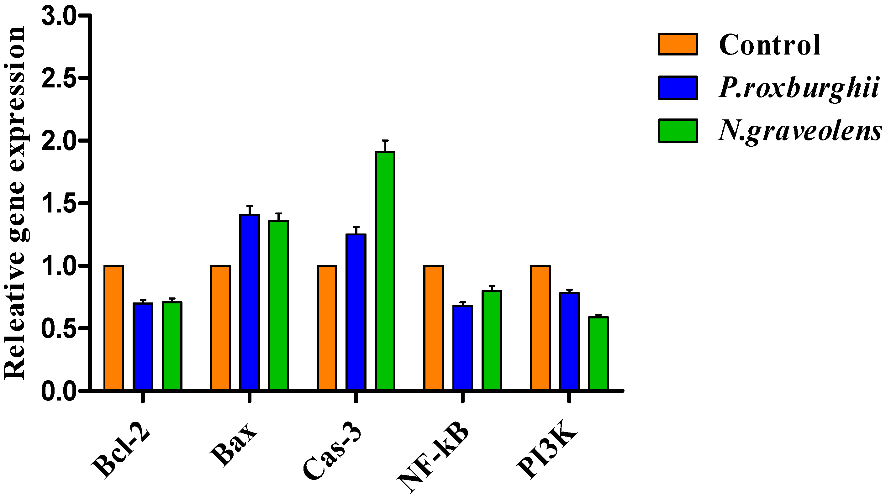
3.5. Gene Expression

The relative expression of the Bcl-2, Cas3, Bax, NF-κB and PI3k genes in a human colon cancer cell line (HCT-116) was investigated, and the results are displayed in Figure 4. Treatment of HCT-116 cells with IC50 doses of P. roxburghii and N. graveolens significantly upregulated BAX expression and downregulated Bcl-2. The Bax/Bcl-2 ratio was elevated upon treatment with IC50 doses of tested extracts with ratios of 1.9 and 4.42, respectively. Moreover, the Cas-3 gene was increased after exposure to the IC50 doses of extracts. On the other hand, the expression levels of the NF-κB and PI3k genes were significantly downregulated.
The expression of signaling biomarker genes was investigated after exposure to the IC50 doses of P. roxburghii and N. graveolens extracts to gain further insights into the mode of cell death. The data showed that Bax and Cas-3 (pro-apoptotic genes) were increased after exposure to the suggested extracts. Moreover, the expression of bcl-2 (an anti-proapoptotic protein) continuously declined. Bax, a member of the Bcl-2 family, has an influential role in the regulation of apoptosis [22]. If Bax is overexpressed, the cell undergoes apoptosis. If the level of bcl-2 increases, the cell undergoes cell survival [61]. Downregulation of bcl-2 and activation of the two genes caspase-3 and Bax eventually inhibited tumor cell growth by enhancing cell apoptosis. The data also illuminated that the Bax/Bcl-2 ratio was elevated after the treatment. The Bax/Bcl-2 ratio can be used as a marker to determine cell susceptibility to apoptosis [62]. Generally, Bcl-2 and Bax proteins govern the activation of Caspase-3 by controlling mitochondrial cytochrome C release in the cytosol, which acts as a critical influencer on Caspase-3 activity regulation [63,64]. Furthermore, NF-κB and PI3k gene expression was downregulated after the IC50 exposure dose of the tested extracts. Suppression of NF-κB through plant-derived phytochemicals has attracted increasing interest in cancer research [65,66]. Blocking NF-κB can halt the proliferation of tumor cells, and the apoptotic pathway is subsequently stimulated [67]. PI3K is influential in treating several types of cancer (Workman et al., 2006). Suppression of PI3K signaling results in decreased cellular proliferation and increased cellular death [68]. Previous studies demonstrated that the down—and upregulation of the selected biomarker genes was accompanied by the presence of bioactive compounds found in both P. roxburghii and N. graveolens plants. Intriguingly, N. graveolens extract is rich in rosmarinic, apigenin-7-glucoside, and cinnamic acid owing to the ability of these active compounds to induce cell cycle arrest and apoptosis [69,70]. This study is supported by previous studies that showed that apigenin administration downregulated Bcl-2 and Bcl-xL and and suppressed NF-κB activation [71]. In the same trend, rosmarinic acid downregulates the expression of Bcl-2 while upregulating the expression of Bax in breast cancer cells (BCSCs), according to Li et al. (2019) [72]. The P. roxburghii extract was considered a rich source of catechin and gallic acid. In agreement with other reports, Moradzadeh et al. (2017) [73] found that epicatechin upregulated caspase-3, increased the Bax/Bcl-2 ratio, and downregulated phosphoinositide-3-kinase protein kinase B (PI3K/Akt) and Bcl-2 in a breast cancer cell line (T47D). Moreover, gallic acid inhibited proliferation and induced apoptosis, which was associated with the downregulation of Bcl-2 and upregulation of Bax [74]. Another work confirmed that the essential oil of P. roxburghii reduced the expression of NF-κB-regulated genes linked to cell survival (Bcl-2 and Bcl-xL), proliferation (Cyclin D1) and metastasis (MMP-9) [56]. The findings also revealed that P. roxburghii and N. graveolens extracts were abundant with polyphenols, like caffeic, kaempferol, syringic, p-coumaric vanillic, and p-hydroxybenzoic acids. These results agree with recent work reporting that caffeic acid intake led to the suppression of colony formation, cell cycle arrest, and induction of apoptosis [75]. Kaempferol prevents angiogenesis by suppressing vascular endothelial growth factor (VEGF) secretion and downregulating extracellular signal-regulated kinase (ERK), NFκB and cMyc expression in ovarian cancer cell lines (OVCAR-3 and A2780/CP70) [76]. Furthermore, Sharma et al. (2019) [77] reported that p-coumaric acid administration caused apoptosis by regulating the Bax/Bcl-2 ratio. Gad et al. (2022) [40] found that supplementation of one-tenth of the LD50 of P. roxburghii and N. graveolens extracts caused a considerable upregulation of the expression of the Bax and Cas-3 genes and downregulation of the expression of the Bcl-2, NF-kB and PI3K genes in BALB/c mice.

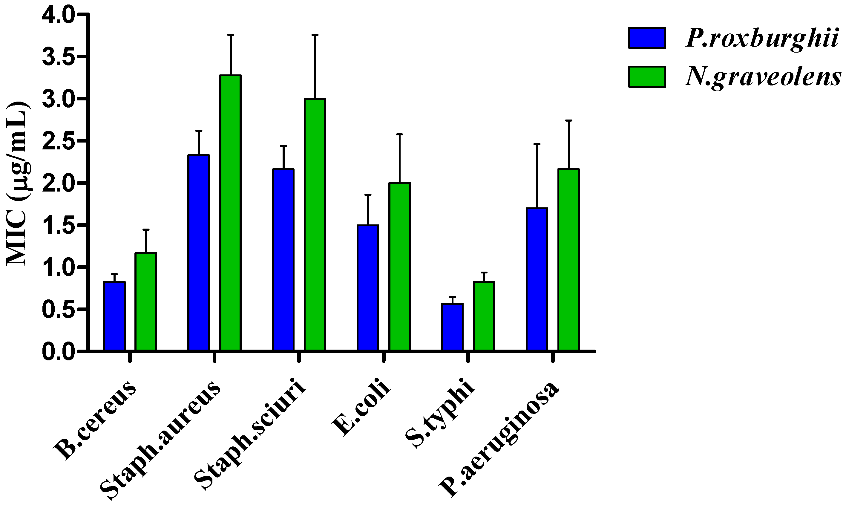
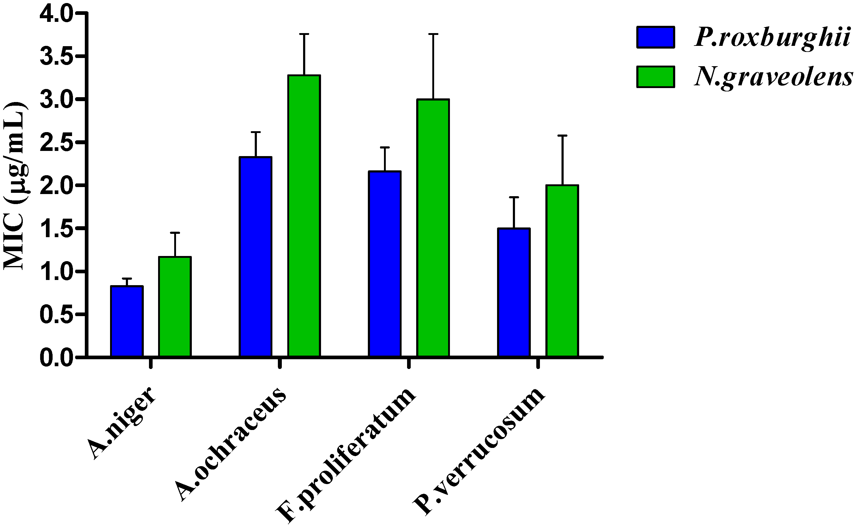
3.6. Antimicrobial Activity

3.6.1. Antibacterial Activity

The inhibitory effects of P. roxburghii and N. graveolens extracts against six bacterial strains are clarified in Table 4. The P. roxburghii extract exhibited high antibacterial capacity against E. coli, S. typhi, and P. aeruginosa with inhibition zones of 12.0, 11.2, and 10.3 mm, respectively. The results also showed that N. graveolens has potent activity against E. coli, S. sciuri and P. aeruginosa with inhibition zones of 11.2, 8.3, and 8.3 mm, respectively.
The MIC values of extracts against 6 strains of foodborne pathogenic bacteria are illustrated in Figure 5. The P. roxburghii extract exhibited the lowest MIC against S. typhi, B. cereus and P. aeruginosa, with values of 0.57, 0.83, and 1.7 µg mL−1, respectively, compared to N. graveolens in the same bacterial strains, with MIC values of 0.83, 1.17 and 2.16 µg mL−1, respectively. The highest MIC observed was against S. aureus, S. sciuri, and P. aeruginosa, with values of 3.28, 3.0, and 2.16 µg mL−1, respectively, by N. graveolens extract. The highest MIC value recorded was versus S. aureus 3.28 µg mL−1 by N. graveolens, and the lowest MIC value recorded was against S. typhi, with a value of 0.57 µg mL−1 by P. roxburghii.

3.6.2. Antifungal Activity

The antifungal activities of P. roxburghii and N. graveolens extracts against four mycotoxigenic fungi are shown in Table 5. The P. roxburghii and N. graveolens extracts have antifungal activity against the growth of all fungal strains. The P. roxburghii extract has strong activity against P. verrucosum and A. ochraceus, with inhibition zones of 9.8 and 8.7 mm, respectively. However, the N. graveolens extract showed strong efficacy against A. niger and P. verrucosum, with inhibition zones of 9.2 and 8.7 mm, respectively.
The MIC values of P. roxburghii and N. graveolens against four strains of mycotoxigenic fungi are represented in Figure 6. The P. roxburghii extract exhibited the lowest MIC against A. niger and P. verrucosum, with values of 0.83 and 1.50 µg mL−1, respectively, compared to N. graveolens in the same fungal strains, with MIC values of 1.50 and 2.0 µg mL−1, respectively. The highest activity of N. graveolens was registered against A. ochraceus, with an MIC value of 3.28 µg mL−1. However, the lowest ability of P. roxburghii was observed versus A. niger, with an MIC value of 0.83 µg mL−1. The MIC values of P. roxburghii were lower than those of N. graveolens.
The MIC was determined for only those microorganisms that displayed a zone of inhibition and were sensitive to the plant extracts. The E. coli strain was the most susceptible to both extracts. In contrast, the B. cereus strain showed the lowest susceptibility against the tested extracts, as indicated by estimating the zone of inhibition diameter. Consistent with the obtained results, Sharma et al. (2016) [48] observed that P. roxburghii extracts significantly prohibited the growth of bacteria against S. aureus, P. aeruginosa and E. coli. Intriguingly, Alrawashdeh et al. (2019) [78] declared that Asteriscus graveolens extracts possessed antibacterial activity against E. coli, Salmonella sp. and E. aerogenes. Concerning the assessment of the antifungal activity of the tested extracts, Table 5 shows that different antifungal activity responses varied according to the fungal strain. F. proliferatum and A. ochraceus were the most susceptible fungal strains against the tested extracts. P. verrucosum and A. niger were the most resistant fungal strains to P. roxburghii and N. graveolens extracts, respectively, as indicated by inhibition zone diameter measurements. These results coincide with those of Satyal et al. (2013) [16], who found that the essential oil of P. roxburghii has antifungal capability against A. niger. Furthermore, Znini et al. (2011) [79] found that the essential oil of Asteriscus graveolens possessed antifungal properties against P. expansum and Alternaria sp.

4. Conclusions

The outcomes of this research elucidated that P. roxburghii and N. graveolens extracts have anticancer activity against human colon cancer HCT-116 cells. The suppression of human colon cancer HCT-116 cells was mediated by optimizing the regulation of apoptotic gene markers. Moreover, the tested extracts showed potential as antioxidant and antimicrobial agents. Nevertheless, additional in vivo research studies are required to clarify the other properties of these plant extracts and their potential application.

Author Contributions

Methodology, M.G., K.M., A.A.A.-R., A.A.Z., A.M.H., A.S.A.-A. and H.Z.H.; software and validation, M.G., M.S.A.-L. and A.A.Z.; formal analysis, M.G., F.A.A., A.A.A.-R. and A.S.A.-A., investigation, M.G. and A.A.Z.; resources, M.G., H.Z.H., A.A.A.-R., A.M.H. and A.A.Z.; data curation, M.G. and A.A.Z.; writing—original draft preparation, M.G., F.A.A., A.A.A.-R. and A.A.Z.; writing—review and editing, J.-H.S., M.G., H.Z.H., K.M., A.A.Z. and E.A.A.-R.; visualization, A.A.Z. and M.G.; project administration, H.Z.H., K.M., M.G., F.A.A., A.A.Z., M.G. and J.-H.S.; funding acquisition, A.A.Z. and M.G. All authors have read and agreed to the published version of the manuscript.

Funding

This research received no external funding.

Data Availability Statement

Not applicable.

Conflicts of Interest

The authors declare no conflict of interest.

References

  1. Mathers, C.D.; Loncar, D. Projections of Global Mortality and Burden of Disease from 2002 to 2030. PLoS Med. 2006, 3, e442. [Google Scholar] [CrossRef] [Green Version]
  2. Ferlay, J.; Ervik, M.; Lam, F.; Colombet, M.; Mery, L.; Pineros, M.; Znaor, A.; Soerjomataram, I.; Bray, F. Global Cancer Observatory: Cancer Today; International Agency for Research on Cancer: Lyon, France, 2018; Available online: https://gco.iarc.fr/today (accessed on 1 December 2020).
  3. World Health Organization (WHO). Global Health Estimates 2020: Deaths by Cause, Age, Sex, by Country and by Region, 2000–2019; WHO: Geneva, Switzerland, 2020; Available online: who.int/data/gho/data/themes/mortality-and-global-health-estimates/ghe-leading-causes-of-death (accessed on 11 December 2020).
  4. Cheng, L.; Eng, C.; Nieman, L.Z.; Kapadia, A.S.; Du, X.L. Trends in colorectal cancer incidence by anatomic site and disease stage in the United States from 1976 to 2005. Am. J. Clin. Oncol. 2011, 34, 573–580. [Google Scholar] [CrossRef] [PubMed]
  5. Wiseman, M. The Second World Cancer Research Fund/American Institute for Cancer Research Expert Report. Food, Nutrition, Physical Activity, and the Prevention of Cancer: A Global Perspective. Proc. Nutr. Soc. 2008, 67, 253–256. [Google Scholar] [CrossRef] [Green Version]
  6. Continuous Update Project Report. Food, Nutrition, Physical Activity, and the Prevention of Colorectal Cancer; American Institute for Cancer Research: Washington, DC, USA, 2011. [Google Scholar]
  7. Sak, K. Chemotherapy and Dietary Phytochemical Agents. Chemother. Res. Pract. 2012, 2012, 282570. [Google Scholar] [CrossRef] [Green Version]
  8. Baskar, R.; Dai, J.; Wenlong, N.; Yeo, R.; Yeoh, K.-W. Biological response of cancer cells to radiation treatment. Front. Mol. Biosci. 2014, 1, 24. [Google Scholar] [CrossRef] [PubMed] [Green Version]
  9. Tsao, A.S.; Kim, E.S.; Hong, W.K. Chemoprevention of Cancer. CA Cancer J. Clin. 2004, 54, 150–180. [Google Scholar] [CrossRef] [PubMed] [Green Version]
  10. Zaky, A.A.; Shim, J.H.; Abd El-Aty, A.M. A Review on extraction, characterization, and applications of bioactive peptides from pressed black cumin seed cake. Front. Nutr. 2021, 8, 743909. [Google Scholar] [CrossRef] [PubMed]
  11. Zaky, A.A.; Simal-Gandara, J.; Eun, J.-B.; Shim, J.-H.; El-Aty, A.M.A. Bioactivities, Applications, Safety, and Health Benefits of Bioactive Peptides from Food and By-Products: A Review. Front. Nutr. 2022, 8, 815640. [Google Scholar] [CrossRef] [PubMed]
  12. Bahnasy, M.I.; Khamis, M.H. Distribution pattern of trees diversity in Orman Botanical Garden. Middle East J. Agric. Res. 2019, 8, 1–11. [Google Scholar]
  13. Kaushik, D.; Kumar, A.; Kaushik, P.; Rana, A.C. Analgesic and Anti-Inflammatory Activity of Pinus roxburghii Sarg. Adv. Pharmacol. Sci. 2012, 2012, 245431. [Google Scholar] [CrossRef] [Green Version]
  14. Puri, A.; Srivastava, A.K.; Singhal, B.; Mishra, S.K.; Srivastava, S.; Lakshmi, V. Antidyslipidemic and antioxidant activity of Pinus roxburghii needles. Med. Chem. Res. 2011, 20, 1589–1593. [Google Scholar] [CrossRef]
  15. Shah, W.A.; Qadir, M.; Banday, J.A. GC-MS analysis, antibacterial, antioxidant and anticancer activity of essential oil of Pinus roxburghii from Kashmir, India. Int. J. Pharm. Res. 2014, 4, 228–232. [Google Scholar] [CrossRef]
  16. Satyal, P.; Paudel, P.; Raut, J.; Deo, A.; Dosoky, N.S.; Setzer, W.N. Volatile constituents of Pinus roxburghii from Nepal. Pharmacogn. Res. 2013, 5, 43–48. Available online: http://www.phcogres.com/text.asp?2013/5/1/43/105650 (accessed on 16 January 2021).
  17. Cheriti, A.; Saad, A.; Belboukhari, N.; Ghezali, S. The essential oil composition of Bubonium graveolens (Forssk.) Maire from the Algerian Sahara. Flav. Frag. J. 2007, 22, 286–288. [Google Scholar] [CrossRef]
  18. Ramdane, F.; Essid, R.; Mkadmini, K.; Hammami, M.; Fares, N.; Mahammed, M.H.; El Ouassis, D.; Tabbene, O.; Limam, F.; Hadj, M.D.O. Phytochemical composition and biological activities of Asteriscus graveolens (Forssk) extracts. Process Biochem. 2017, 56, 186–192. [Google Scholar] [CrossRef]
  19. Chaib, F.; Allali, H.; Bennaceur, M.; Flamini, G. Chemical Composition and Antimicrobial Activity of Essential Oils from the Aerial Parts of Asteriscus graveolens (Forssk.) and Pulicaria incisa (LAM.) DC. Two Asteraceae Herbs Growing Wild in the Hoggar. Chem. Biodiversity 2017, 14, 92. [Google Scholar] [CrossRef] [PubMed]
  20. Aouissi, H.; Gourine, N.; Wang, H.; Chen, X.; Bombarda, I.; Boudjeniba, M.; Yousfi, M. Chemical composition, antioxidative, antimicrobial and anti-cancer activities of Asteriscus graveolens (Forssk) essential oil. Orient. Pharm. Exp. Med. 2018, 18, 217–223. [Google Scholar] [CrossRef] [Green Version]
  21. Guo, X.X.; Li, Y.; Sun, C.; Jiang, D.; Lin, Y.J.; Jin, F.X.; Lee, S.K.; Jin, Y.H. p53-dependent Fas expression is critical for Ginsenoside Rh2 triggered caspase-8 activation in HeLa cells. Protein Cell 2014, 5, 224–234. [Google Scholar] [CrossRef] [Green Version]
  22. Kale, J.; Osterlund, E.J.; Andrews, D.W. BCL-2 family proteins: Changing partners in the dance towards death. Cell Death Differ. 2018, 25, 65–80. [Google Scholar] [CrossRef] [PubMed] [Green Version]
  23. Yip, K.W.; Reed, J.C. Bcl-2 family proteins and cancer. Oncogene 2008, 27, 6398–6406. [Google Scholar] [CrossRef] [Green Version]
  24. Elmore, S. Apoptosis: A review of programmed cell death. Toxicol. Pathol. 2007, 35, 495–516. [Google Scholar] [CrossRef] [PubMed]
  25. Estaquier, J.; Vallette, F.; Vayssiere, J.L.; Mignotte, B. The mitochondrial pathways of apoptosis. Adv. Exp. Med. Biol. 2012, 942, 157–183. [Google Scholar] [CrossRef] [PubMed]
  26. Baeuerle, A.A.P.; Henkel, T. Function and Activation of NF-kappaB in the Immune System. Annu. Rev. Immunol. 1994, 12, 141–179. [Google Scholar] [CrossRef]
  27. Tak, P.P.; Firestein, G.S. NF-κB: A key role in inflammatory diseases. J. Clin. Investig. 2001, 107, 7–11. [Google Scholar] [CrossRef] [PubMed]
  28. Cantley, L.C. The Phosphoinositide 3-Kinase Pathway. Science 2002, 296, 1655–1657. [Google Scholar] [CrossRef] [PubMed]
  29. Gad, M.; Hassouna, H.Z.; Mahmoud, K.; Abd-Rabou, A.A.; Abdel-Azeem, A.S.; Hegazy, A.M.; Lattife, M.S.A.; Ahmed, F.A. Preliminary study on the toxicological impacts of Pinus roxburghii and Nauplius graveolens extracts on albino mice. Egypt. J. Chem. 2021, 64, 3489–3498. [Google Scholar] [CrossRef]
  30. Boulos, L. Flora of Egypt 3: Compositae-Verbenaceae; Al Hadara Publishing: Cairo, Egypt, 2002. [Google Scholar]
  31. Zaky, A.A.; Chen, Z.; Liu, Y.; Li, S.; Jia, Y. Preparation and assessment of bioactive extracts having antioxidant activity from rice bran protein hydrolysates. J. Food Meas. Charact. 2019, 13, 2542–2548. [Google Scholar] [CrossRef]
  32. Zhishen, J.; Mengcheng, T.; Jianming, W. The determination of flavonoid contents in mulberry and their scavenging effects on superoxide radicals. Food Chem. 1999, 64, 555–559. [Google Scholar] [CrossRef]
  33. Moore, J.; Hao, Z.; Zhou, K.; Luther, M.; Costa, J.; Yu, L. Carotenoid, Tocopherol, Phenolic Acid, and Antioxidant Properties of Maryland-Grown Soft Wheat. J. Agric. Food Chem. 2005, 53, 6649–6657. [Google Scholar] [CrossRef]
  34. Zaky, A.A.; Liu, Y.; Han, P.; Ma, A.; Jia, Y. Effect of flavorzyme digestion on the antioxidant capacities of ultra-filtrated rice bran protein hydrolyzates. J. Food Process. Preserv. 2020, 44, e14551. [Google Scholar] [CrossRef]
  35. Zaky, A.A.; Liu, Y.; Han, P.; Chen, Z.; Jia, Y. Effect of Pepsin–Trypsin In Vitro Gastro-Intestinal Digestion on the Antioxidant Capacities of Ultra-Filtrated Rice Bran Protein Hydrolysates (Molecular Weight > 10 kDa; 3–10 kDa, and < 3 kDa). Int. J. Pept. Res. Ther. 2020, 26, 1661–1667. [Google Scholar] [CrossRef]
  36. Zaky, A.A.; Chen, Z.; Qin, M.; Wang, M.; Jia, Y. Assessment of antioxidant activity, amino acids, phenolic acids and functional attributes in defatted rice bran and rice bran protein concentrate. Prog. Nutr. 2020, 22, e2020069. [Google Scholar] [CrossRef]
  37. Thabrew, M.I.; Hughes, R.D.; Mcfarlane, I.G. Screening of Hepatoprotective Plant Components using a HepG2 Cell Cytotoxicity Assay. J. Pharm. Pharmacol. 1997, 49, 1132–1135. [Google Scholar] [CrossRef] [PubMed]
  38. Mosmann, T. Rapid colorimetric assay for cellular growth and survival: Application to proliferation and cytotoxicity assays. J. Immunol. Methods 1983, 65, 55–63. [Google Scholar] [CrossRef] [PubMed]
  39. Prayong, P.; Barusrux, S.; Weerapreeyakul, N. Cytotoxic activity screening of some indigenous Thai plants. Fitoterapia 2008, 79, 598–601. [Google Scholar] [CrossRef]
  40. Gad, M.; Hassouna, H.Z.; Mahmoud, K.; Abd-Rabou, A.A.; Abdel-Azeem, A.S.; Hegazy, A.M.; Abdel-Lattife, M.S.; Ahmed, F.A.; Oz, F.; Proestos, C.; et al. Pinus roxburghii and Nauplius graveolens Extracts Elevate Apoptotic Gene Markers in C26 Colon Carcinoma Cells Induced in a BALB/c Mouse Model. Separations 2022, 9, 277. [Google Scholar] [CrossRef]
  41. Bauer, A.W.; Kirby, W.M.; Sherris, J.C.; Turck, M. Antibiotic susceptibility testing by a standardized single disk method. Am. J. Clin. Pathol. 1966, 45, 493–496. [Google Scholar] [CrossRef]
  42. Medeiros, M.A.N.; De Oliveira, D.C.N.; Dos Prazeres Rodrigues, D.; De Freitas, D.R.C. Prevalence and antimicrobial resistance of Salmonella in chicken carcasses at retail in 15 Brazilian cities. Rev. Panam. Salud Publica 2011, 30, 555–560. [Google Scholar] [CrossRef]
  43. Irith, W.; Kai, H.; Robert, H. Agar and broth dilution methods to determine the minimal inhibitory concentration (MIC) of antimicrobial substances. Nat. Protoc. 2008, 3, 163–175. [Google Scholar] [CrossRef]
  44. Marrez, D.A.; Naguib, M.M.; Sultan, Y.Y.; Higazy, A.M. Antimicrobial and anticancer activities of Scenedesmus obliquus metabolites. Heliyon 2019, 5, e01404. [Google Scholar] [CrossRef] [PubMed] [Green Version]
  45. Sokmen, A.; Gulluce, M.; Akpulat, H.A.; Daferera, D.; Tepe, B.; Polissiou, M.; Sokmen, M.; Sahin, F. The in vitro antimicrobial and antioxidant activities of the essential oils and methanol extracts of endemic Thymus spathulifolius. Food Control 2004, 15, 627–634. [Google Scholar] [CrossRef]
  46. Sultan, Y.Y.; Ali, M.A.; Darwesh, O.M.; Embaby, M.A.; Marrez, D.A. Influence of nitrogen source in culture media on antimicrobial activity of Microcoleus lacustris and Oscillatoria rubescens. J. Pharm. Biol. Chem. Sci. 2016, 7, 1444–1452. Available online: https://www.rjpbcs.com/pdf/2016_7(2)/[197].pdf (accessed on 6 April 2022).
  47. Kumar, G.; Karthik, L.; Rao, K.V.B. Phytochemical composition and in vitro antioxidant activity of aqueous extract of Aerva lanata (L.) Juss. ex Schult. Stem (Amaranthaceae). Asian Pac. J. Trop. Med. 2013, 6, 180–187. [Google Scholar] [CrossRef] [PubMed]
  48. Sharma, A.; Goyal, R.; Sharma, L. Potential biological efficacy of Pinus plant species against oxidative, inflammatory and microbial disorders. BMC Complement. Altern. Med. 2015, 16, 35. [Google Scholar] [CrossRef] [Green Version]
  49. Rawat, U.; Srivastava, B.; Semwal, S.; Sati, O.P. Xanthones from Pinus roxburghii. J. Indian Chem. Soc. 2009, 38, 391–392. [Google Scholar] [CrossRef]
  50. Haddouchi, F.; Chaouche, T.M.; Halla, N. Screening phytochimique, activités antioxydantes et pouvoir hémolytique de quatre plantes sahariennes d’Algérie. Phytothérapie 2016, 16, S254–S262. [Google Scholar] [CrossRef]
  51. Alilou, H.; Asdadi, A.; Hassani, L.M.I.; Gonzalez- Mas, M.C.; Blazquez, M.A.; Akssira, M. Antifungal and Antioxidant Activity of Asteriscus graveolens subsp. odorus Essential Oil. J. Nat. Sci. Res. 2014, 4, 10. [Google Scholar]
  52. Oskoueian, E.; Abdullah, N.; Saad, W.Z.; Omar, A.R.; Kuan, W.B.; Zolkifli, N.A.; Hendra, R.; Ho, Y.W. Antioxidant, anti-inflammatory and anticancer activities of methanolic extracts from Jatropha curcas Linn. J. Med. Plants Res. 2011, 5, 49–57. [Google Scholar]
  53. Badisa, R.B.; Darling-Reed, S.F.; Joseph, P.; Cooperwood, J.S.; Latinwo, L.M.; Goodman, C.B. Selective cytotoxic activities of two novel synthetic drugs on human breast carcinoma MCF-7 cells. Anticancer Res. 2009, 29, 2993–2996. [Google Scholar] [PubMed]
  54. Al-Rashidi, W.; Mat Supri, N.N.; Manshoor, N. Cytotoxic activity of crude extract from Costus malortieanus (Costaceae). Am. Eurasian J. Toxicol. Sci. 2011, 3, 63–66. [Google Scholar]
  55. Kulshrestha, K.; Khan, H.S. Evaluation of anticancer activity of Pinus roxburghii Sarg against A549 human lung cancer cell line. J. Pharmacogn. Phytochem. 2019, 8, 2268–2272. [Google Scholar]
  56. Sajid, A.; Manzoor, Q.; Iqbal, M.; Tyagi, A.K.; Sarfraz, R.A.; Sajid, A. Pinus roxburghii essential oil anticancer activity and chemical composition evaluation. EXCLI J. 2018, 17, 233–245. [Google Scholar] [CrossRef]
  57. Achoub, H.; Mencherini, T.; Esposito, T.; Luca, R.; Aquino, R.; Gazzerro, P.; Zaiter, L.; Benayache, F.; Benayache, S. New sesquiterpenes from Asteriscus graveolens. Nat. Prod. Res. 2019, 35, 2190–2198. [Google Scholar] [CrossRef] [PubMed]
  58. Tayeh, Z.; Dudai, N.; Schechter, A.; Chalifa-Caspi, V.; Barak, S.; Ofir, R. Molecular Mode of Action of Asteriscus graveolens as an Anticancer Agent. Int. J. Mol. Sci. 2018, 19, 2162. [Google Scholar] [CrossRef] [PubMed] [Green Version]
  59. Gurunathan, S.; Han, J.W.; Eppakayala, V.; Jeyaraj, M.; Kim, J.-H. Cytotoxicity of Biologically Synthesized Silver Nanoparticles in MDA-MB-231 Human Breast Cancer Cells. BioMed Res. Int. 2013, 2013, 535796. [Google Scholar] [CrossRef] [PubMed] [Green Version]
  60. Kumari, R.; Saini, A.K.; Kumar, A.; Saini, R.V. Apoptosis induction in lung and prostate cancer cells through silver nanoparticles synthesized from Pinus roxburghii bioactive fraction. JBIC J. Biol. Inorg. Chem. 2019, 25, 23–37. [Google Scholar] [CrossRef] [PubMed]
  61. Parikh, N.; Koshy, C.; Dhayabaran, V.; Perumalsamy, L.R.; Sowdhamini, R.; Sarin, A. The N-terminus and alpha-5, alpha-6 helices of the pro-apoptotic protein Bax, modulate functional interactions with the anti-apoptotic protein Bcl-xL. BMC Cell Biol. 2007, 8, 16. [Google Scholar] [CrossRef]
  62. Raisova, M.; Hossini, A.M.; Eberle, J.; Riebeling, C.; Orfanos, C.E.; Geilen, C.C.; Wieder, T.; Sturm, I.; Daniel, P.T. The Bax/Bcl-2 Ratio Determines the Susceptibility of Human Melanoma Cells to CD95/Fas-Mediated Apoptosis. J. Investig. Dermatol. 2001, 117, 333–340. [Google Scholar] [CrossRef] [PubMed] [Green Version]
  63. Choi, B.-H.; Kim, W.; Wang, Q.C.; Kim, D.-C.; Tan, S.N.; Yong, J.W.H.; Kim, K.-T.; Yoon, H.S. Kinetin riboside preferentially induces apoptosis by modulating Bcl-2 family proteins and caspase-3 in cancer cells. Cancer Lett. 2008, 261, 37–45. [Google Scholar] [CrossRef]
  64. Lee, J.S.; Jung, W.K.; Jeong, M.H.; Yoon, T.R.; Kim, H.K. Sanguinarine induces apoptosis of HT-29 human colon cancer cells via the regulation of Bax/Bcl-2 ratio and caspase-9-dependent pathway. Int. J. Toxicol. 2012, 31, 70–77. [Google Scholar] [CrossRef] [PubMed]
  65. Thakur, V.S.; Deb, G.; Babcook, M.A.; Gupta, S. Plant phytochemicals as epigenetic modulators: Role in cancer chemoprevention. AAPS J. 2014, 16, 151–163. [Google Scholar] [CrossRef] [PubMed] [Green Version]
  66. González-Vallinas, M.; González-Castejón, M.; Rodríguez-Casado, A.; De Molina, A.R. Dietary phytochemicals in cancer prevention and therapy: A complementary approach with promising perspectives. Nutr. Rev. 2013, 71, 585–599. [Google Scholar] [CrossRef]
  67. Escárcega, R.; Fuentes-Alexandro, S.; García-Carrasco, M.; Gatica, A.; Zamora, A. The Transcription Factor Nuclear Factor-kappa B and Cancer. Clin. Oncol. 2007, 19, 154–161. [Google Scholar] [CrossRef] [PubMed]
  68. Hennessy, B.T.; Smith, D.L.; Ram, P.T.; Lu, Y.; Mills, G.B. Exploiting the PI3K/AKT Pathway for Cancer Drug Discovery. Nat. Rev. Drug Discov. 2005, 4, 988–1004. [Google Scholar] [CrossRef]
  69. Niero, E.L.D.O.; Machado-Santelli, G.M. Cinnamic acid induces apoptotic cell death and cytoskeleton disruption in human melanoma cells. J. Exp. Clin. Cancer Res. 2013, 32, 31. [Google Scholar] [CrossRef] [PubMed] [Green Version]
  70. Lee, Y.; Sung, B.; Kang, Y.J.; Kim, D.H.; Jang, J.-Y.; Hwang, S.Y.; Kim, M.; Lim, H.S.; Yoon, J.-H.; Chung, H.Y.; et al. Apigenin-induced apoptosis is enhanced by inhibition of autophagy formation in HCT116 human colon cancer cells. Int. J. Oncol. 2014, 44, 1599–1606. [Google Scholar] [CrossRef] [Green Version]
  71. Shukla, S.; Shankar, E.; Fu, P.; MacLennan, G.T.; Gupta, S. Suppression of NF-κB and NF-κB-Regulated Gene Expression by Apigenin through IκBα and IKK Pathway in TRAMP Mice. PLoS ONE 2015, 10, e0138710. [Google Scholar] [CrossRef] [PubMed]
  72. Li, H.; Ni, F.; Zhang, Y.; Chen, H.-H.; Huang, E.; Zhuang, H.; Li, D. Rosmarinic acid inhibits stem-like breast cancer through hedgehog and Bcl-2/Bax signaling pathways. Pharmacogn. Mag. 2019, 15, 600. [Google Scholar] [CrossRef]
  73. Moradzadeh, M.; Hosseini, A.; Erfanian, S.; Rezaei, H. Epigallocatechin-3-gallate promotes apoptosis in human breast cancer T47D cells through down-regulation of PI3K/AKT and Telomerase. Pharmacol. Rep. 2017, 69, 924–928. [Google Scholar] [CrossRef] [PubMed]
  74. Zhang, T.; Ma, L.; Wu, P.; Li, W.; Li, T.; Gu, R.; Dan, X.; Li, Z.; Fan, X.; Xiao, Z. Gallic acid has anticancer activity and enhances the anticancer effects of cisplatin in non-small cell lung cancer A549 cells via the JAK/STAT3 signaling pathway. Oncol. Rep. 2019, 41, 1779–1788. [Google Scholar] [CrossRef] [Green Version]
  75. Pelinson, L.P.; Assmann, C.E.; Palma, T.V.; da Cruz, I.; Pillat, M.M.; Mânica, A.; Stefanello, N.; Weis, G.C.C.; de Oliveira Alves, A.; De Andrade, C.M.; et al. Antiproliferative and apoptotic effects of caffeic acid on SK-Mel-28 human melanoma cancer cells. Mol. Biol. Rep. 2019, 46, 2085–2092. [Google Scholar] [CrossRef] [PubMed]
  76. Luo, H.; Rankin, G.O.; Li, Z.; DePriest, L.; Chen, Y.C. Kaempferol induces apoptosis in ovarian cancer cells through activating p53 in the intrinsic pathway. Food Chem. 2011, 128, 513–519. [Google Scholar] [CrossRef] [PubMed] [Green Version]
  77. Sharma, S.H.; Rajamanickam, V.; Nagarajan, S. Supplementation of p-coumaric acid exhibits chemopreventive effect via induction of Nrf2 in a short-term preclinical model of colon cancer. Eur. J. Cancer Prev. 2019, 28, 472–482. [Google Scholar] [CrossRef] [PubMed]
  78. Alrawashdeh, I.; Qaralleh, H.; Al-Limoun, M.; Khleifat, K. Antibactrial Activity of Asteriscus graveolens Methanolic Extract: Synergistic Effect with Fungal Mediated Nanoparticles against Some Enteric Bacterial Human Pathogens. J. Basic Appl. Res. Biomed. 2019, 5, 89–98. [Google Scholar] [CrossRef]
  79. Znini, M.; Cristofari, G.; Majidi, L.; Mazouz, H.; Tomi, P.; Paolini, J.; Costa, J. Antifungal Activity of Essential Oil from Asteriscus graveolens against Postharvest Phytopathogenic Fungi in Apples. Nat. Prod. Commun. 2011, 6, 1763–1768. [Google Scholar] [CrossRef]
Figure 1. Impact of P. roxburghii and N. graveolens extracts at different concentrations on a human colon cancer cell line (HCT-116).
Separations 09 00393 g001
Figure 2. Impact of P. roxburghii and N. graveolens extracts at different concentrations on a normal human cell line (BJ-1).
Separations 09 00393 g002
Figure 3. Analysis of DNA fragmentation of P. roxburghii and N. graveolens extracts against HCT-116 cells. Lane 1 untreated as a control; Lane 2 treated with P. roxburghi IC50 = 30.6 µg mL−1; Lane 3 treated with N. graveolens IC50 = 26.5 µg mL−1.
Separations 09 00393 g003
Figure 4. The relative gene expression levels of the Bcl-2, Bax, Cas3, NF-κB and PI3k genes to the GAPDH gene on P. roxburghii and N. graveolens extracts.
Separations 09 00393 g004
Figure 5. Minimum inhibitory concentrations of P. roxburghii and N. graveolens extracts against foodborne pathogenic bacteria.
Separations 09 00393 g005
Figure 6. Minimum inhibitory concentrations of P. roxburghii and N. graveolens extracts against mycotoxigenic fungi.
Separations 09 00393 g006
Table 1. Primer sequences used for qRT–PCR.
PrimersSequences
Bcl-2F:5′CTGCACCTGACGCCCTTCACC3′
R:5′CACATGACCCCACCGAACTCAAAGA3′
BAXF:5′TCCCCCCGAGAGGTCTTTT3′
R:5′CGGCCCCAGTTGAAGTTG3′
Cas-3F:5′GTGGAACTGACGATGATATGGC3′
R:5′CGCAAAGTGACTGGATGAACC3′
NF-κBF:5′ATGGCTTCTATGAGGCTGAG3′
R: 5′GTTGTTGTTGGTCTGGATGC3′
PI3kF: 5′-GCTCTCGGTTGATTCCAACGT-3′
R: 5′-ATGGCTTCTATGAGGCTGAG3′
GAPDHF:5′GTCTCCTCTGACTTCAACAGCG3′
R:5′ACCACCCTGTTGCTGTAGCCAA3′
Table 2. Phytochemical analysis and antioxidant activities of P. roxburghii and N. graveolens extracts.
Plant NameTotal Phenolics (µg GAE/g)Total Flavonoids (Catechin µg/g)Total Carotenoids (µg/g)DPPH (μg TE/g)ABTS (μg TE/g)
P. roxburghii32.70 ± 0.26 b11.67 ± 0.21 b0.11 ± 0.01 b17.66 ± 0.09 b20.95 ± 0.02 b
N. graveolens73.86 ± 0.17 a41.55 ± 0.12 a0.24 ± 0.02 a37.81 ± 0.07 a25.81 ± 0.07 a
Values are expressed as the mean ± SDs. The values with various letters are significantly different (p < 0.05).
Table 3. Phenolic compounds of the P. roxburghii branch and N. graveolens extracts.
CompoundsRT (Min.)Concentration (µg/g Extract)
P. roxburghii ExtractN. graveolens Extract
Gallic acid4.0448.92ND
Protocatechuic acid7.8747.10ND
p-hydroxybenzoic acid12.1913.988.25
Catechin15.081976.4961.86
Chlorogenic acid16.0742.832458.46
Caffeic acid17.0517.4547.93
Syringic acid18.6618.5539.35
Vanillic acid20.619.8837.87
Ferulic acid28.0911.26ND
p-coumaric34.1026.6285.00
Rutin33.38ND474.00
Apigenin-7-glucoside37.19ND712.49
Rosmarinic acid37.99ND168.46
Cinnamic acid44.48ND81.47
Kaempferol54.177.545.04
ND: Not detected.
Table 4. Antibacterial activity of P. roxburghii and N. graveolens extracts against foodborne pathogenic bacteria.
BacteriaZone of Inhibition (mm)
Negative ControlPositive ControlP. roxburghiiN. graveolens
B. cereus027.2 ± 1.89 a7.5 ± 0.50 b7.2 ± 0.28 b
S. aureus028.7 ± 0.58 a9.5 ± 0.86 b8.0 ± 0.50 c
S. sciuri026.5 ± 1.32 a10.2 ± 0.76 b8.3 ± 0.58 c
E. coli011.5 ± 0.86 ab12.0 ± 0.50 a11.2 ± 1.02 b
S. typhi025.2 ± 2.52 a11.2 ± 1.25 b7.2 ± 0.28 c
P. aeruginosa013.0 ± 0.70 a10.3 ± 0.76 b8.3 ± 0.58 c
Means with different letters are significantly different (p < 0.05).
Table 5. Antifungal activity of P. roxburghii and N. graveolens extracts against mycotoxigenic fungi.
FungiZone of Inhibition (mm)
Negative ControlPositive ControlP. roxburghiiN. graveolens
A. niger020.2 ± 1.53 a8.0 ± 0.50 b9.2 ± 1.04 c
A. ochraceus013.2 ± 1.04 a8.7 ± 0.76 b7.7 ± 0.28 c
F. proliferatum010.7 ± 0.76 a7.3 ± 0.58 b7.8 ± 0.28 b
P. verrucosum019.8 ± 2.56 a9.8 ± 0.76 b8.7 ± 0.58 c
Means with various letters are significantly different (p < 0.05).
Publisher’s Note: MDPI stays neutral with regard to jurisdictional claims in published maps and institutional affiliations.

