Article

Molecular Docking Studies on Methanolic Propolis Extracts Collected from Different Regions in Saudi Arabia as a Potential Inhibitor of Topoisomerase IIβ

by
Areej A. Al-Khalaf
1,
Ibrahim Alabdelkareem
2,
Salim S. Al-Rejaie
3,
Mohamed Mohany
3 and
Wael N. Hozzein
4,*
1
Biology Department, Faculty of Science, Princess Nourah Bint Abdulrahman University, Riyadh 11671, Saudi Arabia
2
Health Sciences Research Center, King Abdullah bin Abdulaziz Hospital, Princess Nourah Bint Abdulrahman University, Riyadh 11671, Saudi Arabia
3
Department of Pharmacology and Toxicology, College of Pharmacy, King Saud University, Riyadh 11451, Saudi Arabia
4
Botany and Microbiology Department, Faculty of Science, Beni-Suef University, Beni-Suef 62512, Egypt
*
Author to whom correspondence should be addressed.
Separations 2022, 9(12), 392; https://doi.org/10.3390/separations9120392
Submission received: 5 September 2022 / Revised: 10 November 2022 / Accepted: 11 November 2022 / Published: 24 November 2022
(This article belongs to the Special Issue Extraction and Analysis of Compounds in Food Samples)

Abstract

:
Propolis is a sticky substance made by honeybees from various plant parts that is rich in biologically active substances such as flavonoids, phenolic acids, and phenolics and has a wide range of applications in the food, cosmetics, and pharmaceutical industries. The current study focused on the isolation of honeybee propolis samples from three different locations in Saudi Arabia: Al Hada, Baljurashi, and Rawdat Khuraim, and the evaluation of their anti-cancer effect against human liver cancer cell lines (HeP-G2) and human breast cancer cell lines (MCF-7). Five chemical compounds present in the methanolic extract of propolis honeybee were detected by HPLC. Furthermore, molecular modeling studies were conducted to explain the mechanism of anti-cancer activity exerted by the active compounds. The propolis samples collected from the three isolation sites had anti-cancer activity against MCF-7 and HeP-G2. Samples collected from the Rawdat Khuraim site showed the highest inhibitory activity reaching 81.5% and 83.2% against MCF-7 and HeP-G2, respectively. HPLC detected four main active compounds from propolis samples: pinobanksin, pinocembrin, galangin, and xanthomicrol. The molecular docking technique showed that galangin and pinocembrin had higher anti-cancer activity than xanthomicrol and pinobanksin as the binding affinity of galangin and pinocembrin with the active sites of the topoisomerase IIβ enzyme was much greater.

1. Introduction

Natural products are a rich source of antitumor biomolecules that can be exploited to develop anti-cancer medication prototypes. The abundant biodiversity of the world’s tropical and subtropical climates offers significant bioprospecting potential including propolis [1]. Propolis, often known as bee glue, is a waxy, sticky substance found in beehives that honeybees utilize as cement and to close fractures and open spaces [2]. Because of its antiseptic properties, bees utilize it to prevent viruses, bacteria, and parasites from contaminating the hive as well as to conceal invaders who have died inside the hive to prevent decomposition [3]. Poplar, elm, willow, beech, birch, conifer, alder, and horse-chestnut trees are the best suppliers of propolis [4]. Propolis can be dark brown, yellow, or even clear, depending on its age and provenance [5]. Botanical sources, geographic locations, and bee species all influence the characteristics of distinct types of propolis [6]. Flavonoids including chrysin, pinocembrin, galangin, and pinobanksin are common constituents of propolis in temperate climates. The phenethyl ester of caffeic acid is a key ingredient of temperate propolis and has a wide range of biological activity [7].
This bee product is recommended by modern herbalists for its helpful characteristics in increasing human organisms’ inherent resilience [8]. It is important to remember that bees collect their products for their benefit in the first place, while humans profit from their work. We can have healthy bees contributing to pollination and biodiversity maintenance as well as other secondary features arising from these if we set the apiary in a location rich in the food and material sources they require [9]. The importance of bees in coastal ecosystems and agricultural systems cannot be overstated, and it is also critical to determine whether the location of the apiary influences the features of this vital bee product [10]. Climate change has a wide range of effects on insect communities. As a result of rising temperatures, many biological factors such as range extensions and phenological shifts as well as increased rates of population development, migration, growth, and overwintering, might drive insects to invade new locations [11,12,13,14].
Antihepatotoxic, antioxidative, anticarcinogenic, anti-inflammatory, neuroprotective, antiviral, and antibacterial activities of several propolis formulations have been described. Propolis and some of its active ingredients have also been shown to have strong cytostatic, anticarcinogenic, and anti-cancer activity in both in vitro and in vivo tumor models [15]. With 9.6 million patients in 2018, cancer is the world’s second-largest cause of death. Cancer is responsible for around one-sixth of all deaths worldwide. Low- and middle-income nations account for over 70% of cancer fatalities [16]. Colorectal, breast, and cervical cancers are among the top ten cancers that affect patients worldwide. According to the World Health Organization (WHO) data from 2018, colorectal cancer accounted for 10.2% of patients, breast cancer for 11.6%, and cervical cancer for 3.2% of all new cancer cases [17,18]. Breast, cervical, and colorectal cancers are among the top five cancers identified in Indonesia in 2018. Cervical cancer accounted for 9.3% of all new cancer cases, whereas breast cancer accounted for 16.7%, and colorectal cancer accounted for 8.6%. In Indonesia, the number of cancer patients reached 345,809 in 2018, with 207,210 deaths [17,18].
Molecular docking is an intriguing scaffold for understanding drug biomolecular interactions for logical drug design and discovery as well as in mechanistic studies by placing a compound (ligand) into the ideal binding site of the target specific area of the DNA/protein (receptor) in a non-covalent manner to form a stable complex with potential efficacy and more specificity [19,20]. The free energy, binding energy, and stability of complexes can all be estimated using the data from the docking technique. Currently, the docking technique was used to estimate the ligand–receptor complex’s putative binding characteristics in advance.
Honeybee propolis was obtained from three distinct locations in Saudi Arabia to test its anti-cancer effect on the human breast cancer cell line (MCF-7) and human liver cancer cell lines (HeP-G2) and isolate its active ingredient to identify potential anti-cancer resources from this natural product. To support the putative mechanism of action of the chemicals recovered, molecular modeling investigations were investigated.

2. Materials and Methods

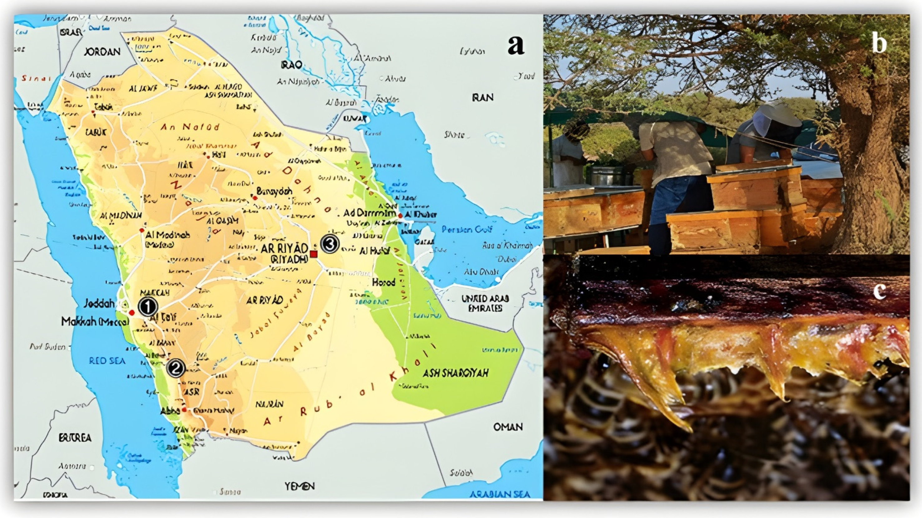
2.1. Propolis Sample Collection and Preparation

In Saudi Arabia, honeybee propolis samples were collected from three different sites: Baljurashi, Baha Province 19.8568154° N, 41.5793896° E (Arabian honeybee Apis mellifera jemenitica); Al Hada, Makkah Province 21°22′07″ N, 40°17′05″ E (Italian honeybee Apis mellifera ligustica); and Rawdat Khuraim, Riyadh Province 25°23′20″ N, 47°17′37″ E (Carniolan honeybee Apis mellifera carnica) (Figure 1). Samples were collected from March 2017–April 2018. Propolis samples were collected monthly from each site by scraping the most two highly active hives.
The active compounds present in the propolis samples were extracted by rinsing the propolis pieces in methanol for 48 h, followed by separation of the methanolic extract from the residue by filtration [21]. The extract was then left to dry, and a part of each extraction was used for chemical analysis. Finally, the powder propolis extract was dissolved in dimethyl sulfoxide (DMSO).

2.2. Cytotoxicity Assay

2.2.1. Cell Lines

The crude propolis extract was evaluated for its anti-cancer activity against the human breast cancer (MCF-7) cell line and human liver cancer cell line (HeP-G2), which was preserved in Dulbecco’s modified Eagle medium (DMEM) supplemented with penicillin-streptomycin (2%). The cancer cell lines were cultured at 37 °C and 95% RH.

2.2.2. Cell Viability

The microculture tetrazolium technique (MTT) was used to determine cell viability. Cell lines at a concentration of (1 × 10⁵ cells/mL) were cultured for 24 h in a 96-well plate. The wells were treated with 100 μL of 5 mg/mL MTT and incubated at 37 °C for another 4 h, then the medium was removed after incubation. Propolis extracts were first dissolved in DMSO (100 µL) before adding to the cell lines. Two types of control were used during the cytotoxicity evaluation, a positive control using cisplatin dissolved in DMSO and a negative control using DMSO only. The plates were incubated in the dark, and the absorption of the purple solution that resulted was measured at 540 nm. The purple solution was easily measured quantitatively using an ELISA plate reader [22,23]. Agilent BioTek Synergy Neo2 laser absorbance reader (Santa Clara, CA, USA) was used in analyzing the ELISA plate.

2.2.3. Statistical Analysis

Results from the current study were analyzed using SPSS program version 16 and presented as the mean S.E.M. Quantitative variables were analyzed using a one-way analysis of variance (ANOVA)
The cytotoxic activity of the propolis samples from various isolation sites was determined by calculating the ratio of viable cells in the tested wells to the negative control.

2.3. Methanolic Propolis Extract Analysis by HPLC

The sample to be chromatographed can either be introduced as a solution or preabsorbed on the packing material. The latter method has been found to be superior, especially for plant extracts, which are often not completely soluble in the initial eluent. The sample is preabsorbed by dissolving it in a suitable low boiling point solvent (e.g., dichloromethane) and silica gel was added (about 1–2 g for every 1 g of sample). The solvent was then removed on a rotary evaporator until the mixture was dry and could move freely in the flask. The last trace of solvent was removed by placing the flask under a high vacuum for 15–30 min. It is important to obtain a completely dry and freely flowing mixture. Depending on the nature of the sample, it may be necessary to increase the proportion of silica gel to sample [24].
LC/MS was carried out using a Dionex 3000 UHPLC pump coupled to an QExactive (Orbitrap) mass spectrometer, Thermo Fisher Scientific (Bremen, Germany). Crude samples and purified compounds were prepared at 1 mg/mL in methanol prior to LC–MS. A reverse-phase 5 µm C18 column (4.6  ×  150 mm) (Hypersil, Thermo) was used, and the elution was carried out using a gradient at a flow rate of 0.3 mL/min, with 0.1% v/v formic acid in water and 0.1% v/v formic acid in acetonitrile (the A and B solvents) making up the mobile phase. The ESI interface in negative ionization permitted the identification of [M-H]-. The spray voltage for the capillary and cone were −4.0 kV and 35 V, respectively. The flow rate of the sheath gas and auxiliary gas were 50 and 15 arbitrary units, respectively. The ion transfer capillary had a temperature of 275 °C, and m/z between 100 and 1500 provided the full scan data. The sample data were acquired and processed with Xcalibur software (Version 4.3, Thermo Fisher Corporation, Hemel Hempstead, UK) [25].

2.4. Molecular Docking of the Propolis Extracted Compounds

To explain the effect of the target compounds from the honeybee propolis samples on the topoisomerase IIβ (TOP IIβ) enzyme compared with the etoposide as a reference control and the experimental reference cisplatin, a docking study was carried out using AutoDock 4.2 [26]. Docked structures were analyzed for the amino acids involved in the ligand binding sites of the enzyme along with the type of interaction such as hydrogen bonding, etc. involved in the docking. This was performed using the Discovery Studio v. 4.5 Visualizer (http://accelrys.com/products/discovery-studio, accessed on 20 May 2022) [27]. For drawing the ligands’ interaction with topoisomerase IIβ (TOP IIβ) enzyme, the Chemdraw 20.0 (CambridgeSoft) (Perkin Inc., Cambridge, MA, USA) program was used with the wave function spartan v 14.0 program to minimize energy. From the protein data bank (PDB) website (http://www.rcsb.org/pdb, accessed on 20 May 2022), the X-ray crystal structure of TOP IIβ with the ligand molecule etoposide (3QX3) was obtained.

3. Results

3.1. Cytotoxicity Assay

Figure 2 and Table 1 show the results of the cytotoxicity assay of the crude propolis samples collected from the three isolation sites in Saudi Arabia together with the positive and negative control. The results demonstrated that the propolis samples collected from the three isolation sites had an inhibitory effect on the human breast cancer cell line (MCF-7) and human liver cancer cell lines (HeP-G2) compared with cisplatin (positive control). The highest inhibitory activity was applied by the propolis samples isolated from the Rawdat Khuraim site, reaching 81.5% and 83.2%, followed by samples from the Al Hada site reaching 81.3% and 79.2% against MCF-7 and HeP-G2, respectively. The lowest anti-cancer activity was from the propolis sample of the Baljurashi site at 48.7% and 59.1% against MCF-7 and HeP-G2, respectively.

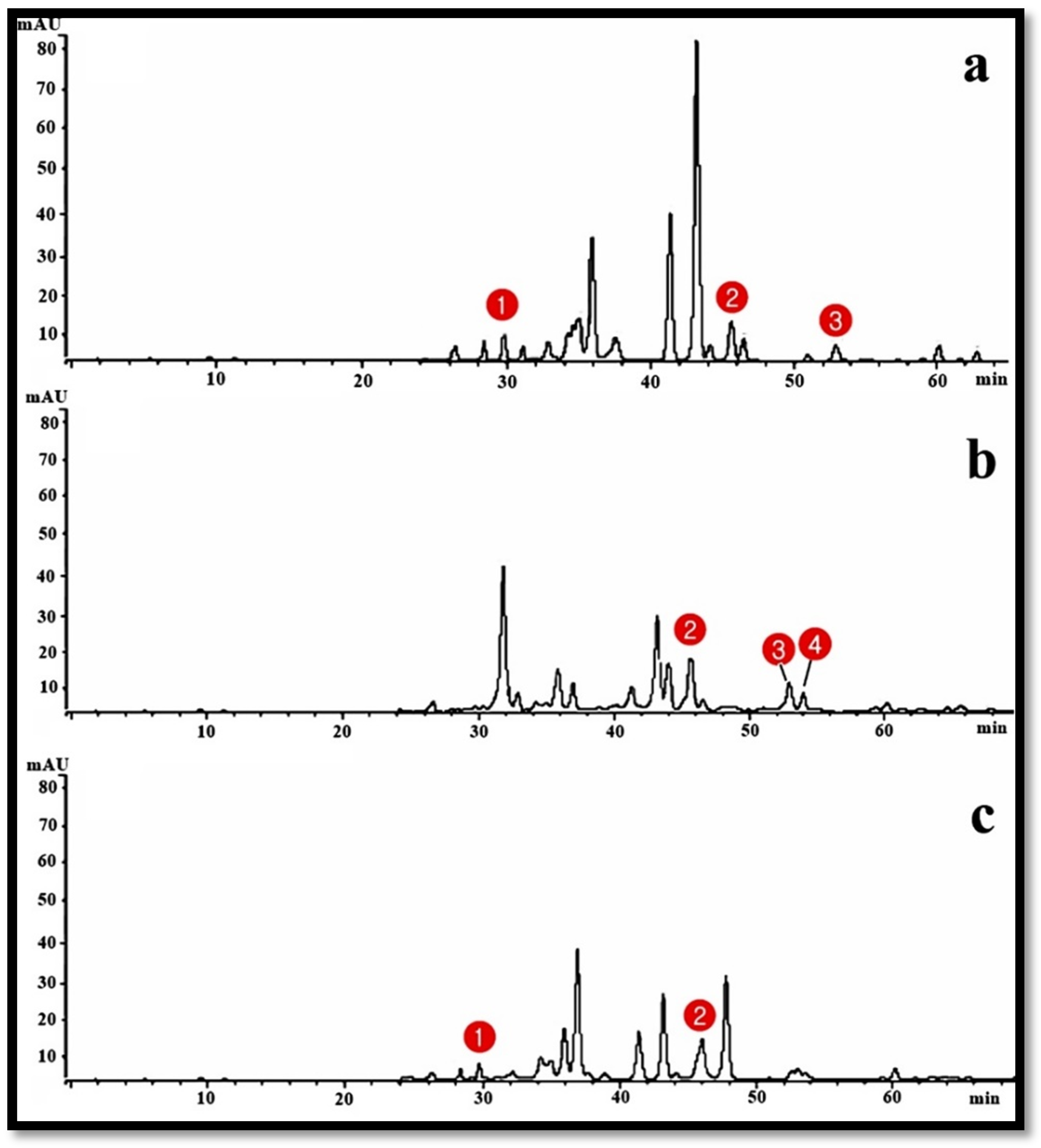
3.2. HPLC Analysis of Propolis Methanolic Extracts

Four active compounds were identified by analyzing the propolis methanolic extracts using HPLC; galangin, xanthomicrol, pinobanksin, and pinocembrin (Table 2) and Table S1. Pinobanksin was found in the propolis extract gathered from Al Hada and Baljurashi isolation sites with a retention time equal to 30 min. Pinocembrin was observed in the extract of propolis isolated from the three different locations of this study with a retention time equivalent to 46 min. Galangin was found in the propolis extract from the Rawdat Khuraim isolation site only, with a retention time equal to 54 min. Xanthomicrol was detected in the extract of propolis isolated from the Al Hada and Rawdat Khuraim isolation sites with a retention time equivalent to 53 min. The analysis of the propolis samples by HPLC showing the four active compounds peaks is shown in Figure 3.

3.3. Molecular Docking of the Propolis Extracted Compounds

Using the control (etoposide) as the reference TOP IIβ inhibitor, the molecular docking simulation studies revealed that the key residues in the major hinge region were Asp479 and Arg503. Three π-interactions with different residues including Arg503 with the aromatic ring of the tetracyclic structure were observed from the etoposide lead binding mode, while there was a H- bonding with Asp479 in the groove of the TOP IIβ binding site formed by phenolic oxygen in the para position of the benzene ring, while the extended chains are solvated and occupy a hydrophobic pocket (Figure 4). The propolis compounds, which were used as ligands, compared to the reference docking with the etoposide, are shown in Figure 5.
As shown in Table 3, the binding mode of etoposide was matched with the propolis compounds. Galangin formed one hydrogen bond with Asp 479 and two pi-alkyl bonds with Arg 503 with a binding energy (−6.25) Kcal/mol as Asp479 and Arg503 are the crucial amino acids as in the reference (epositode) docked, which gave a binding energy of (−6.43) Kcal/mol, and xanthomicrol has one hydrogen bond with one crucial amino acid Arg503. Pinocembrin formed two hydrogen bonds with topoisomerase IIβ Asp 479 and pi alkyl bonds with Arg503 and its binding energy (−7.06) Kcal/mol. Pinocembrin and galangin were the most stable and active compounds in the propolis extract as anti-cancer agents as they had more bonds (hydrogen and other bonds) with crucial amino acids. Pinocembrin had a higher binding energy than the reference (etoposide) while galangin had lower binding energy than pinocembrin. Xanthomicrol was inactive due to fewer hydrogen bonds with crucial amino acids and pinobanksin had a hydrogen bond with noncrucial amino acids (Figure 6). We also compared them with the experimental reference cisplatin, which interacted with three hydrogen bonds and one salt bridge but with non-crucial amino acids Gly793, Gly868, and Glu870 with (−2.46) Kcal/mol with a weaker binding capability (Figure 7).

4. Discussion

Natural cures derived from microorganisms, plants, and animal products have been used in medicine for centuries. They are a diverse group of compounds that have been developed to interact precisely with biological targets and continue to give new and inspirational models for pharmaceutical drug development [26]. Propolis is one of the most fascinating bee products, without a doubt, a vital factor in the success of the important macro-organism of the beehive, and its chemical intricacies make understanding the content and percentage consistency as well as the associated biological activity challenging [27]. For centuries, propolis has been an essential component of apitherapy, and it is now being utilized as a dietary addition or supplement under the umbrella of traditional and alternative medicine [28]. The chemical makeup of propolis varies greatly depending on the geographic area from which it is gathered. Propolis from temperate regions of the world, for example, is high in phenolic compounds produced from poplar tree exudates, but propolis from tropical countries is high in other phytochemicals such as benzophenones and prenylated flavonoids, terpenoids, lignans, and phenolic lipids [29,30].
The extraction solvent affects the biological activity and chemical composition of propolis extracts. The major solvents used to extract propolis for commercial applications are ethanol, glycerol, ether, and water; additional solvents are also available. Ethanol is typically used to produce low wax propolis extracts that are high in physiologically active chemicals [7]. Propolis is added to pharmaceutical and health care products in the form of ethanolic and aqueous extracts [31]. Because of its widespread usage in modern herbal medicine, the chemical makeup of propolis has piqued attention. Different propolis extracts from various sources have been demonstrated to exhibit antibacterial, cytotoxic, anti-inflammatory, immunomodulatory, antiparasitic, and anti-leishmanicidal characteristics in studies from various nations [7].
Cancer is one of the most common diseases facing humans, accounting for one out of every six deaths [32]. Anti-cancer medications that control and/or limit cancer cell proliferation have been the subject of several investigations. The anti-cancer activity of a fraction, extract, or molecule is first examined in vitro using cancer cell lines that have been exposed to the material under research. In diverse cell lines, propolis from several species of stingless bees from throughout the world has shown considerable anti-cancer activity [33]. Breast cancer is one of the most frequent cancers in women, accounting for about half a million deaths each year. In addition, the risk of acquiring breast cancer has been rising in recent years. Breast cancer incidence is much lower among Asian and African women than among Hispanic women, according to statistical and epidemiological studies. Furthermore, due to hormonal involvement, breast cancer incidence rates are increasing in postmenopausal women compared to young women. The chemoprotective efficacy of propolis and its active components was investigated using a variety of breast cancer cell lines (MDA-MB-231, BT-474, MCF-7, and T47D-estrogen receptor) [34]. The results of this study ensured the potency of propolis samples collected from the gathering sites in Saudi Arabia in inhibiting the growth of the breast cancer cell line (MCF-7) and human liver cancer cell line (HeP-G2). The maximum inhibitory activity was accomplished by the propolis collected from Rawdat Khuraim of 81.5% and 83.2% against MCF-7 and HeP-G2, respectively. The cancer cell lines HeLa, MCF-7, and Caco-2 were investigated in [35]. MCF-7 is a human breast cancer cell line that has been extensively studied [36]. Several natural products including stingless bee products (honey, bee pollen, and propolis) have been proven to suppress the proliferation of those cancer cell lines [37]. The cytotoxicity impact of extracts could be linked to a group of chemicals in it. The extract of stingless bee Homotrigona fimbriata contained flavonoid, tannin, and coumarin, among other phytochemicals [38]. The Tetragonula incisa stingless bee propolis extract was tested for cytotoxicity against the human liver (HepG2), colon (SW620), gastric (KATO-III), breast (BT474), and lung (Chago) cancer cell lines [39]. The MTT test is the most widely used method to determine a cell’s metabolic activity and cytotoxic activity [40,41].
High-performance thin-layer chromatography (HPTLC), high-performance liquid chromatography (HPLC), gas chromatography–mass spectrometry (GC–MS), and micellar electrokinetic capillary chromatography (MEKC) have all been used to examine propolis components [42]. More than 180 chemicals, mostly polyphenols, have been discovered as propolis constituents. Flavonoids are the most abundant polyphenols, followed by phenolic acids and esters, ketones, phenolic aldehydes, and other compounds [15]. Chromatographic procedures, particularly HPLC, are used to separate and quantify the specific constituent compounds of the phenolic profile [42]. Four active compounds have been detected from the HPLC analysis of propolis methanolic extracts from Al Hada, Rawdat Khuraim, and Baljurashi isolation sites: xanthomicrol, galangin, pinobanksin, and pinocembrin. The source of galangin could be from the propolis samples collected from the beehives on Helichrysum glumaceum or Helichrysum splendidum plants. The source of the pinocembrin compound can be from the propolis collected from hives on Euphorbia spp. as cactus plants are usually common in Saudi Arabia’s flora while the pinobanksin source may be Helianthus annuus or related plants. Polyphenolic agents such as gallic acid, pinobanksin, caffeic acid, chrysin, protocatechuic acid, rutin, quercetin, galangin, kaempferol, hesperetin, pinocembrin, apigenin, daidzein, luteolin, and caffeic acid phenyl ester (CAPE) have been reported to become the most active phenolic substances in propolis samples [43]. Pinocembrin was detected in 17 samples in concentrations of 0.0664 mg/g to 7.6651 mg/g in a study by Barbarić et al. [44]. Another study also detected pinocembrin on 375 nm by HPLC analysis [42].
The activity of topoisomerase I and II determines the topological states of DNA replication, recombination, transcription, and all biological processes rely on their activity [45]. Anti-cancer medications block the activity of the DNA Topo2 enzyme, which is thought to be the cause of cell death. Even though these drugs have some negative effects, they are nonetheless given as anti-cancer medications. These medicines may influence Topo2b in human cells [45]. Type I topoisomerases (Topo I) create single strand breaks in DNA in an ATP-dependent manner, whereas type II topoisomerases (Topo II) do so by forming double-strand breaks in DNA. Topo II is a well-known anti-cancer target, and Topo II is the target of some of the most potent anti-cancer drugs now on the market. However, topo II chemotherapy (treatment with doxorubicin, etoposide, and analogues) is linked to harmful side effects and subsequent cancers [46]. Topoisomerases are present in different types of cancer cells as in Hep-G2 (liver cancer). Many drug design studies have effectively used a combination of structure- and ligand-based computational approaches [47]. Molecular docking and dynamics are powerful computational tools for discovering new medicine, and as a result, have real-world applicability in drug development [48]. Molecular docking simulation studies on propolis active compounds of this study were evaluated as topoisomerase IIβ inhibitors compared with the control anti-cancer compound etoposide and revealed that galangin and pinocembrin were the most stable and active compounds in propolis samples as the number of hydrogen bonds and other bonds formed with the topoisomerase IIβ enzyme was higher than the two other compounds (pinobanksin and xanthomicrol) and so ensure greater anti-cancer activity.

5. Conclusions

Propolis produced by many species of honeybees is one of the most important substances with many biological activities. Propolis also has an anti-cancer effect. The cytotoxic activity of propolis samples collected from three different locations in Saudi Arabia was studied against human breast cancer cell lines (MCF-7) and human liver cancer cell lines (Hep-G2). The composition of the methanolic propolis extracts was determined by HPLC analysis and revealed the presence of four active compounds (pinobanksin, pinocembrin, galangin, and xanthomicrol). Through the molecular docking technique, the galagnin and pinocembrin compound showed a higher binding affinity with the active sites of topoisomerase IIβ enzyme than xanthomicrol and pinobanksin. It can be concluded that galagnin and pinocembrin from honeybee propolis collected from Saudi Arabia can be further investigated in clinical trials as potential anti-cancer agents.

Supplementary Materials

The following supporting information can be downloaded at: https://www.mdpi.com/article/10.3390/separations9120392/s1. Table S1: the GC mass of propolis extracts.

Author Contributions

Conceptualization, A.A.A.-K., I.A., S.S.A.-R., M.M. and W.N.H.; Methodology, A.A.A.-K., I.A., S.S.A.-R., M.M. and W.N.H.; Software, A.A.A.-K., I.A., S.S.A.-R., M.M. and W.N.H.; Validation, A.A.A.-K., I.A., S.S.A.-R., M.M. and W.N.H.; Formal analysis, A.A.A.-K., I.A., S.S.A.-R., M.M. and W.N.H.; Investigation, A.A.A.-K., I.A., S.S.A.-R., M.M. and W.N.H.; Resources A.A.A.-K., I.A., S.S.A.-R., M.M. and W.N.H.; Data curation, A.A.A.-K., I.A., S.S.A.-R., M.M. and W.N.H.; Writing—original draft preparation, A.A.A.-K. and W.N.H.; Writing—review and editing, A.A.A.-K., I.A., S.S.A.-R., M.M. and W.N.H.; Visualization. All authors have read and agreed to the published version of the manuscript.

Funding

This research was funded by the Health Sciences Research Center, King Abdullah bin Abdulaziz Hospital, Princess Nourah bint Abdulrahman University, through the research funding program [G18-00019].

Data Availability Statement

All data related to the manuscript are available in the manuscript and the supplementary material.

Acknowledgments

We want to extend our thanks to the Health Sciences Research Center, King Abdullah bin Abdulaziz Hospital, Princess Nourah bint Abdulrahman University, through the research funding program.

Conflicts of Interest

The authors declare no conflict of interest.

References

  1. Campos, J.F.; Dos Santos, H.F.; Bonamigo, T.; de Campos Domingues, N.L.; de Picoli Souza, K.; Dos Santos, E.L. Stingless Bee Propolis: New Insights for Anticancer Drugs. Oxidative Med. Cell. Longev. 2021, 2021, 2169017. [Google Scholar] [CrossRef] [PubMed]
  2. Kuropatnicki, A.K.; Szliszka, E.; Krol, W. Historical Aspects of Propolis Research in Modern Times. Evid. -Based Complement. Altern. Med. 2013, 2013, 964149. [Google Scholar] [CrossRef] [PubMed] [Green Version]
  3. Righi, A.A.; Alves, T.R.; Negri, G.; Marques, L.M.; Breyer, H.; Salatino, A. Brazilian red propolis: Unreported substances, antioxidant and antimicrobial activities. J. Sci. Food Agric. 2010, 91, 2363–2370. [Google Scholar] [CrossRef] [PubMed]
  4. Ghisalberti, E.L. Propolis—Review. BeeWorld 1979, 60, 59–84. [Google Scholar] [CrossRef]
  5. Coggshall, W.L.; Morse, R.A. Beeswax: Production, Harvesting, Processing and Products; Wicwas Press: Ithaca, NY, USA, 1984. [Google Scholar]
  6. Huang, S.; Zhang, C.P.; Wang, K.; Li, G.Q.; Hu, F.L. Recent advances in the chemical composition of propolis. Molecules 2014, 19, 19610–19632. [Google Scholar] [CrossRef] [Green Version]
  7. Ibrahim, M.F.M.; El-Samad, G.A.; Ashour, H.; El-Sawy, A.M.; Hikal, M.; Elkelish, A.; El-Gawad, H.A.; El-Yazied, A.A.; Hozzein, W.N.; Farag, R. Regulation of Agronomic Traits, Nutrient Uptake, Osmolytes and Antioxidants of Maize as Influenced by Exogenous Potassium Silicate under Deficit Irrigation and Semiarid Conditions. Agronomy 2020, 10, 1212. [Google Scholar] [CrossRef]
  8. Silici, S.; Kutluca, S. Chemical composition and antibacterial activity of propolis collected by three different races of honeybees in the same region. J. Ethnopharmacol. 2005, 99, 69–73. [Google Scholar] [CrossRef] [PubMed]
  9. Stan, L.; Liviu, A.; Dezmirean, D. Quality Criteria for Propolis Standardization. Sci. Pap. Anim. Sci. Biotechnol. 2011, 44, 137–140. [Google Scholar]
  10. Katekhaye, S.; Fearnley, H.; Fearnley, J.; Paradkar, A. Gaps in propolis research: Challenges posed to commercialization and the need for a holistic approach. J. Apic. Res. 2019, 58, 604–616. [Google Scholar] [CrossRef]
  11. Abou-Shaara, H.; Alashaal, S.A.; Hosni, E.M.; Nasser, M.G.; Ansari, M.J.; Alharbi, S.A. Modeling the Invasion of the Large Hive Beetle, Oplostomus fuligineus, into North Africa and South Europe under a Changing Climate. Insects 2021, 12, 275. [Google Scholar] [CrossRef] [PubMed]
  12. Hosni, E.M.; Nasser, M.; Al-Khalaf, A.A.; Al-Shammery, K.A.; Al-Ashaal, S.; Soliman, D. Invasion of the Land of Samurai: Potential Spread of Old-World Screwworm to Japan under Climate Change. Diversity 2022, 14, 99. [Google Scholar] [CrossRef]
  13. Hosni, E.M.; Al-Khalaf, A.A.; Nasser, M.G.; Abou-Shaara, H.F.; Radwan, M.H. Modeling the Potential Global Distribution of Honeybee Pest, Galleria mellonella under Changing Climate. Insects 2022, 13, 484. [Google Scholar] [CrossRef] [PubMed]
  14. Hosni, E.M.; Nasser, M.G.; Al-Ashaal, S.A.; Rady, M.H.; Kenawy, M.A. Modeling current and future global distribution of Chrysomya bezziana under changing climate. Sci. Rep. 2020, 10, 4947. [Google Scholar] [CrossRef] [PubMed] [Green Version]
  15. Barbarić, M.; Mišković, K.; Bojić, M.; Lončar, M.B.; Smolčić-Bubalo, A.; Debeljak, Ž.; Medić-Šarić, M. Chemical composition of the ethanolic propolis extracts and its effect on HeLa cells. J. Ethnopharmacol. 2011, 135, 772–778. [Google Scholar] [CrossRef]
  16. World Health Organization. Cancer: Key Facts. 2018. Available online: https://www.who.int/newsroom/fact-sheets/detail/cancer (accessed on 1 November 2020).
  17. International Agency for Research on Cancer. Cancer Fact Sheets. World Health Organization, 2018. Available online: https://gco.iarc.fr/today/fact-sheets-cancers (accessed on 1 November 2020).
  18. International Agency for Research on Cancer. Indonesia Global Cancer Observatory. World Health Organization, 2018. Available online: https://gco.iarc.fr/today/data/factsheets/populations/360-indonesia-fact-sheets.pdf (accessed on 1 November 2020).
  19. Rohs, R.; Bloch, I.; Sklenar, H.; Shakked, Z. Molecular flexibility in ab-initio drug docking to DNA: Binding-site and binding-mode transitions in all-atom Monte Carlo simulations. Nucleic Acids Res. 2005, 33, 7048–7057. [Google Scholar] [CrossRef] [Green Version]
  20. Guedes, I.A.; de Magalhães, C.S.; Dardenne, L.E. Receptor-ligand molecular docking. Biophys. Rev. 2014, 6, 75–87. [Google Scholar] [CrossRef] [PubMed] [Green Version]
  21. Escriche, I.; Juan-Borrás, M. Standardizing the analysis of phenolic profile in propolis. Food Res. Int. 2018, 106, 834–841. [Google Scholar] [CrossRef]
  22. Hussain, R.F.; Nouri, A.M.; Oliver, R.T. A new approach for measurement of cytotoxicity using colorimetric assay. J. Immunol. Methods 1993, 160, 89–96. [Google Scholar] [CrossRef] [PubMed]
  23. Mosmann, T. Rapid colorimetric assay for cellular growth and survival: Application to proliferation and cytotoxicity assays. J. Immunol. Methods 1983, 65, 55–63. [Google Scholar] [CrossRef] [PubMed]
  24. Claeson, P.; Tuchinda, P.; Reutrakuv, V. Some empirical aspects on the practical use of flash chromatography and medium pressure uquid chromatography for the isolation of biologically active compounds from plants. ScienceAsia 1993, 19, 073. [Google Scholar] [CrossRef]
  25. Jabborova, D.; Annapurna, K.; Paul, S.; Kumar, S.; Saad, H.A.; Desouky, S.; Ibrahim, M.F.M.; Elkelish, A. Beneficial Features of Biochar and Arbuscular Mycorrhiza for Improving Spinach Plant Growth, Root Morphological Traits, Physiological Properties, and Soil Enzymatic Activities. J. Fungi 2021, 7, 571. [Google Scholar] [CrossRef]
  26. Newman, D.J.; Cragg, G.M. Natural Products as sources of new drugs from 1981 to 2014. J. Nat. Prod. 2016, 79, 629–661. [Google Scholar] [CrossRef] [PubMed] [Green Version]
  27. Pavlovic, R.; Borgonovo, G.; Leoni, V.; Giupponi, L.; Ceciliani, G.; Sala, S.; Bassoli, A.; Giorgi, A. Effectiveness of Different Analytical Methods for the Characterization of Propolis: A Case of Study in Northern Italy. Molecules 2020, 25, 504. [Google Scholar] [CrossRef] [Green Version]
  28. Anjum, S.I.; Ullah, A.; Khan, K.A.; Attaullah, M.; Khan, H.; Ali, H.; Bashir, M.A.; Tahir, M.; Ansari, M.J.; Ghramh, H.A.; et al. Composition and functional properties of propolis (bee glue): A review. Saudi J. Biol. Sci. 2019, 26, 1695–1703. [Google Scholar] [CrossRef] [PubMed]
  29. Raghukumar, R.; Vali, L.; Watson, D.; Fearnley, J.; Seidel, V. Antimethicillin-resistant Staphylococcus aureus (MRSA) activity of ‘pacific propolis’ and isolated prenylflavanones. Phytother. Res. 2010, 24, 1181–1187. [Google Scholar] [CrossRef] [PubMed]
  30. Kardar, M.N.; Zhang, T.; Coxon, G.D.; Watson, D.G.; Fearnley, J.; Seidel, V. Characterisation of triterpenes and new phenolic lipids in Cameroonian propolis. Phytochemistry 2014, 106, 156–163. [Google Scholar] [CrossRef] [PubMed] [Green Version]
  31. Galeotti, F.; Maccari, F.; Fachini, A.; Volpi, N. Chemical composition and antioxidant activity of propolis prepared in different forms and in different solvents useful for finished products. Foods 2018, 7, 41. [Google Scholar] [CrossRef] [PubMed] [Green Version]
  32. WHO. Cancer; World Health Organization: Geneva, Switzerland, 2020. Available online: https://www.who.int/europe/publications/i/item/WHO-EURO-2020-1628-41379-56382 (accessed on 1 November 2022).
  33. Soliman, M.H.; Abdulmajeed, A.M.; Alhaithloul, H.; Alharbi, B.M.; El-Esawi, M.A.; Hasanuzzaman, M.; Elkelish, A. Saponin Biopriming Positively Stimulates Antioxidants Defense, Osmolytes Metabolism and Ionic Status to Confer Salt Stress Tolerance in Soybean. Acta Physiol. Plant. 2020, 42, 114. [Google Scholar] [CrossRef]
  34. Chiu, H.; Han, Y.; Shen, Y.; Golovinskaia, O.; Venkatakrishnan, K.; Wang, C. Chemopreventive and Chemotherapeutic Effect of Propolis and Its Constituents:A Mini-review. J. Cancer Prev. 2020, 25, 70–78. [Google Scholar] [CrossRef] [PubMed]
  35. Arung, E.T.; Ramadhan, R.; Khairunnisa, B.; Amen, Y.; Matsumoto, M.; Nagata, M.; Kusuma, I.W.; Paramita, S.; Sukemi; Yadi; et al. Cytotoxicity effect of honey, bee pollen, and propolis from seven stingless bees in some cancer cell lines. Saudi J. Biol. Sci. 2021, 28, 7182–7189. [Google Scholar] [CrossRef]
  36. Lee, A.V.; Oesterreich, S.; Davidson, N.E. MCF-7 cells changing the course of breast cancer research and care for 45 years. J. Natl. Cancer Inst. 2015, 107, djv073. [Google Scholar] [CrossRef] [PubMed]
  37. Kuppusamy, P.; Yusoff, M.M.; Maniam, G.P.; Ichwan, S.J.A.; Soundharrajan, I.; Govindan, N. Nutraceuticals as potential therapeutic agents for coloncancer: A review. Acta Pharm. Sin. 2014, 4, 173–181. [Google Scholar] [CrossRef] [PubMed] [Green Version]
  38. Saleh, A.A.H.; Abdel-Kader, D.Z.; El Elish, A.M. Role of Heat Shock and Salicylic Acid in Antioxidant Homeostasis in Mungbean (Vigna radiata L.) Plant Subjected to Heat Stress. Am. J. Plant Physiol. 2007, 2, 344–355. [Google Scholar] [CrossRef] [Green Version]
  39. Kustiawan, P.M.; Phuwapraisirisan, P.; Puthong, S.; Palaga, T.; Arung, E.T.; Chanchao, C. Propolis from the stingless bee Trigona incisa from East Kalimantan, Indonesia, induces in vitro cytotoxicity and apoptosis in cancer cell lines. Asian Pac. J. Cancer Prev. 2015, 16, 6581–6589. [Google Scholar] [CrossRef] [Green Version]
  40. Nikzad, S.; Baradaran-Ghahfarokhi, M.; Nasri, P. Dose-response modeling using MTT assay: A short review. Life Sci. J. 2014, 11, 432–437. [Google Scholar]
  41. Adan, A.; Kiraz, Y.; Baran, Y. Cell proliferation and cytotoxicity assays. Curr. Pharm. Biotechnol. 2016, 17, 1213–1221. [Google Scholar] [CrossRef] [PubMed]
  42. Ismail, M.A.; Amin, M.A.; Eid, A.M.; Hassan, S.E.-D.; Mahgoub, H.A.M.; Lashin, I.; Abdelwahab, A.T.; Azab, E.; Gobouri, A.A.; Elkelish, A.; et al. Comparative Study between Exogenously Applied Plant Growth Hormones versus Metabolites of Microbial Endophytes as Plant Growth-Promoting for Phaseolus vulgaris L. Cells 2021, 10, 1059. [Google Scholar] [CrossRef]
  43. Al-Hatamleh, M.A.I.; Boer, J.C.; Wilson, K.L.; Plebanski, M.; Mohamud, R.; Mustafa, M.Z. Antioxidant-based medicinal properties of stingless bee products: Recent progress and future directions. Biomolecules 2020, 10, 923. [Google Scholar] [CrossRef]
  44. Alnusairi, G.S.H.; Mazrou, Y.S.A.; Qari, S.H.; Elkelish, A.A.; Soliman, M.H.; Eweis, M.; Abdelaal, K.; El-Samad, G.A.; Ibrahim, M.F.M.; Elnahhas, N. Exogenous Nitric Oxide Reinforces Photosynthetic Efficiency, Osmolyte, Mineral Uptake, Antioxidant, Expression of Stress-Responsive Genes and Ameliorates the Effects of Salinity Stress in Wheat. Plants 2021, 10, 1693. [Google Scholar] [CrossRef]
  45. Jadhav, A.K.; Karuppayil, S.M. Molecular docking studies on thirteen fluoroquinolines with human topoisomerase II a and b. Silico Pharmacol. 2017, 5, 4. [Google Scholar] [CrossRef] [Green Version]
  46. Ayyamperumal, S.; Dhananjay, D.J.; Tallapaneni, V.; Mohan, S.; Basappa, S.; Selvaraj, J.; Joghee, N.M.; Chandrasekar, M.J. Molecular docking analysis of α-Topoisomerase II with δ-Carboline derivatives as potential anti-cancer agents. Bioinformation 2021, 17, 249–265. [Google Scholar] [CrossRef] [PubMed]
  47. Drwal, M.N.; Agama, K.; Pommier, Y.; Griffith, R. Development of purely structure-based pharmacophores for the topoisomerase I-DNA-ligand binding pocket. J. Comput. Aided Mol. Des. 2013, 27, 1037–1049. [Google Scholar] [CrossRef] [PubMed]
  48. Salmaso, V.; Moro, S. Bridging molecular docking to molecular dynamics in exploring ligand-protein recognition process: An overview. Front. Pharmacol. 2018, 9, 923. [Google Scholar] [CrossRef] [PubMed]
Figure 1. (a) Map of the Kingdom of Saudi Arabia showing the sites of the collected honeybee propolis: (1) Al Hada, Makkah; (2) Baljurashi, Baha; and (3) Rawdat Khuraim, Riyadh. (b) Apiculture activities on Rawdat Khuraim, Riyadh. (c) Edge of a traditional hive with the propolis sample, Baljurashi, Baha.
Separations 09 00392 g001
Figure 2. Inhibitory activity of the propolis samples against the human breast cancer (MCF-7) and human liver cancer cell lines (HeP-G2).
Separations 09 00392 g002
Figure 3. The HPLC chromatogram for the honeybee propolis methanolic extract from the two locations in Saudi Arabia: (a) Al Hada, (b) Rawdat Khuraim, (c) Baljurashi; the red spots indicate the peak numbers of compounds 1. Pinobanksin, 2. Pinocembrin, 3. Xanthomicrol, and 4. Galangin.
Separations 09 00392 g003
Figure 4. Reported etoposide-binding mode in the active site of the TOP IIβ enzyme.
Separations 09 00392 g004
Figure 5. Chemical structure of the ligands (propolis active compounds).
Separations 09 00392 g005
Figure 6. (a). Interaction of galangin with the active site of topoisomerase IIβ (TOP IIβ) in (a) 2D and (b) 3D; (b). Interaction of xanthomicrol with the active site of topoisomerase IIβ (TOP IIβ) in (a) 2D and (b) 3D; (c). Interaction of pinocembrin with the active site of topoisomerase IIβ (TOP IIβ) in (a) 2D and (b) 3D; (d). Interaction of pinobanksin with the active site of topoisomerase IIβ (TOP IIβ) in (b) 2D and (c) 3D.
Separations 09 00392 g006
Figure 7. Interaction of cisplatin (experimentally) with the active site of topoisomerase IIβ (TOP2B) in 3D.
Separations 09 00392 g007
Table 1. The cytotoxicity assay of the propolis sample on the human breast cancer (MCF-7) cell line and human liver cancer cell line (HeP-G2).
Treatment% Inhibitory Activity on MCF-7% Inhibitory Activity on HeP-G2
Cisplatin (positive control)86.286.2
DMSO (negative control)00
Propolis SampleAl Hada site81.379.2
Baljurashi site48.759.1
Rawdat Khuraim site81.583.2
Table 2. The HPLC analysis showing the active compounds of the propolis extract from three isolation sites in Saudi Arabia.
Peak No.Retention TimeCompoundsPropolis Samples
Al HadaRawdat KhuraimBaljurashi
130Pinobanksin+-+
246Pinocembrin+++
353Xanthomicrol++-
454Galangin-+-
Table 3. Molecular interactions of the propolis active compounds with topoisomerase IIβ.
Active Compounds in Propolis SamplesBinding EnergyInhibition Constant (ki)Amino Acids Involved in the Bond with the LigandNumber of Hydrogen Bonds
Pinocembrin−7.066.67Asp 479–Arg 503Formed (2)
Pinobanksin−6.7411.47Arg 503Formed with non-amino acids
Galangin−6.2526.23Asp479–Arg503Formed (2)
Xanthomicrol−6.3422.35Lys486–Arg503Formed (only one with crucial amino acids)
Control (ref original cpd. Docking only) (etoposide)−6.4319. 27Asp 479–Arg 503Formed (2)
Cisplatin (ref. experimentally)−2.4615.7Gly793-Gly868Formed with non-crucial amino acids
Publisher’s Note: MDPI stays neutral with regard to jurisdictional claims in published maps and institutional affiliations.

