Article

Towards a Better Quantification of Cyanotoxins in Fruits and Vegetables: Validation and Application of an UHPLC-MS/MS-Based Method on Belgian Products

by
Wannes Hugo R. Van Hassel
1,2,3,*,
Julien Masquelier
1,
Mirjana Andjelkovic
4 and
Andreja Rajkovic
3
1
Unit Toxins, Organic Contaminants and Additives, Sciensano, Rue Juliette Wytsmanstraat 14, 1050 Brussels, Belgium
2
InBios-Centre for Protein Engineering, Departement of Life Sciences, University of Liège, Allée du six Août 11, 4000 Liège, Belgium
3
Department of Food technology, Food safety and Health, Faculty of Bioscience Engineering, Ghent University, Coupure Links 653, 9000 Gent, Belgium
4
Risk and -Health Impact Assessment, Sciensano, Rue Juliette Wytsmanstraat 14, 1050 Brussels, Belgium
*
Author to whom correspondence should be addressed.
Separations 2022, 9(10), 319; https://doi.org/10.3390/separations9100319
Submission received: 28 September 2022 / Revised: 17 October 2022 / Accepted: 18 October 2022 / Published: 20 October 2022

Abstract

:
Vegetables and fruits can potentially accumulate cyanotoxins after water contaminated with cyanobacteria is used for irrigation. We developed and validated an analytical method to quantify eight microcystin congeners (MCs) and nodularin (NOD) using ultra high-performance liquid chromatography coupled with tandem mass spectrometry (UHPLC-MS/MS) in three different matrices. Strawberries, carrots and lettuce are selected as model matrices to represent the fruits/berries, leafy and root vegetables, sequentially. The validation of a UHPLC-MS/MS method in the strawberry matrix is novel. Matrix effects are observed in all three matrices. Our methodology uses matrix-matched calibration curves to compensate for the matrix effect. The implementation of our method on 103 samples, containing nine different sorts of fruits and vegetables from the Belgian market, showed no presence of MCs or NOD. However, the recoveries of our quality controls showed the effectiveness of our method, illustrating that the use of this method in future research or monitoring as well as in official food controls in fruit and vegetable matrices is valid.

1. Introduction

Cyanotoxins can be produced in eutrophic waterbodies during blooms of photosynthetic, eukaryotic organisms called cyanobacteria [1,2,3,4,5,6,7,8]. Multiple studies and reviews have documented the occurrence of cyanotoxins in different foods [9,10]. The sedentary nature of agriculture allows for the accumulation of these harmful compounds in food crops when vital irrigation water is contaminated with these toxins.
The most common cyanotoxin group, worldwide, is microcystin congeners (MCs). These are hepatotoxins produced by different cyanobacteria species (e.g., Microcystis aeruginosa, Anabaena/Dolichospermum, Planktothrix sp., Oscillatoria and Nostoc) [11]. Nodularin (NOD) has a similar structure to MCs and was initially found in Nodularia sp. [8,12]. A unique structural part of both toxins is the ADDA fragment ((2S,3S,8S,9S)-3-amino-9-methoxy-2,6,8-trimethyl-10-phenyl-4,6-decadienoic acid), which contributes to the interaction with the target proteins [5]. Both toxins inhibit protein phosphatases 1 and 2A (PP1 and PP2A) in eukaryotes after transport in cells, causing cell death. The transport is facilitated by specific organic anion-transporting polypeptides (OATPs) [13,14,15,16]. Cylindrospermopsin (CYN) is another hepatotoxin that inhibits the protein synthesis of the affected cell and, thus, cell growth [17]. This toxin was first found in Australia. CYN is produced by Cylindrospermopsis raciborskii, although other producing cyanobacterial species have been identified [17]. Even though CYN is more commonly reported in warmer climates in the Southern Hemisphere, cases have been found in Europe since its discovery [18].
The effects of the other classes of cyanotoxins, such as neurotoxins, are not well studied in plants. Neurotoxins, such as saxitoxin, are also primarily found in warmer regions [19,20,21], while anatoxin-a is also regularly found in colder regions [6,11,22].
For studying the physiological effects and accumulation of cyanotoxins in plants, two methods are commonly used for the detection and quantification of these toxins. Initially, optimized immunoassays were used to (semi)quantify MCs in plant tissue [23,24,25,26]. Later, more robust and quantitative liquid chromatography (LC) approaches coupled to (tandem) mass spectrometry (MS(/MS)) were used to measure the concentrations of different MCs, CYN and their transformation products in plants [27,28,29,30,31].
The applicability of both the immune assays and the LC-MS(/MS) approaches depends on the goal of the analysis. To determine bound and unbound MCs in plant material, immunoassays might be the best option, as all the available ADDA fragments will be detected [32]. However, immunoassays are known to produce false positive readouts and are not able to identify separate congeners or transformation products [27]. Moreover, insufficient toxicological data are available from a public health point of view to determine the effect and bioavailability of the bound/transformed MCs. Using immunoassays might thus cause an overestimation of the concentration of the bioavailable MCs, resulting in an overestimation of the risk to human health.
Various LC-MS(/MS) approaches have been developed to identify multiple MCs and CYN, separately or concurrently, in plant material [28,30,31]. Usually, multiple reaction monitoring (MRM) is used, which is capable of identifying and quantifying multiple toxins. Yet, due to the selective nature of this method, uncommon congeners or transformation products will not be detected when they are not included in the method a priori. The inclusion of a sufficient number of MCs in a UHPLC-MS/MS method is crucial for accurately assessing the total microcystin concentration and, thus, possible public health risks.
Up till now, different MS methods have been validated. Generally, the methods use 75–80% methanol (MeOH) as an extraction solvent, which in some cases is acidified [27,28,30,31]. Recoveries for MC-LR (56–65%), MC-RR (30–32%), MC-YR (79–81%) and NOD (97–118%) were reported in broccoli roots and stems, separately, using LC-MS selected ion recording (SIR) [27]. Signal enhancement was observed for nearly all toxins [27]. Only MC-LR and MC-YR were affected by signal suppression in the root samples at the highest spiked concentration. Later, a UHPLC-MS/MS method was validated for MC-LR, MC-RR and MC-YR in multiple crop plants (lettuce, turnip, water spinach, potato, cabbage, pumpkin, chio sum, cucumber, carrots and eggplants) [28]. Solid-phase extraction (SPE) was used during the method to purify the extract. A C18 cartridge was used for all crops except for eggplant and cucumber. For the latter, an HLB cartridge had to be used to optimize their recovery. A suppressive matrix effect was observed for all plant materials [28]. Another UHPLC-MS/MS method was validated for MC-LR and MC-RR in lettuce using 50% MeOH in conjunction with an HLB cartridge for the SPE [30]. The UHPLC-MS/MS method was also validated successfully for CYN in lettuce [29]. Furthermore, the quantification of MC-LR, MC-RR, MC-YR and CYN in lettuce was successfully optimized using 80% MeOH extraction, dual (C18 and PGC cartridge) SPE purification and UHPLC-MS/MS. The obtained recoveries were slightly lower compared to the earlier studies, probably due to the inclusion of CYN, which is hydrophilic compared to the more hydrophobic MCs [31]. Clearly, important developments in methods have already occurred. However, most methods still lack multiple chemically diverse microcystin congeners. These MCs could be present in the sample and interact differently with the matrix during analysis. Especially for the more hydrophobic congeners, for example, MC-LF and MC-LW, they are still missing. Other matrices, such as fruits and berries, should be validated separately as different matrix effects could occur.
The plant accumulation of different MCs and CYN through irrigation water has been studied in multiple (crop)plants. Lettuce is probably the best-studied vegetable, often selected based on its relevance in the human diet and its large leaf surface. The presence of MCs was initially detected with an immunoassay in lettuce after spray irrigation with Microcystis aeruginosa containing water [23]. The central leaves significantly retained higher concentrations compared to leaves from the basal or distal zones. Lettuce shots were later also shown to accumulate MCs [33]. The accumulation of MCs in the roots and leaves of lettuce was also shown after irrigation with naturally contaminated water [34,35,36]. The bioaccumulation of different MCs has also been linked to the initial exposure concentrations in lettuce, showing higher accumulation with higher MC concentrations in the water [37,38], while depuration of 75% of the initially accumulated MC-LR was reported for lettuce after seven days [38]. Similar experiments were conducted for CYN, showing accumulation in lettuce [29,39]. Decreasing depuration was observed when the CYN exposure concentration was increased [39]. The combined accumulation of CYN and MCs in lettuce and spinach was shown when these plants were exposed to irrigation water containing both toxins. Only CYN was detected in the leaves, while both MCs and CYN were found in the roots [40]. The accumulation of MCs in root vegetables such as rape, carrots and radishes was also shown and was dependent on the exposure concentration [24,33,41,42]. For rice, the MCs’ accumulation was found in the laboratory as well as in field studies [24,43,44]. MCs have also been observed in tomatoes and chili plants [45,46]. While a lab study with radio-labeled MC-LR only found accumulation in the roots and stems of the tomato plants [45], field studies have shown the accumulation of only MC-RR in the tomatoes when water contaminated with MC-LR and MC-RR was used [46]. In the same field study, MC-LR and MC-RR were found in the seeds of Capsicum annuum (sweet and chill pepper) and only MC-LR in the fruit tissue [46]. The accumulation of MCs in legumes, clover, ryegrass, broccoli and mustard has also been evaluated in the past [27,33,47].
A large field study showed the prevalence of MCs in different vegetables, with the highest concentrations of total MCs found in leafy vegetables, fruits and root vegetables. MC-RR contributed the most to the total MC concentration, while MC-LR and MC-YR combined only contributed up to 30% of the MCs’ concentration in the samples [48].
Moreover, the presence of MCs in irrigation water cannot only endanger public health but also influence the growth of crops. Initially, the conjugation of MC-LR with glutathione by soluble glutathione S-transferases (sGSTs) was shown in the rhizome and stem parts of Phragmites australis [25]. Additionally, a reduction in sGST activity was observed with increasing MC concentrations for lettuce [37]. During development, MCs can influence the growth of different crop plants, resulting in shorter shoots, necrosis or a lack of primary roots and yellowing of the leaves [24,27,33,36,41,47,49,50,51]. However, the effects of MCs are shown to differ between different crop species [33,50]. Furthermore, oxidative stress responses occur when irrigation water is contaminated with MCs but differ depending on the plant [24,37,49,51].
The gas exchange parameters in the lettuce were shown to be elevated when treated with MCs [37]. When lettuce plants were irrigated with MC-contaminated water at different development stages, an overall increase in the photosynthetic rates and a reduction in the root biomass for plants irrigated from the seed stage were shown [36]. In strawberries, carotenoids and chlorophyll-a and -b concentrations were reduced after treatment for 60 days with irrigation water containing 20 µg/L of MCs (primarily MC-LR and MC-RR) [51].
Adult Lycopersicon esculentum was irrigated with pure MC-LR and crude toxic cyanobacteria extract. The treatment with pure MC-LR decreased the plant’s capacity to synthesize ATP and the performance of photosynthesis, while the crude extract caused increases in carbon fixation and decreased carbohydrate metabolism [52].
To provide a tool to accurately quantify the possible accumulation of different MCs in plant materials, we successfully developed and validated a UHPLC-MS/MS method capable of doing this for eight common MCs (i.e., MC-LR, MC-RR, MC-LF, MC-LA, MC-LY, MC-LW, MC-WR and MC-YR) and NOD in lettuce, carrots and strawberries. These crops represent major groups of leaf and root vegetables as well as fruits and berries. Moreover, a validated method has not yet been reported for strawberries up to now. Our method is also validated for the quantification of the highest amount of microcystin congeners in plant materials. To further evaluate the method and show that it is applicable to other vegetable matrices, a screening of nine different vegetables and fruits from The Belgian market was performed in more than 100 independent samples. The recovered concentration for the MCs from a spiked sample in each matrix was used to assess the quality of the method for each of the MCs.

2. Materials and Methods

The solvents used for the mobile phase and extraction were all UPLC/MS grade solvents (Biosolve B.V., Valkenswaard, the Netherlands). The NOD and MC standards were ordered as a solid powder from Enzo Life Sciences (Antwerp, Belgium)®. The initial dilution with 100% methanol was followed by dilutions with 50% methanol with 1% acetic acid to prepare a mixed stock solution. The dissolved cyanotoxin standards were kept at −20 °C.

2.1. Quantification of Cyanotoxins

2.1.1. Sample Preparation and Extraction

Fruit and vegetable samples were cut into small pieces (+/−1 cm3) and mixed with a 1000 W mixer (Moulinex). Consequently, 0.5 g of the samples were stored in a 50 mL plastic tube for analysis. During the analysis, the samples of each sort of vegetable or fruit, which were previously shown to be uncontaminated, were used as blank and quality control (QC) samples. For the QC, 25 µL of a toxin standard solution (100 ng mL−1 per toxin), containing the eight MCs and NOD, was added.
The samples were subjected to extraction with 4.5 mL MeOH (80%) and incubated for 15 min in a sonic disrupter (BRANSON 2510). The samples were then mixed for 30 min in an overhead shaker (Heidolph Reax 2 Mixer) and centrifuged (Sorvall Legend XT centrifuge Thermo Scientific) at 15,303 g for 15 min. The supernatant was evaporated (Evaporator organomation N-EVAP 112) under a nitrogen flow for 90 min at 45 °C, after which some liquid remained.
Solid-phase extraction (SPE) was used to purify the remaining liquid. An Agilent C18 cartridge (6 mL, 500 mg) was conditioned with 6 mL MeOH (100%) and equilibrated with Milli-Q water at pH 11. The remaining liquid was then loaded, and the cartridge was dried under vacuum for 5 min. The toxins were then eluted with 3 mL MeOH (80%). The filtrate was further purified with a 5 mL syringe attached to a 0.2 µm Phenomenex RC-filter.
Purified samples were added to amber glass vials for injection at 10 µL. A matrix-matched standard calibration curve (MCC) was made in a blank matrix in a range between 0.1 and 50 µg L−1.

2.1.2. UHPLC-MS/MS Parameters

The UHPLC-MS/MS method was used earlier in similar methods developed by our lab [53,54,55]. In short, a Waters Acquity UPLC H-class was used in tandem with a Waters XEVO TQ-S. The UHPLC was fitted with a Waters Acquity BEH C18 1.7 µM VANGUARD PRE-Col and Waters Acquity BEH C18 column, 1.7 µm, 2.1 × 100 mm2 for the separation of the toxins. A gradient elution was used where the fraction of acetonitrile (B) in the eluent changed as followed: 0 min, 2% B; 1.00 min, 40% B; 7.00 min, 55% B; 7.20 min, 98% B; 8.00 min, 98% B; 9.00 min; 2% B; 12 min, 2% B. The elution solvent A was made of Milli-Q Water. Both elution solvents were supplemented with 0.025% formic acid, the flow rate was 0.5 mL min−1 and the column was heated to 60 °C.
The parameters for the detection of the different toxins can be found In Table 1. The electrospray was used in the positive mode at a capillary voltage of 1 kV, nebulizer gas pressure of 7.0 bar and a source temperature of 150 °C. The desolvation temperature and desolvation gas flow were 450 °C and 1000 L h−1, respectively.
The cone gas flow and collision gas flow were 150 L h−1 and 0.15 mL min−1, respectively. The elution peaks of the eight MCs and NOD in the three different matrices are shown in Figures S1–S3 in the Supplementary Materials.

2.1.3. Calculations of Toxin Concentration

The quantification of the MCs was performed using the MassLynx TargetLynx software based on a matrix-matched calibration curve for each toxin from 0.1 to 50 ng mL−1, except for MC-RR in strawberries, where 0.1 ng mL−1 was excluded and the curve started at 0.5 ng mL−1. The results were recalculated to µg kg−1 and corrected with the recovery based on the QC.

2.2. Validation Parameters

Three different matrices (i.e., lettuce, strawberries and carrots) were validated for our method at 3 concentration levels (i.e., 1, 5 and 25 µg kg−1). However, the lowest validation point for MC-RR in strawberries did not meet the requirements for the limit of quantification (LOQ). Therefore, the lowest validation point (1 µg kg−1) was replaced by 5 µg kg−1 for MC-RR in strawberries and an additional point was validated at 10 µg kg−1.

2.2.1. Specificity

The specificity was successfully validated if no identifiable signal was observed in the blank samples or lower than 1% of the obtained signal for the analysis of 5 µg kg−1. Further, the signal for both the quantifier and qualifier ions must be present in a spiked or analytic sample to confirm that the signal coincides with the detection of a toxin.

2.2.2. Ion Ratio

The maximum variance of the ion ratios is determined in EU Decision 2002/657/EC [56]. Depending on the contribution of the individual ion to the total peak, a different variation is allowed.

2.2.3. Limit of Detection (LOD) and Limit of Quantification (LOQ)

The LOQ was defined as the lowest concentration where the method was fully validated and the signal-to-noise was higher than 10. The LOD was defined as the lowest concentration and the signal-to-noise was higher than 3.

2.2.4. Linearity and Matrix Effect

The linearity was evaluated based on a seven-point calibration curve ranging from 0.10 to 50 ng mL−1, except for MC-RR in strawberries, where the calibration curve ranged from 0.5 to 50 ng mL−1. Each level was injected twice. The ideal fitting of the curve was determined with Mandel’s fitting test. If a quadratic regression was proposed, the R2 of the linear regression was first evaluated. When this value was equal to or greater than 0.98, a linear regression was chosen over a quadratic one to simplify the subsequent calculations.
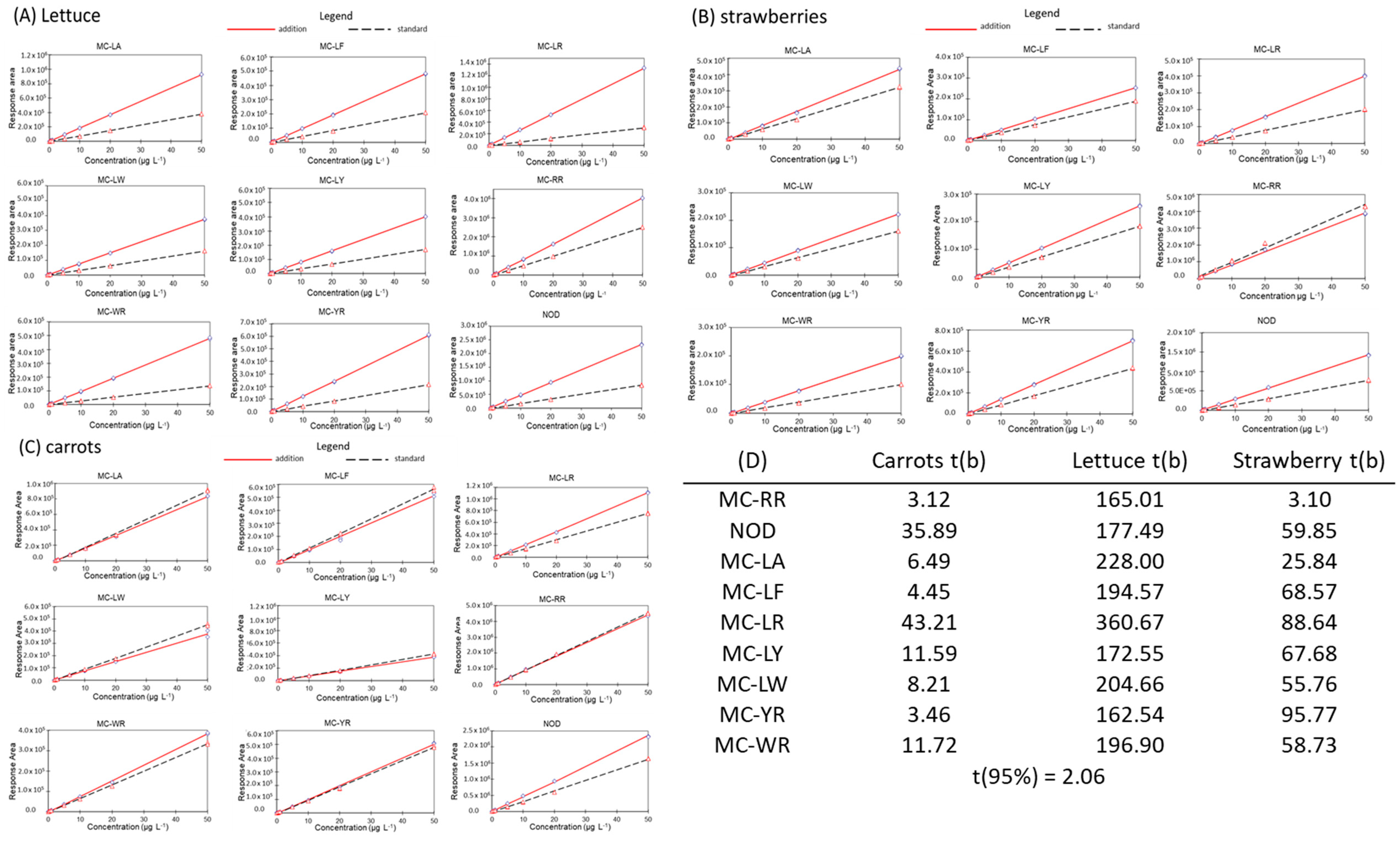
The matrix effect was determined for each toxin by producing a calibration curve combining all the toxins in Milli-Q water and methanol (+1% acidic acid) mix (50:50, v/v) and in a blank matrix (either carrots, strawberries or lettuce). When the fits of the calibration curves for the dilution solvent and blank matrix intersect, there is a matrix effect. A visual inspection of the linear fit was performed to determine the matrix effect. Additionally, the difference in the slope between the dilution solvent and the blank matrix was analyzed using the Student’s t-test. The calculated t-value was compared with the tabulated at the 95% confidence level. If the difference between slopes is significant, a matrix effect is present.

2.2.5. Recovery

Recoveries were calculated based on the different spiked concentrations and should be between 70% and 120%. The recovery was calculated as follows:
Apparent Recovery = Cmm/Cp
where Cmm = the mean of the means of each concentration level (n = 9); Cp = the spiked concentration.

2.2.6. Repeatability, Reproducibility and Measurement Uncertainty

The acceptance criteria for reproducibility and repeatability were established based on EU Decision 2002/657/EC [56]. In this decision, the upper boundaries for both parameters are determined by the Horwitz ratio based on the coefficient of variation (CV) and average variance, respectively. In addition, the boundaries for the measurement uncertainty were set below or equal to 80%. This high amount of variation can be justified by the lack of internal standards. The measurement uncertainty was calculated as twice the coefficient of variation. The statistical analysis was performed according to ISO5725-2 from 1994.

2.3. Sampling from the Belgium Market

A sampling of fruits and vegetables at the point of sale was conducted for two reasons. The first was to further test the developed method. The second was to perform a screening of potential cyanotoxin presence in actual foods bought by the consumers. The selection of fruits and vegetables from the Belgium market was based on multiple criteria. EFSA food consumption data were used to identify the most consumed foods in Europe. Fruits and vegetables were ranked based on the available consumption data (Table 2). Only fruits and vegetables for which consumption data were available were taken into account for sampling. Other selection criteria were raw consumption, contact of edible part with irrigation water, harvest during or after cyanobacteria’s bloom season and scientific and cultural relevance. The last two criteria were based on a lack of data present in the literature and vegetables or fruits typically consumed in Belgium, respectively. Moreover, the included products were preferably harvested in Belgium.
In total, nine different fruits and vegetables were selected based on these criteria. Carrots, chicory, radish, onions and potato were selected and grouped as root vegetables. Strawberries, tomatoes and cherry tomatoes were grouped as fruits. Only lettuce was selected as a leafy vegetable under our selection criteria. Table 2 provides a summary of the criteria applicable to each selected group. A complete overview of all independent samples can be found in Table S2 of the Supplementary Materials. These data also include sampling data and the origin of the samples (as read on the label). The sample analysis for a particular matrix was conformed if the recoveries calculated from the QC samples for this specific matrix adhered to the validation guidelines.

3. Results

3.1. Validation Results for the Different Matrices

The validation for the eight MCs and NOD was successful in lettuce, strawberries and carrots. All toxins showed specific signals for qualifier and quantifier ions in all matrices adhering to the validation criteria. Ion ratios were also found within the boundaries set by EU Decision 2002/657/EC [56], as shown in Table 3.
For the most part, the validation parameters were met for the LOD and LOQ. The LOQ was set at 1 µg kg−1, as this was the lowest validated concentration. The average signal-to-noise values for the LOQ were above 10, as shown in Table S1 of the Supplementary Materials. The LOD was set at the lowest concentration in the calibration curve (0.1 equal to 0.6 µg kg−1) if the signal-to-noise was higher than three, as can also be seen in Table S1 of the Supplementary Materials. All toxins in all matrices adhered to this parameter for the LOD and LOQ, except MC-RR in strawberries, where the average signal-to-noise was not high enough. Therefore, the LOQ and LOD were instead validated at 5 and 3 µg kg−1 (or LOD equal to 0.5 µg L−1), respectively, for MC-RR in strawberries. An additional concentration level (10 µg kg−1) was added specifically for this toxin to assess recovery, repeatability, reproducibility and measurement uncertainty later on during the validation.
The values for the recovery stayed within the preset parameters of 70–120% for all toxins in all matrices, as can be seen in Figure 1 and in Table S1 of the Supplementary Materials. The recovery for the sum of all toxins was also calculated for each concentration level and adhered to the preset parameters.
The linearity of the calibration curves of the different toxins in the different matrices was evaluated with Mandle’s fitting test. The ideal fit (quadratic or linear) always varied for the same toxin in the same matrix due to the R2 values that were similar for both fits. We eventually chose to calculate the concentrations with a linear fit, as the calculations are easier and linearity of the calibration curve is expected when using MS/MS. The R2 values are presented in Table S1 of the Supplementary Materials to illustrate the validity of the linear fit.
The matrix effect was observed for all of the toxins in all of the matrices based on a t-test on the slopes of the calibration curves in the matrix and dilution solvent. Plotting both curves, the presence of the matrix effect could be evaluated based on the intersection. When the lines intersect, a matrix effect is present, as illustrated in Figure 2.
Furthermore, Horwitz ratios determined the maximum repeatability and reproducibility for each toxin and the sum of all toxins in each matrix. The repeatability and reproducibility calculated with the results from the validation were below the maximum values calculated with the Horwitz ratio (Table S1 in the Supplementary Materials).
The measurement uncertainties were also below the boundary of 80% for all the toxins in all the matrices, as can be seen in Table S1 of the Supplementary Materials.

3.2. Method Application on Different Vegetables and Fruits

Multiple vegetables and fruits were collected from the Belgian supermarket from the end of July to the middle of November. Some radishes and potatoes originated from other countries, as during the sampling period, insufficient samples were available from Belgian origins (Table S2 in the Supplementary Materials).
Overall, no MCs or NOD were found in any of the vegetable or fruit samples. An additional quality control (QC) was added to each batch of analysis for each different matrix. The recoveries of the QCs for the matrices were within the boundaries (70–120%) used during validation (Table 4). The successful use of our validated method on vegetables and fruits other than carrots, salad and strawberries shows that our method can be used to analyze diverse matrices in these groups of products.

4. Discussion

We successfully validated a UHPLC-MS/MS method for the highest number of MCs (i.e., eight) described in the literature and NOD in three plant matrices (i.e., carrots, lettuce and strawberries). Moreover, this method describes the first validation for strawberries, where, due to the fact of their different chemical composition compared to the other matrices, it clearly showed a slightly lower recovery for MCs. The matrix effects caused by the strawberries significantly masked the signal of MC-RR, resulting in an increased LOD and LOQ. Chemical diversity among different fruits and vegetables is not uncommon, as Li et al. (2014) already reported the use of an HLB SPE cartridge for cucumbers and eggplants to increase MC recovery instead of the C-18 cartridges used for the validation of other vegetables [28]. The influence of the matrix effect on different methods of analysis has been shown [27,30,31]. In the three matrices we tested, the matrix effect was shown to affect the quantification if the calibration curve was prepared in a solvent. To account for the matrix effect, we opted for a matrix-matched calibration curve.
Separate parameters (LOD, LOQ, recovery and MU) are difficult to compare between the existing validated methods due to the use of different matrices, state of the matrix (dry or fresh weight), methodology or calculation of the parameters. One method used MS [27], while another was developed to extract MCs from different matrices (i.e., fish, soil and vegetables), resulting in a completely different methodology [30]. Similarly, in 2018, Díez-Quijada et al. developed a UHPLC-MS/MS method for CYN and three MCs, altering the methodology using a duplicate SPE, which caused a generally reduced recovery for all compounds [31]. Moreover, they reported a calculated LOD and LOQ, while our LOD and LOQ were based on the signal-to-noise values for the lowest point in the calibration curve and the lowest validated concentration, respectively. Our more experimental approach assured the measurability of the concentrations at the LOD and LOQ, while the calculated LOD and LOQ should be confirmed by the measurements after the validation. However, this confirmation is often not presented in the literature. Li et al. (2014) validated a UHPLC-MS/MS method similar to ours with only three MCs but in multiple matrices [28]. Our recoveries were similar to theirs. Furthermore, their similar recoveries for vegetables were different from our matrices, suggesting that the validation of our three matrices as model matrices for different fruit and vegetable types is valid. Using a QC as a control, we can use our validated method to quantify MCs and NOD in other leaf and root vegetables as well as in fruits and berries without additional validation for each matrix. However, if the recovery of the QC is not sufficient, an additional validation or adjustment to the method could be warranted, as shown for cucumbers and eggplants [28].
Although our study did not find any MCs or NOD in the fruit and vegetables taken from Belgian markets, multiple studies suggest that the accumulation of MCs in crops under real agricultural production is possible [28,34,44]. Up to 3 µg gfreshweight−1 of the total microcystin concentration was found in the edible parts of different plants after irrigation with contaminated water from a groundwater well [34]. MC-RR, MC-LR and MC-YR were also found in lettuce, water spinach, cabbage and chio sum irrigated with contaminated lake water, with the total microcystin concentrations ranging up to 108.2 µg kgfreshweight−1 [28]. MC-LR was also detected in the edible tissues of two rice variants (20.97 and 18.19 µg kg−1) and Ipomoea aquatica (132.86 µg kg−1) [44]. These results suggest that when sampling vegetables for monitoring of MCs, the number of samples per species and variety of samples should be increased. Additionally, for more targeted analysis, one could collect crops produced near locations where (toxic) blooms occur (i.e., “hot spots”). This approach requires the availability of reliable and rich data on bloom monitoring in various types of waterbodies, including those that could be used for the irrigation of plants and crops. However, only recreational waterbodies are monitored for toxic blooms in Belgium [55], but they cannot be exploited for irrigation purposes. Therefore, reliable monitoring of cyanotoxin accumulation in crops has to coincide with an increased scope of monitoring of blooms in waterbodies. Alternatively, the establishment of citizen science initiatives to report blooms might increase awareness of this issue and allow for a more accurate assessment of the presence of MCs in crops.

5. Conclusions

Our UHPLC-MS/MS method quantified eight MCs and NOD in three crops (i.e., carrots, lettuce and strawberries). Moreover, the quantification of MCs in strawberries as a model matrix for fruits is novel. Together with the validation of our uniform method in leafy and root vegetables, these matrices can be used as models for the quantification of the eight MCs and NOD in other edible plants. This approach does require adequate quality controls.
Following the initial screening of nine different vegetables and fruits from Belgian markets, it appears that there is currently no accumulation of MCs. Despite a lack of detection of MCs or NOD in the market samples, the quality control of the developed method was acceptable and reliable. Therefore, the methodology is adequate for future support of food market monitoring and MCs research in general.

Supplementary Materials

The following supporting information can be downloaded at: Http://https://www.mdpi.com/article/10.3390/separations9100319/s1, Table S1: Validation results for eight microcystin congeners (MCs) and nodularin (NOD) in carrots, lettuce and strawberries at three concentration levels and on average. The included parameters are recovery, repeatability, reproducibility, measurement uncertainty (MU), average signal-to-noise for LOD, average signal-to-noise for LOQ and R²; Table S2: Overview of the samples taken from Belgian markets showing separate results, origin, sample type and sample annotation. Figure S1: The elution peaks for eight microcystin congeners and NOD in carrot matrix at validation level 5 ng g−1. The peaks are presented together at representable ratios based on the peak intensities by overlaying the chromatograms of the different toxins. However, during analysis, the chromatograms for each toxin are analyzed separately. Figure S2: elution peaks for eight microcystin congeners and NOD in lettuce matrix at validation level 5 ng g−1. The peaks are presented together at representable ratios based on the peak intensities by overlaying the chromatograms of the different toxins. However, during analysis, the chromatograms for each toxin are analyzed separately. Figure S3: elution peaks for eight microcystin congeners and NOD in lettuce matrix at validation level 5 ng g−1. The peaks are presented together at representable ratios based on the peak intensities by overlaying the chromatograms of the different toxins. However, during analysis, the chromatograms for each toxin are analyzed separately.

Author Contributions

W.H.R.V.H., Conceptualization, methodology, validation, formal analysis, investigation, data curation, writing—original draft preparation, writing—review and editing, visualization and project administration; J.M., conceptualization, methodology, resources, writing—original draft preparation, writing—review and editing, supervision, project administration and funding acquisition; M.A., conceptualization, methodology, writing—original draft preparation, writing—review and editing, supervision and funding acquisition; A.R., conceptualization, resources, writing—original draft preparation, writing—review and editing, supervision, project administration and funding acquisition. All authors have read and agreed to the published version of the manuscript.

Funding

This study is financially supported by FPS Public Health, Safety of the Food Chain and Environment (SP 21/5 CYANTIR 1) and the EU Imptox project (Grant agreement 965173).

Data Availability Statement

Not applicable.

Conflicts of Interest

The authors declare no conflict of interest.

References

  1. Falconer, I.R.; Buckley, T.; Runnegar, M.T.C. Biological Half-Life, Organ Distribution and Excretion of 125I-Labelled Toxic Peptide from the Blue-Green Alga Microcystis aeruginosa. Aust. J. Biol. Sci. 1986, 39, 17–21. [Google Scholar] [CrossRef] [PubMed]
  2. Mahmood, N.A.; Carmichael, W.W. Anatoxin-a(s), an Anticholinesterase from the Cyanobacterium Anabaena Flos-Aquae NRC-525-17. Toxicon 1987, 25, 1221–1227. [Google Scholar] [CrossRef]
  3. Mahmood, N.A.; Carmichael, W.W. Paralytic Shellfish Poisons Produced by the Freshwater Cyanobacterium Aphanizomenon Flos-Aquae NH-5. Toxicon 1986, 24, 175–186. [Google Scholar] [CrossRef]
  4. Namikoshi, M.; Rinehart, K.L.; Sakai, R.; Stotts, R.R.; Dahlem, A.M.; Beasley, V.R.; Carmichael, W.W.; Evans, W.R. Identification of 12 Hepatotoxins from a Homer Lake Bloom of the Cyanobacteria Microcystis Aeruginosa, Microcystis Viridis, and Microcystis Wesenbergii: Nine New Microcystins. J. Org. Chem. 1992, 57, 866–872. [Google Scholar] [CrossRef]
  5. Welker, M.; Brunke, M.; Preussel, K.; Lippert, I.; von Döhren, H. Diversity and Distribution of Microcystis (Cyanobacteria) Oligopeptide Chemotypes from Natural Communities Studied by Single-Colony Mass Spectrometry. Microbiology (Reading) 2004, 150, 1785–1796. [Google Scholar] [CrossRef] [Green Version]
  6. Edwards, C.; Beattie, K.A.; Scrimgeour, C.M.; Codd, G.A. Identification of Anatoxin-A in Benthic Cyanobacteria (Blue-Green Algae) and in Associated Dog Poisonings at Loch Insh, Scotland. Toxicon 1992, 30, 1165–1175. [Google Scholar] [CrossRef]
  7. Harada, K.I.; Ohtani, I.; Iwamoto, K.; Suzuki, M.; Watanabe, M.F.; Watanabe, M.; Terao, K. Isolation of Cylindrospermopsin from a Cyanobacterium Umezakia Natans and Its Screening Method. Toxicon 1994, 32, 73–84. [Google Scholar] [CrossRef]
  8. Dahlmann, J.; Rühl, A.; Hummert, C.; Liebezeit, G.; Carlsson, P.; Granéli, E. Different Methods for Toxin Analysis in the Cyanobacterium Nodularia Spumigena (Cyanophyceae). Toxicon 2001, 39, 1183–1190. [Google Scholar] [CrossRef]
  9. Testai, E.; Buratti, F.M.; Funari, E.; Manganelli, M.; Vichi, S.; Arnich, N.; Fessard, V.; Sialehaamoa, A. Review and Analysis of Occurrence, Exposure and Toxicity of Cyanobacteria Toxins in Food. EFSA Extern. Sci. Rep. 2016, 13, 998E. [Google Scholar] [CrossRef]
  10. Abdallah, M.F.; Van Hassel, W.H.R.; Andjelkovic, M.; Wilmotte, A.; Rajkovic, A. Cyanotoxins and Food Contamination in Developing Countries: Review of Their Types, Toxicity, Analysis, Occurrence and Mitigation Strategies. Toxins 2021, 13, 786. [Google Scholar] [CrossRef]
  11. Mantzouki, E.; Lürling, M.; Fastner, J.; de Senerpont Domis, L.; Wilk-Wo´zniak, E. Temperature Effects Explain Continental Scale Distribution of Cyanobacterial Toxins. Toxins 2018, 10, 156. [Google Scholar] [CrossRef] [PubMed] [Green Version]
  12. Mazur-Marzec, H.; Sutryk, K.; Kobos, J.; Hebel, A.; Hohlfeld, N.; Błaszczyk, A.; Toruńska, A.; Kaczkowska, M.J.; Łysiak-Pastuszak, E.; Kraśniewski, W.; et al. Occurrence of Cyanobacteria and Cyanotoxin in the Southern Baltic Proper. Filamentous Cyanobacteria versus Single-Celled Picocyanobacteria. Hydrobiologia 2013, 701, 235–252. [Google Scholar] [CrossRef] [Green Version]
  13. MacKintosh, C.; Beattie, K.A.; Klumpp, S.; Cohen, P.; Codd, G.A. Cyanobacterial Microcystin-LR Is a Potent and Specific Inhibitor of Protein Phosphatases 1 and 2A from Both Mammals and Higher Plants. FEBS Lett. 1990, 264, 187–192. [Google Scholar] [CrossRef] [Green Version]
  14. Runnegar, M.T.; Kong, S.; Berndt, N. Protein Phosphatase Inhibition and in Vivo Hepatotoxicity of Microcystins. Am. J. Physiol. -Gastrointest. Liver Physiol. 1993, 265, G224–G230. [Google Scholar] [CrossRef] [PubMed]
  15. Fischer, A.; Hoeger, S.J.; Stemmer, K.; Feurstein, D.J.; Knobeloch, D.; Nussler, A.; Dietrich, D.R. The Role of Organic Anion Transporting Polypeptides (OATPs/SLCOs) in the Toxicity of Different Microcystin Congeners in Vitro: A Comparison of Primary Human Hepatocytes and OATP-Transfected HEK293 Cells. Toxicol. Appl. Pharmacol. 2010, 245, 9–20. [Google Scholar] [CrossRef] [Green Version]
  16. Feurstein, D.; Holst, K.; Fischer, A.; Dietrich, D.R. Oatp-Associated Uptake and Toxicity of Microcystins in Primary Murine Whole Brain Cells. Toxicol. Appl. Pharmacol. 2009, 234, 247–255. [Google Scholar] [CrossRef] [Green Version]
  17. Hawkins, P.R.; Chandrasena, N.R.; Jones, G.J.; Humpage, A.R.; Falconer, I.R. Isolation and Toxicity of Cylindrospermopsis Raciborskii from an Ornamental Lake. Toxicon 1997, 35, 341–346. [Google Scholar] [CrossRef]
  18. Antunes, J.T.; Leão, P.N.; Vasconcelos, V.M. Cylindrospermopsis Raciborskii: Review of the Distribution, Phylogeography, and Ecophysiology of a Global Invasive Species. Front Microbiol. 2015, 6, 473. [Google Scholar] [CrossRef] [Green Version]
  19. Merel, S.; Villarín, M.C.; Chung, K.; Snyder, S. Spatial and Thematic Distribution of Research on Cyanotoxins. Toxicon 2013, 76, 118–131. [Google Scholar] [CrossRef]
  20. Hoeger, S.J.; Shaw, G.; Hitzfeld, B.C.; Dietrich, D.R. Occurrence and Elimination of Cyanobacterial Toxins in Two Australian Drinking Water Treatment Plants. Toxicon 2004, 43, 639–649. [Google Scholar] [CrossRef]
  21. Clemente, Z.; Busato, R.H.; Oliveira Ribeiro, C.A.; Cestari, M.M.; Ramsdorf, W.A.; Magalhães, V.F.; Wosiack, A.C.; Silva de Assis, H.C. Analyses of Paralytic Shellfish Toxins and Biomarkers in a Southern Brazilian Reservoir. Toxicon 2010, 55, 396–406. [Google Scholar] [CrossRef] [PubMed]
  22. Ballot, A.; Fastner, J.; Lentz, M.; Wiedner, C. First Report of Anatoxin-a-Producing Cyanobacterium Aphanizomenon Issatschenkoi in Northeastern Germany. Toxicon 2010, 56, 964–971. [Google Scholar] [CrossRef] [PubMed]
  23. Codd, G.A.; Metcalf, J.S.; Beattie, K.A. Retention of Microcystis Aeruginosa and Microcystin by Salad Lettuce (Lactuca Sativa) after Spray Irrigation with Water Containing Cyanobacteria. Toxicon 1999, 37, 1181–1185. [Google Scholar] [CrossRef]
  24. Chen, J.; Song, L.; Dai, J.; Gan, N.; Liu, Z. Effects of Microcystins on the Growth and the Activity of Superoxide Dismutase and Peroxidase of Rape (Brassica Napus L.) and Rice (Oryza Sativa L.). Toxicon 2004, 43, 393–400. [Google Scholar] [CrossRef]
  25. Pflugmacher, S.; Wiegand, C.; Beattie, K.A.; Krause, E.; Steinberg, C.E.W.; Codd, G.A. Uptake, Effects, and Metabolism of Cyanobacterial Toxins in the Emergent Reed Plant Phragmites Australis (CAV.) Trin. Ex Steud. Environ. Toxicol. Chem. 2001, 20, 846–852. [Google Scholar] [CrossRef]
  26. McElhiney, J.; Lawton, L.A.; Leifert, C. Investigations into the Inhibitory Effects of Microcystins on Plant Growth, and the Toxicity of Plant Tissues Following Exposure. Toxicon 2001, 39, 1411–1420. [Google Scholar] [CrossRef]
  27. Järvenpää, S.; Lundberg-Niinistö, C.; Spoof, L.; Sjövall, O.; Tyystjärvi, E.; Meriluoto, J. Effects of Microcystins on Broccoli and Mustard, and Analysis of Accumulated Toxin by Liquid Chromatography–Mass Spectrometry. Toxicon 2007, 49, 865–874. [Google Scholar] [CrossRef]
  28. Li, Y.-W.; Zhan, X.-J.; Xiang, L.; Deng, Z.-S.; Huang, B.-H.; Wen, H.-F.; Sun, T.-F.; Cai, Q.-Y.; Li, H.; Mo, C.-H. Analysis of Trace Microcystins in Vegetables Using Solid-Phase Extraction Followed by High Performance Liquid Chromatography Triple-Quadrupole Mass Spectrometry. J. Agric. Food Chem. 2014, 62, 11831–11839. [Google Scholar] [CrossRef]
  29. Prieto, A.I.; Guzmán-Guillén, R.; Díez-Quijada, L.; Campos, A.; Vasconcelos, V.; Jos, Á.; Cameán, A.M. Validation of a Method for Cylindrospermopsin Determination in Vegetables: Application to Real Samples Such as Lettuce (Lactuca Sativa L.). Toxins 2018, 10, 63. [Google Scholar] [CrossRef] [Green Version]
  30. Manubolu, M.; Lee, J.; Riedl, K.M.; Kua, Z.X.; Collart, L.P.; Ludsin, S.A. Optimization of Extraction Methods for Quantification of Microcystin-LR and Microcystin-RR in Fish, Vegetable, and Soil Matrices Using UPLC–MS/MS. Harmful Algae 2018, 76, 47–57. [Google Scholar] [CrossRef]
  31. Díez-Quijada, L.; Guzmán-Guillén, R.; Prieto Ortega, A.I.; Llana-Ruíz-Cabello, M.; Campos, A.; Vasconcelos, V.; Jos, Á.; Cameán, A.M. New Method for Simultaneous Determination of Microcystins and Cylindrospermopsin in Vegetable Matrices by SPE-UPLC-MS/MS. Toxins 2018, 10, 406. [Google Scholar] [CrossRef] [PubMed] [Green Version]
  32. Lee, S.; Jiang, X.; Manubolu, M.; Riedl, K.; Ludsin, S.A.; Martin, J.F.; Lee, J. Fresh Produce and Their Soils Accumulate Cyanotoxins from Irrigation Water: Implications for Public Health and Food Security. Food Res. Int. 2017, 102, 234–245. [Google Scholar] [CrossRef] [PubMed]
  33. Crush, J.R.; Briggs, L.R.; Sprosen, J.M.; Nichols, S.N. Effect of Irrigation with Lake Water Containing Microcystins on Microcystin Content and Growth of Ryegrass, Clover, Rape, and Lettuce. Environ. Toxicol. 2008, 23, 246–252. [Google Scholar] [CrossRef] [PubMed]
  34. Mohamed, Z.A.; Al Shehri, A.M. Microcystins in Groundwater Wells and Their Accumulation in Vegetable Plants Irrigated with Contaminated Waters in Saudi Arabia. J. Hazard. Mater. 2009, 172, 310–315. [Google Scholar] [CrossRef]
  35. Hereman, T.C.; Bittencourt-Oliveira, M.d.C. Bioaccumulation of Microcystins in Lettuce. J. Phycol. 2012, 48, 1535–1537. [Google Scholar] [CrossRef]
  36. Levizou, E.; Statiris, G.; Papadimitriou, T.; Laspidou, C.S.; Kormas, K.A. Lettuce Facing Microcystins-Rich Irrigation Water at Different Developmental Stages: Effects on Plant Performance and Microcystins Bioaccumulation. Ecotoxicol. Environ. Saf. 2017, 143, 193–200. [Google Scholar] [CrossRef]
  37. do Bittencourt-Oliveira, M.C.; Cordeiro-Araújo, M.K.; Chia, M.A.; Arruda-Neto, J.D.d.T.; de Oliveira, Ê.T.; dos Santos, F. Lettuce Irrigated with Contaminated Water: Photosynthetic Effects, Antioxidative Response and Bioaccumulation of Microcystin Congeners. Ecotoxicol. Environ. Saf. 2016, 128, 83–90. [Google Scholar] [CrossRef]
  38. Cordeiro-Araújo, M.K.; Chia, M.A.; de Arruda-Neto, J.D.T.; Tornisielo, V.L.; Vilca, F.Z.; do Bittencourt-Oliveira, M.C. Microcystin-LR Bioaccumulation and Depuration Kinetics in Lettuce and Arugula: Human Health Risk Assessment. Sci. Total Environ. 2016, 566–567, 1379–1386. [Google Scholar] [CrossRef]
  39. Cordeiro-Araújo, M.K.; Chia, M.A.; Bittencourt-Oliveira, M.d.C. Potential Human Health Risk Assessment of Cylindrospermopsin Accumulation and Depuration in Lettuce and Arugula. Harmful Algae 2017, 68, 217–223. [Google Scholar] [CrossRef]
  40. Llana-Ruiz-Cabello, M.; Jos, A.; Cameán, A.; Oliveira, F.; Barreiro, A.; Machado, J.; Azevedo, J.; Pinto, E.; Almeida, A.; Campos, A.; et al. Analysis of the Use of Cylindrospermopsin and/or Microcystin-Contaminated Water in the Growth, Mineral Content, and Contamination of Spinacia Oleracea and Lactuca Sativa. Toxins 2019, 11, 624. [Google Scholar] [CrossRef]
  41. Machado, J.; Campos, A.; Vasconcelos, V.; Freitas, M. Effects of Microcystin-LR and Cylindrospermopsin on Plant-Soil Systems: A Review of Their Relevance for Agricultural Plant Quality and Public Health. Environ. Res. 2017, 153, 191–204. [Google Scholar] [CrossRef] [PubMed] [Green Version]
  42. Levizou, E.; Papadimitriou, T.; Papavasileiou, E.; Papadimitriou, N.; Kormas, K.A. Root Vegetables Bioaccumulate Microcystins-LR in a Developmental Stage-Dependent Manner under Realistic Exposure Scenario: The Case of Carrot and Radish. Agric. Water Manag. 2020, 240, 106274. [Google Scholar] [CrossRef]
  43. Chen, J.; Han, F.X.; Wang, F.; Zhang, H.; Shi, Z. Accumulation and Phytotoxicity of Microcystin-LR in Rice (Oryza Sativa). Ecotoxicol. Environ. Saf. 2012, 76, 193–199. [Google Scholar] [CrossRef] [PubMed]
  44. Wijewickrama, M.M.; Manage, P.M. Accumulation of Microcystin-LR in Grains of Two Rice Varieties (Oryza Sativa L.) and a Leafy Vegetable, Ipomoea Aquatica. Toxins 2019, 11, 432. [Google Scholar] [CrossRef] [PubMed] [Green Version]
  45. Corbel, S.; Mougin, C.; Nélieu, S.; Delarue, G.; Bouaïcha, N. Evaluation of the Transfer and the Accumulation of Microcystins in Tomato (Solanum Lycopersicum Cultivar MicroTom) Tissues Using a Cyanobacterial Extract Containing Microcystins and the Radiolabeled Microcystin-LR (14C-MC-LR). Sci. Total Environ. 2016, 541, 1052–1058. [Google Scholar] [CrossRef]
  46. Romero-Oliva, C.S.; Contardo-Jara, V.; Block, T.; Pflugmacher, S. Accumulation of Microcystin Congeners in Different Aquatic Plants and Crops – A Case Study from Lake Amatitlán, Guatemala. Ecotoxicol. Environ. Saf. 2014, 102, 121–128. [Google Scholar] [CrossRef]
  47. Peuthert, A.; Chakrabarti, S.; Pflugmacher, S. Uptake of Microcystins-LR and -LF (Cyanobacterial Toxins) in Seedlings of Several Important Agricultural Plant Species and the Correlation with Cellular Damage (Lipid Peroxidation). Environ. Toxicol. 2007, 22, 436–442. [Google Scholar] [CrossRef]
  48. Xiang, L.; Li, Y.-W.; Wang, Z.-R.; Liu, B.-L.; Zhao, H.-M.; Li, H.; Cai, Q.-Y.; Mo, C.-H.; Li, Q.X. Bioaccumulation and Phytotoxicity and Human Health Risk from Microcystin-LR under Various Treatments: A Pot Study. Toxins 2020, 12, 523. [Google Scholar] [CrossRef]
  49. Chen, J.; Dai, J.; Zhang, H.; Wang, C.; Zhou, G.; Han, Z.; Liu, Z. Bioaccumulation of Microcystin and Its Oxidative Stress in the Apple (Malus Pumila). Ecotoxicology 2010, 19, 796–803. [Google Scholar] [CrossRef]
  50. Corbel, S.; Bouaïcha, N.; Nélieu, S.; Mougin, C. Soil Irrigation with Water and Toxic Cyanobacterial Microcystins Accelerates Tomato Development. Env. Chem. Lett. 2015, 13, 447–452. [Google Scholar] [CrossRef]
  51. Haida, M.; El Khalloufi, F.; Mugani, R.; Redouane, E.M.; Campos, A.; Vasconcelos, V.; Oudra, B. Effects of Irrigation with Microcystin-Containing Water on Growth, Physiology, and Antioxidant Defense in Strawberry Fragaria Vulgaris under Hydroponic Culture. Toxins 2022, 14, 198. [Google Scholar] [CrossRef] [PubMed]
  52. Gutiérrez-Praena, D.; Campos, A.; Azevedo, J.; Neves, J.; Freitas, M.; Guzmán-Guillén, R.; Cameán, A.M.; Renaut, J.; Vasconcelos, V. Exposure of Lycopersicon Esculentum to Microcystin-LR: Effects in the Leaf Proteome and Toxin Translocation from Water to Leaves and Fruits. Toxins 2014, 6, 1837–1854. [Google Scholar] [CrossRef] [PubMed] [Green Version]
  53. Van Hassel, W.H.R.; Huybrechts, B.; Masquelier, J.; Wilmotte, A.; Andjelkovic, M. Development, Validation and Application of a Targeted LC-MS Method for Quantification of Microcystins and Nodularin: Towards a Better Characterization of Drinking Water. Water 2022, 14, 1195. [Google Scholar] [CrossRef]
  54. Van Hassel, W.H.R.; Ahn, A.-C.; Huybrechts, B.; Masquelier, J.; Wilmotte, A.; Andjelkovic, M. LC-MS/MS Validation and Quantification of Cyanotoxins in Algal Food Supplements from the Belgium Market and Their Molecular Origins. Toxins 2022, 14, 513. [Google Scholar] [CrossRef]
  55. Van Hassel, W.H.R.; Andjelkovic, M.; Durieu, B.; Marroquin, V.A.; Masquelier, J.; Huybrechts, B.; Wilmotte, A. A Summer of Cyanobacterial Blooms in Belgian Waterbodies: Microcystin Quantification and Molecular Characterizations. Toxins 2022, 14, 61. [Google Scholar] [CrossRef]
  56. P.O. of the E. Union, CELEX1, 2002/657/EC: Commission Decision of 12 August 2002 Implementing Council Directive 96/23/EC Concerning the Performance of Analytical Methods and the Interpretation of Results (Text with EEA Relevance) (Notified under Document Number C(2002) 3044). Available online: http://op.europa.eu/en/publication-detail/-/publication/ed928116-a955-4a84-b10a-cf7a82bad858/language-en (accessed on 30 November 2020).
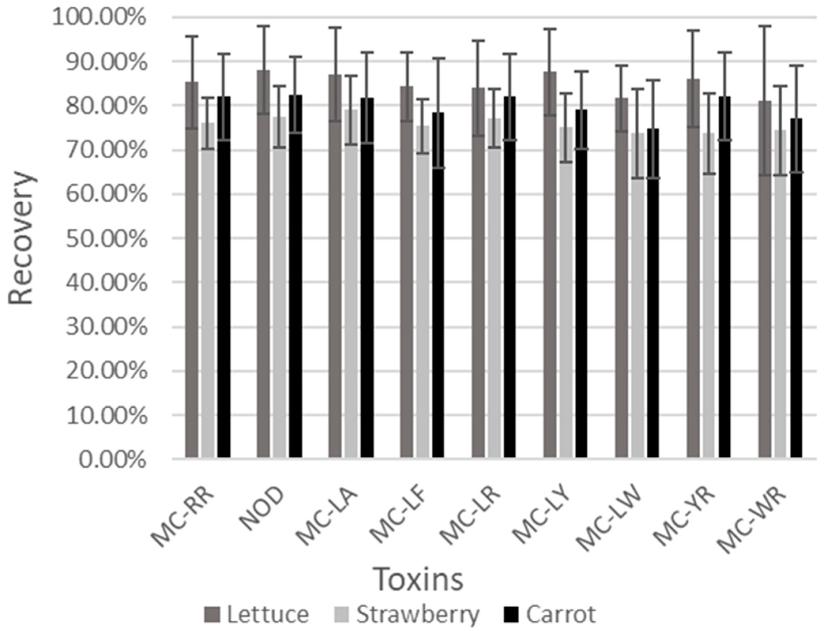
Figure 1. Average recoveries (%) obtained during the validation of the eight microcystin congeners (MCs) and nodularin (NOD) in the validated matrices (i.e., lettuce, carrots and salads). The average recovery for each toxin in each matrix was calculated from the recoveries at the different concentration levels.
Separations 09 00319 g001
Figure 2. Visual assessment of the matrix effect for the eight microcystins and nodularin in lettuce (A), strawberries (B) and carrots (C). Table (D) presents the calculated t(b) values of the Student’s t-tests compared to t(95%), with t(b) < t(95%) if no significant difference was present between the slopes of the curve and, thus, no matrix effect.
Separations 09 00319 g002
Table 1. Mass-to-charge ratio (m/z) of the precursor, quantifier and qualifier ions for the eight microcystin congeners and nodularin. The collision energy (eV) and cone voltage (V) are shown for the quantifier and qualifier ions.
ToxinPrecursor Ion (m/z)Quantifier Ion (m/z)Collision Energy (eV)Cone Voltage (V)Qualifier Ion (m/z)Collision Energy (eV)Cone Voltage (V)
MC-LR995.4135.07080213.16080
MC-RR519.8134.83050107.26050
MC-YR1045.5135.38060212.96060
MC-WR1068.4135.370100213.160100
MC-LY1002.4135.36050213.05050
MC-LA910.3135.16050107.18050
MC-LF986.3135.06070213.16070
MC-LW1025.4134.96060213.15060
NOD825.25134.95080102.79080
Table 2. Criteria for the sample matrix selection: food consumption rank, raw consumption, (direct) contact with irrigation water, harvested during cyanobacteria’s bloom period and scientific relevance and cultural relevance.
Sample MatrixConsumption Rank *Eaten Raw Contact with WaterHarvested during Bloom PeriodScientific RelevanceCultural Relevance
Carrots4X X
Onions5X X
Potato5X XX
Chicory3XXXXX
Radish2X XXX
Strawberries4XXXX
Tomato4XXX
Cherry Tomato4XXX
Lettuce4XXX
* Rank 1 = no consumer data; rank 2 > 1 consumer; rank 3 > 100 consumers; rank 4 > 1000 consumers; rank 5 > 2000 consumers.
Table 3. Results for the average ion ratio (%) and standard deviation (%) in carrots, lettuce and strawberries for the eight microcystin congeners and nodularin.
CarrotsLettuceStrawberries
Average Ion Ratio Standard Deviation Ion RatioAverage Ion Ratio Standard Deviation Ion RatioAverage Ion Ratio Standard Deviation Ion Ratio
MC-RR15.285.4316.155.8213.132.63
NOD42.664.6941.241.6546.582.29
MC-LA47.367.0751.466.0740.281.78
MC-LF40.451.7741.142.1439.310.89
MC-LR31.981.2032.572.8131.751.68
MC-LY50.013.8749.586.2651.805.08
MC-LW45.362.0544.852.3245.192.00
MC-YR36.453.9835.844.4938.884.65
MC-WR43.0510.7441.224.0338.932.21
Table 4. Average recoveries (%) of quality controls in the market samples for the different matrices.
Sample TypeMC-RRNODMC-LAMC-LFMC-LRMC-LYMC-LWMC-YRMC-WR
Chicory98.5397.0795.8796.9399.93105.20110.2789.4794.80
Onion84.0587.5089.8583.7087.4083.6075.4587.6580.60
Cherry Tomato87.3885.7890.3879.3386.7088.5870.1583.6880.38
Raspberry80.1079.5095.1081.2082.1090.5072.8086.5086.30
Tomato91.4099.20106.80100.3089.5098.30104.40108.20109.50
Carrot81.5886.0291.05103.1596.2884.70101.6581.9080.15
Potato79.3888.0589.5897.6095.4885.5396.7587.8780.57
Strawberries79.9391.1384.4775.0787.7382.1384.4780.0093.07
Lettuce70.0%82.6789.0798.47105.6777.0783.1376.0070.87
Publisher’s Note: MDPI stays neutral with regard to jurisdictional claims in published maps and institutional affiliations.