Article

A Novel Photo-Responsive Molecularly Imprinted Silica as a Sustainable Solid-Phase Extraction Filler for Highly Selective Adsorption of Chlorogenic Acid

1
Engineering Research Center of Forest Bio-Preparation, Ministry of Education, Northeast Forestry University, Harbin 150040, China
2
College of Food and Health, Northeast Forestry University, Harbin 150040, China
3
Key Laboratory of Forest Plant Ecology, Ministry of Education, Northeast Forestry University, Harbin 150040, China
4
Heilongjiang Provincial Key Laboratory of Ecological Utilization of Forestry-Based Active Substances, Northeast Forestry University, Harbin 150040, China
*
Authors to whom correspondence should be addressed.
These authors contributed equally to this work.
Separations 2026, 13(7), 200; https://doi.org/10.3390/separations13070200
Submission received: 24 May 2026 / Revised: 29 June 2026 / Accepted: 30 June 2026 / Published: 9 July 2026

Abstract

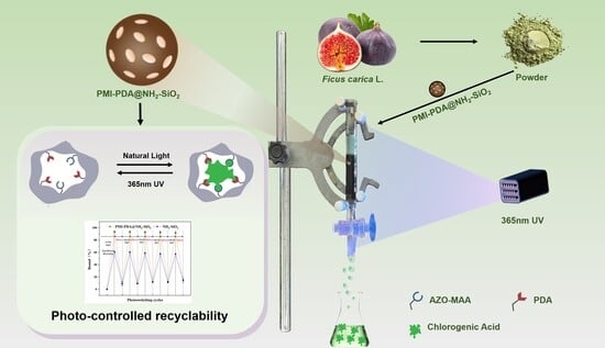
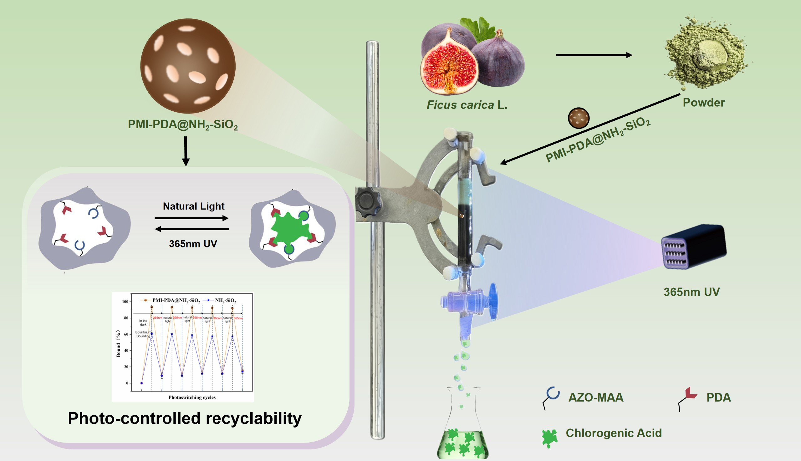
Chlorogenic acid (CA) is an important natural antioxidant component and holds strong potential for health food and cosmetic applications. In this study, a silica-based photo-responsive molecular imprinting material (PMI-PDA@NH2-SiO2) was designed as a solid-phase extraction (SPE) adsorption filler and applied for the efficient separation of CA in Ficus carica L. Using polydopamine-modified NH2-SiO2 silica as the base, combined with the photo-responsive monomer 4-methacryloyloxyazobenzene (AZO-MAA), a molecular imprinting layer with photo-responsive regulation function was constructed. Under 365 nm ultraviolet light irradiation, the azobenzene group was isomerized to the cis structure, causing the imprint cavity to shrink, thereby enabling controlled release of CA, with complete desorption within 40 min, and a desorption rate of 94.33%. Importantly, the material retained 85.56% of its initial adsorption capacity and 91.38% of its original desorption efficiency after 6 consecutive adsorption/desorption cycles, confirming robust operational stability and reproducibility. PMI-PDA@NH2-SiO2 was applied to the extract of Ficus carica L., achieving an adsorption rate of 92.3% for CA and a desorption rate of 87.27%. Density functional theory (DFT) calculations and NOESY spectroscopic analyses revealed that CA interacted with functional monomers via hydrogen bonding and van der Waals forces. This study advances a green separation strategy for bioactive phytochemicals in complex natural matrices.

Graphical Abstract

1. Introduction

Ficus carica L. belongs to the Ficus genus of the Moraceae family and has been highly valued as a resource with both medicinal and edible properties since ancient times [1]. Chlorogenic acid (CA) is an important phenolic compound in F. carica fruits, which possesses various biological activities such as antioxidant, anti-inflammatory, and antiviral properties [2]. To enable the broader application of CA in the fields of cosmetics, healthcare, and chemical engineering, it is imperative to establish an efficient strategy for its separation [3]. Current separation methods for CA primarily include membrane separation [4], polyamide column chromatography [5], macroporous adsorption resin chromatography [6], and high-performance liquid chromatography (HPLC) [7]. However, these methods suffer from poor selectivity and are associated with several practical limitations, including high operational costs, substantial solvent consumption, prolonged pretreatment times, and low separation efficiency [8].
Molecular imprinting technology (MIT) provides an efficient solution to the challenge of low selectivity in natural compound separation [9]. This technique involves initiating the copolymerization of functional monomers and cross-linkers on the surface of a support matrix in the presence of a template molecule, yielding molecularly imprinted polymers (MIPs) [10]. Endowed with pre-engineered recognition sites, MIPs have emerged as pivotal materials in separation technology, owing to their exceptional selectivity and robust binding affinity toward target analytes [9,11].For the separation of CA, a range of high-performance MIP-based materials have been developed [12,13,14]. Among these materials, surface molecularly imprinted polymers (SMIPs) are constructed by forming an imprinted layer on the surface of nano- or microscale carriers. These materials, which possess a high specific surface area, well-defined morphology, enhanced binding capacity, accelerated mass transfer kinetics, and a high density of recognition sites, all of which contribute to significantly improved separation performance [15].
Silica nanoparticles (SiO2) are ideal MIP carriers, as their large pore volumes, high specific surface areas, and tunable mesopore sizes can enhance the adsorption properties of imprinted materials. Further functionalization with amino groups produces amino-functionalized mesoporous silica nanoparticles (NH2-SiO2), which not only retain high specific surface areas and mesopore capacities but also feature abundant surface amino groups that can act as direct adsorption sites or undergo further modification to boost adsorption efficiency [16]. The silica framework is inherently polar due to its dense silanol groups (-SiOH), and the introduction of strongly polar amino groups (-NH2) further amplifies the surface polarity and chemical reactivity, facilitating more intimate interactions with polar target molecules [17,18]. In recent years, NH2-SiO2 has demonstrated substantial application potential across fields including drug delivery, catalysis, and functional material engineering.
Stimuli-responsive polymers (SRPs), which exhibit dynamic changes in morphology, chemical structure, and physicochemical properties in response to external cues (e.g., light, temperature, pH, and solvent polarity), have emerged as a prominent research focus in the field of molecular imprinting technology [19,20]. Light has garnered increasing attention from researchers relative to traditional stimuli, owing to its distinct advantages of being clean, non-polluting, highly controllable, and non-contact.
Photo-responsive molecularly imprinted polymers (P-MIPs) incorporate photosensitive moieties (e.g., azobenzene) that undergo cis-trans isomerization upon irradiation with specific wavelengths of light. This structural transition enables dynamic regulation of the spatial configuration and chemical microenvironment of imprinted cavities, facilitating reversible adsorption and release of target compounds and thereby significantly enhancing the recyclability of the imprinted material [21,22]. A photo-responsive functional monomer, 4-methacryloyloxyazobenzene (Azo-MAA), was successfully synthesized by conjugating azobenzene with methacryloyl chloride, capitalizing on the intrinsic photo-responsive properties of azobenzene derivatives [23].
Polydopamine (PDA) is a superior functional monomer that is formed through the self-polymerization of dopamine (DA), a molecule rich in catechol and amine functional groups, under optimized conditions. Beyond its exceptional adhesive properties, PDA can be further chemically modified to introduce additional functional moieties, thereby enhancing the density of imprinting sites and endowing the material with expanded functionality [24,25]. Thus, employing PDA and Azo-MAA as dual functional monomers for molecularly imprinted materials is anticipated to integrate the advantages of high imprinting site density and photo-responsive behavior [26,27,28], offering a novel material platform for the efficient and intelligent separation of CA.
Photo-responsive molecularly imprinted composites represent an advanced class of materials that integrate selective recognition with stimuli-regulated binding behavior. The incorporation of photo-responsive functional monomers serves as the key responsive component of the photo-responsive molecularly imprinted (PMI) layer. These materials not only achieve high-affinity capture of target analytes but also enable spatiotemporally controlled release through light irradiation, offering a sustainable and precise approach for separation processes. In this work, the developed PMI-PDA@NH2-SiO2 composite exhibits outstanding affinity and selectivity toward CA, allowing reversible and efficient adsorption/desorption cycling under mild photo-stimulation with minimal loss of capacity. Compared to conventional isolation methods for CA [12,13,14], this material demonstrates enhanced recognition sensitivity, accelerated binding kinetics, and remarkable cycling stability, positioning it as a promising and green alternative for the selective extraction and controllable recovery of CA from complex samples.
The elucidation of the adsorption mechanism provides a critical theoretical foundation for optimizing material design and enhancing adsorption efficiency. Density functional theory (DFT) is a quantum mechanical computational approach that uses electron density as the fundamental variable to investigate the structure of multi-electron systems, and it stands as one of the most powerful tools for studying intermolecular interactions [29,30]. In addition, two-dimensional nuclear magnetic resonance-nuclear Overhauser effect spectroscopy (2D NMR-NOESY) enables structural characterization of complex organic molecules, with its 2D correlation signals clearly resolving proton connectivity. As a robust analytical technique for characterizing intermolecular interactions, it offers experimental evidence to support the recognition mechanism.
In this study, a photo-responsive amino-functionalized silica gel molecularly imprinted material (PMI-PDA@NH2-SiO2) was rationally designed using NH2-SiO2 as the support, Azo-MAA and PDA as dual functional monomers, ethylene glycol dimethacrylate (EGDMA) as the crosslinker, azobisisobutyronitrile (AIBN) as the initiator, and dimethyl sulfoxide (DMSO) as the porogen. This material enables highly selective and reproducible separation of CA from F. carica extracts. Furthermore, PDA modification introduced additional imprinting sites to the support, and the photoisomerization property of Azo-MAA conferred upon the imprinted material the ability to rapidly and reversibly regulate adsorption and desorption processes of CA. Finally, the mechanisms underlying the adsorption and desorption of CA by PMI-PDA@NH2-SiO2 were elucidated through density functional theory (DFT) calculations and 2D 1H-1H NOESY spectroscopy.

2. Materials and Methods

2.1. Reagents and Materials

Ficus carica L. fruits were harvested from Chengshan Town, Rongcheng City, Shandong Province, China. Fresh fruits were dried at 40 °C to a constant weight, ground into powder, and passed through a 60-mesh sieve. The fine powder was stored under dry, light-proof conditions at room temperature until analysis. NH2-SiO2 was purchased from Zhengzhou Hecheng New Materials Technology Co., Ltd. (Zhengzhou, China). Triethylamine (99.0%) was sourced from Shanghai Saen Chemical Technology Co., Ltd. (Shanghai, China). Dopamine hydrochloride (DA, 98.0%) was provided by Shanghai Debai Biotechnology Co., Ltd. (Shanghai, China). Rutin (RU, 98.0%), epicatechin (EC, 98.0%), and chlorogenic acid (CA, 98.0%) were procured from Shanghai Yuanye Bio-Technology Co., Ltd. (Shanghai, China). Acetonitrile (99.9%) and phosphoric acid (99.0%) were acquired from Shanghai Macklin Biochemical Technology Co., Ltd. (Shanghai, China). All other analytical grade reagents were provided by Shanghai Macklin Biochemical Technology Co., Ltd. (Shanghai, China). Ultrapure water was prepared using an ultrapure water system from Research Water Purification Technology Co., Ltd. (Xiamen, China).

2.2. Synthesis of PMI-PDA@NH2-SiO2 MIPs

2.2.1. Preparation of AZO-MAA

First, 4.96 g of 4-hydroxyazobenzene, 4.30 g of triethylamine, and 0.15 g of N,N-dimethyl aminopyridine were dissolved in 150 mL of anhydrous acetonitrile. The solution was cooled to 0–5 °C in an ice bath. Under continuous stirring, 4.5 g of methacryloyl chloride was added dropwise. After the addition was completed, the reaction was stirred continuously at 40 °C for 24 h, then cooled to room temperature, followed by the addition of 50 mL of saturated NaOH solution. The precipitated solid was collected by suction filtration, washed successively with 2.0 mol/L HCl solution, and finally purified by recrystallization from an ethanol–water mixed solvent to afford the photo-responsive monomer AZO-MAA.

2.2.2. Preparation of PDA@NH2-SiO2

First, 1.0 g of dried NH2-SiO2 was immersed in 150 mL of 0.1 mol/L Tris-HCl buffer solution. Then, 0.25 g of DA was added to the suspension and magnetically stirred at 500 rpm until completely dissolved. The polymerization reaction was carried out at 25 °C and 150 rpm under light-shielded conditions (wrapping the reaction vessel with aluminum foil) for 12 h, allowing a uniform polydopamine film to form on the surface of NH2-SiO2 via dopamine self-polymerization. After the reaction, the product was collected by vacuum filtration and repeatedly washed with deionized water until the filtrate became colorless. The final product was dried at 40 °C for 24 h to yield polydopamine-modified particles, denoted as PDA@NH2-SiO2.

2.2.3. Preparation of PMI-PDA@NH2-SiO2

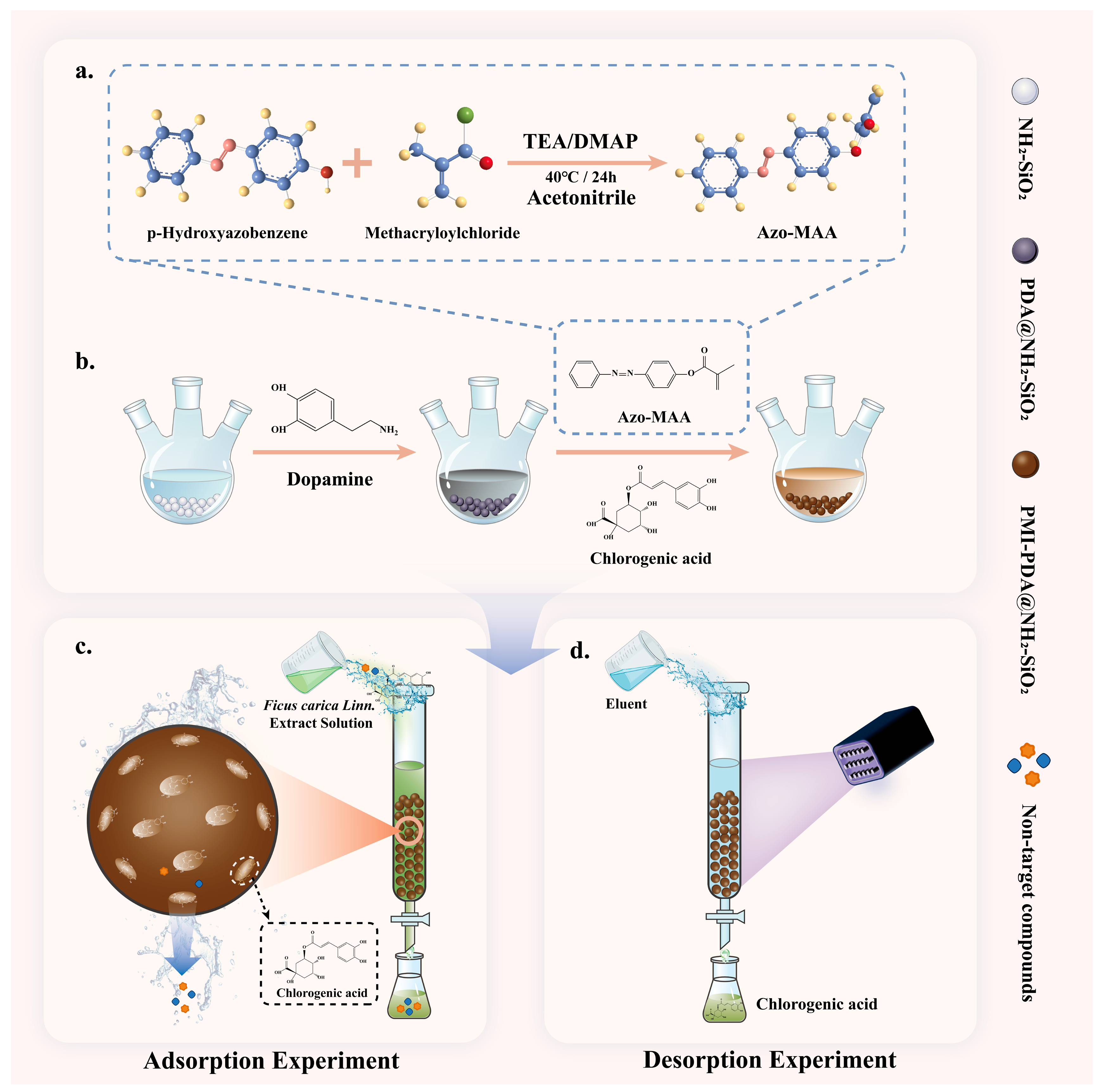
First, 0.1 mmol of CA and 0.4 mmol Azo-MAA were dissolved in 100 mL of DMSO and pre-assembled at 25 °C for 6 h. Next, 1.0 g of PDA@NH2-SiO2 and 2.0 mmol of EGDMA were added to the pre-assembled solution in sequence and sonicated until dissolved. Then, the solution was degassed under nitrogen protection for 10 min, and 0.05 mmol of AIBN was added. The entire system was continuously reacted for 24 h under a 60 °C water bath and nitrogen protection. After the reaction, the resulting product was collected and washed exhaustively with distilled water and a methanol/acetic acid mixture (9/1, v/v) to remove the template molecule and finally washed with a large amount of methanol to remove the residual acetic acid. The product was dried to obtain the target molecularly imprinted material PMI-PDA@NH2-SiO2. Ultimately, a non-imprinted polymer (PNI-PDA@NH2-SiO2) was prepared as the control material following the identical synthetic protocol except that CA was not added. The preparation process of PMI-PDA@NH2-SiO2 is shown in Figure 1.

2.3. Sample Characterization

The successful synthesis of the photo-responsive monomer AZO-MAA was verified using ultraviolet-visible (UV-vis) spectroscopy (Hitachi U-3900 spectrophotometer, High-Technologies Corporation, Tokyo, Japan) and electrospray ionization mass spectrometry (ESI-MS, Thermo Fisher Scientific TSQ Fortis Plus system, Thermo Fisher Scientific Inc., Waltham, MA, USA). The surface topographies of NH2-SiO2, PDA@NH2-SiO2, and PMI-PDA@NH2-SiO2 were visualized via scanning electron microscopy (SEM, Thermo Fisher Scientific Apreo C, Thermo Fisher Scientific Inc., Waltham, MA, USA) at an accelerating voltage of 5 kV. Fourier-transform infrared spectroscopy (FT-IR, Thermo Fisher Scientific Nicolet iS50, Thermo Fisher Scientific Inc., Waltham, MA, USA) was conducted to characterize the chemical structure of AZO-MAA and identify changes in surface functional groups of NH2-SiO2, PDA@NH2-SiO2, and PMI-PDA@NH2-SiO2.

2.4. HPLC Analysis

Quantification was performed using the external standard method. The quantification of sample and standard was conducted using high-performance liquid chromatography (HPLC, Agilent 1260, Agilent Technologies, Inc., Santa Clara, CA, USA) equipped with a HiQ Sil C18 column (4.6 mm× 250 mm, 5 μm). The sample solution consisted of a methanol extract of Ficus carica L., and the HPLC mode employed was isocratic elution. The mobile phase for HPLC consisted of a 30% A phase (0.2% aqueous phosphoric acid) and a 70% B phase (acetonitrile). The flow rate was maintained at 1.0 mL/min, the column temperature was set to 30 °C, the detection wavelength was fixed at 275 nm, and the injection volume was 20 μL.

2.5. Photo-Controlled Adsorption/Desorption Experiments

This experiment investigates the photo-controlled adsorption/desorption performance of PMI-PDA@NH2-SiO2. First, 20 mg of PMI-PDA@NH2-SiO2 was added to 20 mL of a 0.1 mg/mL CA standard solution. The mixture was then placed in a constant temperature shaker under dark conditions at 25 °C and 100 rpm until adsorption equilibrium was reached. The initial concentration of CA (C0) in the supernatant was determined by HPLC. Subsequently, the sample was alternately exposed to 365 nm ultraviolet light and natural light for 1 h each time. After each light exposure, the supernatant was centrifuged (5000 rpm) and filtered (0.22 μm filter membrane), and the CA concentration in the supernatant was measured by HPLC. The binding rate (D%) of PMI-PDA@NH2-SiO2 to CA was calculated according to Equation (1). This process was repeated for 6 cycles of alternating ultraviolet/natural light exposure. The CA concentration in each cycle was determined. All experiments were conducted in triplicate.
D % = ( 1 C e C 0 )   ×   100 %
where C0 (mg/L) represents the initial concentration of CA during the adsorption process, and Ce (mg/L) represents the equilibrium concentration of CA.

2.6. Rebinding Experiment

2.6.1. Adsorption Kinetics

First, 0.1 g of PMI-PDA@NH2-SiO2 was dispersed in 50 mL of 0.1 mg mL CA standard solution and incubated in a thermostatic shaker (150 rpm, 25 °C) to initiate the adsorption process. Aliquots (2 mL) were withdrawn at predefined time intervals (1, 2, 3, 4, 5, 10, 15, 30, 60, 90, and 120 min) within 0–120 min. Each sample was immediately centrifuged at 8000 rpm for 5 min and filtered through a 0.22 μm membrane to remove solid particles. The residual CA concentration in the supernatant was quantified via HPLC. The adsorption capacity at time t (Qt, mg/g) was calculated using Equation (2):
Q e   =   ( C 0 C t ) V m
where C0 (mg/L) and Ct (mg/L) represent the initial and real-time concentration of CA, V (L) represents the volumes of CA solutions, and m (g) represents the mass of the adsorbent.
To elucidate the adsorption mechanism, the kinetic data were fitted to the pseudo-first-order kinetic model Equation (3) and pseudo-second-order kinetic model Equation (4). The equations of models are as follows:
ln ( Q e Q t ) = ln Q e k 1 t
t Q t = 1 k 2 Q e 2 + t Q e
where Qe (mg/g) and Qt (mg/g) represent the adsorption capacity at adsorption equilibrium and at time t (min), respectively. k1 (min−1) and k2 (g/mg/min) represent the constants in Equations (3) and (4).

2.6.2. Adsorption Isotherms

First, 20 mg of PMI-PDA@NH2-SiO2 was added to 5 mL of CA standard solutions with different concentrations (0.02, 0.04, 0.06, 0.08, 0.10, 0.12, 0.14, 0.16, 0.18, and 0.2 mg/mL), followed by shaking at 200 rpm for 60 min. Subsequently, the concentration of CA was determined by HPLC. The performance and adsorption behavior of PMI-PDA@NH2-SiO2 were evaluated and predicted by Langmuir and Freundlich isothermal adsorption models. The Langmuir model and Freundlich model are represented by Equations (5) and (6):
Q e   =   Q m K L C e 1   +   K L C e
ln Q e = ln K F + 1 n ln C e
where Qe (mg/g) and Ce (mg/mL) represent the adsorption capacity of the material and the equilibrium concentration of CA, respectively; Qm (mg/g) represents the maximum theoretical adsorption capacity; n stands for the empirical constant; and KF and KL denote the Freundlich constant and Langmuir constant respectively [31].

2.6.3. Adsorption Selectivity

To evaluate the adsorption selectivity of PMI-PDA@NH2-SiO2 for CA, rutin (RU) and epicatechin (EC) were used as competitive adsorbates. The chemical structures of RU and EC are provided in Figure S2. A mixed solution, with an initial concentration of 0.1 mg/mL for CA, RU, and EC, was prepared. First, 20 mg of PMI-PDA@NH2-SiO2 was added to the mixed solution, and the adsorption experiment was carried out in a constant-temperature shaker for 60 min. The concentrations of CA, RU, and EC in the solution were determined by HPLC. The imprinting factor (IF), distribution coefficient (Kd), selectivity coefficient (ksel), and relative selectivity coefficient (krel) of PMI-PDA@NH2-SiO2 were expressed by Equations (7)–(10).
I F   = Q P M I - P D A @ N H 2 - S i O 2 Q P N I - P D A @ N H 2 - S i O 2
where Q P M I - P D A @ N H 2 - S i O 2 (mg/g) and Q P N I - P D A @ N H 2 - S i O 2 (mg/g) are the equilibrium adsorption capacities of PMI-PDA@NH2-SiO2 (molecularly imprinted polymer, MIP) and PNI-PDA@NH2-SiO2 (non-imprinted polymer, NIP) for CA, respectively.
K   d = Q e C e
k s e l = K d ,   t e m p l a t e   m o l e c u l e K d ,   r e f e r e n t   m o l e c u l e
k r e l = k M I P s s e l k N I P s s e l
where K d (mL/g) is the distribution coefficient, Qe (mg/g) is the equilibrium adsorption capacity of PMI-PDA@NH2-SiO2 for the target compound or analog, and Ce (mg/mL) is the equilibrium concentration of the solute in the solution, Kd, template molecule is the partition coefficient of the template molecule; Kd, referent molecule is the partition coefficient of the competitor; kselMIPs is the selectivity coefficient of MIPs; and kselNIPs is the selectivity coefficient of NIPs [32,33].

2.7. Reusable Performance Test

To evaluate the reusability of the prepared PMI-PDA@NH2-SiO2 for CA, a cyclic adsorption/desorption experiment was conducted. The specific steps were as follows: 20 mg of PMI-PDA@NH2-SiO2 was added to a 0.1 mg/mL CA standard solution, and the adsorption experiment was carried out in a constant-temperature shaker at 25 °C and 100 rpm until adsorption equilibrium was reached. Subsequently, the adsorption-saturated PMI-PDA@NH2-SiO2 was separated from the solution and desorption was performed under 365 nm ultraviolet light until desorption equilibrium was achieved. After desorption, the resin was washed with deionized water, dried, and reused. The content of CA in each cycle was determined, and the adsorption capacity and desorption rate of each adsorption/desorption process were calculated to evaluate the recyclability of PMI-PDA@NH2-SiO2.

2.8. Recognition Mechanism

2.8.1. DFT Simulation

The interaction mode, charge information, and interaction force of PDA, Azo-MAA, and CA systems were studied by DFT simulation, and the recognition mechanism of PMI-PDA@NH2-SiO2 was clarified. All calculations were performed using Gaussian 16 software. Firstly, the geometric structures of PDA, Azo-MAA, and CA and their complexes were optimized to reach the stable configuration with the lowest energy, and the 3D visualization structures were verified by Gauss View 6.0. The intermolecular charge transfer characteristics were investigated based on Mulliken atomic charge distribution, and the donor–acceptor regions were characterized by electrostatic potential mapping. The energy gap values of the system were calculated using the frontier molecular orbital theory (HOMO-LUMO) to evaluate the reactivity and stability of the system. Moreover, non-covalent interactions (NCIs) in the system were characterized using electron density analysis, and RDG isosurfaces generated with Multiwfn 3.7 were visualized in real space to reveal weak intermolecular interactions.

2.8.2. NMR Measurements

2D nuclear magnetic resonance (2D-NMR) is a key technique in modern structural analysis, capable of precisely assigning spectral peaks for complex organic compounds. In the 2D 1H-1H NOESY spectrum, when the spatial distance between two protons is less than 5 Å, corresponding cross-peaks will appear in the spectrum, and this signal can be used to infer the stereostructure and spatial conformation of the molecule. In this study, the interaction between the functional monomer DA and the template CA was analyzed using the 2D 1H-1H NOESY spectrum. DA and the template CA were mixed at a molar ratio of 1:1. Then, the mixture was dissolved in DMSO-d6 for NMR characterization. The NMR measurements were carried out using a nuclear magnetic resonance spectrometer (NMR, Qone AS400, Wuhan Zhongke Niujin Wave Spectrum Technology Co., Ltd., Wuhan, China).

2.9. Purification of CA from Ficus carica L. Using PMI-PDA@NH2-SiO2 SPE Column

Using PMI-PDA@NH2-SiO2 as the solid-phase extraction (SPE) column packing material, the filling volume was controlled at approximately 2/3 of the column volume. After completing the column packing, 30 mL of the recovered F. carica L. extract was loaded onto the solid-phase extraction column at a flow rate of 2 BV/h. The column was allowed to stand overnight, and the eluate I was collected. Then, a mixed elution solution composed of methanol and acetic acid in a ratio of 9:1 (v/v) was prepared. This elution solution was used to desorb CA from the stationary phase, with a volume controlled with a volume of 4 BV/h. Eluate II was collected and its volume was recorded. Finally, eluate II was subjected to rotary evaporation in an oil bath to concentrate the CA content by evaporating the solvent. Following rotary evaporation, the crude product (meluate) was accurately weighed. The adsorption recovery rate (Rads) of CA by the PMI-PDA@NH2-SiO2 extraction column, the desorption recovery rate (Rdes) of CA in the final elution solution, and the purity of the obtained CA(PCA) are calculated using the following Equations (11)–(13):
R a d s   =   C 0     C I   ×   V 0 C 0   ×   V 0 × 100 %
where Rads (%) represents the adsorption efficiency of PMI-PDA@NH2-SiO2 extraction column for CA, C0 (mg/mL) is the concentration of CA in the extract solution, CI (mg/mL) is the concentration of CA in effluent I, and V 0 (mL) is the volume of the extract solution.
R d e s   = C I I   ×   V I I C 0 -   C I ×   V 0 × 100 %  
where Rdes (%) represents the desorption efficiency of PMI-PDA@NH2-SiO2 extraction column for CA, CII (mg/mL) represents the concentration of CA in the elution solution II, and VII (mL) denotes the volume of the eluent II.
P C A = C I I ×   V I I m e l u a t e × 100 %
where PCA (%) represents the purity of the obtained CA, and meluate (mg) refers to the total mass after the rotary evaporation of eluate II.

3. Results and Discussion

3.1. Characterization of AZO-MAA

3.1.1. UV-Vis Spectroscopic Analysis of Azo-MAA

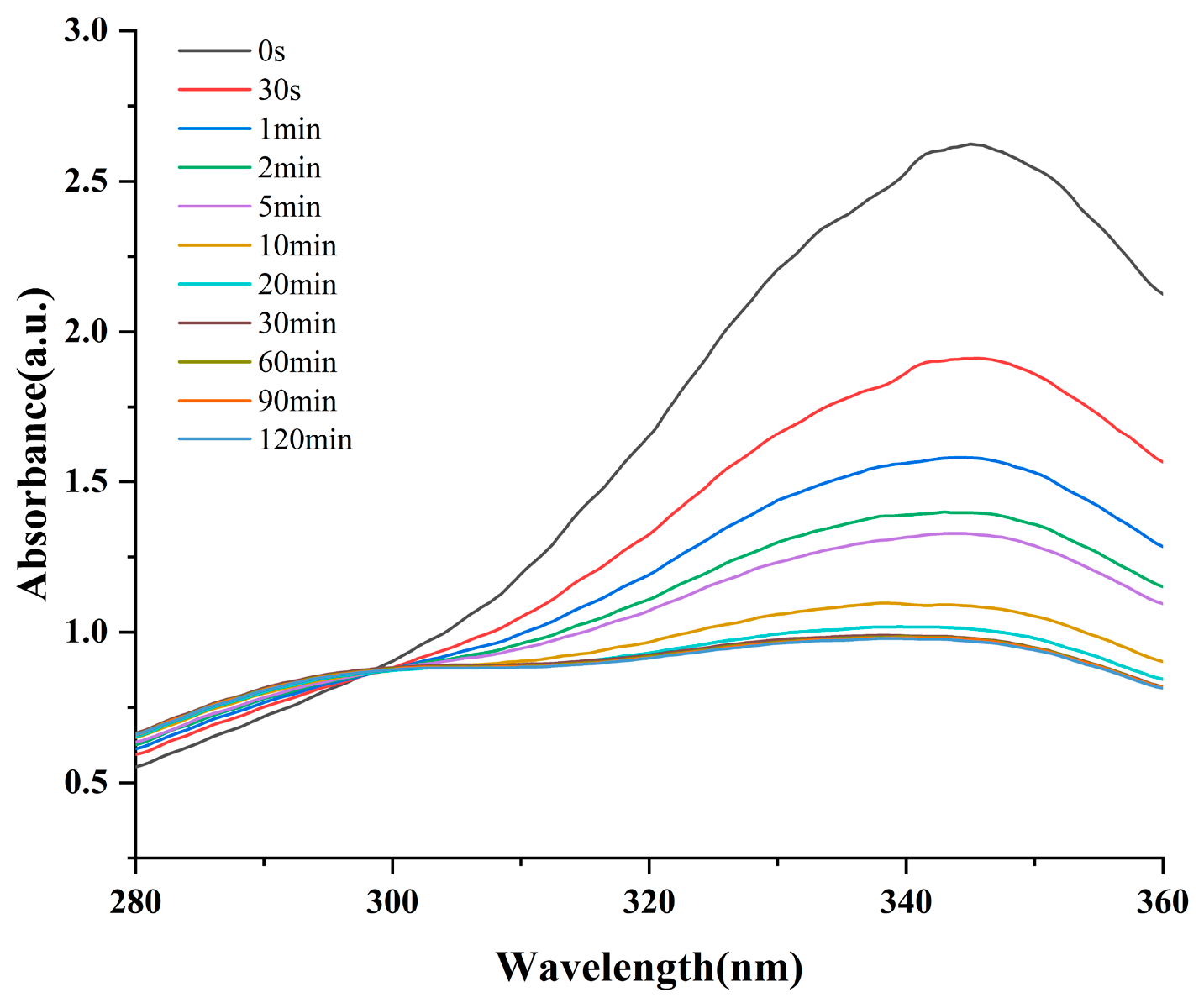
The photo-responsive behavior of Azo-MAA was characterized by UV-Vis absorption spectroscopy, with the results presented in Figure 2. In the absence of UV irradiation, Azo-MAA displayed a prominent absorption peak at 340 nm, which is ascribed to the π→π* transition of the trans-Azo group [34]. Upon exposure to UV light, the trans-Azo underwent photoisomerization to the cis configuration via light energy absorption. As the UV irradiation duration increased, the relative abundance of the trans-Azo moiety decreased, leading to a gradual attenuation of the absorption peak intensity. When the irradiation time reached 20 min, the system reached a photostationary state. Further extension of the irradiation time did not induce significant changes in the peak shape or position of the absorption curve.
Notably, the spectral data revealed that UV irradiation only altered the absorption peak intensity without generating new peaks or causing peak disappearance. This observation confirms that UV light exclusively triggered the photoisomerization of the Azo group rather than inducing side reactions such as photocrosslinking or photodegradation.

3.1.2. FT-IR Spectroscopy of Azo-MAA

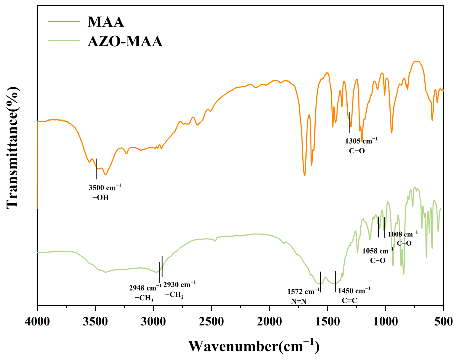
To confirm the chemical structure of Azo-MAA and the successful incorporation of target functional groups, the as-synthesized Azo-MAA was characterized by FT-IR spectroscopy, with the results presented in Figure 3. The C-O bond of the carboxyl group on MAA disappeared at 1305 cm−1. A distinct peak appeared at 1008 and 1058 cm−1, corresponding to the asymmetric stretching vibration of the ether bond (C-O-C) in the ester group. This confirmed that an esterification reaction occurred between MAA and Azo. Meanwhile, the stretching vibration peak of the hydroxyl group (-OH) at 3500 cm−1 was significantly attenuated. This phenomenon is primarily ascribed to the consumption of hydroxyl groups during covalent bond formation. The disappearance of the C-O peak, the appearance of the C-O-C peak, and the attenuation of the -OH peak indicated the successful synthesis of Azo-MAA [31]. Additionally, the C–H stretching vibration peaks of methylene (–CH2–) and methyl (–CH3) were observed at 2930 and 2948 cm−1, respectively, together with the aromatic C=C stretching at 1572 cm−1 and the –N=N– stretching at 1450 cm−1, further confirming the successful synthesis of Azo-MAA [35].

3.1.3. LC-MS Analysis of Azo-MAA

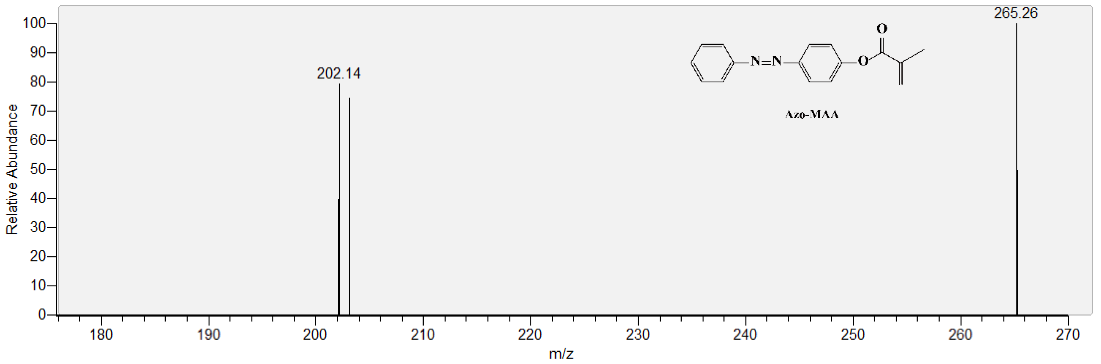
Liquid chromatography mass spectrometry (LC-MS) was employed for the qualitative characterization of Azo-MAA, with the results presented in Figure 4. In the negative ion mode ([M-H]), a distinct quasi-molecular ion peak was detected at a mass-to-charge ratio (m/z) of 265.26. This observed m/z value is in excellent agreement with the theoretical molecular weight of the deprotonated Azo-MAA ([M-H]), which confirms the successful synthesis of Azo-MAA. Further structural elucidation of fragmentation patterns was achieved through MS/MS analysis, as detailed in Figure S1.

3.2. Characterization of PMI-PDA@NH2-SiO2

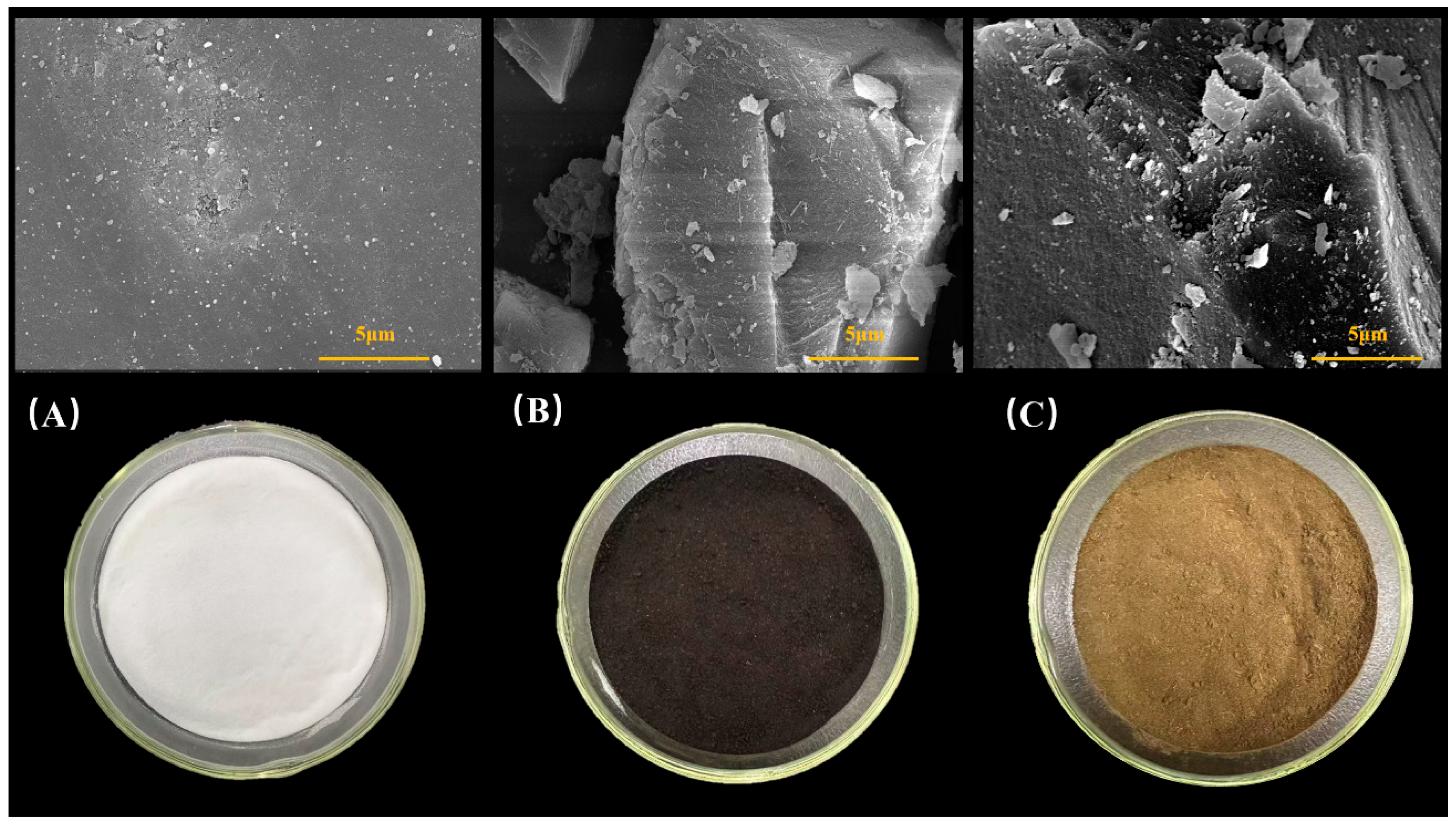
The surface morphology of the materials was characterized via scanning electron microscopy (SEM), with the results presented in Figure 5. The unmodified NH2-SiO2 appeared as irregular white powder particles with a relatively smooth surface, dense structure, and high specific surface area (Figure 5A) [32]. After PDA modification, PDA@NH2-SiO2 transformed into irregular black powder particles with a significantly increased surface roughness (Figure 5B). This morphological transformation confirms the successful deposition of PDA onto the NH2-SiO2 surface.
The final PMI-PDA@NH2-SiO2 exhibited a dark brown appearance (Figure 5C). While retaining the intrinsic framework of NH2-SiO2, its surface roughness was further enhanced, with granular substances observed locally adhering between the layered structures. Collectively, the SEM results indicate that the surface morphology of the NH2-SiO2 support evolved from smooth and dense to a rougher texture after stepwise synthesis and modification. The observed surface irregularities are consistent with the presence of template-specific cavities typically formed during molecular imprinting, which are considered essential for selective molecular recognition.
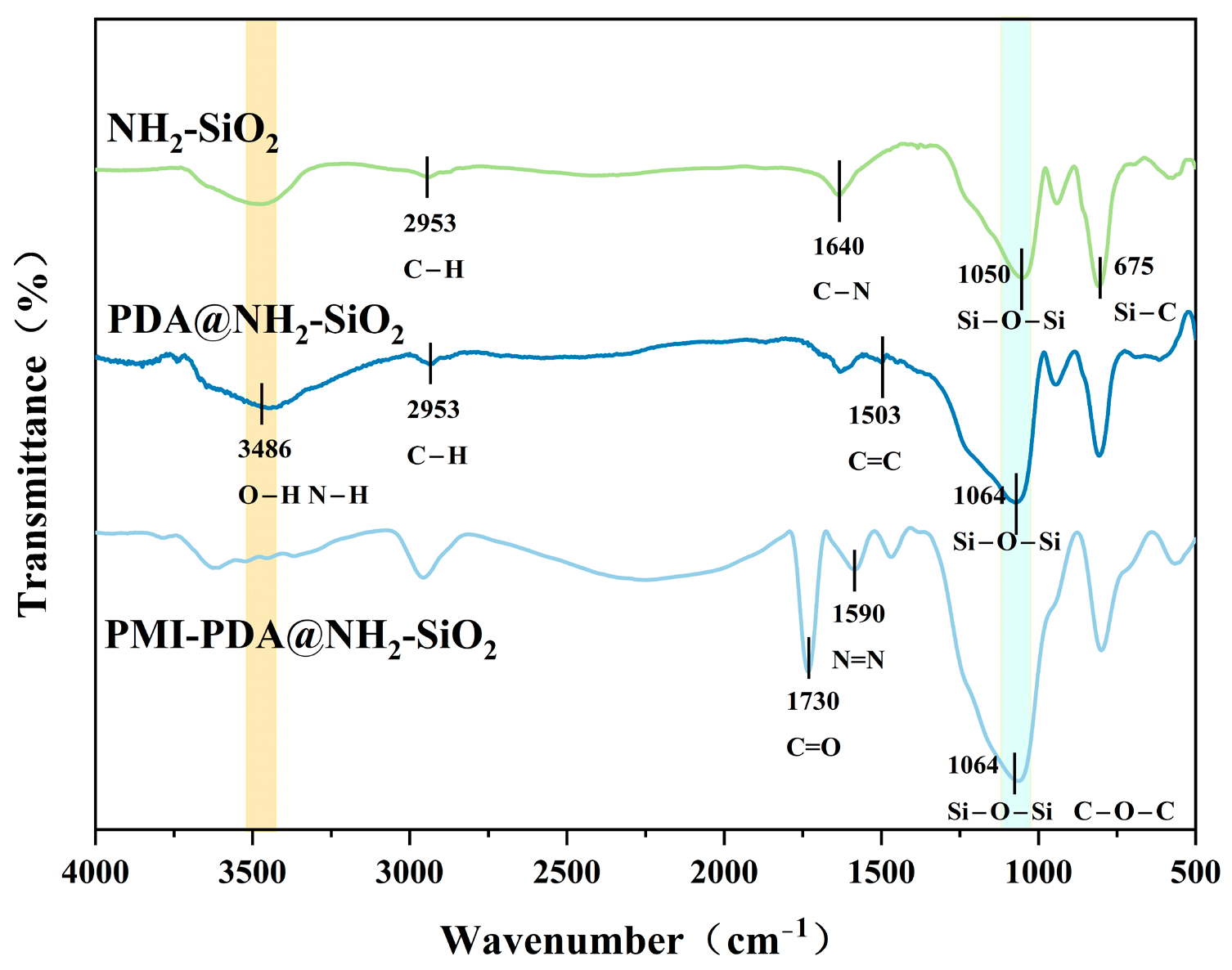
The stepwise preparation process of PMI-PDA@NH2-SiO2 was characterized by tracking the infrared characteristic absorption peaks of key functional groups during the preparation process. The corresponding FT-IR spectra are presented in Figure 6. NH2-SiO2 showed a peak at 2953 cm−1, attributed to the stretching vibration of aliphatic C-H; a broad peak in the range of 3400–3100 cm−1 was attributed to the stretching vibrations of N-H and O-H; and an asymmetric stretching vibration peak of Si-O-Si at 1050 cm−1 was consistent with the reported framework [36]. After modification with PDA, PDA@NH2-SiO2 exhibited a broader peak in the range of 3400–3100 cm−1, attributed to the superposition of the stretching vibrations of O-H and N-H of PDA. Simultaneously, a bending vibration peak of the benzene ring C=C at 1503 cm−1 confirmed the successful anchoring of PDA [37]. For PMI-PDA@NH2-SiO2, the introduction of Azo-MAA was confirmed by the C=O stretching vibration peak at 1730 cm−1 and the N=N stretching vibration peak at 1590 cm−1. Additionally, the broadening and intensification of the absorption peak at 1064 cm−1 might be the result of the superposition of the stretching vibration peak of Si-O-Si and the stretching vibration peak of the ether bond (C-O-C) in Azo-MAA, further confirming the successful introduction of the Azo-MAA [31]. The broad peak in the range of 3400–3100 cm−1 significantly weakened after the formation of the molecularly imprinted cavity, indicating that the phenolic hydroxyl and amino groups on the PDA surface as active sites might have participated in the imprinting polymerization process, thereby providing crucial spectroscopic evidence for the successful in situ polymerization of the molecularly imprinted layer on the PDA surface.

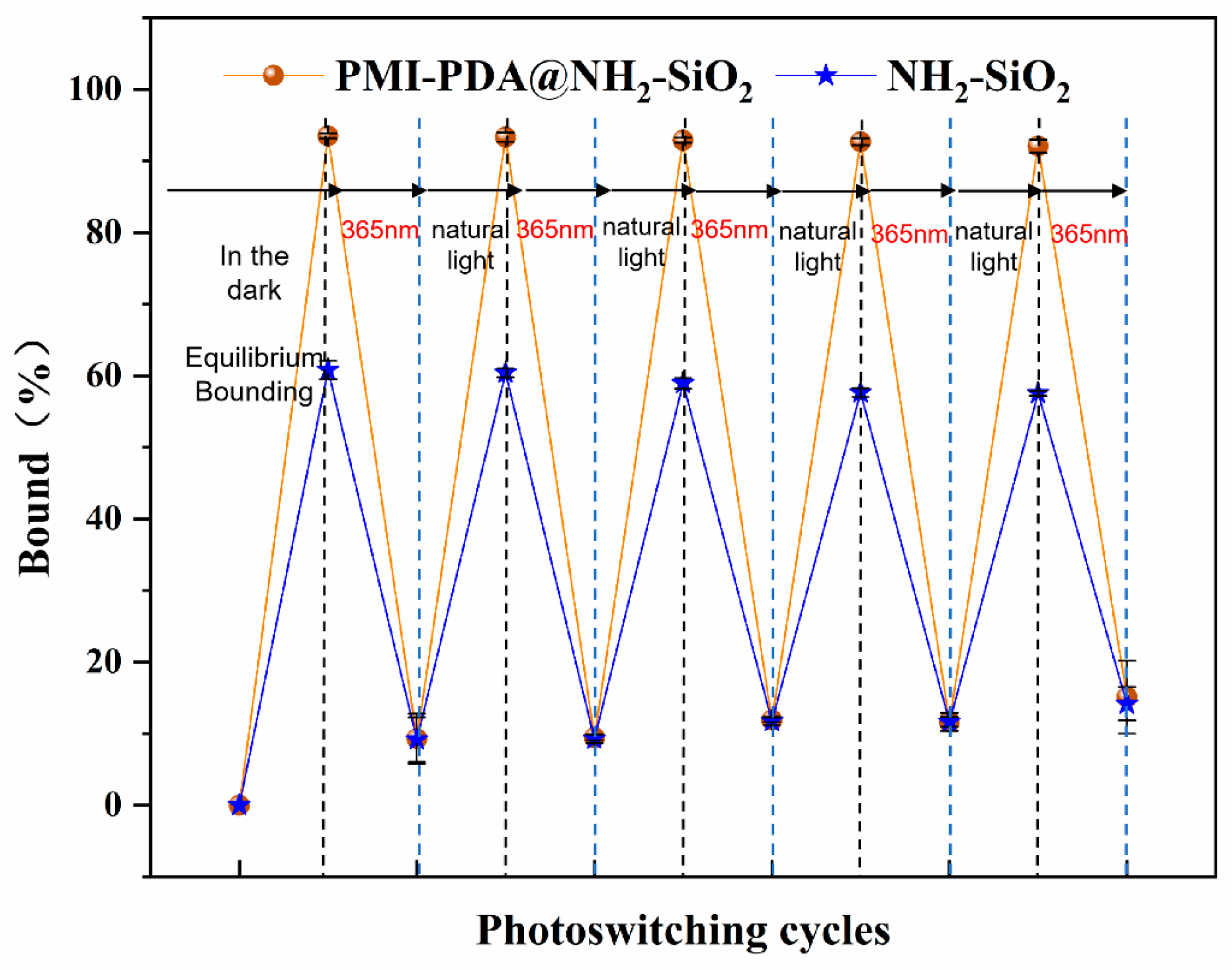
3.3. Results of the Photo-Controlled Adsorption/Desorption Experiments

To evaluate the photo-regulated adsorption/desorption performance of PMI-PDA@NH2-SiO2, photo-controlled adsorption/desorption curves (Figure 7) were constructed based on the variation of binding rates of PMI-PDA@NH2-SiO2 and NH2-SiO2 toward CA with irradiation time under different wavelengths. At adsorption equilibrium, the adsorption rate of PMI-PDA@NH2-SiO2 for CA reached up to 93.5%. The total adsorption capacity of NH2-SiO2 for CA was 60.8%, indicating that 32.7% of CA was adsorbed via the specific recognition sites of the molecularly imprinted material. Under 365 nm UV light irradiation, 84.2% of CA was released from the imprinted material into the solution. Upon switching to natural light, 80.0% of CA was re-adsorbed. In contrast, the binding capacity of NH2-SiO2 for CA decreased from 60.8% to 14.14% under alternating light irradiation.
The observed light-controlled adsorption/desorption behavior is primarily attributed to the cis-trans isomerization of the Azo chromophore. Isomerization of Azo induces reversible conformational changes in the specific molecularly imprinted cavities. Owing to the sufficient free space within the material and the higher energy required for Azo isomerization compared to the interaction energy between the template molecule and receptor sites, CA is driven to release from the imprinted sites [38]. These results confirm that PMI-PDA@NH2-SiO2 exhibits significantly superior light-regulated adsorption/desorption performance relative to NH2-SiO2.
After five cycles of cis-trans isomerization, PMI-PDA@NH2-SiO2 retained a high adsorption capacity for CA, demonstrating excellent reversibility and cycling stability of the material’s specific receptor sites and imprinting affinity. No significant attenuation of specific recognition ability was observed during multiple light-controlled cycles.

3.4. Results of the Adsorption Experiments

3.4.1. Adsorption Kinetics

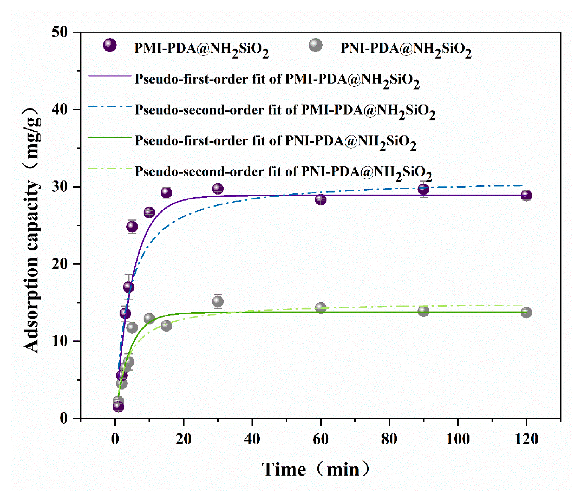
To investigate the adsorption behavior of PMI-PDA@NH2-SiO2 and PNI-PDA@NH2-SiO2 toward CA, adsorption kinetic experiments were performed. The adsorption kinetic results are presented in Figure 8. The adsorption capacity of PNI-PDA@NH2-SiO2 for CA increased rapidly within the first 10 min, followed by a slower rate of increase, and stabilized at 20 min. In contrast, the adsorption capacity of PMI-PDA@NH2-SiO2 for CA increased more rapidly within the first 20 min and reached equilibrium at 30 min. Although both silica-based materials exhibited similar trends in adsorption capacity, their saturated adsorption capacities differed significantly: the saturated adsorption capacity of PMI-PDA@NH2-SiO2 was approximately twice that of PNI-PDA@NH2-SiO2. This discrepancy is attributed to the abundant molecularly imprinted cavities on the surface of PMI-PDA@NH2-SiO2, which are structurally matched to CA, thereby conferring superior adsorption performance for CA.
The kinetic fitting results are summarized in Table 1. The pseudo-first-order model (R2 = 0.949) demonstrated a higher correlation coefficient than the pseudo-second-order model (R2 = 0.877), and the theoretical equilibrium adsorption capacity (Qe1 = 28.85 mg/g) was close to the experimental value. These results suggest that adsorption occurs mainly on the surface, aligning with the surface imprinting feature of PMI-PDA@NH2-SiO2. This is because the surface-located recognition sites promote rapid diffusion of the target to the active sites. This further confirms the affinity and selectivity of PMI-PDA@NH2-SiO2 for CA [39].

3.4.2. Adsorption Isotherms

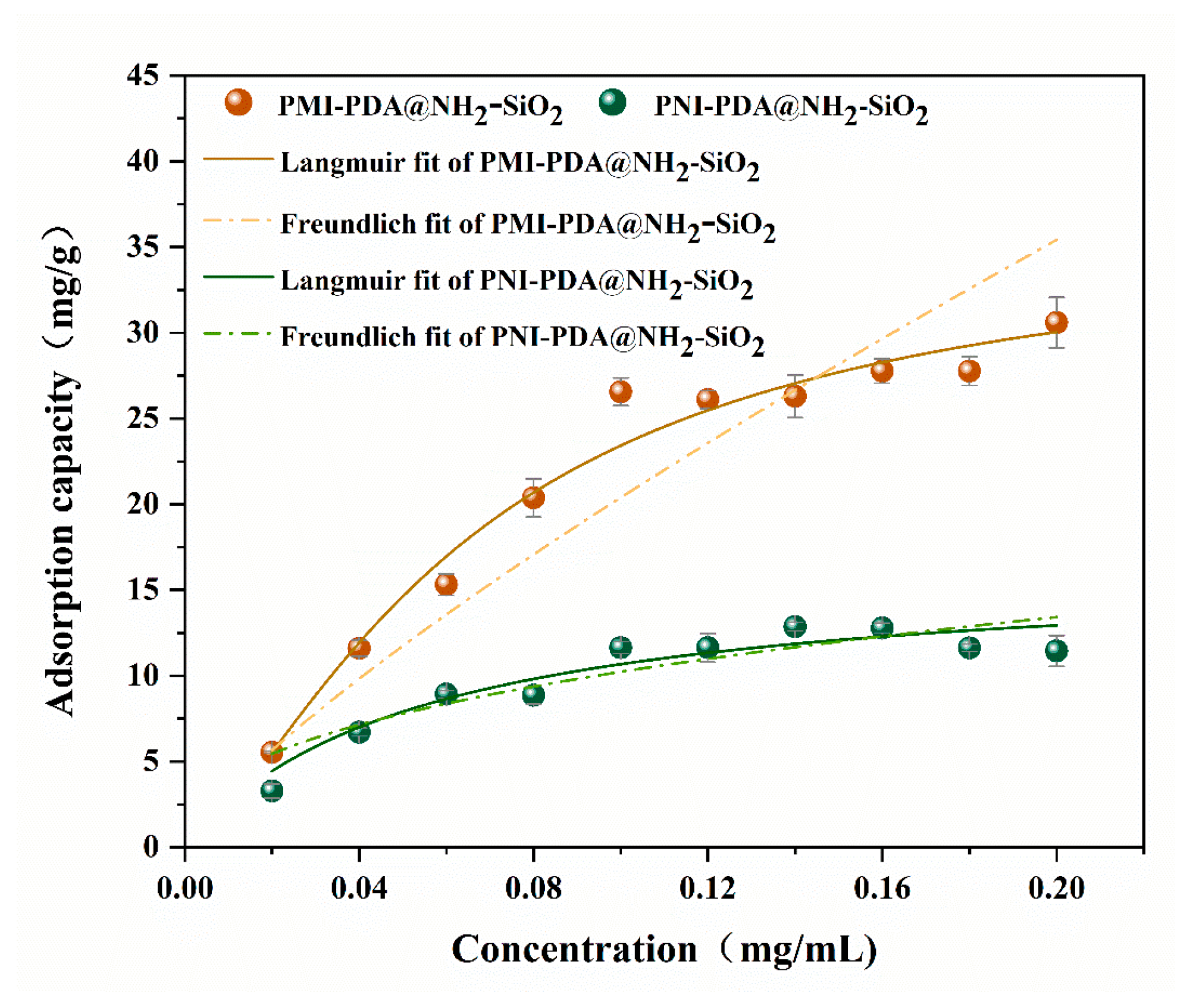
The adsorption capacities of PMI-PDA@NH2-SiO2 and PNI-PDA@NH2-SiO2 in CA solutions of varying concentrations were evaluated through adsorption isotherm experiments, and the adsorption data were fitted and analyzed using the Langmuir and Freundlich models. As shown in Figure 9 and Table 2, the results indicate that the adsorption capacities of both PMI-PDA@NH2-SiO2 and PNI-PDA@NH2-SiO2 increased with rising initial CA concentration, reaching saturation at approximately 0.10 mg/mL. Notably, PMI-PDA@NH2-SiO2 exhibited significantly superior adsorption performance compared to PNI-PDA@NH2-SiO2, with a maximum adsorption capacity of 35.90 mg/g, approximately twice that of PNI-PDA@NH2-SiO2. This enhanced performance suggests that PMI-PDA@NH2-SiO2 possesses specific recognition sites capable of forming interactions with CA molecules, resulting in higher adsorption capacity and selectivity [40]. The Langmuir model provided a better fit for PMI-PDA@NH2-SiO2 (R2 = 0.992), indicating that CA adsorption on PMIP-PDA@NH2-SiO2 primarily occurs as monolayer adsorption, with a uniform surface and consistent adsorption site sizes.

3.4.3. Adsorption Selectivity

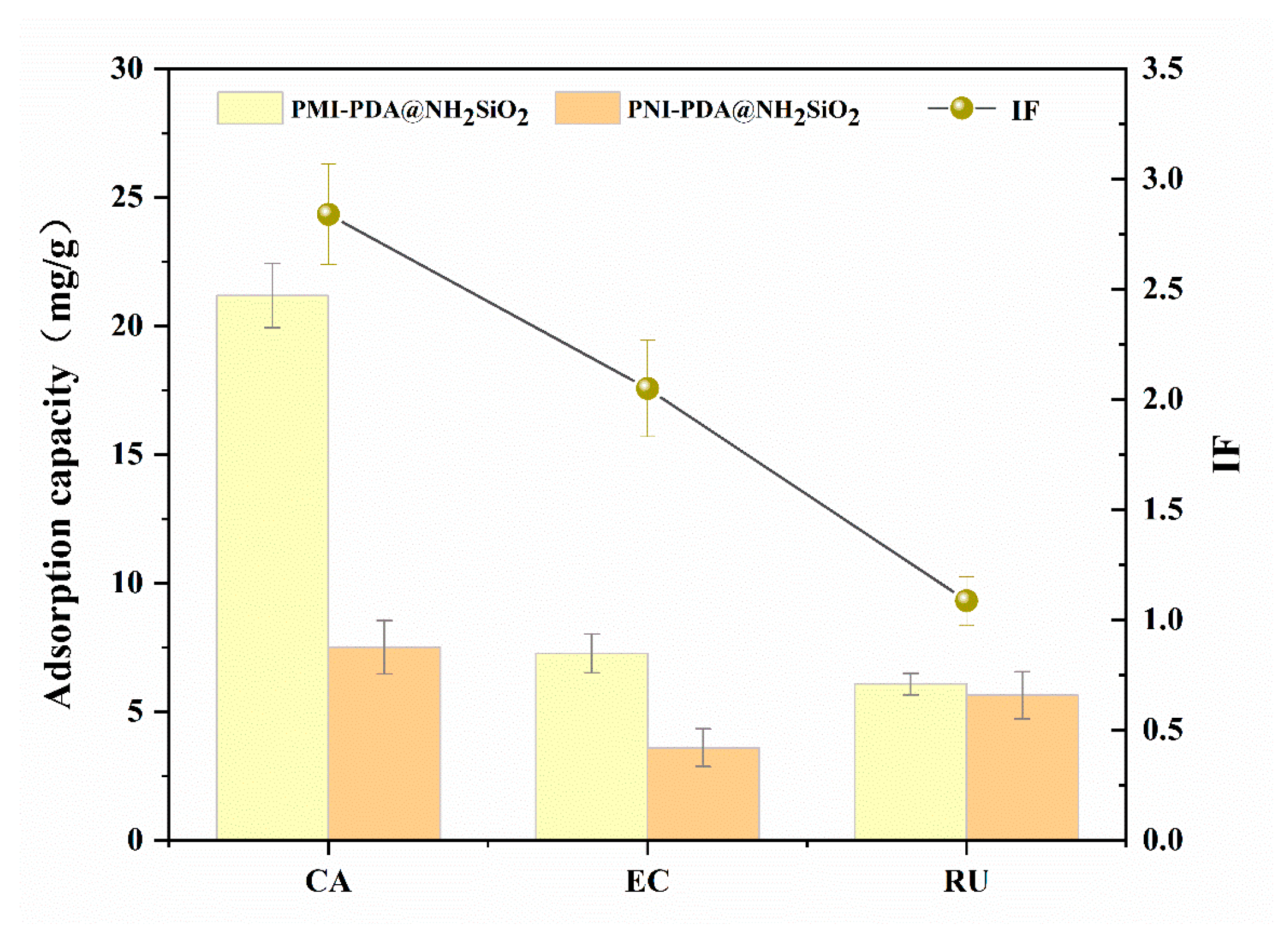
To further assess the selective adsorption capability of PMI-PDA@NH2-SiO2 toward chlorogenic acid (CA) and its structural analogues (epicatechin, EC; rutin, RU), selective adsorption experiments were performed and the IF values were calculated. The results are presented in Figure 10 and Table 3. The adsorption capacities of PMI-PDA@NH2-SiO2 for CA, EC, and RU were 21.18 mg/g, 7.27 mg/g, and 6.08 mg/g, respectively, with corresponding IF values of 2.84, 2.05, and 1.09. The selectivity factors for CA relative to EC and RU were 5.08 and 6.29, respectively, indicating that the adsorption capacity of PMI-PDA@NH2-SiO2 for CA was significantly higher than that for its structural analogs. This enhanced selectivity is attributed to the presence of specific imprinted sites on the surface of PMI-PDA@NH2-SiO2, which are precisely matched to the molecular structure of CA. In contrast, PNI-PDA@NH2-SiO2, which lacks specific recognition cavities, exhibited substantially lower adsorption capacities compared to PMI-PDA@NH2-SiO2.
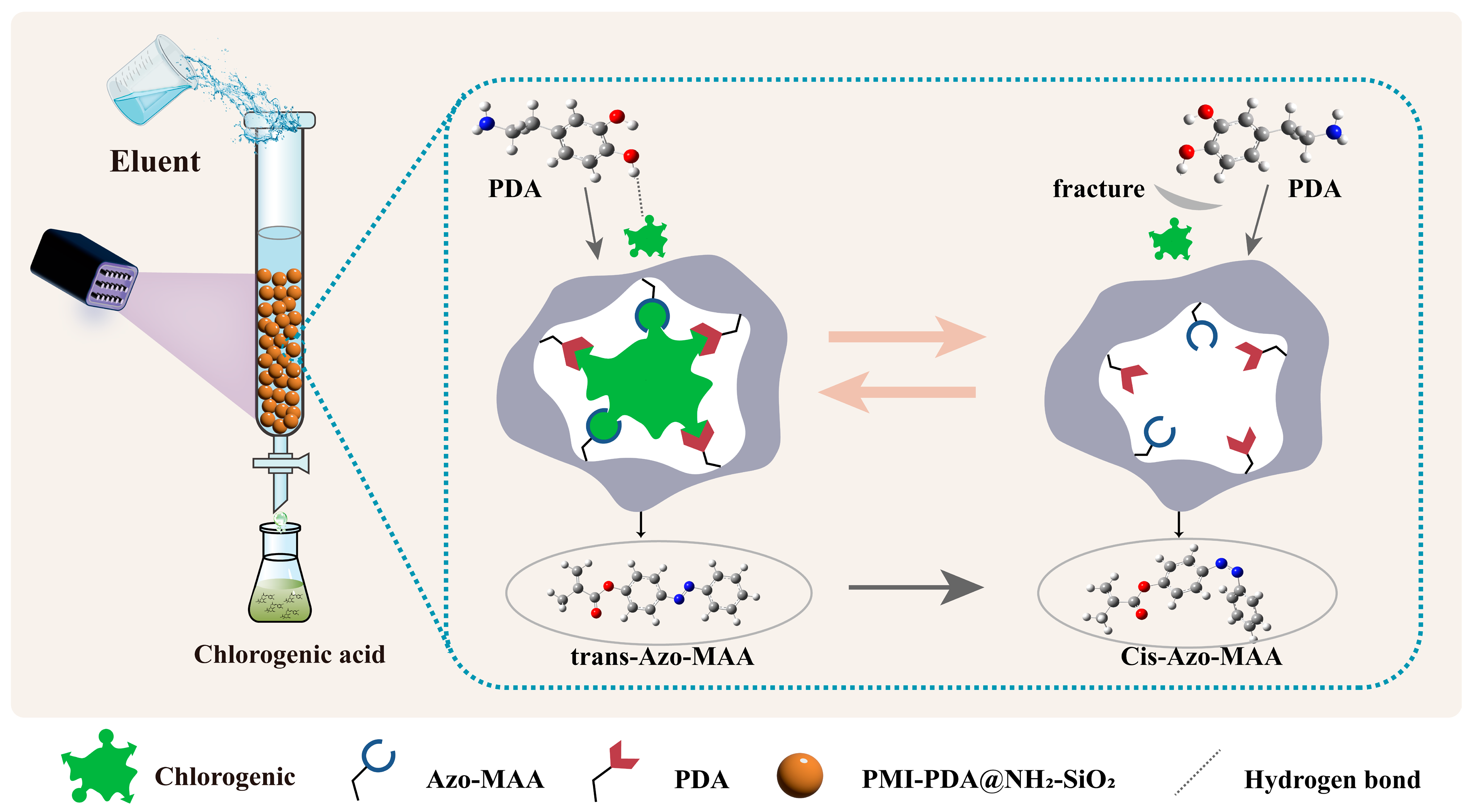
The adsorption mechanism of PMI-PDA@NH2-SiO2 for CA is illustrated in Figure 11. During the molecular imprinting process, CA binds to PDA to form cavity structures with memory functionality. After CA removal, these cavities retain the spatial configuration features of CA, analogous to the “key-and-lock” specific recognition principle [41]. Thus, when CA re-interacts with PMI-PDA@NH2-SiO2, it can precisely fit into these cavities and exhibit strong adsorption.

3.4.4. Recyclability Experiments

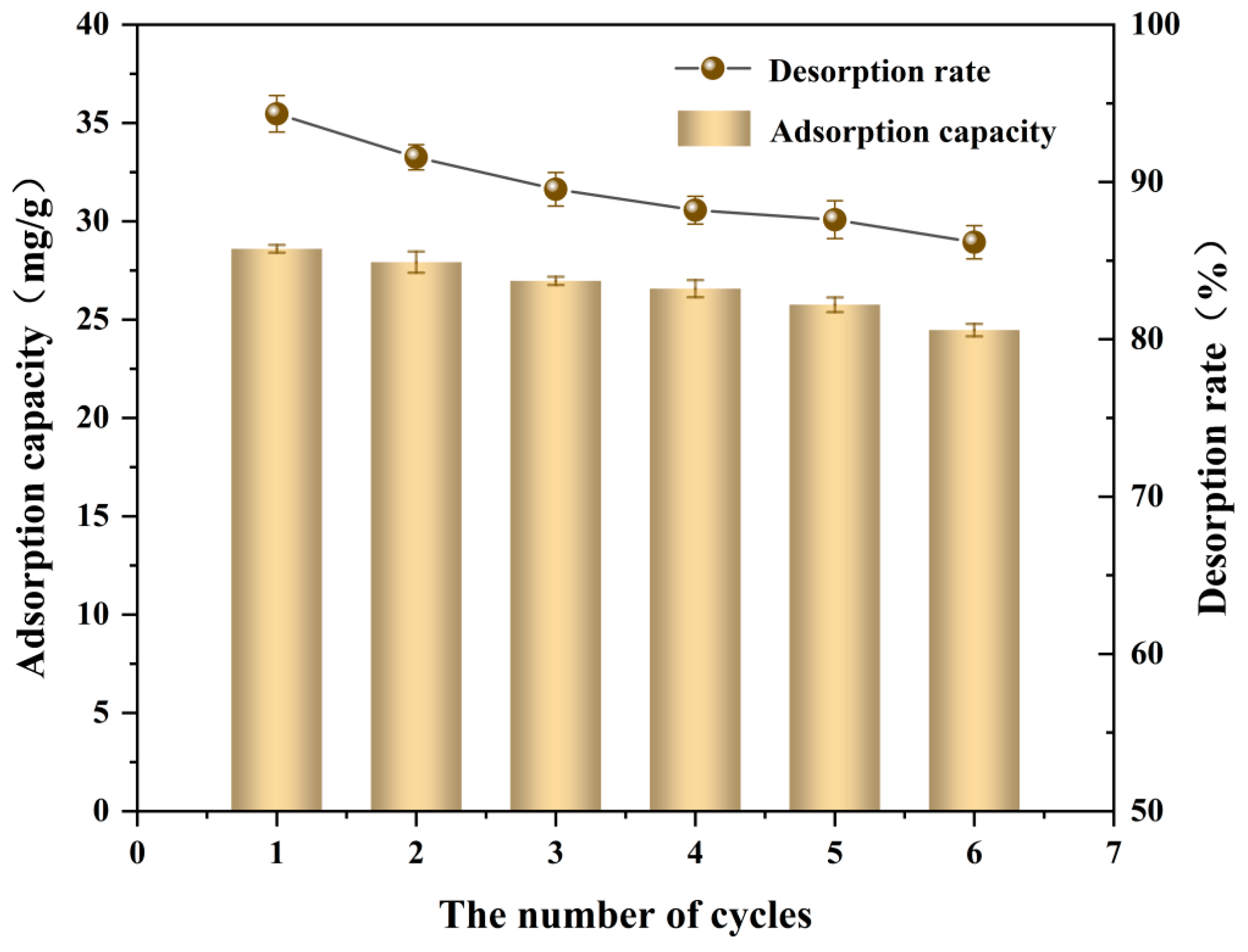
Six consecutive adsorption/desorption cycles of CA were performed using PMI-PDA@NH2-SiO2, and the results are presented in Figure 12. The experimental data reveal that both the adsorption capacity and desorption efficiency of PMI-PDA@NH2-SiO2 toward CA exhibit a gradual downward trend with increasing cycle numbers. After six cycles, the adsorption capacity of PMI-PDA@NH2-SiO2 still retains 85.56% of its initial maximum capacity, while the desorption efficiency remains at 91.38% of its initial level. These findings confirm that PMI-PDA@NH2-SiO2 can be effectively recycled for at least six cycles to achieve efficient separation of CA, indicating that the material not only possesses excellent adsorption performance but also maintains stable separation capability over multiple cycles.

3.5. Analysis of the Recognition Mechanism

3.5.1. Mulliken Atomic Charge Distribution

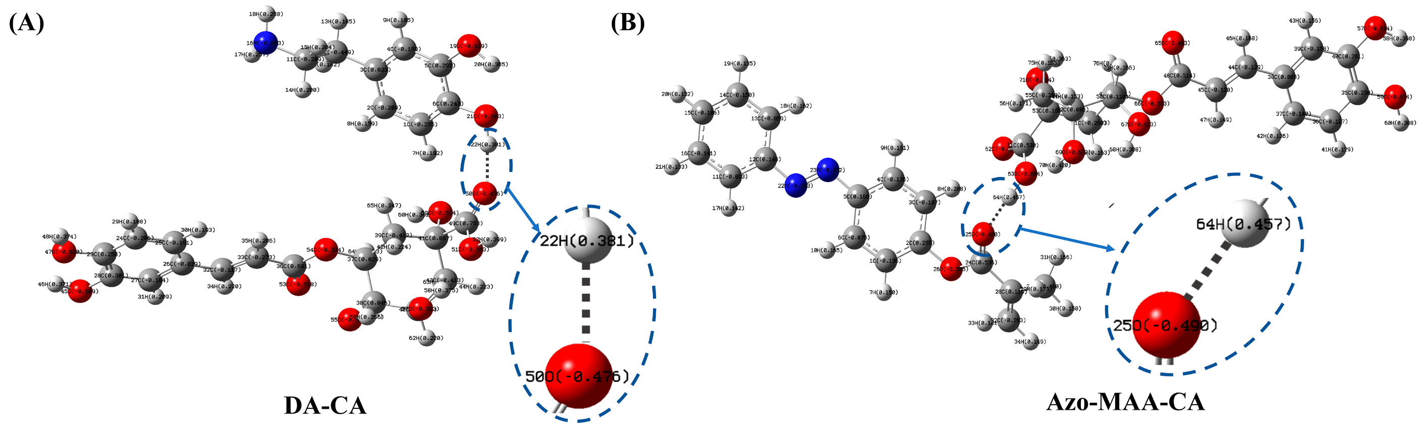
The optimized structure of the interaction system between the template molecule (CA) and the functional monomer (DA) is depicted in Figure 13A. The mechanism underlying the intermolecular interactions between CA and DA was elucidated via Mulliken charge distribution analysis. As illustrated in Figure 13A, the 50th oxygen (O) atom in CA bears a pronounced negative charge, acting as an electron acceptor, whereas the 22nd hydrogen (H) atom in DA carries a distinct positive charge, functioning as an electron donor. Concurrently, the optimized structure of the interaction system between CA and Azo-MAA is presented in Figure 13B. The 69th O atom in CA exhibits a significant negative charge (–0.527), serving as an electron acceptor, while the 30th H atom in Azo-MAA possesses a notable positive charge (0.440), acting as an electron donor. This complementary charge distribution suggests that intermolecular hydrogen bonds (O50···H22, O69···H30) can be formed between the two molecules via electrostatic interactions. Electrostatic potential analysis further validates the prominent charge polarization effect within the system. The electron-rich regions surrounding oxygen atoms in CA and the electron-deficient regions around hydrogen atoms in the two functional monomers exhibit a clear spatial correspondence.

3.5.2. IGMH Analysis

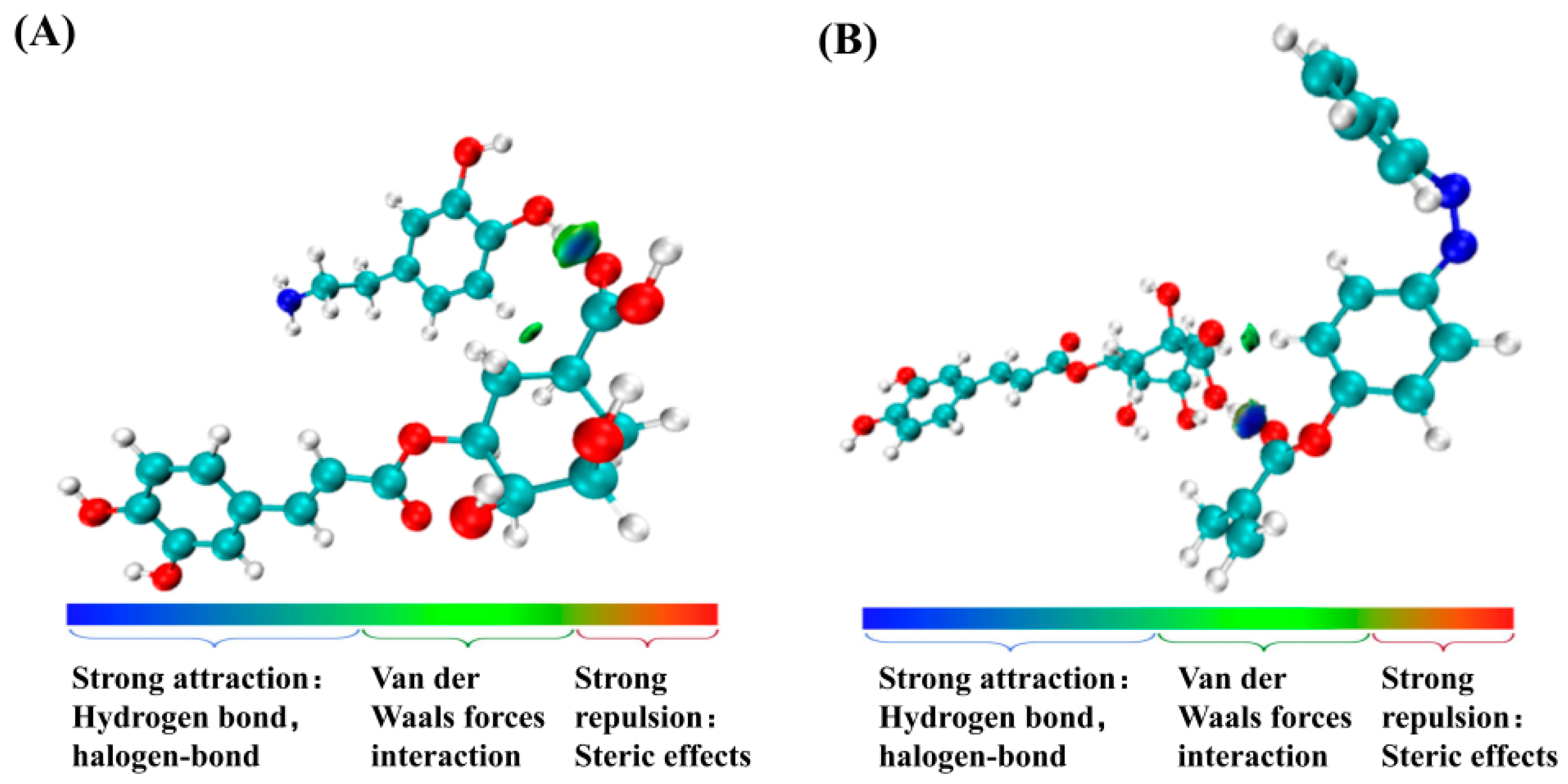
The independent gradient model based on Hirshfeld partitioning (IGMH) was employed to probe the non-covalent interaction mechanisms within the CA-PDA and CA-Azo-MAA systems [42]. By computing the electron density distribution and its gradient variations, IGMH delineates the spatial distribution and intensity of intermolecular interactions, including hydrogen bonds, van der Waals forces, and repulsive forces. As illustrated in Figure 14, in the IGMH isosurface plots of the CA-PDA and CA-Azo-MAA systems, blue regions denote attractive interactions, green regions correspond to van der Waals interactions, and red regions signify repulsive interactions. Notably, blue and green isosurface features were observed between the C=O group of CA and the O-H groups of DA and Azo-MAA, indicating the existence of strong hydrogen bonds and van der Waals forces. This thus validates the selective binding mechanism between CA and DA/Azo-MAA mediated by hydrogen bonds and van der Waals forces [43].

3.5.3. Molecular Orbital Theory

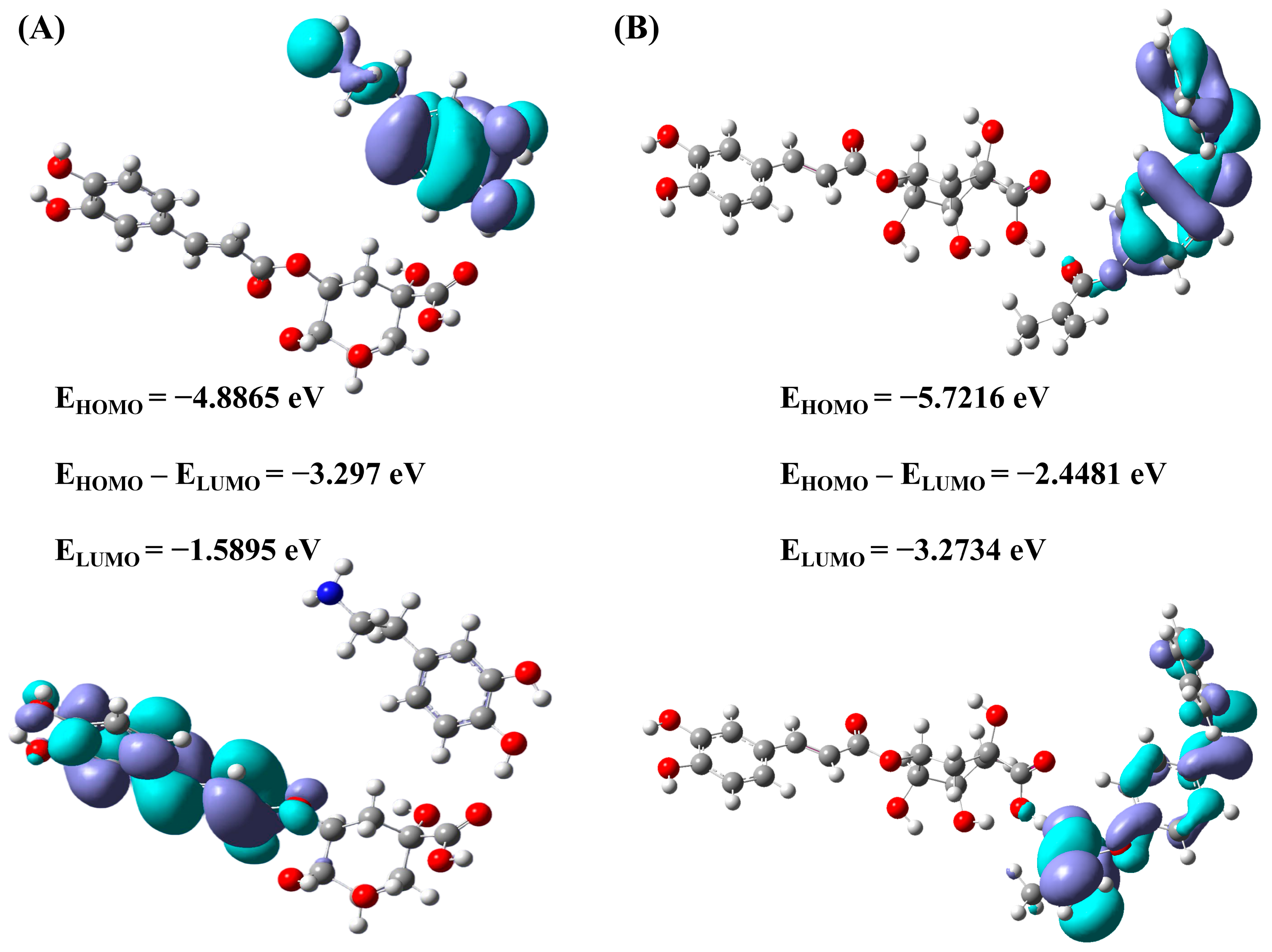
Based on molecular orbital theory, the reactivity and stability of the CA-DA system were analyzed. The frontier molecular orbitals (FMOs) of the CA-DA system are depicted in Figure 15A. The highest occupied molecular orbital (HOMO) and the lowest unoccupied molecular orbital (LUMO) energies of the CA-DA system are –5.2197 eV and –1.8806 eV, respectively, corresponding to a HOMO–LUMO energy gap of 3.3391 eV. The frontier molecular orbitals (FMOs) of the CA-Azo-MAA system are displayed in Figure 15B. For CA-Azo-MAA, the HOMO and LUMO energies are –5.7216 eV and –2.4481 eV, respectively, giving a HOMO–LUMO gap (ΔE) of 3.2734 eV, which reflects a relatively small separation between the occupied and unoccupied orbitals. A narrower energy gap is generally associated with a higher probability of electronic transitions and enhanced electron transport properties [44,45], further corroborating the excellent stability of CA-PDA and CA-Azo-MAA systems.

3.5.4. NMR Analysis

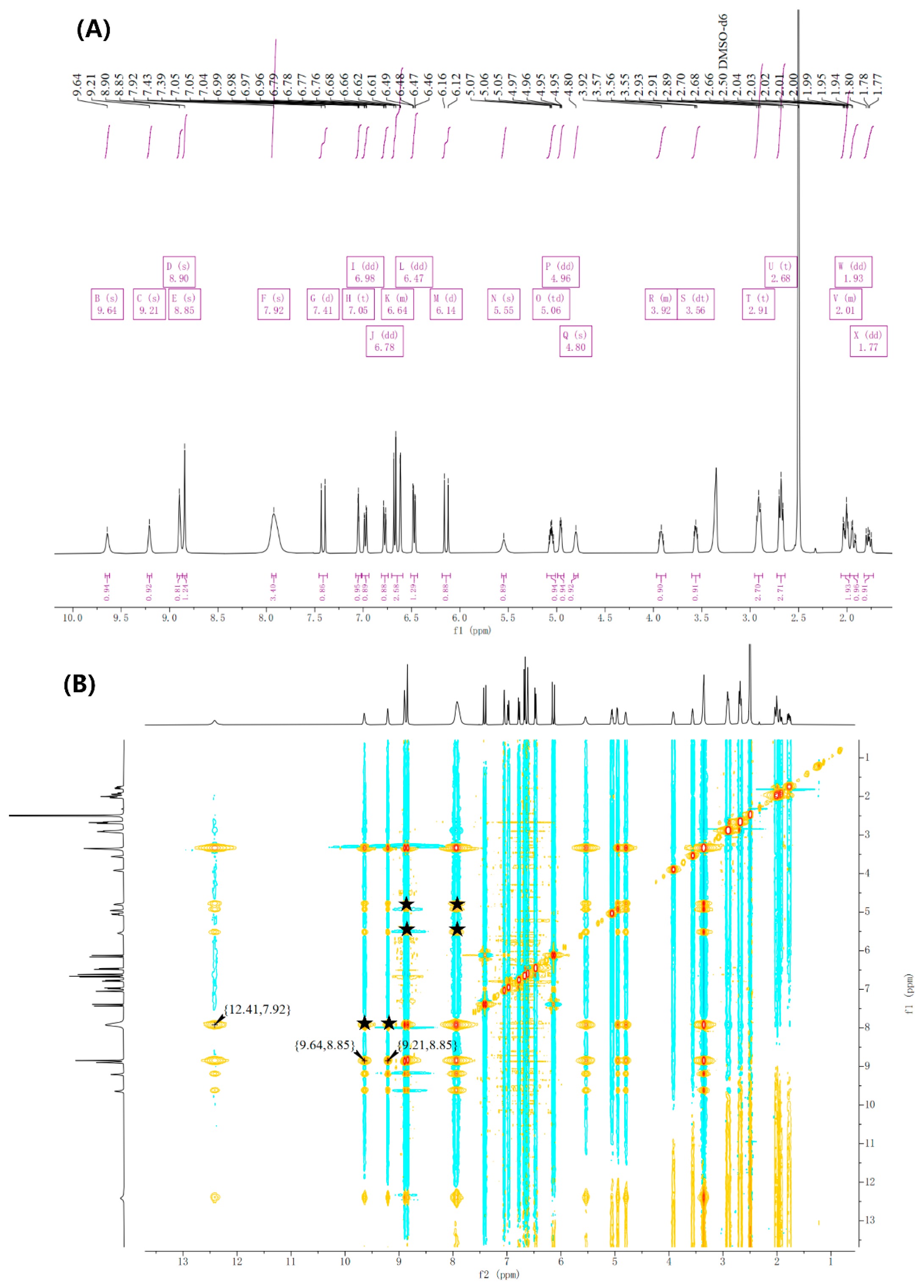
Nuclear magnetic resonance (NMR) spectroscopy, particularly 1H NMR and 2D NOESY, serves as a robust tool for investigating intermolecular interactions [46]. In this study, a 1:1 molar ratio of CA to DA was employed to probe their molecular interactions. The 1H NMR spectrum of the CA-DA complex is presented in Figure 16A, and corresponding proton assignments are presented in the Supporting Information (Tables S1 and S2). Comparative analysis against the spectra of the pure components revealed significant chemical shift perturbations for the 3-OH and 4-OH protons of DA, as well as the 1-OH, 2-OH, NH2, 2′-OH, and 3′-OH protons of CA.
2D NOESY spectral analysis of the complex further elucidated its binding mode (Figure 16B). Distinct cross-peaks were observed between δ 9.21 and 8.85 and between δ 9.64 and 8.85, indicating spatial proximity between the aromatic ring of DA and the phenolic hydroxyl groups of CA, attributed to π-π stacking interactions. Additionally, the cross-peak at δ 7.92 and δ 12.41 confirmed hydrogen bond formation between the amino group of DA and the carboxyl proton of CA.
Other hydrogen bond interactions were validated by cross-peaks between δ 9.21/δ 9.64/δ 4.80/δ 5.55 and δ 7.92, corresponding to interactions between the hydroxyl groups of CA and the amino group of DA. Furthermore, the cross-peak between δ 5.55/δ 4.80 and δ 8.90 suggested the formation of a hydrogen bond network between the hydroxyl groups of CA and the aromatic hydroxyl groups of DA. Collectively, these findings indicate a potential synergistic interplay between hydrogen bonding and van der Waals forces in the CA-DA interaction.

3.6. Comparison of PMI-PDA@NH2-SiO2 with Other Adsorbents

The prepared adsorbent was systematically compared with other molecularly imprinted materials reported in the literature for CA separation, and the results are shown in Table 4. Specifically, 3D-MMIPs use M-GO-MWNTs as a carrier, significantly increasing the specific surface area and improving selectivity. Unlike 3D-MMIPs that require repeated strong-acid elution, our PMI-PDA@NH2-SiO2 material eliminates this step and is therefore more sustainable. Fe3O4-Cu@Lyz-MIPs employ phase-transformed lysozyme as the MIP imprinting shell, exhibiting good acid resistance and structural stability. CGA-MIP uses [BMIM]BF4–DMSO as a porogen, enabling a distinct imprinting effect even at a low template-to-functional monomer molar ratio of 1:240. Nevertheless, none of these materials possess photo-responsive properties. In contrast, the material developed in this work achieves adsorption and desorption through a photo-responsive mechanism, avoiding the use of large amounts of organic solvents required in conventional elution methods. Moreover, the light-controlled process does not require direct contact with the material, operates under mild conditions, and exhibits excellent recyclability, offering clear advantages in green separation and sustainability.

3.7. Purification of CA from Ficus carica L. Using PMI-PDA@NH2-SiO2 SPE Column

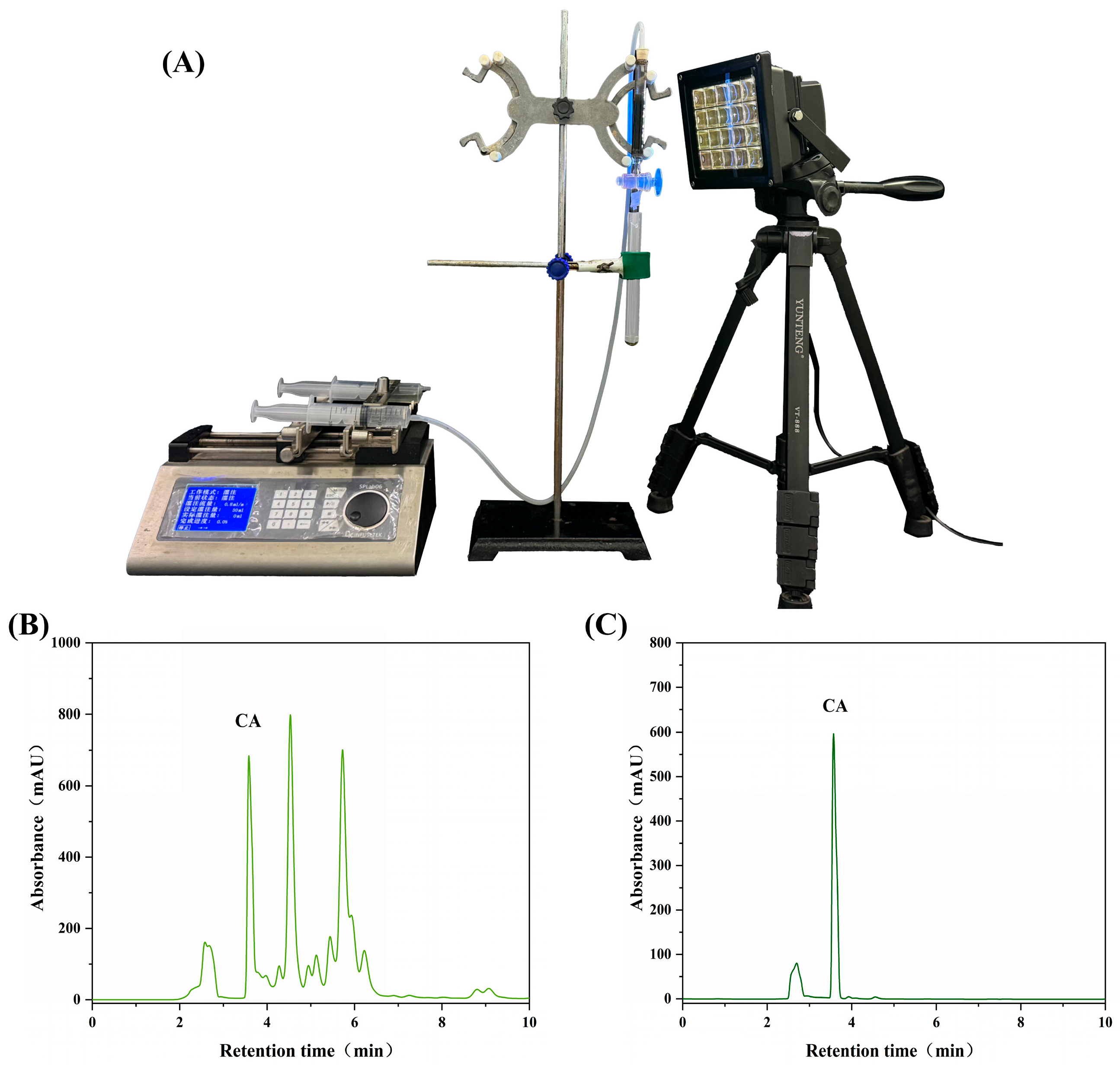
The as-prepared PMI-PDA@NH2-SiO2 was packed into a solid-phase extraction (SPE) column for the selective separation and purification of CA from F. carica L. extracts, with the setup illustrated in Figure 17A. The SPE procedure was carried out as follows. First, PMI-PDA@NH2-SiO2 was loaded into the SPE column, followed by the introduction of the F. carica L. extract sample for dynamic adsorption. Once PMI-PDA@NH2-SiO2 reached adsorption saturation, the adsorbed CA was eluted using an appropriate eluent. The elution mechanism involves the competition between free H+ ions in acetic acid and the -OH groups of CA. This competitive interaction disrupts the hydrogen bonding between CA and PMI-PDA@NH2-SiO2, thereby facilitating the release of CA.
HPLC chromatograms of the F. carica L. extract before adsorption and the eluent after adsorption are presented in Figure 17B and Figure 17C, respectively, with quantitative analysis performed via HPLC. In the HPLC profile of the F. carica L. extract, a characteristic peak corresponding to CA was observed at a retention time of 3.58 min. The PMI-PDA@NH2-SiO2-packed SPE column exhibited an adsorption rate (Rads) of 92.3%, a desorption rate (Rdes) of 87.27%, a recovery rate of 80.55%, and a CA purity (PCA) of 82.62% in the eluent. This confirms the outstanding adsorption performance of PMI-PDA@NH2-SiO2 toward CA. These results demonstrate that PMI-PDA@NH2-SiO2 can serve as an efficient SPE packing filler for the separation of CA from F. carica L. fruits.

3.8. Greenness Assessment and AGREEMIP Analysis

AGREEMIP is a measurement tool and software for evaluating and comparing the environmental sustainability of the MIP synthesis process [48].This study assesses the greenness of the PMI-PDA@NH2-SiO2 molecular imprinting process using the AGREEMIP software (Version 1.0) metric. Each of its 12 principles is scored based on experimental data, normalized, and weighted to obtain an overall greenness value. As seen in Figure S3, PMI-PDA@NH2-SiO2 can be categorized as good greenness based on the final score of 0.76. As shown in Table S3, the favorable greenness performance is mainly attributed to several high-scoring criteria. In particular, the removal of polymerization inhibitors, template usage, porogen/solvent selection, other reagents/adjuvants/carriers, and core/particle preparation or surface modification all received scores of 1.0, indicating low environmental burden in these aspects. The template elution solvent also showed a high score of 0.92, suggesting relatively limited solvent-related hazard during template removal. In addition, the functional monomer and cross-linking agent obtained moderate-to-high scores of 0.65 and 0.62, respectively, further supporting the overall acceptable sustainability of the synthesis route. Although some steps, such as polymerization initiation, particle size control, template elution technique, and final product reusability, showed comparatively lower scores, their influence did not prevent the process from achieving an overall good greenness rating.
Therefore, the AGREEMIP evaluation demonstrates that the PMI-PDA@NH2-SiO2 molecular imprinting process has favorable environmental sustainability, especially in terms of reagent consumption, solvent use, and template elution solvent safety.

4. Conclusions

A photo-responsive molecularly imprinted polymer (PMI-PDA@NH2-SiO2) was successfully prepared. In this system, DA served a dual function by self-polymerizing on the NH2-SiO2 surface to form a PDA layer and acting as a green functional monomer to facilitate the formation of the imprinted layer. This dual functionality thereby increased adsorption sites and conferred excellent selectivity and affinity toward the template molecule CA. Additionally, Azo-MAA was incorporated as a photo-responsive monomer, endowing the polymer with photo-responsive properties and enhancing the efficiency of its adsorption/desorption processes.
Subsequently, PMI-PDA@NH2-SiO2 was employed as an efficient adsorbent filler for solid-phase extraction (SPE) columns, enabling the effective separation of CA from F. carica L. extracts. The maximum adsorption capacity of PMI-PDA@NH2-SiO2 for CA reached 28.6 mg/g at a CA concentration of 0.1 mg/mL. The SPE column filled with PMI-PDA@NH2-SiO2 exhibited an adsorption rate of 92.33% and a desorption rate of 87.27% for CA in F. carica L. extracts, confirming its superior adsorption performance for CA. Moreover, PMI-PDA@NH2-SiO2 demonstrated high selectivity and affinity for CA, with an IF of 2.84.
After six consecutive adsorption/desorption cycles, PMI-PDA@NH2-SiO2 retained robust adsorption performance and reusability. Its adsorption capacity remained at 86.17%, with only a 4.13 mg/g decrease in adsorption amount. Furthermore, the recognition mechanism of PMI-PDA@NH2-SiO2 was elucidated to be primarily the synergistic effect of hydrogen bonding and van der Waals forces between the template molecule CA and the functional monomer DA. This work proposes a novel photo-responsive, non-contact regulation strategy for the green and efficient separation of CA. The photo-responsive reversibility of this material significantly improves its reusability, promoting the greening and sustainable development of chemical separation processes and demonstrating considerable potential for industrial application.

Supplementary Materials

The following supporting information can be downloaded at: https://www.mdpi.com/article/10.3390/separations13070200/s1. Table S1: 1H NMR chemical shifts and peak assignments of DA and CA (400 MHz, DMSO-d6); Table S2: 1H NMR chemical shifts and peak assignments of CA mixing with DA (400 MHz, DMSO-d6); Table S3: AGREEMIP scoring breakdown and weight distribution; Figure S1: MS/MS spectrum of Azo-MAA; Figure S2: The chemical structures of RU and EC; Figure S3. Pictogram of AGREEMIP.

Author Contributions

Conceptualization, Y.Y., X.X., M.L. and C.L. (Chunying Li); methodology, Y.Y., X.X., J.Z. and C.L. (Chunying Li); software, X.X., J.S., C.L. (Chuancheng Lin) and C.L. (Chunying Li); validation, M.L., W.L. and C.Z.; formal analysis, Y.Y., X.X., C.L. (Chuancheng Lin) and C.L. (Chunying Li); investigation, Y.Y., X.X., J.S. and C.L. (Chunying Li); resources, Y.Y., X.X., J.Z. and C.L. (Chunying Li); data curation, Y.Y., X.X., J.S. and C.L. (Chunying Li); writing—original draft preparation, Y.Y., X.X. and C.L. (Chunying Li); writing—review and editing, Y.Y., X.X. and C.Z.; visualization, Y.Y., X.X., C.L. (Chuancheng Lin), W.L. and C.Z.; supervision, C.Z. and C.L. (Chunying Li); project administration, C.L. (Chunying Li) and C.Z.; funding acquisition, C.L. (Chunying Li) and C.Z. All authors have read and agreed to the published version of the manuscript.

Funding

Funding was provided by the financial support of the National Natural Science Foundation (32471844).

Data Availability Statement

The original contributions presented in this study are included in the article or supplementary material. Further inquiries can be directed to the corresponding authors.

Acknowledgments

The authors express their sincere gratitude for the work of the editors and anonymous reviewers.

Conflicts of Interest

The authors declare no conflicts of interest.

References

  1. Yang, Q.; Liu, Y.; Guo, Y.; Jiang, Y.; Wen, L.; Yang, B. New insights of fig (Ficus carica L.) as a potential function food. Trends Food Sci. Technol. 2023, 140, 104146. [Google Scholar] [CrossRef]
  2. Arvaniti, O.S.; Samaras, Y.; Gatidou, G.; Thomaidis, N.S.; Stasinakis, A.S. Review on fresh and dried figs: Chemical analysis and occurrence of phytochemical compounds, antioxidant capacity and health effects. Food Res. Int. 2019, 119, 244–267. [Google Scholar] [CrossRef] [PubMed]
  3. Hu, S.; Zhao, R.; Chi, X.; Chen, T.; Li, Y.; Xu, Y.; Zhu, B.; Hu, J. Unleashing the power of chlorogenic acid: Exploring its potential in nutrition delivery and the food industry. Food Funct. 2024, 15, 4741–4762. [Google Scholar] [CrossRef] [PubMed]
  4. Luo, Z.; Xu, Y.; Tian, M.; Gul, Z.; Qiao, B.; Jia, K.; Li, C.; Zhao, C. An intelligent solvent-responsive surface molecularly imprinted membrane with switchable adsorption/desorption performance for selective separation of psoralen and recognition mechanism. Ind. Crops Prod. 2024, 207, 137347. [Google Scholar] [CrossRef]
  5. Sun, L.; Liu, D.; Sun, J.; Yang, X.; Fu, M.; Guo, Y. Simultaneous separation and purification of chlorogenic acid, epicatechin, hyperoside and phlorizin from thinned young Qinguan apples by successive use of polyethylene and polyamide resins. Food Chem. 2017, 230, 362–371. [Google Scholar] [CrossRef] [PubMed]
  6. Naseem, S.; Rizwan, M.; Qahtani, W.H.A.; Sadiqa, A.; Ahmad, A. Purification of ultrasonic assisted extracted chlorogenic acid from Citrullus lanatus rind using macroporous adsorption resin (MPAR). Food Chem. X 2025, 27, 102374. [Google Scholar] [CrossRef] [PubMed]
  7. Ling, X.; Hu, Y.; Hu, Y.; Meng, J. Analysis of chlorogenic acid and two flavonoids in mulberry leaves of different harvest periods and origins and HPLC fingerprint study for quality control. J. Food Compost. Anal. 2024, 132, 106284. [Google Scholar] [CrossRef]
  8. Fu, Q.B.; Sun, X.; Liu, L.; Zhao, R.S.; Wang, X. Current Progress of Molecularly Imprinted Polymers for Emerging Pollutants Analysis in Food. J. Agric. Food Chem. 2025, 73, 25103–25127. [Google Scholar] [CrossRef] [PubMed]
  9. Arabi, M.; Ostovan, A.; Li, J.H.; Wang, X.Y.; Zhang, Z.Y.; Choo, J.; Chen, L.X. Molecular Imprinting: Green Perspectives and Strategies. Adv. Mater. 2021, 33, 2100543. [Google Scholar] [CrossRef] [PubMed]
  10. Wang, S.L.; Zhang, L.; Zeng, J.; Hu, X.F.; Wang, X.P.; Yu, L.; Wang, D.; Cheng, L.; Ahmed, R.; Romanovski, V.; et al. Multi-templates molecularly imprinted polymers for simultaneous recognition of multiple targets: From academy to application. Trac-Trends Anal. Chem. 2023, 166, 117173. [Google Scholar] [CrossRef]
  11. Dikici, E.; Ay, D.; Gök, T.; Karakoç, V.; İnanan, T.; Acet, B.Ö.; Odabaşı, M.; Acet, Ö. The rise of molecularly imprinted polymers in food analysis: Selective, reliable, and cost-effective approaches. Food Chem. 2025, 497, 146960. [Google Scholar] [CrossRef] [PubMed]
  12. Gao, Y.; Tang, Y.; Gao, L.; Niu, Y.; Gao, R.; Chen, X.; Hao, Y.; Wang, S. Fabrication of acid-resistant imprinted layer on magnetic nanomaterials for selective extraction of chlorogenic acid in Honeysuckle. Anal. Chim. Acta 2021, 1161, 338475. [Google Scholar] [CrossRef] [PubMed]
  13. Sun, Y.K.; Sun, G.Y.; Jia, M.; Yang, J.; Liu, Z.S.; Huang, Y.P.; Aisa, H.A. Cost-effective imprinting to minimize consumption of template in room-temperature ionic liquid for fast purification of chlorogenic acid from the extract of E. ulmoides leaves. Anal. Bioanal. Chem. 2019, 411, 1261–1271. [Google Scholar] [CrossRef] [PubMed]
  14. Gan, T.; Li, J.B.; Zhao, A.X.; Xu, J.H.; Zheng, D.Y.; Wang, H.B.; Li, Y.M. Detection of theophylline using molecularly imprinted mesoporous silica spheres. Food Chem. 2018, 268, 1–8. [Google Scholar] [CrossRef] [PubMed]
  15. Zhang, Y.F.; Zhang, H.Q.; Shi, L.Q. Progress in the preparation of advanced molecularly imprinted polymers via surface template-anchoring methods. Trac-Trends Anal. Chem. 2026, 195, 118591. [Google Scholar] [CrossRef]
  16. Zhang, W.; Zhang, H.; Liu, S.; Zhang, X.; Li, W. Preparation and crystallization behavior of sensitive thermochromic microencapsulated phase change materials. Appl. Energy 2024, 362, 122993. [Google Scholar] [CrossRef]
  17. Li, Z.; Dong, G.; Pan, J.; Chen, X.; Lou, X.; Huang, L.; Guo, K. Preparation and properties of bio-based PA56/SiO2–NH2 composite nanofiber membranes for high efficiency oil-water separation. J. Membr. Sci. 2025, 713, 123400. [Google Scholar] [CrossRef]
  18. Li, J.; Lou, J.; Wang, Z.; Wang, L.; Liu, F.; Pu, X.; Hu, J.; Wang, S.; Zhao, C. Facilitating Proton Transport with Enhanced Water Conservation Membranes for Direct Methanol Fuel Cells. ACS Sustain. Chem. Eng. 2020, 8, 5880–5890. [Google Scholar] [CrossRef]
  19. Wei, D.; Sun, Y.; Zhu, H.; Fu, Q. Stimuli-Responsive Polymer-Based Nanosystems for Cancer Theranostics. ACS Nano 2023, 17, 23223–23261. [Google Scholar] [CrossRef] [PubMed]
  20. Kang, T.W.; Park, K.; Kim, M.S. Advances in stimuli-responsive polymers for biomedical and environmental applications. Mater. Sci. Eng. R 2026, 168, 101140. [Google Scholar] [CrossRef]
  21. Zhu, J.; Guo, T.; Wang, Z.; Zhao, Y. Triggered azobenzene-based prodrugs and drug delivery systems. J. Control. Release Off. J. Control. Release Soc. 2022, 345, 475–493. [Google Scholar] [CrossRef] [PubMed]
  22. Li, Y.; Xue, B.; Yang, J.; Jiang, J.; Liu, J.; Zhou, Y.; Zhang, J.; Wu, M.; Yuan, Y.; Zhu, Z.; et al. Azobenzene as a photoswitchable mechanophore. Nat. Chem. 2023, 16, 446–455. [Google Scholar] [CrossRef] [PubMed]
  23. Liang, R.; Zhang, M.; Yuan, B.; Feng, W. Regulation of Molecular Solar Thermal Energy Storage Capacity of Azobenzene and Acrylic Ester Copolymers through Hydrogen Bonding and Application in Solar Thermal Cells. ACS Appl. Mater. Interfaces 2025, 17, 42256–42267. [Google Scholar] [CrossRef] [PubMed]
  24. Takwa, B.; Sourour, I.; Ali, A.A.; Queenie, F.; Heba, I.; Renuka, B.; Shahab, U.; Said, D.; Maysaloun, M.; Nashiru, B. Applications of polydopaminic nanomaterials in mucosal drug delivery. J. Control. Release 2023, 353, 842–849. [Google Scholar] [CrossRef] [PubMed]
  25. Zhao, D.; Wang, Y.; Sun, P.; Kang, Y.; Xiao, Z.; Niu, Y.; Wang, Y. Encapsulation technique and application progress of polydopamine-based stimulus-responsive microcapsules. Chem. Eng. J. 2024, 500, 157357. [Google Scholar] [CrossRef]
  26. Wu, S.D.; Chen, Z.Z.; Sun, W.J.; Shi, L.Y.Y.; Shen, A.K.; Cao, J.J.; Liu, Z.; Lambert, C.J.; Zhang, H.L. Boosting the Photoresponse of Azobenzene Single-Molecule Junctions via Mechanical Interlock and Dynamic Anchor. ACS Nano 2024, 18, 31547–31558. [Google Scholar] [CrossRef] [PubMed]
  27. Zhang, M.; Chen, K.; Tian, M.; Yang, X.; Gong, J.; Liang, Q.; Xie, X.; Li, C.; Zhao, C. A boronate-affinity thermoresponsive molecularly imprinted resin as a green solid-phase extraction filler for highly selective adsorption of psoralen. Chem. Eng. J. 2025, 517, 164219. [Google Scholar] [CrossRef]
  28. Wei, Y.B.; Tang, Q.; Gong, C.B.; Lam, M.H.W. Review of the recent progress in photoresponsive molecularly imprinted polymers containing azobenzene chromophores. Anal. Chim. Acta 2015, 900, 10–20. [Google Scholar] [CrossRef] [PubMed]
  29. Yu, H.; Wang, S.; Sun, Y.; Zhang, W.; Li, R.; Kang, X. Pyrolysis mechanism law of β-O-4 lignin dimer model compounds: A density functional theory study. Ind. Crops Prod. 2022, 180, 114746. [Google Scholar] [CrossRef]
  30. Sang, S.; Zhou, L.; Ye, J.; Chen, P.; Xiang, Z. Computational insights of noncovalent interactions in xylan hydrate crystal. Carbohydr. Polym. 2026, 376, 124766. [Google Scholar] [CrossRef] [PubMed]
  31. Liu, M.; Xiang, B.; Li, H.; He, X.; Li, H.; Du, K.; Li, X. A sustainable Poly (deep eutectic solvents) based molecular imprinting strategy with experimental and theoretical elucidation: Application for removal of atrazine in agricultural wastewater. Sep. Purif. Technol. 2025, 353, 128637. [Google Scholar] [CrossRef]
  32. Yao, Q.T.; Yu, Y.Z.; Li, M.F. Electrochemical response of MoS2 nanosheets/SiO2–NH2 nanoparticles modified glassy carbon electrode to phenylacetic acid. Rare Met. 2023, 42, 4142–4152. [Google Scholar] [CrossRef]
  33. Teixeira, S.P.B.; Reis, R.L.; Peppas, N.A.; Gomes, M.E.; Domingues, R.M.A. Epitope-imprinted polymers: Design principles of synthetic binding partners for natural biomacromolecules. Sci. Adv. 2021, 7, eabi9884. [Google Scholar] [CrossRef] [PubMed]
  34. Sun, Z.; Xu, Z.; Ding, M.; Wang, L.; Zhao, L.; Sui, P.; Li, G.; Jin, H.; Zhou, Y.; Lin, S. Ultrathin Polymersomes with Controllable Light-Responsivity via Adjusting the Electronic Effect from Para-Substituents of Azobenzene. Angew. Chem. (Int. Ed. Engl.) 2025, 64, 202503104. [Google Scholar] [CrossRef] [PubMed]
  35. Arshad, A.; Tahir, H.M.; Ajaz, H.M.; Ruqia, T.U.; Gulzar, M.; Irfan, A.; Zajif, H.S.; Irshad, H.; Naveed, A. A pH responsive and superporous biocomposite hydrogel of Salvia spinosa polysaccharide-co-methacrylic acid for intelligent drug delivery. RSC Adv. 2023, 13, 4932–4948. [Google Scholar] [CrossRef] [PubMed]
  36. Zhao, S.; Yang, X.; Zhao, H.T.; Dong, A.J.; Wang, J.; Zhang, M.; Huang, W.W. Water-compatible surface imprinting of ‘Saccharin sodium’ on silica surface for selective recognition and detection in aqueous solution. Talanta 2015, 144, 717–725. [Google Scholar] [CrossRef] [PubMed]
  37. Wu, Y.Y.; Guo, Y.Y.; Xue, T.W.; Shao, Z.Y.; Xu, L.Z.; Kuang, J.H.; Li, R.Q.; Xu, G.K.; Chen, P.; Hao, W.L.; et al. Light-Promoted Efficient Gold Recovery Enabled by a Polydopamine-Functionalized Covalent Organic Framework. Angew. Chem.-Int. Ed. 2026, 26042. [Google Scholar] [CrossRef] [PubMed]
  38. Wang, L.Y.; Liang, K.S.; Tang, L.; Gong, H.; Chen, C.Y.; Cai, C.Q. Photonic and Magnetic Dual-Responsive Molecularly Imprinted Sensor for Highly Specific Recognition of Enterovirus 71. ACS Sens. 2021, 6, 3715–3723. [Google Scholar] [CrossRef] [PubMed]
  39. Sun, Y.; Gu, Y.P.; Zha, Q.Y. A novel surface imprinted resin for the selective removal of metal-complexed dyes from aqueous solution in batch experiments: ACB GGN as a representative contaminant. Chemosphere 2021, 280, 130611. [Google Scholar] [CrossRef] [PubMed]
  40. Lee, J.; Kim, C.; Liu, C.; Wong, M.S.; Cápiro, N.L.; Pennell, K.D.; Fortner, J.D. Ultra-high capacity, multifunctional nanoscale sorbents for PFOA and PFOS treatment. npj Clean. Water 2023, 6, 62. [Google Scholar] [CrossRef]
  41. Ali, M.M.; Liu, X.G.; Amin, F.R.; Zhou, J.T.; Hu, L.H. Recent advances in molecularly imprinted polymers for glycosylated molecule analysis. Trac-Trends Anal. Chem. 2025, 185, 118168. [Google Scholar] [CrossRef]
  42. Li, X.; Liu, M.; Sun, W.; Meng, X.; Du, K.; Row, K.H.; Chang, Y. Visual strategies of molecularly imprinted sensors for rapid warning of pesticide residues: Smartphone-Based visual detection. TrAC Trends Anal. Chem. 2025, 193, 118472. [Google Scholar] [CrossRef]
  43. Peng, J.; Deng, F.; Shi, H.; Wang, Z.; Li, X.; Zou, J.; Luo, X. Target recognition and preferential degradation of toxic chemical groups by innovative group-imprinted photocatalyst with footprint cavity. Appl. Catal. B Environ. 2024, 340, 123179. [Google Scholar] [CrossRef]
  44. Li, H.; Zhang, R.Y.; Kui, Y.T.; Wang, Y.T.; Zhao, Y.W.; Sang, X.S.; Shen, J.L.; Zhang, Y.H.; Kong, L.Q.; Cao, Q.E.; et al. Molecularly Imprinted Photoelectrochemical Sensor Designed by Molecular Dynamics Simulation Calculations for Highly Selective and Ultrasensitive Detection of Illicit Drugs. Anal. Chem. 2026, 98, 3286–3295. [Google Scholar] [CrossRef] [PubMed]
  45. Han, X.Y.; Wang, S.; Cui, T.T.; Yang, Y.M.; Li, H.X.; Zhang, H.W.; Zhou, H.H.; Li, X.X. Sustainable Defect-Engineered Imprinted UiO-66-NH2 for Selective Removal of Aristolochic Acids from Medicinal Herbs. Acs Sustain. Chem. Eng. 2025, 13, 11113–11127. [Google Scholar] [CrossRef]
  46. Kim, S.; Ju, J.; Jo, J.; Choi, D.; Yoo, Y.; Kang, D. Advancing CO2 absorbents through structural innovation: Implementation of the aromatic multi-ring based nucleophilic structure. Chem. Eng. J. 2025, 512, 162328. [Google Scholar] [CrossRef]
  47. Yan, L.; Yin, Y.; Lv, P.; Zhang, Z.; Wang, J.; Long, F. Synthesis and Application of Novel 3D Magnetic Chlorogenic Acid Imprinted Polymers Based on a Graphene-Carbon Nanotube Composite. J. Agric. Food Chem. 2016, 64, 3091–3100. [Google Scholar] [CrossRef] [PubMed]
  48. Marć, M.; Wojnowski, W.; Pereira, F.P.; Tobiszewski, M.; Esteban, A.M. AGREEMIP: The Analytical Greenness Assessment Tool for Molecularly Imprinted Polymers Synthesis. ACS Sustain. Chem. Eng. 2024, 12, 12516–12524. [Google Scholar] [CrossRef] [PubMed]
Figure 1. Preparation process of PMI-PDA@NH2-SiO2. (a) preparation of AZO-MAA; (b) preparation of PMI-PDA@NH2-SiO2; (c) adsorption experiment; (d)desorption experiment.
Separations 13 00200 g001
Figure 2. The photo-responsive performance of Azo-MAA under 365 nm UV irradiation.
Separations 13 00200 g002
Figure 3. Infrared spectra of MAA and Azo-MAA.
Separations 13 00200 g003
Figure 4. The mass spectrum of Azo-MAA.
Separations 13 00200 g004
Figure 5. SEM images of NH2-SiO2 (A), PDA@NH2-SiO2, (B) and PMI-PDA@NH2-SiO2 (C).
Separations 13 00200 g005
Figure 6. The FT-IR spectra of NH2-SiO2, PDA@NH2-SiO2, and PMI-PDA@NH2-SiO2.
Separations 13 00200 g006
Figure 7. Photo-controlled adsorption/desorption curves of PMI-PDA@NH2-SiO2 and NH2-SiO2 for CA. (Dashed lines mark cycle boundaries; arrows indicate transition direction.)
Separations 13 00200 g007
Figure 8. Adsorption isothermal curves, pseudo-first-order fit, and pseudo-second-order fit of PMI-PDA@NH2-SiO2 and PNI-PDA@NH2-SiO2.
Separations 13 00200 g008
Figure 9. Adsorption isothermal curves and Langmuir and Freundlich fitting curves of PMI-PDA@NH2-SiO2 and PNI-PDA@NH2-SiO2.
Separations 13 00200 g009
Figure 10. Selective absorption of PMI-PDA@NH2-SiO2 and PNI-PDA@NH2-SiO2 for CA, EC, and RU.
Separations 13 00200 g010
Figure 11. The potential recognition mechanism of PMI-PDA@NH2-SiO2 towards CA.
Separations 13 00200 g011
Figure 12. Adsorption capacity and desorption rate of PNI-PDA@NH2-SiO2 in adsorption/desorption cycles.
Separations 13 00200 g012
Figure 13. The combination of the template molecule and functional monomer of DA-CA (A) and AZO-MAA-CA (B).
Separations 13 00200 g013
Figure 14. The IGMH diagram of the CA-PDA (A) and CA-Azo-MAA (B) system.
Separations 13 00200 g014
Figure 15. Frontier molecular orbital diagrams. (A) CA-DA; (B) CA-Azo-MAA.
Separations 13 00200 g015
Figure 16. 1H NMR of CA-DA (A) and NOESY spectra of CA-DA (B) in DMSO-d6. (Note: The asterisks are merely for highlighting and have no special meaning.)
Separations 13 00200 g016
Figure 17. Schematic diagram of CA purification from F. carica L. using a PMI-PDA@NH2-SiO2 solid-phase extraction column (A); HPLC chromatograms of eluents before (B) and after (C) adsorption of F. carica L. extract.
Separations 13 00200 g017
Table 1. Pseudo-first-order and pseudo-second-order adsorption kinetics model fitting parameters of PMI-PDA@NH2-SiO2 and PNI-PDA@NH2-SiO2.
SamplePseudo-First-OrderPseudo-Second-Order
Qe1
(mg/g)
k1
(min−1)
R2Qe2
(mg/g)
k2
(g/mg/min)
R2
PMI-PDA@NH2-SiO228.85 ± 0.750.195 ± 0.0300.94931.14 ± 1.580.008 ± 0.0030.877
PNI-PDA@NH2-SiO213.72 ± 0.590.236 ± 0.0320.95215.13 ± 0.960.019 ± 0.0050.918
Note: The reported values represent the mean ± standard deviation from three independent determinations (n = 3).
Table 2. The fitting parameters of the Langmuir and Freundlich models of PMI-PDA@NH2-SiO2 and PNI-PDA@NH2-SiO2.
TypesLangmuir ModelFreundlich Model
Qm
(mg/g)
KL
(mL/mg)
R2KF
(mg1 − n · mLn · g−1)
1/nR2
PMI-PDA@NH2-SiO235.90 ± 3.5952.770 ± 33.460.992127.628 ± 14.630.797 ± 0.0350.961
PNI-PDA@NH2-SiO216.43 ± 1.2718.561 ± 4.160.90825.223 ± 3.700.392 ± 0.0660.833
Note: The reported values represent the mean ± standard deviation from three independent determinations (n = 3).
Table 3. Kd, ksel, and krel of PMI-PDA@NH2-SiO2 and PNI-PDA@NH2-SiO2 for CA, EC, and RU.
Analytes K d ,   P M I s
(g/L)
k PMIs sel K d ,   P N I s
(g/L)
k PNIs sel krel
CA0.453 ± 0.057-0.093 ± 0.025--
EC0.089 ± 0.0115.172 ± 1.1250.040 ± 0.0092.424 ± 0.6672.162 ± 0.161
RU0.072 ± 0.0146.279 ± 0.3950.066 ± 0.0121.420 ± 0.1444.470 ± 0.456
Note: The reported values represent the mean ± standard deviation from three independent determinations (n = 3).
Table 4. Comparison with previously reported molecularly imprinted materials for CA.
AdsorbentsAdsorption Capacity (mg/g)Imprinting Factor (IF)Synthetic AgentsRecycle TimeReferences
CGA-MIPC14.729.684-VP, EDMA, AIBN, [BMIM]BF4–DMSO-[13]
3D-MMIPs10.883.28M-GO-MWNTs, MAA, EGDMA, AIBN5[47]
Fe3O4-Cu@Lyz-MIPs10.822.85Fe3O4-Cu NPs, PT Lyz10 [12]
PMIP-PDA@NH2-SiO228.852.84AZO-MAA, EGDMA, AIBN, NH2-SiO26This work
Disclaimer/Publisher’s Note: The statements, opinions and data contained in all publications are solely those of the individual author(s) and contributor(s) and not of MDPI and/or the editor(s). MDPI and/or the editor(s) disclaim responsibility for any injury to people or property resulting from any ideas, methods, instructions or products referred to in the content.