Article

Separation and Identification of Non-Volatile Sour and Bitter Substances in Amomum villosum L. by Ultra-Performance Liquid Chromatography/Quadrupole Time-of-Flight Mass Spectrometry and Electronic Tongue Analysis, as Well as Their In Vitro Anti-Tumor Activity

1
Hubei Provincial Key Laboratory of Quality and Safety of Traditional Chinese Medicine Health Food, Jing Brand Research Institute, Jing Brand Co., Ltd., Daye 435100, China
2
Hubei Traditional Chinese Medicine Formula Granule Engineering & Technology Research Center, Huangshi 435000, China
*
Author to whom correspondence should be addressed.
Separations 2025, 12(4), 77; https://doi.org/10.3390/separations12040077
Submission received: 6 March 2025 / Revised: 24 March 2025 / Accepted: 26 March 2025 / Published: 28 March 2025

Abstract

:
Amomum villosum L. is a perennial herbaceous belonging to the ginger family. Due to its unique aroma, it is widely used in alcoholic beverages and food processing. Unfortunately, issues with bitterness and sourness occur, which affect the taste and quality of processed products. In this study, the non-volatile sour and bitter substances in Amomum villosum L. were systematically isolated, purified, and characterized through a combination of chromatographic separation techniques and ultra-performance liquid chromatography–quadrupole time-of-flight mass spectrometry (UPLC/Q-TOF-MS). The results indicate that three sour compounds (DL-malic acid, protocatechuic acid, and p-hydroxybenzoic acid) and one bitter compound (catechin) were identified for the first time in Amomum villosum L. The in vitro anti-tumor activity was screened and determined using Cell Counting Kit-8 (CCK-8) assays, a 5-Ethynyl-2′-deoxyuridine (EdU) staining experiment, and scratch assays. The results reveal that the bitter substance of catechin (25–100 μg/mL) exhibited significant inhibitory effects, which inhibited the proliferation and migration of human non-small cell lung cancer A549 cells through dose-dependent mechanisms. This investigation also reveals the influence of different traditional extraction solvents on the degree of bitterness and sourness in Amomum villosum extracts, providing a theoretical basis for improving the quality and pharmacological utilization of Amomum villosum extracts.

1. Introduction

Amomum villosum L., a medicinal and edible plant, is widely distributed in subtropical and tropical regions, such as Laos, Vietnam, and China, where it is mainly used as a food additive and a pharmaceutical raw material, enhancing the aroma of food and the efficacy of medicine [1]. The bioactive components of Amomum villosum L. mainly include volatile oil and polysaccharide and flavonoid compounds, which have antioxidant [1], anti-inflammatory [2], and antibacterial effects [3]. Unfortunately, the sourness and bitterness of Amomum villosum extracts can be a serious problem for their application in food processing. Bitterness is not a pleasant taste, and in nature, there are many more bitter organic and inorganic substances than sweet substances [4]. According to previous research reports, bitterness is the most easily perceived among the five recognized basic taste sensations, and alkaloids [5], glycosides [6], coumarin compounds [7], and the bile of animals [8] are considered to be the main sources of bitter flavors found in food. For example, six coumarin-like bitter components have been identified in Hangbaizhi (Angelica dahurica) by a sensory-guided method, and their secondary mass spectrometry fragment data and a possible cleavage pathway were also revealed [9]. These results emphasize that the bitter substances in Hangbaizhi are obvious obstacles in its application. In addition, in adult perception, sourness is generally considered to be aversive, and enjoyment disappears as it increases [10]. Recently, the relationship between coffee acidity and consumer enjoyment was closely studied, and the research results indicated that when there was an excess of organic acids in coffee beverages, their flavor was not accepted by the consumers [11].
As a functional food additive, Amomum villosum L. is widely used in alcoholic beverages, dishes, and local snacks in China [12]. At present, there are many studies on the volatile aroma components of Amomum villosum L., especially on their volatile oil components, which are mainly used in pharmacology. Pintatum et al. [13] pointed out that the volatile oil in Amomum villosum L., as a non-toxic substance, has strong antioxidant activity. Although the current research has focused predominantly on the pharmacological activities of Amomum villosum L., studies on its flavor compounds are still needed. In recent decades, there have been many ways to utilize Amomum villosum L., which can mainly be divided into the following two forms: adding Amomum villosum L. directly to liquid food to enhance its aroma; extracting and drying Amomum villosum L. to prepare a powder as a raw material for solid health food. Powdered extracts are typically utilized as a raw material for food additives and incorporated into health food ingredient blends during the manufacturing phase to formulate solid dietary supplements. Consistent with this pattern, the powder extract of Amomum villosum L. has been reported to be formulated into nutraceuticals exhibiting anti-obesity effects [14]. It is well known that the volatile components of the powdered extracts are largely removed during the preparation process, and the taste of the extract is mainly determined by the dominant non-volatile substances [15]. In the sensory evaluation, we found that the powdered extract of Amomum villosum L. exhibited pronounced acidity and bitterness, which might affect the quality of solid health food and cause people sensitive to bitterness and sourness to develop an aversion. In industrial production, improvement in product taste is mainly achieved by embedding unpleasant substances, but these embedded products often encountered some application problems, such as poor alcohol solubility or unstable dissolution [16]. Determining an effective approach for eliminating the undesirable taste components in extraction-based products has emerged as a critical technical challenge. Fortunately, some scholars have pointed out that the contents and types of unpleasant taste components in extracts prepared by different extraction solvents are different, which might provide an effective method to solve the problem [17]. Scholars have also indicated that the bitter compounds derived from natural products could be specifically removed through enzymatic methods when the specific structural characteristics of the bitter compounds are identified. Gao et al. [18] used alpha-L-rhamnosidases, beta-glucosidases, and limoninases to remove the bitterness from ougan juice. As the bitter substances naringin and limonoid were degraded, the bitterness of ougan juice gradually decreased with their decomposition. However, the composition of active ingredients in a product might undergo a certain degree of change through biological enzyme debittering. It is a common finding that the chemical active ingredients in natural products determine their pharmacological effects. The pharmacological activity of ougan juice after enzyme treatment has not been further compared or studied, and it might lead to an irreversible decline in its pharmacological activity. Given the functional properties of Amomum villosum extracts in food applications, it is also necessary to conduct pharmacological activity studies on the bitter and sour substances isolated from Amomum villosum extracts.
So far, there have been no scientific reports delineating the specific types and structural characteristics of bitter substances and sour constituents in Amomum villosum extracts. Furthermore, the effects of different extraction solvents on the yields of non-volatile bitter and sour substances in Amomum villosum extracts remain undetermined. Due to the variation in solvent polarity used for extract preparation, we hypothesized that different extraction solvents would significantly affect the extraction efficiency of sour constituents and bitter substances in the medicinal herb of Amomum villosum L. In this study, we aim to identify the non-volatile sour and bitter substances in Amomum villosum L. using UPLC/Q-TOF-MS and an electronic tongue, as these taste-active components hinder the application of Amomum villosum extracts in solid health products. Building on the characterization of these sour and bitter substances, product developers can enable personalized customization of solid nutraceutical products by modulating the types and concentrations of the identified compounds, thereby addressing individual health requirements and taste preferences. Two methods of preparing Amomum villosum extract are also demonstrated, with the aim of revealing the relationship between different extraction solvents and the yield of specific substances. In addition, the in vitro anti-tumor activity of the non-volatile bitter and sour substances in Amomum villosum L. was determined through CCK-8 assays, EdU staining experiments, and scratch assays. The findings of this study provide a useful reference for enhancing the product quality of Amomum villosum extract and expanding its application avenues.

2. Materials and Methods

2.1. Chemicals and Materials

Fresh Amomum villosum L. was purchased from Hengfeng Industrial Co., Ltd., Yangchun City, Guangdong Province, China, in October 2023. P-hydroxybenzoic acid (HPLC grade, Product Code: 101149-202405, 100% pure), protocatechuic acid (HPLC grade, Product Code: 110809-202207, 97.5% pure), catechin (HPLC grade, Product Code: 110871-201604, 99.2% pure), DL-malic acid (HPLC grade, Product Code: 190013-201702, 100% pure), and paclitaxel (HPLC grade, Product Code: 100382-201904, 99.8% pure) were purchased from the National Institutes for Food and Drug Control (Beijing, China). HPLC-grade methanol (>99.9%), formic acid (>99.5%), acetonitrile (>99.95%), and phosphoric acid (>85%) were obtained from Thermo Fisher Scientific (Shanghai, China). DMEM high-glucose cell culture medium was purchased from Thermo Fisher Scientific. Fetal bovine serum (Batch Number: A11HO5K) was purchased from the Gemini Corporation in New York, NY, USA. The CCK-8 Cytotoxicity Test Kit was purchased from Dojindo, Tokyo Japan. The BeyClickTM EdU Cell Proliferation Kit was purchased from Biyuntian Biotechnology Co., Ltd. (Shanghai, China).
The human non-small cell lung cancer A549 cell line was purchased from Wuhan Punosai Life Technology Co., Ltd. (Wuhan, China). After thawing the frozen cells in a 37 °C water bath, the cells were resuspended in DMEM complete culture medium containing 10% fetal bovine serum and cultured in an incubator with saturated humidity and a constant temperature of 37 °C, with 5% CO2. Cell passage was performed when the cell density reached 70–80%.

2.2. Preparation of Samples

The fresh Amomum villosum L. was transferred to a Taisite 101-2AB drying oven (Tianjin, China) for drying at 60 °C. After drying, the Amomum villosum L. was ground using a laboratory Benchen BF-10 grinder (Shijiazhuang, China). The coarsely crushed Amomum villosum L. was sieved (50 mesh), with the passed powder retained for experiments. To align with the daily applications of Amomum villosum L., two preparation methods were used to obtain the experimental samples. The WAVE (Amomum villosum L. extracted by pure water) was prepared as follows: powdered seeds of Amomum villosum L. (300 g) were extracted with 2700 mL of ultrapure water for 3 h at 65 °C. The extraction solution of Amomum villosum L. was collected and centrifuged at 2795× g for 15 min using a desktop low-temperature H2500R centrifuge (Wangcheng, China). The supernatant was collected and dried thoroughly under reduced pressure with a N-1100D-W rotary evaporator (Tokyo, Japan) at 65 °C to remove volatile components from the extract as much as possible. Finally, the dried Amomum villosum extracts (500 mg) were dissolved in 50% methanol (100 mL) using an ultrasonic instrument, and the solution was filtered through a 0.45 μM PVDF filter before separating the bitter and sour components.
The EAVE (Amomum villosum L. extracted by ethanol solution) was prepared following Dong et al.’s method with minor modifications [19]. Briefly, 300 g of Amomum villosum powder was added to 50% ethanol solution (3000 mL) and extracted at 70 °C for 2 h. Similarly, the Amomum villosum extraction solution was collected and centrifuged at 2795× g for 15 min using a desktop low-temperature H2500R centrifuge (Wangcheng, China). The supernatant was concentrated to remove the ethanol and reconstituted to its original volume with deionized water. An equal volume of petroleum ether was then added to the solution to remove fat-soluble pigments. The defatted sample solution was dried thoroughly under reduced pressure using a N-1100D-W rotary evaporator (Tokyo, Japan) at 65 °C. Finally, the powder of Amomum villosum extract was dissolved and filtered according to the preparation method for the WAVE.

2.3. Constructing Detection Methods for Sour and Bitter Substances

The sour and bitter components in the Amomum villosum sample were preliminarily detected using an Agilent 1260 HPLC (Agilent Technologies Inc., Santa Clara, CA, USA) equipped with a DAD detector. On the basis of the preliminary experimental results, HPLC detection methods for the chemical components in the WAVE and EAVE were established separately. An Agilent ZORBAX SB Aq (4.6 × 250 mm, 5 μM) was used as a chromatographic column for detection. The mobile phase consisted of two parts: A (methanol) and B (0.05% formic acid solution). A sample solution (10 μL) was prepared for component analysis each time. The column temperature was set at 30 °C, with a flow rate of 1.0 mL/min. To optimize the detection sensitivity and signal stability, the detection wavelength was set to 260 nm. Both samples employed an identical gradient program: 0 min, 90% solvent B; 6 min, 90% solvent B; 10 min, 88% solvent B; 20 min, 88% solvent B; 60 min, 85% solvent B.

2.4. Separation, Collection, and Enrichment of Sour and Bitter Substances in Samples

The bitter and sour substances in Amomum villosum L. were separated, purified, and enriched using preparative high-performance liquid chromatography (prep-HPLC) (APP2000 series, Beijing Qingbohua Technology Co., Ltd., China) on a FLA-2024-10-120A-C18 column (20 mm × 250 mm, 7 μM, Beijing Qingbohua Technology Co., Ltd., Beijing, China). Solvent A (acetonitrile) and solvent B (0.1% phosphoric acid) were selected as the mobile phases. The sample loading volume was 8 mL, the column temperature was maintained at 25 °C, and the elution flow rate was 15 mL/min. The chemical composition in the sample was scanned at 360 nm, and the gradient elution procedure was as follows: 0 min, 90% solvent B; 20 min, 75% solvent B; 40 min, 60% solvent B; 60 min, 60% solvent B.
In order to obtain sufficient experimental material, the sample preparation process was repeatedly carried out. High-purity monomers corresponding to different retention time ranges were prepared and collected in manual mode by the sample-receiving instrument of the prep-HPLC. Fractions with identical retention times were combined for enrichment purposes. The combined fractions were concentrated and dried by a N-1100D-W rotary evaporator (Tokyo, Japan) to remove volatile organic solvents. Finally, the powdered samples were collected and dried through a LYOQUEST vacuum freeze dryer (Telstar Technologies S.L.U., Shanghai, China).

2.5. Electronic Tongue Evaluations

The taste characteristics of the samples were detected using a TS-5000Z electronic tongue (Intelligent Sensor Technology Co., Ltd., Tokyo, Japan). This instrument consists of five lipid membrane taste sensors and three reference electrodes, designed to collect taste data for umami (AAE), saltiness (CT0), sourness (CA0), bitterness (C00), and astringency (AE1). The reference solution was prepared using a tasteless sample containing 30 mM of potassium chloride and 0.3 mM of tartaric acid. After freeze-drying, each fraction was separately dissolved in deionized water as a stock solution with a concentration of 150 μg/mL. The taste profiles were measured using the electronic tongue system in accordance with a previously reported method, with minor modifications [17], and the numbers of sour and bitter fractions were recorded. In addition, the WAVE and EAVE were also dissolved in ultrapure water at a concentration of 10 mg/mL, and the taste information of the two sample solutions was measured three times. The electronic tongue was cleaned and calibrated prior to use, following this procedure: sensor calibration in solution for 30 s, sample measurement for 30 s, and cleaning (2 cycles × 3 s each).

2.6. Qualitative and Quantitative Analysis of Sour and Bitter Substances

The qualitative analysis of sour and bitter fractions in Amomum villosum extract was performed on a Waters ACQUITY UPLSTM system (Waters Technology Co., Ltd., Beverly, MA, USA) combined with a quadrupole time-of-flight mass spectrometer (Waters Technology Co., Ltd., Beverly, Massachusetts, USA). Separation was achieved using an ACQUITY UPLC HSS T3 column (2.1 mm × 100 mm, 1.8 μm, Waters) with a mobile phase consisting of A (acetonitrile) and B (0.1% formic acid). The sample solution (1 μL) was prepared for component analysis. The column temperature was set at 35 °C with a flow rate of 0.4 mL/min. The detection wavelength was set to 260 nm. The optimized gradient elution program was as follows: 0–1 min, 99% A; 1–6 min, 99–95% A; 6–20 min, 95–50% A; 20–28 min, 50–10% A; 28–30 min, 10–100% A; 30–33min, 100% A. The mass spectrometry system was operated in both positive and negative ion modes, with ESI as the ion source. The range of scanning quality was m/z 50–1200, and data acquisition was performed in continuous mode. The capillary voltage was 3.0 kV, the first stage cone voltage and the second stage cone voltage were 40.0 V and 1.0 V, respectively. The ion source temperature was 120 °C, the solvent removal gas temperature was 500 °C, the solvent removal gas flow rate was 800 L/h, and the collision energy was 15–50 eV.
The quantification of sour and bitter substances in the sample was performed using an external standard quantification method. Based on the qualitative results, reference standards corresponding to each sour and bitter substance were dissolved in 50% methanol solution for calibration. The standard solution was serially diluted to 5 concentration gradients, with a 2-fold concentration relationship between adjacent gradients, and the liquid chromatography conditions were consistent with the HPLC-DAD conditions. Afterward, a linear regression equation of the sample was constructed, which was used to calculate the concentrations of sour and bitter substances.

2.7. Cell Viability Detection Through a CCK-8 Experiment

Four monomeric compounds (DL-malic acid, protocatechuic acid, p-hydroxybenzoic acid, and catechin) were isolated and purified from Amomum villosum L. as experimental samples for in vitro anti-tumor studies, with purities of 100%, 98.6%, 100%, and 99.8%, respectively. The CCK-8 assay was employed to evaluate the effects of four monomeric compounds on the cell viability of A549 cells. DMEM complete culture medium and paclitaxel (5 μg/mL) were designated as the blank control and the positive control, respectively. The experimental groups were treated with solutions of four monomeric compounds at low (25 μg/mL), medium (50 μg/mL), and high (100 μg/mL) doses. Cells in logarithmic growth phase were digested with trypsin and seeded onto a 96-well plate at 5000 cells/well. After 24 h of incubation, the culture medium was aspirated and replaced with sour and bitter monomer compounds at concentrations of 25 μg/mL, 50 μg/mL, and 100 μg/mL (with triplicate wells per concentration), respectively. After 48 h of drug treatment, CCK-8 reagent (10 μL) was added to each well, followed by 2 h of incubation at 37 °C. The OD value was measured at 450 nm using an enzyme-linked immunosorbent assay (ELISA) reader. The cell viability rate was calculated as follows:
C e l l   v i a b i l i t y   r a t e = A B C B × 100 %
where A is the OD value of dosing holes on a 96-well plate, B is the OD value of blank holes on a 96-well plate, and C is the OD value of control holes on a 96-well plate.

2.8. Cell Proliferation Rate Detection Through EdU Staining Experiment

The EdU staining assay was employed to assess the effect of catechin, a bitter-tasting compound, on the proliferation of A549 cells. DMEM complete culture medium and paclitaxel (5 μg/mL) were designated as the blank control and the positive control, respectively. The experimental groups were treated with solutions of catechin at low (25 μg/mL), medium (50 μg/mL), and high (100 μg/mL) doses, respectively. The cell suspension was seeded onto a 12-well plate at a density of 2 × 10⁵ cells/mL for per well and cultured in a constant temperature incubator at 37 °C for 24 h. After the cells returned to normal, the old culture medium was discarded, and the cells were treated with bitter monomer (catechin) solutions at concentrations of 25 μg/mL, 50 μg/mL, and 100 μg/mL for 48 h, respectively. EdU staining solution (10 μmol/L) was prepared using cell culture medium and then added to a 12-well plate for further incubation for 2 h. Post-staining, the culture medium was removed, and 500 μL of fixative was added to a 12-well plate for 15 min. Afterward, the fixative was removed, and washing solution (500 μL) was used to wash the cells for 5 min. Following buffer removal, 500 μL of permeable solution was added to each well of the 12-well plate and incubated at room temperature for 15 min. After the cells were infiltrated and washed, 200 μL of Click-iT reaction solution was added and incubated for 30 min. The Click-iT reaction solution was then removed by washing three times with washing solution. Hoechst 33342 staining solution (5 μg/mL) was added to a 12-well plate and incubated at a room temperature in the dark for 10 min. Finally, the Hoechst 33342 staining solution was removed by washing three times with washing solution. Cell proliferation was observed using an IX-73 inverted fluorescence microscope (Olympus Corporation, Japan). The image field of view was randomly selected and photographed for image counting analysis, and the cell proliferation was evaluated using the following formula.
E d U   p o s i t i v e   c e l l   r a t e = t h e   n u m b e r   o f   E d U   p o s i t i v e   c e l l s t h e   t o t a l   n u m b e r   o f   c e l l s × 100 %

2.9. Cell Migration Ability Detection

Cell migration ability was tested using a scratch assay. The lung cancer A549 cells in the logarithmic growth phase were diluted and then inoculated into 12-well plates (3 × 10⁵ cells/well), followed by 24 h of incubation at 37 °C. A scratch wound was generated using a sterile pipette tip, after which the experimental group cells were administered catechin at graded concentrations (25 μg/mL, 50 μg/mL, and 100 μg/mL). DMEM complete culture medium and paclitaxel (5 μg/mL) were designated as the blank control and the positive control, respectively. Afterward, the cells were placed in a constant-temperature incubator and cultured for 48 h. The fields of view were randomly selected at 0 and 24 h, and the scratch healing rate was calculated using the following formula:
T h e   s c r a t c h   h e a l i n g   r a t e = A 0 A 1 A 0 × 100 %
where A0 represents the scratch distance at 0 h; A1 represents the scratch distance at 24 h.

2.10. Statistical Analysis

SPSS 20.0 software was used to analyze the data, which are expressed as the mean ± standard deviation. The comparison between the two groups was conducted using a t-test, with p < 0.01 indicating statistically significant differences.

3. Results and Discussion

3.1. Separation and Identification of Sour and Bitter Substances from Amomum villosum L.

The HPLC-DAD method was used to optimize the chromatographic conditions for the purity determination and separation of each compound in the Amomum villosum samples obtained via two preparation methods. Subsequently, the prep-HPLC separation conditions for each compound in the Amomum villosum sample were established by referencing the liquid-phase conditions of HPLC-DAD. As shown in Figure 1A,B, distinct components in the two samples were effectively separated and enriched through multiple repeated operations under the same chromatographic conditions. This clearly indicates that six fractions in the EAVE sample and seven fractions in the WAVE sample were obtained through prep-HPLC. Subsequently, the electronic tongue system was used to detect the taste of various fractions and two types of Amomum villosum samples. The analytical data reveal that fractions 1, 2, 5, and 6 isolated and purified from the WAVE sample had a certain sourness and bitterness. Interestingly, the same results were also found for the EAVE sample (Table 1 and Figure 1). We speculated that the dissolution categories of the sour and bitter components in Amomum villosum L. were not affected under the conditions of the two extraction solvents (pure water and ethanol solution). However, the electronic tongue radar chart (Figure 1C) reveals that there were marked disparities in bitterness intensity and sourness parameters between the two Amomum villosum samples, despite their identical nominal concentrations. This might indicate that the contents of sour and bitter substances in the Amomum villosum extracts were determined by the type of extraction solvent used.

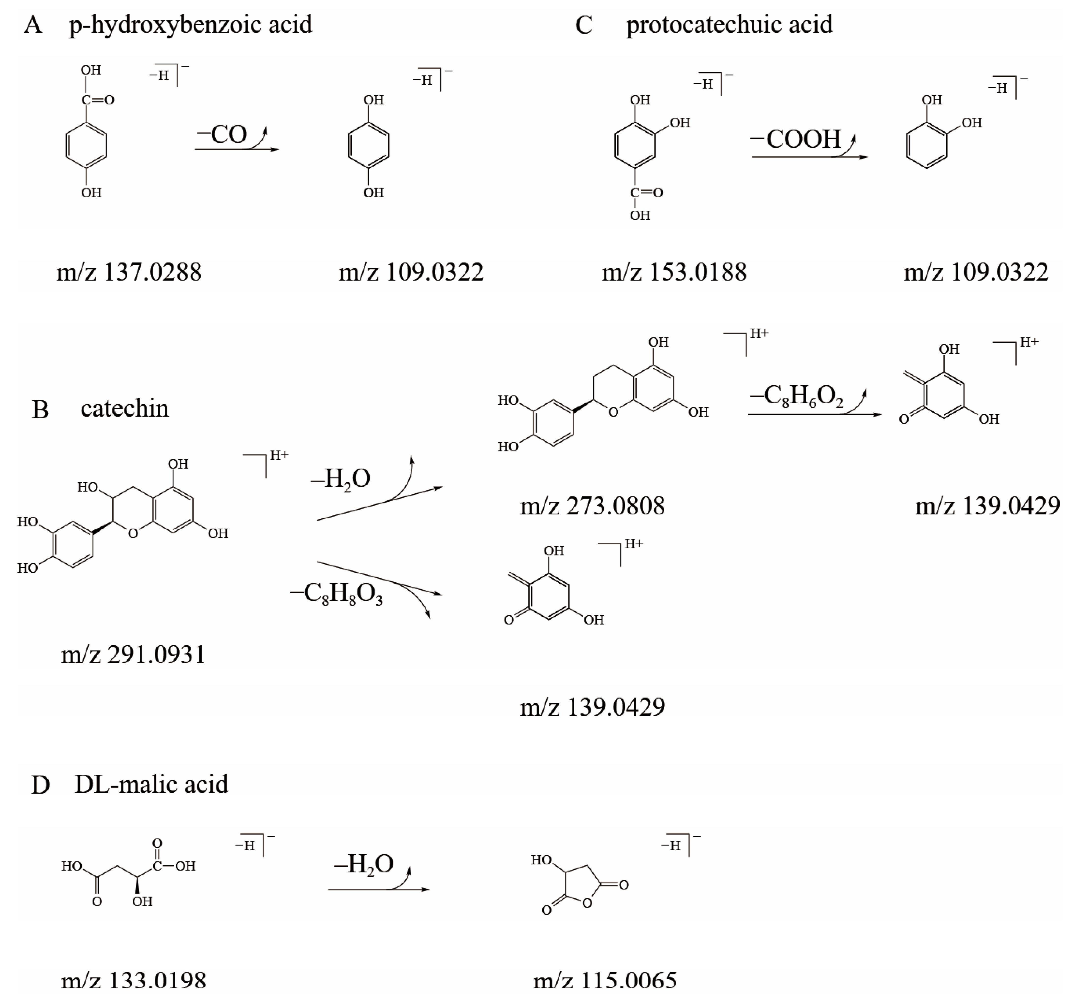
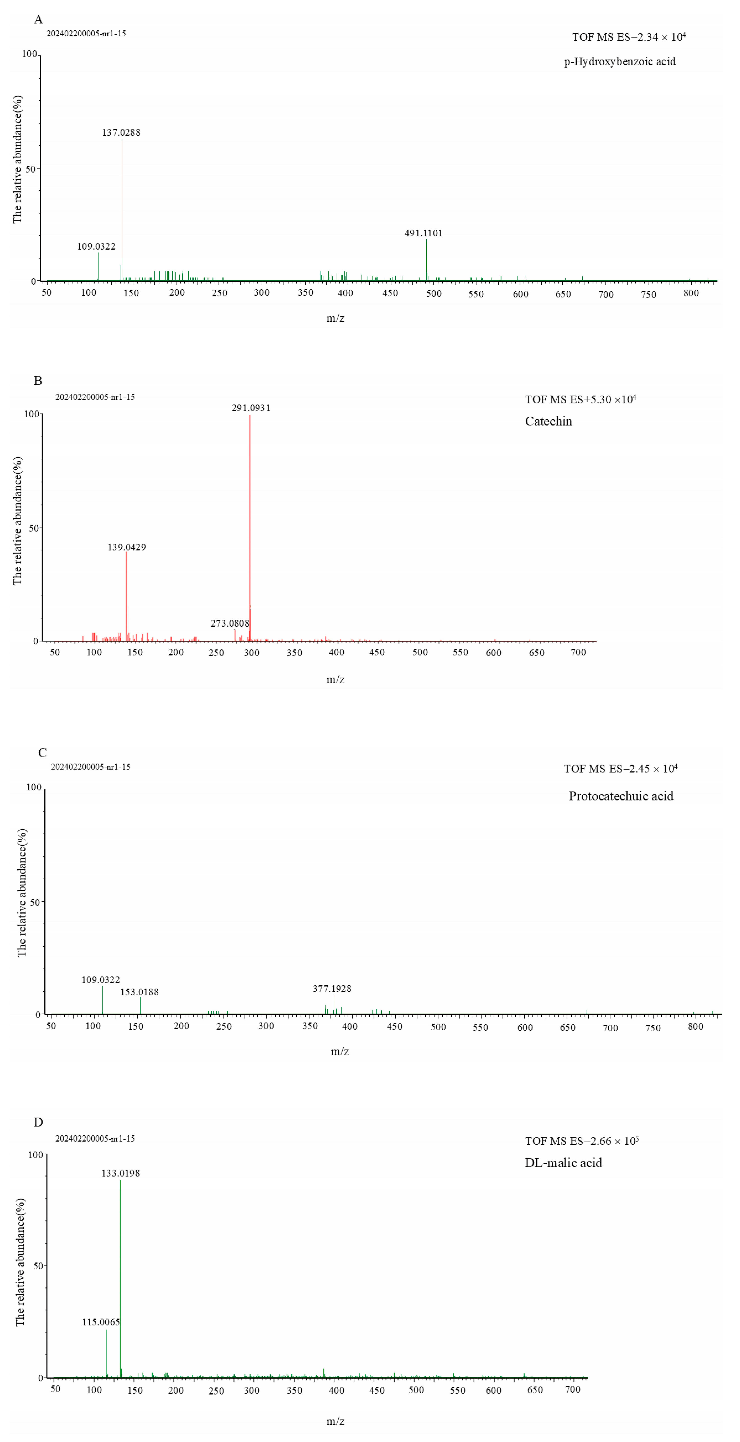
3.2. Qualitative Analysis of Sour and Bitter Fractions in Amomum villosum Extract

The sour and bitter substances in Amomum villosum extract were preliminarily characterized based on HPLC-DAD ultraviolet absorption spectroscopy (Table 2). It is possible that the four monomeric components identified were organic acids and polyphenolic substances [20,21]. Organic acids and polyphenols are important components of Amomum villosum L. and are typically classified as primary metabolites and secondary metabolites of natural plants, respectively. These results indicate that the four monomer components were not newly formed substances during the extraction process. In order to further clarify the four monomer components, their respective fragment ions were obtained by UPLC/Q-TOF-MS. The parent ions and main characteristic fragments are presented in Table 2. The possible mass spectrometry fragmentation modes and secondary mass spectrometry data of the four monomer substances (fractions 1, 2, 5, and 6) are shown in Figure 2, Table 2, and Figure 3, respectively.
Fraction 1: The retention time of fraction 1 during the sample testing was 0.96 min, as shown in Table 2, and the main fragment of its quasi-molecular ion peak was m/z 133.0198 ([M−H]), as shown in Table 2 and Figure 3D. After losing one H2O molecule (18 u), the main fragment ion of m/z 115.0065 ([M−H−H2O]) was obtained, as shown in Figure 3D. Therefore, we speculated that fraction 1 was DL-malic acid.
Fraction 2: The parent ion of fraction 2 was m/z 153.0188 ([M−H]), and the main fragment ion of m/z 109.0322 ([M−H−COOH]) was obtained by losing one molecule of the carboxyl group (44 u), as shown in in Table 2 and Figure 2C and Figure 3C. Therefore, fraction 2 was identified as protocatechuic acid.
Fraction 5: The mass spectrometry information of fraction 5 is shown in Table 2 and Figure 2B and Figure 3B. In positive ion mode, the parent ion of m/z 291.0931 ([M+H]+) was clearly observed. The main fragment ions of m/z 273.0808 ([M+H−H2O]+) and m/z 139.0429 ([M+H−C8H8O3]+) were formed through the retro-Diels–Alder reaction of the parent ion of m/z 291.0931 ([M+H]+). In summary, fraction 5 was identified as catechin.
Fraction 6: The retention time of fraction 6 was 8.54 min, as shown in Table 2. The main ion fragment of m/z 109.0322 ([M−H−CO]) was formed by the loss of a molecule of CO (28 u) from the parent ion of m/z 137.0288 ([M−H]), as shown in Figure 2A and Figure 3A. Fraction 6 was considered to be p-hydroxybenzoic acid.
To further validate the accuracy of the UPLC/Q-TOF-MS qualitative results, four monomer fractions and their corresponding commercially available standards were detected by HPLC and UPLC/Q-TOF-MS under the same chromatographic and mass spectrometry conditions. The results reveal that the four monomer fractions in Amomum villosum L. had the same peak time and MS fragmentation data as the commercially available standards. Based on the above experiments, we believe that the identification results can be fully trusted.

3.3. Quantitative Analysis of Sour and Bitter Fractions in Amomum villosum Extract

The sour and bitter substances in the Amomum villosum extracts were quantitatively analyzed using the external standard quantification method (Table 3). In the EAVE and WAVE samples, the content of DL-malic acid was the highest, while the content of p-hydroxybenzoic acid was the lowest. Compared with the WAVE samples, the content of catechin in the EAVE samples was higher (63.79 ± 0.42 mg/g), which can be explained by the fact that catechin, a polyphenol, is more easily extracted by organic solvents, such as ethanol. Interestingly, the contents of the three acidic substances in the EAVE sample were also higher than in the WAVE sample, indicating that the acidity value of the EAVE sample was similarly higher than that of the WAVE sample. The electronic tongue data of the two samples also reflect the same results, as shown in Figure 1C. In the food industry, an electronic tongue is an excellent instrument for reflecting the taste characteristics of substances, including sourness, bitterness, astringency, aftertaste-B, aftertaste-A, umami, richness, and saltiness. For the taste characteristics of the two samples, sourness and bitterness were more easily detected through an electronic tongue. As shown in Figure 1C, compared with the WAVE sample, the bitterness value (18.76 ± 1.23) and acidity value (15.83 ± 1.47) of the EAVE sample were significantly higher. This indicate that the taste characteristics of non-volatile substances in Amomum villosum extract could be influenced by different extraction solvents.
It is well known that sour and bitter substances in medicinal and edible plants could affect their application in the food industry. Some scholars have also attempted to improve the taste of products by removing unpleasant taste components from extracts. Bansode et al. [22] found that the bitter substances (naringin and limonin) in orange peel powder were significantly reduced by plasma-activated water pretreatment combined with debittering treatment, and the taste of orange peel powder was improved. However, studies have shown that naringin and limonin both have certain pharmacological activities [23]. Unfortunately, the pharmacological activities of sweet orange peel powder after removing bitterness have not been further studied, which might lead to a decrease in its pharmacological effects, such as antioxidant and anti-inflammatory effects [24]. Therefore, in order to provide data support for product developers to balance the trade-off between taste and functionality, the pharmacological activities of substances with unpleasant tastes should be studied. For the above reasons, it is necessary to conduct preliminary studies on the pharmacological activities of sour and bitter substances in Amomum villosum L., such as the anti-tumor activities currently focused on by researchers.

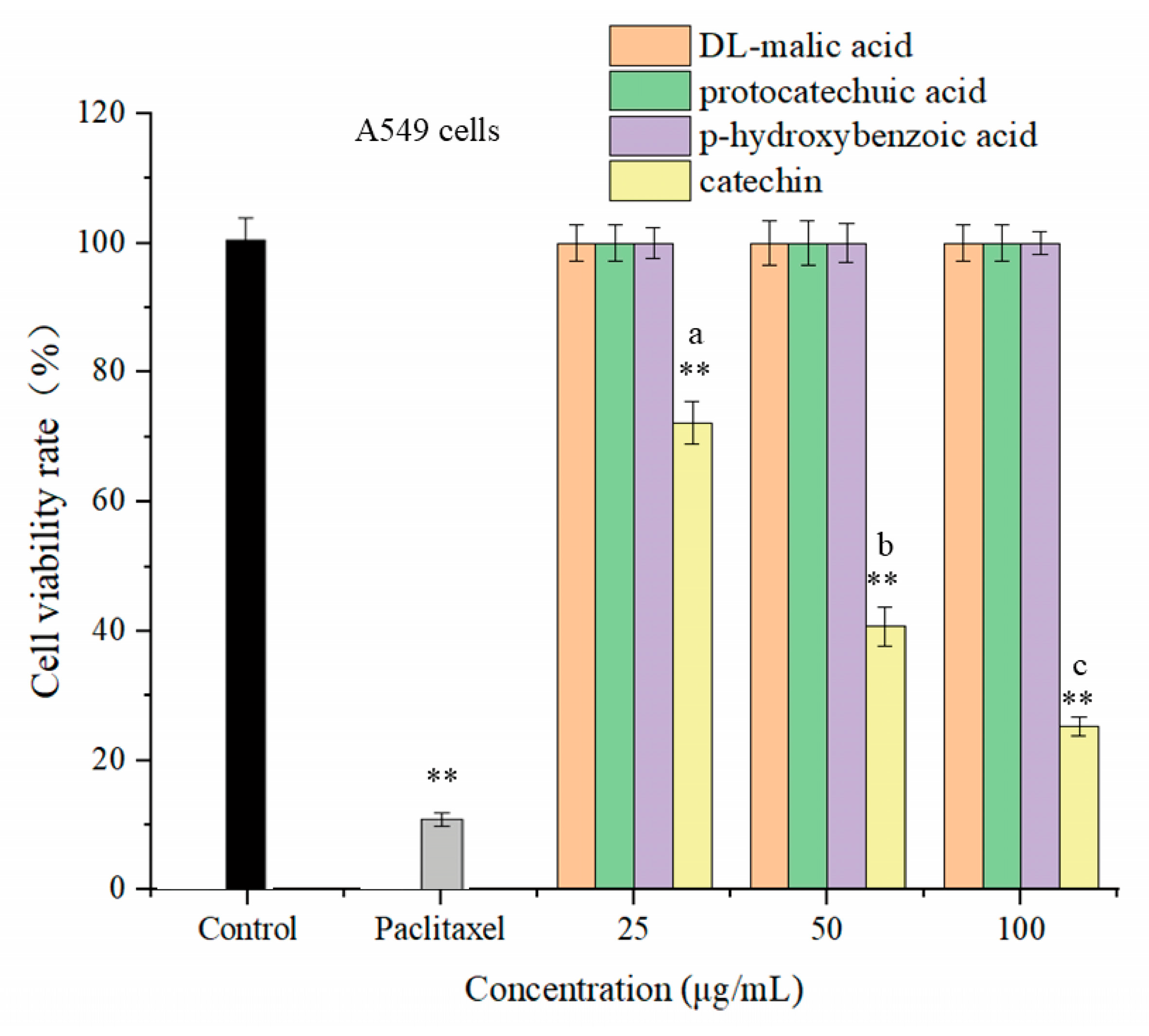
3.4. The In Vitro Anti-Tumor Activity of Sour and Bitter Substances

3.4.1. The Effects of Four Monomeric Substances on the Cell Viability of Non-Small Cell Lung Cancer A549 Cells

In recent years, the anti-tumor activity of natural products has been one of the research hotspots in the academic community. Paclitaxel, a highly effective, non-toxic, and broad-spectrum natural anticancer drug derived from plants, is often used as a positive control in experiments to evaluate the anti-tumor activity of samples. In this study, the CCK-8 assay was employed to preliminarily screen the cell viability of human non-small cell lung cancer A549 cells treated with four monomeric compounds at low (25 μg/mL), medium (50 μg/mL), and high (100 μg/mL) concentrations. The experimental results shown in Figure 4 indicate that, compared with the blank control group, only catechin significantly inhibited the cell viability of human non-small cell lung cancer A549 cells (p < 0.01). In addition, we clearly observed the dose-dependent phenomenon that the anti-tumor activity of catechin increased with rising concentrations. Compared with catechin, the anti-tumor activity of the three acidic substances was not detected at different concentrations, indicating that only catechin had a significant anti-tumor effect.

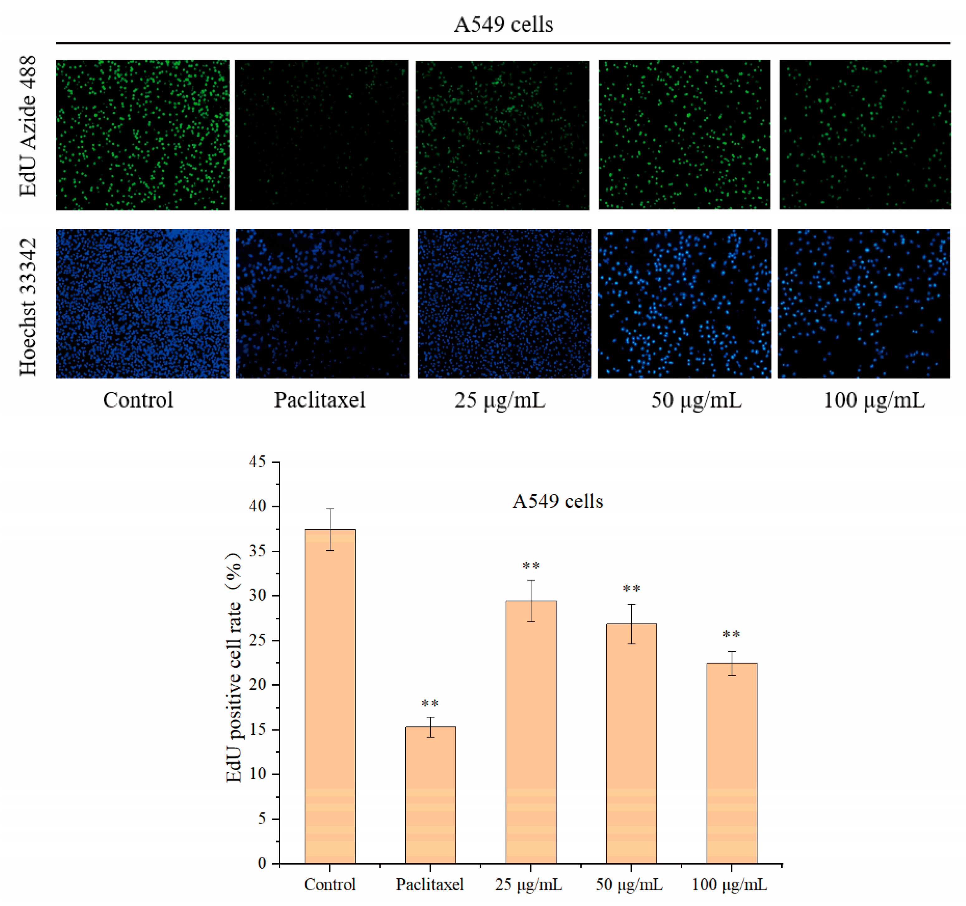
3.4.2. The Effect of Catechin on the Proliferation of A549 Cells

The inhibitory effect of natural products on cancer cell proliferation is also an important indicator for evaluating their anti-tumor activity. Based on the results of the cell viability experiments, catechin was selected as the experimental subject to detect its effect on the proliferation of human non-small cell lung cancer A549 cells. As shown in Figure 5, paclitaxel (5 μg/mL) had a strong antiproliferative effect on human non-small cell lung cancer A549 cells, while the catechin samples had varying degrees of an inhibitory effect on the proliferation of A549 cells within the concentration range of 25–100 μg/mL. Strikingly, catechin at 100 μg/mL markedly suppressed A549 cell proliferation, as evidenced by a diminished Edu positive rate (22.48 ± 1.37%) compared to the control group (37.46 ± 2.32%, p < 0.01). The above results indicate that catechin could inhibit the proliferation activity of A549 cells, which is similar to the effect of nordamnacanthal derived from the roots of Morinda scabrida Craib [25].

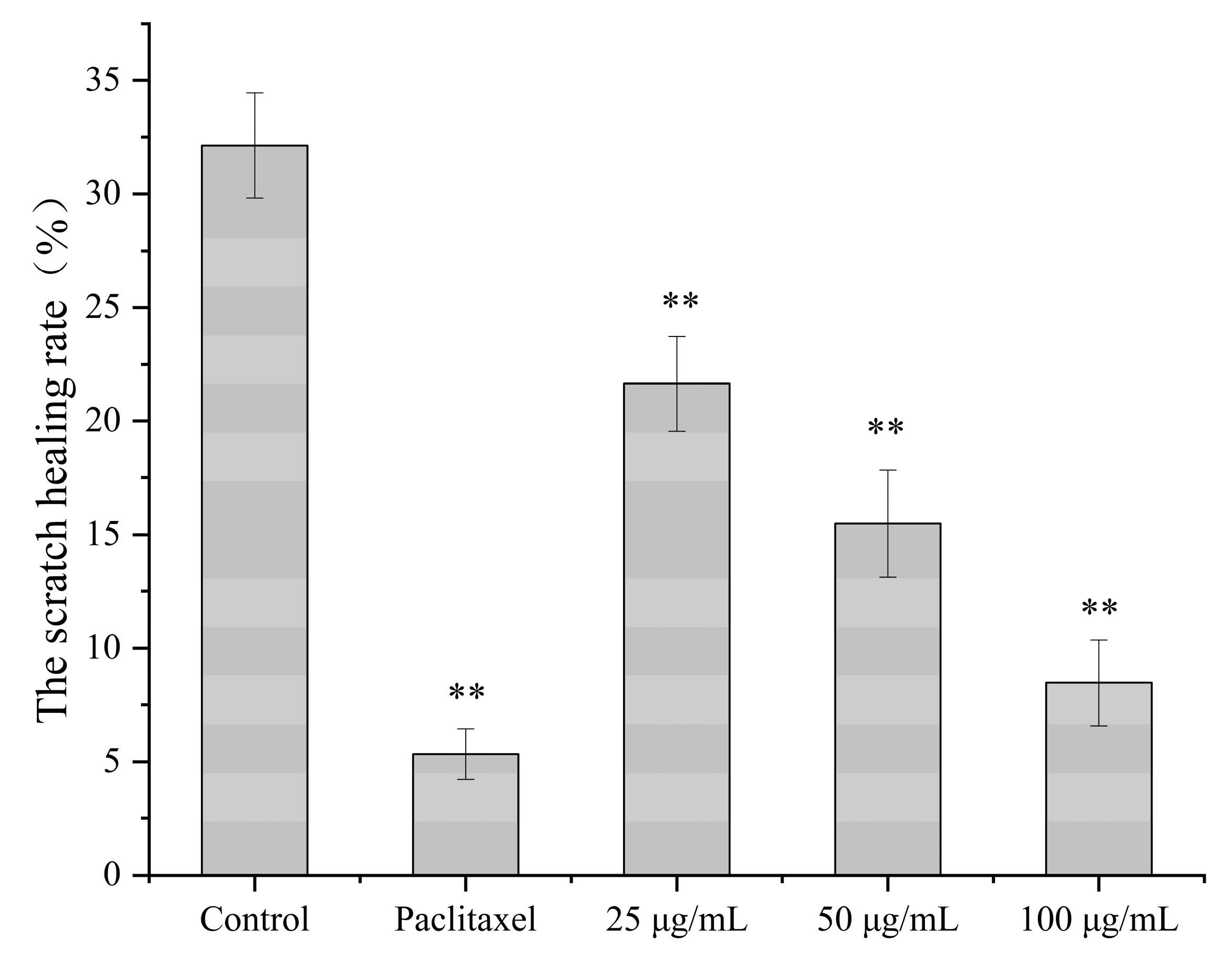
3.4.3. The Effect of Catechin on the Migration Ability of A549 Cells

The spread of cancer cells is caused by the migration of tumor cells, which is usually accompanied by further deterioration of the cancer. The cancer caused by these tumors is difficult to cure with conventional treatment methods due to the high migration ability of the cancer cells [26]. Previous studies have shown that natural product extracts, such as furanocoumarin in the roots of Angelica dahurica, have a good inhibitory effect on the migration of human non-small cell lung cancer A549 cells [27]. In particular, when inhibiting the migration of cancer cells, the cytotoxic effects of most natural product extracts on normal cells have not been discovered. To investigate whether catechin had an effect on the migration of human non-small cell lung cancer A549 cells, the cell scratch assay was used to detect their migration ability. The experimental results are shown in Figure 6. After treating A549 cells with low-dose (25 μg/mL), medium-dose (50 μg/mL), and high-dose (100 μg/mL) catechin solutions, the cells were observed at 0 and 24 h, respectively. The results show that compared with the control group, the migration distance of A549 cells was significantly shortened after treatment with different doses of catechin. It is worth noting that compared with the control group, when the A549 cells were treated with catechin (100 μg/mL) for 24 h, the scratch healing rate of the control group (32.14 ± 2.32%) was 3.8 times higher than that of the catechin-treated group (8.47 ± 1.89%). The cell scratch assay results indicate that the catechin solution (25–100 μg/mL) significantly inhibited the migration of A549 cells and demonstrated potential anti-tumor effects, showing comparable efficacy to exiguaflavanone A and exiguaflavanone B derived from Sophora exigua root extract [28].

4. Conclusions

In this study, the non-volatile sour substances (DL-malic acid, protocatechuic acid, and p-hydroxybenzoic acid) and the bitter substance (catechin) in Amomum villosum samples (EAVE, WAVE) were isolated, purified, and identified for the first time through chromatographic analysis and UPLC/Q-TOF-MS. The taste characteristics of the four monomeric substances were clearly obtained using an electronic tongue, and the results show that compared with the WAVE, the bitterness and sourness values of the EAVE were significantly higher when ethanol solution was used as the extraction solvent. The reason for this result might be that the bitter and sour substances from Amomum villosum L. are more easily extracted by organic solvents, such as ethanol and methanol. Of course, the pharmacological effects of these substances responsible for undesirable taste profiles cannot be ignored. The CCK-8 assay results indicate that compared with the blank control group, only catechin significantly inhibited the cell viability of human non-small cell lung cancer A549 cells (p < 0.01). The EdU staining experiment results demonstrate that catechin at 100 μg/mL markedly suppressed A549 cell proliferation, as evidenced by a diminished EdU positive rate (22.48 ± 1.37%) compared to the control group (37.46 ± 2.32%, p < 0.01). The scratch assay results show that catechin solution (25–100 μg/mL) significantly inhibited the migration of A549 cells (p < 0.01). In conclusion, based on the results of the in vitro anti-tumor evaluation, the bitter component catechin (25–100 μg/mL) from Amomum villosum L. exhibited potential anti-tumor activity (p < 0.01), thereby qualifying it as an anti-tumor bioactive raw material for incorporation into health foods or pharmaceutical products to utilize its pharmacological benefits. As we continue to research the pharmacology of catechin, we will further elucidate the mechanism by which catechin inhibits A549 cells, to broaden the application scope of Amomum villosum extract. The principal aim of this research endeavor was to identify the specific sour and bitter substances in Amomum villosum L. and to emphasize the correlation between the dissolution amount of specific components in natural products and the categories of extraction solvents. Furthermore, it is important to note that food manufacturers should carefully consider the balance between taste profiles and pharmacological efficacy when incorporating Amomum villosum extracts for the functional enhancement of food products. In short, this study is beneficial for the application of Amomum villosum L. in the food and pharmaceutical industries.

Author Contributions

Conceptualization, Y.C. and Z.L.; methodology, Y.C. and W.T.; software, Z.L.; validation, W.T., W.L. and J.X.; formal analysis, Y.C.; investigation, W.L.; resources, Z.W.; data curation, Z.W.; writing—original draft preparation, Y.C. and Z.L.; writing—review and editing, Q.Y. and W.T.; visualization, Q.Y.; supervision, J.X.; project administration, Z.W.; funding acquisition, Q.Y. and Z.W. All authors have read and agreed to the published version of the manuscript.

Funding

This research was funded by the Joint Fund Project of the Natural Science Foundation of Hubei Provincial Department of Science and Technology (2022CFD076).

Data Availability Statement

All relevant data are provided in this published article.

Acknowledgments

All authors express their gratitude to Yang Liu from the Hubei Key Laboratory of Quality and Safety of Traditional Chinese Medicine Health Food for help in investigating the Amomum villosum samples.

Conflicts of Interest

All authors declare that they have no known conflicts of interest.

References

  1. Feng, L.; Wang, Z.; Lei, Z.; Zhang, X.; Zhai, B.; Sun, J.; Guo, D.; Wang, D.; Luan, F.; Zou, J.; et al. Amomum villosum Lour.: An insight into ethnopharmacological, phytochemical, and pharmacological overview. J. Ethnopharmacol. 2024, 335, 118615. [Google Scholar] [CrossRef]
  2. Zhang, Q.Q.; Liu, K.X.; Li, Y.X.; Sun, D.J.; Li, H.; Chen, L.X.J.C. Three New Labdane-Type Diterpenoids from the Fruits of Amomum villosum and Their Anti-Inflammatory Activities. Chem. Biodivers. 2023, 20, e202301014. [Google Scholar] [CrossRef]
  3. Zhang, K.; Cao, F.; Zhao, Y.; Wang, H.; Chen, L.J.P. Antibacterial Ingredients and Modes of the Methanol-Phase Extract from the Fruit of Amomum villosum Lour. Plants 2024, 13, 834. [Google Scholar] [CrossRef] [PubMed]
  4. Salto, L.; Muniyandi, K.; Porat, R.; Goldenberg, L.; Carmi, N.; Maoz, I. Bitterness and flavanone composition of ‘Redson’ fruit: A new red-fleshed pomelo×grapefruit hybrid. J. Food Compos. Anal. 2024, 133, 106366. [Google Scholar] [CrossRef]
  5. Zhong, D.W.; Cheng, H.L.; Hong, C.L.; Tao, L.; Lun, Z.Y.; Da, B.R.; Ying, G.; Shuo, W. Quantification of caffeine and catechins and evaluation of bitterness and astringency of Pu-erh ripen tea based on portable near-infrared spectroscopy. J. Food Compos. Anal. 2024, 125, 105793. [Google Scholar]
  6. Meng, S.L.; Yuan, Y.; Dong, D.L.; Ying, J.X.; Hui, G.; Qing, C.Y.; Jian, L.C.; Peng, X. Does saponin in quinoa really embody the source of its bitterness. Food Chem. 2024, 437, 137872. [Google Scholar]
  7. Diána, K.; Attila, H.; Norbert, K.; Lajos, B.B.; Nikoletta, S.; Gabriella, S.; Dezső, C. Coumarins, furocoumarins and limonoids of Citrus trifoliata and their effects on human colon adenocarcinoma cell lines. Heliyon 2022, 8, 10453. [Google Scholar]
  8. Florian, Z.; Alexandra, S.; Antonella, D.P.; Maik, B. Physiological activation of human and mouse bitter taste receptors by bile acids. Commun. Biol. 2023, 6, 612. [Google Scholar]
  9. Yu, M.; Li, T.; Raza, A.; Wang, L.; Song, H.; Zhang, Y.; Li, L.; Hua, Y. Sensory-guided identification of bitter compounds in Hangbaizhi (Angelica dahurica). Food Res. Int. 2020, 129, 108880. [Google Scholar] [CrossRef]
  10. Spinelli, S.; Hopfer, H.; Moulinier, V.; Prescott, J.; Monteleone, E.; Hayes, J.E. Distinct sensory hedonic functions for sourness in adults. Food Qual Prefer. 2024, 116, 105152. [Google Scholar] [CrossRef]
  11. Santanatoglia, A.; Angeloni, S.; Caprioli, G.; Fioretti, L.; Ricciutelli, M.; Vittori, S.; Alessandroni, L. Comprehensive investigation of coffee acidity on eight different brewing methods through chemical analyses, sensory evaluation and statistical elaboration. Food Chem. 2024, 454, 139717. [Google Scholar] [CrossRef] [PubMed]
  12. Jing, J.W.; Yong, K.L.; Qin, W.L.; Qing, Y.H.; Ping, H.L.; Yao, W.Y.; Guo, D.L.; Hui, X.; Hui, R.T. Drying temperature affects essential oil yield and composition of black cardamom (Amomum tsao-ko). Ind. Crops Prod. 2021, 168, 113580. [Google Scholar]
  13. Aknarin, P.; Surat, L. Volatile constituents of Amomum argyrophyllum Ridl. and Amomum dealbatum Roxb. and their antioxidant, tyrosinase inhibitory and cytotoxic activities. Arabian J. Chem. 2022, 15, 104148. [Google Scholar]
  14. HaeLim, K.; SungKwon, L.; DaEun, M.; BongKeun, C.; DongRyung, L. Anti-Obesity Effects of a Mixture of Atractylodes macrocephala and Amomum villosum Extracts on 3T3-L1 Adipocytes and High-Fat Diet-Induced Obesity in Mice. Molecules 2022, 27, 906. [Google Scholar] [CrossRef] [PubMed]
  15. Renata, B.; Rimantas, V.P.; Ona, R. Valorisation of Roman chamomile (Chamaemelum nobile L.) herb by comprehensive evaluation of hydrodistilled aroma and residual non-volatile fractions. Food Res. Int. 2022, 160, 111715. [Google Scholar]
  16. Meng, Y.; Jiang, H.; Ji, H.; Li, X.; Julian, M.D.; Sang, S.; Jin, Z.; Wang, J.; Qiu, C. Co-encapsulation of curcumin and piperine in whey protein isolate fibrils improves their water dispersibility and antioxidant activity. Food Biosci. 2024, 58, 103750. [Google Scholar] [CrossRef]
  17. Chen, Y.; Liao, Z.; Wang, Z.; Shi, W.; Xu, J. Isolation and Identification of Bitter Compounds in Ginseng (Panax ginseng C. A. Mey.) Based on Preparative High Performance Liquid Chromatography, UPLC-Q-TOF/MS and Electronic Tongue. Separations 2024, 11, 114. [Google Scholar] [CrossRef]
  18. Xian, L.G.; Tuo, F.; Ermeng, L.; Pei, S.; Zhan, K.Z.; Lan, L.; Haile, M. Ougan juice debittering using ultrasound-aided enzymatic hydrolysis: Impacts on aroma and taste. Food Chem. 2021, 345, 128767. [Google Scholar]
  19. Li, D.; Shan, H.Z.; Lina, C.; Jun, L.; Fu, H.Z.; Tie, M.L.; Jin, L.W.; Jia, Y.H.; Yuan, H.M.; Zheng, F.Q.; et al. In vivo anti-hyperuricemia and anti-gouty arthritis effects of the ethanol extract from Amomum villosum Lour. Biomed. Pharmacother. 2023, 161, 114532. [Google Scholar]
  20. Ilhem, R.; Anouar, B.S.; Jazia, S.; Anouar, F.; Sana, N.; Salah, A.M.; Najla, H.; Sami, S. HPLC-DAD identification of polyphenols from ethyl acetate extract of Amaranthus spinosus leaves and determination of their antioxidant and antinociceptive effects. Inflammopharmacology 2019, 27, 975–984. [Google Scholar]
  21. Violeta, I.P.; Dragana, P.; Sasa, M. Rapid and Simple Method for Determination of Target Organic Acids in Wine Using HPLC-DAD Analysis. Food Anal. Methods 2020, 13, 1078–1087. [Google Scholar]
  22. Bansode, V.; Jaddu, S.; Panda, T.C.; Dalbhagat, C.G.; Seth, D.; Roy, S.S.; Gosh, D.; Pradhan, R.C.; Dwivedi, M. Hurdle approach of plasma-activated water pretreatment with debittering treatment on naringin and limonin content of sweet orange peel powder. Food Biosci. 2024, 60, 104431. [Google Scholar]
  23. Kankılıç, N.A.; Küçükler, S.; Gür, C.; Akarsu, S.A.; Akaras, N.; Şimşek, H.; İleritürk, M.; Kandemir, F.M. Naringin protects against paclitaxel-induced toxicity in rat testicular tissues by regulating genes in pro-inflammatory cytokines, oxidative stress, apoptosis, and JNK/MAPK signaling pathways. J. Biochem. Mol. Toxicol. 2024, 38, 23751. [Google Scholar]
  24. Wang, J.; Jing, X.; Liu, X.; Chen, F.; Ge, Z.; Liu, X.; Yang, H.; Guo, Y.; Cui, X. Naringin safeguards vertebral endplate chondrocytes from apoptosis and NLRP3 inflammasome activation through SIRT3-mediated mitophagy. Int. Immunopharmacol. 2024, 140, 112801. [Google Scholar] [PubMed]
  25. Sorachai, S.-l.; Lukana, N.; Kriengsak, L.; Thamrongjet, P.; Sutida, M.; Poomsith, T.; Withsakorn, S.; Jisnuson, S.; Pitak, C. Anthraquinones from the roots of Morinda scabrida Craib exhibit antiproliferative activity against A549 lung cancer cells and antitubulin polymerization. Fitoterapia 2024, 173, 105781. [Google Scholar]
  26. Kunte, M.; Desai, K. The Inhibitory Effect of C-phycocyanin Containing Protein Extract (C-PC Extract) on Human Matrix Metalloproteinases (MMP-2 and MMP-9) in Hepatocellular Cancer Cell Line (HepG2). Protein J. 2017, 36, 186–195. [Google Scholar]
  27. Lee, S.H.; Han, A.-R.; Kang, U.; Kim, J.-B.; Seo, E.K.; Jung, C.-H. Inhibitory Effects of Furanocoumarins From the Roots of Angelica dahurica on Ionizing Radiation-Induced Migration of A549 Human Non-Small Cell Lung Cancer Cells. Nat. Prod. Commun. 2020, 15, 1934578X20915036. [Google Scholar]
  28. Punnida, A.; Kamonwan, S.; Warathit, S.; Sonthaya, U.; Lapamas, R.; Aroonchai, S.; Methee, R.; Trinnakorn, K.; Songyot, A.; Pornngarm, D. Suppression of inflammation-induced lung cancer cells proliferation and metastasis by exiguaflavanone A and exiguaflavanone B from Sophora exigua root extract through NLRP3 inflammasome pathway inhibition. Front. Pharmacol. 2023, 14, 1243727. [Google Scholar]
Figure 1. (A) Six fractions from EAVE through prep-HPLC chromatogram. (B) Seven fractions from WAVE through prep-HPLC chromatogram. *: The corresponding fraction had a sour or bitter taste. (C) The electronic tongue radar diagram for the two types of samples.
Separations 12 00077 g001
Figure 2. (AD) The possible mass spectrometry fragmentation pathways of the sour and bitter substances in Amomum villosum L.
Separations 12 00077 g002
Figure 3. (A) The secondary mass spectrometry of fraction 6. (B) The secondary mass spectrometry of fraction 5. (C) The secondary mass spectrometry of fraction 2. (D) The secondary mass spectrometry of fraction 1.
Separations 12 00077 g003
Figure 4. The screening of anti-tumor effects of sour and bitter substances in Amomum villosum L. on the cell viability of A549 cells. Compared with the blank control group, ** p < 0.01 indicates a significant differences, while the letters a to c indicate significant differences detected in different concentrations of the same samples.
Separations 12 00077 g004
Figure 5. The effect of catechin on the proliferation ability of human non-small cell lung cancer A549 cells. ** p < 0.01 when compared with the control group.
Separations 12 00077 g005
Figure 6. The effects of catechin on the migration ability of human non-small cell lung cancer A549 cells. ** p < 0.01 when compared with the control group.
Separations 12 00077 g006
Table 1. The taste analysis of sour and bitter fractions in Amomum villosum extract samples a using an electronic tongue system.
Tasteless Point of Bitterness bTasteless Point of Sourness bCode cBitternessSournessCode dBitternessSourness
0−1310.07 ± 0.0210.74 ± 0.78 *10.06 ± 0.0110.21 ± 0.66 *
0−1320.04 ± 0.0214.67 ± 0.34 *20.05 ± 0.0214.38 ± 0.74 *
0−13 30.07 ± 0.01−14.10 ± 0.44
0−1340.09 ± 0.03−15.14 ± 1.0440.08 ± 0.02−15.12 ± 0.83
0−13512.97 ± 0.48 *−14.84 ± 0.97512.68 ± 0.55 *−15.03 ± 0.74
0−1360.13 ± 0.045.78 ± 0.55 *60.11 ± 0.045.69 ± 0.75 *
0−1370.07 ± 0.03−14.17 ± 1.0270.08 ± 0.02−14.21 ± 1.14
The results of the sourness and bitterness values of each fraction are displayed as the mean ± SD (n = 3). a: The two samples of WAVE and EAVE; b: the taste value of the reference solution was defined as the tasteless point; c: 6 fractions from EAVE; d: 7 fractions from WAVE. Compared with the tasteless group, p < 0.05 * indicates significant statistical difference.
Table 2. Identification of sour and bitter fractions in Amomum villosum extracts a.
Code bRT (min)UV (nm) cParent IonMain Characteristic FragmentsIdentification
10.96214[M−H] 133.0198115.0065DL-malic acid
26.25260; 294[M−H] 153.0188109.0322protocatechuic acid
59.79280[M+H]+ 291.0931273.0808; 139.0429catechin
68.54254[M−H] 137.0288109.0322p-hydroxybenzoic acid
a: Two samples of WAVE and EAVE; b: the numbering of sour and bitter fractions in Figure 1; c: the wavelength corresponding to the characteristic absorption peak.
Table 3. Parameters of HPLC analytical methods of sour and bitter substances in Amomum villosum extracts.
CategoryCompoundsLinear Regression EquationR2Linearity Range (mg/L)LOD (μg/mL)LOQ (μg/mL)Quantitative Result of EAVE (mg/g)Quantitative Result of WAVE (mg/g)
DL-malic acidY = 0.0090X + 0.00371.0007.648~2447.52.23176.3827356.67 ± 4.59284.87 ± 3.78
Organic acidsprotocatechuic acidY = 0.6115X + 0.18390.99995.698~91.1620.47811.39139.37 ± 0.787.04 ± 0.05
p-hydroxybenzoic acidY = 0.9070X + 0.53800.99997.821~125.1251.12413.21495.25 ± 0.264.17 ± 0.34
PolyphenolscatechinY = 0.3336X + 0.03811.00005.968~95.4801.07063.094063.79 ± 0.4213.67 ± 3.28
The quantitative results of the four compounds in WAVE and EAVE are presented as the mean ± SD (n = 3).
Disclaimer/Publisher’s Note: The statements, opinions and data contained in all publications are solely those of the individual author(s) and contributor(s) and not of MDPI and/or the editor(s). MDPI and/or the editor(s) disclaim responsibility for any injury to people or property resulting from any ideas, methods, instructions or products referred to in the content.

