Review

Methods of Analysis of Phytoestrogenic Compounds: An Up-to-Date of the Present State

by
Ines Adam-Dima
1,
Andreea Alexandra Olteanu
2,*,
Octavian Tudorel Olaru
3,*,
Daniela Elena Popa
4 and
Carmen Purdel
1
1
Department of Toxicology, Faculty of Pharmacy, “Carol Davila” University of Medicine and Pharmacy, Traian Vuia 6, 020956 Bucharest, Romania
2
Department of Analytical Chemistry, Faculty of Pharmacy, “Carol Davila” University of Medicine and Pharmacy, Traian Vuia 6, 020956 Bucharest, Romania
3
Department of Pharmaceutical Botany and Cell Biology, Faculty of Pharmacy, “Carol Davila” University of Medicine and Pharmacy, Traian Vuia 6, 020956 Bucharest, Romania
4
Department of Drug Control, Faculty of Pharmacy, “Carol Davila” University of Medicine and Pharmacy, Traian Vuia 6, 020956 Bucharest, Romania
*
Authors to whom correspondence should be addressed.
Separations 2024, 11(7), 205; https://doi.org/10.3390/separations11070205
Submission received: 1 June 2024 / Revised: 27 June 2024 / Accepted: 28 June 2024 / Published: 1 July 2024

Abstract

:
Phytoestrogens, natural compounds with structural similarity to 17-β-estradiol, are known to have potential health benefits, including in hormone-dependent malignancies. The therapeutic interest and some safety concerns observed triggered the need to develop accurate methods to assess their level in different matrices. This narrative review examines the existing analytical methods used to isolate, quantify, and characterize phytoestrogens and their metabolites in plants, foods, and biological samples. Different conventional and modern extraction techniques, such as ultrasonication-assisted extraction, supercritical fluid extraction, or enzyme-assisted extraction, were presented and compared. The advantages and limitations of the existing analytical methods, such as high-performance liquid chromatography using different sensitive detectors, gas chromatography often coupled with mass spectrometry, and immunoassay methods, are highlighted, along with the need for ongoing research to improve the sensitivity and selectivity of the analysis.

1. Introduction

Phytoestrogens are natural compounds that have gained significant attention due to their structural resemblance to 17-β-estradiol and their capability to mimic or modulate estrogenic activity in the human body [1]. They have been extensively studied for their potential health benefits, including the prevention of hormone-dependent cancers, cardiovascular diseases, osteoporosis, and the alleviation of menopausal symptoms [2,3].
The therapeutic interest and some safety concerns that phytoestrogens raised triggered the need to develop accurate methods to assess and understand their bioavailability, metabolism, and biological effects. Surprisingly, in the last few years, only a few reviews have compared the methods used to assess the content of phytoestrogens in different matrices [4,5,6].
The first methods used to isolate the phytoestrogens from food or plant material involved techniques like Soxhlet extraction, maceration, or sonication, using alcohols or acetone as organic solvents [7]. These methods are time-consuming and sometimes require large volumes of solvents that endanger the environment and expose lab workers [8]. Moreover, during the extraction, the high temperatures cause glycoside hydrolysis, change the isoflavone profile by increasing the concentration of aglycones in the sample, and limit the information obtained [9]. As progress has been made throughout the years, these limitations have been overcome. Therefore, new techniques, such as supercritical fluid extraction (SFE) or solid phase extraction (SPE), which are also applicable to phytoestrogens‘ metabolites, were developed. These methods exhibit enhanced efficiency and high selectivity compared to the classical techniques [10,11].
Various analytical methods have been developed for the separation, identification, and quantification of phytoestrogens and their metabolites, including chromatographic techniques like high-performance liquid chromatography (HPLC) using different sensitive detectors, such as ultraviolet (UV), fluorescence, or electrochemical detectors, and gas chromatography (GC), often coupled with mass spectrometry (MS) and recently with tandem mass spectrometry (MS/MS). Also, immunoassay methods, specifically enzyme-linked immunosorbent assay (ELISA) and time-resolved fluoroimmunoassay (TR-FIA), have been established. The latest analytical techniques are particularly valuable for in vivo analysis and epidemiological studies.
Matrix interference, or the interference of structurally similar compounds, natural variability of phytoestrogen content, and method validation are among the greatest challenges in the analysis of phytoestrogens.
This narrative review aims to summarize the current state of knowledge on the phytoestrogen assay from different samples, including herbal products, foods, and biological fluids, highlighting the sample preparation, the chosen analytical technique, and the method’s validation parameters.

2. Methods

For this narrative review, a literature survey was performed On PubMed to find the most relevant articles reporting the analytical methods used to determine phytoestrogenic compounds. Articles were limited to those published in English, focusing on the recent works between 2014 and 2024. Additionally, the literature was reviewed to ascertain the key aspects of phytoestrogens’ characteristics.
The keywords and MeSH terms used were: “phytoestrogens” AND “assay” AND “analytical methods”AND “biological samples” OR “food” OR “herbal”.
Three researchers independently screened the titles and abstracts first, and disagreements were solved by discussion and consensus. Studies where the main text was unavailable were excluded. Then, the full text of the papers was reviewed to retrieve the relevant information, and the most relevant papers were selected. The following information about each study was recorded: year of publication, name of the first author, tested sample and analytical method, and main findings. A total of 128 papers regarding chemical structure premises and analytical methods were selected after eligibility analysis, cross-checking, and removing duplicates.

3. Characterization and Classification of Phytoestrogens

Phytoestrogens are compounds that are found naturally in some plant species. Traditionally, phytoestrogens have been defined as substances that have an effect on the nervous system, induce estrus, and influence the development of the genital tract in females [2]. However, over time, the definition of phytoestrogens has expanded, now including any chemical that shows signs of estrogenicity. Therefore, for a substance to be classified as an estrogen and its plant-derived form to be identified as a phytoestrogen, it must exhibit mechanisms like binding to estrogen receptors, activating specific estrogen-responsive genes, and stimulating the growth of ER-positive breast cancer cells [1]. In general, phytoestrogens are classified into three types: isoflavones, coumestans, and lignans, all of which have structural similarities to both natural and synthetic estrogens, as well as antiestrogens [12], but different compounds belonging to other classes, such as chromene derivatives, chalconoids, prenylated flavonoids, arylbenzofurans, and ginsenosides, were identified [13]. Plants synthesize these compounds, most probably as defense mechanisms against herbivore predation, directly affecting herbivore fertility [14]. The main classes of phytoestrogens found in food products are isoflavones, coumestans, and lignans, and the representative structures are illustrated in Figure 1.
Based on the phylogenetic analysis [15], plants with phytoestrogens belong mainly to the botanical orders Rosales, Fabales, Malpighiales, Apiales, and Lamiales, or Brassicales. The Rosales order contains the Cannabaceae family (Humulus lupulus L.), and the Fabales order contains the Fabaceae family with the Caesalpiniaceae and Papilionaceae subfamilies. Malpighiales contains the Linaceae family, while Lamiales includes the Lamiaceae and Verbenaceae subfamilies [16].

3.1. Isoflavones

Isoflavones are a subset of the flavonoid group of phytoestrogens and are phenolic compounds primarily distributed almost exclusively to the Fabaceae family and, more specifically, to the subfamily Papilionaceae and less in the other subfamilies such as Mimosaceae and Caesalpiniaceae. In plants, isoflavones can often be found as glycosides but, during isolation and analysis, are readily degraded to the corresponding aglycones [17].
Isoflavones have a diphenylpropane structure with a C6–C3–C6 configuration. The primary structural distinction between isoflavones and another subgroup, flavones, is the positioning of the B-ring on the flavonoid skeleton’s C-ring. In isoflavones, the B-ring is attached at the C-3 position, whereas in flavones, it is positioned at C-2 [18].
These compounds include genistein, daidzein, glycitein, biochanin A, and formononetin [19]. Known as the most potent phytoestrogens, isoflavones can exist in various forms, including aglycons and glycosides [17].
Common sources include soybean (Glycine max (L.) Merr.), rich in daidzein, genistein, and glycitein, and red clover (Trifolium pratense L.), which provides formononetin and biochanin A. They are synthesized from flavanone precursors such as liquiritigenin and naringenin, leading to the formation of different glycosides in plants. Other raw products derived from kudzu (Pueraria montana (Lour.) Merr.) and red clover are also widely used [20]. Isoflavones were also identified in mung bean (Vigna radiata L.), kudzu root (P. lobata), chickpea (Cicer arietinum L.), and psoralea (Psoralea corylifolia L.) [21,22].
In Europe, genistein and daidzein are the most extensively studied isoflavones. The primary sources of isoflavones include soy products, flaxseed, lentils, kidney beans, cereals like oats and wheat (especially bran), and certain vegetables and fruits [23]. Isoflavones are also found in various dietary supplements containing soy and its derivatives, which include soybean concentrate, soy germ extract, fermented soy, and dried soybean extract; different extracts of P. montana from the Fabaceae family are utilized as radix Pueraria (kudzu root); red clover flowers from T. pratense and black cohosh from Actaea racemosa L. of the Ranunculaceae family also serve as rich sources of isoflavones. The isoflavone content in these sources varies widely, ranging from 10.25 to 88.48 mg/g [24].

3.2. Coumestans

Coumestans are plant-derived polycyclic aromatic secondary metabolites with a coumestan skeleton, being considered a particular subclass of isoflavonoids that feature an oxygen-containing heterocyclic four-ring system. This system includes a coumarin unit and a benzofuran unit linked by a carbon-carbon bond, representing the oxidized derivatives of pterocarpan [25,26]. Although many coumestans are known, not all exhibit estrogenic activity. The main sources of coumestans are plants belonging to the Leguminosae, Asteraceae, Mimosaceae, Mniaceae, Apocynaceae, Solanaceae, Moraceae, and Myrtaceae families [25]. Among coumestans, coumestan is the most well-known since it is contained by alfalfa, which was initially isolated in 1957. Other sources are various legumes and vegetables like soybeans, clover, and Brussels sprouts [27].

3.3. Lignans

Lignans are naturally occurring polyphenolic compounds derived from phenylalanine. Matairesinol, secoisolariciresinol, pinoresinol, isolariciresinol, and lariciresionol can be found in large quantities in sources such as flaxseed, cereals, dry seeds, coffee, tea, and vegetables [28,29]. Other lignans with estrogenic activity are enterodiol and enterolactone, provided by colonic bacteria from phytoestrogens during several biotransformations [30,31].

3.4. Possible Mechanisms of Action

Phytoestrogens interact with estrogen receptors (ERα and ERβ), exhibiting a range of activities from pure agonism to antagonism or a combination of both, depending on various factors. Despite binding less strongly to estrogen receptors compared to estradiol, phytoestrogens can exhibit both estrogenic and antiestrogenic effects, as demonstrated in various cancer cell line screenings [32]. The estrogenic activity of these compounds has been intensively researched in the last decades, showing that phytoestrogens are influencing both ERα and ERβ receptors to various extents and being reviewed in the comprehensive work of Kiyama [33]. Coumestrol has shown a high affinity for both ERα and ERβ, contributing to its role as a potent phytoestrogen useful in cancer chemoprevention and providing neuroprotection [34,35]. Moreover, it exhibits a strong affinity for ERβ, an effect that is comparable to or greater than that of estradiol, while its affinity for ERα is relatively weaker [25]. Equol, a derivative of daidzein, stands out for its strong estrogenic activity, which is particularly effective in treating menopausal symptoms and supporting cardioprotective functions through interactions with ERα and Erβ [36]. Biochanin A and Formononetin, prominently sourced from red clover, are studied for their vasculoprotective and cancer-chemopreventive properties [37]. Biochanin A acts on ERα and ERβ with estrogenic effects, while formononetin shows similar interactions but with varied impacts depending on the target tissue [38]. Glycitein, Daidzein, and Genistein, primarily found in soy, are well-known for their diverse physiological roles. Glycitein acts on ERα and ERβ, showing potential for modulating cancer risk. Daidzein interacts similarly but also features prominently in studies related to osteoprotection and menopausal syndrome. Genistein is well-documented for its multifaceted roles in cancer chemoprevention, osteoprotection, and endocrine disruption, affecting both ERα and Erβ [3].

4. Phytoestrogens Analysis from Plant Material

Isoflavones are mainly found as glycosides in the subfamily Papilionaceae (Glycine max, Glycrrhiza glabra, Medicago sativa, Phaseolus vulgaris, P. lobata, T. pratense) and less in the other subfamilies such as Mimosaceae and Caesalpiniaceae (Senna obtusifolia) [17]. Coumestans can be found mainly in Medicago sativa (alfalfa), T. pratense (red clover), and Vigna radiata (mung bean), while lignans are commonly found in Linum usitatissimum L. (flaxseed) or Curcubita pepo (pumpkin) seeds [17]. Flaxseed contains mainly secoisolariciresinol (370 mg/100 g dry weight) and less matairesinol, isolariciresinol, and pinoresinol, while pumpkin seeds contain secoisolariciresinol (21 mg/100 g dry weight) and only trace amounts of lariciresinol [39]. Relevant levels of lignans (185–2321 µg/100 g fresh edible weight) have also been detected in the Brasicales order [39], such as the Brassica oleracea plant, or in the Gentianales order (such as Coffea arabica) [40].
The isolation of phytoestrogens from plant material is relatively simple and involves mainly liquid extraction with polar or apolar solvents, depending on whether the compounds are in free form or as glycosides. Before extraction, the first step in the sample preparation is sometimes to dry the herbal material, as flavonoids, particularly glycosides, can be degraded by enzymes when the plant material is fresh [41]. Moreover, during the extraction, the high temperatures cause glycoside decomposition, change the isoflavone profile, and limit the information obtained [9]. The most common conversions are the decarboxylation of malonyl glucosides to acetyl glucosides and the ester hydrolysis of malonyl and acetyl glucosides to underivatized glucosides. It is also possible for conjugates to generate the aglycone form by cleavage of the glucosidic bond [42].
In addition, chemical hydrolysis leads to a marked increase in the concentration of aglycones present in the sample at the expense of the glucosides and, hence, an increase in the available amount of aglycones to be extracted. Therefore, mild techniques, such as maceration or negative pressure cavitation, are often used to extract the conjugated forms of isoflavonoids, while more drastic methods, such as accelerated solvent extraction or microwave-assisted extraction, can be performed to extract aglycones [41]. Furthermore, Zhao et al. investigated the molecular interaction between genistein and the extraction solvent, especially methanol, during soy extraction. The results indicated that interactions between methanol and genistein were more stable under alkaline conditions, thus increasing the extraction efficiency [43].
Surprisingly, in the last few years, only a few new analytical methods have been developed to assess the content of phytoestrogens in plant material. A selection of the most important methods is included in Table 1.
This is justified as the content of phytoestrogens has only limited value, as in vivo potency and efficacy are more relevant for these compounds. Nevertheless, the content and chemical profile are signs of potential therapeutic use and a marker of the stability of phytoestrogens.
Humulus lupulus L. (hops) flos
Prenylated flavanones are identified as minor constituents of female flowers of hops occurring at concentrations 10–100 fold lower than chalcones. 8-prenylnaringerin (8-PN) has been identified in an amount of 0.002% of the dry matter of hops [62], as has 6- prenylnaringerin (6-PN), which is present in slightly higher amounts (0.01%) [44]. Both are generally stable compounds, and no significant issues regarding their stability have been reported in the literature.
Prencipe et al. developed a complex dynamic maceration with MeOH-HCOOH (99:1, v/v) and an HPLC method with diode array and electrospray ionization–mass spectrometry detection for the fingerprinting of bioactive compounds in hops, including prenylflavonoids and prenylphloroglucinols. Chromatographic conditions used in HPLC/DAD included a fused-core C18 column with a mobile phase of 0.25% formic acid in water and acetonitrile under gradient elution and detection at 290 nm (for prenylflavanones), 330 nm (for bitter acids), and 370 nm (for prenylchalcones). The LoD value was in the range 0.3–1.0 µg/mL for prenylflavonoids and 2.8–5.8 µg/mL for bitter acids, while the LoQ value was in the range 1.3–3.8 µg/mL for prenylflavonoids and 8.1–21.4 µg/mL for bitter acids. HPLC–ESI–MS and MS analyses were performed using the same column, the same applied chromatographic conditions, and an ion trap mass analyzer with an ESI ion source. The flow rate was split 3:1 before the ESI source. The HPLC–ESI–MS system is used both in the positive and negative ion modes. Regarding MS–MS data of prenylflavonoids in the positive ion mode, 6-PN and 8-PN generated a strong fragment at m/z 285, which is attributed to the loss of the prenyl group. In the negative ion mode, the RDA cleavages of prenylflavonoids were the dominant fragmentation pathways, with the [1,3 A] ion occurring at m/z 356 for 8-PN and 6-PN [44].
Ceslova et al. also developed two HPLC methods, one with the Star RP-8e column and the gradient of aqueous acetonitrile containing 0.3% formic acid optimized for the separation of low-polar polyphenolic compounds, and a second one with the Zorbax SB-CN column for more polar components. The quantitation of prenylflavonoids and bitter acids was conducted using two detection techniques (APCI–MS and UV detection), which provided comparable results. In total, 49 low-polar and 37 polar compounds were determined. Both compound classes (prenylflavonoids and bitter acids) were separated and quantitated in a single HPLC run. LoD and LoQ for 8-PN corresponded to 0.006 mg/L and 0.02 mg/L for MS detection and 0.03 mg/L and 0.1 mg/mL for UV detection [45].
Buckett et al. recently developed a stable isotope dilution analysis (SIDA–LC–MS/MS) to investigate the major prenylated flavonoids in hop tea and hops. The method was validated with LoD and LoQs for all analytes between 0.04 and 3.2 µg/L. SIDA proved to be a simple, rapid, and robust analysis of prenylated flavonoids that allows the prospect of measuring the compounds in vivo in clinical samples [46].
Capillary electrophoresis (CE) has been used to analyze prenylated flavonoids in hop extracts [63], but only the nonaqueous reverse-polarity capillary electrophoretic method of Kac et al. reported quantitative data. The LoQ for xanthohumol was reported to be 0.15 μg/mL, while the LoD corresponds to 0.05 mg/L. CE methods suffer from poor reproducibility and require careful buffer selection for successful quantitative analysis [47].
Linum usitatissimum L. (flax)
Flaxseed is one of the richest sources of lignans, such as secoisolariciresinol, matairesinol, and isolariciresinol [16]. Secoisolariciresinol (SECO) cannot be directly determined in the flaxseed extract because it is present as a glucoside (SDG), which is further ester-linked with 3-hydroxy-3-methyl-glutaric acid to form SDG oligomers with a molecular weight of around 4000. Secoisolariciresinol and SDG-containing oligomers are usually extracted by liquid–liquid extraction with ethyl acetate or diethyl ether and less by SPE. Hydrolysis of these oligomers is one of the critical steps in sample preparation for lignan analysis.
Various methods have been developed to identify and quantify flaxseed lignans (free or glucosides). Popova et al. developed an HPLC with high-resolution time-of-flight MS (TOF–MS) to separate and determine both aglycones and unhydrolyzed glucosides. The LoDs for secoisolariciresinol and SDG-containing oligomers correspond to 0.008 pg, demonstrating high sensitivity [48].
Also, Li et al. developed a gradient reversed-phase HPLC for the separation and determination of lignans extracted from defatted flaxseed. The LoDs for SDG oligomers, SDG, and SECO correspond to 0.065 µg/mL, 0.087 µg/mL, and 0.039 µg/mL, respectively, while LoQs correspond to 0.217 µg/mL, 0.288 µg/mL, and 0.130 µg/mL [49].
Using gas chromatography coupling with an ion trap MS detector (GC/IT-MS), Sicilia et al. [39] investigated the chemical lignans profile of two types of flaxseed (L. usitatissimum L. and L. flavum) under enzymatic and acidic hydrolysis, respectively. GC–MS analysis revealed secoisolariciresinol as the main compound, and traces of lariciresinol were also detected in both flaxseeds. Artificial products and loss of original lignans under acidic hydrolysis were observed, and the stereochemistry of flaxseed lignans was analyzed by chiral HPLC, showing that secoisolariciresinol, matairesinol, and lariciresinol consisted predominantly of one enantiomorph. The plant samples were defatted with n-hexane and then hydrolyzed with an enzyme or acid. Then, hydrolyzed samples were extracted with diethyl ether–n-hexane (1:1) before HPLC separation. The HPLC fractions were then derivated with N,O-bis (trimethylsilyl)-trifluoroacetamide (BSTFA) to increase the efficiency and selectivity of GC–MS analysis.
Also, other techniques, such as online high-performance liquid chromatography with nuclear magnetic resonance spectroscopy and mass spectrometry (LC–NMR–MS), were successfully used by Fritsche et al. to elucidate the structures of secoisolariciresinol diglucoside diastereomers, liberated through alkaline hydrolysis and extraction from flaxseeds [64].
Coffea arabica L.
Three lignans, secoisolariciresinol, lariciresinol, and matairesinol, can be found in coffee seeds. Angeloni et al. investigated the content of these lignans using an HPLC MS/MS triple quadrupole method. The lignans are better hydrolyzed using enzymatic digestion and then percolation, with an average of 95.2% extraction yield. The method demonstrated good sensitivity, with LOQs ranging from 5 to 10 µg/L and good linearity [51].
Kuhnle et al. analyzed ground coffee samples using the LC–MS/MS method that involved three steps of isolation: extraction with 10% of methanol in sodium acetate (0.1%, pH 5), followed by enzymatic digestion, and a final SPE extraction. The LoD of this method is 1.5 µg/100 g of dry weight. Lignans were coffee’s main class of phytoestrogens, with an average content of 12 µg/100 g [50].
Trifolium pratense L. (red clover)
The estrogenic activity of red clover is caused by isoflavones such as genistein, daidzein, biochanin A, and formononetin and less by the small amounts of coumestans [65]. The chemical profile of isoflavones in red clover and three other related species was carried out using an HPLC/UV/ESI–MS method [53]. For the qualitative study, the finely ground herbal material was extracted with methanol using sonication, while for the quantitative analysis, the samples were acidically hydrolyzed during the solvent extraction. Under optimized conditions, isoflavones were analyzed and identified by using LC/MSD under MS and MS/MS mode, and 31 isoflavones were detected in red clover extracts, including 9 aglycones, 8 glycosides, and 14 glycoside malonate derivatives. Furthermore, using a reversed-phase HPLC, all isoflavone aglycones, including daidzein, formononetin, genistein, or biochanin A, were successfully separated within 40 min and quantified individually by UV la 254 nm and MS detectors. Validation of this quantitative method led to a LoQ of 24 ng/mL for UV detection and 6 ng/mL for MS detection, respectively.
Red clover aglycones such as daidzein, genistein, formononetin, and biochanin A were isolated by acidic hydrolysis with trifluoroacetic acid during extraction and quantified using reversed-phase HPLC combined with a UV detector at 254 nm [54]. This study used 6-methoxyflavone as the internal standard, and the LoQs values obtained were between 2.0 ng for daidzein and 10.0 ng for formononetin, indicating good sensitivity.
Hloucalová et al. developed an LC–MS method to separate and quantify isoflavones in fresh-cut legume forage. High levels of isoflavone biochanin A and formononetin were found in red clover, with a total isoflavone content of 1.001% dry matter. LoDs and LoQs varied from 0.06 to 1.81 and 0.19 to 6.02 ng/mL, respectively. The isoflavone content decreased with the aging of plants, especially for biochanin A and formononetin, for which the content decreased from 1.278 to 0.710 mg/g of dry weight and from 1.413 to 0.960 mg/g of dry weight, respectively [52].
De Rijke et al. also determined isoflavones and their glucoside malonate levels in red clover leaf extracts using reversed-phase LC coupled to atmospheric pressure chemical ionization mass spectrometry (APCI–MS), UV, and fluorescence detectors. The content of biochanin A was 0.33 mg/g, and that of daidzin was 0.042 mg/g. LoD biochanin A corresponds to 20 µg/mL, while for daidzin, it was 35 µg/mL. Surprisingly, daidzein and genistein were not detected in red clover leaves [55].
Medicago sativa L. (alfalfa)
Metabolic profiling using UHPLC–mass spectrometer (MS)–quadrupole time-of-flight (QToF) was used by Wyse et al. to identify and quantify key phytoestrogens in both fresh and dried lucerne. Six key phytoestrogens, such as coumestrol, present in lucerne in at least two methylated forms, 3′ and 4′-methoxycoumestrol, daidzein, formononetin, and genistein, were identified. Quantitative and qualitative analysis revealed that phytoestrogens varied among selected samples, with the fresh samples containing higher concentrations of coumestans or isoflavones than the dried lucerne. Total phytoestrogen concentration exceeded 300 mg/kg in fresh and 180 mg/kg in dried samples [66].
Generally, the analysis of coumestrol in plant material is complex, as it often requires time-consuming sample handling and hydrolysis of the coumestrol glucosides. Nevertheless, some analytical methods are rapid. Coumestrol levels in alfalfa can be determined using a rapid CE method with a borate buffer at pH 9.6 as an electrolyte and diode-array detection. The method was validated in the concentration range of 0.76–140 mg.dm−3, with a LoD calculated for S/N = 3 of 0.39 mg.dm−3. The levels of coumestrol in dry matter were between 148 and 248 mg/kg [56].
Also, an HPLC method with detection at 260 nm was used, and it was carried out by Tucak et al., which observed significant genetic variation of phytoestrogens between alfalfa populations. The most dominant phytoestrogen was genistein, with a share of 30.33% in the total amount, while slightly lower levels were found for kaempferol (26.84%) and coumestrol (25.02%). The presence of daidzein was even lower, less than 3% of the total amount of phytoestrogens [67,68].
Soto-Zarazúa et al. studied the profile and the content of isoflavones in two new alfalfa-derived products. Isoflavones were investigated by the HPLC/DAD method, with a mobile phase in a gradient of 0.1% formic acid (A) and 0.1% formic acid in methanol (B), at a flow rate of 1 mL/min, with detection at 254 nm.
The values obtained for LoD and LoQ were in the range of 0.03–3.72 µg/mL and 0.10–11.27 µg/mL, respectively. The results showed the presence of daidzein, genistein, genistin, and daidzin in most samples, while glycitein, formononetin, and biochanin A were not detected. Significant differences between isoflavone contents were found when different solvent systems were used for extraction. Water was the best option for extracting daidzein, whereas the water–methanol–formic acid mixture efficiently extracted genistein. The obtained results demonstrate that isoflavones with similar structures, such as genistein or daidzein, have different behaviors in the same solvents, and these aspects should be considered in the current analysis of isoflavones [57].
Isoflavones were also quantified in other plants. For example, Bettaiah et al. developed and validated a rapid and sensitive UPLC–APCI–TOF MS method to identify and quantify genistein from the selected seeds of Apiaceae (as Coriandrum sativum, Apium graveolens, Cuminum cyminum, or Foeniculum vulgare). The optimal conditions for the extraction of genistein were the extraction solvent mixture methanol–water–dimethyl sulfoxide (50:25:25, v/v/v) at 80 °C for one hour. LoD and LoQ for genistein in the UPLC MS/MS system correspond to 1.30 ng/mL and 4.45 ng/mL, respectively, indicating a high sensitivity of the method. The developed method has the advantages of direct sample injection for analysis without any primary purification step, increased sensitivity and specificity, and less time consumption (only 8 min) [69].
Glycrrhiza glabra (licorice)
The roots of liquorice (Glycyrrhiza glabra) are a rich source of flavonoids, particularly prenylated flavonoids. Isoflavones, such as glabridin and glabridin derivatives, or isoflav-3-enes, such as glabrene, were identified in liquorice root extracts [70]. The species-specific marker of G. glabra is glabridin, ranging between 0.08% and 0.35% in roots. Isoliquiritigenin also has estrogen-like activity, suggesting that it may be cyclized to liquiritigenin, which is an active flavonoid [71].
Simons et al. developed ultra-high-performance liquid chromatography (UHPLC) with electrospray ionization mass spectrometry (ESI–MS) to rapidly screen prenylated flavonoids in different extracts. The analysis is based on the fragmentation of prenyl substituents of flavonoids, involving a neutral loss of 56 u, alone or in combination with 42 u. The screening of 70% aq. ethanol, ethanol, and ethyl acetate extracts indicated the presence of 70 mono- and di-prenylated flavonoids, of which 18 were assigned to known compounds such as isoflavones (glabrone), isoflavans (glabridin, hispaglabridin A and B), or isoflav-3-ene (glabrene) [72].
Recently, Celano et al. used an untargeted high-resolution UHPLC–MS/MS analysis, which represents a powerful tool for metabolomic profiling, to define the phytochemical profiles of different plant materials of G. glabra. The results revealed a different, distinctive chemical profile. Roots showed glabridin and prenylated phenolic compounds (erybacin B, kanzonol Y, mulberrofuran K isomers, 3-hydroxyglabrol, and glabrol), while aerial parts as prenylated flavanones contained licoflavanone and glabranin [73]. The results are in agreement with other metabolic profiling studies [74,75].
Glycine max (soy)
Soy is one of the most studied plants, associated with high quantities of phytoestrogens. At least twelve different phytoestrogens have been detected in soybeans, including daidzein, genistein, and glycitein. It has been shown that these are usually found in soybean seed in glycosidic form as malonyl or acetyl glycosides, and the malonylated isoflavone glycosides are major compounds [76].
Although a significant number of analytical methods have been developed and validated to quantify phytoestrogens from soybean or soy food products, the stability and conservation of isoflavones in the samples are still under investigation. The most frequently observed degradation of isoflavones during sample conservation is the decarboxylation of malonyl glucosides to acetyl glucosides and ester hydrolysis of malonyl and acetyl glucosides to glucosides. These changes affect isoflavone chemical profiles and result in avoidable analytical errors [9]. Therefore, the recent trend is to use non-hydrolytic methods.
Hsu et al. developed a two-stage adsorption/desorption chromatography to isolate isoflavones from soybeans. Dianion HP-20 macroporous resin separates daidzein, genistein, and their glucoside conjugates, and then individual compounds are isolated through aluminum oxide by selective adsorption of 5-hydroxy isoflavone. The procedure achieved high recovery (82–97%) and purity (92–95%) of the four isoflavones, which confirms a high separating efficiency [77]. Other modern techniques, such as microwave-assisted, ultrasound-assisted, or supercritical-fluid extraction, are proposed based on their high recovery and low sample degradation [78,79,80]. Other advantages of these techniques include the elimination of additional sample purification and concentration steps before analysis. The most recently developed methods are summarized below and in Table 1.
Sakamoto et al. developed an open sandwich fluorescence-linked immunosorbent assay (os-FLISA) for the detection of daidzin and genistin by taking advantage of enhanced interactions between variable regions of heavy and light chain domains in the presence of daidzin (antigen). The linearity of the methods was determined in the range of 0.1–12.5 μg/mL daidzin. Validation analysis demonstrated that the method was sufficiently reliable and accurate for detecting total isoflavone glycosides [58]. The same researchers produced the monoclonal antibody against daidzin and used it to develop an indirect competitive enzyme-linked immunosorbent assay (icELISA) to simultaneously determine daidzein and genistin [59]. The icELISA involves five steps (antigen coating, blocking the wells, reaction with the primary antibody, reaction with the secondary antibody, and reaction with the substrate) and requires almost 5 h to complete. The method proved to be sensitive, with a LoD of 1.95 ng/mL.
For the rapid detection of daidzin and genistin, the same laboratory established a one-step indirect competitive immunochromatographic assay (ICA) using gold nanoparticles conjugated with a monoclonal antibody against daidzin. The analysis duration corresponds to 15 min, and the detection limit for the total isoflavone glycosides corresponds to 125 ng/mL [60].
Since only isoflavone aglycone is considered to be biologically active, ultra-high-pressure liquid chromatography (UHPLC) was developed for the determination of total isoflavone aglycones (daidzein, glycitein, and genistein) after enzymatic hydrolysis with Helix pomatia juice. The aglycones were separated within 3 min only, and the LoD and LoQ correspond to 67 pg and 223 pg (for daidzein), 55 ng and 184 ng (glycitein), and 94 ng and 314 ng for genistein. The method proved sufficient sensitivity for adequate quantification of all aglycones [61].

5. Phytoestrogens Analysis in Food

The most significant source of phytoestrogens in the human diet is soybeans, but they can also be found in other sources such as fruits (berries, grapes, apples, pears, and plums), other vegetables (beans, peas, chickpeas, lentils, red cabbage, broccoli, zucchini, garlic, and carrot), cereals (wheat bran, barley, and rye bran), and drinks (wine, tea, and beer) [81,82].
All categories of phytoestrogens are included in human foods. The main origin of isoflavonoids in the diet is derived from soy-based foods. The isoflavone glucosides undergo hydrolysis by intestinal β-glucosidases, resulting in aglycones. Daidzin and genistin (glycoside forms), along with their respective aglycone forms, daidzein and genistein, are the most abundant phytoestrogens alongside glycitein and biochanin A [83,84].
The principal lignans present in diet include artigenin, enterodiol, enterolactone, sesamin, syringaresinol, medioresinol, (−)-matairesinol, (−)-secoisolariciresinol, (+)-lariciresinol, and (+)-pinoresinol, among others. The primary source of lignans is represented by oilseeds (flaxseed, sesame seed, sunflower seed), nuts (almond, cashew, hazelnut, peanut, pistachio), whole grain cereals (bread, pasta, whole grain flours: wheat, oat, rye), and various fruits (berries, citrus fruits, apricot, nectarine, watermelon, apple, banana, papaya), legumes (cabbage, avocado, cucumber, pumpkin, carrot, radish, potatoes) [85].
Among all phytoestrogens, coumestans, including coumestrol, wedelolactone, and plicadin, are less common in the human diet. They are found in split peas, pinto and lima beans, spinach, broccoli, brussels, and soybean sprouts [86].
The sample matrix’s complexity, diverse forms of bioactive phytoestrogens, and their interactions with other cellular components present a notable challenge for achieving optimized extraction and precise estimation [87]. For example, isoflavonoids can interact with proteins in the food matrix, influencing their extraction process [88].
Liquid chromatography (LC) and/or gas chromatography (GC), coupled with sensitive detectors, are now the primary methods for detecting phytoestrogens in plant-based foods due to their accuracy, sensitivity, and high-throughput capabilities. However, to maintain the effectiveness of these methods, careful selection of sample pretreatment procedures is crucial. These procedures help eliminate interferents and enhance analyte pre-enrichment, thus ensuring optimal sensitivity, accuracy, reproducibility, and detection reliability. Given the presence of various interferents like proteins and lipids in plant-based foods, proper pretreatment is essential to prevent additional interfering peaks and signal masking [89].

5.1. Extraction of Phytoestrogens from Food Matrix

The extraction method is influenced by the nature of the food matrix, target compounds, and analytical techniques used for subsequent analysis. A selection of these techniques is presented in Table 2.
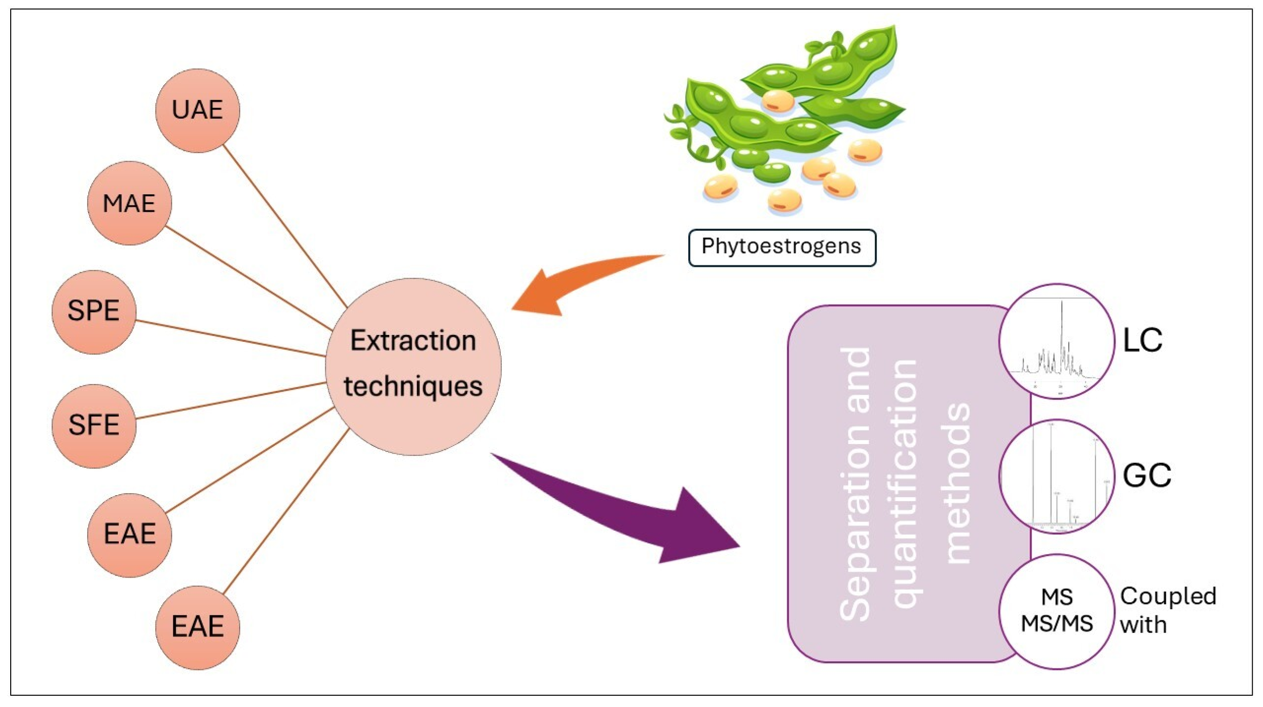
In the literature, various techniques for phytoestrogen extraction were identified: conventional solvent extraction, ultrasonication-assisted extraction (UAE), microwave-assisted extraction (MAE), supercritical fluid extraction (SFE), solid-phase extraction (SPE), pressurized liquid extraction (PLE), or enzyme-assisted extraction (EAE).
Solvent extraction involves maceration or Soxhlet extraction using organic solvents such as ethanol, methanol, or acetone. The UAE utilizes high-frequency ultrasound waves to enhance the penetration of solvents into plant tissues, improving extraction efficiency. MAE employs microwave energy to accelerate extraction, reducing extraction time and solvent consumption. SFE uses carbon dioxide (CO2) above its critical point to extract phytoestrogens from food matrices, offering advantages such as high selectivity, minimal solvent use, and ease of solvent removal. SPE involves the selective adsorption of phytoestrogens onto a solid sorbent material, followed by elution with a suitable solvent, allowing for the purification and concentration of target compounds. PLE applies heat and pressure to enhance the extraction efficiency of phytoestrogens, utilizing a diverse range of solvents to extract compounds from solid samples. EAE employs enzymes (e.g., cellulase, pectinase) to degrade plant cell walls and release phytoestrogens, facilitating enhanced extraction efficiency [41,104]
Therefore, various factors can influence the extraction efficiency of phytoestrogens, such as the type and concentration of the extraction solvent, temperature and pressure conditions, particle size and homogenization of the sample, extraction time and method, and the presence of interfering compounds in the food matrix.

5.2. Analytical Techniques for Phytoestrogens in Food Matrix

For food analysis, the methods that have been applied to phytoestrogen detection and quantification include chromatographic methods such as gas chromatography–mass spectrometry (GS–MS), high-performance liquid chromatography (HPLC) with UV detection, fluorescence detection, electrochemical detection or mass spectrometry, and capillary electrophoresis. Spectroscopic methods include UV–vis and IR spectroscopy, MALDI–TOF–MS, and immunoassay methods such as radioimmunoassay, enzyme-linked immunosorbent assay (ELISA), and time-resolved fluoroimmunoassay. Recently, hyphenated techniques—LC–MS/MS (liquid chromatography–tandem mass spectrometry) or GS–MS–MS (gas chromatography tandem mass spectrometry)—have been developed for enhanced sensitivity and selectivity [4,5]. The main techniques used for the extraction, separation, and quantification of phytoestrogens from plant and food samples are illustrated in Figure 2.

5.2.1. Gas Chromatography

A sensitive and selective method using GC–MS was developed and validated by Kim et al. for the analysis of 21 phytoestrogens, including biochanin A (BIO), daidzein (DAI), genistein (GEN), formononetin (FOR), kaempferol (KAE), equol (EQU), enterodiol (EDO), enterolactone (ELT), α-zearalanol (α-ZLA), β-zearalanol (β-ZLA), zearalenone (ZEN), and coumestrol (COU). The source of phytoestrogens was health functional foods (HFFs)—59 types, including 30 liquid-type and 29 tablet-type HFFs—collected from the market in Korea, used to relieve menopausal symptoms. HFF samples were enzymatically hydrolyzed with β-glucuronidase/arylsulfatase due to the high sensitivity to the acidic hydrolysis of certain phytoestrogens. Before GC–MS, the samples were derivatized to their trimethylsilyl derivatives by incubation with N,O-bis(trimethylsilyl) trifluoroacetamide with 1% trimethylchlorosilane for 30 min at 80 °C. All phytoestrogens were well separated and displayed symmetric peaks in the total ion chromatogram, allowing their identification, confirmation, and quantitation. The retention times for 24 phytoestrogens, including three internal standards, spanned from 6.3 min to 19.8 min within a total run time of 25.5 min. The calibration curves demonstrated excellent linearity (R2 > 0.99) within the relevant concentration ranges for each analyte. The limits of detection (LoD) and quantification (LoQ) for the 21 phytoestrogens were 0.2–20 ng/mL and 0.1–50 ng/mL, respectively. Intra-day precision was 2.2–7.9%, while accuracy was 91.0–129.1%, and inter-day precision and accuracy were 2.8–15.7 and 78.4–129.2%, respectively. Among all phytoestrogens, KAE was present in all the samples at concentrations of 0.284–1382.497 μg/mL in liquids and 1.106–1117.760 mg/g in tablets. DES showed high detection frequencies above 80%, although generally at very low concentrations, 0.001–1.785 μg/mL for liquids and 0.021–0.794 mg/g for tablets. BIO, DAI, GEN, and FOR also present high detection rates (>40%) in both sample types [105].
Benedetti et al. developed and optimized a GS–MS/MS method suitable for the determination of five phytoestrogens: formononetin, daidzein, coumestrol, genistein, and biochanin A in soymilk. For derivatization, three silylating reagents were tested, including N,O-bis-(trimethylsilyl) trifluoroacetamide (BSTFA) mixed with trimethylchlorosilane (TMCS) 90:10, derivatizing mixes N-methyl-N-(trimethylsilyl)-trifluoroacetamide (MSTFA) activated with ethanethiol and ammonium iodide, and N-(tert-butyldimethylsilyl)-Nmethyltrifluoroacetamide (MtBSTFA) with 1% of tertbutyldimethylchlorosilane (tBDMCS). Among the options, BSTFA was selected as the most appropriate derivatization reagent. GC–MS/MS has been demonstrated to be a more sensitive method than GC–MS. Therefore, other instrumental parameters were studied: source temperature, tandem MS detection parameters, and collision energies (CE) applied for the fragmentation of the ions. The optimum parameters chosen for this technique were: PTV injection mode with a temperature program ranging from 45 to 280 °C; GC–MS/MS analysis with an ion source temperature of 250 °C, an ion isolation width of m/z = x ± 1, a collision energy (CE) of 1.5 V for FORM, BIOCH, GEN, and COUM, and 3 V for DAID. The linearity was verified in the 8–500 μg/L range, with a good determination coefficient (R2) for FORM, DAID, and COUM. Intra-day and inter-day precision were in the range of 4.7–6.1% and 12.1–13.5%, respectively. Other methods must be considered for BIOCH and GEN. Sensitivity parameters LoD and LoQ were in the range of 0.1–17 μg/L and 0.3–59 μg/L, respectively. The developed method was applied to determine three commercial soy based drinks on the Italian market. GEN and DAID were present in high concentrations, ranging from 4 to 16 mg/L, while FORM and COUM were less abundant and under the method limit of detection in soymilk [106].

5.2.2. Liquid Chromatography

High-performance liquid chromatography (HPLC) is widely used for the separation of phytoestrogens and quantification due to its sensitivity and versatility.
Palma-Duran et al. developed an LC–MS method for the quantification of 16 phytoestrogens, including secoisolariciresinol, Biochanin A, matairesinol, enterolactone, enterodiol, equol, genistein, formononetin, daidzein, and coumestrol in North Mexico regional food. SPE was used to extract phytoestrogens from food samples. The sample analysis was performed by an LC diode-array detector (DAD) coupled with a single quadrupole MS with electrospray ionization (ESI) in negative mode. The method was validated in terms of selectivity, sensibility, recovery, accuracy, and precision. The linearity was tested in the range of 0.008 to 1200 ng/mL, and the calibration curves showed a good correlation coefficient except for enterodiol. LoD and LoQ were in the range of 0.002–1.061 ng/mL and 0.008–3.541, respectively. The accuracy expressed as CV was less than 15%, except for enterodiol. The inter-assay precision was between 0.44 and 11.21%, while the intra-assay precision was between 1.97 and 13.16%. The food samples analyzed were raw lettuce and mandarin, beefsteak tomato and pinto beans (cooked meals), tomato (puree), and turkey ham. The concentrations of phytoestrogens were below the LoD for most food items. Nevertheless, the levels of daidzein, kaempferol, naringenin, and genistein were notably high in turkey ham, beans, mandarins, and tomato products [98].
In work conducted by Suji Lee et al., thirty-eight isoflavone derivatives were identified and quantified in raw, steamed, and fermented seed samples from four soybean cultivars extracted by solid phase extraction using high-resolution UPLC-DAD-QToF/MS, referenced against established LC–MS libraries and a flavonoid database. The derivatives were categorized into various acylated groups and phosphorylated derivatives (Phos). Among them, the last one, succinyl-glucosides (Suc-Glu), were newly generated during Cheonggukjang fermentation by Bacillus subtilis AFY-2. The research team realized the first characterization of Phos in fermented soy products with Bacillus species.
Isoflavone quantification was accurately assessed by considering factors like soybean variety, cropping environment, storage duration, and extraction solvent system [107]. A total of 67 phytoestrogens (e.g., daidzin, equol, formononetin, genistin, glycitein, glycitin, secoisolariciresinol, and coumestrol) extracted from soy-based meal, fish fillet, and chicken meat were identified and quantified by high-performance liquid chromatography coupled to tandem mass spectrometry (LC–MS/MS), a method developed and validated by Myrtsi et al. The soy-based meal samples were prepared by solvent extraction, and the chicken and fish samples were prepared by ultrasound-assisted extraction. The samples were identified and quantified using an HPLC system coupled with a triple-quadrupole mass spectrometer. The MS/MS determination was conducted using the Electro Spray Ionization (ESI) technique in both positive and negative ion polarities. The validation process included the determination of selectivity, sensibility, recovery, precision, accuracy, detection, and quantification limits for each analyte. The linearity showed a good correlation coefficient (R2 > 0.99). The LoD and LoQ were in the range of 1.8 to 120.5 ng/mL and 5.5 to 365.1 ng/mL, respectively. The exception was equol, which displayed poor sensitivity. The determined repeatability within intra- and inter-day analyses was expressed using the relative standard deviation (RSD), ranging from 0.2% to 19.8% for run-to-run precision and from 0.04% to 25.7% for day-to-day precision.
The soy-based meal contained 21 phytoestrogens, consisting mainly of isoflavones like genistein, daidzein, and daidzin, as well as lignans such as coumestrol. Glycitin, glycitein, and genistin were detected in lower concentrations.
Regarding poultry and fish meals, poultry meals were determined to contain the highest total amount of phytoestrogens. The isoflavones identified were genistein, daidzein, glycitein, or daidzin, present in quantities less than 7.0 mg/kg [108].
The UHPLC–MS/MS method was performed for the determination of five isoflavone compounds, including daidzein, genistein, biochanin A, daidzin, and genistin, in lentils and other legumes (soybeans, chickpeas, “cannellini” beans, “fava” beans, “borlotti” beans, peas, and mixtures of legumes) by Vila-Donat et al. The samples were processed by solvent extraction, followed by a clean-up step consisting of freezing overnight at −24 °C to precipitate proteic or lipidic residues that can interfere during UHPLC–MS/MS analysis and solid phase extraction. The samples were analyzed using an HPLC system coupled with a triple-quadrupole mass spectrometer with an ESI source operating in negative and positive ionization modes. Detection was performed by electrospray ionization (ESI)–MS in the “multiple reaction monitoring” (MRM) mode. Linearity was tested at concentrations ranging from 0.1 to 500 μg/L for biochanin A, genistin, and daidzin and from 1 to 250 μg/L for genistein and daidzein. The calibration curve showed good correlation coefficients. Intraday and inter-day repeatability expressed in terms of relative standard deviations (RSD%) ranged from 0.4% to 1.1% and from 4.3% to 11.5%, respectively. LoD limits were between 0.033 and 0.33 μg/L, while the LoQ limits were between 0.1 and 1 μg/L. Several lentil samples collected from different locations of the Umbro-Marchean Apennines were analyzed using principal component analysis (PCA), with the five studied isoflavones as variables. They exhibited significant variance in isoflavone content that was correlated with the altitude at which the samples were grown. The concentration of total isoflavones in lentils ranged from 1.1 to 95.6 μg/L. The other samples consisted of soybeans, chickpeas, “cannellini” beans, “borlotti” beans, “fava” beans, peas, “Pardina” Spanish lentils, a mixture of lentils and beans, and a mixture of beans and spell, which were purchased from a local supermarket. The highest amount of isoflavones was determined in soybeans, followed by chickpeas. The mixtures of beans with lentils or beans with spelled beans showed a higher isoflavone content compared to the individual samples [99].
Recently, Gu et al. developed a combined ultraperformance liquid chromatography with tandem mass spectrometry (UPLC–MS/MS) method for efficient quantitation of isoflavones in soy protein-based infant food. In the study, only three aglycone isoflavones (daidzein, glycitein, and genistein) were used to quantify all isoflavones. Three distinct β-glucosidases were assessed for the hydrolysis of all glucoside isoflavones, among which Thermotoga maritima proved to be the most efficient in hydrolyzing all glucoside isoflavones into their corresponding aglycones. However, resistance was encountered with malonyl and acetyl glucosides. An extraction solution of 80% methanol with 0.3% NaOH was used to tackle this challenge. The samples were assessed using a UPLC instrument coupled with a triple quadrupole mass spectrometer. The ion source was electrospray ionization (ESI). The compounds were ionized in positive mode. Multiple reaction monitoring (MRM) mode was used for phytoestrogen quantitation. All linear correlation coefficients (R2) were above 0.999, indicating good linearity. The LoD and LoQ limits were in the range of 3–69 μg/kg and 10–231 μg/kg. Three commercially available infant foods based on soy protein from distinct regions were analyzed using the developed method. Additionally, a dietary soymilk powder was simultaneously tested for comparison. The levels of total isoflavones in these infant foods showed minimal variation. In contrast, the ISF content in soymilk powder was approximately twice as high as that in infant foods [109].

5.2.3. Enzyme-Linked Immunosorbent Assay (ELISA)

A recent study focused on developing an efficient soybean extraction method for isoflavones. One step of the analysis involved monitoring the genistein and daidzein content variations through statistical analysis of the data from an ELISA and a Folin–Ciocalteu assay. The sample preparation included the digestion of glycosylated isoflavones by β-glucuronidase arylsulfatase from Helix pomatia, followed by the extraction of aglycone compounds. The antibodies were obtained by functionalizing isoflavones that were coupled in C2 to bovine serum albumin or swine thyroglobulin using a previous method validated by Bennetau-Pelissero et al. and Houerou et al. [110].
Another recent study conducted by Fujii et al. used an indirect competitive enzyme-linked immunosorbent assay (icELISA) to determine formononetin (FMN) content in food samples using a monoclonal antibody (mAb) against FMN produced by a newly established hybridoma cell line. After assessing the compatibility of the monoclonal antibody (mAb) with its cell line culture and determining its titer, a stable and optimal hybridoma cell line named 6C3 was successfully identified. The precision of the icELISA with mAb 6C3 was assessed through intra-assay and inter-assay analyses. The maximum coefficient of variation (CV) values within intra- and inter-assays were 5.244% and 5.079%, respectively, with all values at 5%. The food samples, including Phaseolus radiatus, Garbanzo bean, soybean (dried product), Licorice tea, and Licorice candy, were treated with simple acid hydrolysis, and FMN was quantified by performing the developed icELISA method. FMN was not detectable in soybean and Licorice candy [111].
The analytical methods for the determination of phytoestrogens in various food samples have certain limitations in terms of sensitivity and detection, quantification challenges, validation, and standardization. The sensitivity of analytical techniques may be insufficient for detecting low concentrations of phytoestrogens in complex food matrices. Also, high background noise and interference from matrix components can limit the detection limits of spectroscopic and chromatographic methods. Accurately measuring phytoestrogens can be challenging due to matrix effects, calibration issues, and the lack of certified reference materials. Variability in phytoestrogen content within food samples and between different sample batches can affect the reliability of quantitative results.

6. Phytoestrogens Analysis in Biological Samples

Phytoestrogens present in human food are comprised mainly of isoflavones and lignans. Nutrition-based studies have highlighted a linear dose-response relationship between the intake and urinary excretion of isoflavones. Low-fat, high-fiber, and carbohydrate diets determine increased levels of excreted lignans; therefore, lignan levels are considered a possible marker for healthier dietary models.
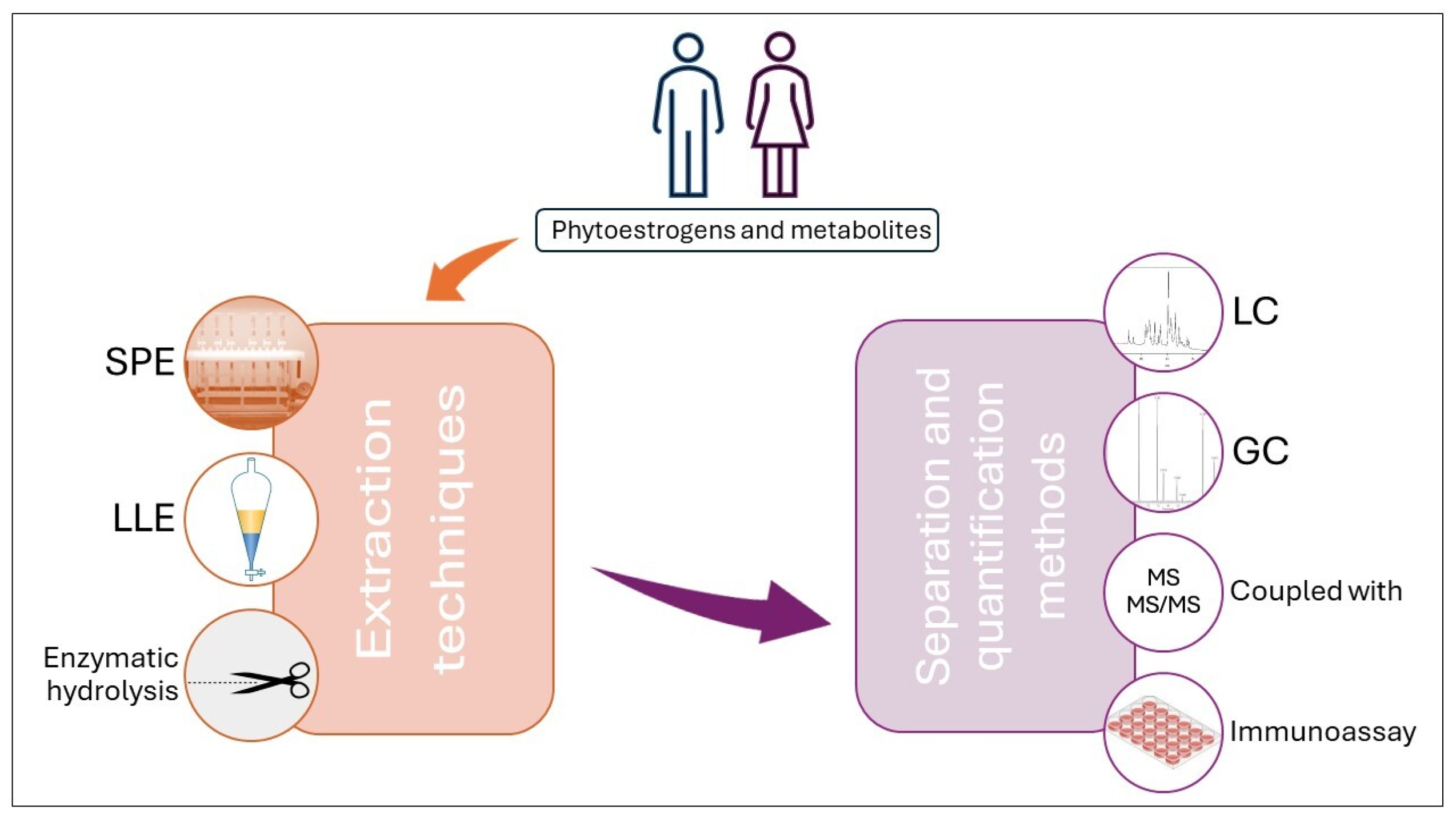
The standard approach for quantifying phytoestrogens in urine and plasma involves chromatographic separation combined with various detection methods. The main techniques used for the extraction, separation, and quantification of phytoestrogen biological samples are illustrated in Figure 3. Alternative techniques like HPLC–DAD and GC/FID necessitate a significant initial urine volume of 20 mL that shall be subsequently concentrated 10–20 times for the analysis. These methods are most appropriate for research scenarios anticipating elevated phytoestrogen levels, such as interventions involving phytoestrogen-rich foods or high-dose pharmacokinetic investigations. Immunoassay techniques utilizing radiolabeled, enzyme-linked, or fluorescently tagged monoclonal antibodies have been devised to quantify isoflavones, genistein, daidzein, or enterolignan enterolactone levels in serum, plasma, or urine. These methods are mainly suitable for efficiently analyzing large sample cohorts [112].

6.1. Liquid Chromatography

HPLC–DAD methods were tested to quantify different isoflavones and lignans in urine, plasma, and human milk. The mobile phase composition optimization for one compound determined a reduced yield of separation and detection for the others; for example, by replacing acetic acid–water co-solvents from a mixture of acetonitrile with methanol and dichloromethane, better separation and detection parameters were obtained for the metabolites equol and O-desmethylangolensin (O-DMA), but those for daidzein, genistein, and coumestrol were reduced. The HPLC–DAD method was applied to human milk, urine, or plasma samples, and the LoD for genistein and daidzein, obtained in different studies, were similar for the three types of biological fluids (around 26.6 and 54.3 nmol/L) [5].
Years ago, Franke and Custer developed a rapid, sensitive, and precise reversed-phase HPLC-DAD method for the assessment of the main isoflavones in the human urine, namely daidzein, genistein, formononetin, and biochanin-A, as well as their metabolites, equol and O-DMA, and of the phytoestrogen coumestrol, after administration of pharmacological or dietary doses of soybeans. Solid-phase extraction (SPE) was performed, followed by HPLC separation and identification through retention times and UV scans. A fluorometric response was also used to identify coumestrol. Acetonitrile and acetic acid–water (10/90, v/v) were used as the mobile phase. Spiking recovery depended on the initial and spiked amount of compound, but it ranged from 76.3% for genistein to 100.7% for O-DMA. The recovery of the total amount of phytoestrogens in the urine samples of the 11 human subjects was highly variable, between 5% and 24% of the dose. The obtained LoDs were below 10 nmol/L for daidzein, genistein, and formononetin (5.15, 8.75, and 7.25 nmol/L, respectively) and about 100 times higher for the metabolites equol and O-DMA (623 and 780 nmol/L, respectively) [113].
Another HPLC–UV–DAD method that used SPE for extraction was set up for the determination in plasma of intact 16 metabolites issued from daidzein and genistein, with a special interest in the highly polar glucuronic acid and/or sulfuric acid conjugated ones. Ammonium acetate and acetonitrile were used as the mobile phase under gradient mode, and luteolin-3′,7-di-O-glucoside was used as an internal standard. The recovery rates of isoflavone metabolites from plasma ranged from 76.6% to 109.4%. The lower LoQ for the studied metabolites ranged between 21.1 and 23.4 ng/mL, and the lower LoDs were between 7.9 and 9.4 ng/mL [114]. This method was applied to monitor the presence of the 16 metabolites in plasma samples collected from healthy human subjects who had consumed kinako, a baked soybean powder dish. Only 2% of the original compounds were identified in plasma (daidzein and genistein) [115].
An update from classical HPLC–UV–DAD was the method established by Redruello et al. that used reverse-phased UHPLC with a spectrophotometric photodiode array/fluorescence (FLR) detection system to determine the concentration of equol (a metabolite of daidzein with high estrogenic activity), daidzein, and genistein (the main soy isoflavones) in human urine. As in the case of the HPLC–UV–DAD methods, SPE was employed to isolate the compounds. The mobile phase contained orthophosphoric acid in water and methanol. Recovery ranged between 70% (daidzein) and 114% (genistein). The inter-day coefficient of variation (CV) was below 15%, and CV in the intra-day tests was lower than 7.5%. The LoDs were low for daidzein and equol (4.75 nmol/L and 2.93 nmol/L, respectively), while the one for genistein (15.17 nmol/L) [116] was higher than those obtained in the mentioned HPLC–UV–DAD [113,114]. Compared to traditional HPLC methods, the rapid method permits a rapid screening of more urine samples to monitor the patient’s compliance with isoflavone oral therapy by evaluating the equol issued from daidzein biotransformation. In this regard, this method was used to evaluate the phytoestrogen profile of menopausal women taking soy-based supplements [116].

6.2. Liquid Chromatography Coupled with Mass Spectrometry Methods

For better results in terms of individual compound identification, the HPLC was coupled with mass spectrometry using a single quadrupole mass spectrometer with atmospheric pressure chemical ionization (APCI). This made possible the quantitative assessment of 13 phytoestrogens in one assay, dietary constituents, and metabolites (including genistein, daidzein, equol, O-DMA, and coumestrol) in human urine or serum samples. After enzymatic deconjugation, liquid–liquid extraction (LLE) was performed for the urine samples, and SPE was performed for the serum ones. For the mobile phase, a combination of water and methanol–acetonitrile (80:20, w/w) was used, acidified with 0.025% (v/v) formic acid. The average recovery rate of phytoestrogens in urine samples was higher than 67%, while for serum samples, it was superior to 83%. The inter-assay variation (CV) was below 18.8% in urine and lower than 15.7% in serum, while the intra-assay CV for urine and serum samples was generally lower than 10%. Good LoDs were attained for both analyzed biological fluids, with even lower values for urine samples (0.2 ng/mL–7.7 ng/mL in urine versus 1.4 ng/mL–20.4 ng/mL in plasma). For daidzein, genistein, and equol, the LoDs in urine and serum samples were much improved compared to the previously mentioned methods (1, 0.8, 5.8 ng/mL in urine and 1.9, 2.2, and 13.8 ng/mL in serum) [117].
Valentin-Blasini et al. had previously quantitatively analyzed seven phytoestrogens from urine and serum through an LC–MS/MS technique, and the LoDs for equol were 2.4 ng/mL in serum and 1.1 ng/mL in urine. The best LoDs obtained with this method were 0.1 ng/mL for enterolactone in serum and 0.2 ng/mL for matairesinol in urine. Advanced sensitivity could not be managed for daidzein in urine (highest value for LoD, 9.3 ng/mL). After SPE, the recovery was between 88.2% for matairesinol and 98.6% for O-DMA from serum, and between 91.7% for coumestrol and 103.7% for enterolactone from urine. The intraday and interday variation was assessed, and good results were reported for enterolactone in serum (CV = 4.5%) or O-DMA in urine (CV = 5.5%), but increased variation was recorded for genistein or equol (CV 19.3% and 13.7%, respectively) in serum or for coumestrol and matairesinol (13.8% and 12.6%, respectively) in urine [118].
Tandem LC–MS/MS gave better selectivity for quantification than LC–MS, as the patterns of the ions resulting from fragmentation were monitored. Recoveries were very satisfying, with more than 90%. The inter-assay variability depended on the compound; for daidzein and genistein in plasma samples, it was below 10%, which was also the value for most phytoestrogens analyzed from urine; for low-concentration genistein in serum samples, it was calculated to have an inter-assay variability of 19% [5].
A study comparing APCI with electrospray ionization (ESI) regarding the performance in the HPLC–MS/MS quantitative analysis of six phytoestrogens, primary and metabolites, showed that ESI gave better results for most analytical parameters. SPE performed extraction, and a combination of ammonium acetate buffer pH 6.5 and methanol/acetonitrile (1:1) was chosen for the mobile phase. The mean recovery rates were 99% for APCI (95–105%) and 100% for ESI, but with a wider range (86–114%). From the point of view of inter-assay variation, a 5.3–8.9% CV was calculated for both APCI and ESI for the six compounds spiked in high concentrations; ESI usually induces lower variations (CV) than APCI in cases of medium and low analyte concentrations (4.4–9.2% versus 13–30% for low concentrations and 6.1–12% versus 5.3–26% for medium concentrations). ESI mode provided lower LoDs than APCI for genistein and equol (0.06 versus 4 ng/mL and 0.3 versus 2.7 ng/mL, respectively) [119], which were better than the ones reported in the HPLC–MS method using APCI [117]. APCI proved to be more sensitive for enterolactone (0.4 ng/mL versus 0.3 ng/mL), and equal LoDs were recorded for daidzein and O-DMA [119]. Previously, lower sensitivity for enterolactone was obtained with ESI using 13C3 labeled isotopes after enzymatic hydrolysis and ether extraction, with a LoD of 0.55 nM (164 ng/mL) [120].
A rapid LC–MS/MS method was developed to quantify as many as 11 phytoestrogens, including their metabolites, from human serum samples after their extraction with diethyl ether through LLE. Recovery rates were not very satisfying, with a minimum of 57.48% for genistein and a maximum of 88.43% for dihydrodaidzein. The intra-day CV values were up to 11.64% for glycitein and as low as 1.52% for enterolactone; the inter-day variations ranged within a narrower interval, from 2.82% (for hihydrodaidzein) to 10.13% for equol. The declared LoD was 1 ng/mL for all analytes except dihydrodaidzein (2 ng/mL), while the upper LoQ was set to 5000 ng/mL [121].
For the simultaneous quantitative assay of the isoflavones puerarin and daidzein, a UPLC–MS/MS method was optimized. As daidzein also results from the gut microbiota enzyme hydrolysis of puerarin, the method is intended to investigate the presence of both compounds in plasma samples collected from consumers of Gegen, the dried root of Pueraria lobata. After LLE, the chromatographic separation was performed using a mobile phase comprising 0.05% acetic acid in water and 0.05% acetic acid in methanol. The recovery rate for puerarin was relatively low, 14.8 ± 2.5%, while a better value was acquired for daidzein, 88.4% ± 6.2%. The inter-batch variation (CV) was 10.82% for puerarin and 12.16% for daidzein, while the intra-batch one was below 8.36% and 12.11%, respectively. As for the sensitivity of the method applied to human plasma samples, the LoQ was a satisfying 0.2 ng/mL for both phytoestrogens [122].
Lower intra- and inter-day variation values and narrower ranges were observed for another LC–MS/MS method that used SPE with enzymatic hydrolysis for the investigation of phytoestrogens from human and urine samples. Thus, the intra-assay variation ranged between 1.1% for daidzein and 7.1% for glycitein, while the interval for inter-assay variation was 4.2% (for enterodiol) and9.8% (for secoisolariciresinol). Excellent recovery rates were provided through this method, from the lowest 82% for genistein to the highest 103% for secoisolariciresinol and enterolactone. All the mentioned parameters were calculated for urine. The authors mentioned a LoD for all analytes in both biological media of 0.1 nm/mL, with a maximum quantifiable concentration of 2000 ng/mL [123].
The hydrolysis of phytoestrogen conjugates from urine and plasma was investigated and improved for better results. Although the process of conjugate hydrolysis is slow, an increase in temperature from 37 °C to 45 °C decreased the reaction time in both biological media: 40 to 20 min for equol, 120 to 100 min for enterodiol in urine, 8 h to 2 h for O-desmethylangolensin, and 80 to 40 min for equol, 40 to 20 min for glycitein in plasma. Another strategy for increasing the yield of hydrolysis was to double the amount of enzyme (beta-glucuronidase from Helix pomatia) used. Thus, the time was halved from 40 to 20 min for daidzein, enterolactone, equol, and glycitein and reduced from 120 min to 100 min for enterodiol in urine; in plasma, a spectacular decrease from 8 h to 160 min was obtained for O-DMA, but also relevant results were noted for equol (80 min to 20 min) and for genistein (40 min to 20 min) [124].
An LC–MS/MS method for direct quantification of the conjugated and free forms of the phytoestrogen enterolactone from human plasma was established, allowing the investigation of a pattern of conjugation of enterolactone in humans after SPE. Recovery rates were superior to 90% except for enterolactone sulfate (65–68%). The intra- and interbatch variations (CV) were lower than 7.7% and 8.9%, respectively. The lower LoQ were 86 pM for free enterolactone and 16 pM and 26 pM for the conjugates sulfate and the glucuronide. The analysis was performed on almost 4000 samples and proved that the conjugation pattern is similar between sexes, and the primary path is glucuronidation [11].
In order to investigate the profile of phytoestrogens in populations from different Asian countries, an LC–MS/MS method was used to quantify their presence in urine samples. The extraction was performed by LLE; afterward, the HPLC separation was possible using methanol and 2 mM ammonium acetate/methanol 10% as the mobile phase. Recovery rates for the native compounds were between 71.5 and 83.4%. However, the corrected recoveries for the internal standards ranged from 96.7% to 115% [125], similar to those obtained by Rybak et al. [119]. The intra-assay variation had a low value of 0.82–3.4% of CV, depending on the analyte; for the inter-assay variation, all values ranged from 1.7% for genistein to 5.3% for coumestrol [125], marking an improvement in method development by comparison to the before-mentioned reports. The LoDs ranged from 0.08 ng/mL for enterolactone to 0.32 ng/mL for genistein and 0.35 ng/mL for daidzein. The method allowed us to investigate if the different degrees of biotransformation of daidzein to equol depend on the population [125].

6.3. Gas Chromatography

The analytical investigations have applications not only for ingested phytoestrogen quantification but also for studies centered on their biotransformation and the subsequent metabolites. Some metabolites are more active than the parent compound, such as equol versus daidzein. GC–MS was considered the most appropriate technique for studying the isoflavones and lignans metabolites. By 2002, the lowest LoD reported for a GC–MS method was 0.04 nmol/L. Depending on the studied compound, the inter-assay variations (CV) between 2-year and 1.5-year experiments where the GC–MS method was applied to serum samples containing phytoestrogens ranged from 6% (for daidzein and genistein in an average concentration of 570 nmol/L) to 11% (for 280 nmol/L isoflavone) [5].
One of the first teams reporting the results of a GC–MS method used for assessing phytoestrogens in human biological samples was that of Fotsis et al., who quantified enterolactone and enterodiol in human urine collected from men and women aged 22 to 62. The extraction of the mono-glucuronides from the urine was conducted on a small reversed-phase cartridge of octadecylsilane-bonded silica, and the isolation was performed using anion exchange chromatography. Afterward, enzymatic hydrolysis was used to release the lignans, which were re-extracted and quantified by capillary column GC. Two gas chromatographs with different columns were used; both had flame ionization detectors and worked with hydrogen as a carrier gas. The recovery rate calculated using radiolabeled estriol was 89–90%. The intra-assay CV was between 5.7% and 7.2% for both compounds. The lowest level of enterodiol (81.6 μg/L) corresponded to a 29-year-old (y.o.) male subject, while the highest concentration (656.7 μg/L) was found in a 61 y.o. female urine sample. For enterolactone, the lowest level (224.2 μg/L) was dosed in a 57 y.o. urine sample, while the most abundant sample was quantified in a urine sample collected from a 61 y.o. female (2370.4 μg/L) [126]. There was no correlation between the levels of enterodiol and those of enterolactone in the analyzed samples.
Later, another GC–MS method was used to investigate the presence of different phytoestrogens in human urine collected from Finnish subjects, both lignans (enterolactone, enterodiol, and matairesinol) and isoflavonoids (daidzein, equol, O-DMA, and genistein). After SEP and chromatographic isolation of the conjugated forms, enzymatic hydrolysis was performed using a Helix pomatia extract to obtain the compounds’ free form. For the intra-assay variation, the calculated values of CV were in the range of 0.8% (for equol)—15.2% (for matairesinol), while the inter-assay variation ranged from 4.1% for enterolactone to 13.9% for matairesinol; an extreme value of 129% resulted for O-DMA, which was found in very low concentrations. In Finnish male subjects aged 25–70, matairesinol was poorly excreted in the urine, often below 5 nmol/24 h; for genistein, it was noticed that it is excreted in a higher concentration than daidzein. It was concluded that there are different patterns of phytoestrogen metabolism according to the established levels in urine samples. The largest range of concentrations in urine resulted in enterodiol, 3.8–2580 nmol/24 h [127].
Lignans enterolactone, enterodiol, and isoflavones daidzein, O-DMA, equol, genistein, and glycitein were also quantified from human urine samples in another GC–MS study. For sample preparation, hydrolysis with a β-glucuronidase/aryl sulfatase extract of Helix pomatia was followed by SPE. The recovery rates ranged from 89.6% ± 1.9% for O-DMA to 104.3% ± 6.7% for glycitein. The intra-assay CV had values in the 1.8% (for equol)–6.5% (for glycitein) interval, while the inter-assay CV ranged from 3.2% (for enterodiol) to 26.5% (for glycitein). The lowest LoD was recorded for enterodiol (1.2 ng/mL) and the highest for O-DMA (5 ng/mL). The 234 analyzed urine samples were collected from healthy women included in a prospective study of diet and cancer in the UK for 10 years. The lowest mean concentration in urine corresponded to equol (6.1 nm/mL) and the highest one to enterolactone (1104.5 nm/mL) [128].
The abundance of daidzein, equol, and genistein was investigated from urine samples collected from German adults with a typical Western diet using a GC–MS method. The urine samples underwent hydrolysis of the conjugates with β-glucuronidase and sulfatase isolated from E. coli and SPE. The recovery rates for different concentrations of spiked samples ranged from 92% for daidzein to 104% for daidzein and genistein for intra-assay evaluation and from 90% for equol to 127% for daidzein for inter-assay assessment. The CV in the intra-assay was given as an interval for each compound, namely 1–7% for daidzein, 3–15% for genistein, and a very narrow range of 5–6% for equol; the inter-assay variation was calculated as 8% for daidzein, 16% for genistein, and 18% for equol. The recorded limits of detection were 4 ng/mL for equol and daidzein and 5 ng/mL for genistein. The average levels of phytoestrogens in the 16 collected urine samples were 62, 24, and 65 μg/day for daidzein, equol, and genistein, respectively, and equol, the metabolite of daidzein, was found in more than 50% of the samples [10].
Another study viewing phytoestrogen patterns of metabolism and excretion in the German population was keen on children and adolescents aged 6–18; the analyzed urine samples came from 90 subjects, 43 girls and 47 boys, who provided 510 urine samples. The method followed the steps previously described [10], and the established limits of detection were 3.5 ng/mL for daidzein, 5.3 ng/mL for genistein, and 3.8 ng/mL for equol (the daidzein metabolite). Unlike the previous study [10], the proportion of the population excreting equol in all urine samples was 31%, but the greater number of subjects in this case must be considered (90 versus 16). The urine concentrations and the excretion rates of daidzein, equol, and genistein were similar between girls and boys; for the 6–12-year-old subjects, an increase in the total isoflavone excretion was observed, followed by a constant trend during adolescence [129].

6.4. Immunoassay

Satisfying features were obtained for immunoassays used for phytoestrogen assessment. With a recovery average of 92% and intra- and inter-assay variability below 10%, the LOD was over 3 nmol/L only in an ELISA method for genistein (185 nmol/L). The drawback of these methods could be the cross-reactivity, which resulted in better cross-reactions with other phytoestrogens than those intended to be measured. This was observed mainly in TR-FIA (time-resolved fluorescence immunoassay) methods applied to urine samples (for enterolactone and genistein, for example), where there are numerous phytoestrogen metabolites or other classes of compounds [5].
Enterolactone, genistein, and daidzein were quantified in human urine samples using the TR-FIA method and the europium-labeled compound as tracers. 5′-carboxymethoxy derivative of enterolactone and 4′-O-carboxymethyl derivatives of genistein and daidzein were synthesized, which were eventually coupled with bovine serum albumin and used for immunization of rabbits. Urine samples were enzymatically hydrolyzed and subjected to quantification of the targeted phytoestrogens. The intra-assay variation (CV) ranged from 1.9% for enterolactone to 5% for daidzein, while the inter-assay variation coefficient had values between 2.4% for enterolactone and 9.7% for genistein. The enterolactone level in 24 h urine was below 7 nmol for more than 60% of the 215 urine samples collected from healthy Finnish women. The lowest concentrations in the working range were 1.6, 1.7, and 1 nmol/L for enterolactone, genistein, and daidzein, respectively. The mean values for enterolactone, genistein, and daidzein concentrations corresponding to vegetarians were significantly higher than the ones found in omnivores’ urine samples (more than three times higher for genistein and daidzein) [130].

7. Discussion

As phytoestrogens, primarily isoflavones and lignans, are compounds of interest in nutritional research due to their potential health benefits, their level in food and herbal products and possible metabolic fate are frequently investigated. This monitoring requires precise extraction methods and analytical techniques to identify phytoestrogen patterns and quantify their levels. Therefore, in recent years, modern extraction techniques such as PLE, SFE, EAE, and UAE have developed due to their improved efficiency and reproducibility and are often combined with high-throughput analyses like UHPLC–MS/MS for more accurate results on phytoestrogen profiles. These modern techniques have been extensively used in isolating isoflavonoids from different matrices, especially soybean and soy derivatives. UAE improves extraction efficiency, and MAE speeds up the process; nevertheless, they may induce compound degradation. SFE provides high selectivity, while EAE is recommended for phytoestrogen extraction from the herbal matrix. SPE is often used for biological samples when matrix concentration is needed.
HPLC, GC, LC–MS, and CE are the main validated analytical methods that play an essential role in characterizing phytoestrogen patterns and quantifying phytoestrogens in a variable matrix. HPLC–DAD is suitable for food and biological samples, providing high recovery rates for isoflavone metabolites, but the sensitivity is lower than HPLC–MS. Although GC–MS methods are highly sensitive and selective, the equipment is more expensive, and the sample preparation requires a significant amount of time.
Furthermore, the chemical profile revealed by these methods can be used as a marker of the stability of phytoestrogens in food or herbal products.
The most frequently used methods for the assessment of phytoestrogens are various forms of LC, often coupled with mass spectrometry (LC–MS), which are preferred due to their high sensitivity and versatility. HPLC is particularly noted for its widespread use in the analysis of phytoestrogens. Better sensitivity was usually achieved for LC–MS methods when referring to the detection and quantification of a certain phytoestrogen, especially from the herbal or food matrix. For example, a much higher sensitivity was obtained by the LC–MS method developed by Palma-Duran et al. for the quantification of various phytoestrogens, including genistein, in regional foods from North Mexico (LoD: 0.002–1.061 ng/mL and LoQ: 0.008–3.541 ng/mL) than that reached in the GC–MS/MS method developed by Benedetti et al. for the quantification of genistein in soymilk (LoD: 0.1–17 µg/L and LoQ: 0.3–59 µg/L, which translates to 0.1 ng/mL and 0.3 ng/mL, respectively).
Isoflavones, such as daidzein, genistein, and biochanin A, are analyzed through GC–MS or LC–MS methods from food matrices and biological samples [10,105,109,122]. The commonly used methods for lignans include HPLC–TOF–MS for both aglycones and unhydrolyzed glucosides or HPLC–MS/MS for the lignans fraction in Coffea arabica seeds [51], but also gradient reversed-phase HPLC or GC–MS for those existing in flaxseed [39,49]. Coumestans (like coumestrol) were assessed in biological samples using an HPLC–DAD method [113], while for the analysis of resveratrol, a natural stilbene, an HPLC method following SPE was applied [98].
The most sensitive method appears to be the HPLC–TOF–MS developed to separate and determine both aglycones and unhydrolyzed glucosides, where the LoDs for secoisolariciresinol and secoisolariciresinol diglucoside-containing oligomers were as low as 0.008 pg. The most sensitive method used for the assessment of phytoestrogens in biological samples was an LC–MS/MS technique when low LoDs were attained for various compounds, such as 0.1 ng/mL for enterolactone in serum and 0.2 ng/mL for matairesinol in urine.
Also, immunoassays, including ELISA and TR-FIA, are commonly used for high-throughput screening of phytoestrogens and their metabolites in biological samples. These assays offer high sensitivity, specificity, and relatively low costs, making them valuable tools for phytoestrogen in vivo analysis, which is more relevant as the potency and efficacy of phytoestrogens depend on their metabolites. ELISA efficiently quantifies phytoestrogens like genistein and daidzein in urine samples, albeit with a potentially higher LoD compared to chromatography. TR-FIA, utilizing europium-labeled tracers, offers high sensitivity and specificity for compounds such as enterolactone, genistein, and daidzein.
However, these methods have known limitations, such as matrix effects and calibration issues, which can affect the reliability of quantitative results; complex matrices, as there are in food, may induce a lack of sensitivity and detection problems. Food matrices are also challenging because of intra- and inter-batch phytoestrogen composition variability. In terms of biological samples, urine has to be concentrated when analysis is performed through HPLC-DAD or GC-FID techniques; if a TR-FIA method is considered for urine samples, special attention should be paid to the interference of structurally similar metabolites.

8. Conclusions

There are various analytical methods for phytoestrogens, most of which offer sensitivity in the low ng/mL range. Analytical methods such as GC, HPLC, and CE provide accurate identification and quantification of phytoestrogens, complemented by spectroscopic and mass spectrometric techniques for characterization. LC–MS and GC–MS are indispensable for analyzing phytoestrogens in biological samples. Immunoassays offer high-throughput screening capabilities for phytoestrogens. Overall, integrating these advanced techniques facilitates comprehensive studies of phytoestrogens and a better understanding of their health benefits, safety, and potential applications in medicine and nutrition. Despite advancements, challenges are present, including sensitivity limitations, matrix effects, calibration issues, and variability in phytoestrogen content within food and plant samples. Overcoming these challenges requires the ongoing refinement and standardization of analytical methods to ensure accurate quantification and reliable results in phytoestrogen analysis.

Author Contributions

Conceptualization, writing—original draft preparation, C.P. and I.A.-D.; writing—original draft preparation, A.A.O., O.T.O. and D.E.P.; methodology, I.A.-D.; formal analysis, investigation, writing—review and editing: C.P., I.A.-D. and A.A.O.; visualization: I.A.-D.; supervision, C.P. All authors have read and agreed to the published version of the manuscript.

Funding

Part of this work was financially supported by “Carol Davila” University of Medicine and Pharmacy Bucharest, Romania, through Contract no. 33PFE/30.12.2021 funded by the Ministry of Research and Innovation within PNCDI III, Program 1—Development of the National RD System, Subprogram 1.2—Institutional Performance—RDI Excellence funding projects.

Data Availability Statement

Data are contained within the article.

Conflicts of Interest

The authors declare no conflicts of interest.

References

  1. Chavda, V.P.; Chaudhari, A.Z.; Balar, P.C.; Gholap, A.; Vora, L.K. Phytoestrogens: Chemistry, potential health benefits, and their medicinal importance. Phytother. Res. 2024, 38, 3060–3079. [Google Scholar] [CrossRef] [PubMed]
  2. Sirotkin, A.V.; Harrath, A.H. Phytoestrogens and their effects. Eur. J. Pharmacol. 2014, 741, 230–236. [Google Scholar] [CrossRef] [PubMed]
  3. Cvejić, J.; Bursać, M.; Atanacković, M. Phytoestrogens: “estrogene-like” phytochemicals. Stud. Nat. Prod. Chem. 2012, 38, 1–35. [Google Scholar]
  4. Wang, C.-C.; Prasain, J.K.; Barnes, S. Review of the methods used in the determination of phytoestrogens. J. Chromatogr. B Analyt. Technol. Biomed. Life Sci. 2002, 777, 3–28. [Google Scholar] [CrossRef] [PubMed]
  5. Wilkinson, A.P.; Wähälä, K.; Williamson, G. Identification and quantification of polyphenol phytoestrogens in foods and human biological fluids. J. Chromatogr. B Analyt. Technol. Biomed. Life Sci. 2002, 777, 93–109. [Google Scholar] [CrossRef]
  6. Hoikkala, A.A.; Schiavoni, E.; Wähälä, K. Analysis of phyto-oestrogens in biological matrices. Br. J. Nutr. 2003, 89 (Suppl. 1), S5–S18. [Google Scholar] [CrossRef] [PubMed]
  7. Suhaj, M. Spice antioxidants isolation and their antiradical activity: A review. J. Food Compos. Anal. 2006, 19, 531–537. [Google Scholar] [CrossRef]
  8. Horn-Ross, P.L.; Barnes, S.L.; Lee, M.M.; Coward, L.H.; Mandel, E.M.; Koo, J.; John, E.M.; Smith, M.S. Phytoestrogen content of foods consumed in the United States. J. Natl. Cancer Inst. 2000, 92, 134–146. [Google Scholar]
  9. Rostagno, M.A.; Villares, A.; Guillamón, E.; García-Lafuente, A.; Martínez, J.A. Sample preparation for the analysis of isoflavones from soybeans and soy foods. J. Chromatogr. A 2009, 1216, 2–29. [Google Scholar] [CrossRef]
  10. Moors, S.; Blaszkewicz, M.; Bolt, H.M.; Degen, G.H. Simultaneous determination of daidzein, equol, genistein and bisphenol A in human urine by a fast and simple method using SPE and GC-MS. Mol. Nutr. Food Res. 2007, 51, 787–798. [Google Scholar] [CrossRef]
  11. Nørskov, N.P.; Kyrø, C.; Olsen, A.; Tjønneland, A.; Knudsen, K.E. High-throughput LC-MS/MS method for direct quantification of glucuronidated, sulfated, and free enterolactone in human plasma. J. Proteome Res. 2016, 15, 1051–1058. [Google Scholar] [CrossRef] [PubMed]
  12. Qin, W.; Zhu, W.; Shi, H.; Hewett, J.E.; Ruhlen, R.L.; MacDonald, R.S.; Rottinghaus, G.E.; Chen, Y.C.; Sauter, E.R. Soy isoflavones have an antiestrogenic effect and alter mammary promoter hypermethylation in healthy premenopausal women. Nutr. Cancer 2009, 61, 238–244. [Google Scholar] [CrossRef]
  13. Lóránd, T.; Vigh, E.; Garai, J. Hormonal action of plant derived and anthropogenic non-steroidal estrogenic compounds: Phytoestrogens and xenoestrogens. Curr. Med. Chem. 2010, 17, 3542–3574. [Google Scholar] [CrossRef]
  14. Reed, K.F.M. Fertility of herbivores consuming phytoestrogen-containing Medicago and Trifolium species. Agriculture 2016, 6, 35. [Google Scholar] [CrossRef]
  15. Daly, D.C.; Cameron, K.M.; Stevenson, D.W. Plant systematics in the age of genomics. Plant Physiol 2001, 127, 1328–1333. [Google Scholar] [CrossRef] [PubMed]
  16. Ososki, A.L.; Kennelly, E.J. Phytoestrogens: A review of the present state of research. Phytother. Res. 2003, 17, 845–869. [Google Scholar] [CrossRef]
  17. Ibarreta, D.; Daxenberger, A.; Meyer, H.H. Possible health impact of phytoestrogens and xenoestrogens in food. APMIS 2001, 109, 161–184. [Google Scholar] [CrossRef]
  18. Jung, Y.S.; Rha, C.S.; Baik, M.Y.; Baek, N.I.; Kim, D.O. A brief history and spectroscopic analysis of soy isoflavones. Food Sci. Biotechnol. 2020, 29, 1605–1617. [Google Scholar] [CrossRef]
  19. Křížová, L.; Dadáková, K.; Kašparovská, J.; Kašparovský, T. Isoflavones. Molecules 2019, 24, 1076. [Google Scholar] [CrossRef]
  20. Aboushanab, S.A.; Khedr, S.M.; Gette, I.F.; Danilova, I.G.; Kolberg, N.A.; Ravishankar, G.A.; Ambati, R.R.; Kovaleva, E.G. Isoflavones derived from plant raw materials: Bioavailability, anti-cancer, anti-aging potentials, and microbiome modulation. Crit. Rev. Food Sci. Nutr. 2023, 63, 261–287. [Google Scholar] [CrossRef]
  21. Popa, D.-S.; Rusu, M.E.; Popa, D.-S.; Rusu, M.E. Isoflavones: Vegetable sources, biological activity, and analytical methods for their assessment. In Superfood and Functional Food-The Development of Superfoods and Their Roles as Medicine; BoD–Books on Demand: Norderstedt, Germany, 2017. [Google Scholar]
  22. Alipour, M.R.; Karimi-Sales, E. Molecular mechanisms of protective roles of isoflavones against chemicals-induced liver injuries. Chem. Biol. Interact. 2020, 329, 109213. [Google Scholar] [CrossRef] [PubMed]
  23. Fletcher, R.J. Food sources of phyto-oestrogens and their precursors in Europe. Br. J. Nutr. 2003, 89 (Suppl. 1), S39–S43. [Google Scholar] [CrossRef] [PubMed]
  24. Delmonte, P.; Rader, J.I. Analysis of isoflavones in foods and dietary supplements. J. AOAC Int. 2006, 89, 1138–1146. [Google Scholar] [CrossRef]
  25. Tu, Y.; Yang, Y.; Li, Y.; He, C. Naturally occurring coumestans from plants, their biological activities and therapeutic effects on human diseases. Pharmacol. Res. 2021, 169, 105615. [Google Scholar] [CrossRef] [PubMed]
  26. Goel, A.; Kumar, A.; Raghuvanshi, A. Synthesis, stereochemistry, structural classification, and chemical reactivity of natural pterocarpans. Chem. Rev. 2013, 113, 1614–1640. [Google Scholar] [CrossRef] [PubMed]
  27. Murkies, A.L.; Wilcox, G.; Davis, S.R. Clinical review 92: Phytoestrogens. J. Clin. Endocrinol. Metab. 1998, 83, 297–303. [Google Scholar] [CrossRef] [PubMed]
  28. Ghisoni, S.; Chiodelli, G.; Rocchetti, G.; Kane, D.; Lucini, L. UHPLC-ESI-QTOF-MS screening of lignans and other phenolics in dry seeds for human consumption. J. Funct. Foods 2017, 34, 229–236. [Google Scholar] [CrossRef]
  29. Liggins, J.; Grimwood, R.; Bingham, S.A. Extraction and quantification of lignan phytoestrogens in food and human samples. Anal. Biochem. 2000, 287, 102–109. [Google Scholar] [CrossRef] [PubMed]
  30. Stojanov, S.; Kreft, S. Gut Microbiota and the Metabolism of Phytoestrogens. Rev. Bras. De Farmacogn. 2020, 30, 145–154. [Google Scholar] [CrossRef]
  31. Landete, J.M.; Arqués, J.; Medina, M.; Gaya, P.; de Las Rivas, B.; Muñoz, R. Bioactivation of Phytoestrogens: Intestinal Bacteria and Health. Crit. Rev. Food Sci. Nutr. 2016, 56, 1826–1843. [Google Scholar] [CrossRef]
  32. Wang, L.Q. Mammalian phytoestrogens: Enterodiol and enterolactone. J. Chromatogr. B Analyt. Technol. Biomed. Life Sci. 2002, 777, 289–309. [Google Scholar] [CrossRef] [PubMed]
  33. Kiyama, R. Estrogenic flavonoids and their molecular mechanisms of action. J. Nutr. Biochem. 2023, 114, 109250. [Google Scholar] [CrossRef] [PubMed]
  34. Zafar, A.; Singh, S.; Naseem, I. Cytotoxic activity of soy phytoestrogen coumestrol against human breast cancer MCF-7 cells: Insights into the molecular mechanism. Food Chem. Toxicol. 2017, 99, 149–161. [Google Scholar] [CrossRef] [PubMed]
  35. Gorzkiewicz, J.; Bartosz, G.; Sadowska-Bartosz, I. the potential effects of phytoestrogens: The role in neuroprotection. Molecules 2021, 26, 2954. [Google Scholar] [CrossRef] [PubMed]
  36. Setchell, K.D.; Clerici, C. Equol: Pharmacokinetics and biological actions. J. Nutr. 2010, 140, 1363S–1368S. [Google Scholar] [CrossRef] [PubMed]
  37. Raheja, S.; Girdhar, A.; Lather, V.; Pandita, D. Biochanin A: A phytoestrogen with therapeutic potential. Trends Food Sci. Technol. 2018, 79, 55–66. [Google Scholar] [CrossRef]
  38. Ji, Z.N.; Zhao, W.Y.; Liao, G.R.; Choi, R.C.; Lo, C.K.; Dong, T.T.; Tsim, K.W. In vitro estrogenic activity of formononetin by two bioassay systems. Gynecol. Endocrinol. 2006, 22, 578–584. [Google Scholar] [CrossRef]
  39. Sicilia, T.; Niemeyer, H.B.; Honig, D.M.; Metzler, M. Identification and stereochemical characterization of lignans in flaxseed and pumpkin seeds. J. Agric. Food Chem. 2003, 51, 1181–1188. [Google Scholar] [CrossRef]
  40. Mazur, W.M.; Wähälä, K.; Rasku, S.; Salakka, A.; Hase, T.; Adlercreutz, H. Lignan and isoflavonoid concentrations in tea and coffee. Br. J. Nutr. 1998, 79, 37–45. [Google Scholar] [CrossRef]
  41. Tzanova, M.; Atanasov, V.; Yaneva, Z.; Ivanova, D.; Dinev, T. Selectivity of current extraction techniques for flavonoids from plant materials. Processes 2020, 8, 1222. [Google Scholar] [CrossRef]
  42. Murphy, P.A.; Barua, K.; Hauck, C.C. Solvent extraction selection in the determination of isoflavones in soy foods. J. Chromatogr. B Analyt. Technol. Biomed. Life Sci. 2002, 777, 129–138. [Google Scholar] [CrossRef] [PubMed]
  43. Zhao, H.; Song, X.; Zhang, Y.; Sheng, X. Molecular interaction between MeOH and genistein during soy extraction. RSC Adv. 2019, 9, 39170–39179. [Google Scholar] [CrossRef] [PubMed]
  44. Prencipe, F.P.; Brighenti, V.; Rodolfi, M.; Mongelli, A.; dall’Asta, C.; Ganino, T.; Bruni, R.; Pellati, F. Development of a new high-performance liquid chromatography method with diode array and electrospray ionization-mass spectrometry detection for the metabolite fingerprinting of bioactive compounds in Humulus lupulus L. J. Chromatogr. A 2014, 1349, 50–59. [Google Scholar] [CrossRef] [PubMed]
  45. Ceslová, L.; Holcapek, M.; Fidler, M.; Drsticková, J.; Lísa, M. Characterization of prenylflavonoids and hop bitter acids in various classes of Czech beers and hop extracts using high-performance liquid chromatography-mass spectrometry. J. Chromatogr. A 2009, 1216, 7249–7257. [Google Scholar] [CrossRef] [PubMed]
  46. Buckett, L.; Schinko, S.; Urmann, C.; Riepl, H.; Rychlik, M. Stable isotope dilution analysis of the major prenylated flavonoids found in beer, hop tea, and hops. Front. Nutr. 2020, 7, 619921. [Google Scholar] [CrossRef]
  47. Kac, J.; Zakrajsek, J.; Mlinaric, A.; Kreft, S.; Filipic, M. Determination of xanthohumol in hops (Humulus lupulus L.) by nonaqueous CE. Electrophoresis 2007, 28, 965–969. [Google Scholar] [CrossRef] [PubMed]
  48. Popova, I.E.; Hall, C.; Kubátová, A. Determination of lignans in flaxseed using liquid chromatography with time-of-flight mass spectrometry. J. Chromatogr. A 2009, 1216, 217–229. [Google Scholar] [CrossRef] [PubMed]
  49. Li, X.; Yuan, J.P.; Xu, S.P.; Wang, J.H.; Liu, X. Separation and determination of secoisolariciresinol diglucoside oligomers and their hydrolysates in the flaxseed extract by high-performance liquid chromatography. J. Chromatogr. A 2008, 1185, 223–232. [Google Scholar] [CrossRef] [PubMed]
  50. Kuhnle, G.G.; Dell’Aquila, C.; Aspinall, S.M.; Runswick, S.A.; Mulligan, A.A.; Bingham, S.A. Phytoestrogen content of beverages, nuts, seeds, and oils. J. Agric. Food Chem. 2008, 56, 7311–7315. [Google Scholar] [CrossRef]
  51. Angeloni, S.; Navarini, L.; Khamitova, G.; Sagratini, G.; Vittori, S.; Caprioli, G. Quantification of lignans in 30 ground coffee samples and evaluation of theirs extraction yield in espresso coffee by HPLC-MS/MS triple quadrupole. Int. J. Food Sci. Nutr. 2020, 71, 193–200. [Google Scholar] [CrossRef]
  52. Hloucalová, P.; Skládanka, J.; Horký, P.; Klejdus, B.; Pelikán, J.; Knotová, D. Determination of phytoestrogen content in fresh-cut legume forage. Animals 2016, 6, 43. [Google Scholar] [CrossRef] [PubMed]
  53. Wu, Q.; Wang, M.; Simon, J.E. Determination of isoflavones in red clover and related species by high-performance liquid chromatography combined with ultraviolet and mass spectrometric detection. J. Chromatogr. A 2003, 1016, 195–209. [Google Scholar] [CrossRef] [PubMed]
  54. Krenn, L.; Unterrieder, I.; Ruprechter, R. Quantification of isoflavones in red clover by high-performance liquid chromatography. J. Chromatogr. B Analyt. Technol. Biomed. Life Sci. 2002, 777, 123–128. [Google Scholar] [CrossRef] [PubMed]
  55. De Rijke, E.; Zafra-Gómez, A.; Ariese, F.; Brinkman, U.A.; Gooije, C. Determination of isoflavone glucoside malonates in Trifolium pratense L. (red clover) extracts: Quantification and stability studies. J. Chromatogr. A 2001, 932, 55–64. [Google Scholar] [CrossRef] [PubMed]
  56. Moravcová, J.; Kleinová, T.; Loučka, R. The determination of coumestrol in alfalfa (Medicago sativa) by capillary electrophoresis. Rostl. Vyrob. 2002, 48, 224–229. [Google Scholar] [CrossRef]
  57. Soto-Zarazúa, M.G.; Rodrigues, F.; Pimentel, F.B.; Bah, M.M.; Oliveira, M.B. The isoflavone content of two new alfalfa-derived products for instant beverage preparation. Food Funct. 2016, 7, 364–371. [Google Scholar] [CrossRef] [PubMed]
  58. Sakamoto, S.; Uchiyama, H.; Yusakul, G.; Kyokong, N.; Pongkitwitoon, B.; Putalun, W.; Tanaka, H.; Morimoto, S. Open sandwich fluorescence-linked immunosorbent assay for detection of soy isoflavone glycosides. Food Chem. 2021, 361, 129829. [Google Scholar] [CrossRef] [PubMed]
  59. Sakamoto, S.; Yusakul, G.; Pongkitwitoon, B.; Paudel, M.K.; Tanaka, H.; Morimoto, S. Simultaneous determination of soy isoflavone glycosides, daidzin and genistin by monoclonal antibody-based highly sensitive indirect competitive enzyme-linked immunosorbent assay. Food Chem. 2015, 169, 127–133. [Google Scholar] [CrossRef] [PubMed]
  60. Sakamoto, S.; Yusakul, G.; Pongkitwitoon, B.; Tanaka, H.; Morimoto, S. Colloidal gold-based indirect competitive immunochromatographic assay for rapid detection of bioactive isoflavone glycosides daidzin and genistin in soy products. Food Chem. 2016, 194, 191–195. [Google Scholar] [CrossRef]
  61. Fiechter, G.; Opacak, I.; Raba, B.; Mayer, H.K. A new ultra-high pressure liquid chromatography method for the determination of total isoflavone aglycones after enzymatic hydrolysis: Application to analyze isoflavone levels in soybean cultivars. Food Res. Int. 2013, 50, 586–592. [Google Scholar] [CrossRef]
  62. Yazaki, K.; Sasaki, K.; Tsurumaru, Y. Prenylation of aromatic compounds, a key diversification of plant secondary metabolites. Phytochemistry 2009, 70, 1739–1745. [Google Scholar] [CrossRef]
  63. Arráez-Román, D.; Cortacero-Ramírez, S.; Segura-Carretero, A.; Martín-Lagos Contreras, J.A.; Fernández-Gutiérrez, A. Characterization of the methanolic extract of hops using capillary electrophoresis-electrospray ionization-mass spectrometry. Electrophoresis 2006, 27, 2197–2207. [Google Scholar] [CrossRef]
  64. Fritsche, J.; Angoelal, R.; Dachtler, M. On-line liquid-chromatography–nuclear magnetic resonance spectroscopy–mass spectrometry coupling for the separation and characterization of secoisolariciresinol diglucoside isomers in flaxseed. J. Chromatogr. A 2002, 972, 195–203. [Google Scholar] [CrossRef] [PubMed]
  65. Beck, V.; Rohr, U.; Jungbauer, A. Phytoestrogens derived from red clover: An alternative to estrogen replacement therapy? J. Steroid Biochem. Mol. Biol. 2005, 94, 499–518. [Google Scholar] [CrossRef] [PubMed]
  66. Wyse, J.M.; Latif, S.; Gurusinghe, S.; Berntsen, E.D.; Weston, L.A.; Stephen, C.P. Characterization of phytoestrogens. Metabolites 2021, 11, 550. [Google Scholar] [CrossRef]
  67. Tucak, M.; Horvat, D.; Cupic, T.; Krizmanic, G.; Tomas, V.; Ravlic, M.; Popovic, S. Forage legumes as sources of bioactive phytoestrogens for use in pharmaceutics: A review. Curr. Pharm. Biotechnol. 2018, 19, 537–544. [Google Scholar] [CrossRef] [PubMed]
  68. Tucak, M.; Čupić, T.; Horvat, D.; Popović, S.; Krizmanić, G.; Ravlić, M. Variation of phytoestrogen content and major agronomic traits in alfalfa (Medicago sativa l.) populations. Agronomy 2020, 10, 87. [Google Scholar] [CrossRef]
  69. Bettaiah, A.; Prabhushankar, H.B. Screening of novel source for genistein by rapid and sensitive UPLC-APCI-TOF mass spectrometry. Int. J. Food Sci. 2021, 2021, 5537917. [Google Scholar] [CrossRef] [PubMed]
  70. Simons, R.; Vincken, J.P.; Mol, L.A.; The, S.A.; Bovee, T.F.; Luijendijk, T.J.; Verbruggen, M.A.; Gruppen, H. Agonistic and antagonistic estrogens in licorice root (Glycyrrhiza glabra). Anal. Bioanal. Chem. 2011, 401, 305–313. [Google Scholar] [CrossRef]
  71. Pastorino, G.; Cornara, L.; Soares, S.; Rodrigues, F.; Oliveira, M. Liquorice (Glycyrrhiza glabra): A phytochemical and pharmacological review. Phytother. Res. 2018, 32, 2323–2339. [Google Scholar] [CrossRef]
  72. Simons, R.; Vincken, J.P.; Bakx, E.J.; Verbruggen, M.A.; Gruppen, H. A rapid screening method for prenylated flavonoids with ultra-high-performance liquid chromatography/electrospray ionisation mass spectrometry in licorice root extracts. Rapid Commun. Mass Spectrom. 2009, 23, 3083–3093. [Google Scholar] [CrossRef]
  73. Celano, R.; Docimo, T.; Piccinelli, A.L.; Rizzo, S.; Campone, L.; Di Sanzo, R.; Carabetta, S.; Rastrelli, L.; Russo, M. Specialized metabolite profiling of different Glycyrrhiza glabra organs by untargeted UHPLC-HRMS. Ind. Crops Prod. 2021, 170, 113688. [Google Scholar] [CrossRef]
  74. Mahrous, R.S.; Fathy, H.; Ibrahim, R.S. Metabolic bioprofiling of different Glycyrrhiza glabra solvent fractions for the identification of anti-adenoviral compounds using LC-HRMS/MS and in-vitro cytopathic assay coupled with chemometry. BMC Complement. Med. Ther. 2023, 23, 259. [Google Scholar] [CrossRef] [PubMed]
  75. Docimo, T.; Celano, R.; Lambiase, A.; Di Sanzo, R.; Serio, S.; Santoro, V.; Coccetti, P.; Russo, M.; Rastrelli, L.; Piccinelli, A.L. Exploring influence of production area and harvest time on specialized metabolite content of glycyrrhiza glabra leaves and evaluation of antioxidant and anti-aging properties. Antioxidants 2024, 13, 93. [Google Scholar] [CrossRef] [PubMed]
  76. Bursać, M.; Krstonošić, M.A.; Miladinović, J.; Malenčić, Đ.; Gvozdenović, L.; Cvejić, J.H. Isoflavone composition, total phenolic content and antioxidant capacity of soybeans with colored seed coat. Nat. Prod. Commun. 2017, 12, 1934578X1701200417. [Google Scholar] [CrossRef]
  77. Hsu, C.; Wang, S.-T.; Wu, B.-Y.; Hung, Y.-T.; Su, N.-W. Isolation of individual isoflavone species from soybean by solvent extraction followed by the combination of macroporous resin and aluminium oxide separation. Food Chem. 2020, 331, 127312. [Google Scholar] [CrossRef] [PubMed]
  78. Lante, A.; Barion, G.; Zannoni, S.; Pastore, M.R.; Tinello, F.; Dal Cortivo, C.; Vamerali, T.; Mosca, G. An ecofriendly procedure to extract isoflavones from soybean seeds. J. Clean. Prod. 2018, 170, 1102–1110. [Google Scholar] [CrossRef]
  79. Magiera, S.; Sobik, A. Ionic liquid-based ultrasound-assisted extraction coupled with liquid chromatography to determine isoflavones in soy foods. J. Food Compos. Anal. 2017, 57, 94–101. [Google Scholar] [CrossRef]
  80. Bustamante-Rangel, M.; Delgado-Zamarreño, M.M.; Pérez-Martín, L.; Carabias-Martínez, R. QuEChERS method for the extraction of isoflavones from soy-based foods before determination by capillary electrophoresis-electrospray ionization-mass spectrometry. Microchem. J. 2013, 108, 203–209. [Google Scholar] [CrossRef]
  81. Bacciottini, L.; Falchetti, A.; Pampaloni, B.; Bartolini, E.; Carossino, A.M.; Brandi, M.L. Phytoestrogens: Food or drug? Clin. Cases Miner. Bone Metab. 2007, 4, 123–130. [Google Scholar]
  82. Viggiani, M.T.; Polimeno, L.; Di Leo, A.; Barone, M. Phytoestrogens: Dietary intake, bioavailability, and protective mechanisms against colorectal neoproliferative lesions. Nutrients 2019, 11, 1709. [Google Scholar] [CrossRef]
  83. de Pascual-Teresa, S.; Hallund, J.; Talbot, D.; Schroot, J.; Williams, C.M.; Bugel, S.; Cassidy, A. Absorption of isoflavones in humans: Effects of food matrix and processing. J. Nutr. Biochem. 2006, 17, 257–264. [Google Scholar] [CrossRef] [PubMed]
  84. Domínguez-López, I.; Yago-Aragón, M.; Salas-Huetos, A.; Tresserra-Rimbau, A.; Hurtado-Barroso, S. Effects of dietary phytoestrogens on hormones throughout a human lifespan: A review. Nutrients 2020, 12, 2456. [Google Scholar] [CrossRef] [PubMed]
  85. Rodríguez-García, C.; Sánchez-Quesada, C.; Toledo, E.; Delgado-Rodríguez, M.; Gaforio, J.J. Naturally lignan-rich foods: A dietary tool for health promotion? Molecules 2019, 24, 917. [Google Scholar] [CrossRef] [PubMed]
  86. Ionescu, V.S.; Popa, A.; Alexandru, A.; Manole, E.; Neagu, M.; Pop, S. Dietary phytoestrogens and their metabolites as epigenetic modulators with impact on human health. Antioxidants 2021, 10, 1893. [Google Scholar] [CrossRef] [PubMed]
  87. Luthria, D.L.; Natarajan, S.S. Influence of sample preparation on the assay of isoflavones. Planta Med. 2009, 75, 704–710. [Google Scholar] [CrossRef]
  88. Achouri, A.; Boye, J.I.; Belanger, D. Soybean isoflavones: Efficacy of extraction conditions and effect of food type on extractability. Food Res. Int. 2005, 38, 1199–1204. [Google Scholar] [CrossRef]
  89. Lin, X.; Duan, N.; Wu, J.; Lv, Z.; Wang, Z.; Wu, S. Potential food safety risk factors in plant-based foods: Source, occurrence, and detection methods. Trends Food Sci. Technol. 2023, 138, 511–522. [Google Scholar] [CrossRef]
  90. Fahmi, R.; Khodaiyan, F.; Pourahmad, R.; Emam-Djomeh, Z. Effect of ultrasound assisted extraction upon the Genistin and Daidzin contents of resultant soymilk. J. Food Sci. Technol. 2014, 51, 2857–2861. [Google Scholar] [CrossRef]
  91. Benedetti, B.; Di Carro, M.D.; Magi, E. Phytoestrogens in soy-based meat substitutes: Comparison of different extraction methods for the subsequent analysis by liquid chromatography-tandem mass spectrometry. J. Mass Spectrom. 2018, 53, 862–870. [Google Scholar] [CrossRef]
  92. da Silva, B.; Kupski, L.; Badiale-Furlong, E. Central composite design-desirability function approach for optimum ultrasound-assisted extraction of daidzein and genistein from soybean and their antimycotoxigenic potential. Food Anal. Methods 2019, 12, 258–270. [Google Scholar] [CrossRef]
  93. Rostagno, M.A.; Palma, M.; Barroso, C.G. Microwave assisted extraction of soy isoflavones. Anal. Chim. Acta 2007, 588, 274–282. [Google Scholar] [CrossRef] [PubMed]
  94. Nemes, S.M.; Orsat, V. Microwave-assisted extraction of secoisolariciresinol diglucoside-method development. Food Bioprocess Technol. 2011, 4, 1219–1227. [Google Scholar] [CrossRef]
  95. Nemes, S.M.; Orsat, V. Evaluation of a microwave-assisted extraction method for lignan quantification in flaxseed cultivars and selected oil seeds. Food Anal. Methods 2012, 5, 551–563. [Google Scholar] [CrossRef]
  96. Rostagno, M.A.; Arau´jo, J.M.A.; Sandi, D. Supercritical fluid extraction of isoflavones from soybean flour. Food Chem. 2002, 78, 111–117. [Google Scholar] [CrossRef]
  97. Pyo, D.; Yoo, J.; Surh, J. Comparison of supercritical fluid extraction and solvent extraction of isoflavones from soybeans. J. Liq. Chromatogr. Relat. Technol. 2009, 32, 923–932. [Google Scholar] [CrossRef]
  98. Palma-Duran, S.A.; Caire-Juvera, G.; Campa-Siqueiros, M.M.; Chávez-Suárez, K.M.; Robles-Burgueño, M.d.R.; Gutiérrez-Coronado, M.L.; Bermúdez-Almada, M.d.C.; Saucedo-Tamayo, M.d.S.; Grajeda-Cota, P.; Valenzuela-Quintanar, A.I. A comprehensive HPLC-DAD-ESI-MS validated method for the quantification of 16 phytoestrogens in food, serum and urine. Appl. Sci. 2020, 10, 8147. [Google Scholar] [CrossRef]
  99. Vila-Donat, P.; Caprioli, G.; Maggi, F.; Ricciutelli, M.; Torregiani, E.; Vittori, S.; Sagratini, G. Effective clean-up and ultra high-performance liquid chromatography-tandem mass spectrometry for isoflavone determination in legumes. Food Chem. 2015, 174, 487–494. [Google Scholar] [CrossRef]
  100. Nørskov, N.P.; Knudsen, K.E.B. Validated LC-MS/MS method for the quantification of free and bound lignans in cereal-based diets and feces. J. Agric. Food Chem. 2016, 64, 8343–8351. [Google Scholar] [CrossRef] [PubMed]
  101. Rostagno, M.A.; Palma, M.; Barroso, C.G. Pressurized liquid extraction of isoflavones from soybeans. Anal. Chim. Acta 2004, 522, 169–177. [Google Scholar] [CrossRef]
  102. Angeloni, S.; Navarini, L.; Sagratini, G.; Torregiani, E.; Vittori, S.; Caprioli, G. Development of an extraction method for the quantification of lignans in espresso coffee by using HPLC-MS/MS triple quadrupole. J. Mass Spectrom. 2018, 53, 842–848. [Google Scholar] [CrossRef]
  103. de Queirós, L.D.; Dias, F.F.G.; de Ávila, A.R.A.; Macedo, J.A.; Macedo, G.A.; Leite Nobrega de Moura Bell, J.M. Effects of enzyme-assisted extraction on the profile and bioaccessibility of isoflavones from soybean flour. Food Res. Int. 2021, 147, 110474. [Google Scholar] [CrossRef]
  104. Arya, P.; Munshi, M.; Kumar, P. Diosgenin: Chemistry, extraction, quantification and health benefits. Food Chem. Adv. 2023, 2, 100170. [Google Scholar] [CrossRef]
  105. Kim, Y.R.; Pyo, H.S.; Chung, B.C.; Moon, M.H.; Lee, J. GC-MS Analysis of various phytoestrogens in health functional foods. Bull. Korean Chem. Soc. 2017, 38, 448–458. [Google Scholar] [CrossRef]
  106. Benedetti, B.; Di Carro, M.; Mirasole, C.; Magi, E. Fast derivatization procedure for the analysis of phytoestrogens in soy milk by gas chromatography tandem mass spectrometry. Microchem. J. 2018, 137, 62–70. [Google Scholar] [CrossRef]
  107. Lee, S.; Kwon, R.H.; Kim, J.H.; Na, H.; Lee, S.J.; Choi, Y.M.; Yoon, H.; Kim, S.Y.; Kim, Y.S.; Lee, S.H.; et al. Changes in isoflavone profile from soybean seeds during cheonggukjang fermentation based on high-resolution UPLC-DAD-QToF/MS: New succinylated and phosphorylated conjugates. Molecules 2022, 27, 4120. [Google Scholar] [CrossRef] [PubMed]
  108. Myrtsi, E.D.; Koulocheri, S.D.; Haroutounian, S.A. A novel method for the efficient simultaneous quantification of 67 phytoestrogens in plants and foodstuffs. Food Biosci. 2023, 56, 103357. [Google Scholar] [CrossRef]
  109. Gu, W.; Pang, Y.H.; Li, Y.L.; Shen, X.F.; Liu, J. Alkaline-enzymatic hydrolysis for improved quantitation of total isoflavones in soy protein-based infant food by UPLC-MS/MS. ACS Food Sci. Technol. 2024, 4, 263–271. [Google Scholar] [CrossRef]
  110. Bensaada, S.; Chabrier, F.; Ginisty, P.; Ferrand, C.; Peruzzi, G.; Valat, M.; Bennetau-Pelissero, C. Improved food-processing techniques to reduce isoflavones in soy-based foodstuffs. Foods 2023, 12, 1540. [Google Scholar] [CrossRef]
  111. Fujii, S.; Ohta, T.; Ehama, R.; Irikida, M.; Nomura, S.; Shoyama, Y.; Uto, T. Development of an indirect competitive enzyme-linked immunosorbent assay for formononetin and its application in a cell-based assay using MC3T3-E1 cells. Food Chem. 2023, 403, 134339. [Google Scholar] [CrossRef]
  112. Lampe, J.W. Isoflavonoid and lignan phytoestrogens as dietary biomarkers. J. Nutr. 2003, 133 (Suppl. 3), 956S–964S. [Google Scholar] [CrossRef]
  113. Franke, A.A.; Custer, L.J. High-performance liquid chromatographic assay of isoflavonoids and coumestrol from human urine. J. Chromatogr. B Biomed. Appl. 1994, 662, 47–60. [Google Scholar] [CrossRef]
  114. Hosoda, K.; Furuta, T.; Ishii, K. Simultaneous determination of glucuronic acid and sulfuric acid conjugated metabolites of daidzein and genistein in human plasma by high-performance liquid chromatography. J. Chromatogr. B Analyt. Technol. Biomed. Life Sci. 2010, 878, 628–636. [Google Scholar] [CrossRef] [PubMed]
  115. Hosoda, K.; Furuta, T.; Yokokawa, A.; Ishii, K. Identification and quantification of daidzein-7-glucuronide-4’-sulfate, genistein-7-glucuronide-4’-sulfate and genistein-4’,7-diglucuronide as major metabolites in human plasma after administration of kinako. Anal. Bioanal. Chem. 2010, 397, 1563–1572. [Google Scholar] [CrossRef] [PubMed]
  116. Redruello, B.; Guadamuro, L.; Cuesta, I.; Álvarez-Buylla, J.R.; Mayo, B.; Delgado, S. A novel UHPLC method for the rapid and simultaneous determination of daidzein, genistein and equol in human urine. J. Chromatogr. B Analyt. Technol. Biomed. Life Sci. 2015, 1005, 1–8. [Google Scholar] [CrossRef] [PubMed]
  117. Wyns, C.; Bolca, S.; De Keukeleire, D.; Heyerick, A. Development of a high-throughput LC/APCI-MS method for the determination of thirteen phytoestrogens including gut microbial metabolites in human urine and serum. J. Chromatogr. B Analyt. Technol. Biomed. Life Sci. 2010, 878, 949–956. [Google Scholar] [CrossRef]
  118. Valentín-Blasini, L.; Blount, B.C.; Rogers, H.S.; Needham, L.L. HPLC-MS/MS method for the measurement of seven phytoestrogens in human serum and urine. J. Expo. Anal. Environ. Epidemiol. 2000, 10, 799–807. [Google Scholar] [CrossRef]
  119. Rybak, M.E.; Parker, D.L.; Pfeiffer, C.M. Determination of urinary phytoestrogens by HPLC-MS/MS: A comparison of atmospheric pressure chemical ionization (APCI) and electrospray ionization (ESI). J. Chromatogr. B Analyt. Technol. Biomed. Life Sci. 2008, 861, 145–150. [Google Scholar] [CrossRef] [PubMed]
  120. Kuijsten, A.; Buijsman, M.N.; Arts, I.C.; Mulder, P.P.; Hollman, P.C. A validated method for the quantification of enterodiol and enterolactone in plasma using isotope dilution liquid chromatography with tandem mass spectrometry. J. Chromatogr. B Analyt. Technol. Biomed. Life Sci. 2005, 822, 178–184. [Google Scholar] [CrossRef]
  121. Prasain, J.K.; Arabshahi, A.; Moore, D.R.; Greendale, G.A.; Wyss, J.M.; Barnes, S. Simultaneous determination of 11 phytoestrogens in human serum using a 2 min liquid chromatography/tandem mass spectrometry method. J. Chromatogr. B Analyt. Technol. Biomed. Life Sci. 2010, 878, 994–1002. [Google Scholar] [CrossRef]
  122. Jung, H.R.; Kim, S.J.; Ham, S.H.; Cho, J.H.; Lee, Y.B.; Cho, H.Y. Simultaneous determination of puerarin and its active metabolite in human plasma by UPLC-MS/MS: Application to a pharmacokinetic study. J. Chromatogr. B Analyt. Technol. Biomed. Life Sci. 2014, 971, 64–71. [Google Scholar] [CrossRef]
  123. Grace, P.B.; Mistry, N.S.; Carter, M.H.; Leathem, A.J.; Teale, P. High throughput quantification of phytoestrogens in human urine and serum using liquid chromatography/tandem mass spectrometry (LC-MS/MS). J. Chromatogr. B Analyt. Technol. Biomed. Life Sci. 2007, 853, 138–146. [Google Scholar] [CrossRef] [PubMed]
  124. Taylor, J.I.; Grace, P.B.; Bingham, S.A. Optimization of conditions for the enzymatic hydrolysis of phytoestrogen conjugates in urine and plasma. Anal. Biochem. 2005, 341, 220–229. [Google Scholar] [CrossRef] [PubMed]
  125. Kunisue, T.; Tanabe, S.; Isobe, T.; Aldous, K.M.; Kannan, K. Profiles of phytoestrogens in human urine from several Asian countries. J. Agric. Food Chem. 2010, 58, 9838–9846. [Google Scholar] [CrossRef] [PubMed]
  126. Fotsis, T.; Heikkinen, R.; Adlercreutz, H.; Axelson, M.; Setchell, K.D. Capillary gas chromatographic method for the analysis of lignans in human urine. Clin. Chim. Acta 1982, 121, 361–371. [Google Scholar] [CrossRef] [PubMed]
  127. Adlercreutz, H.; Fotsis, T.; Bannwart, C.; Wähälä, K.; Brunow, G.; Hase, T. Isotope dilution gas chromatographic-mass spectrometric method for the determination of lignans and isoflavonoids in human urine, including identification of genistein. Clin. Chim. Acta 1991, 199, 263–278. [Google Scholar] [CrossRef] [PubMed]
  128. Grace, P.B.; Taylor, J.I.; Botting, N.P.; Fryatt, T.; Oldfield, M.F.; Bingham, S.A. Quantification of isoflavones and lignans in urine using gas chromatography/mass spectrometry. Anal. Biochem. 2003, 315, 114–121. [Google Scholar] [CrossRef]
  129. Degen, G.H.; Blaszkewicz, M.; Shi, L.; Buyken, A.E.; Remer, T. Urinary isoflavone phytoestrogens in German children and adolescents—A longitudinal examination in the DONALD cohort. Mol. Nutr. Food Res. 2011, 55, 359–367. [Google Scholar] [CrossRef]
  130. Uehara, M.; Lapcík, O.; Hampl, R.; Al-Maharik, N.; Mäkelä, T.; Wähälä, K.; Mikola, H.; Adlercreutz, H. Rapid analysis of phytoestrogens in human urine by time-resolved fluoroimmunoassay. J. Steroid Biochem. Mol. Biol. 2000, 72, 273–282. [Google Scholar] [CrossRef]
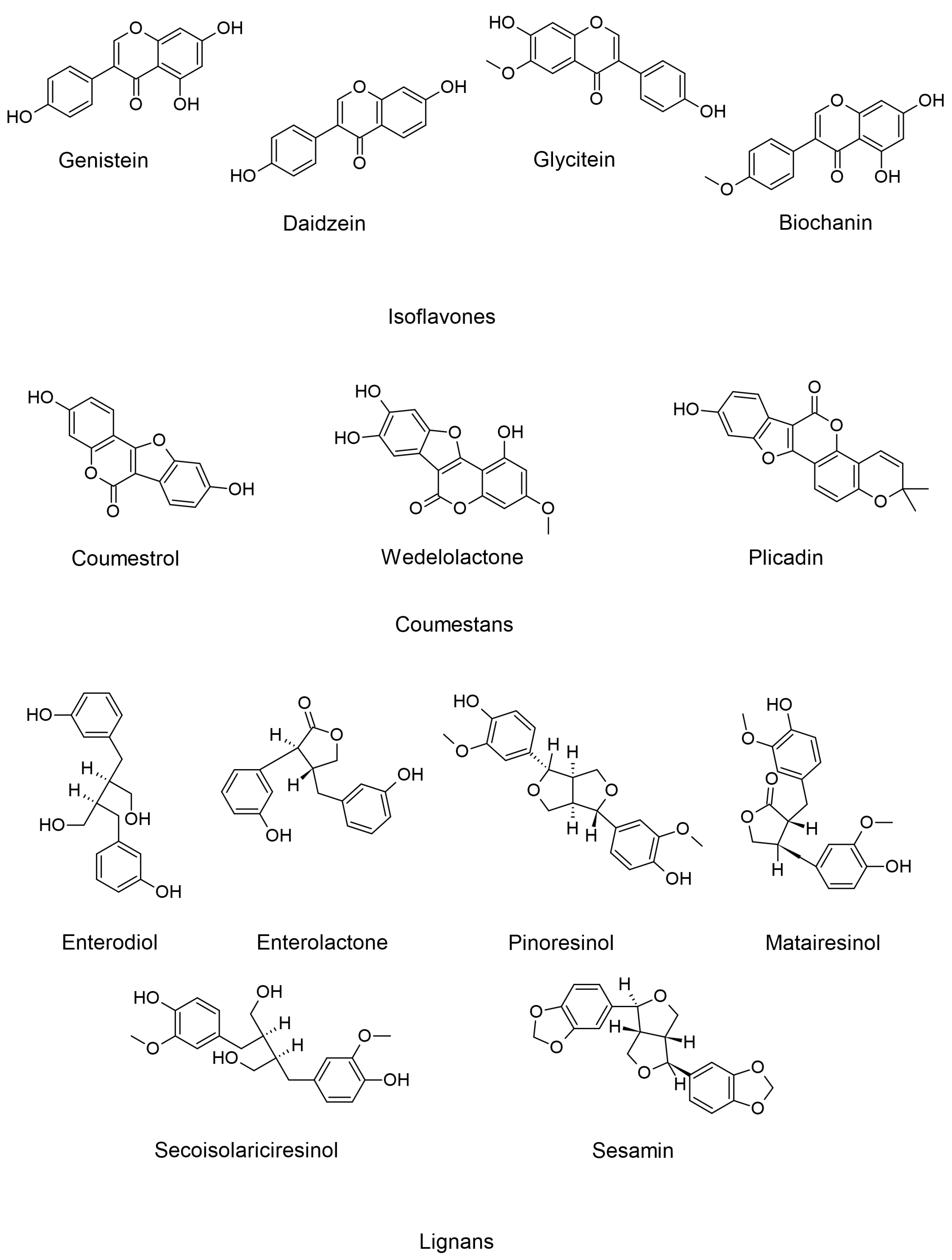
Figure 1. Representative structures of phytoestrogens.
Separations 11 00205 g001
Figure 2. Summarized graphical representation of the processes involved in the analysis of phytoestrogens in plant and food samples; UAE = ultrasonication-assisted extraction; MAE = microwave-assisted extraction; SFE = supercritical fluid extraction; PLE = pressurized liquid extraction; EAE = enzyme-assisted extraction; LC = liquid chromatography; GC = gas chromatography; MS = mass spectrometry; MS/MS = tandem mass spectrometry.
Separations 11 00205 g002
Figure 3. Summarized graphical representation of the processes involved in the analysis of phytoestrogens from human biological samples; SPE = solid-phase extraction; LLE = liquid–liquid extraction; LC = liquid chromatography; GC = gas chromatography; MS = mass spectrometry; MS/MS = tandem mass spectrometry.
Separations 11 00205 g003
Table 1. Selection of analytical methods for the analysis of phytoestrogens in plant material.
Botanical NamePhytoestrogensMethodLoD/LoQReference
Humulus lupulus L.Prenylated flavonoids (8-PN; 6-PN; Xanthohumol)HPLC/DAD
(assay)
HPLC–ESI–MS (fingerprinting)
LoD: 0.3 to 1.0 µg/mL
LoQ: 1.3 to 3.8 µg/mL (HPLC/DAD)
[44]
Prenylflavonoids and bitter acidsHPLC (APCI–MS and UV detection)For 8-PN with MS detection: LoD: 0.006 mg/L; LoQ: 0.02 mg/L;
For 8-PN with UV detection: LoD: 0.03 mg/L; LoQ: 0.1 mg/mL
[45]
Prenylated flavonoidsSIDA–LC–MS/MSLoD: 0.04 µg/L;
LoQ: 3.2 µg/L
[46]
Xanthohumol Nonaqueous reversed polarity CE For xanthohumol
LoD: 0.05 mg/L;
LoQ: 0.15 μg/mL,
[47]
Linum usitatissimum L. Lignans (SECO, SDG-oligomers) TOF–MS LoDs for SECO and SDG-containing oligomers: 0.008 pg [48]
SECO, SDG, SDG-oligomers reversed-phase HPLC LoDs for SDG oligomers, SDG and SECO: 0.065 µg/mL, 0.087 µg/mL and 0.039 µg/mL,
LoQs: 0.217 µg/mL, 0.288 µg/mL and 0.130 µg/ml
[49]
Coffea arabica L.Lignans (SECO, lariciresinol, matairesinol) LC–MS/MSLoD: 1.5 µg/100 g of dry weight[50]
SECO, lariciresinol, matairesinol HPLC–MS/MS SECO: LoD: 2 µg/L; LoQ: 5 µg/L
Lariciresinol: LoD: 2 µg/L; LoQ: 5 µg/L
Matairesinol: LoD: 3 µg/L; LoQ: 10 µg/L
[51]
Trifolium pratense L. Isoflavones (genistein, daidzein, biochanin A, and formononetin); coumestans LC–MS LoDs: 0.06 to 1.81 ng/mL
LoQs: 0.19 to 6.02 ng/mL
[52]
Daidzein, genistein, biochanin A, formononetin HPLC/UV/ESI–MS LoQ(UV detection): 24 ng/mL
LoQ(MS detection): 6 ng/ml
[53]
Daidzein, genistein, biochanin A, formononetin HPLC/UV with internal standard
(TFA hydrolysis)
LoQ daidzein: 2.0 ng
LoQ formononetin 10.0 ng
[54]
Biochanin A, formononetin, genistein, ononin, sissotrin, daidzein LC–MS LoD: 0.06 to 1.81 ng/mL
LoQs: 0.19 to 6.02 ng/mL
[52]
Isoflavones and their glucoside malonates Reversed-phase LC (APCI–MS), DAD, and fluorescence detectorsLC-DAD: LoD (biochanin A): 20 µg/mL, LoD (daidzin): 35 µg/mL.[55]
Medicago sativa L.Coumestrol CE/diode-array detection LoD: 0.39 mg.dm−3[56]
IsoflavonesHPLC/DAD methodLoD: 0.03 to 3.72 µg/ mL
LoQ: 0.10 to 11.27 µg/mL
[57]
Glycine max L. Merr. Daidzin and genistin os-FLISA LoQ: 0.1 μg/mL daidzin[58]
Daidzin and genistin icELISA LoD: 1.95 ng/ml[59]
daidzin and genistin ICA LoD: 125 ng/mL[60]
Daidzein, glycitein, genistein UHPLC
(with enzymatic hydrolysis)
LoD: 67 pg; LoQ: 223 pg (for daidzein)
LoD: 55 pg; LoQ: 184 pg (for glycitein)
LoD: 94 pg; LoQ: 314 pg (for genistein)
[61]
Abbreviations: 8-prenylnaringerin (8-PN); 6-prenylnaringerin (6-PN); secoisolariciresinol (SECO); secoisolariciresinol glucoside (SDG); GC—gas chromatography; IT—ion trap; MS—mass spectrometry; os-FLISA—open sandwich fluorescence-linked immunosorbent assay; icELISA—indirect competitive enzyme-linked immunosorbent assay; ICA—immunochromatographic assay; UHPLC—ultra-high pressure liquid chromatography.
Table 2. Extraction techniques for phytoestrogens from foods.
Extraction TechniquePhytoestrogenSourceParametersReference
UAEDaidzin, genistin, daidzein, genisteinsoymilkUltrasound frequency 35 kHz, 135 kHz
Time: 20 to 60 min
Temperature: 20 to 40 °C
[90]
UAEDaidzin, genistin,
formononetin, biochaninA, coumestrol
soy burgersTime:15 min
Temperature: 30 °C
[91]
UAEDaidzin, genistinsoybeansUltrasound frequency 20 to 90 kHz
Time 10 to 50 min
Temperature: 32 to 168 °C (hydrolysis temperature)
[92]
MAEDaidzein, genistein,
glycitein,
daidzin, genistin,
glycitin
soybeansMicrowave power: 500 W
Solvents: EtOH, MeOH (50–70%)
Temperature: 50 to 150 °C
Extraction time: 10–30 min
[93]
MAESecoisolariciresinol diglucosideflaxseedMicrowave power: 30 to 360 W;
Time: 1 to 25 min;
Solvent: NaOH 0.25 to 1 M; Pmode: power on 30 and 60 s/min.
[94]
MAESecoisolariciresinol diglucosideflaxseed cultivars, flaxseed hulls, sesame seeds,
chia seeds,
almonds,
sunflower seeds
Microwave power: 135 W;
Time: 3 min;
Solvent: NaOH 0.5 M;
Pmode: power on 30 s/min.
[95]
SFEGenistin,
genistein
daidzein
soybean flourTemperature: 40–70 °C, Pressure: 200–360 bar,
Modifier: 0, 5, and 10 mol % of MeOH 70% in water
Time: 30 min
[96]
SFEDaidzein
genistein
soybeanTemperature: 45–65 °C, Pressure: 80–120 bar,
Modifier: 6.5–8.5% EtOH
Time: 120 min
[97]
SPEBiochanin A, secoisolariciresinol, matairesinol, enterodiol, enterolactone, equol, quercetin, genistein, glycitein, luteolin, naringenin, kaempferol, formononetin, daidzein, resveratrol and coumestrolboiled rice potatoesC18 SPE
120 μL, 65% water, 35% MeOH
[98]
SPEDaidzein,
Genistein,
Biochanin A
lentilsC18 SPE
4 mL, MeOH
[99]
SPESecoisolariciresinol, Enterodiol,
Matairesinol,
Enterolactone
breadC18 SPE
1.6 mL, 25% ACN
[100]
PLEDaidzein,
Genistein,
Biochanin A
soybeansSolvent: MeOH, EtOh (30–80%)
Temperature: 60–200 °C
Pressure: 100–200 atm
[101]
EAESecoisolariciresinolcoffee4 Different enzymes:
Taka-Diastase (Aspergillus oryzae), Clara-Diastase (papaya latex),
Papain (papaya latex),
Protease(Rhizopus sp)
[102]
EAEDaidzin, genistin, daidzein, genisteinsoybean flourProtease derived from Bacillus subtilis[103]
Disclaimer/Publisher’s Note: The statements, opinions and data contained in all publications are solely those of the individual author(s) and contributor(s) and not of MDPI and/or the editor(s). MDPI and/or the editor(s) disclaim responsibility for any injury to people or property resulting from any ideas, methods, instructions or products referred to in the content.