Article

Brassica oleracea var. capitata L. Alleviates Indomethacin-Induced Acute Gastric Injury by Enhancing Anti-Inflammatory and Antioxidant Activity

1
Department of Veterinary Surgery, College of Veterinary Medicine, Kyungpook National University, Daegu 41566, Korea
2
Department of Pre-Korean Medicine, College of Korean Medicine, Daegu Haany University, Gyeongsan 38610, Korea
3
Central Research Laboratory, Nutracore Co., Ltd., Suwon 16514, Korea
4
Research Laboratory, Echo Trading Co., Seongnam 13596, Korea
*
Authors to whom correspondence should be addressed.
S.H.R. and I.J.C. contributed equally to this work.
Processes 2021, 9(2), 372; https://doi.org/10.3390/pr9020372
Submission received: 26 January 2021 / Revised: 12 February 2021 / Accepted: 14 February 2021 / Published: 17 February 2021

Abstract

:
Brassica oleracea var. capitata L. (white cabbage) is a valuable vegetable with diverse nutraceutical benefit. Present study aimed to investigate the preventive effects of B. oleracea extract (BOE) standardized by vitamin U on indomethacin (IND)-induced acute gastric injury in Sprague-Dawley rats. Pre-administration of three different doses of BOE (12.5–50 mg/kg) for 14 days significantly decreased visible ulcerative lesions in the gastric tissue. In addition, BOE alleviated IND-mediated increase in histological score with inhibiting invaded percentage of lesion and restoring mucosa thickness in peri-ulcerative region. BOE increased the gastric tissue bound to Alcian blue and inhibited the decrease in hexose, sialic acid, and collagen levels by IND, suggesting that BOE protects the gastric tissue through preserving mucus and mucosal integrity. Moreover, BOE pre-administration blocked the reduction of prostaglandin E2 and down-regulated histamine and mRNA expression related to secret gastric acid. Furthermore, BOE mitigated inflammatory responses in the gastric tissue by decreasing activity of myeloperoxidase and expression of nuclear factor-κB-dependent inflammatory genes. BOE also suppressed malondialdehyde with preventing the reduction of glutathione, superoxide dismutase, and catalase in the gastric tissue. Therefore, results from present study suggest that BOE will have a potential for preventing gastric injury.

1. Introduction

Gastric ulcer affects about 5–10% of the general population during their lifetime [1]. Although gastric ulcer is caused by imbalance between multiple aggressive and cytoprotective factors, the use of non-steroidal anti-inflammatory drugs (NSAIDs) and infection with Helicobacter pylori are considered as the major risk factors for accelerating hypersecretory acidic environment in the gastric tissue. Especially, NSAIDs have been prescribed extensively because of their analgesic, anti-inflammatory and antipyretic activities, but the use of NSAIDs increases relative risk of gastric ulcer by 4.7 times [2]. Although NSAIDs vary in potential to provoke adverse effects in the gastric tissue [2,3], the major mechanism correlated with the gastrointestinal toxicity is the inhibition of prostaglandin biogenesis, which impairs gastric mucosa through reducing the secretion of mucus and bicarbonate [1]. Moreover, NSAIDs also damage the mucosal integrity by infiltrating inflammatory cells into the gastric tissue and inducing oxidative stress [1,4,5]. Thus, in order to prevent gastric ulcer caused by NSAIDs, great attention has been paid to natural resource exhibiting anti-inflammatory and antioxidant activities, and several edible plants and those-derived phytochemicals have been suggested as promising alternative candidates [6,7,8,9].
Brassica oleracea var. capitata L. (general name, white cabbage), which belongs to Brassicaceae family, has been cultivated as one of the most valuable vegetables worldwide due to its long-term storage ability. In addition, B. oleracea has been traditionally consumed for managing diverse diseases related to inflammation and gastrointestinal disorders [10]. Modern nutraceutical studies have suggested that B. oleracea possesses wide range of beneficial activities. For instance, B. oleracea induces apoptosis of certain types of cancer [11], relieves hydrogen peroxide-mediated oxidative stress in cardiomyoblast cells [12], protects the liver from carbon tetrachloride-induced injury [13], improves serum glucose and lipid profiles in alloxan-induced diabetic rabbits [14], and inhibits the activity of enzymes associated with neurodegeneration [15]. Especially, Cheney and colleagues reported that supplementation with fresh cabbage juice or its concentrated extract accelerates the healing process in patient with gastric ulcer [16,17]. In addition, Cheney suggested that vitamin U, S-methylmethionine sulfonium chloride, is the major active ingredient correlated with anti-ulcerogenic activity in cabbage juice [18]. However, only a few studies using experimental animals have been conducted to prove the gastroprotective nature of cabbage. For instance, it has been reported that post-administration of B. oleracea aqueous extract for 5–7 days shortened the length of ulcerogenic lesions and alleviated acidity of gastric juice in aspirin-administered rats [9,19]. Therefore, the prophylactic effect of B. oleracea on gastric ulcer as well as relevant gastroprotective mechanisms remain to be further established.
As the process for discovering novel gastroprotective candidates, we recently reported that pre-administration of B. oleracea extract (BOE) standardized by vitamin U could protect the gastric tissue from acidified ethanol-induced ulcer in mice [20]. However, for reaching general conclusions and expanding indications, it is necessary to evaluate the efficacy of BOE in more than one experimental model. Among diverse etiologies to induce gastric ulcer, about 25% of urgent gastric ulcer are known to be associated with NSAIDs administration [21]. Thus, in the present study, indomethacin (IND) was chosen as the second experimental model for gastric ulcer because it is a representative NSAID with a high relative risk for gastrointestinal toxicity [2,3]. Therefore, the purpose of the present study was to evaluate the prophylactic effects of BOE on IND-mediated gastric ulcer and explore its mechanisms involved. Furthermore, present study used omeprazole (OM; an irreversible proton pump inhibitor) as a reference drug for comparing gastroprotective effects of BOE.

2. Materials and Methods

2.1. Materials

Certified BOE powder was provided by grow® company (Ridgefield, NJ, USA) via Echo Trading Co. (Seongnam, Korea). Vitamin U was obtained from Tokyo Chemical Industry (Tokyo, Japan). Enzyme-linked immunosorbent assay (ELISA) kits for rat histamine, prostaglandin E2 (PGE2), tumor necrosis factor-α (TNF-α), interleukin (IL)-1β, IL-6, and IL-18 were supplied by Mybiosource (San Diego, CA, USA). Trizol was obtained from Thermo Fisher Scientific (Rockford, IL, USA) and a High-capacity cDNA reverse transcription kit was from Applied Biosystems (Foster City, CA, USA). IND, OM, hematoxylin, eosin, Alcian blue, palmityltrimethylammonium bromide, o-dianisidine dihydrochloride, hydrogen peroxide, orcinol, thiobarbiturate, Ehrlich’s reagent, 1,1,3,3-tetramethoxypropane, Ellman’s reagent, nitroblue tetrazolium chloride, and other reagents were purchased from Merck (Darmstadt, Germany).

2.2. Measurement of Vitamin U in BOE

Concentration of vitamin U in BOE was analyzed by high-performance liquid chromatography (HPLC), as reported previously [20]. Briefly, an appropriate amount of BOE or standard (vitamin U) was dissolved in distilled water, lyophilized by vacuum evaporator, and re-dissolved in 0.2 N sodium citrate (pH 2.2). The BOE or serially diluted vitamin U was separated on an analytical column, Capcell Pak C18 UG120 (size, 250 × 4.6 mm; pore size 5 μm; Shiseido, Tokyo, Japan) at 40 °C. Mobile phase was 40 mM NaH2PO4 (pH 7.8) (solution A) and acetonitrile:methanol:water (45:45:10%)(solution B). The eluent initially consisted of 95:5% (solution A:solution B). During 0 to 31 min, the amount of solution B increased linearly to 56%, and then remained the same for 2 min. Finally, between 33 and 34 min, the amount of solution B increased to 100%, and then stayed for an additional 4 min. Flow rate was 1.5 mL/min, and vitamin U was detected at 338 nm using a photodiode array detector in Agilent LC system (Agilent Technologies, Palo Alto, CA, USA).

2.3. Animals and Treatment

Sixty SPF/VAF Outbred Crl:CD (Sprague-Dawley) rats (gender, male; age, 6 weeks old) were provided from OrientBio (Seongnam, Korea), acclimatized for 9 days, and then divided into the following six groups (N = 10 per group): vehicle, IND, IND + OM, IND + BOE-H, IND + BOE-M, and IND + BOE-L. BOE, OM, and IND were dissolved in distilled water. Using a zonde attached to 3 mL syringe, three different doses of BOE (e.g., 50 mg/kg for BOE-H, 25 mg/kg for BOE-M, and 12.5 mg/kg for BOE-L) were orally administered to the rats once daily for 14 consecutive days. In case of vehicle and IND groups, equal volume of water was administered for 14 days to provide same stress from oral gavage. For OM group, 10 mg/kg OM was orally administered to the rat once on day 14 after administration of water for 13 days (i.e., the first administration of test material = day 1). To induce gastric injury, 25 mg/kg IND was orally administered at 30 min after the last treatment of test material. Rats in vehicle group was administered to equal volume of water instead of IND. The drug concentration for OM and IND was chosen according to the previous report [6]. At 6 h after IND treatment, all animals were euthanized to collect bloods and gastric tissues. Plasma was obtained by centrifugation of blood at 12,000× g for 10 min, and all samples were stored at −150 °C until analysis.

2.4. Histopathology

The stomach was opened along with greater curvature and washed mildly to remove gastric contents and blood clots. After capturing image of gastric mucosa, visible ulcerative area was calculated as mm2 using an image analyzer software (isolution FL ver9.1, IMT i-solution Inc; Burnaby, BC, Canada). For microscopical observation, the fundus/body regions that had been fixed in 10% neutral-buffered formalin was embedded in paraffin, sectioned, and stained with hematoxylin and eosin, as described previously [6,19]. Histopathological analyses of the gastric tissue were conducted using an image analyzer software (isolution FL ver9.1, Burnaby, BC, Canada). Briefly, invaded lesion (%) was calculated as the percentage of lesion length to fundus/body’s wall length, and mucosa thickness from luminal mucosal surface to muscularis mucosa (mm) was calculated on the peri-ulcerative region. In addition, histological score was semi-quantitatively estimated as the following four grades: 0, normal intact mucosa; 1, slight surface erosive damage; 2, moderate mucosa damage; 3, severe total mucosa damage.

2.5. Alcian Blue Binding Assay

To quantify gastric mucus, the gastric tissue was stained with Alcian blue binding solution consisting 0.02% Alcian blue, 0.16 M sucrose, and 0.05 M sodium acetate buffer (pH 5.8) for 24 h, and then centrifuged at 3000× g for 10 min. Using an UV/VIS spectrophotometer (OPTIZEN POP, Mecasys; Daejeon, Korea), absorbance of resulting supernatant was determined at 620 nm, and amount of mucus was calculated as mg/g tissue, as described previously [20,22].

2.6. Measurement of Total Hexose, Sialic Acid, and Collagen

Gastric mucosa including mucus was collected by scraping, homogenized in 10 volumes of phosphate-buffered saline using TacoTMPrep bead beater (3 cycles, 30 sec/cycle)(GeneReach Biotechnology; Taichung, Taiwan) and SONICS Vibra CellTM sonicator (3 cycles, 1 min/cycle, 20% amplitude)(Sonics & Materials Inc., Newtown, CT, USA), and centrifuged at 12,000× g for 10 min. The resulting supernatant was used as gastric mucosal homogenate. As described previously [20,23], the amount of total hexose, sialic acid, and collagen in the gastric mucosal homogenate was calorimetrically quantified. Briefly, level of total hexose was determined at 425 nm after the tissue homogenate was reacted with orcinol in sulfuric acid. In addition, level of sialic acid was read at 550 nm based on periodate-thiobarbiturate assay. For quantifying collagen, mucosal proteins were hydrolyzed using 6 M hydrogen chloride, and the level of hydroxyproline was determined at 565 nm after the hydrolyzed samples were further reacted with Ehrlich’s reagent. The amount of all mucosal components was normalized by tissue weight.

2.7. ELISA

Level of histamine in the plasma and levels of PGE2 and pro-inflammatory cytokines in the gastric mucosal homogenate were quantified using commercial ELISA kits (Mybiosource) and microplate reader (Sunrise, Tecan; Männedorf, Switzerland).

2.8. Quantitative Polymerase Chain Reaction (qPCR)

After total RNA isolating from the gastric tissue was reverse transcribed to cDNA, qPCR was carried out using a CFX96 thermal cycler (Bio-Rad; Hercules, CA, USA). Expression level of specific mRNA was normalized by that of glyceraldehyde 3-phosphate dehydrogenase (GAPDH), as endogenous control, as described previously [20]. Primers for amplifying specific genes were listed in Table 1.

2.9. Measurement of Myeloperoxidase (MPO) Activity

MPO activity in the gastric tissue was determined, as described previously [6,20]. Briefly, the gastric tissue was lysed in 50 mM potassium phosphate (pH 6.0) and 0.5% palmityltrimethylammonium bromide and centrifuged at 12,000× g for 10 min. After the resulting pellet was further incubated with hydrogen peroxide and o-dianisidine dihydrochloride, MPO activity determined at 460 nm and normalized by tissue weight.

2.10. Lipid Peroxidation Assay

For estimating lipid peroxidation, the gastric mucosal homogenate was boiled for 1 h with thiobarbiturate, sodium dodecyl sulfate, and acetic acid, followed by measuring optical intensity at 532 nm. The malondialdehyde concentration (nM) in the homogenate was calculated by interpolation to the standard curve of 1,1,3,3-tetramethoxypropane and normalized by tissue weight.

2.11. Measurement of Glutathione Concentration

After the gastric mucosal homogenate was precipitated by adding trichloroacetic acid, supernatant was reacted with Ellman’s reagent and measured absorbance at 412 nm. Glutathione concentration (nM) was normalized by tissue weight.

2.12. Measurement of Catalase and Superoxide Dismutase Activities

Catalase and superoxide dismutase activities were determined by reacting the gastric mucosal homogenate with hydrogen peroxide and nitroblue tetrazolium chloride, as described previously [6,20]. Enzyme activities were normalized by tissue weight.

2.13. Statistical Analyses

Numerical results were expressed as mean ± standard deviation (SD). Levene’s test was conducted to evaluate the equality of variances. When the numerical results indicated the homogeneity of variance, the group means were compared with One-way analysis of variance. As post hoc analysis, the Tukey honestly significant difference test was used to assess mean difference among experimental groups. For numerical results showing significance in Levene’s test, Welch’s test was followed by Dunnett’s T3 test to compare the means among groups. All statistical analyses were performed using SPSS Statistics for Windows (Version 23; SPSS Inc., Chicago, IL, USA), and p < 0.05 was considered as significant.

3. Results

3.1. HPLC Analysis for Quantifying Vitamin U in BOE

Prior to evaluate gastroprotective effect of BOE, we quantified the concentration of vitamin U, a representative gastroprotective compound dissolved in B. oleracea [18], in BOE. For obtaining calibration curve, serially diluted vitamin U was injected into the HPLC system. After the area of the peak with similar retention time in the chromatogram eluting with BOE was interpolated into the calibration curve of vitamin U, we calculated that BOE used in the present study contained 44.97 mg/g of vitamin U, which was similar to previous report [20].

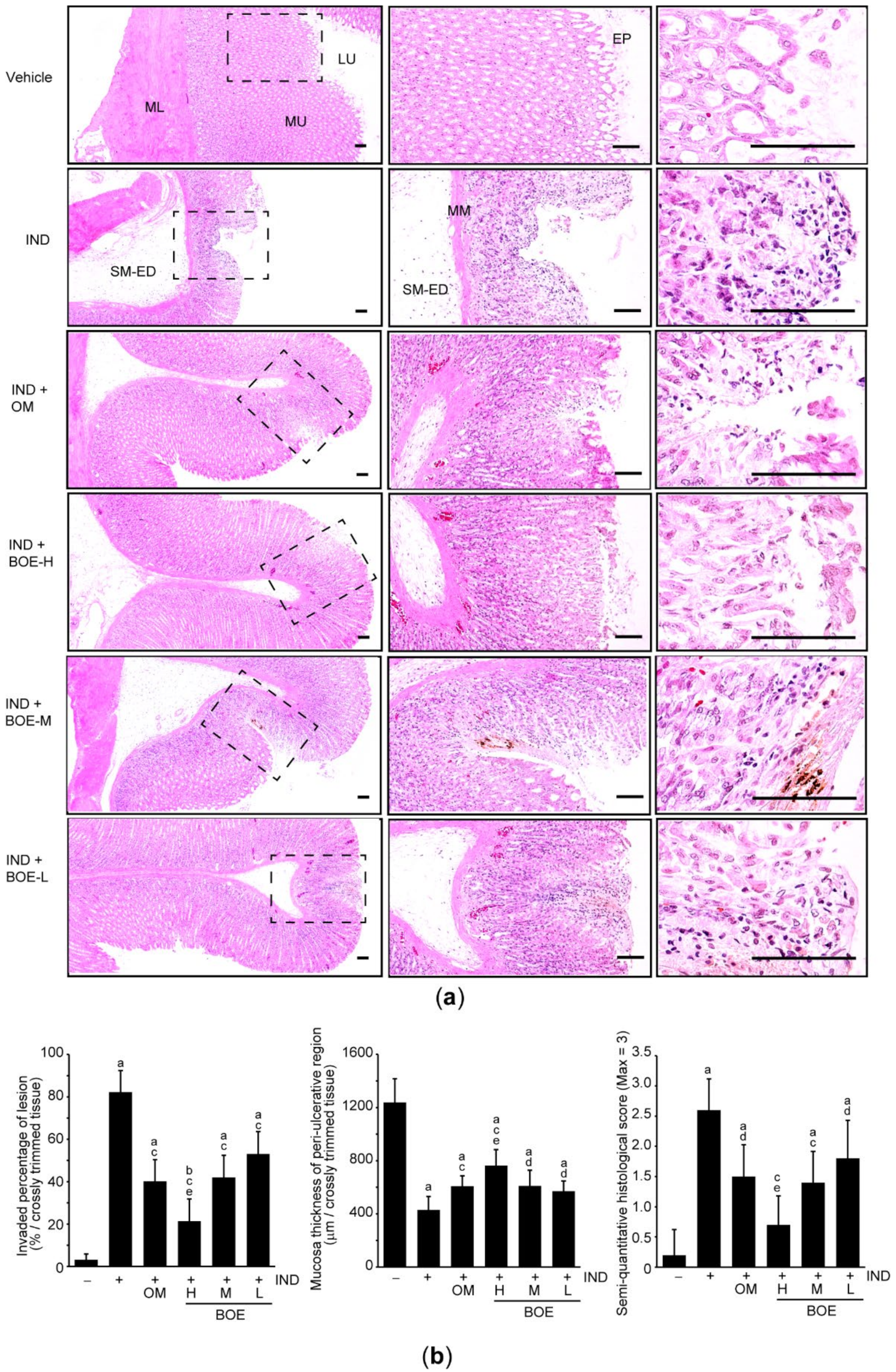
3.2. Pre-Administration of BOE Alleviates IND-Mediated Gastric Injury in Rats

To explore whether BOE has prophylactic effects against IND-mediated gastric injury, rats that had been orally administered with three different doses of BOE (12.5–50 mg/kg) for 14 days were treated with 25 mg/kg IND for 6 h. OM (10 mg/kg) was used as a reference drug and was orally administered once on day 14 (i.e., the first BOD administration = day 1). During the experimental period, there were no statistical differences in body weight among experimental groups (data not shown). When the stomach was opened along with greater curvature, focal hemorrhagic ulcerative lesions were shown throughout whole gastric mucosa in rats treated with IND. However, those lesions were decreased in rats treated with three different doses of BOE and OM (Figure 1a). When macroscopic visible lesion on the gastric mucosa was quantified using an image analyzer, the ulcerative area was significantly increased in IND-treated rats as compared to vehicle group. However, pre-administration of three different doses of BOE as well as OM was significantly prevented the IND-induced increase in gross ulcerative area. There were no statistical differences in gross lesion area between IND + OM and IND + BOE-M groups (Figure 1b).
Next, the fundus/body region of the stomach was further stained with hematoxylin and eosin for conducting histopathological evaluation among experimental groups. Desquamation of focal epithelium, congestion, infiltration of inflammatory cells, necrosis of gastric glands, and submucosal edema were found in the rats treated with IND, which is parallel with previous observation that IND provokes severe ulcerative lesions [6,7,8]. However, those histopathological changes were reduced by three different doses of BOE and OM (Figure 2a). Especially, results from histomorphometric and semi-quantitative analyses indicated that three different doses of BOE significantly suppressed the IND-mediated increase in the ulcerative histological score, which was caused by increasing the percentage of invaded lesion and decreasing mucosa thickness in peri-ulcerative region. Pre-administration of OM also inhibited significantly the histomorphometric changes induced by IND, while the magnitude of OM-mediated reduction in the histomorphometric analyses was lesser than that associated with BOE-H. There were no statistical differences in histomorphometric results between IND + OM and IND + BOE-M (/BOE-L) groups (Figure 2b).

3.3. Pre-Administration of BOE Restores Integrity of the Gastric Mucus Impaired by IND

To explore whether BOE protects the gastric tissue through maintaining gastric mucus, the mucus was quantified by Alcian blue binding assay. Compared to vehicle-treated rats, IND significantly decreased the gastric mucus. Pre-administration of three different doses of BOE and OM prevented the mucus loss induced by IND. There were no statistical differences in the mucus contents among IND + OM and three IND + BOE groups (Figure 3a). Next, we further quantified the level of monosaccharide in the gastric mucosal homogenates to investigate glycoprotein level in the gastric mucus. In parallel with results from Alcian blue binding assay, pre-administration of three different doses of BOE as well as OM significantly attenuated the IND-mediated reduction of total hexose and sialic acid in the gastric mucosal homogenate (Figure 3b). Moreover, rats pre-administered with BOE-H, BOE-M, and OM significantly blocked the collagen loss by IND (Figure 3c). There were no statistical differences in the total hexose, sialic acid, and collagen contents among IND + OM and three IND + BOE groups, except for sialic acid and collagen contents, which the preventive effects by BOE-H were greater than those seen by OM (Figure 3b,c).

3.4. Pre-Administration of BOE Downregulates Essential Factors for Secreting Gastric Acid

Because IND injures the gastric mucosa by enhancing the secretion of gastric acid [24], we explored the effect of BOE on essential factors associated with secreting gastric acid. As expected, the level of histamine in the plasma was significantly increased in IND-treated group compared to vehicle-treated group. Pre-administration of BOE significantly decreased the level of plasma histamine in a doses-dependent manner, while OM administration did not change the histamine level increased by IND. When comparing histamine levels among IND + OM and three IND + BOE groups, magnitude of the reduction by BOE-H and BOE-M was more potent than that by OM (Figure 4a). In addition, pre-administration of BOE-H, BOE-M, or OM significantly suppressed IND-mediated decrease in PGE2 level in the gastric mucosal homogenates. Statistical significance was only seen in rats receiving BOE-H when comparing PGE2 level to OM-treated rats (Figure 4b). Moreover, IND-mediated increases in the mRNA expression of H+/K+ ATPase, H2R, and CCK2R were significantly inhibited by pre-administration of three different doses of BOE, except for H+/K+ ATPase mRNA, which did not differ between IND + BOE-L and IND group. Of three mRNAs expression, H+/K+ ATPase mRNA was only decreased by pre-administration of OM, and the mRNA reduction of H2R and CCK2R by BOE-H and BOE-M was statistically significant as compared to that by OM (Figure 4c).

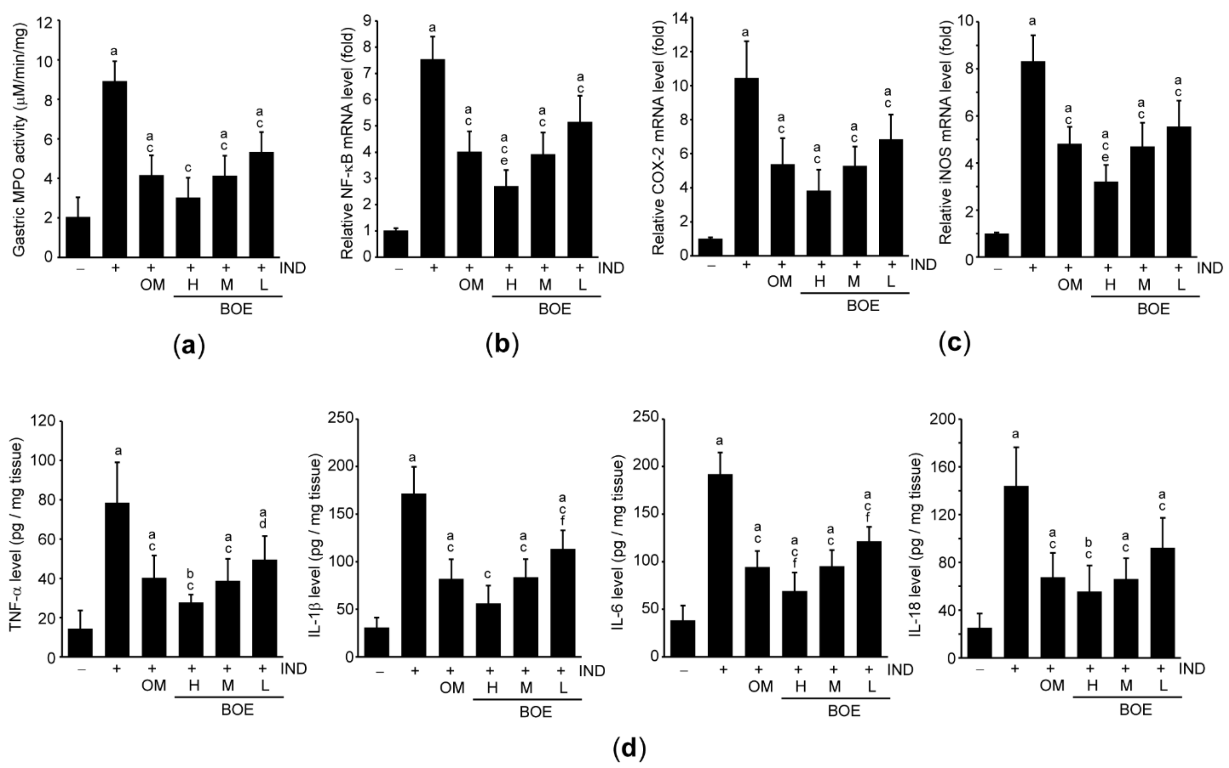
3.5. Pre-Administration of BOE Attenuates IND-Mediated Gastric Inflammation

To explore whether BOE protects the gastric tissue by alleviating inflammatory responses induced by IND, we measured MPO activity in the gastric tissue. IND significantly increased MPO activity as compared to vehicle, and pre-administration of three different doses of BOE and OM significantly reduced the IND-mediated increase in MPO activity. There were no statistical differences in MPO activity among IND + OM and three IND + BOE groups (Figure 5a). In addition, results from qPCR indicated that IND significantly increased the mRNA level of NF-κB, which is a pivotal transcription factor for regulating genes associated with inflammation in the gastric tissue [25]. However, IND-mediated increase in NF-κB mRNA was significantly attenuated in rats administered with three different doses of BOE and OM. Although magnitude of the reduction in NF-κB mRNA by BOE-H was greater than that by OM, the inhibitory effect seen in response to BOE-M and BOE-L did not differ from that by OM (Figure 5b). Moreover, three different doses of BOE as well as OM significantly prevented IND-mediated expression of inflammatory enzymes (e.g., COX-2 and iNOS) (Figure 5c) and cytokines (e.g., TNF-α, IL-1β, IL-6, and IL-18). Inhibitory effects of inflammatory enzymes and cytokines in three BOE-treated groups did not differ from those by OM-treated group, except for iNOS mRNA level between IND + BOE-H and IND + OM, IL-1β protein level between IND + BOE-L and IND + OM, IL-6 protein level between IND + BOE-H (/BOE-L) and IND + OM (Figure 5d).

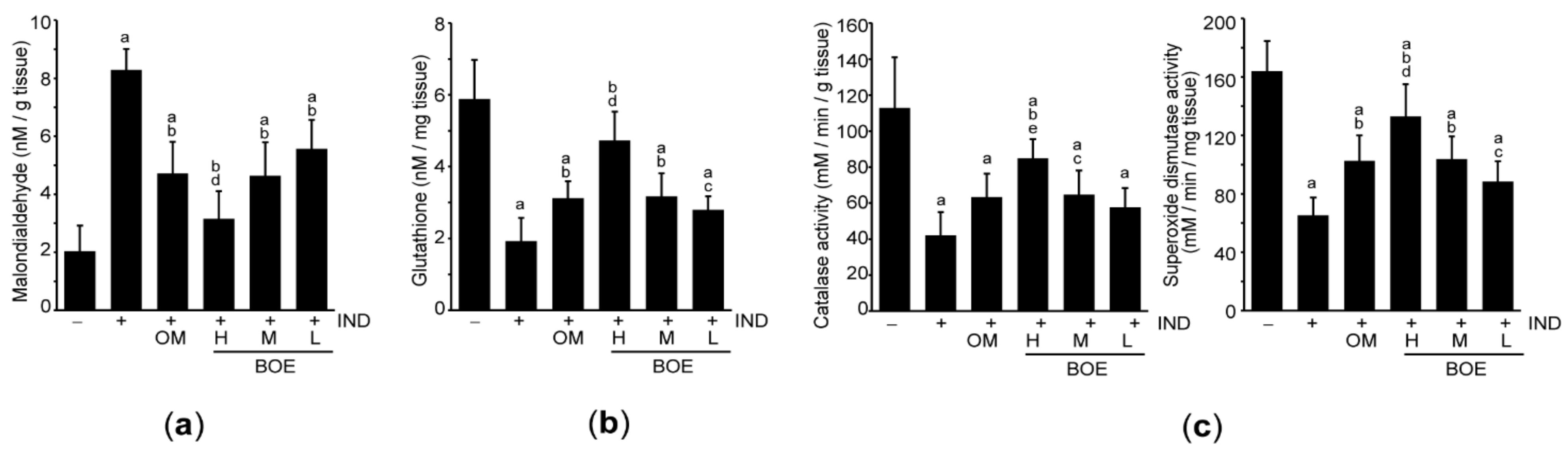
3.6. Pre-Administration of BOE Suppresses Lipid Peroxidation by Enhancing Antioxidant Activities

To explore the effect of BOE on IND-mediated oxidative stress, lipid peroxidation was monitored by quantifying level of malondialdehyde in the gastric tissue. IND increased the level of malondialdehyde, suggesting that IND induces oxidative stress in the gastric tissue. However, pre-administration of three different doses of BOE and OM significantly decreased the malondialdehyde level. Although reduction of malondialdehyde by BOE-H administration was greater than that in response to OM, there were no statistical differences between BOE-M (/BOE-L) and OM (Figure 6a). In addition, three different doses of BOE and OM significantly prevented IND-mediated depletion of glutathione as well as decreases in catalase and superoxide dismutase activities, except for catalase activity in OM-administered group. Especially, the magnitudes of restoration in antioxidant activities by BOE-H were greater than those by OM (Figure 6b,c).

4. Discussion

Throughout macroscopic and microscopic histopathological evaluation, results from present study proposed that B. oleracea extract standardized by vitamin U could prevent acute gastric injury induced by IND. In addition, gastroprotective effects of three different doses of BOE were compared to those of OM. Although most of gastroprotective effects by BOE-H were more potent than those by OM, there were no statistical differences in gastroprotective effects among BOE-M, BOE-L, and OM groups.
Cheney suggested that vitamin U is major bioactive compound that contributes to the gastroprotection of cabbage juice [18], and studies have reported that effective dosage of vitamin U for managing gastric ulcer ranges from 100 to 500 mg/kg [26,27]. Because present results from HPLC analysis showed that BOE contained about 4.5% of vitamin U, 12.5–50 mg/kg of BOE used for the animal experiments in the present study is equivalent to 0.56–2.25 mg/kg of vitamin U. Thus, concentration of vitamin U in BOE seems to be too low to reflect the potent gastroprotective effects of BOE. In addition to vitamin U, our HPLC chromatogram also showed that at least three unidentified peaks absorbing 338 nm of light were existed in BOE. Moreover, it has been reported that B. oleracea contains various bioactive compounds including glucosinolates (e.g., sinigrin, glucoiberin, glucoraphanin, and glucobrassicin), flavonoids (e.g., quercetin, kaempferol, apigenin, and rutin), phenolic acids (e.g., caffeic acid, p-coumaric acid, ferulic acid, and sinapic acid), carotenoids (e.g., tocopherol, α-carotene, and β-carotene) and vitamins (e.g., vitamin K, vitamin C, and folate) [10,28,29]. Moreover, several bioactive compounds (e.g., kaempferol, quercetin, rutin, apigenin, caffeic acid, p-coumaric acid, ferulic acid, α-tocopherol, and β-carotene) and those-derived hydrolysis byproducts (e.g., allyl isothiocyanate, indole-3-carbinol, and sulforaphane) have been reported to exhibit beneficial effects on gastric ulcer in experimental animal models [30,31,32,33,34,35,36,37,38,39,40,41]. Ameliorating oxidative stress, reducing acute inflammation, and restoration of endogenous cytoprotective molecules are proposed mechanisms associated with the prevention of gastric ulcer by the aforementioned bioactive compounds. Although the concentration of individual bioactive compounds in BOE is not sufficient to protect the gastric tissue, combinations of bioactive compounds may synergistically contribute to protecting the gastric tissue from IND. Further studies are needed to explore other bioactive compounds responsible for BOE-mediated gastroprotection.
Because mucus secreted from mucous cells of the gastric tissue is able to separate chemical and biological toxins in the lumen from epithelium, the mucus has been recognized as the first line of defense to protect the stomach from gastric injury [42]. Mucin, which is the major building blocks comprising mucus, is protein complex with over 80% carbohydrate [43]. Moreover, collagen is one of the representative proteins in extracellular matrix for providing mechanical strength of (sub)mucosal and muscular layer [8]. Therefore, present results from Alcian blue binding assay and measurement of gastric carbohydrate/collagen levels imply that BOE-mediated protection of mucus as well as mucosal integrity contributes to protecting gastric tissue from IND.
The present results showed that pre-administration of BOE significantly inhibited the histamine release and mRNA expression associated with secreting gastric acid. Histamine released from neuroendocrine and paracrine system (e.g., enterochromaffin-like cells in the gastric tissue) stimulates secretion of gastric acid through H2R activation in parietal cells. In addition, gastrin also stimulates the acid secretion through direct activation of CCK2R in parietal cells or CCK2R-mediated histamine release in enterochromaffin-like cells. Finally, histamine binding to the H2R in the parietal cells pumps out H+ ion via cAMP-dependent activation of H+/K+ ATPase [44]. More importantly, we further verified that pre-administration of BOE significantly inhibited the reduction of PGE2 caused by IND. Studies using specific PGE2 receptor agonist/antagonist as well as mice deficient specific receptor demonstrate that PGE2-mediated EP1 activation plays a critical role on gastric protection against NSAIDs [45,46]. In addition, it has been reported that PGE2 inhibits acid secretion, neutrophil migration, and hypermotility of the gastric tissue. Moreover, PGE2 increases bicarbonate secretion, mucus production, and mucosal blood flow [47], which provide an evidence that PGE2 is the guardian to protect the stomach from necrotizing agents including IND. Therefore, present results imply that modulation of histamine and PGE2 levels in response to BOE can be attributed to protect the gastric mucosa.
MPO, an abundant enzyme expressed in primary azurophilic granules of neutrophils, facilitates acute inflammation by catalyzing the formation of reactive oxygen species [48]. In addition, mice injected with an antibody against CD11/CD18 or mice lacking with fucosyltransferase VII showed marked reduction of IND-mediated gastric ulcer and preserved mucosal integrity [4,49], which provide an evidence that adherence and infiltration of neutrophil are required to accelerate IND-mediated ulcerogenesis. Therefore, present results showing BOE pre-administration inhibited IND-mediated MPO activity in the gastric tissue suggest that inhibition of infiltrating neutrophils by BOE may be one of the plausible mechanisms accounting for anti-ulcerogenic activity of BOE in IND-treated rats. In addition, results from present study also showed BOE significantly reduced NF-κB as well as several inflammatory mediators. NF-κB is a ubiquitous transcription factor which initiates a vast array of biological processes including inflammation [25]. It has been reported that NF-κB mRNA is increased in patient with gastric ulcer [50]. On the contrary, genetic ablation of NF-κB subunit (e.g., p50) ameliorates cold stress-mediated mucosal injury in conjunction with the reduced expression of inflammatory cytokines and adhesion molecules [51]. Cellular debris released from damaged tissue promotes infiltration of inflammatory cells (e.g., neutrophils) and triggers inflammatory responses by producing inflammatory enzymes (e.g., iNOS, and COX-2) and inflammatory cytokines (e.g., TNF-α, IL-1β, IL-6, and IL-18) which are known to be mainly regulated by NF-κB [25,52,53]. Therefore, not only inhibition of infiltrating neutrophils but also suppression of NF-κB mRNA collaboratively contributes to preventing the gastric ulcer caused by IND.
Mice deficient in gp91phox mitigate IND-mediated gastric ulcer [4], which imply that oxidative stress from infiltrated inflammatory cells is another pathological mechanism for aggravating mucosal damage during ulcerogenesis. Consistent with previous reports [6], we also showed that IND depleted glutathione and decreased catalase and superoxide dismutase activities, resulting in accumulating oxidative stress in the gastric tissue. However, pre-administration of BOE significantly mitigated IND-induced oxidative stress. Glucosinolates and those hydrolyzed byproducts possess potent antioxidant activity via activating nuclear factor E2-related factor 2 (Nrf2) [54]. Nrf2 activators disrupt protein–protein interaction between Nrf2 and Keap1 and allow Nrf2 to translocate into the nucleus, where Nrf2 transactivates many antioxidant genes (e.g., superoxide dismutase, catalase, and glutamate-cysteine ligase) to cope with oxidative stress [54,55]. More interestingly, in silico study has shown that vitamin U is able to bind to the ETGE motif of Nrf2, which facilitates the dissociation of Nrf2 from Keap1 [56]. Furthermore, Nrf2 also perturbs the expression of inflammatory cytokines via direct binding to the proximal promoter of those genes [57]. Although more studies are needed on the role of Nrf2 in BOE-mediated gastroprotection, present results suggest that BOE may protect the gastric tissue via activating Nrf2-dependent signaling pathway.

5. Conclusions

In conclusion, present results showed that pre-administration of BOE standardized by vitamin U efficiently prevented IND-mediated acute gastric injury via maintaining mucus and mucosal integrity, regulating factors related to gastric acid secretion, inhibiting inflammation, and enhancing antioxidant activities. Although results from present study provide a direct evidence that BOE can prevent the gastric ulcer, several studies including (1) chemical characterization of BOE beyond vitamin U, (2) major bioactive compounds that reflect the gastroprotective effects of BOE, (3) detailed molecular targets of BOE, (4) efficacy of BOE on other gastric ulcer models (e.g., H. pylori and other NSAIDs), and (5) toxicity and safety of BOE must be further addressed to develop BOE as an alternative gastroprotective agent. If appropriate studies related to BOE are successfully performed, BOE will be a potential candidate for development as a gastroprotective nutraceutical.

Author Contributions

Conceptualization, Y.S.K. and S.K.K.; methodology, S.H.R. and S.K.K.; formal analysis, S.H.R., B.-R.C., M.B.K., I.J.C., and S.K.K.; investigation, S.H.R., I.J.C., and S.K.K.; resources, B.-R.C. and M.B.K.; data curation, I.J.C., Y.S.K., and S.K.K.; writing—original draft preparation, S.H.R., I.J.C., Y.S.K., and S.K.K.; writing—review and editing, Y.S.K., and S.K.K.; visualization, I.J.C. and S.K.K.; supervision, Y.S.K. and S.K.K. All authors have read and agreed to the published version of the manuscript.

Funding

This research received no external funding.

Institutional Review Board Statement

After receiving approval from IACUC at Daegu Haany University (Approval No. DHU2019-090), animal experiment was conducted according to the national regulations regarding the use and welfare of laboratory animals.

Informed Consent Statement

Not applicable.

Data Availability Statement

The data presented in this study are available on request from the corresponding author.

Conflicts of Interest

The authors declare no conflict of interest.

References

  1. Lanas, A.; Chan, F.K.L. Peptic ulcer disease. Lancet 2017, 390, 613–624. [Google Scholar] [CrossRef]
  2. Melcarne, L.; García-Iglesias, P.; Calvet, X.; Luigi, M.; Pilar, G.-I.; Xavier, C. Management of NSAID-associated peptic ulcer disease. Expert Rev. Gastroenterol. Hepatol. 2016, 10, 723–733. [Google Scholar] [CrossRef]
  3. Coxib and traditional NSAID Trialists’ (CNT) Collaboration; Bhala, N.; Emberson, J.; Merhi, A.; Abramson, S.; Arber, N.; Baron , J.A.; Bombardier, C.; Cannon, C.; Farkouh, M.E.; et al. Vascular and upper gastrointestinal effects of non-steroidal an-ti-inflammatory drugs: Meta-analyses of individual participant data from randomised trials. Lancet 2013, 382, 769–779. [Google Scholar] [CrossRef] [Green Version]
  4. Beck, P.L.; Xavier, R.; Lu, N.; Nanda, N.N.; Dinauer, M.; Podolsky, D.K.; Seed, B. Mechanisms of NSAID-induced gastroin-testinal injury defined using mutant mice. Gastroenterology 2000, 119, 699–705. [Google Scholar] [CrossRef]
  5. Villegas, I.; Martin, M.J.; La Casa, C.; Motilva, V.; De La Lastra, C.A. Effects of meloxicam on oxygen radical generation in rat gastric mucosa. Inflamm. Res. 2000, 49, 361–366. [Google Scholar] [CrossRef]
  6. Lim, J.-M.; Song, C.-H.; Park, S.-J.; Park, D.-C.; Jung, G.-W.; Cho, H.-R.; Bashir, K.M.I.; Ku, S.K.; Choi, J.-S. Protective effects of triple fermented barley extract (FBe) on indomethacin-induced gastric mucosal damage in rats. BMC Complement. Altern. Med. 2019, 19, 49. [Google Scholar] [CrossRef] [PubMed] [Green Version]
  7. Katary, M.A.; Salahuddin, A. Gastroprotective effect of vanillin on indomethacin-induced gastric ulcer in rats: Protective pathways and anti-secretory mechanism. Clin. Exp. Pharmacol. Physiol. 2017, 7, 100232. [Google Scholar] [CrossRef] [Green Version]
  8. Sharma, A.V.; Ganguly, K.; Paul, S.; Maulik, N.; Swarnakar, S. Curcumin Heals Indomethacin-Induced Gastric Ulceration by Stimulation of Angiogenesis and Restitution of Collagen Fibers via VEGF and MMP-2 Mediated Signaling. Antioxidants Redox Signal. 2012, 16, 351–362. [Google Scholar] [CrossRef]
  9. De Carvalho, C.A.; Fernandes, K.M.; Matta Pinto, S.L.; da Silva, M.B.; de Oliveira, L.L.; Fonseca, C.C. Evaluation of antiulcerogenic activity of aqueous extract of Brassica oleracea var. capitata (cabbage) on wistar rat gastric ulceration. Arq. Gastroenterol. 2011, 48, 276–282. [Google Scholar] [CrossRef] [Green Version]
  10. Šamec, D.; Pavlović, I.; Salopek-Sondi, B. White cabbage (Brassica oleracea var. capitata f. alba): Botanical, phytochemical and pharmacological review. Phytochem. Rev. 2017, 16, 117–135. [Google Scholar] [CrossRef]
  11. Thangam, R.; Suresh, V.; Rajkumar, M.; Vincent, J.D.; Gunasekaran, P.; Anbazhagan, C.; Kaveri, K.; Kannan, S. Antioxidant and In Vitro Anticancer Effect of 2-Pyrrolidinone Rich Fraction of Brassica oleracea var. capitata Through Induction of Apoptosis in Human Cancer Cells. Phytotherapy Res. 2013, 27, 1664–1670. [Google Scholar] [CrossRef]
  12. Yang, D.K. Cabbage (Brassica oleracea var. capitata) Protects against H2O2-Induced Oxidative Stress by Preventing Mitochondrial Dysfunction in H9c2 Cardiomyoblasts. Evid. Based Complement. Altern. Med. 2018, 2018, 1–10. [Google Scholar] [CrossRef] [PubMed] [Green Version]
  13. Morales-López, J.; Centeno-Álvarez, M.; Nieto-Camacho, A.; López, M.G.; Pérez-Hernández, E.; Pérez-Hernández, N.; Fernández-Martínez, E. Evaluation of antioxidant and hepatoprotective effects of white cabbage essential oil. Pharm. Biol. 2016, 55, 233–241. [Google Scholar] [CrossRef]
  14. Assad, T.; Khan, R.A.; Feroz, Z. Evaluation of hypoglycemic and hypolipidemic activity of methanol extract of Brassica oleracea. Chin. J. Nat. Med. 2014, 12, 648–653. [Google Scholar] [CrossRef]
  15. Oboh, G.; Ademiluyi, A.O.; Ogunsuyi, O.B.; Oyeleye, S.I.; Dada, A.F.; Boligon, A.A. Cabbage and cucumber extracts exhibited anticholinesterase, antimonoamine oxidase and antioxidant properties. J. Food Biochem. 2017, 41, e12358. [Google Scholar] [CrossRef]
  16. Cheney, G. Rapid Healing of Peptic Ulcers in Patients Receiving Fresh Cabbage Juice. Calif. Med. 1949, 70, 10–15. [Google Scholar]
  17. Cheney, G.; Waxler, S.H.; Miller, I.J. VITAMIN U THERAPY OF PEPTIC ULCER—Experience at San Quentin Prison. Calif. Med. 1956, 84, 39–42. [Google Scholar]
  18. Cheney, G. Anti-peptic ulcer dietary factor (vitamin “U”) in the treatment of peptic ulcer. J. Am. Diet. Assoc. 1950, 26, 668–672. [Google Scholar]
  19. Ben Hadda, T.; ElSawy, N.A.; Header, E.A.M.; Mabkhot, Y.N.; Mubarak, M.S. Effect of garlic and cabbage on healing of gastric ulcer in experimental rats. Med. Chem. Res. 2014, 23, 5110–5119. [Google Scholar] [CrossRef]
  20. Kim, M.-R.; Kim, T.-I.; Choi, B.-R.; Kim, M.B.; Cho, I.J.; Lee, K.-W.; Ku, S.K. Brassica oleracea Prevents HCl/Ethanol-Induced Gastric Damages in Mice. Appl. Sci. 2020, 11, 16. [Google Scholar] [CrossRef]
  21. Russell, R.I. Non-steroidal anti-inflammatory drugs and gastrointestinal damage-problems and solutions. Postgrad. Med. J. 2001, 77, 82–88. [Google Scholar] [CrossRef] [Green Version]
  22. Ribeiro, A.R.S.; Valença, J.D.D.N.; Santos, J.D.S.; Boeing, T.; Da Silva, L.M.; De Andrade, S.F.; Albuquerque-Júnior, R.L.; Thomazzi, S.M. The effects of baicalein on gastric mucosal ulcerations in mice: Protective pathways and anti-secretory mechanisms. Chem. Interact. 2016, 260, 33–41. [Google Scholar] [CrossRef]
  23. Cho, S.-Y.; Song, C.-H.; Lee, J.-E.; Choi, S.H.; Ku, S.-K.; Park, S.-J. Effects of platycodin D on reflux esophagitis due to modulation of antioxidant defense systems. Evid. Based Complement. Alternat. Med. 2018, 2018, 7918034. [Google Scholar] [CrossRef] [Green Version]
  24. Matsui, H.; Shimokawa, O.; Kaneko, T.; Nagano, Y.; Rai, K.; Hyodo, I. The pathophysiology of non-steroidal anti-inflammatory drug (NSAID)-induced mucosal injuries in stomach and small intestine. J. Clin. Biochem. Nutr. 2011, 48, 107–111. [Google Scholar] [CrossRef] [PubMed] [Green Version]
  25. Sokolova, O.; Naumann, M. NF-κB signaling in gastric cancer. Toxins 2017, 9, 119. [Google Scholar] [CrossRef] [PubMed]
  26. Ichikawa, T.; Ito, Y.; Saegusa, Y.; Iwai, T.; Goso, Y.; Ikezawa, T.; Ishihara, K. Effects of combination treatment with famotidine and methylmethionine sulfonium chloride on the mucus barrier of rat gastric mucosa. J. Gastroenterol. Hepatol. 2009, 24, 488–492. [Google Scholar] [CrossRef] [PubMed]
  27. Watanabe, T.; Ohara, S.; Ichikawa, T.; Saigenji, K.; Hotta, K. Mechanisms for cytoprotection by vitamin U from ethanol-induced gastric mucosal damage in rats. Dig. Dis. Sci. 1996, 41, 49–54. [Google Scholar] [CrossRef]
  28. Park, S.; Arasu, M.V.; Jiang, N.; Choi, S.-H.; Lim, Y.P.; Park, J.-T.; Al-Dhabi, N.A.; Kim, S.-J. Metabolite profiling of phenolics, anthocyanins and favonols in cabbage (Brassica oleracea var. capitata). Ind. Crops Prod. 2014, 60, 8–14. [Google Scholar] [CrossRef]
  29. Park, S.; Arasu, M.V.; Lee, M.-K.; Chun, J.-H.; Seo, J.M.; Lee, S.-W.; Al-Dhabi, N.A.; Kim, S.-J. Quantification of glucosinolates, anthocyanins, free amino acids, and vitamin C in inbred lines of cabbage (Brassica oleracea L.). Food Chem. 2014, 145, 77–85. [Google Scholar] [CrossRef]
  30. Kolgazi, M.; Cilingir, S.; Yilmaz, O.; Gemici, M.; Yazar, H.; Ozer, S.; Acikel-Elmas, M.; Arbak, S.; Suyen, G.G. Caffeic acid attenuates gastric mucosal damage induced by ethanol in rats via nitric oxide modulation. Chem. Interact. 2020, 334, 109351. [Google Scholar] [CrossRef] [PubMed]
  31. Costa, P.; Boeing, T.; Somensi, L.B.; Cury, B.J.; Espíndola, V.L.; França, T.C.S.; De Almeida, M.O.; Arruda, C.; Bastos, J.K.; Da Silva, L.M.; et al. Hydroalcoholic extract from Baccharis dracunculifolia recovers the gastric ulcerated tissue, and p -coumaric acid is a pivotal bioactive compound to this action. BioFactors 2019, 45, 479–489. [Google Scholar] [CrossRef]
  32. Li, Q.; Hu, X.; Xuan, Y.; Ying, J.; Fei, Y.; Rong, J.; Zhang, Y.; Zhang, J.; Liu, C.; Liu, Z. Kaempferol protects ethanol-induced gastric ulcers in mice via pro-inflammatory cytokines and NO. Acta Biochim. Biophys. Sin. 2018, 50, 246–253. [Google Scholar] [CrossRef] [PubMed] [Green Version]
  33. AlKushi, A.G.R.; ElSawy, N.A.M. Quercetin attenuates, indomethacin-induced acute gastric ulcer in rats. Folia Morphol. 2017, 76, 252–261. [Google Scholar] [CrossRef] [PubMed] [Green Version]
  34. Masuda, S.; Masuda, H.; Shimamura, Y.; Sugiyama, C.; Takabayashi, F. Improvement Effects of Wasabi (Wasabia japonica) Leaves and Allyl Isothiocyanate on Stomach Lesions of Mongolian Gerbils Infected with Helicobacter pylori. Nat. Prod. Commun. 2017, 12, 595–598. [Google Scholar] [CrossRef] [Green Version]
  35. Zeren, S.; Bayhan, Z.; Kocak, F.E.; Kocak, C.; Akcılar, R.; Bayat, Z.; Simsek, H.; Duzgun, S.A. Gastroprotective effects of sul-foraphane and thymoquinone against acetylsalicylic acid-induced gastric ulcer in rats. J. Surg. Res. 2016, 203, 348–359. [Google Scholar] [CrossRef] [PubMed]
  36. A El-Shinnawy, N.; A Abd-Elmageid, S.; A Alshailabi, E.M. Evaluation of antiulcer activity of indole-3-carbinol and/or omeprazole on aspirin-induced gastric ulcer in rats. Toxicol. Ind. Heal. 2012, 30, 357–375. [Google Scholar] [CrossRef]
  37. Huilgol, S.V.; Kumar, V.H. Evaluation of antiulcerogenic potential of antioxidant α-tocopherol in pylorus-ligated albino rats. J. Basic Clin. Physiol. Pharmacol. 2014, 25, 81–85. [Google Scholar] [CrossRef]
  38. Abdel-Raheem, I.T. Gastroprotective Effect of Rutin against Indomethacin-Induced Ulcers in Rats. Basic Clin. Pharmacol. Toxicol. 2010, 107, 742–750. [Google Scholar] [CrossRef]
  39. De Barros, M.P.; Lemos, M.; Maistro, E.L.; Leite, M.F.; Sousa, J.P.B.; Bastos, J.K.; De Andrade, S.F. Evaluation of antiulcer activity of the main phenolic acids found in Brazilian Green Propolis. J. Ethnopharmacol. 2008, 120, 372–377. [Google Scholar] [CrossRef]
  40. Min, Y.S.; Yim, S.H.; Bai, K.L.; Choi, H.J.; Jeong, J.H.; Song, H.J.; Park, S.Y.; Ham, I.; Whang, W.K.; Sohn, U.D. The effects of apigenin-7-O-beta-d-glucuronopyranoside on reflux oesophagitis and gastritis in rats. Auton. Autacoid Pharmacol. 2005, 25, 85–91. [Google Scholar] [CrossRef]
  41. Singh, P.; Bhargava, V.K.; Garg, S.K. Effect of melatonin and beta-carotene on indomethacin induced gastric mucosal injury. Indian J. Physiol. Pharmacol. 2002, 46, 229–234. [Google Scholar]
  42. Wallace, J.L. Prostaglandins, NSAIDs, and Gastric Mucosal Protection: Why Doesn’t the Stomach Digest Itself? Physiol. Rev. 2008, 88, 1547–1565. [Google Scholar] [CrossRef]
  43. Johansson, M.E.V.; Sjövall, H.; Hansson, G.C. The gastrointestinal mucus system in health and disease. Nat. Rev. Gastroenterol. Hepatol. 2013, 10, 352–361. [Google Scholar] [CrossRef] [Green Version]
  44. Engevik, A.C.; Kaji, I.; Goldenring, J.R. The Physiology of the Gastric Parietal Cell. Physiol. Rev. 2020, 100, 573–602. [Google Scholar] [CrossRef]
  45. Kunikata, T.; Tanaka, A.; Miyazawa, T.; Kato, S.; Takeuchi, K. 16,16-Dimethyl prostaglandin E2 inhibits indomethacin-induced small intestinal lesions through EP3 and EP4 receptors. Dig. Dis. Sci. 2002, 47, 894–904. [Google Scholar] [CrossRef]
  46. Suzuki, K.; Araki, H.; Mizoguchi, H.; Furukawa, O.; Takeuchi, K. Prostaglandin E inhibits indomethacin-induced gastric lesions through EP-1 receptors. Digestion 2001, 63, 92–101. [Google Scholar] [CrossRef]
  47. Takeuchi, K.; Amagase, K. Roles of cyclooxygenase, prostaglandin E2 and EP receptors in mucosal protection and ulcer healing in the gastrointestinal tract. Curr. Pharm. Des. 2018, 24, 2002–2011. [Google Scholar] [CrossRef] [PubMed]
  48. Aratani, Y. Myeloperoxidase: Its role for host defense, inflammation, and neutrophil function. Arch. Biochem. Biophys. 2018, 640, 47–52. [Google Scholar] [CrossRef] [PubMed]
  49. Wallace, J.L.; Arfors, K.-E.; McKnight, G. A monoclonal antibody against the CD18 leukocyte adhesion molecule prevents indomethacin-induced gastric damage in the rabbit. Gastroenterology 1991, 100, 878–883. [Google Scholar] [CrossRef]
  50. Żebrowska-Nawrocka, M.; Agnieszka, W.; Jacek, P.; Jeleń, A.; Adrian, K.; Dagmara, S.-K.; Sałagacka-Kubiak, A.; Balcerczak, E. NFKB2 gene expression in patients with peptic ulcer diseases and gastric cancer. Mol. Biol. Rep. 2020, 47, 2015–2021. [Google Scholar] [CrossRef] [PubMed] [Green Version]
  51. Ye, B.; Zhou, P.-Y.; Jia, M.; Cheng, X.-S.; Jia, Y.-T.; Xu, S.-G. Absence of NF-κB subunit p50 ameliorates cold immobilization stress-induced gastric ulcers. Biochem. Biophys. Res. Commun. 2013, 434, 547–551. [Google Scholar] [CrossRef] [PubMed]
  52. Kim, J.K.; Lee, J.E.; Jung, E.H.; Jung, J.Y.; Jung, D.H.; Ku, S.K.; Cho, I.J.; Kim, S.C. Hemistepsin A ameliorates acute inflam-mation in macrophages via inhibition of nuclear factor-κB and activation of nuclear factor erythroid 2-related factor 2. Food Chem. Toxicol. 2018, 111, 176–188. [Google Scholar] [CrossRef] [PubMed]
  53. Grandjean-Laquerriere, A.; Antonicelli, F.; Gangloff, S.C.; Guenounou, M.; Le Naour, R. UVB-induced IL-18 production in human keratinocyte cell line NCTC 2544 through NF-kappaB activation. Cytokine 2007, 37, 76–83. [Google Scholar] [CrossRef] [PubMed]
  54. Houghton, C.A.; Fassett, R.G.; Coombes, J.S. Sulforaphane and other nutrigenomic Nrf2 activators: Can the clinician’s ex-pectation be matched by the reality? Oxid. Med. Cell. Longev. 2016, 2016, 7857186. [Google Scholar] [CrossRef] [Green Version]
  55. Kobayashi, M.; Yamamoto, M. Nrf2–Keap1 regulation of cellular defense mechanisms against electrophiles and reactive oxygen species. Adv. Enzym. Regul. 2006, 46, 113–140. [Google Scholar] [CrossRef] [PubMed]
  56. Oztay, F.; Tunali, S.; Kayalar, O.; Yanardag, R. The protective effect of vitamin U on valproic acid-induced lung toxicity in rats via amelioration of oxidative stress. J. Biochem. Mol. Toxicol. 2020, 34, 22602. [Google Scholar] [CrossRef]
  57. Kobayashi, E.H.; Suzuki, T.; Funayama, R.; Nagashima, T.; Hayashi, M.; Sekine, H.; Tanaka, N.; Moriguchi, T.; Motohashi, H.; Nakayama, K.; et al. Nrf2 suppresses macrophage inflammatory response by blocking proinflammatory cytokine transcription. Nat. Commun. 2016, 7, 11624. [Google Scholar] [CrossRef]
Figure 1. B. oleracea extract (BOE) reduces indomethacin (IND)-mediated gross gastric ulcer in rats. Sprague-Dawley rats were orally administered with BOE, omeprazole (OM), and IND, as described in method section. (a) Representative images of the stomach at 6 h after IND treatment. Scale bars indicate 8 mm. (b) Using an image analyzer, macroscopic visible lesion was calculated as gross lesion area (mm2). Results are expressed as mean ± standard deviation (SD) of ten rats (a p < 0.01, b p < 0.05 versus vehicle group; c p < 0.01 versus IND-treated group; d p < 0.01, e p < 0.05 between IND + OM and IND + BOE groups). Car., cardiac region; Fun., fundus/body region; Pyl., pylorus region.
Processes 09 00372 g001
Figure 2. BOE alleviates microscopic ulcerative lesions in IND-treated rats. (a) Representative fundus/body region stained with hematoxylin and eosin. Dashed squares in the left column are enlarged in the middle and right columns. Scale bars indicate 80 μm. (b) Histomorphometric analyses. Invaded percentage of lesion and mucosa thickness were calculated using an image analyzer. Histological score was estimated on four grades from 0 to 3. Results are expressed as mean ± SD of ten rats (a p < 0.01, b p < 0.05 versus vehicle group; c p < 0.01, d p < 0.05 versus IND-treated group; e p < 0.01 between IND + OM and IND + BOE groups). ED, edematous change; EP, surface epithelium; LU, lumen; ML, muscular layer; MM, muscularis mucosa; MU, mucosa layer; SM, submucosa.
Processes 09 00372 g002
Figure 3. BOE restores integrity of the gastric mucus. (a) Gastric mucus was quantified after the gastric tissue was stained with Alcian blue. (b) Glycoprotein in the mucus was measured as total hexose and sialic acid levels in the gastric mucosal homogenates. (c) Collagen was determined as quantifying hydroxyproline level in the gastric mucosal homogenates. Results are expressed as mean ± SD of ten rats (a p < 0.01, b p < 0.05 versus vehicle group; c p < 0.01, d p < 0.05 versus IND-treated group; e p < 0.01 between IND + OM and IND + BOE groups).
Processes 09 00372 g003
Figure 4. BOE down-regulates essential factors associated with secreting gastric acid. Levels of plasma histamine (a) and PGE2 in the gastric mucosal homogenate (b) were determined using commercial ELISA kits. (c) mRNA level associated with secreting gastric acid was quantified by qPCR. Results are expressed as mean ± SD of ten rats (a p < 0.01, b p < 0.05 versus vehicle group; c p < 0.01, d p < 0.05 versus IND-treated group; e p < 0.01, f p < 0.05 between IND + OM and IND + BOE groups).
Processes 09 00372 g004
Figure 5. BOE attenuates IND-mediated inflammation in the gastric tissue. (a) Myeloperoxidase (MPO) activity was determined after gastric tissues were incubated with hydrogen peroxide and o-dianisidine dihydrochloride. mRNA levels of NF-κB (b) and inflammatory enzymes (c) and protein levels of cytokine (d) in the gastric tissue were measured using qPCR and commercial ELISA, respectively. Results are expressed as mean ± SD of ten rats (a p < 0.01, b p < 0.05 versus vehicle group; c p < 0.01, d p < 0.05 versus IND-treated group; e p < 0.01, f p < 0.05 between IND + OM and IND + BOE groups).
Processes 09 00372 g005
Figure 6. BOE suppresses lipid peroxidation and enhances antioxidant activities in the gastric tissue. (a) Malondialdehyde level in the gastric tissue was measured using a thiobarbiturate. (b) Glutathione was quantified in the supernatant after removing proteins from gastric mucosal homogenate. (c) Catalase and superoxide dismutase activities were determined after incubating the gastric mucosal homogenate with hydrogen peroxide and nitroblue tetrazolium chloride. Results are expressed as mean ± SD of ten rats (a p < 0.01 versus vehicle group; b p < 0.01, c p < 0.05 versus IND-treated group; d p < 0.01, e p < 0.05 between IND + OM and IND + BOE groups).
Processes 09 00372 g006
Table 1. Oligonucleotide sequences used in the present study.
Gene NameNucleotide Sequence (5′→3′)GenBank
Accession No.
Amplicon
Size (bp)
H+/K+ ATPaseSenseATCATTGGACGCATCGCCTCTCTGGNM_012509.1420
AntisenseGTCTTCTGTGGTGTCCGCCGTGTGG
H2RSenseATGGAGCCCAATGGCACAGNM_012965.3105
AntisenseGCCAGCAATGGTGATGAGGA
CCK2RSenseCAGCAGGCCGGTGATAAGAD50608.1245
AntisenseGGTGGACATGAGAAGGTGT
NF-κBSenseGCGCATCCAGACCAACAATAALC369719.1425
AntisenseGCCGAAGCTGCATGGACACT
COX-2SenseTGCGATGCTCTTCCGAGCTGTGCTNM_017232472
AntisenseTCAGGAAGTTCCTTATTTCCTTTC
iNOSSenseCACCACCCTCCTTGTTCAACU26686.1132
AntisenseCAATCCACAACTCGCTCCAA
GAPDHSenseTGGTGAAGGTCGGTGTGAACNM_017008.4190
AntisenseTTCCCATTCTCAGCCTTGAC
H2R, histamine H2 receptor; CCK2R, cholecystokinin 2 receptor; NF-κB, nuclear factor-κB; COX-2, cyclooxygenase-2; iNOS, inducible nitric oxide synthase.
Publisher’s Note: MDPI stays neutral with regard to jurisdictional claims in published maps and institutional affiliations.

