Article

Effects and Risk Assessment of the Polycyclic Musk Compounds Galaxolide® and Tonalide® on Marine Microalgae, Invertebrates, and Fish

by
Friday Ojie Ehiguese
1,*,
M. Judit González-Delgado
1,
Carmen Garrido-Perez
2,
Cristiano V. M. Araújo
3 and
M. Laura Martin-Diaz
1
1
Physical Chemistry Department, University Institute of Marine Research (INMAR), International Campus of Excellence of the Sea (CEI.MAR), University of Cádiz, 11510 Puerto Real (Cádiz), Spain
2
Environmental Technology Department, University Institute of Marine Research (INMAR), International Campus of Excellence of the Sea (CEI.MAR), University of Cádiz, 11510 Puerto Real (Cádiz), Spain
3
Department of Ecology and Coastal Management, Institute of Marine Sciences of Andalusia (ICMAN-CSIC), 11510 Puerto Real (Cádiz), Spain
*
Author to whom correspondence should be addressed.
Processes 2021, 9(2), 371; https://doi.org/10.3390/pr9020371
Submission received: 30 December 2020 / Revised: 11 February 2021 / Accepted: 14 February 2021 / Published: 17 February 2021
(This article belongs to the Section Environmental and Green Processes)

Abstract

:
The current research investigated the potential environmental risk of the polycyclic musk compounds, Galaxolide® (HHCB) and Tonalide® (AHTN), in the marine environments. These substances are lipophilic, bioaccumulated, and potentially biomagnified in aquatic organisms. To understand the toxicity of HHCB and AHTN, acute toxicity tests were performed by exposing marine microalgae (Phaeodactylum tricornutum, Tretraselmis chuii, and Isochrysis galbana), crustaceans (Artemia franciscana), echinoderms (Paracentrotus lividus), bivalves (Mytilus galloprovincialis), fish (Sparus aurata), and a candidate freshwater microalga (Raphidocelis subcapitata) to environmentally relevant concentrations (0.005–5 µg/L) following standardized protocols (US EPA, Environment Canada and OECD). P. tricornutum and I. galbana were sensitive to both substances and for P. tricornutum exposed to HHCB and AHTN, the IC10 values (the inhibition concentration at which 10% microalgae growth inhibition was observed) were 0.127 and 0.002 µg/L, respectively, while IC10 values calculated for I. galbana were 5.22 µg/L (a little higher than the highest concentration) and 0.328 µg/L, for HHCB and AHTN, respectively. Significant (p < 0.01) concentration dependent responses were measured in P. lividus and M. galloprovincialis larvae developments, as well as S. aurata mortality tested with HHCB. The effect of HHCB on P. lividus larvae development was the most sensitive endpoint recorded, producing an EC50 value (the effect concentration at which 50% effect was observed) of 4.063 µg/L. Considering the risk quotients both substances seem to represent high environmental risk to P. tricornutum and M. galloprovincialis in marine environments.

1. Introduction

Polycyclic musk compounds (PMCs) have been included in the priority lists of the European Commission existing substances regulation [1]. PMCs are synthetic chemicals, produced in large quantities as a replacement for nitro musks, which have been banned because of their environmental persistence and adverse effects on humans and the environment [2]. They have wide applicability in household and personal care products such as detergents, shampoos, lotions, perfumes, as well as additives in cigarettes and fish baits [2,3]. Due to their high water solubility, inherent lipophilicity, and biological stability, coupled with external application and the fact that they do not undergo biotransformation, it is not surprising to find them as contaminants in aquatic ecosystems at concentrations ranging from ng/L to µg/L [4].
In particular, the PMCs marketed as Galaxolide® (HHCB) and Tonalide® (AHTN) represent about 95% of total fragrance materials in the perfume industry [5] and are the most commonly detected PMCs in environmental compartments and biological tissues [6,7,8,9,10,11,12]. Fromme et al. [8] detected HHCB and AHTN in surface water in Berlin (Germany) at concentration values ranging from 70 to 1590 ng/L and 20 to 530 ng/L, respectively. Similarly, Heberer [3] also reported high levels of HHCB and AHTN in surface water in Berlin at concentrations ranging from 20 to 12,500 ng/L and 30 to 1100 ng/L, respectively. Moreover, these substances have been detected in surface waters in almost every country in Europe [8,12,13]. Although most studies were for the freshwater environment, there are data confirming their presence in the marine environment [6,7,11,14], and reported values over 5 µg/L depending on the proximity to a sewage treatment plants (STP). Sumner et al. [11] studied the transport of PMCs from an STP effluent into coastal waters and reported that the concentrations of HHCB and AHTN in the open sea depends on the distance from STP and the dilution power towards the open sea [11]. For instance, the authors measured the concentration of HHCB in STP effluent ranging from 987 to 2098 ng/L, being diluted towards the sea over 2 km, to 6 to 28 ng/L. However, this level could be higher near urban coasts where STP effluents are directly discharged into coastal waters, which could represent a potential threat to the survival of nearshore organisms, an important part of the marine ecosystems.
Bioaccumulation of PMCs, particularly HHCB and AHTN, have also been reported in marine organisms including crustaceans, bivalves, fish, marine birds, and mammals [9,15,16,17] at levels of environmental concern. The concern over the potential effects is not only related to the environment, but also the impacts on food safety and, consequently, public health. Therefore, Vandermeersch et al. [18] reviewed emerging contaminants in seafood, acknowledging that HHCB and AHTN were the most commonly detected PMCs, with concentrations reaching 160 and 45 µg/g lipid weight in mollusks and fish, respectively [19]. Similarly, in the framework of the FP7 ECsafefood project, where 62 commercial seafood samples (mackerel, tuna, salmon, seabream, cod, monkfish, crab, shrimp, octopus, perch, and plaice) in the European Union were analyzed for residues from personal care products, HHCB was detected at concentrations ranging from 2.5 to 414.4 µg/kg dry weight, with the highest concentration measured in fish (sole) and AHTN found at concentrations ranging from 2.5 to 12.2 µg/kg dry weight [20]. Furthermore, HHCB and AHTN have the potential to elicit adverse effects in marine organisms due to the bio-concentration factors (based on lipid content) of 3504 and 5017 [8] as consequence of high octanol water partition coefficients (LogKow) of 5.9 and 5.7 [21], respectively.
Data on the acute, sub-chronic, and chronic aquatic toxicity of HHCB and AHTN are available for algae, crustaceans, mollusks, bivalves, and various fish [22,23,24,25,26,27,28,29,30,31]. Although the majority of the studies were focused on freshwater ecosystems, Breitholtz et al. [22] and Wollenberger et al. [30] investigated the acute, sub-lethal and lethal effects of these substances on the marine copepods, Nitocra spinipes and Acartia tonsa, respectively. While Wollenberger et al. [30] concluded that HHCB and AHTN inhibited larval development in A. tonsa at low concentration and should be considered very toxic, Breitholtz et al. [22] reported low adverse effects in N. spinipes.
Several attempts have been made to assess the environmental risk of HHCB and AHTN in the aquatic environment. Balk and Ford [5] provided an insight by using acute and chronic toxicity data from freshwater and terrestrial organisms to estimate the environmental risk, but with no reference to the marine environment. Other environmental risk assessments (ERA) performed so far [1,32,33,34] revealed a paucity of ecotoxicity data for the marine environment needed to effectively assess the risk of these substances.
The aim of this research was to assess the potential environmental risk of HHCB and AHTN in the marine environment by exposing organisms from different trophic levels at early life stages to environmental concentrations of HHCB and AHTN. Therefore, acute toxicity tests were performed using marine organisms such as microalgae (Phaeodactylum tricornutum, Tretraselmis chuii and Isochrysis galbana), crustaceans (Artemia franciscana), echinoderms (Paracentrotus lividus), bivalves (Mytilus galloprovincialis) and fish (Sparus aurata), and a candidate freshwater alga—Raphidocelis subcapitata. Secondly, the potential risks these substances might exert on the marine ecosystems were estimated following European Chemical Agency (ECHA) guidelines [35]. Furthermore, understanding the toxicity of HHCB and AHTN in lower trophic organisms will help to predict possible bottom-up and top-down effects, which could lead to a functional and structural disruption of the ecosystems [36,37,38,39]. The species used in the current study have been used to examine the effects of effluents from sewage treatment plants [40,41], metals [42,43], organic solvents [44], and inorganic chemicals [45,46] in marine environments. Additionally, these species have been endorsed by international organizations for ERA of contaminants due to their sensitivity [35,47,48,49,50].

2. Materials and Methods

2.1. Chemical Selections

Analytical grade HHCB and AHTN were purchased from Sigma–Aldrich, Spain. The physiochemical properties, preparations in organic solvent and determination of exposure concentrations followed details found in Ehiguese et al. [51]. In brief, HHCB and AHTN were dissolved in dimethyl sulfoxide (DMSO) (0.001% v/v) in glass vials to form a stock solution. Concentrations (0.005, 0.05, 0.5, and 5.0 μg/L) of each substance were prepared by diluting the stock solutions in 18.2 MΩ-cm Nanopure water.

2.2. Acute Toxicity Test

2.2.1. Microalgae Growth Inhibition Test

Microalgae growth inhibition tests were performed using three marine and one freshwater species, following the procedure reported by Garrido-Perez et al., [52]. Inocula of P. tricornutum, T. chuii and I. galbana (marine species) were provided by the Laboratory of Marine Culture of the University of Cádiz, Spain, and R. subcapitata (strain 61.81) was provided by the Culture Collection of Algae at Goettingen University, Germany. Seawater microalgae culture media was prepared by adding nutrients (macro- and micronutrients and vitamins) according to f/2 medium [53] to synthetic seawater according to the formula from USEPA [50]. For the freshwater species, the culture media were prepared according to the concentrations of COMBO Media [54] in Nanopure water. Before exposure to HHCB and AHTN, an inoculum from each species of microalgae was cultured in fresh media and in the same chamber of assay (19 ± 1 °C; 11,000 lux; photoperiod 14/10 light/day). Inocula were maintained for three days to reach the exponential growth phase, in order to provide acclimatized and healthy cells for the tests.
An aliquot of 0.25 mL of each inoculum was added to 3.75 mL of the culture media spiked with different concentrations of contaminants. The exposure was performed in triplicates including two controls: (1) culture media without contaminants (normal growing of the microalga in a fresh medium), and (2) culture media without contaminants but including DMSO (to test the toxic effects of the organic solvent). The addition time of the microalgae was as short as possible, to ensure that they had similar biomass at the initial time. After 30 min inoculation, the initial biomass (B0) was measured, representing time zero (T0). The biomass was measured indirectly through absorbance at 680 nm (maximum chlorophyll peak in a fresh culture of microalga), with a TECAN 2000 micro-well plate reader. This measurement was repeated at 24, 48, and 72 h. The endpoint observed in this test was the inhibition of biomass growth at 72 h compared to the control. The minimal growth acceptable for the control was 16 times higher than the initial density.

2.2.2. Artemia Toxicity Test

The cysts of A. franciscana are certified biological material (AF450, INVE) and were acquired from Acuazul, S.C. (Spain). Before starting the trial, dehydrated artemia cysts were induced to hatch. In a one-liter Erlenmeyer flask containing 300 mL of natural seawater (35 g/L salinity), approximately 100 mg of cysts were resuspended and left with aeration and continuous illumination throughout the hatching time, which occurs within 24 to 36 h. Once the artemia hatched, the aeration was removed. The hatched nauplii were placed in clean seawater to avoid moving empty or unhatched cysts. Therefore, all nauplii used in the test belong to the same cohort. The test was carried out in triplicates using Petri dishes made of glass, and 60 mm diameter. On each plate 10 nauplii were placed using a glass Pasteur pipette to avoid stress to the nauplii. Subsequently, seawater spiked with each contaminant was added to a total volume of 10 mL. The test was conducted at 20 ± 0.4 °C and in the dark to minimize the swimming of the nauplii and the consumption of energy. The test lasted for 72 h and records were taken at 24, 48, and 72 h. The Artemia were counted using a stereomicroscope at a magnification of 2× and 4×, identifying dead specimens (mortality was recorded when they showed no sign of any movement of their limbs for 30 s) and those that presented motility problems such as movements in circles, asynchronous, among others. The test was considered acceptable when survival of over 80% was recorded in the control group after 72 h.

2.2.3. Sea Urchin Toxicity Test

The sea urchin P. lividus fertilization and larval development tests were performed following procedures described by Fernandez and Beiras [55], and Environment Canada [48]. Individuals were collected from the uncontaminated rocky subtidal environment off the Bay of Cádiz (Spain) at 1.5–2 m depth. They were immediately transported to the laboratory in a cooler box. Matured individuals were dissected, and eggs and sperm were collected using a micropipette. For the fertilization test, 10 µL of sperm were added to the aliquots containing 10 mL of the test concentrations arranged in sequence for 10 min; then, 1 mL of eggs was added to each test tube, swirled gently, and allowed to proceed for 10 min. The endpoint for fertilization success was the presence of a fertilization membrane. A larval development test was performed by adding 1 mL of fertilized eggs to beakers containing test solutions in triplicate, including the controls (seawater and DMSO), in dark conditions for 48 h. The test was considered valid when development in the control was ≥80% and the result expressed as percentage of normal pluteus stage, normalized to the corresponding seawater control. Both fertilization and larval development tests were conducted in a controlled chamber at a temperature of 20 °C.

2.2.4. Mussels Larvae Development Test

The mussels M. galloprovincialis were purchased from an aquaculture farm in north-western Spain. The depurated mussels were transported to the laboratory at 8 °C. Animals with matured gonads were held at 9 °C for two weeks to acclimate to the laboratory condition. The toxicity test was performed following the ASTM protocol for acute toxicity of saltwater bivalves [56]. Mussels were placed each in a beaker containing filtered seawater and induced to spawn by thermal stimulation at 19 °C. Eggs and sperm were filtered to remove debris using 75 and 37 µm screens, respectively. Before fertilization, the egg and sperm quality and density were evaluated under a microscope. Aliquots of 10 µL of sperm were added to the eggs for fertilization (106/egg) and fertilization success was assessed under the microscope. The fertilized eggs were added to test solutions at 50 eggs/mL and incubated for 48 h at 16 ± 0.2 °C. The test was performed in triplicate including seawater and solvent controls (DMSO). Samples were fixed with 40% formalin at the end of the test and 100 larvae were counted under the microscope (×40 magnification) per replicate distinguishing between normal developed larvae (D-shaped) and malformed larvae. Test results were accepted when normal developed larvae in control was ≥ 80%.

2.2.5. Fish Larva Mortality Test

The effects of environmental concentrations of HHCB and AHTN on the survival of seabream (S. aurata) larvae was tested following OECD guidelines for fish early-life stage toxicity test [47]. Yolk-sac larvae (3–5 h post hatch) of S. aurata were obtained from the laboratory of Marine Culture, Faculty of Marine and Environmental Sciences, University of Cádiz, Spain. Individuals (n = 70) of the yolk-sac larvae were added to each beaker (Pyrex©) containing 600 mL of seawater spiked with the concentrations of contaminants, and each treatment was run in triplicate. Seawater and solvent (DMSO) controls were also tested. The exposure proceeded for 96 h, physiochemical parameters were monitored using the CRISON CM35+ and 40MM+ multiparameter probes (Crison–Hachs Lange S.L.U., Spain) and values recorded during the exposure were: Temperature (16.01 ± 0.2 °C), salinity (34.2 ± 0.3‰), pH (7.7 ± 0.2), and oxygen (>5 mg/L). The test was considered valid if mortality did not exceed 10% in the control group. Mortality in each treatment group was recorded and data were expressed as the percentage of survived larvae at each experimental condition.

2.3. Statistical Analysis

Statistical analysis was performed using IBM SPSS Statistics, version 24.0. Significant differences between organisms exposed to the different concentrations of PMCs, or to the organisms from control, were checked using a one-way ANOVA followed by multiple comparisons of Turkey’s or Dunnett’s test. Statistically significant differences were set at p < 0.05. Spearman’s rank order of correlation was calculated between the measured effects and the exposure concentrations for pairwise comparison, setting significance levels at p < 0.05 and p < 0.01. In addition, the SPSS Probit response model and PriProbit 1.63 software [57] were used to calculate the ICx/ECx (concentrations that cause growth inhibition or any effect, respectively, to x% of the population).
The Risk Quotient (RQ) was derived from the ratio of the measured environmental concentrations (MEC) listed in Table 1 to the predicted no effect concentration (PNEC). PNEC is the effect concentrations (EC) or inhibition concentration (IC) obtained from the tests divided by an assessment factor (European Comission Joint Research Centre, 2003).
RQ = MEC PNEC   =   MEC EC 1000   or   MEC IC / 10
where:
If RQ < 1 = no risk expected, and no further evaluation required
If RQ > 1 = potential risk and further evaluation is required

3. Results

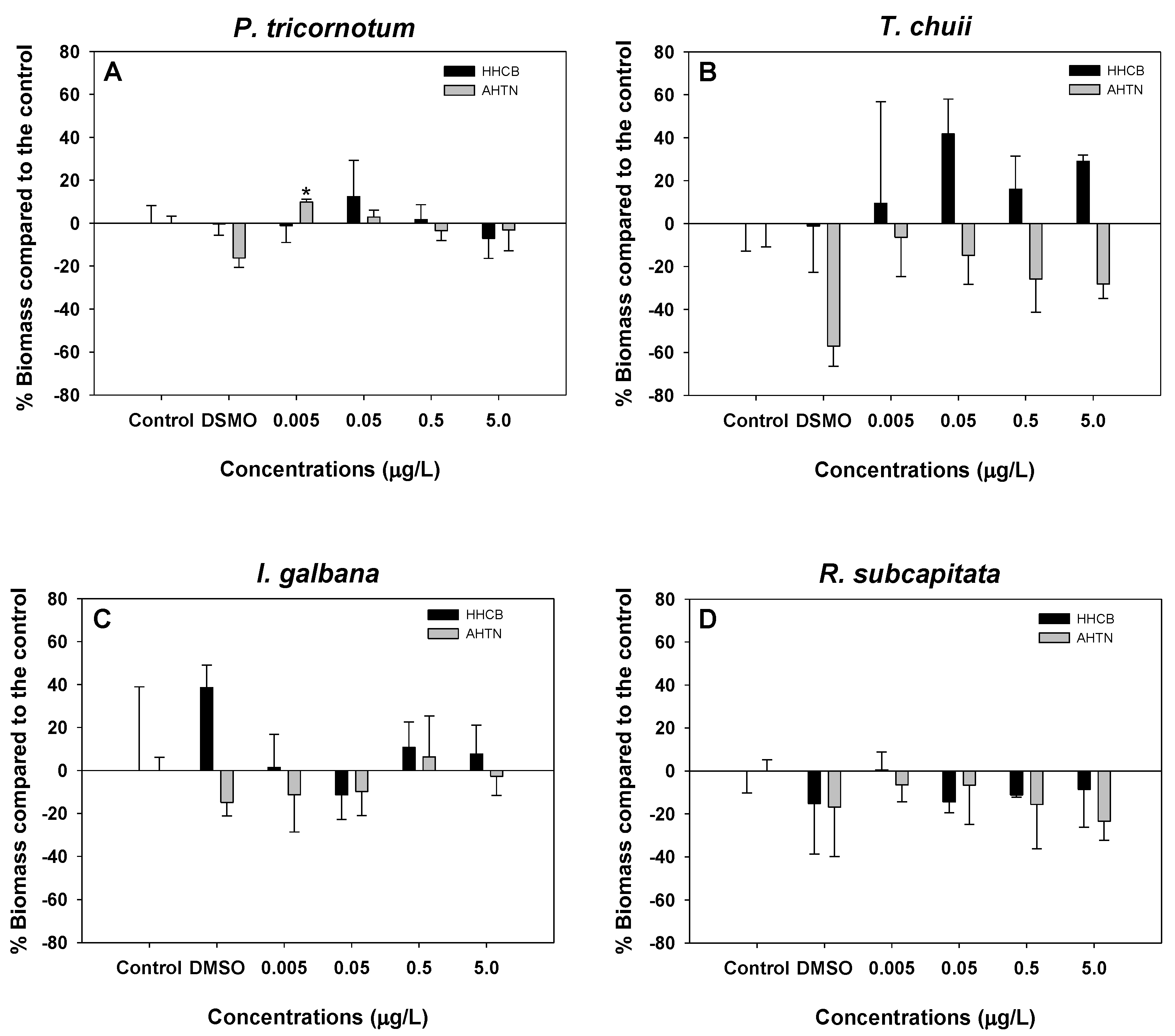
3.1. Microalgae Growth Inhibition Tests

Data of growth inhibition for microalgae are presented in Figure 1. There was no critical difference between microalgae exposed to seawater control and solvent control, although microalgal growth response was observed in the group treated with DMSO. In brief, it is possible to observe that toxicity of both compounds was minimal for the four microalgae, although AHTN seemed to be slightly more toxic. For P. tricornutum, growth inhibition for both compounds were observed in some concentrations. However, significant differences (p < 0.05) in growth in relation to the control only occurred with exposure to AHTN at 0.005 μg/L by 16.2% (Figure 1A). On the one hand, T. chuii growth was inhibited by AHTN and the decrease in biomass was significantly (p < 0.01) concentration dependent (Table S1 in Supplementary Materials). On the other hand, the exposure to HHCB produced a stimulation (hormesis) in the growth (Figure 1B) and similar biphasic responses was seen in I. galbana biomass after exposure to both musk compounds (Figure 1C). The freshwater microalgae, R. subcapitata growth decreased after exposure to HHCB and AHTN. In the case of AHTN, significant (p < 0.05) concentration dependent growth inhibition of R. subcapitata was observed (Table S1 in Supplementary Materials), and the highest inhibition occurred at 5 μg/L by 23.5%. In contrast, R. subcapitata growth inhibition by HHCB was more severe at 0.05 μg/L by 14.5% in relation to the control (Figure 1D).

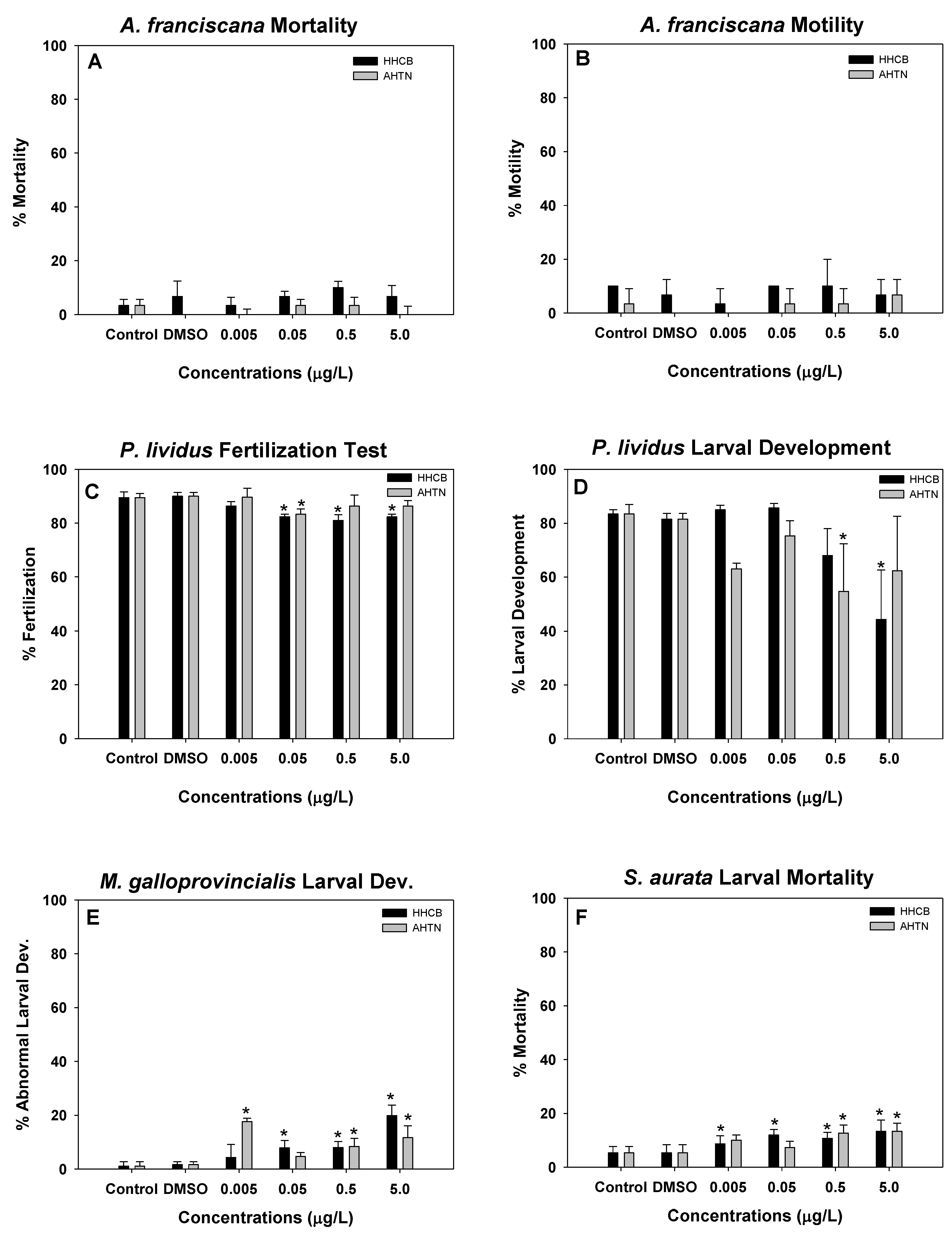
3.2. Artemia, Sea Urch, Mussels, and Fish Early Life Stage Toxicity Tests

For the organisms tested for fertilization, larvae development, motility, and mortality, the validity criteria for the control experiments (seawater control and solvent control—DMSO) were not exceeded.
Moreover, there was no significant difference (p < 0.05) in responses detected in organisms exposed to seawater control and DMSO.
The effects of HHCB and AHTN on survival and motility of A. franciscana are shown in Figure 2A,B, respectively. Both responses showed no significant difference (p < 0.05) compared to the control. For the A. franscana mortality test, percentage mortalities in the controls were 3.33 and 6.67% for seawater and DMSO, respectively. The effect of HHCB on the survival of A. franciscana was very minimal with the highest mortality of 10% recorded at a 0.5 µg/L HHCB. The effect of AHTN on A. franciscana mortality was also very low, with only 3.33% mortality at 0.05 and 0.5 µg/L. Other concentrations of AHTN tested had no effect on A. franciscana survival. Artemia motility test was not sensitive to either compound (Figure 2B).
The effects of HHCB and AHTN on P. lividus fertilization and larval development tests are presented in Figure 2C,D, respectively. The percentage of sea urchin that were able to fertilize in the controls were 89.50 and 90.00% for seawater and DMSO, respectively. Percentage fertilization of sea urchin tested with 0.005, 0.05, 0.5, and 5 µg/L HHCB were 86.33, 82.33, 81.00, and 82.33%, respectively. Under exposure to AHTN, sea urchin fertilization success recorded at 0.005, 0.05, 0.5, and 5 µg/L were 89.00, 83.33, 86.33, and 86.33%, respectively. Although the effect is minimal, P. lividus fertilization failure induced by both substances was more pronounced for HHCB with significant effects at 0.05–5 µg/L, while a significant difference (p < 0.05) was only observed at 0.05 µg/L AHTN compared to control (Figure 2C). The results for the sea urchin larval development tested with HHCB and AHTN are presented in Figure 2D. The percentages of larval development in the seawater and solvent controls were 83.50 and 81.50%, respectively. The number of oocytes that were able to develop to pluteus stage was significantly (p < 0.01) concentration dependent (Table S2 in Supplementary Materials) and a significant (p < 0.05) decrease in larvae development was recorded at 5 µg/L HHCB compared to the control (Figure 2D). The percentage of larval that were able to develop from morula stage to normal pluteus stage for 0.005, 0.05, 0.5, and 5 µg/L HHCB were 85.00, 85.00, 68.00, and 44.33%, respectively, while similar concentrations of AHTN reduced larvae development by 63.00, 75.33, 54.67, and 62.33%, respectively.
The results for the larval development toxicity test for M. galloprovincialis exposed to HHCB and AHTN are presented in Figure 2E. The number of fertilized eggs that developed after 48 h to D-veliger stage in the control was 98%. However, the embryotoxicity of HHCB to mussels was significantly (p < 0.01) concentration dependent (Table S2 in Supplementary Materials). From 0.05 to 5 µg/L HHCB, the percentage of abnormal larvae development was significantly (p < 0.05) different from the control (Figure 2E) and the percentage effect increased to 19.88% in the highest concentration tested (5 µg/L). Similarly, significant toxicity of AHTN to embryos of M. galloprovincialis was observed in 0.5 and 5.0 µg/L with percentages of abnormal larvae of 8.36 and 11.63%, respectively (Figure 2E).
The results of the effect of HHCB and AHTN on the survival of yolk-sac larvae of S. aurata larval after 96 h exposure was significantly (p < 0.01) concentration dependent (Table S2 in Supplementary Materials) and the measured effect is presented in Figure 2F. After 96 h, the percentage mortality of yolk-sac larvae of S. aurata in seawater and DMSO controls was 5.33% each. Percentages of mortality in fish exposed to 0.005, 0.05, 0.5, and 5 µg/L of HHCB were 8.67, 12.00, 10.67, and 13.33%, respectively. On the other hand, the percentage mortality of fish tested with the same range of concentrations of AHTN were 10.00, 7.33, 12.67, and 13.33%, respectively.

3.3. Risk Quotient (RQ)

Quantitative risk estimation of chemicals in the environment is achieved using monitoring data of MEC and PNEC, giving a risk quotient necessary for risk characterization. A literature survey of MEC for both polycyclic musk compounds were undertaken, and the values are presented in Table 1. Due to minimal effects detected in this study for all the species of microalgae, Artemia, sea urchin, mussels, and fish, it was not possible to calculate the EC50 values and their, respective, confidence intervals, except for P. lividus larvae development tested with HHCB, producing an EC50 value and 95% confidence interval of 4.063 (0.963–120.731) µg/L. The IC10/EC10 calculated for P. tricornutum, I. galbana, P. lividus, and M. galloprovincialis, including the risk quotients using the MEC–PNEC ratio are presented in Table 2. The results of the microalgae growth inhibition and larval development tests showed that HHCB and AHTN posed high risk to the growth of P. tricornutum and M. galloprovincialis larval development at environmental relevant concentrations. Additionally, environmental concentrations of HHCB and AHTN pose ecological risk to P. lividus larval development and I. galbana growth, respectively (Table 2).

4. Discussion

The aim of this study was to evaluate the impacts of environmentally relevant concentrations of HHCB and AHTN on marine microalgae, Artemia, sea urchin, and mussels after short-term exposure and, where possible, characterize the risk following the ECHA guideline for ERA [35] Several in vitro and in vivo toxicity tests have been performed with these compounds using freshwater species [5,24,25,28,59,60] and the only tests with marine species used high concentrations [22,30], making it difficult to compare such results with actual environmental impacts. Furthermore, for evaluation of toxicity of substances, it is imperative to use a varied battery of tests because organisms are not equally susceptible to the same toxic substance.
Due to the instability of these substances under laboratory exposure, it is difficult to estimate the exact concentration causing toxic effects. Although we could not measure the concentrations of the exposure water and bioaccumulation because of practical constraints, studies have shown that after 3 h of exposure, over 30% was lost and this reduced to ca. 80% after 96 h [22,30,51,61]. Again, there are currently no techniques to measure the concentrations of these substances in-situ; therefore, there is a probability that reported environmental concentrations are a fraction of the actual concentration in the marine environments eliciting biological effects, given that some amount could be lost before or during sample analysis in the laboratory. Consequently, risk estimation was based on maximum concentration reported.
The effects of HHCB and AHTN on P. tricornutum, T. chuii, I. galbana, and R. subcapitata showed that both compounds have limited effects (significantly similar to the control treatment) on microalgae growth (Figure 1). Although the statistical analysis indicated no differences between control with and without DMSO, the results for microalgae should be considered with caution, because in some situations inhibition or enrichment due to DMSO was observed. Therefore, we cannot reject the possibility that the effects observed in the presence of HHCB or AHTN was partially caused by DMSO.
Microalgae have been used in water quality assessments as in-situ bio-monitors because they are primary producers at the base of the ecological trophic arrangement and the basic supplier of oxygen in aquatic ecosystems [62]. Microalgae toxicity tests are useful in ERA and have gained international recognition leading to development of test guidelines for reliable and relevant toxicity data [63]. The potential of HHCB and AHTN to inhibit the growth of microalgae in the aquatic ecosystem have been scarcely reported. Previous studies revealed that significant acute toxicity of microalgae occurred at concentration greater than 100 or 500 µg/L depending on the species [5,64] but our data suggest that even at low concentrations microalgae growth might be inhibited.
The microalgae growth inhibition tests were mildly sensitive to HHCB and AHTN; IC10 values for most of the microalgae could not be calculated, except for P. tricornutum and I. galbana exposed to HHCB with IC10 values of 0.127 and 5.220 µg/L, respectively (Table 2). In contrast, previous studies have reported higher EC50 values of 0.050 mg/L for Navicula spp. and 0.336 mg/L for Scendesmus quadricauda exposed to HHCB [64]. This is because microalgae are not equally sensitive to contaminants. For example, in all the microalgae tested, we found that differential sensitivity was exhibited, the order of sensitivity to HHCB was P. tricornutum > I. galbana, and to AHTN was T. chuii > P. tricornutum > I. galbana. Similarly, differential sensitivity of two microalgae to HHCB was recently reported, whilst the microalgae, Navicula spp. are more sensitive to HHCB than S. quadricauda [64]. The basis for the differential sensitivity of microalgae to these contaminants is beyond the scope of the current investigation. Furthermore, HHCB and AHTN are lipophilic and known to bioaccumulate in marine organisms [5,15,64]. Remarkably, the effects of these compounds on microalgae growth has been adduced to bioaccumulation, altering antioxidant enzymes and biochemical processes, resulting to decrease microalgae growth [64].
Artemia was not sensitive to either substance since the endpoints measured were not critically different from the control (Figure 2A,B). This is because Artemia is known to be fairly resistant to toxic substances in comparison to other invertebrates and they respond differently to different environmental contaminants [65,66].
HHCB significantly reduced the fertilization success of P. lividus as the concentrations increased. Meanwhile, AHTN had no significant effects on sea urchin fertilization success (Figure 2C). Sea urchin fertilization success depends on sperm fitness, motility potentiated by the axonemal engine in the flagellum, morphology and chemotaxic navigation [67]. For organoleptic substances, such as HHCB and AHTN, the latter might be the most compelling factor inducing reduction in sea urchin fertilization success since the exposure procedure involved prior treatment of sperm with the contaminants before the introduction of eggs. Between the two contaminants tested, HHCB had more significant effects on P. lividus fertilization, causing a 19% reduction at 0.5 µg/L, and we observed that only 0.05 µg/L AHTN significantly reduced sea urchin fertilization by 16.67% (Figure 2C). Importantly, the effect exerted by both contaminants on sea urchin fertilization success was below 20%, presenting less toxic effects compared to other contaminants of emerging concern. For example, 500 ng/L propanol, 500 ng/L 17α-ethinylestradiol and 5000 ng/L gemfibrozil reduced sea urchin fertilization success by 24.1, 36.9, and 26.9%, respectively [68]. Similarly, other contaminants of emerging concern belonging to pharmaceutical and personal care products have been reported to significantly affect sea urchin fertilization success at concentrations detected in the environment [69,70].
Ecotoxicity studies with early life stages of aquatic organisms have been recommended as a faster and more cost-effective means of examining chemicals and environmental samples, because newly hatched larvae are sensitive to exogenous substances as the embryos lose their protective membranes and are fully exposed to potential xenobiotics [71]. P. lividus and M. galloprovincialis are well recognized in toxicity bioassays and are applied globally for the evaluation of toxicity of marine contaminants by the exposure of gametes to aqueous phases, such as surface waters and pore waters [69, 72] and elutriates [73]. Critical effects were detected in P. lividus and M. galloprovincialis larvae exposed to HHCB and AHTN compared to the control (Figure 2D and E). The effects of HHCB on sea urchin larvae development was significantly (p < 0.01) concentration dependent (Table S2 in Supplementary Materials) and significantly different (p < 0.05) compared to the control, with only 44.33% of larvae able to develop to pluteus stage after 48 h exposure to 5 µg/L HHCB. The sensitivity of sea urchins to environmental chemicals is widely reported and significant evidence showed that they represent an important biomonitoring tool for ecosystems health. Similarly, HHCB and AHTN significantly (p < 0.05) affected the development of M. galloprovincialis larvae when compared to the control (Figure 2E). However, when considered in relation to the number of zygotes exposed to each contaminant, the effects were minimal with the highest percentage of deformed and undeveloped zygotes being 19.88 and 17.60% for HHCB and AHTN, respectively. Although P. lividus and M. galloprovincialis larvae development tests were similar, the effects recorded were more pronounced in sea urchin larvae than mussels.
Significant (p < 0.01) concentration dependent responses (Table S2 in Supplementary Materials) were observed in S. aurata mortality tests with HHCB and AHTN (Table S2 in Supplementary Materials). Notwithstanding, the percentage mortality of S. aurata exposed to both fragrances not up to 20%, the highest effect being 13% for HHCB and AHTN at the highest concentration of 5 µg/L. Although this species and endpoints proved to be sensitive to other contaminants at low concentrations [40,68], the impacts of environmental concentrations of the tested fragrances in this study were low. For chemical prioritization, fish early-life stage toxicity test is endorsed [47] because it is a reliable and reproducible risk assessment tool that requires shorter exposure time and lower cost to perform. However, the sensitivity of fish embryotoxicity to some emerging contaminants remain doubtful as previous studies reported low sensitivity [68,74].
Risk characterization of contaminants is quantified using MEC–PNEC ratio and for aquatic environments (freshwater and marine), PNEC is estimated by dividing the ECx value by an assessment factor of 1.000 for acute toxicity test and 10 for chronic toxicity test [35]. Although short term toxicity tests were performed in this study, an assessment factor of 1000 was used only for larval development test while an assessment factor of 10 was used for the microalgae because the exponential phase of microalgae growth was regarded as a full life stage and therefore considered as a chronic test. Analyzing the RQs, HHCB represented potential high risk for the marine environment based on the EC50 estimated for P. lividus larvae development. In addition, HHCB and AHTN posed high ecological risk to M. galloprovincialis larval development at environmental relevant concentrations. The larval development of P. lividus and M. galloprovincialis seem to be very sensitive to chemical exposure because previous studies of environmental contaminants in coastal waters have reported significant toxicity of industrial and domestic effluent [40], pharmaceutically active products [68,74], UV-filters [70] and organic pollutants [46,72] to sea urchin and mussels larvae development. The quantitative risk estimate of HHCB obtained in this study for P. lividus using EC10 value (1200–57,500) was higher than that of propanol (0.02–17.29), previously reported by Capolupo et al. [68]. We also found that both compounds posed high risk to P. tricornutum and I. galbana based on IC10 values recorded (Table 2). Microalgae have broadly been used in evaluation of ERA for other emerging contaminants. For example, I. galbana have been reported to be significantly affected by UV filters and pharmaceutical active ingredients [70,74]. Fragrances have been demonstrated to pose high risk to microalgae in the marine environment, of which HHCB and AHTN were more toxic than musk xylene and musk ketone [7]. The adversity of measured environmental concentrations of HHCB and AHTN to P. tricornutum and I. galbana deserve attention because microalgae occupy the lowest trophic level of the marine food chain and, therefore, serve as food for higher trophic organisms. Again, HHCB and AHTN impacts on microalgae growth represent a potential bottom-up effect that might result in structural and functional disruption of the ecosystems [37]. Therefore, more studies are required to fully understand the environmental effects of these contaminants in the marine environments.

5. Conclusions

This study evaluated the potential risk of comparable measured environmental concentrations of HHCB and AHTN to microalgae and early life stages of marine organisms, including A. franciscana, P. lividus, M. galloprovincialis, and S. aurata, adding to the dearth of information regarding the adverse effects of HHCB and AHTN in the marine environment. For all the species of microalgae, Artemia, sea urchin, mussels, and fish tested, differential sensitivity was observed. Artemia motility and survival were the least sensitive endpoints affected by both substances and P. lividus larvae development was the most sensitive species/endpoint. From our data, the environmental risk of HHCB and AHTN was characterized as high for P. tricornutum, I. galbana growth, P. lividus and M. galloprovincialis larvae development. Therefore, more studies are required to understand the sub-lethal effects of these compounds in the marine environment.

Supplementary Materials

The following are available online at https://www.mdpi.com/2227-9717/9/2/371/s1, Table S1: Spearman rank order of correlation (r) values recorded for the effect of galaxolide (HHCB) and tonalide (AHTN) on microalagae exposed for 72 h (n = 3). Asterisk(s) * & ** represent significant levels at p < 0.05 and 0.01, respectively, Table S2: Spearman rank order of correlation (r) values recorded for the effect of galaxolide (HHCB) and tonalide (AHTN) on marine organisms exposed for 72 h (n = 3). Asterisk(s) * & ** represent significant levels at p < 0.05 and 0.01, respectively; (-) represent values not determined.

Author Contributions

F.O.E.: Conceptualization, methodology, samples analyses, data curation, project management, original draft preparation, writing and editing of manuscript. M.J.G.-D.: Methodology, samples analyses, data curation and original draft preparation. C.G.-P.: Conceptualization, methodology, samples analyses, data curation, revision of manuscript. C.V.M.A.: Conceptualization, methodology, samples analyses, data curation, revision of manuscript. M.L.M.-D.: Conceptualization, methodology, samples analyses, data curation, project management and revision of manuscript. All authors have read and agreed to the published version of the manuscript.

Funding

This research received no external funding.

Institutional Review Board Statement

Not applicable.

Informed Consent Statement

Not applicable.

Data Availability Statement

Data will be made available to the public at the completion of the project.

Acknowledgments

This research was supported by the Spanish Ministry of Economy and Competitiveness (Ref. CTM2012-37591), ERDF support from European Union and also partially funded by the Spanish Ministry of Science, Innovation and Universities through the CaPETHaS project (#CGL2017-92160-EXP). CVM Araújo is grateful for the Ramón y Cajal contract (RYC-2017-22324) from the Spanish Ministry of Science and Innovation.

Conflicts of Interest

The authors declare that there is no conflicts of interest for the publication of this present work and are willing to provide any clarification whenever called upon to do so.

References

  1. OSPAR. Musk Xylene and Other Musks. Hazard. Subst. Ser. 2004, 1–160. Available online: https://www.ospar.org/documents?v=6978 (accessed on 15 February 2021).
  2. Gao, Y.; Ji, Y.; Li, G.; Mai, B.; An, T. Bioaccumulation and ecotoxicity increase during indirect photochemical transformation of polycyclic musk tonalide: A modeling study. Water Res. 2016, 105, 47–55. [Google Scholar] [CrossRef] [PubMed]
  3. Heberer, T. Occurrence, fate, and removal of pharmaceutical residues in the aquatic environment: A review of recent research data. Toxicol. Lett. 2002, 131, 5–17. [Google Scholar] [CrossRef]
  4. Dietrich, D.R.; Hitzfeld, B.C. Bioaccumulation and ecotoxicity of synthetic musks in the aquatic environment. Handb. Environ. Chem. 2004, 3, 233–244. [Google Scholar] [CrossRef]
  5. Balk, F.; Ford, R.A. Environmental risk assessment for the polycyclic musks, AHTN and HHCB II. Effect assessment and risk characterisation. Toxicol. Lett. 1999, 111, 81–94. [Google Scholar] [CrossRef]
  6. Bester, K.; Hühnerfuss, H.; Lange, W.; Rimkus, G.G.; Theobald, N. Results of non target screening of lipophilic organic pollutants in the German Bight II: Polycyclic musk fragrances. Water Res. 1998, 32, 1857–1863. [Google Scholar] [CrossRef]
  7. Díaz-Garduño, B.; Pintado-Herrera, M.; Biel-Maeso, M.; Rueda-Márquez, J.; Lara-Martín, P.; Perales, J.; Manzano, M.; Garrido-Pérez, C.; Martín-Díaz, M. Environmental risk assessment of effluents as a whole emerging contaminant: Efficiency of alternative tertiary treatments for wastewater depuration. Water Res. 2017, 119, 136–149. [Google Scholar] [CrossRef] [PubMed]
  8. Fromme, H.; Otto, T.; Pilz, K. Polycyclic musk fragrances in different environmental compartments in Berlin (Germany). Water Res. 2001, 35, 121–128. [Google Scholar] [CrossRef]
  9. Kannan, K.; Reiner, J.L.; Yun, S.H.; Perrotta, E.E.; Tao, L.; Johnson-Restrepo, B.; Rodan, B.D. Polycyclic musk compounds in higher trophic level aquatic organisms and humans from the United States. Chemosphere 2005, 61, 693–700. [Google Scholar] [CrossRef] [PubMed]
  10. Nakata, H. Occurrence of synthetic musk fragrances in marine mammals and sharks from Japanese coastal waters. Environ. Sci. Technol. 2007, 41, 2216–2222. [Google Scholar] [CrossRef]
  11. Sumner, N.R.; Guitart, C.; Fuentes, G.; Readman, J.W. Inputs and distributions of synthetic musk fragrances in an estuarine and coastal environment; a case study. Environ. Pollut. 2010, 158, 215–222. [Google Scholar] [CrossRef]
  12. Villa, S.; Assi, L.; Ippolito, A.; Bonfanti, P.; Finizio, A. First evidences of the occurrence of polycyclic synthetic musk fragrances in surface water systems in Italy: Spatial and temporal trends in the Molgora River (Lombardia Region, Northern Italy). Sci. Total. Environ. 2012, 416, 137–141. [Google Scholar] [CrossRef]
  13. Dsikowitzky, L.; Schwarzbauer, J.; Littke, R. Distribution of polycyclic musks in water and particulate matter of the Lippe River (Germany). Org. Geochem. 2002, 33, 1747–1758. [Google Scholar] [CrossRef]
  14. Pintado-Herrera, M.G.; González-Mazo, E.; Lara-Martín, P.A. Environmentally friendly analysis of emerging contaminants by pressurized hot water extraction–stir bar sorptive extraction–derivatization and gas chromatography–mass spectrometry. Anal. Bioanal. Chem. 2013, 405, 401–411. [Google Scholar] [CrossRef] [PubMed]
  15. Moon, H.-B.; Lee, D.-H.; Lee, Y.S.; Kannan, K. Occurrence and accumulation patterns of polycyclic aromatic hydrocarbons and synthetic musk compounds in adipose tissues of Korean females. Chemosphere 2012, 86, 485–490. [Google Scholar] [CrossRef] [PubMed]
  16. Nakata, H.; Sasaki, H.; Takemura, A.; Yoshioka, M.; Tanabe, S.; Kannan, K. Bioaccumulation, Temporal Trend, and Geographical Distribution of Synthetic Musks in the Marine Environment. Environ. Sci. Technol. 2005, 39, 3430–3434. [Google Scholar] [CrossRef]
  17. Rüdel, H.; Böhmer, W.; Schröter-Kermani, C. Retrospective monitoring of synthetic musk compounds in aquatic biota from German rivers and coastal areas. J. Environ. Monit. 2006, 8, 812–823. [Google Scholar] [CrossRef] [PubMed]
  18. Vandermeersch, G.; Lourenço, H.M.; Alvarez-Muñoz, D.; Cunha, S.; Diogène, J.; Cano-Sancho, G.; Sloth, J.J.; Kwadijk, C.; Barcelo, D.; Allegaert, W.; et al. Environmental contaminants of emerging concern in seafood—European database on contaminant levels. Environ. Res. 2015, 143, 29–45. [Google Scholar] [CrossRef]
  19. Gatermann, R.; Biselli, S.; Hühnerfuss, H.; Rimkus, G.G.; Hecker, M.; Karbe, L. Synthetic Musks in the Environment. Part 1: Species-Dependent Bioaccumulation of Polycyclic and Nitro Musk Fragrances in Freshwater Fish and Mussels. Arch. Environ. Contam. Toxicol. 2002, 42, 437–446. [Google Scholar] [CrossRef]
  20. Cunha, S.; Trabalón, L.; Jacobs, S.; Castro, M.; Fernandez-Tejedor, M.; Granby, K.; Verbeke, W.; Kwadijk, C.; Ferrari, F.; Robbens, J.; et al. UV-filters and musk fragrances in seafood commercialized in Europe Union: Occurrence, risk and exposure assessment. Environ. Res. 2018, 161, 399–408. [Google Scholar] [CrossRef] [PubMed]
  21. Balk, F.; Ford, R.A. Environmental risk assessment for the polycyclic musks AHTN and HHCB in the EU. I. Fate and exposure assessment. Toxicol. Lett. 1999, 111, 57–79. [Google Scholar] [CrossRef]
  22. Breitholtz, M.; Wollenberger, L.; Dinan, L. Effects of four synthetic musks on the life cycle of the harpacticoid copepod Nitocra spinipes. Aquat. Toxicol. 2003, 63, 103–118. [Google Scholar] [CrossRef]
  23. Carlsson, G.; Orn, S.; Andersson, P.L.; Söderström, H.; Norrgren, L. The impact of musk ketone on reproduction in zebrafish (Danio rerio). Mar. Environ. Res. 2000, 50, 237–241. [Google Scholar] [CrossRef]
  24. Carlsson, G.; Norrgren, L. Synthetic musk toxicity to early life stages of zebrafish (Danio rerio). Arch. Environ. Contam. Toxicol. 2004, 46, 102–105. [Google Scholar] [CrossRef] [PubMed]
  25. Gooding, M.P.; Newton, T.J.; Bartsch, M.R.; Hornbuckle, K.C. Toxicity of Synthetic Musks to Early Life Stages of the Freshwater Mussel Lampsilis cardium. Arch. Environ. Contam. Toxicol. 2006, 51, 549–558. [Google Scholar] [CrossRef] [PubMed] [Green Version]
  26. Chen, C.; Zhou, Q.; Liu, S.; Xiu, Z. Acute toxicity, biochemical and gene expression responses of the earthworm Eisenia fetida exposed to polycyclic musks. Chemosphere 2011, 83, 1147–1154. [Google Scholar] [CrossRef] [PubMed]
  27. Luckenbach, T.; Epel, D. Nitromusk and Polycyclic Musk Compounds as Long-Term Inhibitors of Cellular Xenobiotic Defense Systems Mediated by Multidrug Transporters. Environ. Heal. Perspect. 2005, 113, 17–24. [Google Scholar] [CrossRef]
  28. Parolini, M.; Magni, S.; Traversi, I.; Villa, S.; Finizio, A.; Binelli, A. Environmentally relevant concentrations of galaxolide (HHCB) and tonalide (AHTN) induced oxidative and genetic damage in Dreissena polymorpha. J. Hazard. Mater. 2015, 285, 1–10. [Google Scholar] [CrossRef]
  29. Pedersen, S.; Selck, H.; Salvito, D.; Forbes, V. Effects of the polycyclic musk HHCB on individual- and population-level endpoints in Potamopyrgus antipodarum. Ecotoxicol. Environ. Saf. 2009, 72, 1190–1199. [Google Scholar] [CrossRef] [Green Version]
  30. Wollenberger, L.; Breitholtz, M.; Kusk, K.O.; Bengtsson, B.-E. Inhibition of larval development of the marine copepod Acartia tonsa by four synthetic musk substances. Sci. Total. Environ. 2003, 305, 53–64. [Google Scholar] [CrossRef]
  31. Yamauchi, R.; Ishibashi, H.; Hirano, M.; Mori, T.; Kim, J.-W.; Arizono, K. Effects of synthetic polycyclic musks on estrogen receptor, vitellogenin, pregnane X receptor, and cytochrome P450 3A gene expression in the livers of male medaka (Oryzias latipes). Aquat. Toxicol. 2008, 90, 261–268. [Google Scholar] [CrossRef]
  32. European Commission. European Union Risk Assessment Report on AHTN. Rep. AHTN 2008. [Google Scholar] [CrossRef]
  33. European Commission. European Union Risk Assessment Report on HHCB. Rep. HHCB 2008. [Google Scholar] [CrossRef]
  34. Human & Environmental Risk Assessment on ingredients of Household Cleaning Products (HERA). Polycyclic musks AHTN (CAS 1506-02-1) and HHCB (CAS 1222-05-05). Scenario 2004. [Google Scholar]
  35. ECHA. Chapter R.10: Characterisation of dose [concentration]-response for environment. In Guidance on Information Requirements and Chemical Safety Assessment; European Chemicals Agency: Helsinki, Finland, 2008; pp. 1–65. [Google Scholar]
  36. De Lange, H.; Sala, S.; Vighi, M.; Faber, J. Ecological vulnerability in risk assessment—A review and perspectives. Sci. Total. Environ. 2010, 408, 3871–3879. [Google Scholar] [CrossRef] [PubMed]
  37. Fleeger, J.W.; Carman, K.R.; Nisbet, R.M. Indirect effects of contaminants in aquatic ecosystems. Sci. Total. Environ. 2003, 317, 207–233. [Google Scholar] [CrossRef]
  38. Johnston, E.L.; Roberts, D.A. Contaminants reduce the richness and evenness of marine communities: A review and meta-analysis. Environ. Pollut. 2009, 157, 1745–1752. [Google Scholar] [CrossRef]
  39. Brink, P.J.V.D. Ecological Risk Assessment: From Book-Keeping to Chemical Stress Ecology. Environ. Sci. Technol. 2008, 42, 8999–9004. [Google Scholar] [CrossRef] [Green Version]
  40. Díaz-Garduño, B.; Rueda-Márquez, J.; Manzano, M.; Garrido-Pérez, C.; Martín-Díaz, M. Are combined AOPs effective for toxicity reduction in receiving marine environment? Suitability of battery of bioassays for wastewater treatment plant (WWTP) effluent as an ecotoxicological assessment. Mar. Environ. Res. 2016, 114, 1–11. [Google Scholar] [CrossRef]
  41. Meriç, S.; De Nicola, E.; Iaccarino, M.; Gallo, M.; Di Gennaro, A.; Morrone, G.; Warnau, M.; Belgiorno, V.; Pagano, G. Toxicity of leather tanning wastewater effluents in sea urchin early development and in marine microalgae. Chemosphere 2005, 61, 208–217. [Google Scholar] [CrossRef] [PubMed]
  42. Moreno-Garrido, I.; Campaña, O.; Lubián, L.; Blasco, J. Calcium alginate immobilized marine microalgae: Experiments on growth and short-term heavy metal accumulation. Mar. Pollut. Bull. 2005, 51, 823–829. [Google Scholar] [CrossRef]
  43. Yu, Y.; Kong, F.; Wang, M.; Qian, L.; Shi, X. Determination of short-term copper toxicity in a multispecies microalgal population using flow cytometry. Ecotoxicol. Environ. Saf. 2007, 66, 49–56. [Google Scholar] [CrossRef] [PubMed]
  44. Okumura, J.K.Y.; Koyama, J.; Takaku, H.; Satoh, H.; Okumura, Y. Influence of Organic Solvents on the Growth of Marine Microalgae. Arch. Environ. Contam. Toxicol. 2001, 41, 123–128. [Google Scholar] [CrossRef]
  45. Latała, A.; Nędzi, M.; Stepnowski, P. Acute toxicity assessment of perfluorinated carboxylic acids towards the Baltic microalgae. Environ. Toxicol. Pharmacol. 2009, 28, 167–171. [Google Scholar] [CrossRef]
  46. Mariani, L.; De Pascale, D.; Faraponova, O.; Tornambè, A.; Sarni, A.; Giuliani, S.; Ruggiero, G.; Onorati, F.; Magaletti, E. The use of a test battery in marine ecotoxicology: The acute toxicity of sodium dodecyl sulfate. Environ. Toxicol. 2006, 21, 373–379. [Google Scholar] [CrossRef]
  47. OECD. No. 210: Fish, Early-Life Stage Toxicity Test. OECD Test 1992, 2, 1–24. [Google Scholar] [CrossRef]
  48. Environment Canada. Biological test method: Fertilization assay using echinoids (sea urchins and sand dollars). In Method Dev. Appl. Sect.; 2011; p. 111. Available online: http://publications.gc.ca/collections/collection_2011/ec/En49-7-1-27-eng.pdf (accessed on 15 February 2021).
  49. US EPA. Guidelines for Exposure Assessment. Risk Assess. Forum 1992, 57, 22888–22938. [Google Scholar]
  50. US EPA. Methods for Measuring the Acute Toxicity of Effluents and Receiving Waters to Freshwater and Marine Organisms, 5th ed.; U.S. Environmental Protection AgencyOffice of Water: Washington, DC, USA, 2002. [Google Scholar]
  51. Ehiguese, F.O.; Fernandez, M.D.C.C.; Lara-Martín, P.A.; Martín-Díaz, M.L.; Araújo, C.V. Avoidance behaviour of the shrimp Palaemon varians regarding a contaminant gradient of galaxolide and tonalide in seawater. Chemosphere 2019, 232, 113–120. [Google Scholar] [CrossRef]
  52. Garrido-Perez, M.C.; Perales-VargasMachuca, J.A.; Nebot-Sanz, E.; Sales-Márquez, D.; Garrido-Pérez, M.D.C. Effect of the test media and toxicity of LAS on the growth of Isochrysis galbana. Ecotoxicol. 2008, 17, 738–746. [Google Scholar] [CrossRef]
  53. Guillard, R.R.; Ryther, J.H. Studies of marine planktonic diatoms. I. Cyclotella nana hustedt, and Detonula confervacea (CLEVE) Gran. Can. J. Microbiol. 1962, 8, 229–239. [Google Scholar] [CrossRef] [PubMed]
  54. Kilham, S.S.; Kreeger, D.A.; Lynn, S.G.; Goulden, C.E.; Herrera, L. COMBO: A defined freshwater culture medium for algae and zooplankton. Hydrobiol. 1998, 377, 147–159. [Google Scholar] [CrossRef]
  55. Beiras, R.; Vázquez, E.; Bellas, J.; Lorenzo, J.; Fernández, N.; Macho, G.; Mariño, J.; Casas, L. Sea-urchin Embryo Bioassay for in situ Evaluation of the Biological Quality of Coastal Seawater. Estuarine, Coast. Shelf Sci. 2001, 52, 29–32. [Google Scholar] [CrossRef]
  56. ASTM International. Standard guide for conducting acute toxicity test starting with embryos of four species of saltwater bivalve molluscs. 2012, 21, E724–E798. [Google Scholar] [CrossRef]
  57. Yoshioka, K.; Saito, M.; Oh, K.-B.; Nemoto, Y.; Matsuoka, H.; Natsume, M.; Abe, H. Intracellular Fate of 2-NBDG, a Fluorescent Probe for Glucose Uptake Activity, inEscherichia coliCells. Biosci. Biotechnol. Biochem. 1996, 60, 1899–1901. [Google Scholar] [CrossRef]
  58. Zhang, H.; Bayen, S.; Kelly, B.C. Multi-residue analysis of legacy POPs and emerging organic contaminants in Singapore’s coastal waters using gas chromatography–triple quadrupole tandem mass spectrometry. Sci. Total. Environ. 2015, 523, 219–232. [Google Scholar] [CrossRef]
  59. Schreurs, R.H.; Quaedackers, M.E.; Seinen, W.; Van Der Burg, B. Transcriptional Activation of Estrogen Receptor ERα and ERβ by Polycyclic Musks Is Cell Type Dependent. Toxicol. Appl. Pharmacol. 2002, 183, 1–9. [Google Scholar] [CrossRef]
  60. Składanowski, A.; Stepnowski, P.; Kleszczyński, K.; Dmochowska, B. AMP deaminase in vitro inhibition by xenobiotics. Environ. Toxicol. Pharmacol. 2005, 19, 291–296. [Google Scholar] [CrossRef] [PubMed]
  61. Tumová, J.; Šauer, P.; Golovko, O.; Ucun, O.K.; Grabic, R.; Máchová, J.; Kroupová, H.K. Effect of polycyclic musk compounds on aquatic organisms: A critical literature review supplemented by own data. Sci. Total. Environ. 2019, 651, 2235–2246. [Google Scholar] [CrossRef] [PubMed]
  62. Sibila, M.; Garrido, M.; Perales, J.; Quiroga, J. Ecotoxicity and biodegradability of an alkyl ethoxysulphate surfactant in coastal waters. Sci. Total. Environ. 2008, 394, 265–274. [Google Scholar] [CrossRef] [PubMed]
  63. OECD. Proposal for updating guideline 201 freshwater alga and cyanobacteria, growth inhibition test. In OECD Guidelines for Testing of Chemicals; OECD: Paris, France, 2002; pp. 1–21. [Google Scholar]
  64. Ding, T.; Li, W.; Cai, M.; Jia, X.; Yang, M.; Yang, B.; Li, J. Algal toxicity, accumulation and metabolic pathways of galaxolide. J. Hazard. Mater. 2020, 384, 121360. [Google Scholar] [CrossRef]
  65. Ferreira, C.S.G.; Nunes, B.A.; Henriques-Almeida, J.M.D.M.; Guilhermino, L. Acute toxicity of oxytetracycline and florfenicol to the microalgae Tetraselmis chuii and to the crustacean Artemia parthenogenetica. Ecotoxicol. Environ. Saf. 2007, 67, 452–458. [Google Scholar] [CrossRef] [PubMed]
  66. Hadjispyrou, S.; Kungolos, A.; Anagnostopoulos, A. Toxicity, Bioaccumulation, and Interactive Effects of Organotin, Cadmium, and Chromium on Artemia franciscana. Ecotoxicol. Environ. Saf. 2001, 49, 179–186. [Google Scholar] [CrossRef] [PubMed]
  67. Hussain, Y.H.; Guasto, J.S.; Zimmer, R.K.; Stocker, R.; Riffell, J.A. Sperm chemotaxis promotes individual fertilization success in sea urchins. J. Exp. Biol. 2016, 219, 1458–1466. [Google Scholar] [CrossRef] [Green Version]
  68. Capolupo, M.; Díaz-Garduño, B.; Martín-Díaz, M.L. The impact of propranolol, 17α-ethinylestradiol, and gemfibrozil on early life stages of marine organisms: Effects and risk assessment. Environ. Sci. Pollut. Res. 2018, 25, 32196–32209. [Google Scholar] [CrossRef] [PubMed]
  69. Aguirre-Martínez, G.V.; DelValls, A.T.; Martín-Díaz, M.L. Yes, caffeine, ibuprofen, carbamazepine, novobiocin and tamoxifen have an effect on Corbicula fluminea (Müller, 1774). Ecotoxicol. Environ. Saf. 2015, 120, 142–154. [Google Scholar] [CrossRef]
  70. Paredes, E.; Perez, S.; Rodil, R.; Quintana, J.; Beiras, R. Ecotoxicological evaluation of four UV filters using marine organisms from different trophic levels Isochrysis galbana, Mytilus galloprovincialis, Paracentrotus lividus, and Siriella armata. Chemosphere 2014, 104, 44–50. [Google Scholar] [CrossRef]
  71. Beiras, R.; Fernandez, N.; Bellas, J.; Besada, V.; González-Quijano, A.; Nunes, T. Integrative assessment of marine pollution in Galician estuaries using sediment chemistry, mussel bioaccumulation, and embryo-larval toxicity bioassays. Chemosphere 2003, 52, 1209–1224. [Google Scholar] [CrossRef]
  72. Bellas, J.; Granmo, Å.; Beiras, R. Embryotoxicity of the antifouling biocide zinc pyrithione to sea urchin (Paracentrotus lividus) and mussel (Mytilus edulis). Mar. Pollut. Bull. 2005, 50, 1382–1385. [Google Scholar] [CrossRef]
  73. Losso, C.; Picone, M.; Novelli, A.A.; Delaney, E.; Ghetti, P.F.; Ghirardini, A.V. Developing Toxicity Scores for Embryotoxicity Tests on Elutriates with the Sea Urchin Paracentrotus lividus, the Oyster Crassostrea gigas, and the Mussel Mytilus galloprovincialis. Arch. Environ. Contam. Toxicol. 2007, 53, 220–226. [Google Scholar] [CrossRef]
  74. Aguirre-Martínez, G.; Owuor, M.; Garrido-Pérez, C.; Salamanca, M.; Del Valls, T.; Martín-Díaz, M. Are standard tests sensitive enough to evaluate effects of human pharmaceuticals in aquatic biota? Facing changes in research approaches when performing risk assessment of drugs. Chemosphere 2015, 120, 75–85. [Google Scholar] [CrossRef] [PubMed]
Figure 1. A-1D: Microalgae exposed to galaxolide and tonalide for growth inhibition tests after 72 h. Biomass (%) compared to the control of (A) P. tricornotum, (B) T. chuii, (C) I. galbana, and (D) R. subcapitata (negative values corresponding to % growth inhibition). Asterisks (*) indicate significant differences (p < 0.05) in relation to control.
Processes 09 00371 g001
Figure 2. A. franciscana 72 h mortality and motility, P. lividus fertilization and 48 h larval development, M. galloprovincialis 48 h larval development and S. aurata 96 h larval mortality tests exposed to galaxolide and tonalide. Asterisks (*) indicate significant differences (p < 0.05) in relation to control.
Processes 09 00371 g002
Table 1. Measured Environmental Concentrations (MEC) of Galaxolide (HHCB) and Tonalide (AHTN) in seawater from different locations. Measured values are in ng/L. Maximum values (bold) were used in calculating risk quotient (RQ).
Country/LocationHHCB AHTN Reference
Germany (North Sea)0.09–4.80.08–2.6[6]
Germany (Elbe Estuary)95–13665–200[6]
United Kingdom (Tamar Estuarine—Plym Sound)6.00–303.00–15[11]
Spain (Bay of Cadiz)230 ± 0.1NA[14]
Singapore (Coastal water)1.66–21.80.244–1.85[58]
NA = Data not available.
Table 2. Median lethal concentration (EC50) and EC10 (µg/L) and their respective confidence interval (CI), and estimated risk quotients of Galaxolide (HHCB) and Tonalide (AHTN) as the MEC (maximum value per location)—PNEC ratio.
Microalgae GrowthEmbryo-Larval Development
P. tricornutumI. galbanaP. lividusM. galloprovincialis
HHCBEC50NCNC4.063 (0.963–120.731)NC
EC100.127(NC)5.22(NC)0.004 (0.000–0.025)0.188(0.074–0.390)
MEC/PNEC0.378–18.1100.009–0.4401200–5750025.532–1223.404
RiskYesNoYesYes
ANTHEC50NCNCNCNC
EC100.002(0.000 – 0.014)0.328(NC) 0.006 (NC)
MEC/PNEC24–11500.146–7.0122NC800–38333.330
RiskYesYesNCYes
NC = Not calculated.
Publisher’s Note: MDPI stays neutral with regard to jurisdictional claims in published maps and institutional affiliations.

