Article

In Vitro Bioadsorption of Cd2+ Ions: Adsorption Isotherms, Mechanism, and an Insight to Mycoremediation

1
Department of Biotechnology, Maharishi Markandeshwar (Deemed to be University), Mullana (Ambala), Haryana 133207, India
2
Department of Environment Studies, Panjab University, Chandigarh 160014, India
3
Department of Chemistry, Faculty of Science and Arts, Najran University, Najran 11001, Saudi Arabia
4
Promising Centre for Sensors and Electronic Devices (PCSED), Najran University, Najran 11001, Saudi Arabia
5
Department of Bio and Nano Technology Guru Jambheshwar University of Science and Technology, Hisar 125001, India
6
Division of Soil and Crop Management, Central Soil Salinity Research Institute, Karnal 132001 India
7
Department of Agri-Business Management, Maharishi Markandeshwar (Deemed to be University), Mullana (Ambala), Haryana 133207, India
8
Department of Chemistry, College of Science, King Saud University, P.O. Box 2445, Riyadh 11451, Saudi Arabia
*
Authors to whom correspondence should be addressed.
Processes 2020, 8(9), 1085; https://doi.org/10.3390/pr8091085
Submission received: 1 August 2020 / Revised: 22 August 2020 / Accepted: 29 August 2020 / Published: 2 September 2020
(This article belongs to the Special Issue Various Adsorbents for Water Purification Processes)

Abstract

:
The objective of this paper is to establish the significance of the mycoremediation of contaminants such as Cd2+ to achieve sustainable and eco-friendly remediation methods. Industries such as electroplating, paint, leather tanning, etc. release an enormous amount of Cd2+ in wastewater, which can drastically affect our flora and fauna. Herein, we report on the in vitro bioadsorption of Cd2+ ions using fungal isolates obtained from different contaminated industrial sites. The detailed studies revealed that two fungal species, i.e., Trichoderma fasciculatum and Trichoderma longibrachiatum, were found to be most effective against the removal of Cd2+ when screened for Cd2+ tolerance on potato dextrose agar (PDA) in different concentrations. Detailed adsorption studies were conducted by exploring various experimental factors such as incubation time, temperature, pH, inoculum size, and Cd2+ salt concentrations. Based on optimum experimental conditions, T. fasciculatum exhibited approximately 67.10% removal, while T. longibrachiatum shows 76.25% removal of Cd2+ ions at pH 5.0, 120 h incubation time, at 30°C. The inoculum sizes for T. fasciculatum and T. longibrachiatum were 2.5% and 2.0%, respectively. Finally, the morphological changes due to Cd2+ accumulation were examined using scanning electron microscopy (SEM). Further, Fourier transform infrared spectroscopy (FTIR) spectroscopy reveals the presence of various functional groups (-CH, –C=O, NH and –OH), which seem to be responsible for the efficient binding of Cd2+ ions over the fungal surfaces.

1. Introduction

The emerging worldwide environmental problems of the past decades are mostly related to the superfluous increase in metallic contaminants in the environment [1,2,3,4,5,6]. Inorganic contaminants such as heavy metals released into the environment through various industrial, agricultural, and domestic practices [5,6]. Heavy metals may also enter into the environment by natural sources, but the extent of such exposures is non-significant. Heavy metals are often present together with organic pollutants in industrial wastewater [7]. The industrial effluents are rich in dissolved salts of Cr, Cu, Ni, Zn, and Cd, etc., and can cause a serious problem to the surrounding environment. Heavy metals pollution causes serious problems due to their non-biodegradable nature [8,9]. Although these metals are vital for the proper functioning of biological systems but only in trace concentrations, higher concentrations may lead to disturbed functioning of bio-geo-chemical cycles [10,11,12]. In addition, serious health issues may arise if such metal ions can enter into the food chains and their corresponding products. Thus, even 1.0–10 mg/L can cause health problems in human beings such as jaundice, facial edema, blue lungs, kidney damage, hearing disorder, skin cancer, asthma, protein metabolism, and bronchial cancer, etc. [13,14]. Therefore, a number of traditional physiochemical treatment techniques have been reported to remove the heavy metals from wastewater, especially from the industrial wastewater, to name a few, electro-coagulation, solvent extraction, ion-exchange, electro-reduction, reverse osmosis, adsorption, membrane separation, chemical precipitation, and so on [15,16,17,18,19,20,21,22,23,24,25,26,27,28,29]. Even though used widely, these above-mentioned techniques exhibited several disadvantages which include the utilization of high-cost equipment and monitoring systems, the use of various expensive chemicals, the discharge of toxic sludge, long processing time, the production of toxic waste products that need further processing, and so on [23,24,25,26,27,28,29,30,31]. Thus, it is the necessity of the time to develop a new, simple, and inexpensive method for heavy metal removal from wastewater. Recently, by utilizing living/non-living microorganisms and their derivatives to remove the heavy metals from industrial wastewater is considered a promising technique [32,33,34]. Accordingly, the bioremediation can be considered as one of the most important and advantageous process that can efficiently remove the heavy metals from industrial wastewater. The entrapment of the heavy metal ions and their corresponding sorption on the binding cites of the cellular structures leads to the bioadsorption of heavy metal ions from the industrial wastewater [35,36]. As a result of the faster adsorption process, the microbial cells are considered as an important and advantageous scaffold for the biosorption of heavy metals and wastewater treatment.
In this study, different fungal isolates extracted from various contaminated sites (Haryana, India) were cultured and screened for tolerance to Cadmium (Cd2+). Various process factors, i.e., inoculum size, pH, initial metal ion concentration, temperature, and incubation time on the Cd2+ removal by highly efficient Cd2+ tolerant fungal isolates have been investigated and presented in this article. In addition, mechanisms of Cd2+ removal by efficient Cd2+ tolerant fungal isolates have also been studied.

2. Materials and Methods

2.1. Chemicals Used

Potato dextrose broth (PDB) and nutrient media, potato dextrose agar (PDA), were procured from Hi-Media, Mumbai India. Cd(NO3)2, was provided by Fine-Chem Limited, Mumbai, India. The solutions were prepared in triply distilled water (conductivity =0.5 μS cm−1), and the reagents were used without any further purification.

2.2. Sample Collection and Isolation of Fungi

Samples from various sites such as industrial effluents, sewerage discharge, and sludge were collected from Karnal, Panipat, and Sonepat districts of Haryana (India). The samples were stored at 4 °C prior to further processing. Fungal isolates were isolated from samples by the PDA serial dilution method. The serial dilutions were made up to 106 and 1 mL of each dilution i.e., 104 and 106 were poured over PDA plates. An incubation of cultured petri plates was performed at 28 °C for 96 h. The prominently grown fungal colonies were regenerated and purified using the streak plate method.

2.3. Screening and Identification of Efficient Cd2+-Tolerant Fungal Isolate

The isolated fungal isolates were tested for their potential to remove Cd2+ at 25, 50, and 75 mg/L concentrations. The concentrations were prepared from a sterilized 100 mg/L solution of Cd(NO3)2 containing PDA. A loop full of purified isolates was positioned at the Petri plates’ center comprising of PDA (pH 5.0) and the above-mentioned Cd2+ concentrations. The growth of fungal isolates on the pure PDA medium was labeled ‘Control’. All plates were sealed with parafilm and subjected to incubation of 5 days. Based on the visual observation, the growth of fungal isolates was labeled as absent growth (−), least growth (+), less than normal growth (++), slightly less than normal growth (+++), and normal growth (++++). The highly efficient Cd2+-tolerant fungal isolates were sent for identification to the Indian Agricultural Research Institute, New Delhi, India.

2.4. Optimization of Batch Cultures

The highly efficient Cd2+-tolerant fungal culture was tested at different pH, i.e., 3.5, 4.0, 4.5, 5.0, 5.5, and 6.0 for uptake and percentage removal of Cd2+ from PDA containing 20 mg/L of Cd2+. First, 1M HCl and 1M NaOH were used to adjust the pH of PDB. Then, 1mL spore suspension of efficient fungal culture containing 104 spores (1%) was inoculated into 250 mL Erlenmeyer conical flasks containing 100 mL of PDB enriched with 20 mg/L of Cd2+ followed by shaking these flasks for 120 h at 30 °C. For the control, a non-inoculated flask having 20 mg/L of Cd2+ and PDB was used. After 120h, fungal growth was collected through filtration using Whatman Filter Paper No. 1, which was later dried for 48 h at 80 °C in an oven and then weighed. Atomic absorption spectrophotometer (AAS, GBC 932, Dandenong, Australia) was used to evaluate the Cd2+ concentration left in the filtrate. To evaluate the incubation time effect, the incubation time was varied between 24 and 144h under optimum pH. Under optimum pH and incubation time, the effect of inoculum size was investigated by performing batch experiments at different inoculum sizes i.e., 0.5%, 1.0%, 1.5%, 2.0%, 2.5%, and 3.0%. Temperatures of 20, 25, 30, and 35 °C were studied to examine the temperature effect at optimum pH, incubation time, and inoculum size. Under optimum pH, the effect of initial Cd2+ concentration, inoculum size, and incubation time was investigated by changing the concentrations of Cd2+ ions from 5 to 50 mg/L. The process to perform all the experiments was the same as mentioned above. All experiments were performed in triplicate, and data were statistically examined by t-test with the help of SPSS 11.5.
The Cd2+ uptake by fungal biomass was calculated as follows:
Q   =   ν C 0 C f M
where Q is the Cd2+ uptake (mg/g); C0 and Cf are the initial and leftover concentrations of Cd2+ (mg/L), respectively; v is the total volume of the solution (L); and M the dried biomass of fungus (g).
The removal of Cd2+ (%) was calculated using following equation:
%   Removal   of   Cd 2 +   =   C ( i n i t i a l ) C ( f i n a l ) C ( i n i t i a l ) × 100

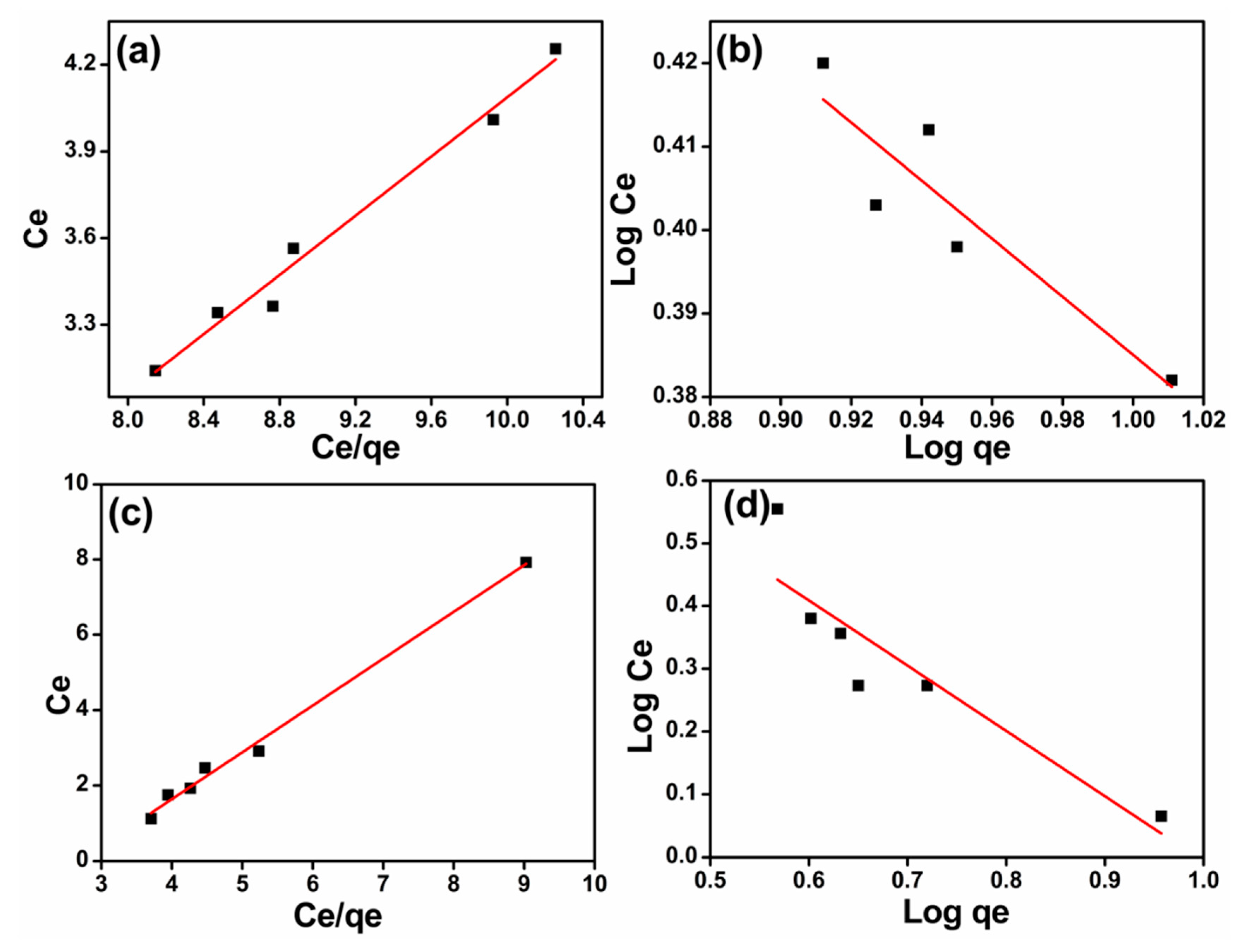
2.5. Equilibrium Isotherms

2.5.1. Langmuir Isotherm

Langmuir isotherm is a mathematical deduction of kinetic equilibrium between the condensation and loss of adsorbed molecules.
Ce/qe =1/qmax b + Ce/qmax
where Ce is equilibrium concentration of Cd2+ (mg/L), qe is amount of Cd2+ adsorbed per gram of fungal biomass (mg/g), qmax is Langmuir constant, and b is the energy of adsorption. The values of b and qmax were evaluated by the intercept and slope of the graph, respectively.

2.5.2. Freundlich Isotherm

This isotherm is the result of empirical consideration and expressed as
qe = Kf Ce1/n
where qe is the Cd2+ adsorbed (mg/g), Ce is the equilibrium concentration of Cd2+ (mg/L), Kf is the adsorption coefficient (mg/g) and directly related to the standard free energy change, and n is the empirical constant.
The linear logarithmic form of the isotherm is:
Log qe = (1/n) log Ce + log Kf
From the straight-line plot between log qe and log Ce, the values of of Kf and 1/n were found.

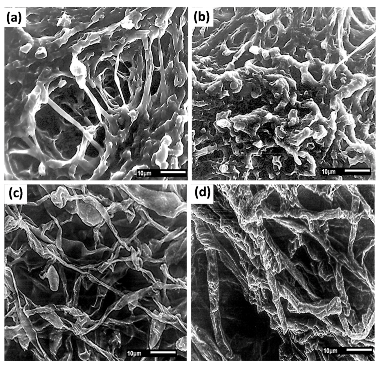
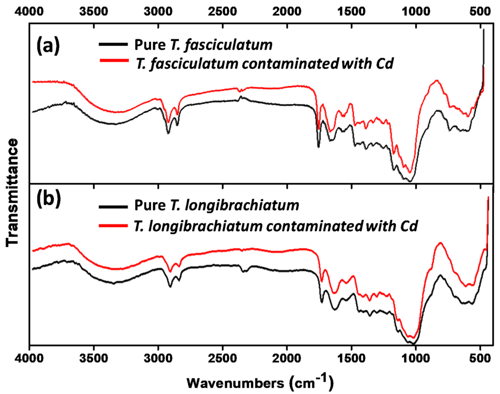
2.6. Scanning Electron Microscopy (SEM) and Fourier Transform Infrared Spectroscopy (FTIR) Analysis

The morphological changes in the fungi without and after incubating with Cd2+ were visualized using Scanning Electron Microscopy (SEM; JSM-6100, JEOL, Tokyo, Japan). The changes in surface chemical profiling were analyzed and interpreted by Fourier transform infrared spectroscopy (FTIR) (Perkin Elmer Spectrum BXII, Waltham, MA, USA, in 4000–400 cm−1 frequency range with a resolution of 1 cm−1).

3. Results and Discussion

3.1. Isolation, Screening, and Identification of Cd2+-Resistant Fungal Biomass

Twenty-five sludge, industrial effluents and sewage samples were collected from Panipat, Sonepat and Karnal districts of Haryana, India to isolate fungal isolates. All the fungi were maintained on PDA slants (pH 4.5) at 30 °C. Screening of these fungal isolates on PDA containing 25, 50, and 75 mg/L of Cd2+ showed that 13 fungal isolates were tolerant to 25 mg/L Cd2+, 7 fungal isolates were tolerant to 50 mg/L, and 3 fungal isolates were tolerant to 75 mg/L (Table 1). Such phenomena indicate that some fungal isolates exhibited inhibition at higher metal ions concentrations. Out of three fungal isolates tolerant to 75 mg/L Cd2+, the highly Cd2+-tolerant fungal isolate FS7 and FS16 were identified as Trichoderma fasciculatum (ITCC 7547) and Trichoderma longibrachiatum (ITCC 7062) by the Indian Culture Collection, Indian Agricultural Research Institute. IARI, New Delhi, India.

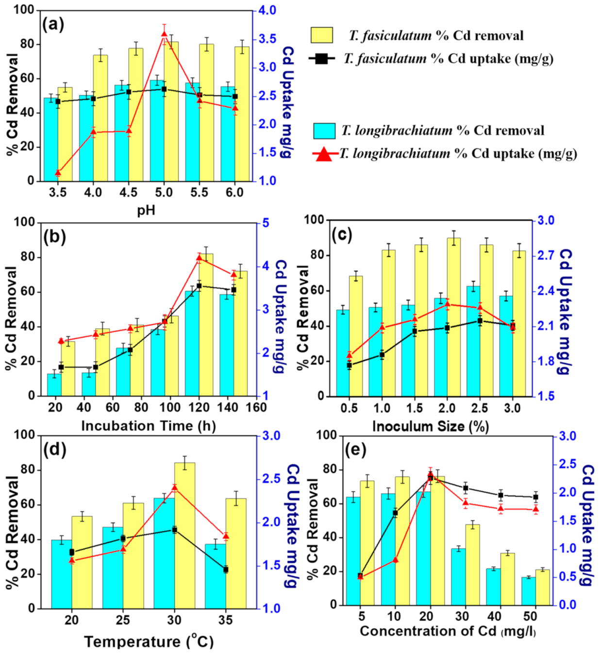
3.2. Optimization of Process Parameters

Various surface and physiochemical properties—for instance, pH, incubation time, inoculum size, temperature, metal ion concentrations, quantity of biosorbents, etc. are affecting the process for the bioaccumulation of heavy metal ions in fungal biomass.

3.2.1. pH Effect

In the process to remove the heavy metal ions from fungi, the pH plays an important and significant role. The observed results demonstrated that there was an enhancement in the Cd2+ ion uptake and percentage removal up to pH 5.0 from the liquid medium by T. fasciculatum and T. longibrachiatum. After pH 5.0, the uptake and percentage removal was significantly decreased with the increasing pH (Figure 1a). The outcomes showed that the ideal pH for uptake and percentage removal of Cd2+ + from the liquid medium by T. fasciculatum and T. longibrachiatum was 5.0.
The observed uptake and percentage removal of Cd2+ from the liquid medium by T. fasciculatum at pH 5.0 were 2.63 mg/g and 59.20%, respectively. In addition, the uptake and percentage removal of Cd2+ by T. longibrachiatum were 3.59 mg/g and 81.60%, respectively. Interestingly, it has been reported that the optimum pH is important to get better results regarding the removal of heavy metal ions using fungal isolates [37,38]. In many cases, the optimal value of pH was 5.0, which shows a maximum removal of heavy metal ions (Pb(II), Cd(II), As(III), and Hg(II)) by fungus isolates such as Penicillium purpurogenum [37,38].

3.2.2. Incubation Time Effect

Figure 1b exhibits the incubation time effect on the Cd uptake and percentage removal from the liquid medium by T. fasciculatum and T. longibrachiatum. It was observed that at optimum pH 5.0, the uptake and percentage removal of Cd2+ ions from the liquid medium by T. fasciculatum and T. longibrachiatum were increased with increasing the incubation time. Interestingly, it was seen the uptake and percentage removal were enhanced from 24 h to 120 h; however, they reduce after 120h. The maximum uptake and percentage removal of Cd2+ by T. fasciculatum at 120 h incubation time were 3.56 mg/g and 60.55%, respectively. Nevertheless, at 120 h incubation time, the uptake and percentage removal of Cd2+ ion by T. longibrachiatum were 4.2 mg/g and 82.2%, respectively.
It is believed by most of the researchers that ion exchange or physical adsorption at the surface of the cell might relate with the initial rapid phase [39]. However, in the presence of salt, the metal ion transport into the cytoplasm across the cell membrane via an active metabolism-dependent transport is responsible for the slower phase [40]. Moreover, in the absence of salt, other biosorption mechanisms that consist of complexation, microprecipitation, etc. due to the diffusion of metal ions into the cell debris are responsible for the second slower phase [41].

3.2.3. Inoculum Size Effect

Under the optimized conditions (pH 5.0, incubation time 120 h), with increasing the inoculum size from 0.5% to 3.0%, the Cd2+ ion uptake and percentage removal was enhanced up to a particular size and then reduced. Figure 1c depicts the relation between the Cd2+ ion uptake and percentage removal and inoculum size. The experimental observations confirmed that the uptake and percentage removal of the Cd2+ ion was increased with increasing the inoculum size from 0.5% to 2.5% for T. fasciculatum and 2.0% for T. longibrachiatum, while it declined afterwards. The maximum uptake and percentage removal of Cd2+ from the liquid medium by T. fasciculatum was approximately 2.15 mg/g and 62.60%, respectively. Moreover, under the same optimized experimental conditions, the Cd2+ ion uptake and percentage removal in the liquid medium by T. longibrachiatum were 2.29 mg/g and 89.85%, respectively. Interestingly, after the inoculum size 2.5% for T. fasciculatum and 2.0% for T. longibrachiatum, the uptake and percentage removal of the Cd2+ ions were decreased significantly. It has been reported that the percentage removal of heavy metal ions increases with enhancing the adsorbent dose due to the fact that it increases the adsorption sites, which leads to high adsorption and high removal efficiency [42,43,44].

3.2.4. Temperature Effect

Interestingly, the adsorption medium temperature is also substantially important in the biosorption of the metal ions by microbial cells for energy-dependent mechanisms. As reported in the literature, mostly, the adsorption is considered as an exothermal process [45]. To understand the effect of temperature on the uptake and percentage removal of Cd2+ ions, temperature-dependent experiments were performed by keeping all other optimized experimental conditions the same, i.e., pH 5, incubation time 120h, inoculum size 2% for T. longibrachiatum. Figure 1d exhibits the relation between the Cd2+ ion uptake and percentage removal and adsorption temperature. The observed results revealed that under optimized conditions, the maximum Cd2+ion uptake and percentage removal from the liquid medium by both fungi were obtained at 30 °C. It was interesting to note that for T. fasciculatum, the maximum Cd2+ uptake and percentage removal was 1.92 mg/g and 63.85%, respectively while for the T. longibrachiatum, the uptake and percentage removal was 2.40 mg/g and 84.25%, respectively.

3.2.5. Initial Metal Concentration Effect

Initial metal ion concentrations also have a particular effect on the adsorption characteristics of heavy metal ions. Figure 1e shows the effect of initial metal ion concentration on the uptake and percentage removal of Cd2+ ions using fungal isolates. To check the initial ion concentrations, several concentrations (5–50 mg/L) were tested, and all the experiments were performed at optimized experimental conditions, i.e., pH 5.0, incubation time (120 h), inoculum size (2.5% for T. fasciculatum, and 2.0% for T. longibrachiatum), and temperature 30 °C. The observed results confirmed that under optimized reaction conditions, for both fungal isolates, the Cd2+ uptake and percentage removal increased with the increasing the Cd2+ concentrations from 5 to 20 mg/L, and afterwards, a decline was seen. At an optimum concentration of 20 mg/L, the uptake and percentage removal of Cd2+ from liquid medium was 2.27 mg/g and 67.10% for T. fasciculatum and 2.34 mg/g and 76.25% for T. longibrachiatum, respectively. This enhancement may be because of an increase in the electrostatic associations (comparative with covalent interactions), including sites of continuously lower affinity for metal ions [46].

3.3. Adsorption Isotherms

The availability of a finite number of binding sites can be expected in the Langmuir model, which equally distributed to the sorbent surface. It presents the same attraction for single-layer sorption with no interaction between sorbed species. The Freundlich equation provides the adsorption data for a limited concentration range and proposed an adsorption sites heterogeneity on the biomass [47]. By changing the pH from 3.5 to 6.0 for Cd2+ ion removal from the liquid medium using T. fasciculatum and T. longibrachiatum, the obtained results were examined, and sorption isotherms were calculated. Figure 2 exhibits the calculated Langmuir and Freundlich isotherms for Cd2+ removal by T. fasciculatum and T. longibrachiatum. As a result of the higher value of correlation coefficients R2 > 0.90, the observed results revealed that the Cd2+ removal using both the fungal isolates, i.e., T. fasciculatum and T. longibrachiatum, better fit with the Langmuir isotherm models compared with the Freundlich isotherm. Various parameters of Langmuir and Freundlich models for the Cd2+ ion sorption on the live cells of T. fasciculatum and T. longibrachiatum are presented in Table 2. As observed, the maximum Cd2+ ion adsorption capacities (qmax) for T. fasciculatum and T. longibrachiatum fungal isolates were 1.90 mg/g and 0.80 mg/g, while the calculated correlation coefficients (R2) were 0.99 and 0.99 for both the fungal isolates, respectively.

3.4. SEM and FTIR Analysis

The morphological changes before and after the Cd2+ ions accumulation in the fungal isolates, i.e., T. fasciculatum and T. longibrachiatum, were further analyzed by scanning electron microscopy (SEM), and the results are presented in Figure 3.
The observation revealed that after 120 h, the hyphae of both the fungi was tube shaped, septate, and diverged with no metal treatment. However, after treatment with 20 mg/L of Cd2+ after 120 h, there was a thorough disruption and dissolution of mycelium of T. fasciculatum and T. longibrachiatum. These metals were consistently intact to the fungal mycelium, and a higher absorption of Cd2+ ions along with flocculation in mycelium was seen. The formation of such complexes was most probably because of the detoxification mechanism, in which normally fungal isolates are used in order to manage the lethal concentrations of heavy metal ions [48].
To understand the metal ions bindings, the attachment of various functional groups on the fungal surface are important to study. To examine the functional groups available on the surfaces of fungal isolates, FTIR studies were performed for both the fungal biomass, i.e., T. fasciculatum and T. longibrachiatum before and after Cd2+ ions treatment. Figure 4 exhibits the typical FTIR spectra in the range of 450–4000 cm−1 for both the fungal biomass, i.e., T. fasciculatum and T. longibrachiatum before and after Cd2+ ions treatment. The Cd2+ free biomass of T. fasciculatum and T. longibrachiatum exhibited several absorption peaks, which show the complex nature of the biomass. The peak that appeared in the range of 3500–3200 cm−1 was due to stretching of the N–H bond of the amino groups and the O–H bond of the hydroxyl group. Upon Cd2+ loading to the biomass of T. fasciculatum and T. longibrachiatum, a significant change in the peak positions was seen, which confirmed the Cd2+ binding with hydroxyl and amino groups (Table 3). The peaks that appeared at 2853 cm−1 and 2922 cm-1 exhibited the presence of C–H methyl groups stretching. Furthermore, the peak shown at 1747 cm−1 represents the native carbonyl stretching, while the peak at 1373 cm−1 represented as CH symmetric/symmetric band. The peaks that appeared at 2853 cm−1, 2922 cm−1, and 1747 cm−1did not show any significant change in samples exposed to Cd2+ except for a peak exhibited at 1373 cm−1, which showed a slight change from its original position.
In case of Cd2+-unloaded spectra of T. Fasciculatum and T. longibrachiatum, the peaks appeared at 1650 and 1544 cm−1 are related with the C=O of amide I and the NH/C=O blend of the amide II bond, respectively confirming the availability of the carboxyl groups [49,50]. It is interesting to see that the peak positioned at 1544 cm−1 expanded in the presence of Cd2+ ions, which confirms the interaction of Cd2+ with carboxyl groups. The above observations revealed the presence of various functional groups such as -CH, -OH, –C=O, and –NH in the binding of Cd2+ions with the fungal biomass, i.e., T. fasciculatum and T. longibrachiatum. The observed results are in agreement with the reported literature [49,50,51].

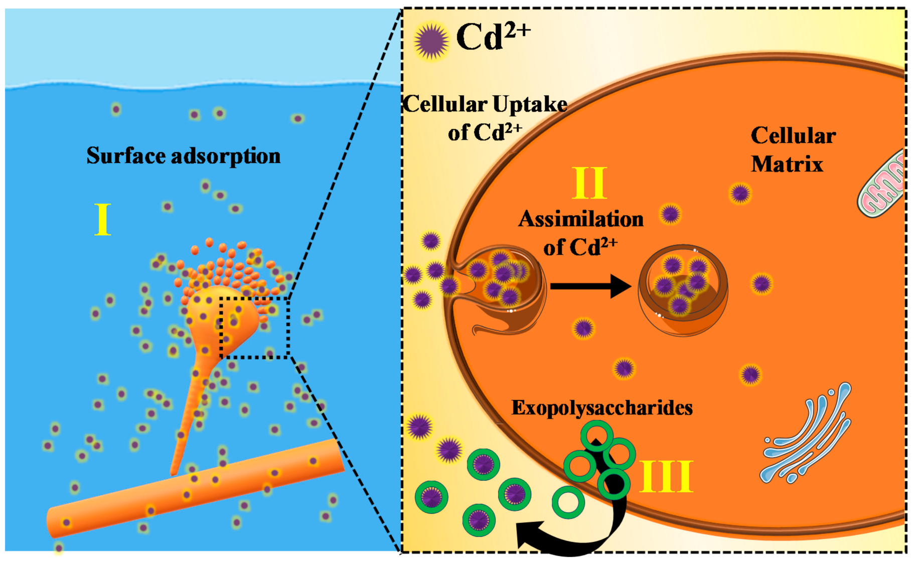
3.5. Proposed Mechanism for Cd2+ Removal

Based on the findings of this work, we proposed the possible mechanism of T. fasciculatum and T. longibrachiatum response to Cd2+ stress (Figure 5). The bioaccumulation of Cd2+ in T. fasciculatum and T. longibrachiatum is a cumulative result of three pathways. First the Cd2+ get adsorbed over the fungal outer membrane [52]. It has been shown in the FTIR results that various functional groups available on the fungal surface showed significant changes in their peak positions, indicating the chemical adsorption of Cd2+ over the membrane. Then, the ions are engulfed in the fungal cytoplasm where they are assimilated and sequestrated by the cellular reactions [53]. In addition, the fungi can release exopolysaccharides (high-molecular-weight polymers composed of sugar residues) to entrap or adsorb the extracellular Cd2+ ions [54].

4. Conclusions

In summary, 25 sludge, industrial effluents and sewage samples were collected to isolate fungal isolates and screened to their tolerance to Cd2+ in PDA medium containing Cd2+ from 25 to 75 mg/L. There was a decline in the number of fungi for their tolerance to Cd2+ with the increment in concentration of Cd2+ from 25 to 75 mg/L. The removal of Cd2+ by highly effective fungi T. fasciculatum and T. longibrachiatum was dependent on various experimental parameters such as temperature, initial metal ion concentrations, pH, incubation time, inoculum size, etc. Under optimized conditions, i.e., pH 5.0, inoculum size (2.5% for T. fasciculatum and 2.0% for T. longibrachiatum), incubation time (120 h), initial Cd2+ concentration (20 mg/L), and temperature (30 °C), the maximum removal of Cd2+ was 67.10% for T. fasciculatum and 76.25% for T. longibrachiatum. The observed data were well fitted with the Langmuir and Freundlich isotherm models. The SEM examinations confirmed the morphological changes in response to the Cd2+ aggregation in T. fasciculatum and T. longibrachiatum. Both the fungal isolate exhibited Cd2+ binding on the cell wall surface as a mechanism of metal tolerance. The FTIR studies confirmed the presence of various functional groups such as –CH, –OH, -C=O and –NH in the binding of Cd2+ ions with the fungal biomass, i.e., T. fasciculatum and T. longibrachiatum. The observed results revealed the potential biosorption tendency of T. fasciculatum and T. longibrachiatum to remove Cd2+ from wastewater.

Author Contributions

Conceptualization, R.K. (Raman Kumar), N.S. and A.U.; methodology, R.K. (Raman Kumar), N.S. and A.U.; software, R.K. (Raman Kumar), N.S., P.S., A.U., R.K. (Rajeev Kumar), P.K.J., F.A.A., A.A.A. and N.A.-Z.; validation, R.K. (Raman Kumar), P.S., A.U., R.K. (Rajeev Kumar), P.K.J., F.A.A., A.A.A., and N.A.-Z.; formal analysis, R.K. (Raman Kumar), N.S. and A.U.; the investigation, R.K. (Raman Kumar), and A.U.; resources, R.K. (Raman Kumar), P.S., A.U., R.K. (Rajeev Kumar), N.S.,P.K.J., F.A.A., A.A.A., and N.A.-Z.; data curation, R.K. (Raman Kumar), P.S., A.U., R.K. (Rajeev Kumar), N.S., P.K.J., F.A.A., A.A.A., and N.A.-Z.; writing—original draft preparation, R.K. (Raman Kumar) and A.U.; writing—review and editing, R.K. (Raman Kumar) and A.U.; visualization, R.K. (Raman Kumar), P.S., A.U., R.K. (Rajeev Kumar), N.S., P.K.J., F.A.A., A.A.A. and N.A.-Z.; supervision, R.K. (Raman Kumar); N.S. project administration, R.K. (Raman Kumar), N.S. and A.U. All authors have read and agreed to the published version of the manuscript.

Funding

The authors extend their appreciation to the Deanship of Scientific Research at King Saud University for funding this work through the research group No. RGP-1441-305.

Acknowledgments

The authors are thankful to Haryana State Council of Science and Technology, Panchkula, Haryana for providing financial support for carrying out research work (Grant No. HSCST/R&D/2018/209R). The authors are thankful to Maharishi Markandeshwar (Deemed to be University), Mullana, India for providing the lab facilities. The authors extend their appreciation to the Deanship of Scientific Research at King Saud University for funding this work through the research group No. RGP-1441-305.

Conflicts of Interest

The authors declare no conflict of interest.

References

  1. Wu, H.-Z.; Meng, L.-F.; Song, W.-G. Glutathione-Stabilized Silver Nanoparticles and Magnetic Nanoparticles Combination for Determination of Lead and Cadmium in Environmental Waters. Sci. Adv. Mater. 2019, 11, 1133–1139. [Google Scholar] [CrossRef]
  2. Hoang, H.-G.; Lin, C.; Tran, H.-T.; Chiang, C.-F.; Bui, X.-T.; Cheruiyot, N.K.; Shern, C.-C.; Lee, C.-W. Heavy metal contamination trends in surface water and sediments of a river in a highly-industrialized region. Environ. Technol. Innov. 2020, 20, 101043. [Google Scholar] [CrossRef]
  3. Song, T.; Li, R.; Li, N.; Gao, Y. Research Progress on the Application of Nanometer TiO2 Photoelectrocatalysis Technology in Wastewater Treatment. Sci. Adv. Mater. 2019, 11, 158–165. [Google Scholar] [CrossRef]
  4. Tan, K.; Ma, W.; Chen, L.; Wang, H.; Du, Q.; Du, P.; Yan, B.; Liu, R.; Li, H. Estimating the Distribution Trend of Soil Heavy Metals in Mining Area from HyMap Airborne Hyperspectral Imagery Based on Ensemble Learning. J. Hazard. Mater. 2020, 401, 123288. [Google Scholar] [CrossRef] [PubMed]
  5. Zhou, X.-Y.; Wang, X.-R. Impact of industrial activities on heavy metal contamination in soils in three major urban agglomerations of China. J. Clean. Prod. 2019, 230, 1–10. [Google Scholar] [CrossRef]
  6. Yao, S.; Li, Q.; Kong, N.; Li, C.; Wei, M.; Song, R.; Ren, X.; Pu, X.; Li, W. Influence of Initial Cu/(Zn+Sn) Concentration Ratio in Cu–Zn–Sn–S Composites on Their Microstructures, Adsorption and Visible-Light-Sensitive Photocatalytic Activities. Sci. Adv. Mater. 2018, 10, 1381–1388. [Google Scholar] [CrossRef]
  7. Gulan, L.; Milenkovic, B.; Zeremski, T.; Milic, G.; Vuckovic, B. Persistent organic pollutants, heavy metals and radioactivity in the urban soil of Priština City, Kosovo and Metohija. Chemosphere 2017, 171, 415–426. [Google Scholar] [CrossRef]
  8. Hassen, A.; Saidi, N.; Cherif, M.; Boudabous, A. Effects of heavy metals on Pseudomonas aeruginosa and Bacillus thuringiensis. Bioresour. Technol. 1998, 65, 73–82. [Google Scholar] [CrossRef]
  9. Di Cesare, A.; Pjevac, P.; Eckert, E.; Curkov, N.; Šparica, M.M.; Corno, G.; Orlić, S.; Andrea, D.C.; Petra, P.; Ester, M.E.; et al. The role of metal contamination in shaping microbial communities in heavily polluted marine sediments. Environ. Pollut. 2020, 265, 114823. [Google Scholar] [CrossRef]
  10. Bost, M.; Houdart, S.; Oberli, M.; Kalonji, E.; Huneau, J.-F.; Margaritis, I. Dietary copper and human health: Current evidence and unresolved issues. J. Trace Elements Med. Boil. 2016, 35, 107–115. [Google Scholar] [CrossRef]
  11. Rayman, M.P. The importance of selenium to human health. Lancet 2000, 356, 233–241. [Google Scholar] [CrossRef] [Green Version]
  12. Guo, J.; Xie, J.; Zhou, B.; Găman, M.-A.; Varkaneh, H.K.; Clark, C.C.; Salehi-Sahlabadi, A.; Li, Y.; Han, X.; Hao, Y.; et al. The influence of zinc supplementation on IGF-1 levels in humans: A systematic review and meta-analysis. J. King Saud Univ. Sci. 2020, 32, 1824–1830. [Google Scholar] [CrossRef]
  13. Jaishankar, M.; Tseten, T.; Anbalagan, N.; Mathew, B.B.; Beeregowda, K.N. Toxicity, mechanism and health effects of some heavy metals. Interdiscip. Toxicol. 2014, 7, 60–72. [Google Scholar] [CrossRef] [Green Version]
  14. Piñón-Gimate, A.; Jakes-Cota, U.; Tripp-Valdez, A.; Casas-Valdez, M.; Almendarez-Hernández, L.C.; Alejandra, P.-G.; Ulianov, J.-C.; Arturo, T.-V.; Margarita, C.-V.; Cesar, A.-H.L. Assessment of human health risk: Copper and lead concentrations in Stone Scorpionfish (Scorpaena mystes) from the coastal region of Santa Rosalia in the Gulf of California, Mexico. Reg. Stud. Mar. Sci. 2020, 34, 101003. [Google Scholar] [CrossRef]
  15. Bi, Y.; Liu, D.; Huang, M. Preparation and Characterization of Nano Hydroxyapatite and Its Adsorption Behavior toward Lead Ions. Sci. Adv. Mater. 2018, 10, 896–903. [Google Scholar] [CrossRef]
  16. Hosseini, S.; Alibakhshi, H.; Jashni, E.; Parvizian, F.; Shen, J.; Taheri, M.; Ebrahimi, M.; Rafiei, N. A novel layer-by-layer heterogeneous cation exchange membrane for heavy metal ions removal from water. J. Hazard. Mater. 2020, 381, 120884. [Google Scholar] [CrossRef]
  17. Da̧browski, A.; Hubicki, Z.; Podkościelny, P.; Robens, E. Selective removal of the heavy metal ions from waters and industrial wastewaters by ion-exchange method. Chemosphere 2004, 56, 91–106. [Google Scholar] [CrossRef] [PubMed]
  18. Tian, D.; Lv, H.; Mao, H.; Wang, D.; Zheng, Y.; Peng, C.; Hou, S. Synthesis, Characterization and Adsorption Behavior of Cd(II) Ion-Imprinted Mesoporous Materials. Sci. Adv. Mater. 2018, 10, 324–330. [Google Scholar] [CrossRef]
  19. Ya, V.; Martin, N.; Chou, Y.-H.; Chen, Y.; Choo, K.-H.; Chen, S.-S.; Li, C.-W. Electrochemical treatment for simultaneous removal of heavy metals and organics from surface finishing wastewater using sacrificial iron anode. J. Taiwan Inst. Chem. Eng. 2018, 83, 107–114. [Google Scholar] [CrossRef]
  20. Azarian, G.; Rahmani, A.R.; Khoram, M.M.; Atashzaban, Z.; Nematollahi, D. New batch electro-coagulation process for treatment and recovery of high organic load and low volume egg processing industry wastewater. Process. Saf. Environ. Prot. 2018, 119, 96–103. [Google Scholar] [CrossRef]
  21. Shih, Y.-J.; Chien, S.-K.; Jhang, S.-R.; Lin, Y.-C. Chemical leaching, precipitation and solvent extraction for sequential separation of valuable metals in cathode material of spent lithium ion batteries. J. Taiwan Inst. Chem. Eng. 2019, 100, 151–159. [Google Scholar] [CrossRef]
  22. Jantunen, N.; Virolainen, S.; Latostenmaa, P.; Salminen, J.; Haapalainen, M.; Sainio, T. Removal and recovery of arsenic from concentrated sulfuric acid by solvent extraction. Hydrometallurgy 2019, 187, 101–112. [Google Scholar] [CrossRef]
  23. Xia, Y.; Tsai, F.-C.; Ma, N.; Zhang, K.-D.; Liu, H.-L.; Jiang, T.; Chiang, T.-C. Green Synthesis of H3O40PW12@MIL-100(Fe): Enhanced and Selective Adsorption of Methylene Blue from Aqueous Solution. Sci. Adv. Mater. 2018, 10, 172–180. [Google Scholar] [CrossRef]
  24. Hao, J.; Ji, L.; Li, C.; Hu, C.; Wu, K. Rapid, efficient and economic removal of organic dyes and heavy metals from wastewater by zinc-induced in-situ reduction and precipitation of graphene oxide. J. Taiwan Inst. Chem. Eng. 2018, 88, 137–145. [Google Scholar] [CrossRef]
  25. Kim, T.; Kim, T.-K.; Zoh, K.-D. Removal mechanism of heavy metal (Cu, Ni, Zn, and Cr) in the presence of cyanide during electrocoagulation using Fe and Al electrodes. J. Water Process. Eng. 2020, 33, 101109. [Google Scholar] [CrossRef]
  26. Fan, Y.; Wang, Y.; Kang, D.; Deng, L.; Li, C. Oil-Tea Shell Derived N-Doped Porous Carbon for Selective Separation of CO2, CH4, and N2. Sci. Adv. Mater. 2019, 11, 1146–1155. [Google Scholar] [CrossRef]
  27. Zhao, X.; Liu, C. Efficient removal of heavy metal ions based on the optimized dissolution-diffusion-flow forward osmosis process. Chem. Eng. J. 2018, 334, 1128–1134. [Google Scholar] [CrossRef]
  28. Sadeghi, M.H.; Tofighy, M.A.; Mohammadi, T. One-dimensional graphene for efficient aqueous heavy metal adsorption: Rapid removal of arsenic and mercury ions by graphene oxide nanoribbons (GONRs). Chemosphere 2020, 253, 126647. [Google Scholar] [CrossRef]
  29. Naseem, K.; Begum, R.; Wu, W.; Usman, M.; Irfan, A.; Al-Sehemi, A.G.; Farooqi, Z.H. Adsorptive removal of heavy metal ions using polystyrene-poly(N-isopropylmethacrylamide-acrylic acid) core/shell gel particles: Adsorption isotherms and kinetic study. J. Mol. Liq. 2019, 277, 522–531. [Google Scholar] [CrossRef]
  30. Gola, D.; Dey, P.; Bhattacharya, A.; Mishra, A.; Malik, A.; Namburath, M.; Ahammad, S.Z. Multiple heavy metal removal using an entomopathogenic fungi Beauveria bassiana. Bioresour. Technol. 2016, 218, 388–396. [Google Scholar] [CrossRef]
  31. Talukdar, D.; Jasrotia, T.; Sharma, R.; Jaglan, S.; Kumar, R.; Vats, R.; Kumar, R.; Mahnashi, M.H.; Umar, A. Evaluation of novel indigenous fungal consortium for enhanced bioremediation of heavy metals from contaminated sites. Environ. Technol. Innov. 2020, 20, 101050. [Google Scholar] [CrossRef]
  32. Rangabhashiyam, S.; Balasubramanian, P. Characteristics, performances, equilibrium and kinetic modeling aspects of heavy metal removal using algae. Bioresour. Technol. Rep. 2019, 5, 261–279. [Google Scholar] [CrossRef]
  33. Qin, H.; Hu, T.; Zhai, Y.; Lu, N.; Aliyeva, J. The improved methods of heavy metals removal by biosorbents: A review. Environ. Pollut. 2020, 258, 113777. [Google Scholar] [CrossRef] [PubMed]
  34. Wei, Y.-Q.; Zhao, Y.; Zhao, X.; Gao, X.; Zheng, Y.; Zuo, H.; Wei, Z. Roles of different humin and heavy-metal resistant bacteria from composting on heavy metal removal. Bioresour. Technol. 2019, 296, 122375. [Google Scholar] [CrossRef]
  35. Bano, A.; Hussain, J.; Akbar, A.; Mehmood, K.; Anwar, M.; Hasni, M.S.; Ullah, S.; Sajid, S.; Ali, I. Biosorption of heavy metals by obligate halophilic fungi. Chemosphere 2018, 199, 218–222. [Google Scholar] [CrossRef]
  36. El Hameed, A.H.A.; Eweda, W.E.; Abou-Taleb, K.A.; Mira, H. Biosorption of uranium and heavy metals using some local fungi isolated from phosphatic fertilizers. Ann. Agric. Sci. 2015, 60, 345–351. [Google Scholar] [CrossRef] [Green Version]
  37. Visoottiviseth, P.; Panviroj, N. Selection of fungi capable of removing toxic arsenic compounds from liquid medium. Sci. Asia 2011, 27, 83–92. [Google Scholar] [CrossRef]
  38. Say, R.; Yılmaz, N.; Denizli, A. Biosorption of Cadmium, Lead, Mercury, and Arsenic Ions by the Fungus Penicillium purpurogenum. Sep. Sci. Technol. 2003, 38, 2039–2053. [Google Scholar] [CrossRef]
  39. Goksungur, Y. Biosorption of cadmium and lead ions by ethanol treated waste baker’s yeast biomass. Bioresour. Technol. 2005, 96, 103–109. [Google Scholar] [CrossRef]
  40. Dönmez, G.; Aksu, Z. Bioaccumulation of copper(II) and nickel(II) by the non-adapted and adapted growing Candida SP. Water Res. 2001, 35, 1425–1434. [Google Scholar] [CrossRef]
  41. Prakasham, R.; Merrie, J.; Sheela, R.; Saswathi, N.; Ramakrishna, S. Biosorption of chromium VI by free and immobilized Rhizopus arrhizus. Environ. Pollut. 1999, 104, 421–427. [Google Scholar] [CrossRef]
  42. Kumar, R.; Negi, S.; Sharma, P.; Prasher, I.; Chaudhary, S.; Dhau, J.S.; Umar, A. Wastewater cleanup using Phlebia acerina fungi: An insight into mycoremediation. J. Environ. Manag. 2018, 228, 130–139. [Google Scholar] [CrossRef] [PubMed]
  43. Chaudhary, S.; Sharma, P.; Kaur, A.; Kumar, R.; Mehta, S.K. Surfactant Coated Silica Nanoparticles as Smart Scavengers for Adsorptive Removal of Naphthalene. J. Nanosci. Nanotechnol. 2018, 18, 3218–3229. [Google Scholar] [CrossRef] [PubMed]
  44. Chaudhary, S.; Sharma, P.; Singh, D.; Umar, A.; Kumar, R. Chemical and Pathogenic Cleanup of Wastewater Using Surface-Functionalized CeO2 Nanoparticles. ACS Sustain. Chem. Eng. 2017, 5, 6803–6816. [Google Scholar] [CrossRef]
  45. Sharma, Y. Cr(VI) removal from industrial effluents by adsorption on an indigenous low-cost material. Colloids Surfaces A Physicochem. Eng. Asp. 2003, 215, 155–162. [Google Scholar] [CrossRef]
  46. Al-Asheh, S.; Duvnjak, Z. Adsorption of copper and chromium by Aspergillus carbonarius. Biotechnol. Prog. 1995, 11, 638–642. [Google Scholar] [CrossRef]
  47. Juang, R.-S.; Wu, F.-C.; Tseng, R.-L. Adsorption Isotherms of Phenolic Compounds from Aqueous Solutions onto Activated Carbon Fibers. J. Chem. Eng. Data 1996, 41, 487–492. [Google Scholar] [CrossRef]
  48. Manna, A.; Sundaram, E.; Amutha, C.; Vasantha, V.S. Efficient Removal of Cadmium Using Edible Fungus and Its Quantitative Fluorimetric Estimation Using (Z)-2-(4H-1,2,4-Triazol-4-yl)iminomethylphenol. ACS Omega 2018, 3, 6243–6250. [Google Scholar] [CrossRef]
  49. Naumann, A.; Navarro-González, M.; Peddireddi, S.; Kües, U.; Polle, A. Fourier transform infrared microscopy and imaging: Detection of fungi in wood. Fungal Genet. Biol. 2005, 42, 829–835. [Google Scholar] [CrossRef]
  50. Jilkine, K.; Gough, K.M.; Julian, R.; Kaminskyj, S.G. A sensitive method for examining whole-cell biochemical composition in single cells of filamentous fungi using synchrotron FTIR spectromicroscopy. J. Inorg. Biochem. 2008, 102, 540–546. [Google Scholar] [CrossRef]
  51. Akar, T.; Tunali, S. Biosorption performance of Botrytis cinerea fungal by-products for removal of Cd(II) and Cu(II) ions from aqueous solutions. Miner. Eng. 2005, 18, 1099–1109. [Google Scholar] [CrossRef]
  52. Morley, G.F.; Gadd, G.M. Sorption of toxic metals by fungi and clay minerals. Mycol. Res. 1995, 99, 1429–1438. [Google Scholar] [CrossRef]
  53. Krämer, U.; Talke, I.N.; Hanikenne, M.; Krämer, U. Transition metal transport. FEBS Lett. 2007, 581, 2263–2272. [Google Scholar] [CrossRef] [PubMed]
  54. Rana, S.; Upadhyay, L.S.B. Microbial exopolysaccharides: Synthesis pathways, types and their commercial applications. Int. J. Biol. Macromol. 2020, 157, 577–583. [Google Scholar] [CrossRef]
Figure 1. Effect of operational parameters on the Cd2+ removal efficiency by T. fasciculatum and T. longibrachiatum (a) pH (b) Incubation Time (c) Inoculum Size (d) Temperature (e) Concentration of Cd.
Processes 08 01085 g001
Figure 2. (a,c) Langmuir and (b,d) Fredundlich isotherms for Cd2+ removal by T. fasciculatum and T. longibrachiatum, respectively.
Processes 08 01085 g002
Figure 3. SEM images of (a) T. fasciculatum control and (b) after treatment with Cd2+, (c) T. longibrachiatum control and (d) after treatment with Cd2+.
Processes 08 01085 g003
Figure 4. Fourier transform infrared spectroscopy (FTIR) spectra of (a) T. fasciculatum and (b) T. longibrachiatum fungal biomass before and after treatment with Cd2+.
Processes 08 01085 g004
Figure 5. Pictorial illustration of Cd2+ removal by T. fasciculatum and T. longibrachiatum.
Processes 08 01085 g005
Table 1. Growth profile of all the fungal isolates on potato dextrose agar (PDA) (pH 5.0, temperature 30˚C) containing 25, 50, and 75 mg/L of Cd2+ after five days.
Sr. No.Fungal Isolate CodeControlCd2+ Ion Concentration (mg/L)
255075
1FS1+++++++++
2FS2+++++++++
3FS3+++++++++
4FS4+++++++++++++
5FS5+++++++++
6FS6+++++++++
7FS7+++++++++++++++
8FS8++++
9FS9+++++++++++++
10FS10+++++++++++++
11FS11++++
12FS12++++++++++++++
13FS13++++
14FS14+++++++
15FS15++++
16FS16+++++++++++++++
17FS17+++++
18FS18+++++
19FS19++++--
20FS20++++++++++
21FS21++++++++++
22FS22+++++++++++++
23FS23++++++++++
24FS24++++
25FS25+++++++
Note: Absent growth (−), least growth (+), less than normal growth (++), slightly less than normal growth (+++), and normal growth (++++).
Table 2. Langmuir and Freundlich isotherm constants for removal of Cd2+ by T. fasciculatum and T. longibrachiatum.
IsothermsParametersT. fasciculatumT. longibrachiatum
Langmuirqmax (mg/g)1.900.80
b (L/mg)−2.23−2.75
R20.990.99
FreundlichKf (L/g)−0.150.02
n−3.11−0.95
R20.840.84
Table 3. FTIR peaks and their corresponding functional groups of T. fasciculatum and T. longibrachiatum before and after the addition of Cd2+.
Sr. no.T. fasciculatumT. longibrachiatumAssigned Groups
ControlCd2+ AdditionControlCd2+ Addition
13332.63320.13363.53325.2-OH, -NH
22923.62924.32923.32923.3-CH, -OH
32853.32853.22854.62854.9-CH
41745.41746.01744.91745.1-C=O, of ester group
51654.81654.31645.51648.1-C=O, COO
61560.21563.01554.51558.8-NH
71373.81375.61372.31373.8-CH

