Article

Kinetics of Luteolin Extraction from Peanut Shells and Reseda luteola for Potential Applications as a Biofunctional Ingredient

by
Efstratios Episkopou
,
Dimitrios Tsimogiannis
,
Maria Giannakourou
* and
Petros Taoukis
*
Laboratory of Food Chemistry and Technology, School of Chemical Engineering, National Technical University of Athens, 5 Heroon Polytechniou Str., 15772 Athens, Greece
*
Authors to whom correspondence should be addressed.
Processes 2025, 13(9), 3009; https://doi.org/10.3390/pr13093009
Submission received: 1 August 2025 / Revised: 9 September 2025 / Accepted: 17 September 2025 / Published: 21 September 2025

Abstract

This study investigates the extraction kinetics of luteolin, a bioactive flavonoid with recognized antioxidant and health-promoting properties, from the aerial parts of Reseda luteola (dyer’s weld), with emphasis on its industrial potential. A comparative analysis with peanut shells (Arachis hypogea) identified R. luteola as a superior source, containing 14 ± 3 mg of LUT/g of material, approximately eight times higher than the amount in peanut shells. Luteolin occurred predominantly as luteolin-7-O-glycoside (57%) and the aglycone (35%). Methanolic semi-batch extraction at 25 °C yielded 9.6 mg LUT/g (70%) within 60 min at a solid-to-liquid ratio of 1:9, demonstrating significantly greater solvent efficiency than conventional Soxhlet or maceration techniques. Kinetic modeling, based on Fick’s second law, revealed a biphasic process with a low rate constant ratio (3:1) between the two stages, indicating the need for process optimization. These results establish R. luteola as a cost-effective and sustainable source of luteolin for dietary supplements and functional foods, while indicating the need to explore alternative solvents and advanced extraction methods to further optimize yield and efficiency.

1. Introduction

Luteolin, also known as 2-(3,4-dihydroxyphenyl)-5,7-dihydroxychromen-4-one, is a flavonoid of the flavone subgroup, commonly found in various herbs, fruits and vegetables as a secondary metabolite. Its structure, shown in Figure 1, consisting of four hydroxyl groups, contributes to a wide variety of biological and biochemical activities. As a result, further studies of the potential health benefits derived from luteolin’s biological activity, as well as its efficient extraction and isolation from plant sources, are gaining increased interest from food and pharmaceutical industries, for the development of luteolin-based supplements and functional foods [1].
Luteolin is a flavonoid with well-documented antioxidant activity, largely attributed to its hydroxylation pattern, particularly the o-di-hydroxy substitution on the B-ring, which enhances hydrogen atom donation to free radicals [2]. Comparative in vitro studies using the DPPH and ABTS methods have shown that luteolin and its non-glycosylated forms display higher antiradical activity than other flavones, such as apigenin and chrysin [2]. Recent findings confirmed this superiority, reporting significantly lower IC50 values for luteolin compared to apigenin and kaempferol [3]. In addition to direct radical scavenging, luteolin exhibits in vivo and in vitro activity against reactive oxygen species, including H2O2, partly through the inhibition of enzymes involved in free radical generation [4].
Luteolin has demonstrated notable anticancer and anti-inflammatory activity in various in vitro and in vivo studies [1,5,6]. Its anticancer effects are associated with the inhibition of abnormal cell proliferation and the induction of apoptosis in several cancer models [1,5,6]. Additionally, luteolin and its glycosides can modulate inflammatory responses by suppressing the production of pro-inflammatory mediators such as interleukins and TNF-α, particularly in the presence of bacterial toxins like lipopolysaccharides [1]. These properties, largely attributed to its antioxidant and signaling regulatory potential, further support its relevance in health-promoting formulations.
Beyond its well-established antioxidant, anti-inflammatory, and anticancer roles, luteolin also demonstrates antimicrobial, cardioprotective, and neuroprotective activities. It inhibits pathogens such as Chlamydia pneumoniae, influenza (H3N2), and SARS-CoV-2, partly through interactions with viral proteins [7,8,9]. The cardiovascular benefits are linked to the attenuation of oxidative stress during ischemia and arrhythmia, while the neuroprotective effects involve the modulation of neuroinflammation and the preservation of cognitive function in conditions such as chemotherapy-related or long-COVID “brain fog” [7,10,11]. Its low toxicity and inclusion in nutraceuticals, including olive pomace oil-based formulations like Neuroprotek (Algonot, Sarasota, FL, USA) and BrainGain (Algonot, Sarasota, FL, USA), highlight its biomedical relevance and the need for efficient extraction methods [9,12,13]. It should be noted that the compound shows low oral bioavailability due to poor solubility, extensive metabolism (e.g., glucuronidation), and rapid elimination [14]. Preclinical studies reported that microemulsions increase the plasma exposure of luteolin metabolites by ~2.2-fold in rodents [15], while nanostructured lipid carriers and microemulsions improve luteolin oral absorption over standard formulations [16]. Advanced formulations—such as liposomes, polymeric nanoparticles, and micelles—further enhance its solubility, stability, and bioavailability by protecting luteolin and enabling its controlled release [17]. Notably, polymeric micelles achieved an increase in its relative bioavailability with extended half-life in vivo [18]. Thus, optimized delivery systems are crucial for luteolin’s nutraceutical and pharmaceutical applications.
Regarding the natural sources of the compound, luteolin is found in a wide variety of plant species such as herbs, fruits, vegetables, and their seeds. However, the majority of plants contain luteolin in low concentrations (<1000 ppm), which thereby limits the number of plant sources suitable for its efficient industrial-scale extraction. In natural sources, luteolin occurs either in the aglycone form or as glycosylated derivatives, with O-glycosides representing the most prevalent type of luteolin derivatives found in plants [7,19].
Among plant species native to Greece and the Mediterranean basin, the most abundant source of luteolin is Reseda luteola, or dyer’s weld plant. Reseda luteola is a non-aromatic plant used for extracting yellow dyes in the past, which thrives in Central and Southern Europe, as well as the Southwestern and Eastern Mediterranean zone [20]. The plant has been previously studied by some researchers in order to determine its chemical composition, as well as the luteolin content present in Reseda luteola varieties. In [21], authors determined that the luteolin content of the plant’s aerial parts is equal to 11 mg/g of material, using an extraction process with a methanol–water mixture of 80:20 for a Reseda luteola variety cultivated in Southern France. Luteolin was mostly identified as the aglycone and as luteolin-7-O-glycoside, with concentrations of 4.5 and 3.5 mg/g of material, respectively. In another study [22], a content of 30 mg of total luteolin/g of material was reported in a Portuguese variety of the plant, with luteolin-7-O-glycoside being the most abundant compound in the plant’s aerial parts, followed by the aglycone, the 4′-O-glycoside, and the 3,7-O-diglycoside. The plant’s rich content in luteolin, alongside its non-aromatic nature, makes it a possible target for luteolin extraction. Reseda luteola was cultivated in the past in various areas of the Mediterranean as a natural weld and has a good agronomic potential, as displayed by Angelini et al. (2002) [23]. In Greece, the plant thrives in parts of Crete, Macedonia, Epirus, and Lemnos as a wild plant [24]. The plant has not been reported as toxic [25], though it is not included in EFSA’s list of approved botanicals. A flavonoid-rich extract (RF-40) from R. luteola demonstrated selective anti-proliferative activity in vitro without causing general cytotoxicity, but comprehensive toxicological studies are still required [26].
A plant source utilized in the food and pharmaceutical industry for the extraction of luteolin consists of the shells from Arachis hypogea. The shells constitute a byproduct of peanut production, which is mostly discarded or used as animal feed or as a fertilizer [27]. It mainly contains flavonoids, with the most common of these being luteolin at concentrations of 1–5 mg/g of material, followed by eriodictyol, as reported in [27,28], where the flavonoid content of peanut shells using methanolic extraction was studied. The compound is found in the peanut hulls only in the aglycone form, which makes the isolation of the pure aglycone easier, without the need of hydrolyzing glycosides [23]. However, the low content of luteolin in the shells makes the process of extraction and isolation less practical and efficient, due to the higher cost needed to extract significant quantities of the compound for use in supplements and functional foods. In Greece, peanuts are cultivated in Macedonia, Crete, and Western Peloponnese [29]. Other agro-industrial byproducts, olive leaves [30], and artichoke processing residues [31] have been reported to contain luteolin derivatives at concentrations of approximately 1–3 mg/g of material, comparable to the concentration found in peanut shells [27]. However, their relatively low content, compared with Reseda luteola, limits their practical potential for efficient industry-scale luteolin recovery.
Studies examining the luteolin content in Reseda luteola and Arachis hypogaea shells have mainly focused on analytical purposes, and pilot-scale recovery processes have not yet been reported [22,27,32,33]. Luteolin is typically extracted from both materials using agitation or reflux, with solid-to-solvent ratios of 1:20–1:30 to maximize the yield [27,32,33]. Laboratory-scale investigations have also explored Soxhlet and Ultrasound-Assisted Extraction [22,27,32,33]. Methanol and methanol–water mixtures are most commonly used, although ethanol–water mixtures have also been reported [21,22,27,33], while ionic liquids remain at an early stage of study [33]. Conventional solvent extraction was therefore chosen for this study, as it enables efficient luteolin recovery, minimizes solvent use, and provides a practical approach suitable for industrial-scale application.
This study aimed to investigate the extraction of luteolin from Mediterranean plant sources and to develop a standardized process for obtaining luteolin-rich extracts suitable for potential use in dietary supplements and functional foods. The aerial parts of Reseda luteola were selected due to their well-documented high luteolin content, and the results were compared with those related to luteolin extraction from Arachis hypogaea (peanut) shells, an industrial byproduct, using the same procedure. Beyond previous reports that primarily focused on the analytical determination of luteolin levels, our approach integrates exhaustive extraction, compositional profiling, and kinetic modeling to provide the first comprehensive evaluation of luteolin recovery potential with an industrial scale-up perspective. A kinetic study of the extraction from R. luteola was performed to monitor luteolin recovery over time and to determine the solvent volumes required at different stages. This combined approach—linking plant selection, process standardization, and kinetic analysis into a unified framework—represents a novel strategy for optimizing luteolin extraction and informing its potential industrial application.

2. Materials and Methods

2.1. Plant Samples

For the extraction of luteolin, two plant sources were used, namely, Reseda luteola and Arachis hypogea (peanut) shells. In total, 1 kg of slightly segmented aerial parts of a Reseda luteola (product of Anatolia, Turkey) was purchased from the local market. The peanut shells used in the study were kindly provided by the industry «Grecian Peanuts» (Serres, Greece).
The Reseda luteola sample exhibited a very heterogeneous form, consisting of a mostly fine material (leaves, flowers, small stems) and a wooden material, coming mostly from the plant stems. The fine fraction was separated from the rest of the material, powdered, and sieved further to create a homogenous powder with a particle size distribution <850 μm, which was used as a source of extraction to determine the plant’s luteolin content.
The peanut hulls were subjected to drying in a Hendi food dehydrator (Hendi, Rhenen, The Netherlands) at 40 °C for 24 h, in order to remove the moisture of the industrial by-product. Then a similar process of pulverization and sieving, as previously described, was followed in order to obtain a homogenous powder with a particle size distribution <850 nm, which was used in the extraction process.

2.2. Chemicals and Reagents

The solvent used for the extractions was methanol pro analysis (Carlo Erba Reagents, Milan, Italy). For the HPLC analyses, the solvents used were water, acetonitrile, and methanol, HPLC-grade, purchased from Fisher Chemical (Leicestershire, UK), as well as trifluoroacetic acid (TFA). Folin–Ciocalteu reagent, gallic acid, sodium carbonate, 2,2-diphenyl-1-picrylhydrazyl radical (DPPH), and 6-hydroxy-2,5,7,8-tetra-methylchroman-2-carboxylic acid (Trolox) were obtained from Merck (Darmstadt, Germany), and standard luteolin form Extrasynthese (Genay Cedex, France).

2.3. Exhaustive Extraction of Reseda luteola and Peanut Shells

The extraction process of the plant sources used in the current work was performed in a 90 mL laboratory-scale extractor, using percolation as the extraction technique. The ground raw material was introduced into the extractor (14 g for peanut shells, 20 g for Reseda luteola), with cotton being inserted in the inlet and outlet of the extractor to prevent solid particles of the materials to enter the extract flow. A peristaltic pump (Percom N-M-II, JP Selecta, Spain) was used to transfer and introduce the solvent into the system’s lower inlet, at a flow of 2.8 mL/min, with the extract being flowed out from the upper outlet. The vertical bottom-to-top flow of the solvent ensured the uniform wetting of the material. Methanol was used as the solvent for the extraction of luteolin and other phenols. For both raw materials, an exhaustive extraction was carried out, meaning that a solid-to-liquid ratio of 1:30 and an extraction time of 180 min at room temperature (25 °C) were sufficient to achieve extensive extraction of the target bioactive compounds. For each type of raw material, the extraction was conducted in duplicate in order to calculate the standard deviations and ensure reproducibility. The obtained extracts were analyzed for their luteolin content, antiradical activity, and total phenolic content.

2.4. Kinetic Study of Luteolin Extraction from Reseda luteola

For the kinetic experiments, two additional extractions of the aerial parts of Reseda luteola were carried out under the same conditions, with samples collected at predetermined time intervals to determine luteolin content (HPLC), total phenolic content (TPC), and extraction kinetics. Samples of 1 mL of extract were collected at specific time intervals (0, 1.5, 3, 5, 9, 15, 25, 45, 60, 90, 105 min). The samples were then analyzed with the Folin–Ciocalteu and HPLC-DAD techniques, in order to determine the TPC and flavonoid extraction progression over time.
The kinetic equation describing the process follows the simplified second Fick’s law:
l n ( C     C t )   =   k   ×   t   +   b
C∞ represents the maximum yield of a bioactive compound recovered from the material, as determined from the exhaustive extraction experiments (Section 2.3). Ct corresponds to the total recovered concentration up to time t. The concentration values are expressed as mg of GAE (Gallic Acid Equivalents) per g of material or mg of luteolin equivalents (LutE) per g of material.

2.5. Analyses and Treatments of the Extracts

2.5.1. Determination of Total Solid Yield (TSY)

The total solid yield (TSY) determination was performed as follows: 10 mL of extract was introduced in a pre-weighed glass vial, which was then placed in an oven at 70 °C for 5 h to evaporate methanol and then at 105 °C overnight so as to remove traces of moisture. After solvent evaporation, the vial was placed in a desiccator with anhydrous Mg(NO3)2 for 15–20 min to stabilize the temperature, then reweighed. The TSY was then determined as mg of solids/g of material.

2.5.2. Determination of Total Phenolic Content (TPC)

The total phenolic content determination was performed using the Folin–Ciocalteu colorimetric method with slight modifications from the original protocol, as previously reported by Papageorgiou et al. (2024) [34]. An aliquot of 7.9 mL of deionized water (DI) was transferred in a test tube, followed by 100 μL of extract and 500 μL of Folin–Ciocalteu reagent. The mixture was stirred, 1.5 mL of saturated Na2CO3 solution was added, and the final solution was thoroughly mixed. A blank sample was prepared with the same methodology as described, but the 100 μL of extract were replaced with an equivalent aliquot of methanol. The test tubes were incubated for 2 h in a dark place at room temperature. After 2 h, the absorbance of the samples was measured via a UV/Vis spectrophotometer (Hitachi, Tokyo, Japan, U-2900 UV/Vis, 200 V) at 765 nm. The number of replications for each sample was n = 2. Two separate calibration curves were used to quantify the TPC in the extracts, one using gallic acid, which is the most common quantification standard for TPC in the literature, and the other using luteolin, to obtain more representative results for this specific extract type. All TPC results were expressed in both mg GAE (Gallic Acid Equivalents)/g of material and mg LutE (luteolin equivalents)/g of material for the examined plant sources. The selectivity of the extraction was calculated as the concentration of the phenolic content in the dried extract and expressed in both mg GAE/g of dried extract and mg LutE/g of dried extract, depending on the calibration standard.

2.5.3. Determination of Antiradical Capacity (AC)

The antiradical capacity of the extracts was evaluated according to the DPPH method, as proposed by Brand-Williams et al. (1995) [35], with some modifications. In total, 100 μL of each properly diluted sample was added in 3.9 mL of 6∙10−5 M DPPH radical solution in methanol, agitated, incubated for 30 min, and then analyzed by a UV-Vis spectrophotometer at 515 nm. Each sample was measured twice, and the measurements were then averaged. For the determination of the antiradical capacity of the extracts, two standard curves were used, one using Trolox, (the most common quantification standard), and the other using luteolin, to obtain more representative results for the respective extracts. The results are expressed in both mg TE/g of material (Trolox Equivalents) and mg of LutE/g of material for the examined plant sources. The selectivity of the extraction in antioxidants was calculated as the concentration of antioxidants in the dried extract and expressed in both mg TE/g of dried extract and mg LutE/g of dried extract, depending on the calibration standard.

2.5.4. Determination of Flavonoids by HPLC-DAD Analyses

Additionally, HPLC-DAD analyses were conducted in order to identify and quantify individual compounds. The determination of the phenolic compounds recovered from the extracted materials was based on the HPLC method of Merken and Beecher (2000) [36], with modifications. The HPLC-DAD system was equipped with an autosampler (Agilent Infinity 1260, Agilent, Santa Clara, CA, USA), a gradient quaternary pump (HP 1100, Waldbronn, Germany), a diode array detector (Hewlett-Packard, Waldbronn, Germany), and a reversed-phase column Hypersil C18 (ODS 5 μm, 250 × 4.6 mm, MZ Analysentechnik, Mainz, Germany). The mobile phase consisted of three solvents, each one containing 0.2% trifluoracetic acid, namely, Solvent A (water), solvent B (methanol), and solvent C (acetonitrile). The flow rate of the mobile phase was 1 mL/min. The gradient program of the mobile phase (changed with linear gradients between time points) is summarized in Table 1. The injection volumes were 20 μL, while the compounds were detected at 280 and 360 nm. Data processing was performed using ChemStation for LC 3D software (version Agilent, Rev. B.04.03, Agilent Technologies, Waldbronn, Germany).

2.5.5. Acid Hydrolysis of Flavonoid Glycosides for the Determination of the Respective Aglycones

In order to verify chromatographically the structure of the phenolic aglycones of the Reseda extracts, a sample of the plant’s extract was subjected to acid hydrolysis, using the method described by Kouri et al. (2007), with some modifications [37]. Condensed sulfuric acid was added to the extract at a ratio of 5 mL of acid per 3 g of extract solids. The mixture was heated at 70 °C for 1 h, and subsequently a Na2CO3 solution was added to adjust the pH at 6, followed by cooling overnight at 4 °C. The precipitated aglycones were collected via filtration and resolubilized in methanol to an volume equal to the volume of the initial extract. A sample of the methanolic solution was filtered with a syringe filter and then analyzed by HPLC-DAD.

2.6. Statistical Analysis

The experimental data were elaborated by using Analysis of Variance (ANOVA) (STATISTICA 7, StatSoft Inc., Tulsa, OK, USA), with significant differences in mean values calculated at the probability threshold of p < 0.05.

3. Results and Discussion

3.1. Comparative Study of the Exhaustive Extractions of Reseda luteola and Peanut Shells

In order to quantify the content of both plant materials (Reseda luteola and peanut shells) as far as luteolin and other bioactive compounds were concerned, a method of exhaustive extraction was chosen, with a solid/solvent ratio of 1:30 for both sources. The solvent used in this work to extract bioactive compounds was methanol, since it is widely reported in the literature as the most efficient solvent for quantitative flavonoid extraction [21,30].
The results obtained by the Folin–Ciocalteu and DPPH methods for the examined extracts of Reseda luteola aerial plants and peanut shells, as well as the selectivity for phenols and antioxidants in each extract are shown in Table 2.
It can be observed that the total solid yield of the methanolic extract of Reseda luteola, expressed as the amount of dried extract obtained after solvent evaporation per g of raw material, was higher than the corresponding yield from peanut shells. Similar findings were obtained when comparing the two sources in terms of total phenolic content and antioxidant capacity, with the Reseda luteola extract exhibiting approximately four times and seven times higher values, respectively. The total phenolic content of the peanut shells, as determined by the Folin–Ciocalteu method (4.6 mg GAE/g material), was comparable to the values reported in studies by Imran et al. (2022) and Yen et al. (1995), which ranged from 2.5 to 9.5 mg GAE/g material [38,39]. Similarly, the antiradical capacity was also in agreement with previous findings reporting a value of 4.6 mg TE/g material [40]. In contrast, Reseda luteola exhibited substantially higher levels of TPC and AC, which were 16 ± 3 mg GAE/g material and 29 ± 4 mg TE/g material, respectively. Selectivity was also 2–5 times higher for the Reseda luteola methanolic extracts in comparison with the respective extracts of Arachis hypogaea shells.
The comparison of the TPC and AC results for both materials expressed as mg GAE/g material and mg TE/g material, respectively, against the respective results in mg LutE/g material revealed informative patterns in the quantification of standard references. Specifically, for the TPC results the GAE/LutE ratios were 0.80 and 0.81 for Reseda luteola and the peanut shells, respectively, indicating a high degree of consistency between the two samples and suggesting that gallic acid tends to underestimate the phenolic content relative to luteolin as a standard. In contrast, the AC results exhibited greater variation, with TE/LutE ratios of 1.32 for Reseda and 1.44 for peanut, highlighting a less consistent relationship between antioxidant capacity values when expressed relative to Trolox versus luteolin, though indicating that Trolox standards tend to overestimate the antioxidant capacity relative to luteolin. These discrepancies underscore the influence of the standard selection on the reported values and suggest that luteolin may be a more representative standard in systems rich in flavonoids such as luteolin derivatives.
The results of these primal analyses of the obtained extracts were further verified by HPLC-DAD analysis of the samples for luteolin content.
The respective chromatograms, monitored at 360 nm, are shown in Figure 2. The compositional simplicity of the peanut shells extract was evident (Figure 2a), since one major peak was detected. Both spectral characteristics and retention time (40.0 min) matched with the respective ones of luteolin. For confirmation, standard luteolin was spiked into the extract, leading to an increased characteristic peak for luteolin. The above finding that luteolin is practically the one and major component of peanut shells extract was verified in the literature; In [27,33], extracted the components of peanut shells were extracted using methanol and an ethanol/water mixture (7:3) as conventional solvents, respectively. Both research teams identified luteolin aglycone as the sole component of the extracts.
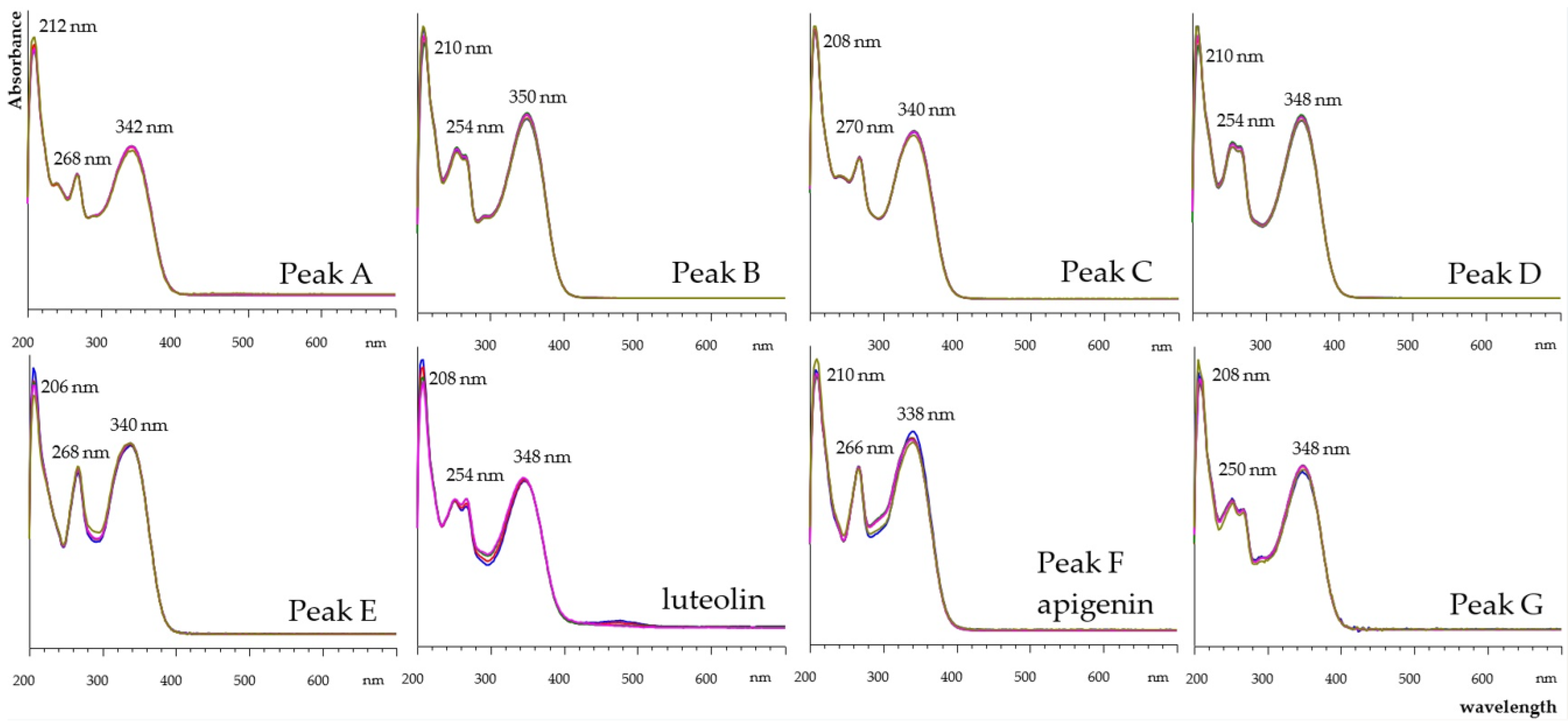
The chromatogram of the Reseda luteola extract (Figure 2b) was more complex than that of the peanut shells. Luteolin aglycone was identified by the same means as that for peanut shells. Beside luteolin aglycone, a second major peak and several minor peaks were detected. Their UV-Vis spectra produced the characteristic pattern of flavonoids belonging to the flavone subgroup. Peaks B, D, and G presented luteolin-type spectra, while peaks A, C, E, and F presented apigenin-type spectra. Spiking with apigenin standard confirmed the identity of peak F, which retained special characteristics similar to those of apigenin. UV-Vis spectra of all peaks are displayed in Figure 3.
These chromatographic findings are in agreement with previous reports. Samples of Reseda were extracted with mixtures of methanol/water and analyzed [21,22]. The chromatographic profiles that they produced were very similar to the one in Figure 2b. There is agreement that the second major peak of their extracts (peak B in the current research) corresponds to luteolin 7-O-glycoside. The UV-Vis spectrum of their identified luteolin 7-O-glycoside, which exhibited the same maxima and pattern as Peak B (Figure 3) [21]. The minor compounds are identified as luteolin and apigenin glycosides [22].
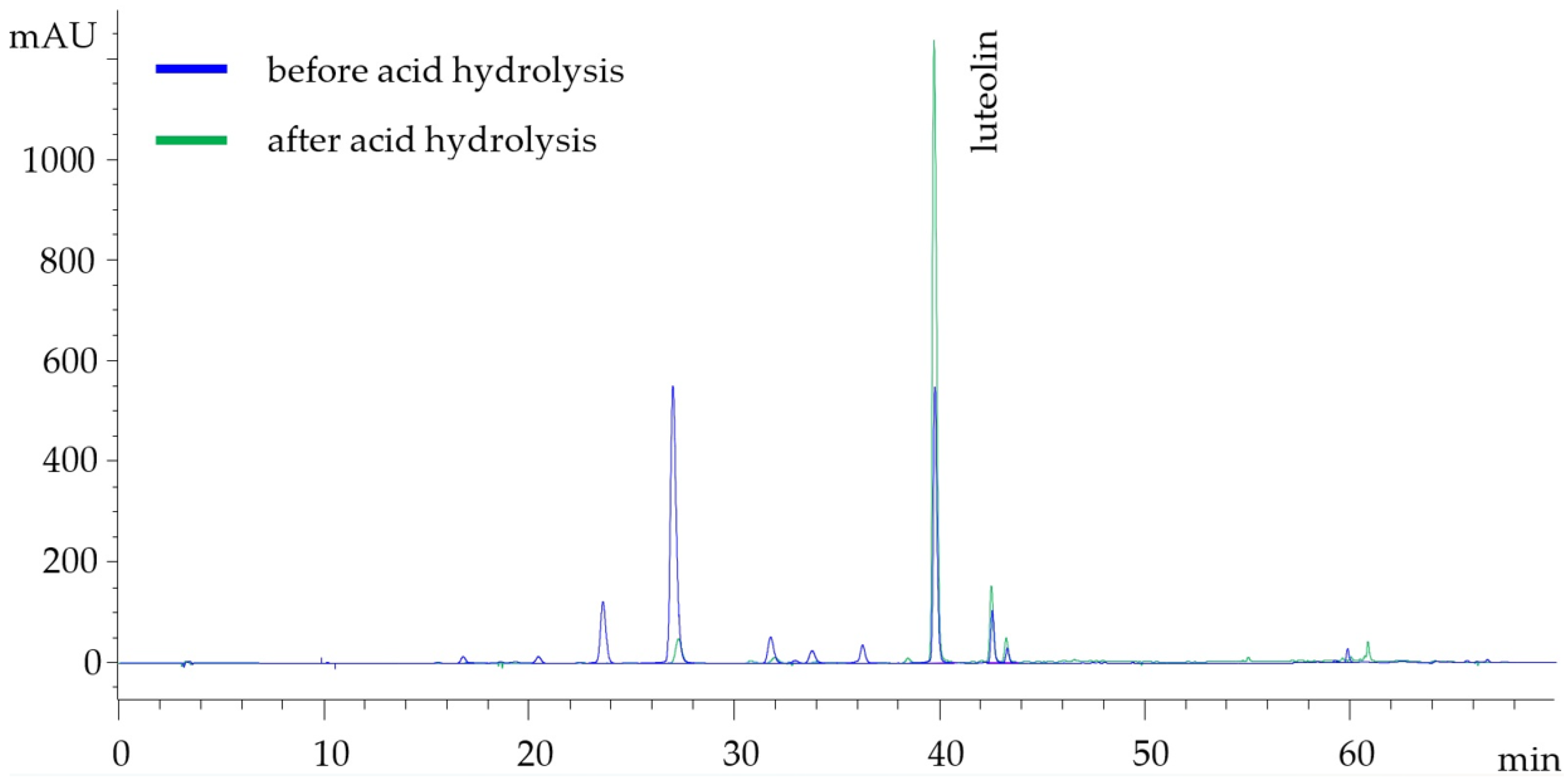
In order to verify the structure of the aglycone part of the glycosides, the extract was subjected to acid hydrolysis and then was re-analyzed by HPLC-DAD. The two comparative chromatograms are displayed in Figure 4. After acid hydrolysis, Peaks A–E could not be detected, except a trace of Peak B. Luteolin aglycone peak increased by almost 160%. Apigenin peak also increased, by 85%; however, it remained a minor peak. The unidentified Peak G did not change, which implies that it corresponds to a flavone aglycone. However, the compound was in trace quantities.
The results from the HPLC analysis of both methanolic extracts, as shown in Table 3, demonstrated a total flavonoid content of 18 mg LutE/g material and a total luteolin aglycone and glycoside content (LAG) of 14 mg LutE/g material for Reseda luteola, confirming the literature data of a high luteolin content in the aerial plant parts of the herb [21,22,23]. A 57% of the contained luteolin was found as the 7-O-glycoside (8.0 ± 0.3 mg LutE/g material), followed by the pure aglycone, representing 35% of the total content of the compound (4.9 ± 0.4 mg LutE/g material). These findings are in agreement with the study of Moiteiro et al. [22], which reported the 7-O-glucoside of luteolin as the main form of the compound in the aerial parts of the plant, accounting for over 50% of the total material, followed by the aglycone. In contrast, Cristea et al. reported the aglycone as the predominant form of luteolin in the examined plant parts, with a yield of 4.5 mg/g material compared to 3.5 mg/g material of the 7-O-glucoside [21]. The results were further verified by the results of acid hydrolysis, which demonstrated a 35–95% hydrolysis yield for the glycosides (Peaks A–E), while luteolin aglycone represented 85% of the total flavonoid content after hydrolysis (12 mg LutE/g material), as demonstrated in Figure 4 and Figure 5.
On the other hand, the peanut hull methanolic extracts demonstrated a relatively low luteolin content (1.5 ± 0.5 mg LutE/g material), alongside a low selectivity for the extraction process. These findings are also in alignment with those of Yen et al. (1995) and Zhang et al. (2023), which demonstrated a low luteolin content for industrial use (1–4 mg LUT/g material) [33,39].
These results show that Reseda luteola is a more adequate source of luteolin compared to peanut shells, confirming the respective literature findings [21,22,23]. The selectivity of the extraction for luteolin recovery—calculated as the concentration of luteolin aglycone and glycosides (LAGs) in the dried extract—was also high, reaching 70 mg LutE/g of dry final extract, i.e., 7% of the mass of total solids in the final product consisted of luteolin and its glycosides. The content in the Reseda plant of the compound far exceeded (by eight times) that in the peanut shells, verifying the potential of the plant as a luteolin extraction source replacing Arachis hypogea byproducts, as shown in Figure 5. It is also a better source of phenols and antioxidants, as demonstrated by its aforementioned superior TPC content and antiradical capacity (four and seven times larger than the respective values for the peanut shells), which are also presented in Figure 5. Thus, the Reseda luteola plant was confirmed to be a remarkable raw material source for luteolin extraction and warrants further studying, especially in terms of the kinetics governing the extraction process.

3.2. Kinetic Study of Reseda luteola Aerial Parts Extraction

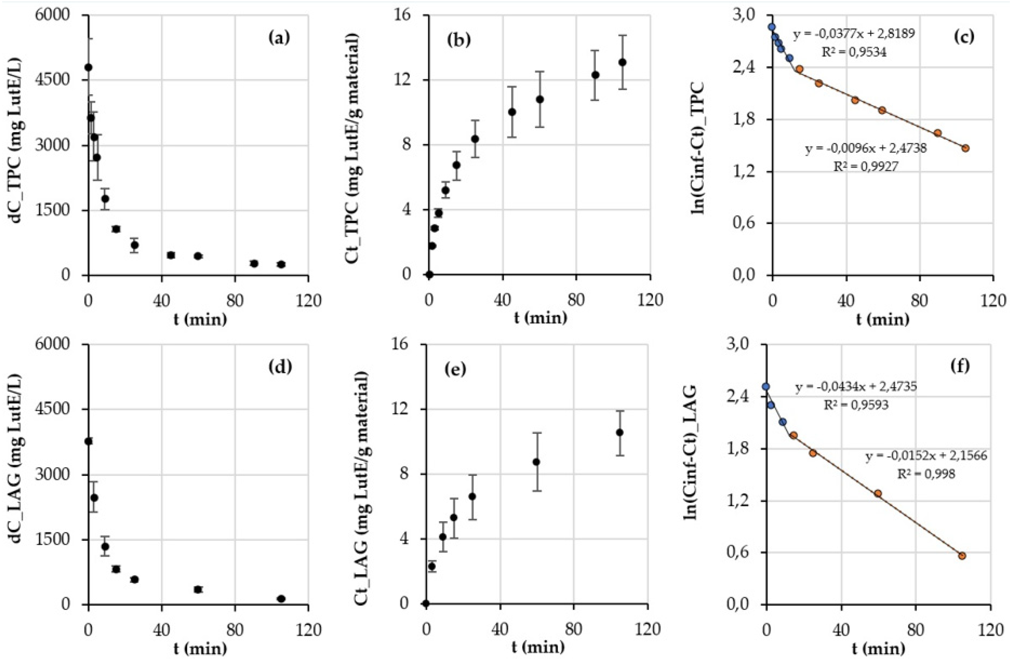
The kinetic experiment for the extraction of Reseda luteola aerial parts was conducted over a 0–105 min period, with a 2.8 mL/min flow, and finally produced 294 mL of methanolic extract. During this time, it was estimated that most of the luteolin content was retrieved. Based on Equation (1) for the semi-batch methanolic extraction of Reseda luteola at room temperature (25 °C), the results of TPC versus time are presented in Figure 6a,b. The rates of the two distinct stages of extraction (washing and diffusion stages) were calculated from the respective slopes of the kinetic equations determined by the second Fick’s law (Figure 6c). The results of the same extraction for luteolin aglycone and glycoside content (LAG), obtained by HPLC analysis, are displayed in Figure 6d–f.
The rates of the washing and diffusion stages of phenolic extraction displayed in Figure 6c were 0.0377 min−1 and 0.0096 min−1, respectively. Therefore, the rate of the washing stage was determined to be 3.9 times higher than that of the diffusion stage. The respective rates according to the HPLC analyses were determined to be 0.0434 min−1 and 0.0152 min−1 (Figure 6f). The ratio of the rates of the two stages (washing/diffusion) amounted to 2.9, quite lower than the ratio determined by the Folin measurements. This disagreement might be attributed to the fact that both luteolin and non-luteolin phenolics responded to the Folin reagent, while in the HPLC analyses, attention was focused exclusively on luteolin and its derivatives.
As far as the leaching time was concerned, the two methods produced similar values, i.e., 12 min according to the Folin method and 11 min according to HPLC. There was also agreement concerning the yields of the washing stage. At the end of the washing stage, the yield of TPC was 39%, and the respective yield for flavonoids was 41%, while the solid-to-solvent ratio of the extraction was approximately 1:2 g of material/mL of solvent. The yields of TPC and flavonoids at the end of the diffusion stage at 105 min were 72% and 87%, respectively, while the m/V ratio rose to approximately 1:15 g of material/mL of solvent.
Because of this low ratio between the rates of extraction, our results suggest that an industrially sustainable yield cannot be achieved merely by the washing stage, as is the case for the extraction of other bioactive compounds [40,41,42]. The relatively low yield during the washing stage may be attributed to the intracellular localization of luteolin and its glycosides. This localization likely slows their diffusion through plant cell wall structures [43], making the diffusion stage necessary to achieve higher yields. As shown in Figure 6e, a sustainable yield of extraction could be achieved at 60 min, at which time 70% of luteolin (or 9.6 mg LutE/g material) was obtained. At that same time, the total yield for TPC extraction amounted to 62%, as displayed in Figure 6b. The solid-to-solvent ratio was 1:9. Prolonging the extraction time to 60 min resulted in an improved solvent efficiency compared to that observed with other conventional luteolin extraction techniques applied to plant materials. Methods such as Soxhlet, maceration, and hot reflux extraction necessitate solid-to-liquid ratios as high as 1:30, according to previous studies [27,32,33,44].
The overall kinetics of the extraction process demonstrated the need to attain the diffusion stage of extraction in order to obtain the majority of the compound contained into the matrix. This affects the solvent requirements, increasing both time and solvent needed for achieving industrially sustainable yields. This obstacle could be surpassed by utilizing assisting extraction methods, such as Ultrasound-Assisted Extraction (UAE) [45,46,47,48], or Microwave-Assisted Extraction (MAE) [42]. A further investigation of other, less toxic solvents for extraction and isolation of the compound, such as ethanol or hydroalcoholic mixtures, should be investigated to evaluate their extraction efficiency [45,46,47,48,49,50].

4. Conclusions

This study demonstrated that Reseda luteola is a superior source of luteolin, containing 14 ± 3 mg LutE/g material—approximately eight times higher than the amount in peanut shells, a current industrial source. Kinetic studies of R. luteola extraction indicated that a 60 min semi-batch process can achieve an industrially sustainable yield, recovering 70% of luteolin (9.6 mg LutE/g material). At this stage, the process operated at a solid-to-solvent ratio of 1:9 (g/mL), reflecting a considerably improved solvent efficiency than that of conventional methods such as Soxhlet or maceration, which necessitate higher solid-to-solvent ratios for efficient luteolin extraction. The initial washing stage allowed for limited luteolin recovery with very low solvent use; however, most luteolin was extracted during the subsequent diffusion stage, which required higher solvent volumes. Overall, these results position Reseda luteola as a promising candidate for sustainable industrial luteolin production, and future studies should explore extraction-assisted techniques to enhance its diffusion while maintaining a low solvent consumption.

Author Contributions

Conceptualization, D.T. and P.T.; methodology, D.T. formal analysis, D.T. and E.E.; investigation, D.T. and E.E.; resources, P.T. and M.G.; data curation, D.T. and E.E.; writing—original draft preparation, E.E. and D.T.; writing—review and editing, D.T., M.G. and P.T.; supervision, P.T. and M.G.; project administration, D.T., P.T. and M.G. All authors have read and agreed to the published version of the manuscript.

Funding

This research received no external funding.

Data Availability Statement

The raw data supporting the conclusions of this article will be made available by the authors on request.

Conflicts of Interest

The authors declare no conflicts of interest.

References

  1. Çetinkaya, M.; Baran, Y. Therapeutic Potential of Luteolin on Cancer. Vaccines 2023, 11, 554. [Google Scholar] [CrossRef]
  2. Lien, E.J.; Ren, S.; Bui, H.-H.; Wang, R. Quantitative Structure-Activity Relationship Analysis of Phenolic Antioxidants. Free Radic. Biol. Med. 1999, 26, 285–294. [Google Scholar] [CrossRef]
  3. Tian, C.; Liu, X.; Chang, Y.; Wang, R.; Lv, T.; Cui, C.; Liu, M. Investigation of the Anti-Inflammatory and Antioxidant Activities of Luteolin, Kaempferol, Apigenin and Quercetin. S. Afr. J. Bot. 2021, 137, 257–264. [Google Scholar] [CrossRef]
  4. Li, K.; Wang, Y.; Liu, W.; Zhang, C.; Xi, Y.; Zhou, Y.; Li, H.; Liu, X. Structure–Activity Relationships and Changes in the Inhibition of Xanthine Oxidase by Polyphenols: A Review. Foods 2024, 13, 2365. [Google Scholar] [CrossRef]
  5. Seelinger, G.; Merfort, I.; Wölfle, U.; Schempp, C.M. Anti-Carcinogenic Effects of the Flavonoid Luteolin. Molecules 2008, 13, 2628–2651. [Google Scholar] [CrossRef] [PubMed]
  6. Tuorkey, M.J. Molecular Targets of Luteolin in Cancer. Eur. J. Cancer Prev. 2016, 25, 65–76. [Google Scholar] [CrossRef] [PubMed]
  7. Lopez-Lazaro, M. Distribution and Biological Activities of the Flavonoid Luteolin. Mini Rev. Med. Chem. 2009, 9, 31–59. [Google Scholar] [CrossRef]
  8. Liu, A.-L.; Liu, B.; Qin, H.-L.; Lee, S.; Wang, Y.-T.; Du, G.-H. Anti-Influenza Virus Activities of Flavonoids from the Medicinal Plant Elsholtzia rugulosa. Planta Med. 2008, 74, 847–851. [Google Scholar] [CrossRef]
  9. Xie, Y.-Z.; Peng, C.-W.; Su, Z.-Q.; Huang, H.-T.; Liu, X.-H.; Zhan, S.-F.; Huang, X.-F. A Practical Strategy for Exploring the Pharmacological Mechanism of Luteolin Against COVID-19/Asthma Comorbidity: Findings of System Pharmacology and Bioinformatics Analysis. Front. Immunol. 2022, 12, 769011. [Google Scholar] [CrossRef]
  10. Liao, P.H.; Hung, L.M.; Chen, Y.H.; Kuan, Y.H.; Zhang, F.B.; Lin, R.H.; Shih, H.C.; Tsai, S.K.; Huang, S.S. Cardioprotective Effects of Luteolin during Ischemia-Reperfusion Injury in Rats. Circ. J. 2011, 75, 443–450. [Google Scholar] [CrossRef]
  11. Manzoor, M.F.; Ahmad, N. Food-Based Phytochemical Luteolin, Their Derivatives, Sources, and Medicinal Benefits. Int. J. Agric. Life Sci. 2017, 3, 195–207. [Google Scholar] [CrossRef]
  12. Theoharides, T.C.; Cholevas, C.; Polyzoidis, K.; Politis, A. Long-COVID Syndrome-Associated Brain Fog and Chemofog: Luteolin to the Rescue. BioFactors 2021, 47, 232–241. [Google Scholar] [CrossRef] [PubMed]
  13. Bangar, P.S.; Kajla, P.; Chaudhary, V.; Sharma, N.; Ozogul, F. Luteolin: A Flavone with Myriads of Bioactivities and Food Applications. Food Biosci. 2023, 52, 102366. [Google Scholar] [CrossRef]
  14. Wang, Z.; Zeng, M.; Wang, Z.; Qin, F.; Chen, J.; He, Z. Dietary luteolin: A narrative review of Focusing on Its Pharmakokinetic Properies and Effects on Glycolipid Metabolism. J. Agric. Food Chem. 2021, 69, 1441–1454. [Google Scholar] [CrossRef] [PubMed]
  15. Miyashita, A.; Ito, J.; Parida, I.S.; Syoji, N.; Fujii, T.; Takahashi, H.; Nakagawa, K. Improving water dispersibility and bioavailability of luteolin using microemulsion system. Sci. Rep. 2022, 12, 11949. [Google Scholar] [CrossRef]
  16. Liu, Y.; Wang, L.; Zhao, Y.; He, M.; Feng, N.P. Nanostructured lipid carriers versus microemulsions for delivery of the poorly water-soluble drug luteolin. Int. J. Pharm. 2014, 476, 169–177. [Google Scholar] [CrossRef]
  17. Mod Razif, M.R.F.; Chan, S.Y.; Chew, Y.-L.; Hassan, M.; Ahmad Hisham, S.; Abdul Rahman, S.; Mai, C.-W.; Teo, M.Y.M.; Kee, P.E.; Khoo, K.S.; et al. Recent Developments in Luteolin-Loaded Nanoformulations for Enhanced Anti-Carcinogenic Activities: Insights from In Vitro and In Vivo Studies. Sci 2024, 6, 68. [Google Scholar] [CrossRef]
  18. Liu, Y.; Cheng, S.; Li, L.; Liu, Y.; Wang, D.; Liu, G. Enhancement of Oral Bioavailability and Anti-Colitis Effect of Luteolin-Loaded Polymer Micelles with RA-SS-mPEG as Carrier. Drug Dev. Ind. Pharm. 2023, 49, 17–29. [Google Scholar] [CrossRef]
  19. Haytowitz, D.B.; Wu, X.; Bhagwat, S. USDA Database for the Flavonoid Content of Selected Foods, Release 3.3; U.S. Department of Agriculture, Agricultural Research Service: Beltsville, MD, USA, 2018. Available online: http://www.ars.usda.gov/nutrientdata/flav (accessed on 26 June 2025).
  20. Abdallah, M.S.; de Wit, H.C.D. The Resedaceae: A Taxonomical Revision of the Family (Final Instalment); Mededelingen Landbouwhogeschool Wageningen: Wageningen, The Netherlands, 1978; pp. 259–262. Available online: https://edepot.wur.nl/287583 (accessed on 22 July 2025).
  21. Cristea, D. Identification and Quantitative HPLC Analysis of the Main Flavonoids Present in Weld (Reseda luteola L.). Dye. Pigm. 2003, 57, 267–272. [Google Scholar] [CrossRef]
  22. Moiteiro, C.; Gaspar, H.; Rodrigues, A.I.; Lopes, J.F.; Carnide, V. HPLC Quantification of Dye Flavonoids in Reseda luteola L. from Portugal. J. Sep. Sci. 2008, 31, 3683–3687. [Google Scholar] [CrossRef]
  23. Angelini, L.G.; Bertoli, A.; Rolandelli, S.; Pistelli, L. Agronomic Potential of Reseda luteola L. as New Crop for Natural Dyes in Textiles Production. Ind. Crops Prod. 2003, 17, 199–207. [Google Scholar] [CrossRef]
  24. Dimopoulos, P.; Raus, T.; Bergmeier, E.; Constantinidis, T.; Iatrou, G.; Kokkini, S.; Strid, A.; Tzanoudakis, D. Vascular Plants of Greece: An Annotated Checklist; Botanic Garden and Botanical Museum Berlin-Dahlem & Hellenic Botanical Society: Berlin, Germany, 2013. [Google Scholar] [CrossRef]
  25. European Food Safety Authority (EFSA). Botanical Summary Report. European Food Safety Authority. Available online: https://www.efsa.europa.eu/en/microstrategy/botanical-summary-report (accessed on 29 July 2025).
  26. Wölfle, U.; Simon-Haarhaus, B.; Merfort, I.; Schempp, C.M. Reseda luteola L. extract displays antiproliferative and pro-apoptotic activities that are related to its major flavonoids. Phytother. Res. 2010, 24, 1033–1036. [Google Scholar] [CrossRef] [PubMed]
  27. Peng, M.; Chen, Z.; Deng, Q.; Zhu, S.; Wang, G. The Roles of Luteolin in Peanut Shell Extract-Mediated Protection of Erythrocytes against Hypoxanthine-Xanthine Oxidase-Induced Toxicity. Food Biosci. 2021, 39, 100826. [Google Scholar] [CrossRef]
  28. Daigle, D.J.; Conkerton, E.J.; Sanders, T.H.; Mixon, A.C. Peanut Hull Flavonoids: Their Relationship with Peanut Maturity. J. Agric. Food Chem. 1988, 36, 1179–1181. [Google Scholar] [CrossRef]
  29. Kotzamanidis, S.T.; Stavropoulos, N.; Ipsilandis, C.G. Classification and Evaluation of Greek Groundnut (Arachis hypogaea L.) Using 17 Main Agronomic and Quality Traits. Pak. J. Biol. Sci. 2006, 9, 1021–1027. [Google Scholar] [CrossRef]
  30. Meirinhos, J.; Silva, B.M.; Valentão, P.; Seabra, R.M.; Pereira, J.A.; Dias, A.; Andrade, P.B.; Ferreres, F. Analysis and quantification of flavonoidic compounds from Portuguese olive (Olea europaea L.) leaf cultivars. Nat. Prod. Res. 2005, 19, 189–195. [Google Scholar] [CrossRef]
  31. Jiménez-Moreno, N.; Cimminelli, M.J.; Volpe, F.; Ansó, R.; Esparza, I.; Mármol, I.; Rodríguez-Yoldi, M.J.; Ancín-Azpilicueta, C. Phenolic Composition of Artichoke Waste and Its Antioxidant Capacity on Differentiated Caco-2 Cells. Nutrients 2019, 11, 1723. [Google Scholar] [CrossRef]
  32. Burger, P.; Monchot, A.; Bagarri, O.; Chiffolleau, P.; Azoulay, S.; Fernandez, X.; Michel, T. Whitening Agents from Reseda luteola L. and Their Chemical Characterization Using Combination of CPC, UPLC-HRMS and NMR. Cosmetics 2017, 4, 51. [Google Scholar] [CrossRef]
  33. Zhang, S.; Niu, L.; Si, X.; Li, L.; Sheng, Z. Microwave-Assisted Extraction of Luteolin from Peanut Shells Using Natural Deep Eutectic Solvents and Its Molecular Mechanism. Ind. Crops Prod. 2025, 225, 120578. [Google Scholar] [CrossRef]
  34. Papageorgiou, C.; Dermesonlouoglou, E.; Tsimogiannis, D.; Taoukis, P. Enrichment of Bakery Products with Antioxidant and Dietary Fiber Ingredients Obtained from Spent Coffee Ground. Appl. Sci. 2024, 14, 6863. [Google Scholar] [CrossRef]
  35. Brand-Williams, W.; Cuvelier, M.E.; Berset, C. Use of a free radical method to evaluate antioxidant activity. LWT Food Sci. Technol. 1995, 28, 25–30. [Google Scholar] [CrossRef]
  36. Merken, H.M.; Beecher, G.R. Liquid chromatographic method for the separation and quantification of prominent flavonoid aglycones. J. Chromatogr. A 2000, 897, 177–184. [Google Scholar] [CrossRef] [PubMed]
  37. Kouri, G.; Tsimogiannis, D.; Bardouki, H.; Oreopoulou, V. Extraction and Analysis of Antioxidant Components from Origanum dictamnus. Innov. Food Sci. Emerg. Technol. 2007, 8, 155–162. [Google Scholar] [CrossRef]
  38. Imran, A.; Humiyion, M.; Arshad, M.U.; Saeed, F.; Arshad, M.S.; Afzaal, M.; Al Jbawi, E. Extraction, Amino Acid Estimation, and Characterization of Bioactive Constituents from Peanut Shell through Eco-Innovative Techniques for Food Application. Int. J. Food Prop. 2022, 25, 2055–2065. [Google Scholar] [CrossRef]
  39. Yen, G.-C.; Duh, P.-D. Antioxidant Activity of Methanolic Extracts of Peanut Hulls from Various Cultivars. J. Am. Oil Chem. Soc. 1995, 72, 1065–1067. [Google Scholar] [CrossRef]
  40. Oreopoulou, A.; Goussias, G.; Tsimogiannis, D.; Oreopoulou, V. Hydro-alcoholic extraction kinetics of phenolics from oregano: Optimization of the extraction parameters. Food Bioprod. Process. 2020, 123, 378–389. [Google Scholar] [CrossRef]
  41. Psarrou, I.; Oreopoulou, A.; Tsimogiannis, D.; Oreopoulou, V. Extraction Kinetics of Phenolic Antioxidants from the Hydro Distillation Residues of Rosemary and Effect of Pretreatment and Extraction Parameters. Molecules 2020, 25, 4520. [Google Scholar] [CrossRef]
  42. Oreopoulou, A.; Alexandraki, A.-C.; Tsimogiannis, D.; Oreopoulou, V. Semi-Batch Extraction of Phenolic Compounds from Rosmarinus officinalis: Kinetic Study and Dimensionless Modeling. J. Food Eng. 2024, 370, 111962. [Google Scholar] [CrossRef]
  43. Fu, Y.J.; Liu, W.; Zu, Y.G.; Tong, M.H.; Li, S.M.; Yan, M.M.; Efferth, T.; Luo, H. Enzyme Assisted Extraction of Luteolin and Apigenin from Pigeonpea (Cajanus cajan (L.) Millsp.) Leaves. Food Chem. 2008, 111, 508–512. [Google Scholar] [CrossRef]
  44. Manzoor, M.F.; Ahmad, N.; Ahmed, Z.; Siddique, R.; Zeng, X.; Rahaman, A.; Aadil, R.M.; Wahab, A. Novel extraction techniques and pharmaceutical activities of luteolin and its derivatives. J. Food Biochem. 2019, 43, e12974. [Google Scholar] [CrossRef]
  45. Abidin, L.; Mujeeb, M.; Mir, S.R.; Khan, S.A.; Ahmad, A. Comparative Assessment of Extraction Methods and Quantitative Estimation of Luteolin in the Leaves of Vitex negundo Linn. by HPLC. Asian Pac. J. Trop. Med. 2014, 7, S289–S293. [Google Scholar] [CrossRef]
  46. Lama-Muñoz, A.; Contreras, M.d.M.; Espínola, F.; Moya, M.; Romero, I.; Castro, E. Optimization of Oleuropein and Luteolin-7-O-Glucoside Extraction from Olive Leaves by Ultrasound-Assisted Technology. Energies 2019, 12, 2486. [Google Scholar] [CrossRef]
  47. Hussain, A.I.; Chatha, S.A.S.; Noor, S.; Arshad, M.; Rathore, H.A. Effect of Extraction Techniques and Solvent Systems on the Extraction of Antioxidant Components from Peanut (Arachis hypogaea L.) Hulls. Food Anal. Methods 2012, 5, 890–896. [Google Scholar] [CrossRef]
  48. Hou, M.; Hu, W.; Wang, A.; Xiu, Z.; Shi, Y.; Hao, K.; Sun, X.; Cao, D.; Lu, R.; Sun, J. Ultrasound-Assisted Extraction of Total Flavonoids from Pteris cretica L.: Process Optimization, HPLC Analysis, and Evaluation of Antioxidant Activity. Antioxidants 2019, 8, 425. [Google Scholar] [CrossRef]
  49. Wang, H.; Yang, L.; Zu, Y.; Zhao, X. Microwave Assisted Simultaneous Extraction of Luteolin and Apigenin from Tree Peony Pod and Evaluation of its Antioxidant Activity. Sci. World J. 2014. [Google Scholar] [CrossRef]
  50. Lama-Muñoz, A.; Contreras, M.d.M.; Espínola, F.; Moya, M.; de Torres, A.; Romero, I.; Castro, E. Extraction of Oleuropein and Luteolin-7-O-Glucoside from Olive Leaves: Optimization of Technique and Operating Conditions. Food Chem. 2019, 293, 161–168. [Google Scholar] [CrossRef]
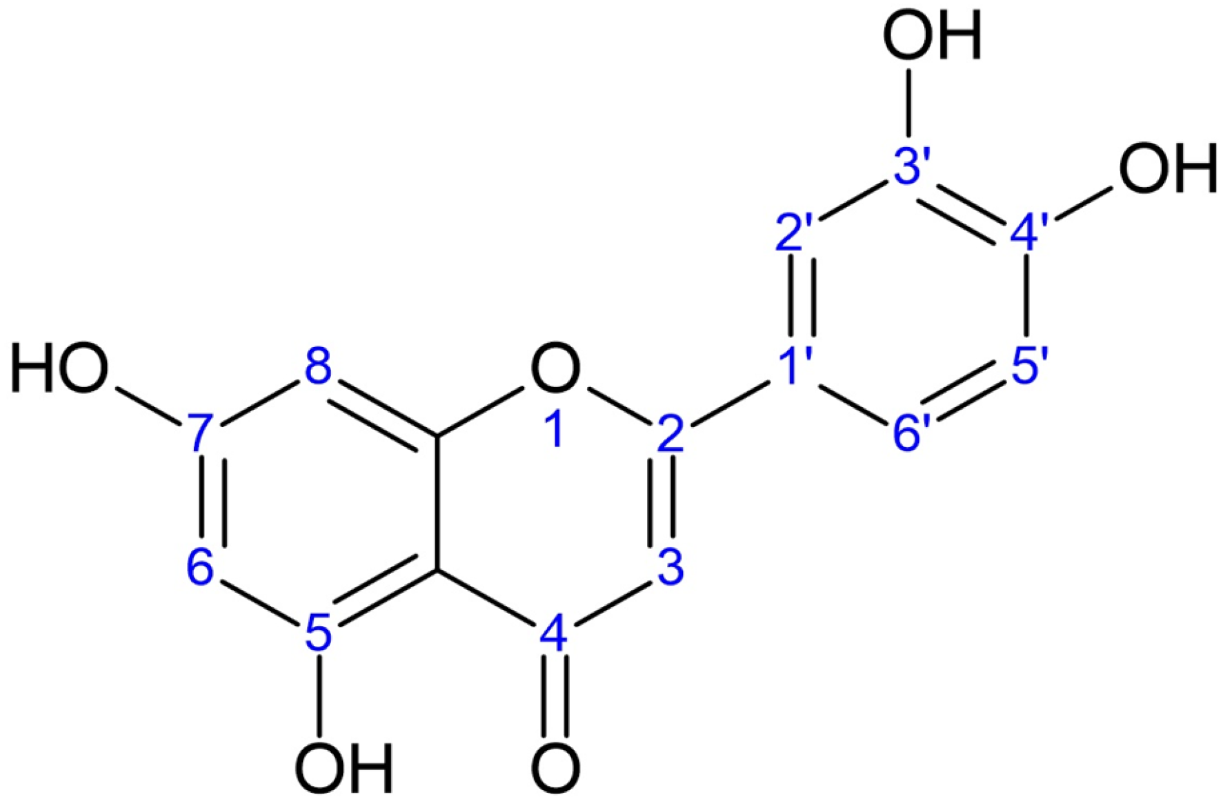
Figure 1. The molecular structure of luteolin aglycone.
Processes 13 03009 g001
Figure 2. The HPLC chromatograms for the methanolic extracts of peanut shells (a) and Reseda luteola (b), monitored at 360 nm.
Processes 13 03009 g002
Figure 3. UV–Vis spectra of the eight HPLC peaks (A–G) detected for the Reseda luteola extract (Figure 2b), illustrating the characteristic absorption profiles used for the characterization of the compounds. Peaks B, D, and G display luteolin-type spectra, while Peaks A, C, E, and F display apigenin-type spectra. The peak identities were confirmed by comparison with the spectra of luteolin and apigenin standards.
Processes 13 03009 g003
Figure 4. Chromatograms of the Reseda luteola extract before (blue line) and after acid hydrolysis (green line), monitored at 360 nm. After hydrolysis, Peaks A–E disappeared (except for a trace of Peak B), indicating that they corresponded to luteolin or apigenin glycosides. The luteolin (D) and apigenin (F) aglycone peaks increased, which confirmed their identity. Peak G remained unchanged, possibly corresponding to some trace flavone aglycone.
Processes 13 03009 g004
Figure 5. Total phenolic content (TPC), antiradical capacity (AC), and luteolin aglycone and glycoside content (LAG) (expressed as mg of luteolin equivalents (LutE)/g material) from Reseda luteola and groundnut shells.
Processes 13 03009 g005
Figure 6. (ac) Total phenolic content (TPC) recovery from Reseda luteola, at 25 °C. (a) TPC obtained from the outlet of the extractor expressed as mg luteolin equivalents (LutE)/L versus time; (b) TPC for total extract expressed as mg LutE/g material versus time; (c) the factor ln(C − Ct) versus the extraction time, approximation curves based on the diffusion model, according to Equation (1); (df) total luteolin aglycone and glycoside content (LAG) recovery from Reseda luteola aerial parts, at 25 °C, (d) LAG obtained from the outlet of the extractor expressed as mg LutE/L versus time; (e) LAG for total extract expressed as mg LutE/g material versus time; (f) the factor ln(C − Ct) versus the extraction time, approximation curves based on a diffusion model, according to Equation (1).
Processes 13 03009 g006
Table 1. Gradient elution program of the HPLC mobile phase.
Time
(min)
%A
(Water + 0.2% TFA)
%B
(Methanol + 0.2% TFA)
%C
(Acetonitrile + 0.2% TFA)
09064
58596
307117.411.6
6008515
Table 2. The basic parameters measured for the extracts of Reseda luteola aerial parts and peanut shells: total solid yield (TSY), total phenolic content (TPC), antiradical capacity (AC), and selectivity of each extraction in phenols and antioxidants. Different superscript letters indicate significant differences between the means as calculated by Analysis of Variance for a significance level of p = 0.05.
Parameters MeasuredReseda luteola
Methanolic Extract
(1:30 Solid-to-Liquid Ratio)
Peanut Hull
Methanolic Extract
(1:30 Solid-to-Liquid Ratio)
TSY
(mg solids/g material)210 a ± 18137 b ± 37
TPC
(mg GAE/g material)16 a ± 34.6 b ± 0.6
(mg LutE/g material)20 a ± 35.7 b ± 0.8
AC
(mg TE/g material)29 a ± 44.6 b ± 0.1
(mg LutE/g material)22 a ± 33.2 b ± 0.1
Selectivity of extraction (Folin)
(mg GAE/g dried extract)77 a ± 1236 b ± 11
(mg LutE/g dried extract)98 a ± 2144 b ± 14
Selectivity of extraction (DPPH)
(mg TE/g dried extract)148 a ± 3435 b ± 10
(mg LutE/g dried extract)114 a ± 2224 b ± 7
Table 3. Flavonoid content in the Reseda luteola aerial parts and peanut shells, as well as selectivity of each extraction for luteolin aglycone and glycoside (LAG) recovery, as determined by HPLC-DAD analyses. Different superscript letters indicate significant differences between the means as calculated by Analysis of Variance for a significance level of p = 0.05.
Parameters MeasuredReseda luteola
Methanolic Extract
(1:30 Solid-to-Liquid Ratio)
Peanut Shell
Methanolic Extract
(1:30 Solid-to-Liquid Ratio)
Total Flavonoid Content
(mg LutE/g material)
18 a ± 31.7 b ± 0.5
Total Luteolin Aglycone and Glycoside Content (LAG)
(mg LutE/g material)
14 a ± 31.5 b ± 0.5
Selectivity of LAG extraction (HPLC) (mg LutE/g dried extract)69 a ± 825 b ± 5
Disclaimer/Publisher’s Note: The statements, opinions and data contained in all publications are solely those of the individual author(s) and contributor(s) and not of MDPI and/or the editor(s). MDPI and/or the editor(s) disclaim responsibility for any injury to people or property resulting from any ideas, methods, instructions or products referred to in the content.