Article

Natural Antioxidant Enrichment of Goat Meat Pates with Portulaca oleracea and Honey Improves Oxidative Stability and Color Properties

Department of Technology of Food and Processing Industries, S. Seifullin Kazakh Agrotechnical Research University, Astana 010000, Kazakhstan
*
Author to whom correspondence should be addressed.
Processes 2025, 13(10), 3213; https://doi.org/10.3390/pr13103213
Submission received: 7 September 2025 / Revised: 2 October 2025 / Accepted: 7 October 2025 / Published: 9 October 2025

Abstract

Oxidative reactions accelerate quality loss in emulsified meats. This study evaluated a clean-label strategy in goat meat pates by co-fortifying Portulaca oleracea powder 1% and honey 4%. Control and treatment batches were cooked to 72 °C and stored as opened packs at ≤6 °C for 10 days. Oxidative stability of lipid and protein was monitored by peroxide value (PV), TBARS, acid value, and baseline protein carbonyls; total antioxidant capacity was assessed by FRAP and DPPH; color was quantified in CIE Lab; fatty acids were profiled by GC-FID; and protein integrity was examined by SDS-PAGE. The treatment modestly increased α-linolenic acid (ALA) (1.2% vs. 0.8%) in the control and markedly enhanced antioxidant status (FRAP 10.5 ± 0.04 mg GAE/g vs. not detected; DPPH 33.02 ± 0.009% vs. 22.33 ± 0.007%; IC50 106.10 ± 10.01 vs. 138.25 ± 11.15 µg/mL). Across storage, PV showed a small, non-significant delay on day 10 (13.0 ± 0.9 vs. 14.0 ± 0.9 meq/kg), while secondary and hydrolytic indices were consistently lower (TBARS day 10: 1.91 ± 0.13 vs. 3.29 ± 0.23 mg MDA/kg; acid value day 10: 7.0 ± 0.5 vs. 8.5 ± 0.6 mg KOH/g). Protein carbonyls at baseline were comparable (99.19 vs. 95.73 nmol/mg). L* and b* remained similar before and after light exposure, with a modest, non-significant reduction in color stability and greater a* loss in the treatment. These results show that purslane–honey co-fortification nutritionally enriches pates and attenuates oxidative spoilage during refrigerated storage, with minor color trade-offs that merit process optimization.

1. Introduction

Cardiometabolic and food-safety considerations have intensified scrutiny of processed meat systems, where lipid and protein oxidation remain primary drivers of quality loss, nutritional depreciation, and off-flavor development across storage and distribution. A state-of-the-art synthesis places lipid peroxidation at the center of discoloration, rancidity, and textural decline in meat matrices, underscoring the need for effective antioxidant strategies and robust markers such as TBARS, peroxide value, and protein carbonyls [1]. Concomitantly, meat intake patterns can exacerbate reactive oxygen species formation in the gastrointestinal tract, reinforcing the rationale for antioxidant-forward reformulation of meat products [2]. In this context, clean-label interventions using plant-derived antioxidants have shown broad efficacy in stabilizing fresh and processed meats while responding to consumer preferences and regulatory expectations [3,4,5].
Contemporary toolkits for meat-quality evaluation integrate instrumental colorimetry (CIE Lab*), chemometric assays of total antioxidant capacity (e.g., FRAP, DPPH), and digital/AI pipelines to enable objective, high-throughput, and reproducible quality control within QbD/PAT frameworks [6,7,8]. Beyond classical wet-chemistry assays, nondestructive sensors—hyperspectral imaging, near-infrared, and Raman spectroscopy—together with computer vision, are bridging the “lab-to-line” gap, delivering real-time monitoring of color, oxidative markers, and freshness, thereby enhancing translational relevance and industrial scalability [9,10,11]. These advances align with clean-label reformulation and the valorization of natural matrices, achieving synergistic stabilization of color and lipid phases [12,13].
Portulaca oleracea (purslane) represents a particularly promising functional plant for meat reformulation due to its high alpha-linolenic acid (ALA, ω-3) content, abundant phenolics/flavonoids, and robust agronomic adaptability under saline, drought, and heat stress—key attributes for sustainability and circular bioeconomy agendas [14,15]. Beyond phenolics and ω-3 ALA, purslane provides both soluble and insoluble dietary fiber and betalain pigments (betacyanins/betaxanthins) that enhance radical-scavenging and metal-chelating capacity and can reinforce interfacial stability in emulsified meats. The fiber fraction may improve water/oil binding and limit oxygen and pro-oxidant mobility, while betalains and phenolics can help protect polyunsaturated fatty acids against peroxidation [14,15,16,17,18,19,20]. Agronomically, P. oleracea is widely distributed, fast-growing, and tolerant to salinity, drought, and heat, enabling low-input cultivation and broad availability at low cost—attributes that support clean-label, scalable adoption [14].
Honey provides a complementary, clean-label matrix rich in phenolic compounds and enzymes, offering multi-modal antioxidant and antimicrobial activities that can modulate TBARS formation, color stability, and microbial ecology in meat emulsions [21,22,23]. Recent functional-food research demonstrates that judicious honey (or honey powder) inclusion can enhance quality attributes during refrigerated storage of poultry products, signaling potential for emulsified red-meat products as well [24]. Broader reviews on leveraging natural antioxidants for shelf-life extension in meat reinforce the strategic fit of honey–botanical combinations for oxidative stabilization and consumer-perceived naturalness [25].
Objective color remains a pivotal quality cue in pates; L*, a*, and b* metrics correlate with consumer acceptance and enable rigorous, reproducible evaluation of color drift during storage and light exposure [6,26]. Integrating instrumental color with TAC assays (FRAP, DPPH) and classical oxidation indices (peroxide value, TBARS) yields a holistic analytical framework suitable for hypothesis-driven testing of natural antioxidant interventions in emulsified goat meat systems [1,27]. In parallel, the goat meat literature indicates responsiveness of small-ruminant matrices to polyphenol-rich interventions, supporting the translational rationale for pate reformulation with plant- and bee-derived actives [28].
Lipid oxidation is a primary non-microbial driver of quality loss in meat systems, manifesting as flavor and aroma deterioration, discoloration, texture softening, and depletion of nutritional value, while accelerating shelf-life decline. Polyunsaturated fatty acids are particularly prone to peroxidation, and the extent and kinetics of oxidation depend on the degree of unsaturation as well as storage and processing conditions (temperature, light, oxygen, pro-oxidant pigments, and metals) [1,29]. Beyond sensory depreciation, secondary lipid-oxidation products such as malondialdehyde and 4-hydroxy-alkenals (e.g., 4-HNE) are biologically active and have been linked to mutagenic, genotoxic, and cytotoxic effects, with the capacity to form protein and DNA adducts [30,31]. In parallel, protein oxidation and carbonyl-amine reactions contribute to pigment instability and color drift through impacts on myoglobin chemistry and matrix functionality, further undermining consumer acceptance [32,33].
Within this context, emulsified goat meat pates represent a susceptible matrix due to comminution-induced exposure of lipids and heme iron; thus, stabilizing both the lipid phase and instrumental color (CIE Lab*) is critical [34]. Clean-label co-fortification with Portulaca oleracea (a source of phenolics and ω-3 ALA) and honey (phenolics, organic acids) is a promising strategy to curb oxidative cascades and preserve color, as suggested by recent applications of purslane in cooked sausages and honey in poultry products [20,24]. Accordingly, in the present study we pair bench-scale antioxidant assays (FRAP, DPPH) with in-matrix oxidation markers (peroxide value, TBARS, acid value) and carbonyls, alongside instrumental colorimetry, and we profile fatty acids to contextualize susceptibility to peroxidation in the control and purslane–honey pates. This integrative approach aligns with best-practice metrics for oxidation and color monitoring in muscle foods [35].
Herein, we posit that synergistic fortification of goat meat pates with Portulaca oleracea powder (ω-3/phenolic source) and honey (phenolics/enzymes; antimicrobial co-benefits) can significantly improve oxidative stability (lipid and protein), augment total antioxidant capacity, and enhance color resilience under refrigerated storage—without compromising technological functionality. This study, therefore, (i) characterizes antioxidant capacity via FRAP and DPPH, (ii) monitors lipid/protein oxidation kinetics (peroxide value, TBARS; carbonyls), (iii) quantifies instrumental color (CIE Lab*) and color stability, and (iv) explores compositional enrichment (fatty acids, minerals, vitamins) attributable to purslane–honey co-fortification, with attention to clean-label positioning and process scalability [1,6,27]. By coupling bench-scale analytics with best-practice metrics and state-of-the-art, non-destructive sensing, the present study advances an evidence-based, reproducible, and industry-relevant framework for clean-label, natural-antioxidant enrichment of goat meat pates, aligned with QbD/PAT principles and standardized protocols for color and oxidation monitoring [36].

2. Materials and Methods

2.1. Materials

Goat meat (leg and shoulder, 24–48 h postmortem) was sourced from a local supplier (Astana, Kazakhstan). Additional food-grade ingredients included onion, carrot, mustard, starch, curing mix, purslane (Portulaca oleracea L.) extract powder, blossom honey, and spices. Common purslane (Portulaca oleracea L.) dried plant material (powder) was purchased from a multivitamin retail store (Astana, Kazakhstan); according to the supplier, the crop was harvested in June 2025. Identity was confirmed at the species level (P. oleracea L.); an infraspecific variety was not assigned (varietal status: not determined). Two formulations were prepared: a control pate and a treatment pate with purslane and honey. The exact composition of each formulation is provided in Table 1. All percentages are expressed as weight per weight (% w/w) of the total batter.
Batches (n = 3 independent productions per formulation) were processed in the pilot plant of S. Seifullin Kazakh Agro-Technical Research University. The ingredients were mixed in a bench-top bowl cutter (Robot Coupe R2, France; S-blade), maintaining the batter temperature below 12 °C; the batter was then filled into heat-resistant containers, cooked to a core temperature of 72 °C, and rapidly cooled to ≤4 °C. Purslane extract was prepared according to a validated in-house protocol and milled to <250 µm particle size. Representative images of the finished goat meat pates are shown in Figure 1.

2.2. Determination of Fatty Acid Composition

The methyl esters of fatty acids were analyzed using an Agilent 7890 gas chromatograph (Agilent Technologies, Andover, MA, USA) equipped with a flame ionization detector and an HP-Innowax capillary column (60 m × 0.32 mm × 0.5 µm) under nitrogen flow. The temperature gradient was set from 100 to 260 °C at a rate of 10 °C/min. A 1 µL sample was injected, with a split ratio of 1:100, and the detector temperature was maintained between 250 and 300 °C. A Supelco No. 47885U standard mixture of fatty acid methyl esters (C6–C24) was used as a reference, with automatic data processing. Quantitative analysis of the fatty acids was conducted using the internal standard method. Results are expressed as the relative percentage of total identified fatty acids (% of total FA; g/100 g FA) calculated by area normalization; values < 0.1 are reported as <0.1%. Class totals (ΣSFA, ΣMUFA, ΣPUFA) were calculated as the sum of individual fatty acids within each class for each replicate and then averaged across replicates. Displayed ‘<0.1%’ values were included in class totals using their measured concentrations. Totals may differ from 100.0% by ≤0.2% due to rounding.

2.3. Determination of Color Characteristics

The samples’ color properties were analyzed with a Konica Minolta CM-2300d spectrophotometer (Konica Minolta Sensing, Inc., Sakai, Osaka, Japan), following the methodology described in the study by Iftikhar et al. [37], which evaluated antioxidant potential in minced beef meat. Before the measurements, the spectrophotometer underwent both zero and white calibration according to the manufacturer’s guidelines. The final result for each sample was derived as the arithmetic mean of the five measurements.
Y = ( 1 L 1 L 2 3 × L 1 + a 1 a 2 3 × a 1 + b 1 b 2 3 × b 1 ) × 100 % ,
where
L1 and L2 are the values of the light index before and after exposure to light.
a1 and a2—the values of the redness index before and after exposure to light.
b1 and b2—the values of the yellowness index before and after exposure to light.
When determining the color resistance to light, the sample was placed under an artificial light source (an incandescent or fluorescent lamp with a power of at least 40 watts). After 1 h after the start of the experiment, the change in color characteristics was instrumentally determined.

2.4. Analysis of Molecular Weight Distribution of Protein Fractions in Samples Carried Out Using One-Dimensional Electrophoresis

A 100 mg sample was mixed with 500 μL of a lysis buffer (4.5 M urea, 2.5% (v/v) β-mercaptoethanol, 1% (v/v) Triton X-100, and 1% (v/v) ampholytes, pH 3–10) and centrifuged at 14,000 rpm for 20 min. The supernatant was carefully separated, and an equal volume of protein sample buffer was added. The protein sample buffer was prepared by combining 1 mL of 10% (w/v) sodium dodecyl sulfate (SDS) stock (10 g/100 mL in deionized water), 250 μL of concentrated β-mercaptoethanol, 625 μL of 0.5 M Tris-HCl, 1.5 g of urea, and bromophenol blue; then, the total volume was adjusted to 5 mL with deionized water (final SDS concentration 2% w/v; β-mercaptoethanol 5% v/v). The mixtures were heated in a boiling water bath for 5 min. Protein visualization was achieved using Coomassie G-250 staining prepared as 10% (v/v) acetic acid, 25% (v/v) isopropanol, and 0.05% (w/v) Coomassie G-250; excess unbound dye was removed with 10% (v/v) acetic acid. One-dimensional electrophoresis gels in their wet state were utilized for densitometric analysis. High-resolution digital images were captured using a Bio-5000 Plus scanner (Serva, Heidelberg, Germany) at 600 ppi in 2D-RGB mode and subsequently refined for analysis. ImageJ version 1.53t was used (https://imagej.net/ij/, accessed on 20 August 2024).

2.5. Determination of Ferric-Reducing Antioxidant Power (FRAP) and Antioxidant Activity Using 2,2-Diphenyl-1-picrylhydrazyl (DPPH) Assay

The ferric-reducing antioxidant power (FRAP) assay was conducted using BHT and α-tocopherol as standard antioxidants. To perform this assay, 1 mL of the extract at varying dilutions was combined with 2.5 mL of phosphate buffer (0.1 M; pH 6.6) and 2.5 mL of potassium ferricyanide solution (1%, w/v). The mixture was incubated at 50 °C for 20 min. Following incubation, 2.5 mL of trichloroacetic acid solution (10%, w/v) was added. From this mixture, 2.5 mL was withdrawn and combined with 2.5 mL of deionized water and 0.5 mL of ferric chloride solution (0.1%, w/v). The solution was allowed to stand for 30 min before the absorbance was measured at 700 nm. The FRAP values were expressed as milligrams of gallic acid equivalents (GAEs) per gram of dry extract (mg GAE/g).
For the DPPH radical-scavenging activity assay analysis, 2 mL of a DPPH solution (0.1 mg/mL in methanol) was mixed with 2 mL of the sample solutions at a concentration of 200 μg/mL. All analyses were performed in triplicate. The reaction mixture was shaken and incubated in the dark at room temperature for 30 min. After incubation, the absorbance was measured at 517 nm against a blank. Ascorbic acid was prepared as a positive control in a similar manner, with the antioxidant solution being replaced accordingly.

2.6. Statistical Analyses

Data are reported as mean ± SD. Two-way ANOVA (factors: formulation and storage time) was applied; when appropriate, one-way ANOVA with Tukey’s post hoc test was used. Normality (Shapiro–Wilk) and homogeneity (Levene) were checked; data were transformed if assumptions were violated. Significance was set at p < 0.05. Analyses were performed in MATLAB R2023b (MathWorks, Natick, MA, USA). Effect sizes (η2 or Cohen’s d) were calculated where relevant.

3. Results and Discussion

3.1. Analysis of Fatty Acid Composition Results

Within this context, emulsified goat meat pates represent a susceptible matrix due to comminution-induced exposure of lipids and heme iron; thus, stabilizing both the lipid phase and instrumental color (CIE Lab*) is critical [34]. Clean-label co-fortification with Portulaca oleracea (a source of phenolics and ω-3 ALA) and honey (phenolics, organic acids) is a promising strategy to curb oxidative cascades and preserve color, as suggested by recent applications of purslane in cooked sausages and honey in poultry products [20,24]. Accordingly, in the present study we pair bench-scale antioxidant assays (FRAP and DPPH) with in-matrix oxidation markers (peroxide value, TBARS, and acid value) and carbonyls, alongside instrumental colorimetry, and we profile fatty acids to contextualize susceptibility to peroxidation in the control and purslane–honey pates. This integrative approach aligns with best-practice metrics for oxidation and color monitoring in muscle foods [35]. Detailed fatty acid compositions for both formulations are provided in Table 2.
Both formulations were dominated by saturated (SFA) and monounsaturated fatty acids (MUFA), with oleic acid C18:1 as the principal MUFA—an expected pattern for small ruminant meat emulsions. This aligns with prior characterizations of sheep/goat processed products showing balanced fat profiles and relatively stable oxidative behavior under proper processing [38]. Compared with the control, the purslane–honey pate showed a modest reduction in total SFA (~1.2 percentage points) and a slight rise in total MUFA (≈0.6 pp). Even small shifts in SFA and MUFA/PUFA can favorably influence health-related lipid indices, which synthesize the physiological implications of the FA pattern rather than single analytes. Although we did not compute these indices here, the direction of change suggests a potential improvement [39,40,41]. The most nutritionally meaningful difference was the 1.5-fold increase in α-linolenic acid ALA and C18:3 n−3 in the purslane–honey pate (1.2% vs. 0.8%). This is consistent with Portulaca oleracea being a strong plant source of n−3 fatty acids—particularly ALA—and with reports that incorporating purslane into comminuted meats elevates ALA in the finished product. From a dietary perspective, incremental ALA enrichment helps to rebalance the n−6/n−3 ratio (a recognized nutritional target) and may support downstream long-chain n−3 pools, even given limited conversion [15,42,43,44]. At the same time, total n−6 PUFA remained broadly comparable between treatments (≈4.6% vs. 4.7%), with linoleic acid as the main contributor; the experimental pate additionally showed minor amounts of arachidonic acid (C20:4 n−6). Lowering the dietary n−6/n−3 ratio toward ~1-5:1 is frequently cited as desirable, and meat systems with higher n−3 content often show improved n−6/n−3 profiles without large changes in total PUFA. While we did not calculate the ratios here, the observed ALA gain would be expected to modestly depress n−6/n−3 [45,46]. Notably, both formulations reported ≈3.8% of trans-C18:1 annotated as elaidic acid. In ruminant meats, the predominant natural trans-C18:1 is typically vaccenic acid (trans-11), and nomenclature in routine FAME outputs can generically label trans-C18:1 as elaidic. Thus, part of the reported elaidic fraction may actually reflect ruminant-derived trans isomers (vaccenic/CLA precursors) rather than industrial hydrogenation products—an analytical caveat with nutritional ramifications [47,48]. Beyond the trans-C18:1 issue, the experimental pate exhibited a richer spectrum of detectable fatty acids (22 vs. 16 >LOQ), including small increases in very-long-chain MUFA [49]. From a product-development lens, incrementally elevating ALA via purslane while keeping SFA slightly lower and MUFA stable is directionally favorable for “clean-label” meat pates. Similar reformulation strategies—whether via animal feeding or plant-based inclusions—have improved lipid quality metrics (PUFA/SFA, h/H ratio) and consumer-relevant attributes (color stability and oxidative resilience) in meat systems. Honey can also contribute phenolics that support oxidative stability and sensory quality during chilled storage, complementing the botanical matrix [24,45,50,51,52].
Additional functional contributions of purslane: In addition to phenolics, the comminuted plant solids likely delivered dietary fiber (soluble and insoluble) and betalains to the pate matrix. Fiber can increase viscosity and water/oil binding and create mild diffusion barriers that reduce oxygen ingress and the mobility of pro-oxidant species; fiber surfaces may also sequester metals. Betalains, together with phenolics, provide complementary radical-quenching and metal-chelating actions that help preserve labile polyunsaturated fatty acids, aligning with the lower late-storage TBARS and acid values observed here despite modest differences in PV. Although we did not quantify fiber or betalain content in the extract used, published characterizations of P. oleracea report appreciable levels of these classes; future work will determine their concentrations in our ingredient and delineate dose–response effects in emulsified goat meat systems [15,16,18,20]. From a techno-economic standpoint, the wide availability and low input requirements of purslane further strengthen its suitability for cost-effective, clean-label reformulation [14].
At the next stage, we evaluated the protective properties of Portulaca oleracea extract powder (1%) in combination with honey (4%) within the matrix of goat meat pates. To simulate a consumer storage scenario, opened packages were held at ≤6 °C for 10 days. Monitoring included lipid oxidation indices and protein oxidation markers (Table 3, Table 4 and Table 5).
Primary oxidation (peroxide value, PV): Across 0–10 days, PV rose in both pates, with numerically lower means in the purslane–honey treatment from day 4 onward, though pairwise differences were not significant (all p > 0.17). This pattern fits the canonical trajectory in meat systems—rapid hydroperoxide accrual followed by decomposition into secondary products—where small numeric PV delays can still translate into later benefits at the secondary-oxidation stage. The PV kinetics observed here are consistent with the widely described progression and dynamics of lipid oxidation in muscle foods [1]. Secondary oxidation (TBARS) diverged sharply after day 4: despite a small early uptick in the treatment at day 4 (0.140 ± 0.014 mg MDA/kg; p = 0.004), the purslane–honey pate thereafter showed substantially lower TBARS than the control at days 6–10 (all p ≤ 0.002), indicating effective suppression of malondialdehyde formation. At day 4, TBARS was higher in the purslane–honey pate despite similar PV up to day 4 and identical TBARS at days 0–2. Given the sugar- and phenolic-rich matrix, which can generate TBA-reactive chromogens under the spectrophotometric TBARS assay, we interpret this as a matrix/assay effect and/or a transient early MDA release; from day 6 onward TBARS is consistently lower in the treated samples. Methodologically, TBARS is a simple, reproducible proxy that tracks sensory rancidity; values around 2.0 mg MDA/kg are frequently cited as the onset threshold for perceivable rancidity in meat. By day 10, the control exceeded this benchmark (3.289 ± 0.230), whereas the treatment remained below it (1.910 ± 0.134), suggesting a practically meaningful mitigation under the “opened-pack, ≤6 °C” scenario [53,54]. Hydrolytic changes (acid value, AV) increased as storage progressed, reflecting lipolysis and hydrolytic rancidity. The treatment exhibited significantly lower AV at days 8 and 10 (p = 0.026 and 0.031), indicating slower accumulation of free fatty acids—a quality-relevant effect because elevated AV negatively influences fat quality and consumer acceptance [55]. The net improvement in secondary oxidation and AV likely reflects complementary roles of purslane and honey. Purslane contributes polyphenols and α-linolenic acid (ALA)—the latter enhancing the nutritional profile while phenolics counterbalance PUFA susceptibility—and honey supplies additional phenolics and redox-active constituents documented to enhance oxidative stability in meat matrices. Together with curing salts/spices, these phytochemicals can chelate pro-oxidant metals, scavenge radicals, and stabilize heme redox states, dampening the propagation phase that generates MDA. Our findings mirror controlled studies where purslane inclusion in meat products elevated ALA and improved oxidative stability, and where honey powder reduced TBARS and extended shelf life in meat products [15,24]. Phytochemicals from purslane (phenolic acids, flavonoids, minor tocopherols, and betalains) and honey (phenolics, organic acids, Maillard reaction reductants/reductones, and trace H2O2 generated by glucose oxidase) likely act synergistically to achieve the following: (i) quench chain radicals via single-electron transfer/hydrogen-atom transfer (SET/HAT) pathways; (ii) chelate Fe2+/Cu2+ and stabilize the heme-iron redox state; (iii) protect the fat–water interfacial region where lipid oxidation is initiated; and (iv) partially scavenge reactive aldehydes (MDA/4-HNE). These mechanisms are consistent with the observed increases in FRAP/DPPH and decreases in TBARS and acid value, with no evidence of protein degradation on SDS-PAGE. Role of onion as a co-ingredient. Both formulations contained onion at 10% (w/w), which is a recognized source of antioxidant flavonols (e.g., quercetin/kaempferol), organosulfur compounds (thiosulfinates/cysteine-sulfoxides), and vitamin C. These constituents can scavenge radicals, chelate pro-oxidant metals, and modulate heme redox chemistry, thereby contributing to baseline oxidative resistance of the pate matrix after cooking. Because onion was present at the same level in control and treatment, its effects are expected to be comparable across groups and thus cannot explain the lower late-storage TBARS and acid values observed in the purslane–honey formulation. Instead, these between-group improvements are attributed to the added phytochemicals from Portulaca oleracea and honey, acting in concert with the shared onion matrix.

3.2. Analysis of Color Characteristic Results

To assess color stability before and after light exposure, instrumental measurements were taken in the CIE L*a*b* space (L* = lightness, a* = redness, b* = yellowness) for both control and purslane–honey pates. The results are summarized in Table 6.
At baseline (“Before”), lightness (L*) did not differ between formulations (p = 0.988), but the purslane–honey pate was overall darker numerically (lower L*), a plausible outcome of plant solids and honey chromophores in the matrix. After light exposure, L* remained statistically comparable (p = 0.316), indicating that illumination induced similar reflectance shifts in both products. Such modest L* drift is typical for cooked, comminuted meats under short light stress [56]. For redness (a*), the formulations differed at both timepoints (p < 0.0001)—the purslane–honey pate started with slightly higher a* (7.48 vs. 7.38) but ended with a lower a* after light (6.07 vs. 6.53), implying a greater loss of redness under illumination. This pattern is consistent with myoglobin redox transitions (OxyMb → MetMb) catalyzed by light, oxygen, and endogenous pro-oxidants; small compositional changes can modulate these trajectories [57]. Plant phenolics can stabilize color by radical scavenging and metal chelation, yet matrix- and dose-dependent effects (including interactions with heme pigments and curing co-factors) can also shift chroma, explaining why improved oxidative metrics do not always translate to higher a* under light challenge [58]. Yellowness (b*) increased upon light exposure in both groups, with no between-formulation differences either before or after light (p = 0.772 and 0.996) [51,58]. The composite color stability index was numerically lower in the purslane–honey pate (88.84% vs. 93.17%), but not significant (p = 0.183). When viewed alongside oxidation data (Table 2, Table 3 and Table 4), this underscores a known nuance in meat systems—secondary lipid oxidation (TBARS) can be attenuated while light-exposed redness still declines, because chromatic outcomes depend on the coupled kinetics of lipid radicals and myoglobin chemistry, illumination geometry, and the microenvironment around heme. Aligning plant-based antioxidants with conventional color-protecting co-factors often yields the most robust gains in both oxidative stability and redness retention [34,38]. Despite a small penalty in a* under light stress, the purslane–honey formulation maintained comparable L* and b* and delivered clear late-storage advantages in TBARS and acid value. Optimizing the inclusion level/particle size of Portulaca oleracea, sweetener type, and co-antioxidants could help recover redness under light while preserving the demonstrated oxidative benefits [59].

3.3. Analysis of Molecular Weight Distribution of Protein Fractions in Samples Using One-Dimensional Electrophoresis

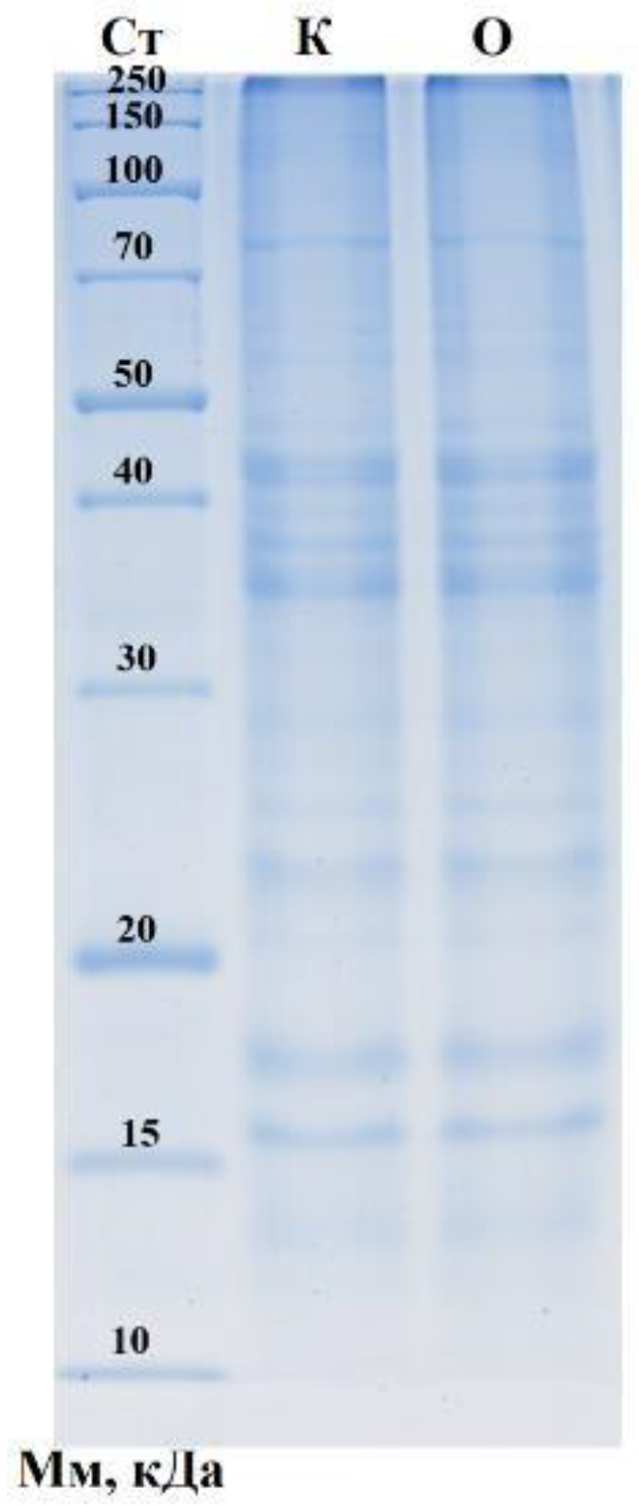
The SDS-PAGE profiles of the control (K) and purslane–honey (O) pates display the typical banding pattern of cooked, comminuted meat systems: a high-molecular-weight (HMW) band near the top consistent with myosin heavy chain (MHC, ~200 kDa), mid-range bands around ~40–45 kDa attributable to actin/troponin-T, and a cluster of ~14–22 kDa bands compatible with myosin light chains/troponin subunits (Figure 2). Overall band topology is comparable between K and O, indicating that reformulation with Portulaca oleracea (1%) and honey (4%) did not induce gross proteolysis or major loss of structural myofibrillar fractions under the extraction conditions applied [60]. Two qualitative cues merit mention. First, no pronounced diffuse smear is evident in the low-MW region, suggesting limited fragmentation of myofibrillar proteins during processing—consistent with our carbonyl data taken at baseline (no difference between groups). Second, there is no clear depletion of the putative MHC band in O relative to K; in oxidation-driven systems, MHC attenuation (and/or appearance of stacking/“gel top” material) is often observed due to covalent cross-linking (dityrosine, Schiff base, and disulfide) or aggregation that reduces gel entry. The absence of a marked MHC loss here implies that any oxidation occurring during cooking/emulsification was not severe enough to restructure the myofibrillar backbone detectably at the gel level—an inference aligned with the lower TBARS and acid-value trajectories previously shown for the purslane–honey pate [32].

3.4. Evaluation of Antioxidant Capacity: Ferric-Reducing Antioxidant Power (FRAP) and 2,2-Diphenyl-1-picrylhydrazyl (DPPH) Assay Results

The purslane–honey formulation markedly enhanced the antioxidant status of goat meat pates. FRAP was undetectable in the control but 10.5 ± 0.04 mg GAE/g in the treatment (p < 0.0001), indicating a substantial gain in electron-donating/reducing capacity attributable to phenolic- and flavonoid-rich botanicals (purslane) and honey constituents. In meat systems, FRAP is widely used as a single-electron transfer (SET) assay and often rises when plant phenolics are incorporated; similar FRAP elevations have been reported for reformulated sausages enriched with purslane or other botanicals [61]. FRAP and DPPH outcomes for both formulations are summarized in Table 7.
At a fixed test concentration, DPPH radical-scavenging increased from 22.33 ± 0.007% (control) to 33.02 ± 0.009% (treatment) (p < 0.0001), aligning with the well-documented capacity of plant extracts and bee products to quench stable free radicals in muscle foods. DPPH complements FRAP by capturing hydrogen atom transfer (HAT) and mixed mechanisms, so concurrent gains across both assays strengthen the case for genuine antioxidant enrichment rather than assay-specific artifacts [62,63].

4. Conclusions

Portulaca oleracea 1% combined with honey 4% is an effective clean-label strategy to enhance antioxidant status and oxidative stability in goat meat pates. The reformulated pate showed a 1.5-fold higher α-linolenic acid (1.2% vs. 0.8%) alongside a strong rise in total antioxidant capacity (FRAP 10.5 ± 0.04 mg GAE/g; control: not detected) and improved radical quenching (DPPH 33.02% vs. 22.33%; IC50 106.10 vs. 138.25 μg/mL). During refrigerated storage of opened packs, secondary lipid oxidation was consistently lower in the treatment group (TBARS day 10: 1.91 ± 0.13 vs. 3.29 ± 0.23 mg MDA/kg), and hydrolytic rancidity progressed more slowly (acid value day 10: 7.0 ± 0.5 vs. 8.5 ± 0.6 mg KOH/g); primary oxidation showed a modest numerical delay (PV day 10: 13.0 ± 0.9 vs. 14.0 ± 0.9 meq/kg). No evidence of increased baseline protein oxidation or gross proteolysis was observed; however, protein-oxidation kinetics during storage were not determined, while color (L*, b*) remained similar between formulations with a small, non-significant penalty in a* under light. Collectively, these outcomes demonstrate that purslane–honey co-fortification both nutritionally enriches pates and reduces oxidative spoilage, offering a practical natural alternative to synthetic additives for maintaining product quality and extending shelf life.

5. Patents

A Kazakhstan utility model patent related to the methods used in this study has been granted: Patent No.: 10414 (Utility Model). Title: Method of Meat Paste Production. Application No.: 2024/1548.2. Filing Date: 27 November 2024. Publication Date: 28 November 2025. Assignee: “S. Seifullin Kazakh Agro-Technical Research University” Non-profit JSC (KZ).

Author Contributions

Conceptualization, K.M. and T.T.; methodology, G.T.; validation, A.M., A.S., and A.K.; formal analysis, K.D.; investigation, N.B.; resources, S.T.; data curation, K.M.; writing—original draft preparation, K.M.; writing—review and editing, G.T.; visualization, K.D.; supervision, K.M.; project administration, T.T. All authors have read and agreed to the published version of the manuscript.

Funding

This research was funded by the Ministry of Science and Higher Education of the Republic of Kazakhstan, grant number BR21882184.

Data Availability Statement

The original contributions presented in this study are included in the article. Further inquiries can be directed to the corresponding author.

Conflicts of Interest

The authors declare no conflicts of interest.

References

  1. Domínguez, R.; Pateiro, M.; Gagaoua, M.; Barba, F.J.; Zhang, W.; Lorenzo, J.M.A. Comprehensive Review on Lipid Oxidation in Meat and Meat Products. Antioxidants 2019, 8, 429. [Google Scholar] [CrossRef] [PubMed]
  2. Macho-González, A.; Garcimartín, A.; López-Oliva, M.E.; Bastida, S.; Benedí, J.; Ros, G.; Nieto, G.; Sánchez-Muniz, F.J. Can Meat and Meat-Products Induce Oxidative Stress? Antioxidants 2020, 9, 638. [Google Scholar] [CrossRef] [PubMed]
  3. Manessis, G.; Kalogianni, A.I.; Lazou, T.; Moschovas, M.; Bossis, I.; Gelasakis, A.I. Plant-Derived Natural Antioxidants in Meat and Meat Products. Antioxidants 2020, 9, 1215. [Google Scholar] [CrossRef] [PubMed]
  4. Selani, M.M.; Herrero, A.M.; Ruiz-Capillas, C. Plant Antioxidants in Dry Fermented Meat Products with a Healthier Lipid Profile. Foods 2022, 11, 3558. [Google Scholar] [CrossRef]
  5. Velázquez, L.; Quiñones, J.; Díaz, R.; Pateiro, M.; Lorenzo, J.M.; Sepúlveda, N. Natural Antioxidants from Endemic Leaves in the Elaboration of Processed Meat Products: Current Status. Antioxidants 2021, 10, 1396. [Google Scholar] [CrossRef]
  6. Bumsted, J.; Ford, E.; Blair, A.; Underwood, K.; Zuelly, S.M.S. Instrumental Color Measurements Have Relationships to Fat Smearing in Fresh Sausage. Foods 2023, 12, 2813. [Google Scholar] [CrossRef]
  7. Echegaray, N.; Pateiro, M.; Munekata, P.E.S.; Lorenzo, J.M.; Chabani, Z.; Farag, M.A.; Domínguez, R. Measurement of Antioxidant Capacity of Meat and Meat Products: Methods and Applications. Molecules 2021, 26, 3880. [Google Scholar] [CrossRef]
  8. Liu, C.; Li, Y.; Sun, W.; Ma, F.; Wang, X.; Yang, Z. New Techniques of Meat Quality Assessment for Detecting Meat Texture. Processes 2025, 13, 640. [Google Scholar] [CrossRef]
  9. Huang, H.; Liu, L.; Ngadi, M. Recent Developments in Hyperspectral Imaging for Assessment of Food Quality and Safety. Sensors 2014, 14, 7248–7276. [Google Scholar] [CrossRef]
  10. Anagnostou, G.; Ferragina, A.; Crofton, E.C.; Frias Celayeta, J.M.; Hamill, R.M. The Development of Optical Sensing Techniques as Digital Tools to Predict the Sensory Quality of Red Meat: A Review. Appl. Sci. 2025, 15, 1719. [Google Scholar] [CrossRef]
  11. Li, W.; Wu, Y.; Du, L.; Shang, X.; Shi, J. Hyperspectral Imaging for Foreign Matter Detection in Foods: Advances, Challenges, and Future Directions. Foods 2025, 14, 3026. [Google Scholar] [CrossRef] [PubMed]
  12. Domínguez, R.; Gullón, P.; Pateiro, M.; Munekata, P.E.S.; Zhang, W.; Lorenzo, J.M. Tomato as Potential Source of Natural Additives for Meat Industry. A Review. Antioxidants 2020, 9, 73. [Google Scholar] [CrossRef] [PubMed]
  13. Munekata, P.E.S.; Pateiro, M.; Domínguez, R.; Pollonio, M.A.R.; Sepúlveda, N.; Andres, S.C.; Reyes, J.; Santos, E.M.; Lorenzo, J.M. Beta vulgaris as a Natural Nitrate Source for Meat Products: A Review. Foods 2021, 10, 2094. [Google Scholar] [CrossRef] [PubMed]
  14. Carrascosa, A.; Pascual, J.A.; Ros, M.; Petropoulos, S.A.; Alguacil, M.d.M. Agronomical Practices and Management for Commercial Cultivation of Portulaca oleracea as a Crop: A Review. Plants 2023, 12, 1246. [Google Scholar] [CrossRef]
  15. Petropoulos, S.A.; Fernandes, Â.; Dias, M.I.; Vasilakoglou, I.B.; Petrotos, K.; Barros, L.; Ferreira, I.C.F.R. Nutritional Value, Chemical Composition and Cytotoxic Properties of Common Purslane (Portulaca oleracea L.) in Relation to Harvesting Stage and Plant Part. Antioxidants 2019, 8, 293. [Google Scholar] [CrossRef]
  16. Gallo, M.; Conte, E.; Naviglio, D. Analysis and Comparison of the Antioxidant Component of Portulaca oleracea Leaves Obtained by Different Solid-Liquid Extraction Techniques. Antioxidants 2017, 6, 64. [Google Scholar] [CrossRef]
  17. Chrysargyris, A.; Xylia, P.; Zengin, G.; Tzortzakis, N. Purslane (Portulaca oleracea L.) Growth, Nutritional, and Antioxidant Status under Different Nitrogen Levels in Hydroponics. Horticulturae 2024, 10, 1007. [Google Scholar] [CrossRef]
  18. Fernández-Poyatos, M.d.P.; Llorent-Martínez, E.J.; Ruiz-Medina, A. Phytochemical Composition and Antioxidant Activity of Portulaca oleracea: Influence of the Steaming Cooking Process. Foods 2021, 10, 94. [Google Scholar] [CrossRef]
  19. Nkhumeleni, Z.; Phoswa, W.; Mokgalaboni, K. Purslane Ameliorates Inflammation and Oxidative Stress in Diabetes Mellitus: A Systematic Review. Int. J. Mol. Sci. 2024, 25, 12276. [Google Scholar] [CrossRef]
  20. Makangali, K.; Tultabayeva, T.; Zamaratskaia, G.; Ospankulova, G.; Tokysheva, G.; Abzhanova, S.; Zhakupova, G.; Ergalikyzy, A. Enhancing the Antioxidant Capacity and Oxidative Stability of Cooked Sausages Through Portulaca oleracea (Purslane) Supplementation: A Natural Alternative to Synthetic Additives. Appl. Sci. 2024, 14, 9986. [Google Scholar] [CrossRef]
  21. Becerril-Sánchez, A.L.; Quintero-Salazar, B.; Dublán-García, O.; Escalona-Buendía, H.B. Phenolic Compounds in Honey and Their Relationship with Antioxidant Activity, Botanical Origin, and Color. Antioxidants 2021, 10, 1700. [Google Scholar] [CrossRef]
  22. Tlak Gajger, I.; Dar, S.A.; Ahmed, M.M.M.; Aly, M.M.; Vlainić, J. Antioxidant Capacity and Therapeutic Applications of Honey: Health Benefits, Antimicrobial Activity and Food Processing Roles. Antioxidants 2025, 14, 959. [Google Scholar] [CrossRef] [PubMed]
  23. Ogwu, M.C.; Izah, S.C. Honey as a Natural Antimicrobial. Antibiotics 2025, 14, 255. [Google Scholar] [CrossRef] [PubMed]
  24. Bogusz, R.; Onopiuk, A.; Chmiel, M.; Piotrowska, A.; Kostyra, E.; Lipińska, E.; Bryś, J.; Samborska, K.; Pietrzak, D. The Effect of Honey Powder Addition on Chosen Quality Properties of Model Chicken Products. Foods 2024, 13, 4163. [Google Scholar] [CrossRef] [PubMed]
  25. Petcu, C.D.; Tăpăloagă, D.; Mihai, O.D.; Gheorghe-Irimia, R.-A.; Negoiță, C.; Georgescu, I.M.; Tăpăloagă, P.R.; Borda, C.; Ghimpețeanu, O.M. Harnessing Natural Antioxidants for Enhancing Food Shelf Life: Exploring Sources and Applications in the Food Industry. Foods 2023, 12, 3176. [Google Scholar] [CrossRef]
  26. Tkacz, K.; Modzelewska-Kapituła, M.; Więk, A.; Nogalski, Z. The Applicability of Total Color Difference ΔE for Determining the Blooming Time in Longissimus Lumborum and Semimembranosus Muscles from Holstein-Friesian Bulls at Different Ageing Times. Appl. Sci. 2020, 10, 8215. [Google Scholar] [CrossRef]
  27. Soulage, C.O.; Pelletier, C.C.; Florens, N.; Lemoine, S.; Dubourg, L.; Juillard, L.; Guebre-Egziabher, F. Two Toxic Lipid Aldehydes, 4-hydroxy-2-hexenal (4-HHE) and 4-hydroxy-2-nonenal (4-HNE), Accumulate in Patients with Chronic Kidney Disease. Toxins 2020, 12, 567. [Google Scholar] [CrossRef]
  28. Salzano, A.; Damiano, S.; D’Angelo, L.; Ballistreri, G.; Claps, S.; Rufrano, D.; Maggiolino, A.; Neglia, G.; De Palo, P.; Ciarcia, R. Productive Performance and Meat Characteristics of Kids Fed a Red Orange and Lemon Extract. Animals 2021, 11, 809. [Google Scholar] [CrossRef]
  29. Dragoev, S.G. Lipid Peroxidation in Muscle Foods: Impact on Quality, Safety and Human Health. Foods 2024, 13, 797. [Google Scholar] [CrossRef]
  30. Schaur, R.; Siems, W.; Bresgen, N.; Eckl, P. 4-Hydroxy-nonenal-A Bioactive Lipid Peroxidation Product. Biomolecules 2015, 5, 2247–2337. [Google Scholar] [CrossRef]
  31. Shabalala, S.C.; Johnson, R.; Basson, A.K.; Ziqubu, K.; Hlengwa, N.; Mthembu, S.X.H.; Mabhida, S.E.; Mazibuko-Mbeje, S.E.; Hanser, S.; Cirilli, I.; et al. Detrimental Effects of Lipid Peroxidation in Type 2 Diabetes: Exploring the Neutralizing Influence of Antioxidants. Antioxidants 2022, 11, 2071. [Google Scholar] [CrossRef]
  32. Domínguez, R.; Pateiro, M.; Munekata, P.E.S.; Zhang, W.; Garcia-Oliveira, P.; Carpena, M.; Prieto, M.A.; Bohrer, B.; Lorenzo, J.M. Protein Oxidation in Muscle Foods: A Comprehensive Review. Antioxidants 2021, 11, 60. [Google Scholar] [CrossRef] [PubMed]
  33. Poveda-Arteaga, A.; Krell, J.; Gibis, M.; Heinz, V.; Terjung, N.; Tomasevic, I. Intrinsic and Extrinsic Factors Affecting the Color of Fresh Beef Meat-Comprehensive Review. Appl. Sci. 2023, 13, 4382. [Google Scholar] [CrossRef]
  34. Silva, S.R.; Teixeira, A.; Guedes, C. Sheep and Goat Meat Processing and Quality. Foods 2023, 12, 2033. [Google Scholar] [CrossRef] [PubMed]
  35. Reitznerová, A.; Šuleková, M.; Nagy, J.; Marcinčák, S.; Semjon, B.; Čertík, M.; Klempová, T. Lipid Peroxidation Process in Meat and Meat Products: A Comparison Study of Malondialdehyde Determination between Modified 2-Thiobarbituric Acid Spectrophotometric Method and Reverse-Phase High-Performance Liquid Chromatography. Molecules 2017, 22, 1988. [Google Scholar] [CrossRef]
  36. Wei, X.; Lam, S.; Bohrer, B.M.; Uttaro, B.; López-Campos, O.; Prieto, N.; Larsen, I.L.; Juárez, M. A Comparison of Fresh Pork Colour Measurements by Using Four Commercial Handheld Devices. Foods 2021, 10, 2515. [Google Scholar] [CrossRef]
  37. Iftikhar, A.; Dupas-Farrugia, C.; De Leonardis, A.; Macciola, V.; Moiz, A.; Martin, D. Antioxidant potential of olive leaf (Olea europaea L.) sustainable extracts evaluated in vitro and minced beef meat. Ital. J. Food Sci. 2024, 36, 305–316. [Google Scholar] [CrossRef]
  38. Teixeira, A.; Silva, S.; Guedes, C.; Rodrigues, S. Sheep and Goat Meat Processed Products Quality: A Review. Foods 2020, 9, 960. [Google Scholar] [CrossRef]
  39. Chen, J.; Liu, H. Nutritional Indices for Assessing Fatty Acids: A Mini-Review. Int. J. Mol. Sci. 2020, 21, 5695. [Google Scholar] [CrossRef]
  40. Tilami, S.K.; Kouřimská, L. Assessment of the Nutritional Quality of Plant Lipids Using Atherogenicity and Thrombogenicity Indices. Nutrients 2022, 14, 3795. [Google Scholar] [CrossRef]
  41. Dal Bosco, A.; Cavallo, M.; Menchetti, L.; Angelucci, E.; Cartoni Mancinelli, A.; Vaudo, G.; Marconi, S.; Camilli, E.; Galli, F.; Castellini, C.; et al. The Healthy Fatty Index Allows for Deeper Insights into the Lipid Composition of Foods of Animal Origin When Compared with the Atherogenic and Thrombogenicity Indexes. Foods 2024, 13, 1568. [Google Scholar] [CrossRef] [PubMed]
  42. Al Juhaimi, F.; Atasoy, Z.B.; Uslu, N.; Özcan, M.M.; Mohamed Ahmed, I.A.; Walayat, N. The Effect of Roasting on Oil Content, Fatty Acids, Bioactive Compounds and Mineral Contents of Purslane (Portulaca oleracea L.) Seeds. Foods 2025, 14, 732. [Google Scholar] [CrossRef] [PubMed]
  43. Gonzalez-Becerra, K.; Barron-Cabrera, E.; Muñoz-Valle, J.F.; Torres-Castillo, N.; Rivera-Valdes, J.J.; Rodriguez-Echevarria, R.; Martinez-Lopez, E. A Balanced Dietary Ratio of n−6:n−3 Polyunsaturated Fatty Acids Exerts an Effect on Total Fatty Acid Profile in RBCs and Inflammatory Markers in Subjects with Obesity. Healthcare 2023, 11, 2333. [Google Scholar] [CrossRef] [PubMed]
  44. Mititelu, M.; Lupuliasa, D.; Neacșu, S.M.; Olteanu, G.; Busnatu, Ș.S.; Mihai, A.; Popovici, V.; Măru, N.; Boroghină, S.C.; Mihai, S.; et al. Polyunsaturated Fatty Acids and Human Health: A Key to Modern Nutritional Balance in Association with Polyphenolic Compounds from Food Sources. Foods 2024, 14, 46. [Google Scholar] [CrossRef]
  45. Minelli, G.; D’Ambra, K.; Macchioni, P.; Lo Fiego, D.P. Effects of Pig Dietary n−6/n−3 Polyunsaturated Fatty Acids Ratio and Gender on Carcass Traits, Fatty Acid Profiles, Nutritional Indices of Lipid Depots and Oxidative Stability of Meat in Medium-Heavy Pigs. Foods 2023, 12, 4106. [Google Scholar] [CrossRef]
  46. Terevinto, A.; Cabrera, M.C.; Zaccari, F.; Saadoun, A. The Oxidative and Color Stability of Beef from Steers Fed Pasture or Concentrate during Retail Display. Animals 2023, 13, 2972. [Google Scholar] [CrossRef]
  47. Sanjulian, L.; Lamas, A.; Barreiro, R.; Martínez, I.; García-Alonso, L.; Cepeda, A.; Fente, C.; Regal, P. Investigating the Dietary Impact on Trans-Vaccenic Acid (Trans-C18:1 n-7) and Other Beneficial Fatty Acids in Breast Milk and Infant Formulas. Foods 2024, 13, 2164. [Google Scholar] [CrossRef]
  48. Pipoyan, D.; Stepanyan, S.; Stepanyan, S.; Beglaryan, M.; Costantini, L.; Molinari, R.; Merendino, N. The Effect of Trans Fatty Acids on Human Health: Regulation and Consumption Patterns. Foods 2021, 10, 2452. [Google Scholar] [CrossRef]
  49. Agnew, M.P.; Craigie, C.R.; Weralupitiya, G.; Reis, M.M.; Johnson, P.L.; Reis, M.G. Comprehensive Evaluation of Parameters Affecting One-Step Method for Quantitative Analysis of Fatty Acids in Meat. Metabolites 2019, 9, 189. [Google Scholar] [CrossRef]
  50. Frunză, G.; Ciobanu, M.-M.; Murariu, O.C.; Radu-Rusu, R.-M.; Boișteanu, P.-C. The Fatty Acid Content, Health Lipid Indices, and Instrumental, Histological, and Sensory Quality of Hare Meat (Lepus europaeus Pallas). Foods 2025, 14, 310. [Google Scholar] [CrossRef]
  51. Martinello, M.; Mutinelli, F. Antioxidant Activity in Bee Products: A Review. Antioxidants 2021, 10, 71. [Google Scholar] [CrossRef]
  52. Zaldivar-Ortega, A.K.; Cenobio-Galindo Ade, J.; Morfin, N.; Aguirre-Álvarez, G.; Campos-Montiel, R.G.; Esturau-Escofet, N.; Garduño-García, A.; Angeles-Hernandez, J.C. The Physicochemical Parameters, Phenolic Content, and Antioxidant Activity of Honey from Stingless Bees and Apis mellifera: A Systematic Review and Meta-Analysis. Antioxidants 2024, 13, 1539. [Google Scholar] [CrossRef] [PubMed]
  53. Abeyrathne, E.D.N.S.; Nam, K.; Ahn, D.U. Analytical Methods for Lipid Oxidation and Antioxidant Capacity in Food Systems. Antioxidants 2021, 10, 1587. [Google Scholar] [CrossRef] [PubMed]
  54. Orădan, A.C.; Tocai, A.C.M.; Rosan, C.A.; Vicas, S.I. Fruit Extracts Incorporated into Meat Products as Natural Antioxidants, Preservatives, and Colorants. Processes 2024, 12, 2756. [Google Scholar] [CrossRef]
  55. Martins, A.A.; Andrade, S.; Correia, D.; Matos, E.; Caetano, N.S.; Mata, T.M. Valorization of Agro-Industrial Residues: Bioprocessing of Animal Fats to Reduce Their Acidity. Sustainability 2021, 13, 10837. [Google Scholar] [CrossRef]
  56. Gagaoua, M.; Bonnet, M.; Picard, B. Protein Array-Based Approach to Evaluate Biomarkers of Beef Tenderness and Marbling in Cows: Understanding of the Underlying Mechanisms and Prediction. Foods 2020, 9, 1180. [Google Scholar] [CrossRef]
  57. Suman, S.P.; Joseph, P. Myoglobin Chemistry and Meat Color. Annu. Rev. Food Sci. Technol. 2013, 4, 79–99. [Google Scholar] [CrossRef]
  58. Munekata, P.E.S.; Gullón, B.; Pateiro, M.; Tomasevic, I.; Domínguez, R.; Lorenzo, J.M. Natural Antioxidants from Seeds and Their Application in Meat Products. Antioxidants 2020, 9, 815. [Google Scholar] [CrossRef]
  59. Cierach, M.; Niedźwiedź, J. Effects of three lighting intensities during display on discolouration of beef semitendinosus muscle. Eur. Food Res. Technol. 2014, 239, 377–383. [Google Scholar] [CrossRef]
  60. Mazumder, M.A.R.; Sujintonniti, N.; Chaum, P.; Ketnawa, S.; Rawdkuen, S. Developments of Plant-Based Emulsion-Type Sausage by Using Grey Oyster Mushrooms and Chickpeas. Foods 2023, 12, 1564. [Google Scholar] [CrossRef]
  61. Munteanu, I.G.; Apetrei, C. Analytical Methods Used in Determining Antioxidant Activity: A Review. Int. J. Mol. Sci. 2021, 22, 3380. [Google Scholar] [CrossRef]
  62. Gulcin, İ.; Alwasel, S.H. DPPH Radical Scavenging Assay. Processes 2023, 11, 2248. [Google Scholar] [CrossRef]
  63. López-Pedrouso, M.; Lorenzo, J.M.; Franco, D. Advances in Natural Antioxidants for Food Improvement. Antioxidants 2022, 11, 1825. [Google Scholar] [CrossRef]
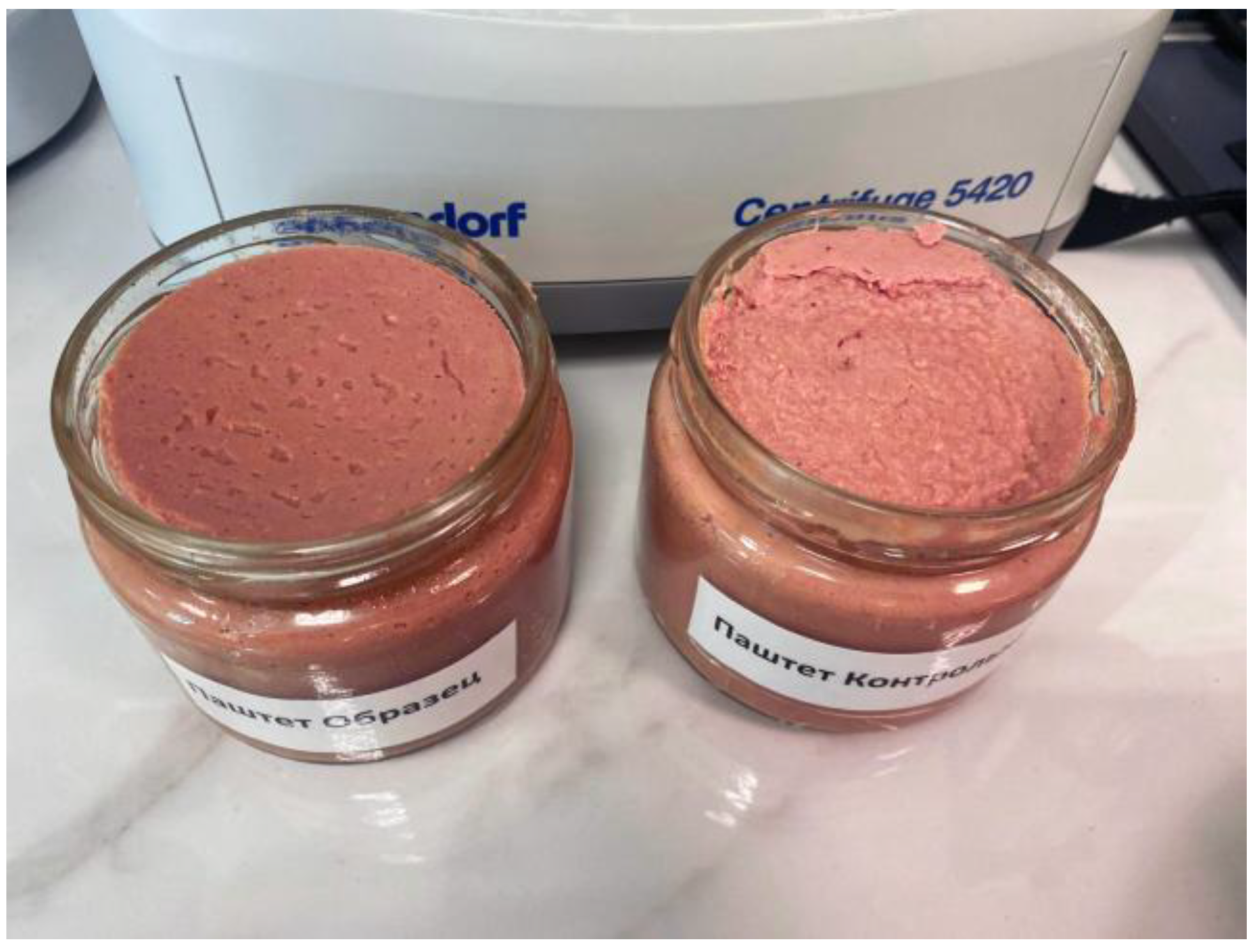
Figure 1. Goat meat pastes.
Processes 13 03213 g001
Figure 2. One-dimensional electropherogram of sausage samples. Cт—molecular weight standards: 250, 150, 100, 70, 50, 40, 30, 20, 15, and 10 kDa (from top to bottom); K—Control pate (without purslane and honey); O—Treatment pate (with purslane and honey).
Processes 13 03213 g002
Table 1. Formulation of goat meat pates (%, w/w of total batter).
IngredientControl PateTreatment Pate (Purslane + Honey)
Goat meat (leg/shoulder)76.071.2
Onion10.010.0
Carrot10.010.0
Mustard2.02.0
Starch2.02.0
Curing mix1.50.8
Purslane extract powder1.0
Honey4.0
Total100.0100.0
Table 2. Fatty acid compositions of meat pate samples with the addition of purslane.
Indicator NameControl Pate (Without Purslane and Honey) (% of Total FA)Treatment Pate (with Purslane and Honey) (% of Total FA)
Butyric, C4:0<0.1<0.1
Caproic, C6:0<0.1<0.1
Caprylic, C8:0<0.1<0.1
Capric, C10:0<0.1<0.1
Undecanoic, C11:0<0.1<0.1
Lauric, C12:0<0.1<0.1
Tridecanoic, C13:0<0.1<0.1
Myristic, C14:04.5 ± 0.44.3 ± 0.4
Pentadecanoic, C15:00.8 ± 0.40.8 ± 0.4
Palmitic, C16:027.1 ± 2.126.5 ± 2.1
Margaric (Heptadecanoic), C17:01.6 ± 0.41.5 ± 0.4
Stearic, C18:021.2 ± 2.119.8 ± 2.1
Arachidic, C20:00.4 ± 0.40.6 ± 0.4
Heneicosanoic, C21:00.8 ± 0.41.0 ± 0.4
Behenic, C22:0<0.10.5 ± 0.4
Tricosanoic, C23:0<0.10.2 ± 0.4
Lignoceric, C24:0<0.1<0.1
Myristoleic, C14:1<0.1<0.1
cis-10-Pentadecenoic, C15:10.4 ± 0.40.5 ± 0.4
Palmitoleic, C16:12.5 ± 0.42.6 ± 0.4
Heptadecenoic, C17:10.8 ± 0.40.9 ± 0.4
Oleic, C18:130.0 ± 2.129.5 ± 2.1
Elaidic (trans-C18:1)3.8 ± 0.43.8 ± 0.4
Gondoic, C20:10.6 ± 0.41.0 ± 0.4
Erucic, C22:1<0.10.2 ± 0.4
Nervonic, C24:1<0.10.3 ± 0.4
α-Linolenic (ALA), C18:3 n−30.8 ± 0.41.2 ± 0.4
Timnodonic (EPA), C20:5 n−3<0.1<0.1
Eicosatrienoic, C20:3 n−3<0.1<0.1
Docosahexaenoic (DHA), C22:6 n−3<0.1<0.1
Linoleic, C18:2 n−64.0 ± 0.43.3 ± 0.4
Linolelaidic (trans-C18:2 n−6)0.5 ± 0.40.6 ± 0.4
Dihomo-γ-linolenic, C20:3 n−6<0.1<0.1
Arachidonic, C20:4 n−6<0.10.5 ± 0.4
Eicosadienoic, C20:2 n−6<0.10.3 ± 0.4
Docosadienoic, C22:2 n−6<0.1<0.1
Results are expressed as the relative percentage of total identified fatty acids (% of total FA; g/100 g FA) calculated by area normalization. Values < 0.1 are reported as <0.1%. Class totals (ΣSFA, ΣMUFA, and ΣPUFA) equal the sum of the individual fatty acids within each class; small deviations from 100.0% reflect rounding.
Table 3. Dynamics of lipid oxidation and baseline protein oxidation during storage (peroxide value accumulation).
Parameter MeasuredUnitControl Pate (Without Purslane and Honey)Treatment Pate (with Purslane and Honey)p-Value, Treatment Within Storage Time
Peroxide value, day 0meq O2/kg fat3.8 ± 0.43.5 ± 0.40.410
Peroxide value, day 2meq O2/kg fat4.1 ± 0.44.3 ± 0.40.573
Peroxide value, day 4meq O2/kg fat5.9 ± 0.35.5 ± 0.30.178
Peroxide value, day 6meq O2/kg fat7.1 ± 0.47.0 ± 0.40.775
Peroxide value, day 8meq O2/kg fat10.1 ± 0.59.7 ± 0.50.383
Peroxide value, day 10meq O2/kg fat14.0 ± 0.913.0 ± 0.90.245
Protein carbonyls (baseline)nmol/mg protein95.7399.190.686
Table 4. Dynamics of lipid oxidation during storage (TBARS accumulation).
Parameter MeasuredUnitControl Pate (Without Purslane and Honey)Treatment Pate (with Purslane and Honey)p-Value, Treatment Within Storage Time
TBARS, day 0mg MDA/kg0.028 ± 0.0030.028 ± 0.0031.000
TBARS, day 2mg MDA/kg0.028 ± 0.0030.028 ± 0.0031.000
TBARS, day 4mg MDA/kg0.028 ± 0.0030.140 ± 0.0140.004
TBARS, day 6mg MDA/kg0.117 ± 0.0120.546 ± 0.0380.001
TBARS, day 8mg MDA/kg1.719 ± 0.1200.766 ± 0.0540.002
TBARS, day 10mg MDA/kg3.289 ± 0.2301.910 ± 0.1340.002
Table 5. Dynamics of lipid oxidation during storage (acid value accumulation).
Parameter MeasuredUnitControl Pate (Without Purslane and Honey)Treatment Pate (with Purslane and Honey)p-Value, Treatment Within Storage Time
Acid value, day 0mg KOH/g2.4 ± 0.22.0 ± 0.20.070
Acid value, day 2mg KOH/g3.0 ± 0.22.6 ± 0.20.070
Acid value, day 4mg KOH/g3.6 ± 0.33.1 ± 0.20.084
Acid value, day 6mg KOH/g4.2 ± 0.33.9 ± 0.30.288
Acid value, day 8mg KOH/g6.5 ± 0.55.2 ± 0.30.026
Acid value, day 10mg KOH/g8.5 ± 0.67.0 ± 0.50.031
Table 6. Color characteristics of pates.
ItemLight ExposureColor Characteristicsp-Value, Color Within Light Exposure
Control Pate (Without Purslane and Honey)Treatment Pate (with
Purslane and Honey)
L-lightnessBefore58.37 ± 0.4451.20 ± 0.510.988
After56.95 ± 0.3951.74 ± 0.480.316
a-rednessBefore7.38 ± 0.137.48 ± 0.58<0.0001
After6.53 ± 0.366.07 ± 0.81<0.0001
b-yellownessBefore25.31 ± 0.3524.35 ± 0.830.772
After26.27 ± 0.2626.53 ± 0.780.996
Color stability, % 93.17 ± 1.9088.84 ± 1.180.183
Table 7. Antioxidant capacity of goat meat pates (FRAP and DPPH results).
IndicatorResultsp-Value
Control Pate (Without
Purslane and Honey)
Treatment Pate (with
Purslane and Honey)
Ferric-reducing antioxidant power (FRAP), mg GAE/gNot detected10.5 ± 0.04<0.0001
DPPH radical-scavenging activity, %22.33 ± 0.00733.02 ± 0.009<0.0001
IC50 of DPPH radical-scavenging activity, µg/mL138.25 ± 11.15106.10 ± 10.010.002
Disclaimer/Publisher’s Note: The statements, opinions and data contained in all publications are solely those of the individual author(s) and contributor(s) and not of MDPI and/or the editor(s). MDPI and/or the editor(s) disclaim responsibility for any injury to people or property resulting from any ideas, methods, instructions or products referred to in the content.