Review

Imaging of Acute Complications of Community-Acquired Pneumonia in the Paediatric Population—From Chest Radiography to MRI

by
Efthymia Alexopoulou
1,
Spyridon Prountzos
1,*,
Maria Raissaki
2,
Argyro Mazioti
1,
Pablo Caro-Dominguez
3,
Franz Wolfgang Hirsch
4,
Jovan Lovrenski
5 and
Pierluigi Ciet
6,7
1
2nd Department of Radiology, University General Hospital “Attikon”, National and Kapodistrian University of Athens, 12462 Athens, Greece
2
University Hospital of Heraklion, Medical School, University of Crete, 70013 Heraklion, Greece
3
Pediatric Radiology Unit, Radiology Department, Hospital Universitario Virgen del Rocío, Avenida Manuel Siurot s/n, 41013 Seville, Spain
4
Department of Pediatric Radiology, University Hospital, Liebigstraße 20a, 04107 Leipzig, Germany
5
Radiology Department, Faculty of Medicine, Institute for Children and Adolescents Health Care of Vojvodina, University of Novi Sad, 21000 Novi Sad, Serbia
6
Department of Radiology and Nuclear Medicine, Erasmus MC—Sophia Children’s Hospital, 3015 CN Rotterdam, The Netherlands
7
Department of Radiology, University of Cagliari, 09124 Cagliari, Italy
*
Author to whom correspondence should be addressed.
Children 2024, 11(1), 122; https://doi.org/10.3390/children11010122
Submission received: 19 December 2023 / Revised: 11 January 2024 / Accepted: 15 January 2024 / Published: 18 January 2024
(This article belongs to the Special Issue Pediatric Respiratory Diseases: Diagnosis, Treatment, and Prevention)

Abstract

:
The most common acute infection and leading cause of death in children worldwide is pneumonia. Clinical and laboratory tests essentially diagnose community-acquired pneumonia (CAP). CAP can be caused by bacteria, viruses, or atypical microorganisms. Imaging is usually reserved for children who do not respond to treatment, need hospitalisation, or have hospital-acquired pneumonia. This review discusses the imaging findings for acute CAP complications and the diagnostic role of each imaging modality. Pleural effusion, empyema, necrotizing pneumonia, abscess, pneumatocele, pleural fistulas, and paediatric acute respiratory distress syndrome (PARDS) are acute CAP complications. When evaluating complicated CAP patients, chest radiography, lung ultrasonography, computed tomography (CT), and magnetic resonance imaging (MRI) can be used, with each having their own pros and cons. Imaging is usually not needed for CAP diagnosis, but it is essential for complicated cases and follow-ups. Lung ultrasound can supplement chest radiography (CR), which starts the diagnostic algorithm. Contrast-enhanced computed tomography (CECT) is used for complex cases. Advances in MRI protocols make it a viable alternative for diagnosing CAP and its complications.

1. Introduction

Pneumonia is one of the most common acute infections and the single most frequent cause of death in children worldwide, accounting for 14% of all deaths in children under 5 years of age [1,2]. Pneumonia killed 740,180 children under the age of 5 in 2019, accounting for 14% of all deaths of children under 5 years old but 22% of all deaths in children aged 1–5 years [2].
Community-acquired pneumonia (CAP) manifests as pneumonia symptoms, such as coughing, shortness of breath, and fever, in a previously healthy child due to an infection contracted outside of a hospital setting [3,4,5]. Laboratory findings useful for diagnosing CAP include the increase in erythrocyte sedimentation rate, white blood cell count, and C-reactive protein [6]. These markers are useful for monitoring treatment response [6].
The underlying causes can be bacterial, viral, or atypical (mycoplasma and chlamydia species). A significant proportion of severe pneumonia cases (up to 34%) result from mixed infections involving both viral and bacterial agents [7,8]. Although pneumonia’s causes include fungi and parasites, these are not common in the setting of CAP. Etiology can be presumed based on the patient’s age; children below 5 years have more frequent viral infections, while older children suffer from bacterial infections [3,4].
The predominant pathogens associated with CAP include the following: Streptococcus pneumoniae is the main etiological agent responsible for bacterial pneumonia in children. Haemophilus influenzae type b (Hib) is the second most prevalent etiological agent causing bacterial pneumonia. Respiratory syncytial virus is the predominant viral aetiology of pneumonia. Pneumocystis jiroveci is a prevalent cause of pneumonia among HIV-infected neonates, accounting for a minimum of 25% of pneumonia-related fatalities in this population [2]. Other bacterial pathogens appear to be less frequent causes of CAP. Group A streptococcal infection is important in terms of severity as, when present, it is more likely to progress to paediatric ICU admission or empyema [4]. Overall, viruses appear to account for 30–67% of CAP cases in childhood [9]. Mycoplasma is an etiological agent that is not unusual in ages 1–5 years old [4].
The diagnosis of CAP is based on clinical and laboratory findings, whereas imaging is not required in an immunocompetent child with uncomplicated CAP that does not need hospitalisation, since it will not change the management and outcome [3,4,10].
Imaging is mandatory when there is no response to outpatient treatment, in cases of hospital admission and in hospital-acquired pneumonia [10].
The potential acute complications of CAP can be pulmonary (pleural effusion, empyema, necrotizing pneumonia, lung abscess, bronchopleural fistula, pneumothorax, and acute respiratory failure), focal extrapulmonary (due to septicaemia and metastatic infection), and systemic (systemic inflammatory response syndrome or hemolytic uremic syndrome) [3].
When evaluating complicated CAP patients, chest radiography (CR), lung ultrasonography (LUS), computed tomography (CT), and magnetic resonance imaging (MRI) can be used, with each having their own pros and cons (Table 1). Imaging is usually not needed for CAP diagnosis, but it is essential for complicated cases and follow-ups.
The scope of this review is to present the imaging findings regarding the potential acute complications of CAP and to describe the role of each imaging modality.

2. Imaging Modalities

2.1. Chest Radiography

Chest radiography (CR) is the most frequently performed imaging modality, which clinicians are more familiar with, for interpreting pathological CAP findings. For a proper assessment, several parameters should be considered, such as the technical parameters of the examination (kV and mAs), projection (AP versus PA), the rotation of the child, and the degree of inspiration. Although radiation exposure in CR is low, proper consideration should be given in order not to overexpose the child. Thus, the use of lateral CR views is usually avoided in the pediatric population for radiation safety, although the Paediatric Infectious Diseases Society and the Infectious Diseases Society of America recommend the use of both projections in their guidelines [3,11,12]. Special consideration should be given to hidden areas by other anatomical structures, such as the heart, the mediastinum, and the diaphragm [12]. Great variations in intra- and interobserver agreement between radiologists in CR interpretation are a limitation when using this modality [11]. Systematic reviews have calculated that the pooled sensitivity and specificity for pediatric pneumonia diagnosed by CXR can be up to 0.91 (95% CI: 0.90 to 0.93), 1.00 (95% CI: 0.99 to 1.00), and the pooled sensitivity and specificity for children with pneumonia diagnosed by LUS were 0.95 (95% CI: 0.94 to 0.96), 0.90 (95% CI: 0.87 to 0.92), respectively [13].

2.2. Lung Ultrasound

Lung ultrasound (LUS) is a bedside modality that can be used when assessing a child with CAP. As a widely available, non-ionising, and low-cost technique, LUS is especially useful when assessing bed-ridden patients in the point-of-care ultrasound (POCUS) setting. This is a real-time examination where the examined child does not need to be sedated, allowing for a full examination of the chest. It is the best technique to evaluate the amount and characteristics of pleural effusion and the mobility of the diaphragm [14]. Rough estimates of the relative position of intrapulmonary lesions can also be obtained. LUS can detect small multifocal consolidations adherent to the subpleural surface [15,16]. Its main disadvantages include the reliance on operator skills for accurate results, the potential for time-intensive procedures (taking up to 20 min per examination), and the presence of hidden areas, like the subscapular or central lung regions, where a comprehensive assessment of parenchyma may not be achievable. Moreover, lesions that are not in touch with the pleura might be missed at the LUS examination [17,18,19,20]. If LUS is used to assess for CAP complications in the lung parenchyma, such as necrotizing areas or lung abscesses, Colour-Doppler (CD) and/or contrast-enhanced ultrasonography (CEUS) may be used [21,22]. Systematic reviews comparing CR and LUS suggest the excellent diagnostic accuracy of the latter, and that LUS is more suitable than CR for detecting pleural effusion, with LUS being able to detect effusions as small as 10 mL, whereas chest X-rays only identify larger effusions (200 mL) [23,24].

2.3. Computed Tomography

Computed tomography (CT) is considered the gold-standard technique in terms of sensitivity in detecting CAP and its acute complications, but its use is limited due to the high radiation doses that are needed compared to CR. In the paediatric population, growth refers to an increase in cell proliferation, resulting in a greater number of cells undergoing mitosis and displaying heightened sensitivity to radiation. This is exacerbated by the fact that in children have a higher percentage of red bone marrow compared to adults, making them three times more radiation-sensitive compared to adults [25]. Advancements in technology, such as a new generation of scanners and the implementation of iterative reconstruction techniques, have made it possible to greatly decrease the radiation exposure during medical examinations. Achievable doses, measured in dose length products (DLPs), can now be as low as 7.66 mGy·cm per examination [26].
Previous generation scanners (the first generation and some of the second generation) required sedation in children younger than 4 years of age. However, third-generation scanners have overcome this problem, enabling high-speed scans with full acquisition of the thorax in less than a second without anaesthesia [27]. This technique is achieved using a dual-source scan with a maximum rotation time and a minimum pitch > 1.5. The use of an intravenous (IV) iodine contrast is mandatory when assessing for acute complications of CAP, such as pleural empyema and parenchymal necrosis. The examination protocol includes a single-phase scan directly after the IV contrast injection, without a non-contrast scan. In these complex clinical situations, contrast-enhanced CT enables image-guided intervention and serves as a pre-operative examination prior to video-assisted thoracoscopy (VATS) or in instances of diagnostic uncertainty (to rule out concurrent malignancy) (Figure 1). CT is suitable when the diagnosis of an abscess or necrotizing pneumonia cannot be confirmed with CR and LUS, in cases of extensive cavitation or loculated empyema, and to exclude the presence of a bronchopleural fistula. In cases where pleural involvement has to be assessed, the timing of the acquisition after intravenous contrast injection should be longer for the better enhancement of pleural folds. Lastly, CT is the technique of choice in children with recurrent pneumonia to determine the cause of recurrent infections, such as aspirated foreign bodies or possible congenital lung abnormalities [10,27,28,29].

2.4. Magnetic Resonance Imaging

Magnetic Resonance Imaging (MRI) is a radiation-free technique that provides excellent soft-tissue contrast and anatomical detail. Still, it comes at a higher cost per examination and is less available in low-income countries. MRI is usually a more time-consuming examination than CT, with an increased need for sedation in children younger than 5 years. However, the recently reported use of real-time MRI for imaging pneumonia is just as fast as a CT scan, and it no longer requires sedation. Movement and breathing by the child do not cause artefacts either [30]. To date, this technique has only been available in research settings and is limited to a single MRI brand [31]. Spatial resolution of MRI is usually lower than CT, but new sequences, like ultra-short and zero echo time sequences, allow isotropic voxel sizes of around 1 mm [32]. In cooperative children, MRI has some advantages over CT. Firstly, MRI does not require the use of contrast to identify necrotic lung tissue from abscesses, which can be detected on T2-weighted images as hyperintense lung regions [33]. Secondly, MRI allows for the detection of septation on complex pleural effusions and empyema, which cannot be seen on CT. This allows for relevant information to be obtained in a single examination instead of using both LUS and CT. Finally, MRI can be repeated at the end of treatment as a follow-up technique to assess residual abnormalities and the effect of treatment. In the study of Konietzke et al., MRI showed similar results to CT in assessing necrotizing pneumonia and pulmonary abscess when both CR and LUS were inconclusive [34]. Contraindications to MRI may include unstable patients, claustrophobia, metallic implants, or a lack of cooperation [27,34,35,36,37]. The basic MRI protocol for lung examinations consists of T1- and T2-weighted sequences in at least two planes, diffusion-weighted (DWI) images, and post-contrast T1 weighted images with fat saturation.

2.4.1. Pleural Effusion—Empyema

A pleural effusion is formed when there is an imbalance between hydrostatic and oncotic pressure between the systemic and pulmonary circulations and the pleural space [38]. Additionally, the obstruction of lymphatic drainage by thick pleural fluid and debris further contributes to the accumulation of the effusion [6,39,40]. Parapneumonic effusion is a pleural effusion associated with an ipsilateral lung infection and can be simple or complicated (in the presence of infecting organisms). An empyema is a thick, purulent pleural effusion, which can be either free or loculated [41]. The progression of pleural effusion to empyema occurs in three stages: (a) the exudative stage, which is a simple parapneumonic effusion; (b) the fibrinopurulent stage, which is a complicated parapneumonic effusion; and (c) the organising phase, in which fibroblastic activity and peel formation occur. The distinction between transudate, exudate, and empyema is also based on chemical analysis. If a child is pyrexial or sick 48–72 h after beginning antibiotics, empyema may be present. Pneumonia and pleural effusion cause dullness to percussion and diminished breath sounds on physical examination. Consolidation increases fremitus. Detecting fremitus in children may be less helpful than detecting fremitus in adults due to age, size, and cooperation. Pleural effusion does not cause bronchial breathing [6].
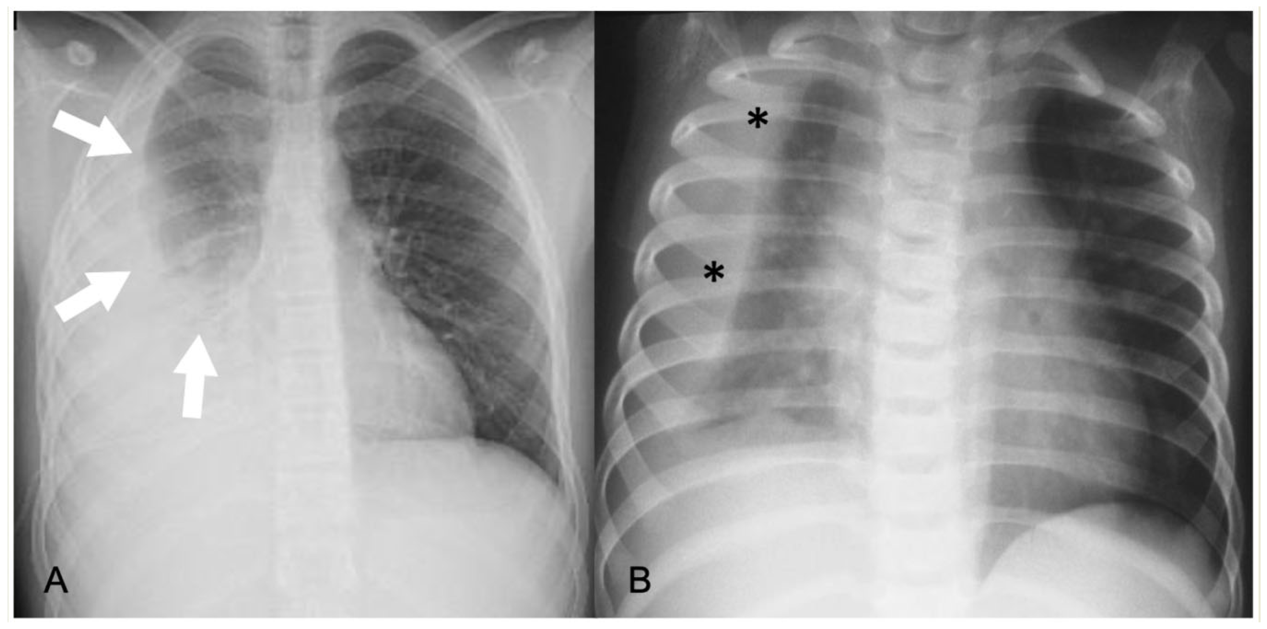
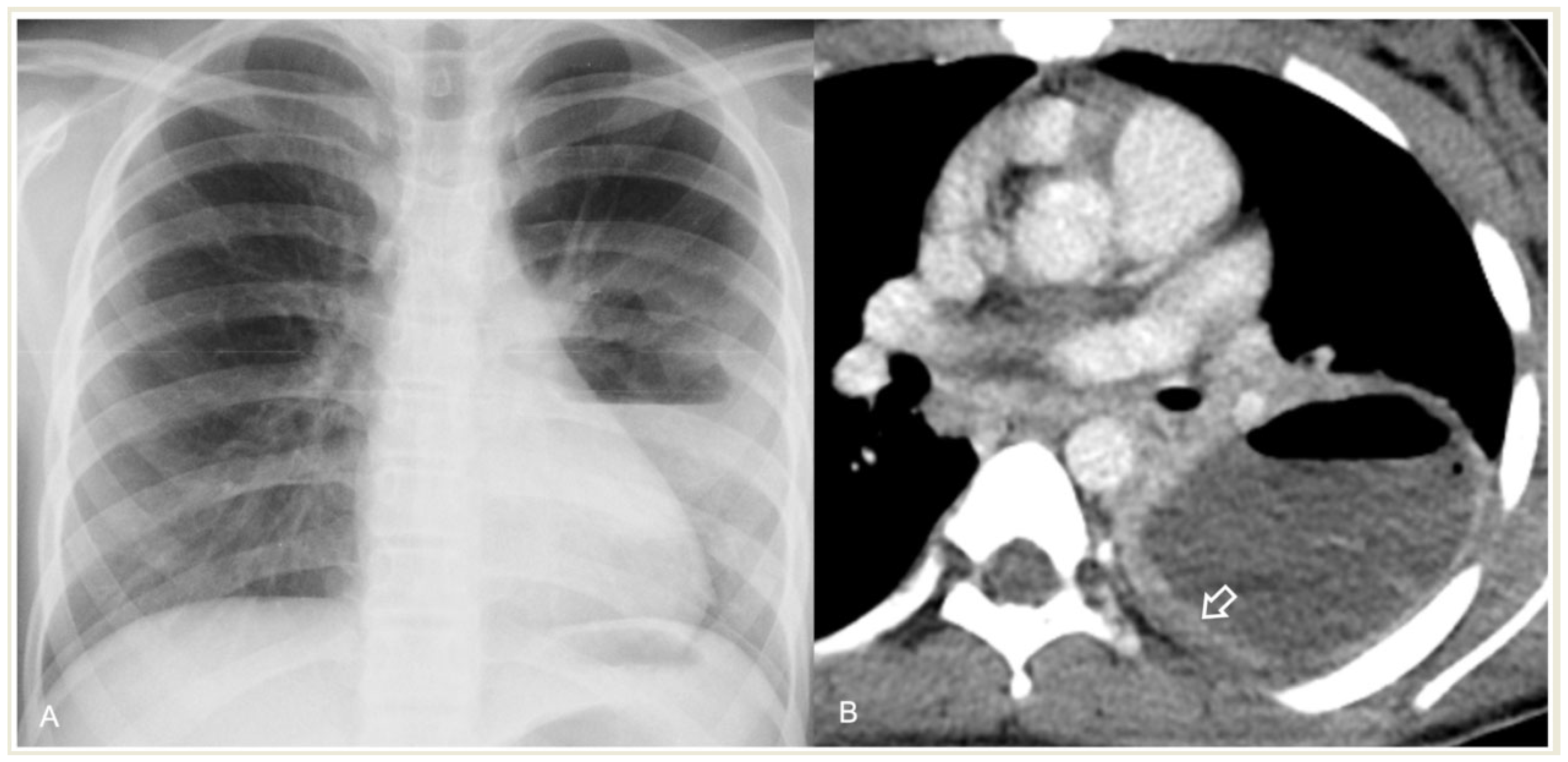
In CR, pleural effusion may have different appearances, such as blunting of the costophrenic angle or the meniscus sign when free fluid is present (Figure 1). A medial convex border to the lung parenchyma indicates a loculated effusion. Complete opacification of the ipsilateral hemithorax is present in large effusions where the underlying parenchyma is obscured. An ipsilateral concave scoliosis and contralateral mediastinal shift may be common associated findings, and further imaging is required to investigate the condition of the underlying lung and pleura (Figure 2).
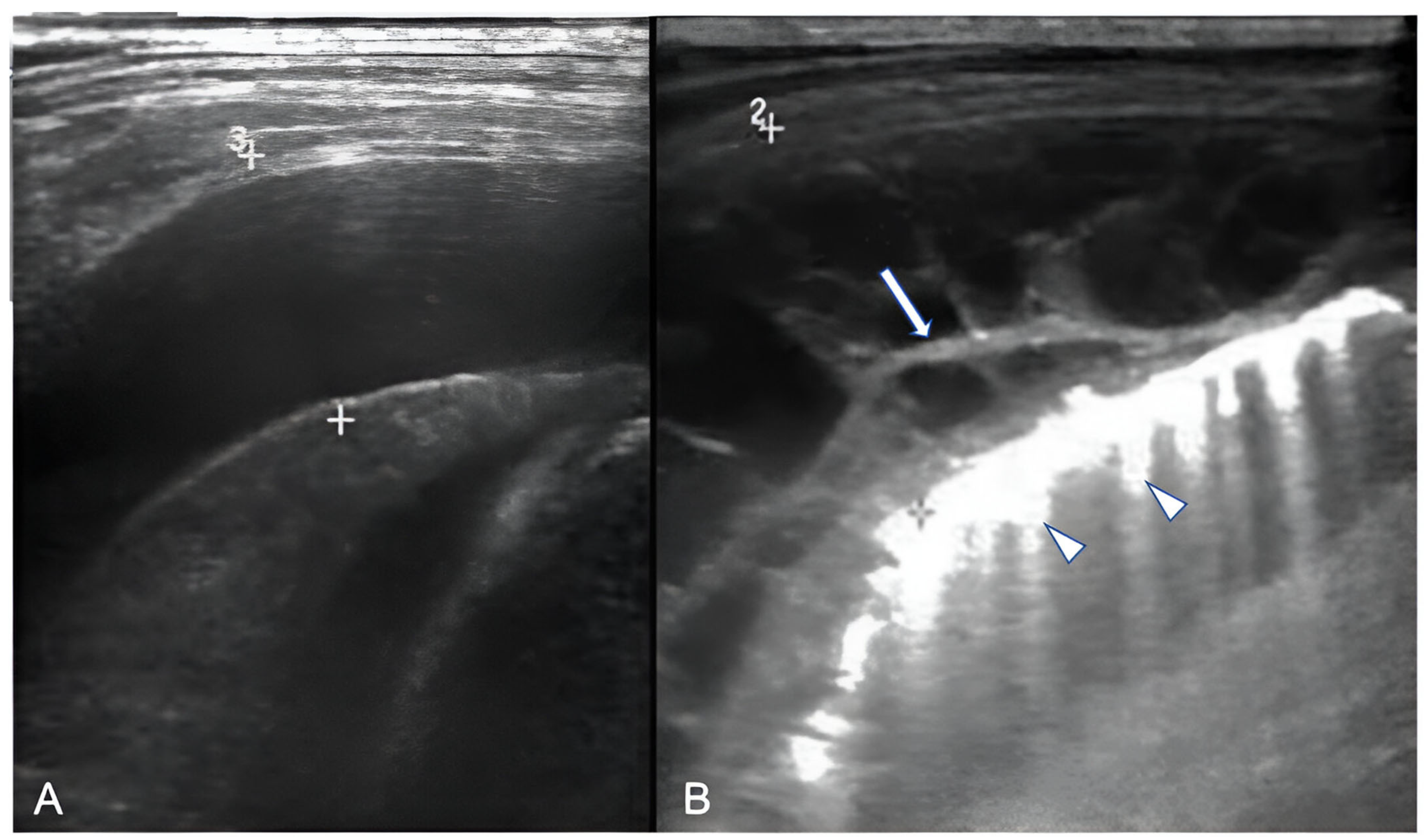
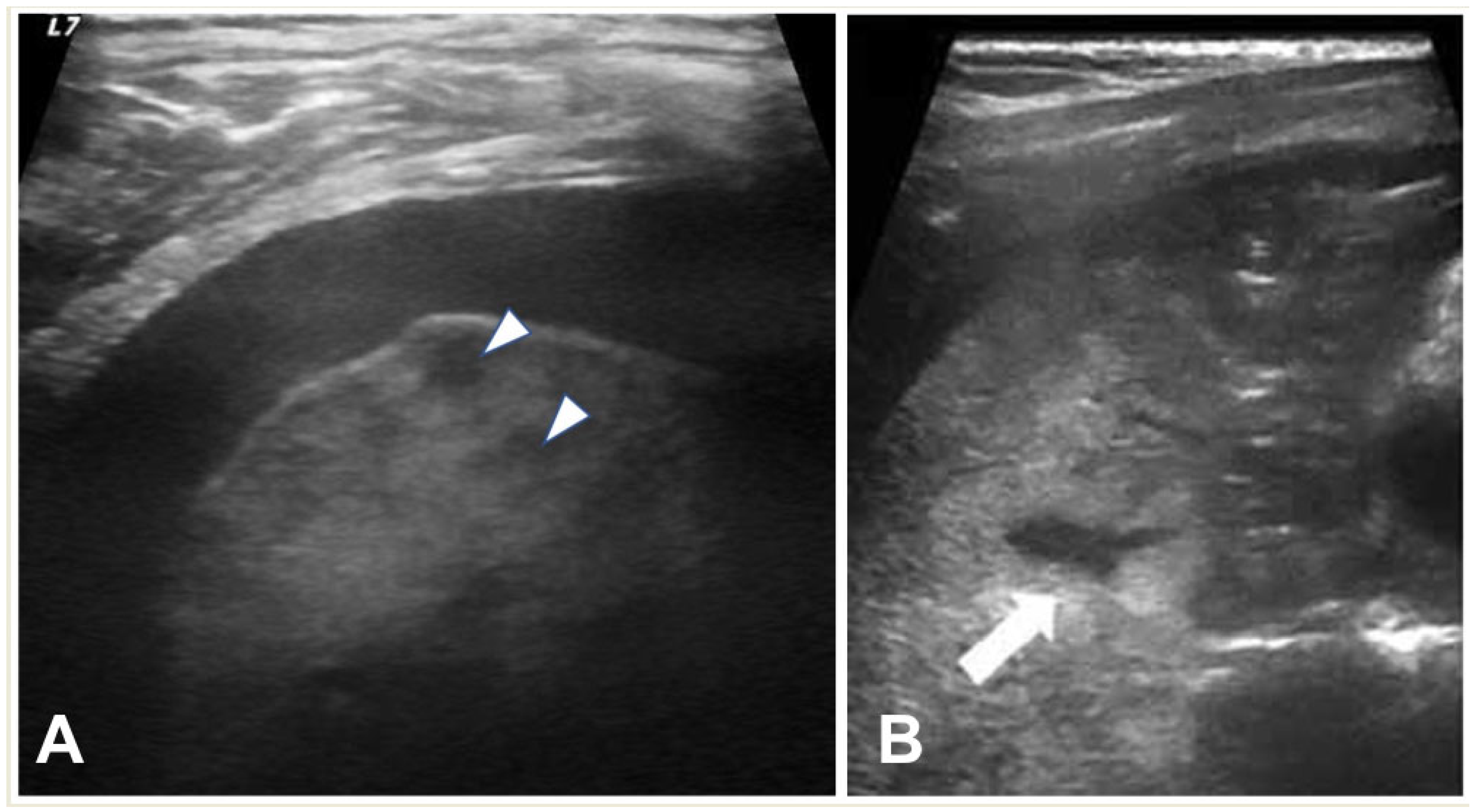
LUS can provide information regarding the volume of the effusion (as small as 10 mL), the presence of fibrinous septations, and the echogenicity grade of the pleural fluid (Figure 3) [21]. LUS can be used to differentiate pleural effusions from consolidated lung and peripheral lung abscesses from empyema. In the appropriate clinical setting, the pulmonologist or thoracic surgeon may decide whether to perform thoracocentesis and put on a chest tube, fibrinolysis, or VATS based on the appearance and volume of the pleural effusion. The indication for performing thoracentesis arises when a pleural effusion, which may vary in volume, shows resistance to IV antibiotic treatment or when the patient continues to experience symptoms [42,43]. A few formulae can be used to calculate pleural effusion volume in adults; however, none of them are truly adapted to children. LUS also provides guidance for the insertion of draining catheters. Intravenous CEUS may be useful to better outline pleural effusions, whereas intracavitary contrast is helpful in determining catheter localization and patency, or the presence of loculation [20,44,45,46,47].
Contrast-enhanced CT can demonstrate the location and volume of the effusion, although it might not show existing septations. The enhancement of thickened parietal pleura is usually depicted in this setting, although its absence does not exclude an infected effusion (Figure 4) [41].
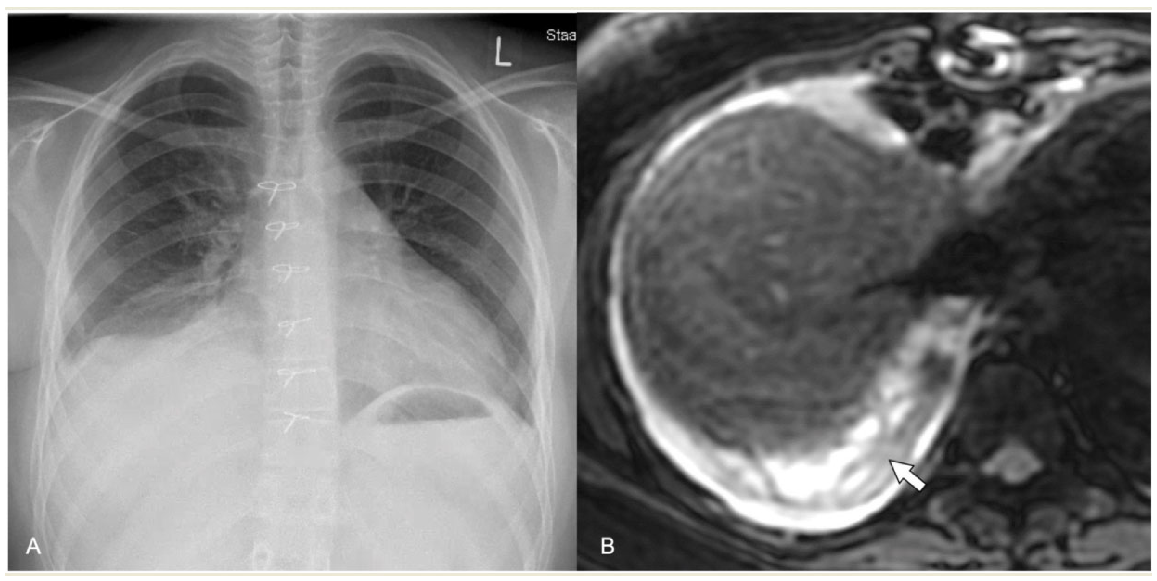
Regarding MRI, simple effusions show crescentic distribution and low signal on T1-weighted sequences, high signal on T2-weighted sequences, and no contrast enhancement. Empyema shows a loculated effusion with thickened, enhancing pleura, septations, and heterogeneous contents on T1-weighted and T2-weighted images due to debris (Figure 5). The use of DWI can help in differentiating between simple effusion and empyema, as the latter shows restricted diffusivity [27,48]. In a recent study, the use of intravenous contrast was equivocal as it provided no added value regarding the differentiation of pleural effusion and empyema, since all empyemas showed septations on T2-weighted sequences [34]. Differentiating fibrinous from non-fibrinous complex pleural effusions is one area where MR spectroscopy may be useful in everyday clinical practice, while its practicality is still debatable [49].

2.4.2. Acute Lung Parenchyma Complications

Three significant acute complications of severe pneumonia result from the extensive destruction and liquefaction of lung tissue caused by vascular thrombosis: necrotizing pneumonia, cavitary necrosis, and the formation of lung abscesses [6].

2.4.3. Necrotizing Pneumonia

Children with necrotizing pneumonia exhibit symptoms such as fever, cough, and tachypnoea that last for many days. Hypoxia, moderate anaemia, and hypoalbuminaemia are frequent symptoms, with pleural effusion commonly visible upon physical examination [6].
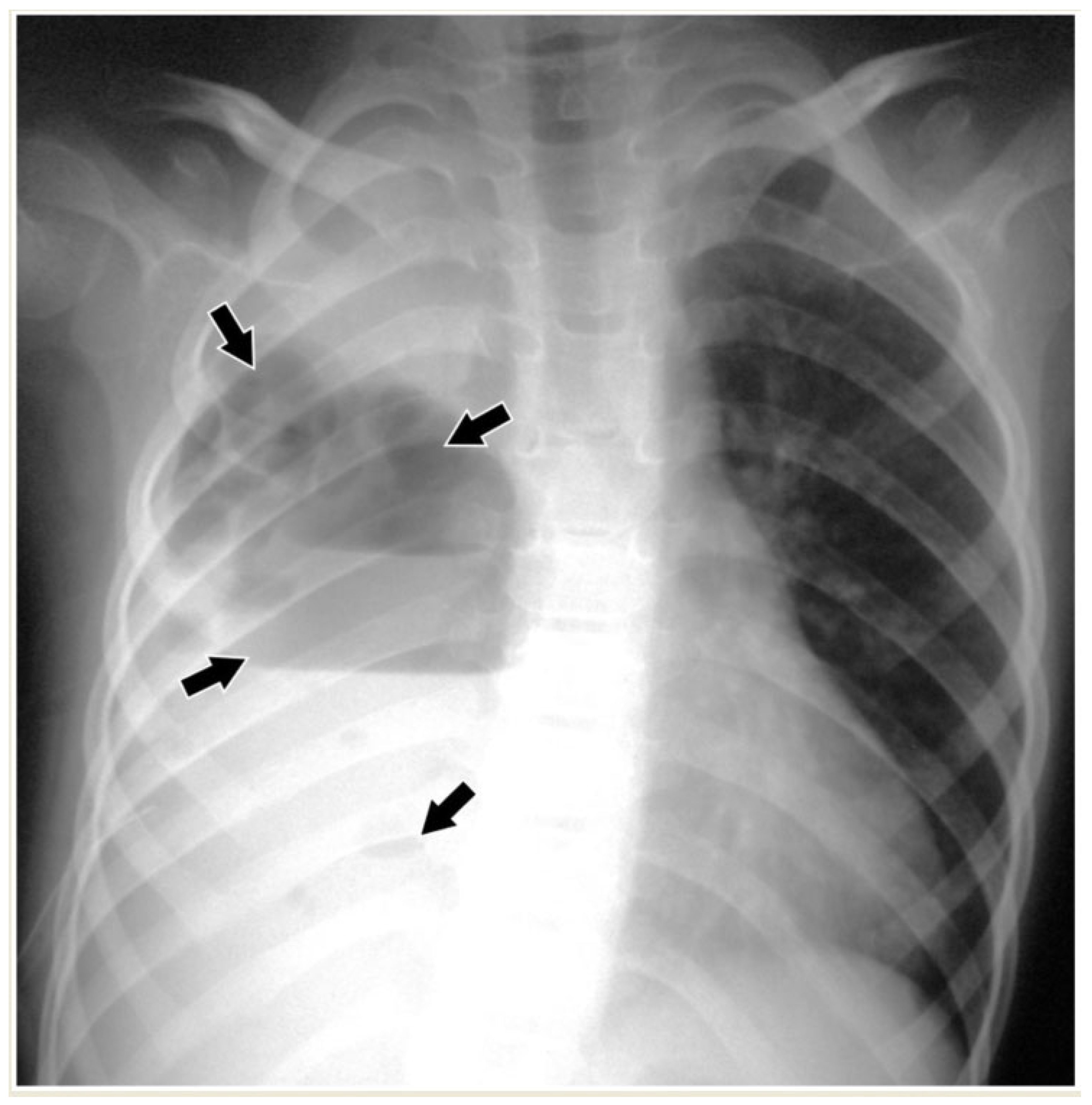
CR is not useful in the early stage of necrotizing pneumonia, as it is unable to depict liquefaction of the lung parenchyma. Cavitary necrosis typically exhibits the presence of air-filled cavities or air–fluid levels, which may require additional distinction from lung abscess or loculated empyema (Figure 6) [6,41].
Necrosis is seen on LUS as ill-defined areas of low echogenicity and diminished vascularity (Figure 7). Attention needs to be paid to the differentiation between echogenic effusions and echogenic lung parenchyma due to pneumonia (Figure 8). When positioned in the peripheral lung, LUS demonstrates comparable accuracy to CT in identifying pneumonia sequelae, such as pleural effusion, necrosis, and abscesses of lung parenchyma. However, the LUS sensitivity is lower than that of CT for centrally located abnormalities. The presence of fluid and static air bronchograms within a consolidated region may indicate the early onset of parenchymal complications [50]. The role of intravenous CEUS in complicated pneumonia remains a subject of controversy. While some researchers have reported on its accurate identification of necrotizing pneumonia and the precise delineation of its extent, contrasting findings have emerged from other authors, who found that no significant additional information was garnered in comparison to the utilisation of Colour-Doppler [20,22,45,46,51].
Upon contrast-enhanced CT (CECT), necrotizing pneumonia is characterised by the presence of several patchy or widespread areas of diminished or absent enhancement, indicating liquefaction of the lung tissue (Figure 8). Cavitary necrosis is distinguished by the absence of a normal lung tissue structure, which is either diminished or lacking enhancement, and the appearance of many thin-walled cavities filled with either fluid or air. These cavities lack an enhancing border (Figure 9).
Pulmonary necrosis on MRI is represented by areas of increased signal intensity on T2-weighed sequences, while cavitary necrosis is represented by fluid-filled and air-filled areas within a consolidation that do not enhance after the injection of contrast (Figure 10). Contrast-enhanced MRI shows a similar CECT pattern to identify necrotic lung parenchyma.

2.4.4. Abscess

Children with lung abscesses often have an extended cough and mild fever, with less common symptoms including chest discomfort, dyspnoea, sputum production, and hemoptysis. A chest exam may show normal or symptoms of consolidation [6].
A lung abscess is a single cavity with a thick wall and purulent content resulting from suppuration and necrosis of the parenchyma. After the initial stage, the abscess can be partially or even completely air-filled. This is an uncommon complication in immunocompetent children with CAP but, when present, it may be associated with a pre-existing pulmonary cavity, such as cystic pulmonary adenomatoid malformations (CPAM). Severe forms of necrotizing pneumonia have also been associated with Staphylococcus aureus strains expressing the Panton–Valentine leukocidin cytotoxic agent [6,52]. A lung abscess on CR appears as a round or oval-shaped area that is denser than the surrounding lung parenchyma. The edges of the abscess might be irregular or ill-defined, and there may be air–fluid levels inside, indicating the presence of gas and fluid. On LUS, a lung abscess typically appears as a hypoechoic area within the lung tissue with irregular borders. Surrounding the hypoechoic area, there might be a hyperechoic rim, which corresponds to the inflamed tissue. On CECT, this appears as a hypodense cavity surrounded by a well-defined enhancing wall without central enhancement and a variable proportion of air and/or fluid content (Figure 11). On MRI, a rounded, T2-hyperintense area surrounded by a hypointense wall can be seen, which is usually thick and irregular and may contain an air–fluid level or be completely air-filled (Figure 12 and Figure 13 and Supplementary Material). A characteristic, restricted diffusion can be seen in DWI sequences, with peripheral wall enhancement after the injection of intravenous contrast [27,34,48,53,54,55].

2.4.5. Pneumatocele

A pneumatocele is a thin-walled intraparenchymal cystic structure that is filled with air (or little-to-no fluid content) and is formed by a valve-type mechanism that allows for the one-way passage of air into the interstitial space. Pneumatocele are often the results of severe infections, trauma, and mechanical ventilation. Pneumatocele appear as bubble-like structures on imaging studies like X-rays or CT scans [56]. Pneumatocele are difficult to identify on LUS because they are usually filled with air and therefore have an anechoic appearance.

2.4.6. Pleural Fistulas

A pleural fistula can form during severe pneumonia as a result of the erosion or perforation of lung tissue that connects the bronchi to the pleural space. Pleural fistula is a serious and potentially life-threatening complication in severe pneumonia, leading to respiratory distress, sepsis, and pneumothorax. The diagnosis of bronchopleural fistula is usually definite on CT when a direct communication between the lung parenchyma and the pleural space is identified. It is also suspected when there is a persistent air leak in the pleural space for more than 24 h (Figure 14) or when pneumothorax recurs following the removal of the drains [57]. CECT is the optimal technique for diagnosing a pleural fistula due to its high resolution, which enables the identification of direct connections between small peripheral bronchi and the pleural space [58].

2.4.7. Pediatric Acute Respiratory Distress Syndrome (PARDS)

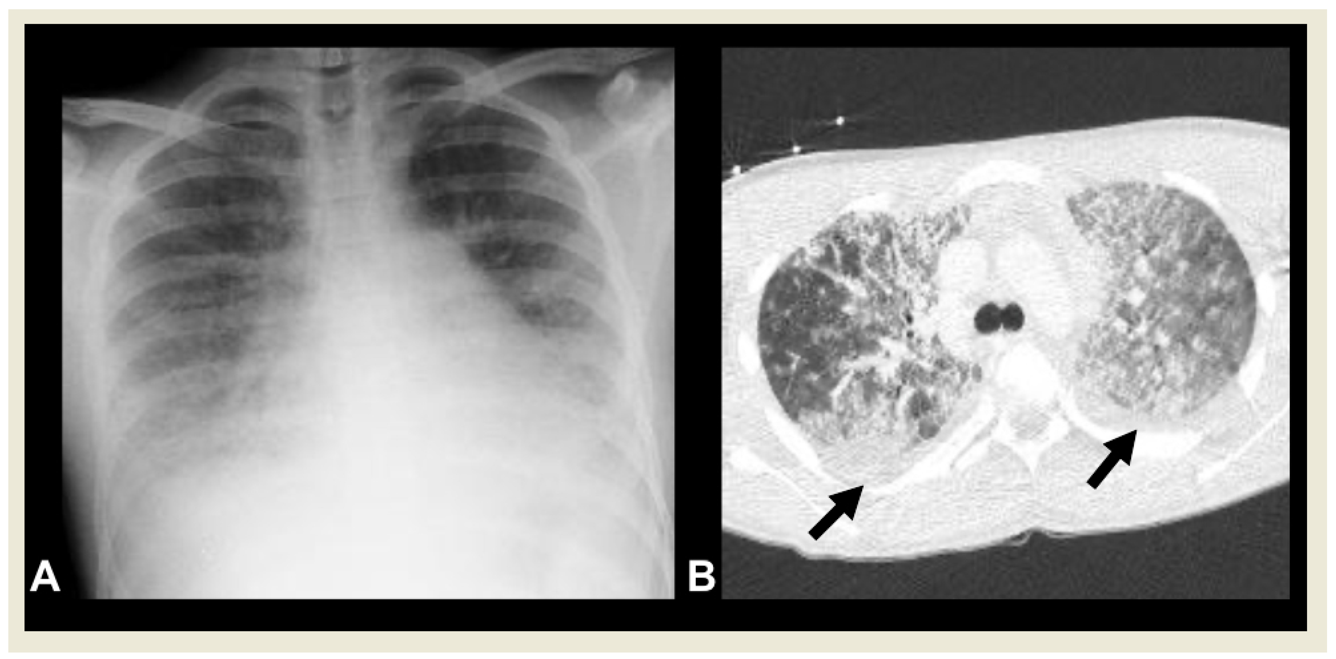
PARDS is defined as the occurrence of signs and symptoms of pulmonary oedema that are not fully explained by cardiac failure or fluid overload within 7 days of a clinical insult [59]. Various epidemiological factors and etologies play a significant role in the development of PARDS. The most common PARDS risk factors include pneumonia or lower respiratory tract infections [60]. One of the clinical and laboratory criteria used to diagnose PARDS is the detection of bilateral lung infiltrates on chest radiography (Figure 15) [59].

3. Conclusions

Imaging is usually not needed for the initial diagnosis of CAP; however, it becomes crucial for identifying acute complications and for follow-up assessments. The diagnostic algorithm always starts with CR which can be supplemented with LUS, particularly to confirm the presence of pleural effusion and empyema. In cases without severe complications, the combined findings from these two techniques are sufficient for patient management.
CECT scanning is reserved for more complicated cases, enabling the assessment of severe parenchymal abnormalities, such as necrotizing pneumonia, cavitary lesions, abscesses, and pleural fistulas, or when surgical intervention is being considered. In skilled hands, LUS can offer valuable insights into complicated pneumonias, potentially obviating the need for more advanced diagnostic modalities like CT. Finally, emerging advancements in MRI protocols, such as real-time MRI and ultra-short and zero echo time sequences, as presented above, have positioned MRI as a viable alternative for the diagnosis of CAP and its complications, particularly in centres equipped with specialized expertise and resources. A suggested imaging algorithm, according to clinical indications, can be found in Table 2.

Supplementary Materials

The following supporting information can be downloaded at: https://www.mdpi.com/article/10.3390/children11010122/s1, Video S1: Real-time MRI sequence in the coronal plane in a 1-year-old girl with left upper lobe pneumonia with an acquisition time of 16 s.

Author Contributions

Conceptualization E.A. and P.C.; writing—original draft preparation E.A. and S.P.; data curation E.A.; writing—review and editing and visualization E.A., S.P., M.R., A.M., P.C.-D., F.W.H., J.L. and P.C. All authors have read and agreed to the published version of the manuscript.

Funding

This research received no external funding.

Institutional Review Board Statement

Not applicable.

Informed Consent Statement

Informed consent was obtained from all subjects involved in the study. All the presented images belong to the authors’ team.

Data Availability Statement

No new data were created or analyzed in this study. Data sharing is not applicable to this article.

Conflicts of Interest

The authors declare no conflict of interests.

References

  1. Wardlaw, T.; Salama, P.; Johansson, E.W.; Mason, E. Pneumonia: The leading killer of children. Lancet 2006, 368, 1048–1050. [Google Scholar] [CrossRef] [PubMed]
  2. Pneumonia in Children. Available online: https://www.who.int/news-room/fact-sheets/detail/pneumonia (accessed on 7 January 2024).
  3. Bradley, J.S.; Byington, C.L.; Shah, S.S.; Alverson, B.; Carter, E.R.; Harrison, C.; Kaplan, S.L.; Mace, S.E.; McCracken, G.H.; Moore, M.R.; et al. The Management of Community-Acquired Pneumonia in Infants and Children Older than 3 Months of Age: Clinical Practice Guidelines by the Pediatric Infectious Diseases Society and the Infectious Diseases Society of America. Clin. Infect. Dis. 2011, 53, e25–e76. [Google Scholar] [CrossRef] [PubMed]
  4. Harris, M.; Clark, J.; Coote, N.; Fletcher, P.; Harnden, A.; Mckean, M.; Thomson, A. British Thoracic Society guidelines for the management of community acquired pneumonia in children: Update 2011. Thorax 2011, 66, ii1–ii23. [Google Scholar] [CrossRef]
  5. Edis, E.C.; Hatipoglu, O.N.; Yilmam, I.; Eker, A.; Tansel, O.; Sut, N. Hospital-Acquired Pneumonia Developed in Non-Intensive Care Units. Respiration 2009, 78, 416–422. [Google Scholar] [CrossRef]
  6. De Benedictis, F.M.; Kerem, E.; Chang, A.B.; Colin, A.; Zar, H.J.; Bush, A. Complicated pneumonia in children. Lancet 2020, 396, 786–798. [Google Scholar] [CrossRef] [PubMed]
  7. Roh, E.J.; Shim, J.Y.; Chung, E.H. Epidemiology and surveillance implications of community-acquired pneumonia in children. Clin. Exp. Pediatr. 2022, 65, 563–573. [Google Scholar] [CrossRef] [PubMed]
  8. Von Mollendorf, C.; Berger, D.; Gwee, A.; Duke, T.; Graham, S.M.; Russell, F.M.; Mulholland, E.K. ARI review group Aetiology of childhood pneumonia in low- and middle-income countries in the era of vaccination: A systematic review. J. Glob. Health 2022, 12, 10009. [Google Scholar] [CrossRef]
  9. Cilla, G.; Oñate, E.; Perez-Yarza, E.G.; Montes, M.; Vicente, D.; Perez-Trallero, E. Viruses in community-acquired pneumonia in children aged less than 3 years old: High rate of viral coinfection. J. Med. Virol. 2008, 80, 1843–1849. [Google Scholar] [CrossRef]
  10. Chan, S.S.; Kotecha, M.K.; Rigsby, C.K.; Iyer, R.S.; Alazraki, A.L.; Anupindi, S.A.; Bardo, D.M.; Brown, B.P.; Chandra, T.; Dorfman, S.R.; et al. ACR Appropriateness Criteria® Pneumonia in the Immunocompetent Child. J. Am. Coll. Radiol. 2020, 17, S215–S225. [Google Scholar] [CrossRef]
  11. Cherian, T.; Mulholland, E.K.; Carlin, J.B.; Ostensen, H.; Amin, R.; De Campo, M.; Greenberg, D.; Lagos, R.; Lucero, M.; Madhi, S.A.; et al. Standardized interpretation of paediatric chest radiographs for the diagnosis of pneumonia in epidemiological studies. Bull. World Health Organ. 2005, 83, 353–359. [Google Scholar] [PubMed]
  12. Andronikou, S.; Lambert, E.; Halton, J.; Hilder, L.; Crumley, I.; Lyttle, M.D.; Kosack, C. Guidelines for the use of chest radiographs in community-acquired pneumonia in children and adolescents. Pediatr. Radiol. 2017, 47, 1405–1411. [Google Scholar] [CrossRef] [PubMed]
  13. Yan, J.-H.; Yu, N.; Wang, Y.-H.; Gao, Y.-B.; Pan, L. Lung ultrasound vs chest radiography in the diagnosis of children pneumonia. Medicine 2020, 99, e23671. [Google Scholar] [CrossRef]
  14. Ibitoye, B.O.; Idowu, B.M.; Ogunrombi, A.B.; Afolabi, B.I. Ultrasonographic quantification of pleural effusion: Comparison of four formulae. Ultrasonography 2018, 37, 254–260. [Google Scholar] [CrossRef] [PubMed]
  15. Brogna, B.; Bignardi, E.; Brogna, C.; Volpe, M.; Lombardi, G.; Rosa, A.; Gagliardi, G.; Capasso, P.F.M.; Gravino, E.; Maio, F.; et al. A Pictorial Review of the Role of Imaging in the Detection, Management, Histopathological Correlations, and Complications of COVID-19 Pneumonia. Diagnostics 2021, 11, 437. [Google Scholar] [CrossRef]
  16. Kogias, C.; Prountzos, S.; Alexopoulou, E.; Douros, K. Lung ultrasound systematic review shows its prognostic and diagnostic role in acute viral bronchiolitis. Acta Paediatr. 2023, 112, 222–232. [Google Scholar] [CrossRef]
  17. Liu, J.; Copetti, R.; Sorantin, E.; Lovrenski, J.; Rodriguez-Fanjul, J.; Kurepa, D.; Feng, X.; Cattaross, L.; Zhang, H.; Hwang, M.; et al. Protocol and Guidelines for Point-of-Care Lung Ultrasound in Diagnosing Neonatal Pulmonary Diseases Based on International Expert Consensus. J. Vis. Exp. 2019, 2019, e58990. [Google Scholar] [CrossRef]
  18. Bhalla, D.; Naranje, P.; Jana, M.; Bhalla, A.S. Pediatric lung ultrasonography: Current perspectives. Pediatr. Radiol. 2022, 52, 2038–2050. [Google Scholar] [CrossRef]
  19. Lovrenski, J. Pediatric lung ultrasound cons—Are they really strong enough? Pediatr. Radiol. 2020, 50, 321–322. [Google Scholar] [CrossRef]
  20. Lovrenski, J. Pediatric lung ultrasound—Pros and potentials. Pediatr. Radiol. 2020, 50, 306–313. [Google Scholar] [CrossRef]
  21. Laya, B.F.; Concepcion, N.D.P.; Garcia-Peña, P.; Naidoo, J.; Kritsaneepaiboon, S.; Lee, E.Y. Pediatric Lower Respiratory Tract Infections: Imaging Guidelines and Recommendations. Radiol. Clin. N. Am. 2022, 60, 15–40. [Google Scholar] [CrossRef]
  22. Dietrich, C.F.; Buda, N.; Ciuca, I.M.; Dong, Y.; Fang, C.; Feldkamp, A.; Jüngert, J.; Kosiak, W.; Mentzel, H.J.; Pienar, C.; et al. Lung ultrasound in children, WFUMB review paper (part 2). Med. Ultrason. 2021, 23, 443–452. [Google Scholar] [CrossRef]
  23. Chidini, G.; Raimondi, F. Lung ultrasound for the sick child: Less harm and more information than a radiograph. Eur. J. Pediatr. 2023. online ahead of print. [Google Scholar] [CrossRef] [PubMed]
  24. Hansell, L.; Milross, M.; Delaney, A.; Tian, D.H.; Ntoumenopoulos, G. Lung ultrasound has greater accuracy than conventional respiratory assessment tools for the diagnosis of pleural effusion, lung consolidation and collapse: A systematic review. J. Physiother. 2021, 67, 41–48. [Google Scholar] [CrossRef] [PubMed]
  25. Nagy, E.; Tschauner, S.; Schramek, C.; Sorantin, E. Paediatric CT made easy. Pediatr. Radiol. 2023, 53, 581–588. [Google Scholar] [CrossRef] [PubMed]
  26. Worrall, M.; Holubinka, M.; Havariyoun, G.; Hodgson, K.; Edyvean, S.; Holroyd, J.; Davis, A.; Dunn, M.; Gardiner, A. Analysis and results from a UK national dose audit of paediatric CT examinations. Br. J. Radiol. 2022, 95, 20210796. [Google Scholar] [CrossRef]
  27. Liszewski, M.C.; Görkem, S.; Sodhi, K.S.; Lee, E.Y. Lung magnetic resonance imaging for pneumonia in children. Pediatr. Radiol. 2017, 47, 1420–1430. [Google Scholar] [CrossRef]
  28. Jaffe, A.; Calder, A.D.; Owens, C.M.; Stanojevic, S.; Sonnappa, S. Role of routine computed tomography in paediatric pleural empyema. Thorax 2008, 63, 897–902. [Google Scholar] [CrossRef]
  29. Odev, K.; Guler, I.; Altinok, T.; Pekcan, S.; Batur, A.; Ozbiner, H. Cystic and Cavitary Lung Lesions in Children: Radiologic Findings with Pathologic Correlation. J. Clin. Imaging Sci. 2013, 3, 60. [Google Scholar] [CrossRef]
  30. Hirsch, F.W.; Sorge, I.; Vogel-Claussen, J.; Roth, C.; Gräfe, D.; Päts, A.; Voskrebenzev, A.; Anders, R.M. The current status and further prospects for lung magnetic resonance imaging in pediatric radiology. Pediatr. Radiol. 2020, 50, 734–749. [Google Scholar] [CrossRef]
  31. Hirsch, F.W.; Sorge, I.; Voit, D.; Frahm, J.; Prenzel, F.; Wachowiak, R.; Anders, R.; Roth, C.; Gräfe, D. Chest examinations in children with real-time magnetic resonance imaging: First clinical experience. Pediatr. Radiol. 2023, 53, 12–20. [Google Scholar] [CrossRef]
  32. Wielpütz, M.O.; Triphan, S.M.F.; Ohno, Y.; Jobst, B.J.; Kauczor, H.-U. Outracing Lung Signal Decay—Potential of Ultrashort Echo Time MRI. RoFo Fortschritte Auf Dem Geb. Der Rontgenstrahlen Und Der Bildgeb. Verfahr. 2019, 191, 415–423. [Google Scholar] [CrossRef] [PubMed]
  33. Liszewski, M.C.; Ciet, P.; Winant, A.J.; Lee, E.Y. Magnetic Resonance Imaging of Pediatric Lungs and Airways. J. Thorac. Imaging 2023, 39, 57–66. [Google Scholar] [CrossRef] [PubMed]
  34. Konietzke, P.; Mueller, J.; Wuennemann, F.; Wagner, W.L.; Schenk, J.-P.; Alrajab, A.; Kauczor, H.-U.; Stahl, M.; Mall, M.A.; Wielpütz, M.O.; et al. The value of chest magnetic resonance imaging compared to chest radiographs with and without additional lung ultrasound in children with complicated pneumonia. PLoS ONE 2020, 15, e0230252. [Google Scholar] [CrossRef]
  35. Peltola, V.; Ruuskanen, O.; Svedström, E. Magnetic resonance imaging of lung infections in children. Pediatr. Radiol. 2008, 38, 1225–1231. [Google Scholar] [CrossRef]
  36. Attenberger, U.; Morelli, J.; Henzler, T.; Buchheidt, D.; Fink, C.; Schoenberg, S.; Reichert, M. 3Tesla proton MRI for the diagnosis of pneumonia/lung infiltrates in neutropenic patients with acute myeloid leukemia: Initial results in comparison to HRCT. Eur. J. Radiol. 2014, 83, e61–e66. [Google Scholar] [CrossRef]
  37. Sodhi, K.S.; Khandelwal, N.; Saxena, A.K.; Singh, M.; Agarwal, R.; Bhatia, A.; Lee, E.Y. Rapid lung MRI in children with pulmonary infections: Time to change our diagnostic algorithms. J. Magn. Reson. Imaging 2016, 43, 1196–1206. [Google Scholar] [CrossRef]
  38. Calder, A.; Owens, C.M. Imaging of parapneumonic pleural effusions and empyema in children. Pediatr. Radiol. 2009, 39, 527–537. [Google Scholar] [CrossRef]
  39. Feller-Kopman, D.; Light, R. Pleural Disease. N. Engl. J. Med. 2018, 378, 740–751. [Google Scholar] [CrossRef]
  40. Lai-Fook, S.J. Pleural Mechanics and Fluid Exchange. Physiol. Rev. 2004, 84, 385–410. [Google Scholar] [CrossRef]
  41. Eslamy, H.K.; Newman, B. Pneumonia in Normal and Immunocompromised Children: An Overview and Update. Radiol. Clin. N. Am. 2011, 49, 895–920. [Google Scholar] [CrossRef]
  42. Tracy, M.C.; Mathew, R. Complicated pneumonia: Current concepts and state of the art. Curr. Opin. Pediatr. 2018, 30, 384–392. [Google Scholar] [CrossRef] [PubMed]
  43. Fischer, G.B.; Mocelin, H.T.; Andrade, C.F.; Sarria, E.E. When should parapneumonic pleural effusions be drained in children? Paediatr. Respir. Rev. 2018, 26, 27–30. [Google Scholar] [CrossRef]
  44. James, C.A.; Lewis, P.S.; Moore, M.B.; Wong, K.; Rader, E.K.; Roberson, P.K.; Ghaleb, N.A.; Jensen, H.K.; Pezeshkmehr, A.H.; Stroud, M.H.; et al. Efficacy of standardizing fibrinolytic therapy for parapneumonic effusion. Pediatr. Radiol. 2022, 52, 2413–2420. [Google Scholar] [CrossRef] [PubMed]
  45. Deganello, A.; Rafailidis, V.; Sellars, M.E.; Ntoulia, A.; Kalogerakou, K.; Ruiz, G.; Cosgrove, D.O.; Sidhu, P.S. Intravenous and Intracavitary Use of Contrast-Enhanced Ultrasound in the Evaluation and Management of Complicated Pediatric Pneumonia. J. Ultrasound Med. 2017, 36, 1943–1954. [Google Scholar] [CrossRef] [PubMed]
  46. Tomà, P. Lung ultrasound in pediatric radiology—Cons. Pediatr. Radiol. 2020, 50, 314–320. [Google Scholar] [CrossRef]
  47. Chen, H.-J.; Yu, Y.-H.; Tu, C.-Y.; Chen, C.-H.; Hsia, T.-C.; Tsai, K.-D.; Shih, C.-M.; Hsu, W.-H. Ultrasound in Peripheral Pulmonary Air-Fluid Lesions. Chest 2009, 135, 1426–1432. [Google Scholar] [CrossRef]
  48. Kapur, S.; Bhalla, A.S.; Jana, M. Pediatric Chest MRI: A Review. Indian J. Pediatr. 2019, 86, 842–853. [Google Scholar] [CrossRef]
  49. Chiu, C.-Y.; Cheng, M.-L.; Wong, K.-S.; Lai, S.-H.; Chiang, M.-H.; Tsai, M.-H.; Lin, G. Metabolomics Reveals Anaerobic Bacterial Fermentation and Hypoxanthine Accumulation for Fibrinous Pleural Effusions in Children with Pneumonia. J. Proteome Res. 2019, 18, 1248–1254. [Google Scholar] [CrossRef]
  50. Musolino, A.M.; Tomà, P.; Supino, M.C.; Scialanga, B.; Mesturino, A.; Scateni, S.; Battaglia, M.; Pirozzi, N.; Bock, C.; Buonsenso, D. Lung ultrasound features of children with complicated and noncomplicated community acquired pneumonia: A prospective study. Pediatr. Pulmonol. 2019, 54, 1479–1486. [Google Scholar] [CrossRef]
  51. Rafailidis, V.; Andronikou, S.; Mentzel, H.-J.; Piskunowicz, M.; Squires, J.H.; Barnewolt, C.E. Contrast-enhanced ultrasound of pediatric lungs. Pediatr. Radiol. 2021, 51, 2340–2350. [Google Scholar] [CrossRef]
  52. Eber Fabio, E.M. (Ed.) ERS Handbook of Paediatric Respiratory Medicine; The European Respiratory Society: Lausanne, Switzerland, 2021. [Google Scholar] [CrossRef]
  53. Gorkem, S.B.; Coskun, A.; Yikilmaz, A.; Zurakowski, D.; Mulkern, R.V.; Lee, E.Y. Evaluation of Pediatric Thoracic Disorders: Comparison of Unenhanced Fast-Imaging-Sequence 1.5-T MRI and Contrast-Enhanced MDCT. Am. J. Roentgenol. 2013, 200, 1352–1357. [Google Scholar] [CrossRef] [PubMed]
  54. Baez, J.C.; Ciet, P.; Mulkern, R.; Seethamraju, R.T.; Lee, E.Y. Pediatric Chest MR Imaging. Magn. Reson. Imaging Clin. N. Am. 2015, 23, 337–349. [Google Scholar] [CrossRef] [PubMed]
  55. Ciet, P.; Tiddens, H.A.W.M.; Wielopolski, P.A.; Wild, J.M.; Lee, E.Y.; Morana, G.; Lequin, M.H. Magnetic resonance imaging in children: Common problems and possible solutions for lung and airways imaging. Pediatr. Radiol. 2015, 45, 1901–1915. [Google Scholar] [CrossRef] [PubMed]
  56. Al-Saleh, S.; Grasemann, H.; Cox, P. Necrotizing Pneumonia Complicated by Early and Late Pneumatoceles. Can. Respir. J. 2008, 15, 129–132. [Google Scholar] [CrossRef]
  57. Andronikou, S.; Goussard, P.; Sorantin, E. Computed tomography in children with community-acquired pneumonia. Pediatr. Radiol. 2017, 47, 1431–1440. [Google Scholar] [CrossRef]
  58. Kapoor, H.; Gulati, V.; Gulati, A.; Donuru, A.; Parekh, M. Comprehensive Imaging Review of Pleural Fistulas from Diagnosis to Management. RadioGraphics 2022, 42, 1940–1955. [Google Scholar] [CrossRef]
  59. Shein, S.L.; Maddux, A.B.; Klein, M.J.; Bhalla, A.; Briassoulis, G.; Dahmer, M.K.; Emeriaud, G.; Flori, H.R.; Gedeit, R.; Ilia, S.; et al. Epidemiology and Outcomes of Critically Ill Children at Risk for Pediatric Acute Respiratory Distress Syndrome: A Pediatric Acute Respiratory Distress Syndrome Incidence and Epidemiology Study. Crit. Care Med. 2022, 50, 363–374. [Google Scholar] [CrossRef]
  60. Kohne, J.G.; Flori, H.R. Risk Factors and Etiologies of Pediatric Acute Respiratory Distress Syndrome. In Pediatric Acute Respiratory Distress Syndrome; Springer: Cham, Switzerland, 2019; pp. 33–46. [Google Scholar] [CrossRef]
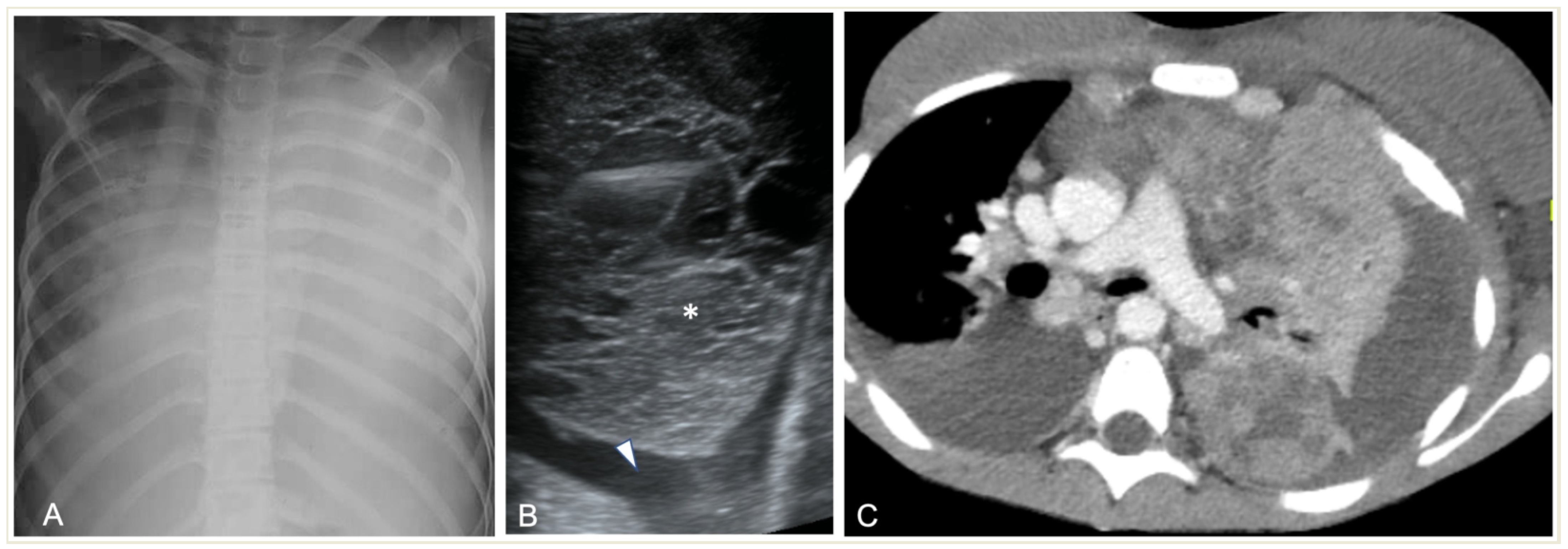
Figure 1. Imaging studies of a 14-year-old girl with respiratory distress, fever, and biopsy-proved non-Hodgkin Lymphoma. (A) Chest radiograph shows complete opacification of left hemithorax with mediastinal shift and some right lung opacities with pleural effusion. (B) LUS shows small left-sided pleural effusion (arrowhead) surrounding a lung mass (*) with multiple fluid-filled cavities. (C) Axial contrast-enhanced CT scan shows an inhomogeneously enhancing left lung mass with multiple necrotic areas, thoracic wall infiltration, bilateral pleural effusions, and enlarged mediastinal lymph nodes.
Children 11 00122 g001
Figure 2. Pleural effusions on chest radiographs: (A) meniscus sign (arrows); (B) loculated effusion with convex borders (*) towards the spine.
Children 11 00122 g002
Figure 3. Ultrasonographic appearances of pleural effusions between cursors: (A) simple (anechoic), amenable for drainage under the appropriate clinical setting; (B) complicated, with internal septae (arrow). Also, note confluent B-lines (arrowheads).
Children 11 00122 g003
Figure 4. A 5.5-year-old boy with complicated pneumonia underwent a CT scan because of a differential diagnostic issue between empyema and lung abscesses on CR and LUS. Axial chest contrast-enhanced CT scan with soft-tissue window (A) and lung window (B) depicts a large multiloculated empyema (arrow), collapsed lung parenchyma with cavitary necrosis (arrowhead), and loculated pneumothorax (stars). Further, optimal treatment was drainage of the empyema based upon the chest CT findings.
Children 11 00122 g004
Figure 5. Complicated pneumonia following heart surgery. (A) Chest radiograph demonstrates the blunting of right pleurodiaphragmatic angle and a possible subpulmonary pleural effusion. (B) Axial MRI, T2-weighted sequence. Note the clear depiction of a pleural effusion with septations (arrow).
Children 11 00122 g005
Figure 6. Necrotizing pneumonia in an 8-year-old boy. Chest radiograph shows opacification of the right hemithorax with ipsilateral concave scoliosis and multiple cavities (arrows) with air–fluid levels of variable size.
Children 11 00122 g006
Figure 7. (A) LUS shows a consolidated lung with multiple hypoechoic areas (arrowheads) consistent with necrotising pneumonia in a 5.5-year-old boy. Pleural effusion is also present. (B) Necrotizing pneumonia in a 7-year-old girl; there is extensive consolidation containing hypoechoic areas with an ill-defined wall (arrow) consistent with necrosis.
Children 11 00122 g007
Figure 8. Complicated pneumonia in a 1-year-old boy. (A) Chest radiograph shows opacification of the right hemithorax with areas of consolidation and collapse. (B) Lung US shows an echogenic pleural effusion, making it difficult to distinguish from adjacent echogenic and necrotic lung parenchyma (C). Post-contrast CT scan images depict a large effusion, a loculated air–fluid level in the pleural space (thick arrow), and adjacent collapsed–consolidated lung parenchyma with necrotic areas (arrowhead).
Children 11 00122 g008
Figure 9. Development of necrotic and cavitary pneumonia in a 7-year-old girl. (A) There is a left basal consolidation with a central hypo-enhancing area, consistent with early necrosis. (B) Follow-up CT scan shows extensive areas of non-enhancement, consistent with extensive necrotic pneumonia. (C,D) Follow-up post-contrast scan, soft tissue, and lung window demonstrate cavitary necrosis containing multiple air–fluid levels.
Children 11 00122 g009
Figure 10. A 6-year-old boy, referred to MRI for exclusion of a lung abscess. (A) T2-weighted sequence shows a right lower lobe consolidation with a single air–fluid cavity (area with high and very low signal intensity), without a clearly depicted wall (area of isointensity surrounding the cavity) and an adjacent small pleural effusion (crescent-shaped line of high signal intensity). (B) Post-contrast T1-weighted sequence shows contrast enhancement by the adjacent consolidated lung parenchyma, consistent with the presence of cavitary necrosis.
Children 11 00122 g010
Figure 11. Lung abscess complicated CAP in a 12-year-old girl: (A) chest radiograph shows a cavity with an air–fluid level; (B) contrast-enhanced CT scan demonstrates a cavity with a thick enhancing wall (arrow) containing an air–fluid level, favoring the diagnosis of an abscess.
Children 11 00122 g011
Figure 12. A 17-year-old girl with a pulmonary abscess following septic embolism. Lung MRI, in an axial T2-weighted image, shows areas of high signal intensity consistent with subpleural septic emboli (arrow) and pleural effusion (very high-signal-intensity area dorsally, mostly on the left side). Note the abscess at the left lung base, exhibiting a low signal and relatively thick wall, and containing an air–fluid level (arrowhead).
Children 11 00122 g012
Figure 13. Real-time MRI sequence in the coronal plane in a 1-year-old girl with left upper-lobe pneumonia. Selected images from the free-breathing scan, which took 16 s to acquire and reconstruct (Supplementary Materials section). Within the isointense pulmonary infiltrate, there are areas of bright signal intensity (arrow) and a round area of very low (almost black) signal intensity, suggestive of air inclusions (arrowhead), with an appearance consistent with cavitary necrosis.
Children 11 00122 g013
Figure 14. A 4-year-old girl with complicated pneumonia and a surgically confirmed broncho-pleural fistula. CT scan and coronal reconstruction show right loculated pneumothorax and collapsed lung parenchyma, and suggest the fistula to be an indentation (arrow) at the visceral pleura adjacent to a pneumatocele.
Children 11 00122 g014
Figure 15. A 15-year-old boy with PARDS. (A) Chest radiography shows bilateral, diffuse lower lung opacities, and pleural effusions. (B) CECT shows bilateral ground-glass and alveolar opacities, and interlobular septal thickening with concomitant pleural effusions (arrows).
Children 11 00122 g015
Table 1. Imaging modalities’ pros and cons.
ModalityProsCons
CRWidely available, very quick examinationRadiation exposure, prone to misinterpretation
LUSWidely available, no ionizing radiation, point-of-care examinationUser-dependent and time-consuming examination, findings are dependent on the location
CTWidely available, quick examination; excellent spatial resolution for chest examinations, with high sensitivityRadiation exposure, patient cooperation needed
MRIExcellent soft tissue contrast, no ionizing radiationNot widely available, patient cooperation needed, time-consuming examination, newer techniques and software needed for lung examinations to improve spatial and temporal resolution
Table 2. Suggested imaging algorithm according to clinical indication.
Complicated CAPUncomplicated CAP
Pleural EffusionEmpyemaNecrotising PneumoniaAbscessPneumatocelePleural FistulaPARDS
US:
usually appropriate
US/CT:
usually appropriate
US/CT:
usually appropriate
US/CT:
usually appropriate
CT:
usually appropriate
CT:
usually appropriate
CR:
usually appropriate
No imaging is necessary
CR/CT:
may be appropriate
MRI:
may be appropriate
MRI:
may be appropriate
MRI:
may be appropriate
CR:
may be appropriate
US/CT:
may be appropriate
MRI:
usually not appropriate
CR:
usually not appropriate
CR:
usually not appropriate
CR:
usually not appropriate
US/MRI:
usually not appropriate
CR/US/MRI:
usually not appropriate
MRI:
usually not appropriate
Disclaimer/Publisher’s Note: The statements, opinions and data contained in all publications are solely those of the individual author(s) and contributor(s) and not of MDPI and/or the editor(s). MDPI and/or the editor(s) disclaim responsibility for any injury to people or property resulting from any ideas, methods, instructions or products referred to in the content.