Article

A Novel Ratiometric Fluorescent Nanosensor Based on N-CDs@UiO-66-NH2 for Sensitive and Selective Detection of Nitrite


1
School of Medical Engineering, Henan Medical University, Xinxiang 453003, China
2
Xinxiang Key Laboratory of Neurobiosensor, Xinxiang 453003, China
3
School of Mathematical Medicine, Henan Medical University, Xinxiang 453003, China
*
Author to whom correspondence should be addressed.
Chemosensors 2026, 14(7), 156; https://doi.org/10.3390/chemosensors14070156

Submission received: 27 May 2026 / Revised: 5 July 2026 / Accepted: 6 July 2026 / Published: 8 July 2026
(This article belongs to the Special Issue Advancements of Chemosensors and Biosensors in China—3rd Edition)

Abstract

Nitrite (NO2) is a crucial environmental and food safety indicator, and excessive intake poses severe threats to human health; thus, highly sensitive and selective detection methods are urgently needed. Herein, a ratiometric fluorescent nanosensor based on N-CDs@UiO-66-NH2 was constructed via a facile assembly strategy for sensitive and selective nitrite detection. The composite demonstrated dual-emission fluorescence at 456 nm and 730 nm under 365 nm excitation, originating from UiO-66-NH2 and N-CDs, respectively, enabling an intrinsic self-referencing signal. The fluorescence intensity ratio exhibited a good linear response toward nitrite in the range of 10–100 μM and 100–450 μM with a limit of detection (LOD) 1.76 μM. The nanosensor showed high selectivity and anti-interference ability against various ions and molecules. Furthermore, it was successfully applied to nitrite detection in lake water samples with satisfactory recoveries (97–101%) and low relative standard deviations (<1.8%). This work provides a simple and effective approach for nitrite monitoring in environmental samples.

1. Introduction

As a key indicator in environmental and food safety monitoring, nitrite (NO2) has long attracted intense attention from researchers and regulatory agencies. The main sources of NO2 include overuse of nitrogen-containing fertilizers, discharge of industrial wastewater, and its application as a preservative and freshness agent in food processing [1]. Although nitrite shows preservative and antibacterial effects at appropriate dosages, excessive intake presents serious hazards to human health [2]. In the acidic gastric environment, NO2 can react with primary and secondary amines to form highly carcinogenic N-nitrosamines, which have been categorized as Group 2A carcinogens by the World Health Organization (WHO) and the International Agency for Research on Cancer (IARC) [3]. Epidemiological investigations have confirmed that high nitrite exposure is closely associated with gastrointestinal cancers, thyroid dysfunction, and cardiovascular diseases [4]. Therefore, the development of sensitive, highly selective, and convenient analytical methods suitable for rapid on-site detection is of great significance for environmental protection, food safety, and public health [5,6,7].
Conventional nitrite detection strategies include colorimetry, ion chromatography, electrochemical, and high-performance liquid chromatography [8,9,10]. Despite their favorable accuracy, these methods often rely on sophisticated instruments, involve tedious sample pretreatment, are time-consuming and make it difficult to realize real-time monitoring, which restricts their application in rapid and portable analysis [11]. In response, fluorescent probe technology has received growing research interest owing to its high sensitivity, simple operation, fast response, and visual detection capability, making fluorescence-based approaches a research hotspot for nitrite determination in recent years [12]. However, most traditional fluorescent nitrite sensors rely on the single-emission signal output, which is vulnerable to matrix interference, background signals, and instrumental fluctuations, thus reducing quantitative accuracy or even causing false positives [13,14]. To overcome these limitations, ratiometric fluorescent probes have become an emerging research direction [15]. By employing the intensity ratio of two independent emission peaks as the response signal, with one channel acting as a built-in reference, ratiometric platforms can effectively eliminate interference from probe concentration, excitation intensity, and environmental factors, significantly improving quantitative reliability and anti-interference performance [16,17]. Moreover, ratiometric probes usually display obvious fluorescence color changes, enabling visual identification and making them highly suitable for on-site detection and food safety monitoring [18,19].
Among various fluorescent nanosensing materials, carbon dots (CDs) have attracted considerable attention owing to their abundant precursors, facile synthesis, and high optical properties, such as high quantum yield, tunable emission wavelengths, and good water solubility [13,20]. In particular, nitrogen-doped carbon dots (N-CDs) can effectively regulate the electronic structure and surface states of carbon dots via nitrogen doping, endowing them with stronger fluorescence emission and higher electron transfer efficiency, thus making them ideal building blocks for fluorescence nanosensing systems [21,22]. On the other hand, metal–organic frameworks (MOFs), especially UiO-66-NH2, have been widely explored as promising fluorescence nanosensing platforms due to their high crystallinity, large specific surface area, high chemical stability, and versatile functionalization [23]. Benefiting from its intrinsic blue fluorescence, UiO-66-NH2 can serve as an ideal response unit; meanwhile, the abundant amino groups on its ligands provide sufficient specific reaction sites for the selective recognition of NO2 through diazotization reactions [24,25]. Therefore, the rational integration of N-CDs and UiO-66-NH2 is expected to combine their complementary advantages to construct a sensitive, selective, and reliable ratiometric fluorescence naosensing platform for NO2 detection [26].
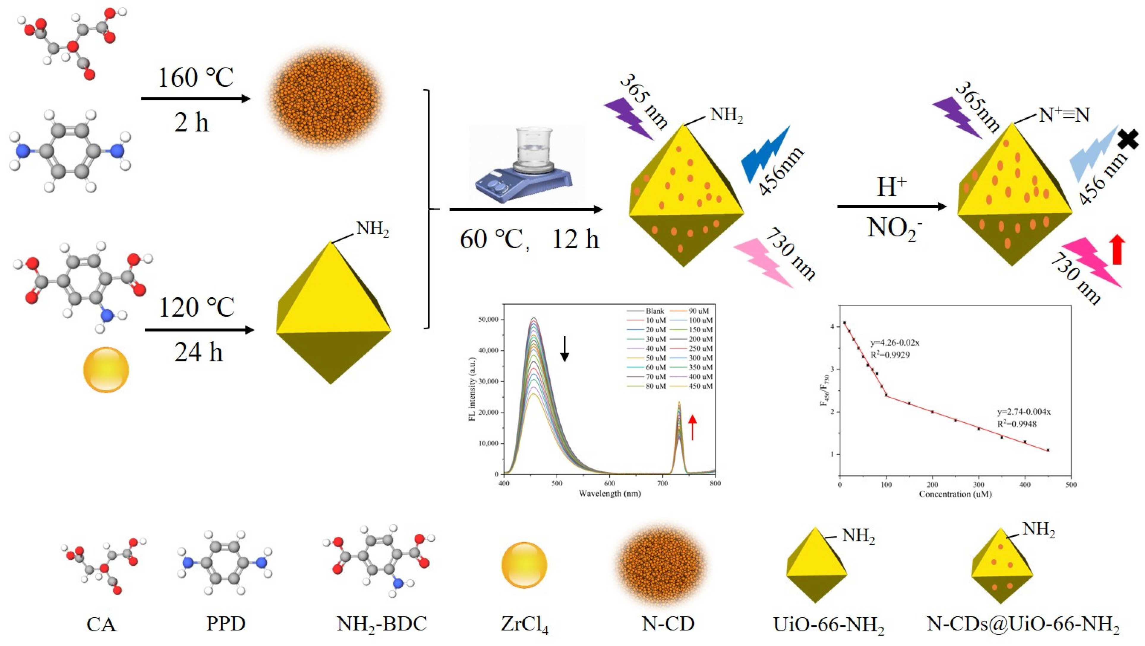
In this work, a ratiometric fluorescent sensor for nitrite detection was developed by incorporating nitrogen-doped carbon dots (N-CDs) into UiO-66-NH2 through a facile one-pot synthesis strategy (Figure 1). The resulting N-CDs@UiO-66-NH2 nanocomposite exhibited two well-resolved fluorescence emission bands centered at 456 and 730 nm under excitation at 365 nm, enabling ratiometric signal output. Upon the addition of NO2 under acidic conditions, a significant decrease in the emission intensity at 456 nm was accompanied by a simultaneous enhancement of the 730 nm emission, generating a distinct concentration-dependent fluorescence ratio response. Furthermore, the fluorescence color gradually changed from blue to red with increasing nitrite concentration, allowing both visual and quantitative detection. Benefiting from the self-calibrating nature of ratiometric sensing, the proposed platform provides a simple, sensitive, and reliable strategy for nitrite determination in environmental monitoring.

2. Materials and Methods

2.1. Materials and Apparatus

Zirconium chloride (ZrCl4), 2-Aminoterephthalic acid (NH2-BDC) with a purity of at least 98%, N, N-dimethylformamide (DMF), Citric acid with a purity of at least 99.5%, p-Phenylenediamine and sodium nitrite, were bought from Aladdin (Shanghai, China). H2SO4 were bought from Damao Chemical Reagent Company (Tianjin, China). Anhydrous ethanol were bought from Hengxing Reagent Company (Tianjin, China). All chemicals were used without further purification.

2.2. Synthesis of N-CDs

N-CDs were synthesized via a hydrothermal method using p-phenylenediamine and citric acid as precursors [27]. Briefly, 0.42 g of citric acid and 0.16 g of p-phenylenediamine were dissolved in 5 mL of deionized water and stirred for 2 min to obtain a homogeneous solution. The mixture was then transferred to a PTFE-lined stainless steel autoclave and reacted at 160 °C for 2 h. After cooling naturally, the obtained N-CDs dispersion was centrifuged at 10,000 rpm for 20 min to remove large particles or precipitates. The supernatant was freeze-dried to obtain solid N-CDs powder, which was stored in a dry environment to avoid moisture-induced performance deterioration.

2.3. Synthesis of UiO-66-NH2

UiO-66-NH2 was prepared via a solvothermal method [14]. Typically, zirconium chloride (ZrCl4, 0.16 g) and 2-aminoterephthalic acid (NH2-BDC, 0.124 g) were dissolved in N,N-dimethylformamide (DMF, 60 mL). After ultrasonic homogenization for 5 min, the mixture was transferred to a 100 mL PTFE-lined stainless steel autoclave and held at 120 °C for 24 h. After cooling to room temperature, the product was collected by centrifugation at 8000 rpm for 5 min. The precipitate was washed with DMF three times and ethanol three times to remove unreacted reagents and residual DMF, followed by vacuum-drying at 60 °C overnight. The final UiO-66-NH2 powder was stored in a desiccator for further use.

2.4. Construction of N-CDs@UiO-66-NH2

Typically, 100 mg of UiO-66-NH2 was dispersed in 8 mL of ultrapure water under sonication until uniform. Then, 2 mL of N-CDs solution (0.5 mg·mL−1) was added, and the mixture was stirred continuously at 60 °C for 12 h. After cooling to room temperature, the precipitate was collected and washed repeatedly with ultrapure water until the washing solution showed no obvious fluorescence signal. Finally, the product was centrifuged and vacuum-dried at 60 °C to obtain N-CDs@UiO-66-NH2. TEM measurements were carried out using an FEI Tecnai G2 F20 transmission electron microscope (FEI Company, Hillsboro, OR, USA). Size measurements (dynamic light scattering, DLS) were performed using a Nano-ZS instrument in water at 25 °C (Malvern, Worcestershire, UK). Fluorescence spectra were acquired using a SHIMADZU RF-6000 fluorescence spectrophotometer (Shimadzu Corporation, Kyoto, Japan) with the excitation wavelength set to 365 nm. Emission signals were scanned over the range of 375–800 nm. For ratiometric quantitative analysis, fluorescence intensities at 456 nm (originating from the linker BDC-NH2 of UiO-66-NH2) and 730 nm (originating from embedded N-CDs) were extracted from the collected spectra. The intensity ratio F456/F730 was defined as the sensing response signal [28].

2.5. Fluorometric Detection of Nitrite

An amount of 0.02 g of N-CDs@UiO-66-NH2 powder was dispersed in 100 mL of ultrapure water under sonication for 20 min to prepare a uniform stock suspension (0.2 mg·mL−1). The resulting colloidal dispersion was optically transparent and remained stable without precipitation for at least 2 h. Before fluorescence measurements, the suspension was gently vortexed to guarantee uniform particle distribution. The sample was transferred directly into a quartz cuvette without centrifugation or filtration. Owing to the small size of the colloidal nanoparticles, light scattering interference was negligible under our measurement conditions, so solid–liquid separation steps were not required [29]. For nitrite detection, 700 μL of deionized water, 100 μL of 0.06 M H2SO4 solution, 100 μL of N-CDs@UiO-66-NH2 suspension, and 100 μL of NO2 solution with different concentrations were mixed sequentially in a test tube. The mixture was incubated for 60 min at room temperature, and its fluorescence emission spectrum was recorded under 365 nm excitation.

2.6. Selectivity Investigation

Selectivity experiments were carried out against various interfering substances, including NaCl, KBr, NaCO3, KI, Pb(NO3)2, CaCl2, Zn(NO3)2, MnCl2, CuSO4, AlCl3, Co(NO3)2, Ni(NO3)2, MgSO4, urea, ammonium, glucose, and ascorbic acid (10 mM for each). For testing, 700 μL of water, 100 μL of 0.06 M H2SO4, 100 μL of N-CDs@UiO-66-NH2 suspension, and 100 μL of interfering substance solution were mixed and incubated for 30 min before fluorescence measurement. A control group containing NO2 (1 mM) was prepared and tested under identical conditions.

2.7. Analysis of Real Samples

Lake water samples were collected from Xinglin Lake outside the North Campus and Boyue Lake within the South Campus of Henan Medical University. All samples were centrifuged and filtered through a 0.45 μm membrane to remove suspended impurities prior to analysis. Standard addition method was employed to evaluate the accuracy by spiking known concentrations of NO2 (0, 10, and 15 μM) into the pretreated water samples. All quantitative tests were conducted following the procedures described in Section 2.5.

3. Results and Discussion

3.1. Characterization of N-CDs@UiO-66-NH2

UiO-66-NH2 was synthesized via a slightly modified literature method [14]. The N-CDs@UiO-66-NH2 nanohybrid was prepared by introducing the as-synthesized N-CDs into a UiO-66-NH2 suspension under vigorous stirring at 60 °C for 12 h to ensure complete integration. To improve dispersion stability and minimize particle aggregation, all samples were subjected to ultrasonic treatment for 20 min prior to optical measurements, electron microscopy sample preparation, and DLS characterization.
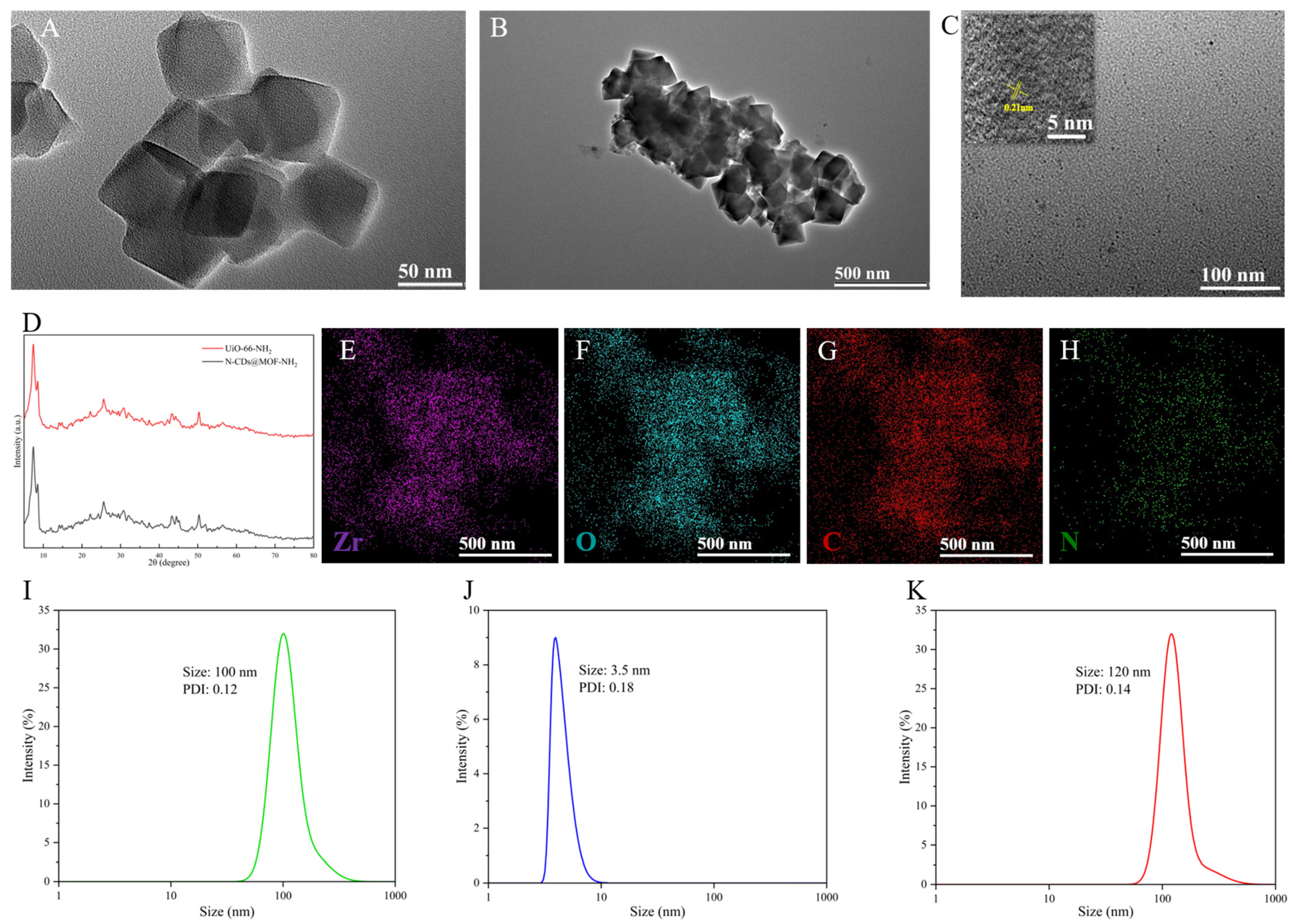
Transmission electron microscopy (TEM) images revealed that UiO-66-NH2 exhibited a well-defined octahedral morphology with an average particle size of approximately 60 nm (Figure 2A). Following N-CDs incorporation, the resulting N-CDs@UiO-66-NH2 retained a similar octahedral architecture, indicating that the hybridization process did not compromise the structural integrity of the MOF scaffold (Figure 2B). The morphology of the pristine N-CDs was examined by TEM, as shown in Figure 2C. The corresponding high-resolution TEM (HRTEM) image (inset of Figure 2C) exhibits distinct lattice fringes with an interplanar spacing of 0.21 nm, corresponding to the (100) plane of graphitic carbon. The presence of clear lattice fringes indicates the formation of highly crystalline N-CDs with a well-developed graphitic structure.
To further elucidate the crystalline phase and chemical composition of the nanohybrid, X-ray diffraction (XRD) and energy-dispersive X-ray spectroscopy (EDS) mapping analyses were performed. The XRD patterns (Figure 2D) demonstrated that N-CDs@UiO-66-NH2 retained the characteristic diffraction peaks of pristine UiO-66-NH2, confirming that the loading of N-CDs did not perturb the crystalline structure of the MOF. Furthermore, EDS elemental mapping images (Figure 2E–H) revealed the homogeneous distribution of Zr, O, C, and N throughout the nanohybrid, collectively verifying the successful construction of the N-CDs@UiO-66-NH2 composite.
Dynamic light scattering (DLS) analysis provided additional evidence for the successful fabrication of the composite. The hydrodynamic diameter increased from 100 nm (UiO-66-NH2) to 120 nm (N-CDs@UiO-66-NH2), with a polydispersity index (PDI) of 0.14, indicating uniform dispersion of N-CDs on the MOF surface without significant aggregation. The N-CDs alone exhibited a peak diameter of 3.5 nm (PDI = 0.18) (Figure 2I–K). Although partial aggregation could be observed in the TEM images due to solvent evaporation during sample preparation, the DLS results obtained from freshly ultrasonicated suspensions indicated that the composite maintains good dispersion in aqueous media.
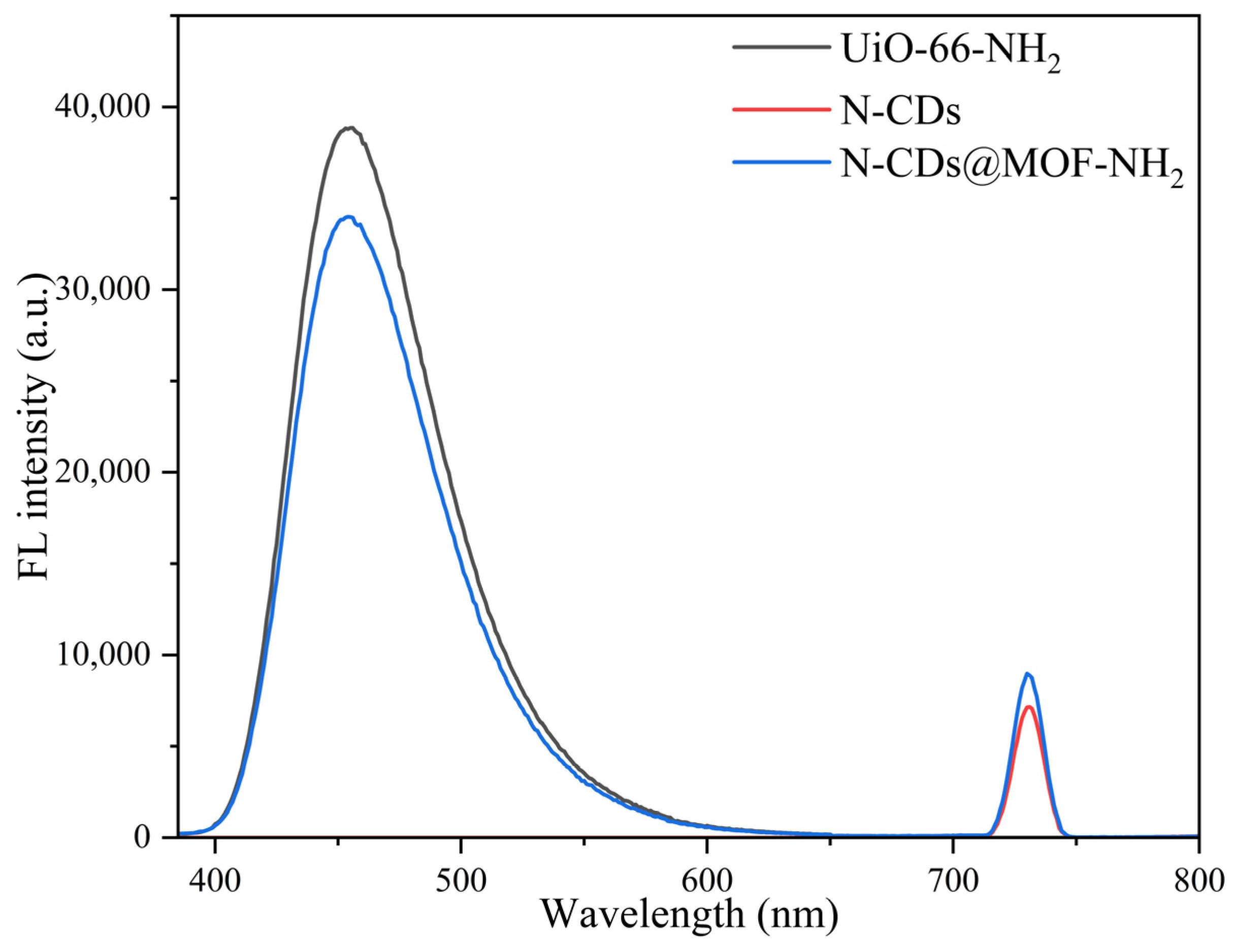
The optical properties of the as-prepared materials were subsequently characterized by fluorescence spectroscopy. Under 365 nm excitation, pristine N-CDs displayed a characteristic near-infrared emission centered at 730 nm, whereas UiO-66-NH2 exhibited a distinct emission peak at 456 nm (Figure 3). Consistent with the preservation of both emissive components, the N-CDs@UiO-66-NH2 nanohybrid simultaneously presented dual emission at 456 nm and 730 nm, establishing the spectroscopic foundation for ratiometric detection.
Notably, the observed emission band at 730 nm coincided with twice the excitation wavelength (2 × 365 nm), raising the possibility that it may originate from second-order Rayleigh scattering rather than intrinsic fluorescence. To verify the origin of this emission, two complementary spectroscopic experiments were performed. First, excitation spectra were recorded by monitoring the emission at 456 and 730 nm, respectively (Figure S1). Both excitation spectra exhibited similar spectral profiles with a common excitation maximum at approximately 365 nm, indicating that the two emission bands originate from the same luminescent system within the N-CDs@UiO-66-NH2 composite rather than from random scattering artifacts. Second, emission spectra were collected under different excitation wavelengths (345, 355, 365, 375, 385, and 395 nm) as shown in Figure S2. As the excitation wavelength varied, the emission maximum at 730 nm remained unchanged, whereas the theoretical second-order Rayleigh scattering position (2λex) shifted linearly with the excitation wavelength. This distinct behavior confirmed that the 730 nm band originated from intrinsic fluorescence of the embedded N-CDs rather than from second-order Rayleigh scattering.

3.2. Feasibility of N-CDs@UiO-66-NH2 for Detecting NO2

Inspired by the well-established diazotization chemistry of aromatic amines, the N-CDs@UiO-66-NH2 nanohybrid was employed as a ratiometric fluorescent probe for nitrite detection. Considering the reaction demands an acidic environment, the pH of the solution was adjusted to 2.0 with sulfuric acid [30]. The dual-emission profile of the nanohybrid (λem = 456 and 730 nm under 365 nm excitation) was found to be sensitive to proton concentration; notably, the fluorescence intensity ratio decreased markedly upon acidification. Consequently, the acidic conditions were systematically optimized prior to quantitative measurements.
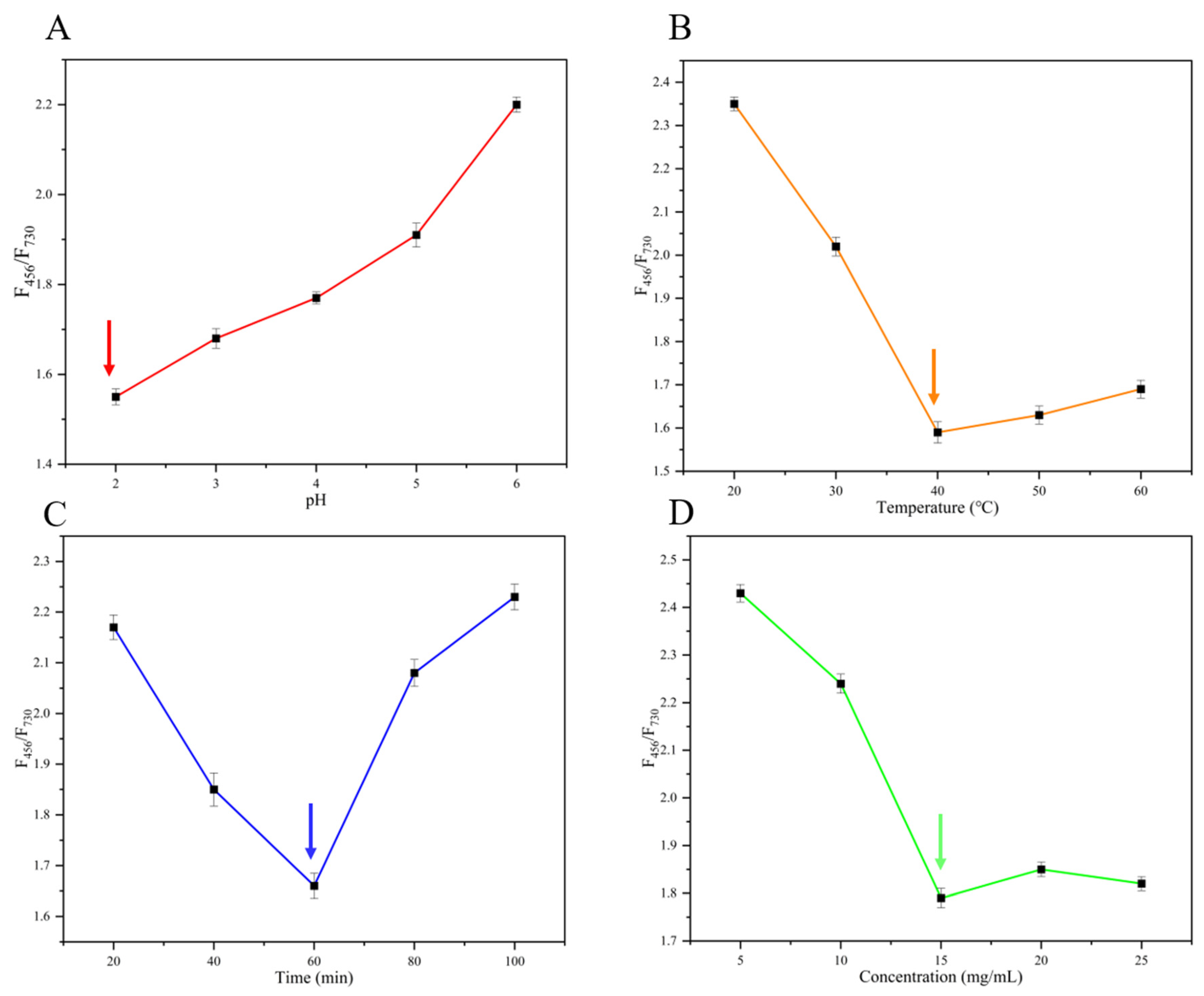
To achieve optimal detection performance for nitrite, four key experimental parameters including pH, reaction time, temperature and material concentration were systematically optimized for the N-CDs@UiO-66-NH2 sensing system. As shown in Figure 4A, the value of F456/F730 gradually increased as the pH increased from 2 to 6, with the minimum ratio observed at pH 2. This indicated that the diazotization reaction between nitrite and the amino groups of UiO-66-NH2 proceeded most efficiently under strongly acidic conditions, leading to the most significant quenching of the 456 nm emission and the largest response difference. Thus, pH 2 was selected as the optimal condition for subsequent experiments. The effect of temperature on the sensing performance was presented in Figure 4B. The ratio F456/F730 decreased sharply from 20 °C to 40 °C and then fluctuated slightly at higher temperatures. The lowest ratio was achieved at 40 °C, suggesting that moderate heating accelerates the diazotization reaction without causing significant thermal quenching or decomposition of the nanoprobe. Therefore, 40 °C was chosen as the optimal reaction temperature. The influence of reaction time was illustrated in Figure 4C. The ratio decreased rapidly from 20 to 60 min, reaching its minimum at 60 min, and then gradually increased with further extension of the reaction time. This trend indicated that the reaction reached equilibrium at 60 min, which was thus selected as the optimal incubation time to ensure complete reaction. Finally, the effect of N-CDs@UiO-66-NH2 concentration was evaluated (Figure 4D). The ratio F456/F730 decreased significantly with increasing nanoprobe concentration, reaching the lowest value at 15 mg/mL, and then remained relatively stable. A concentration of 15 mg/mL was therefore selected as the optimal probe concentration to balance signal sensitivity and consumption. Based on the above optimization results, the optimal conditions for nitrite detection were determined as pH 2, reaction temperature of 40 °C, reaction time of 60 min, and probe concentration of 15 mg/mL, which were adopted for all subsequent quantitative experiments.
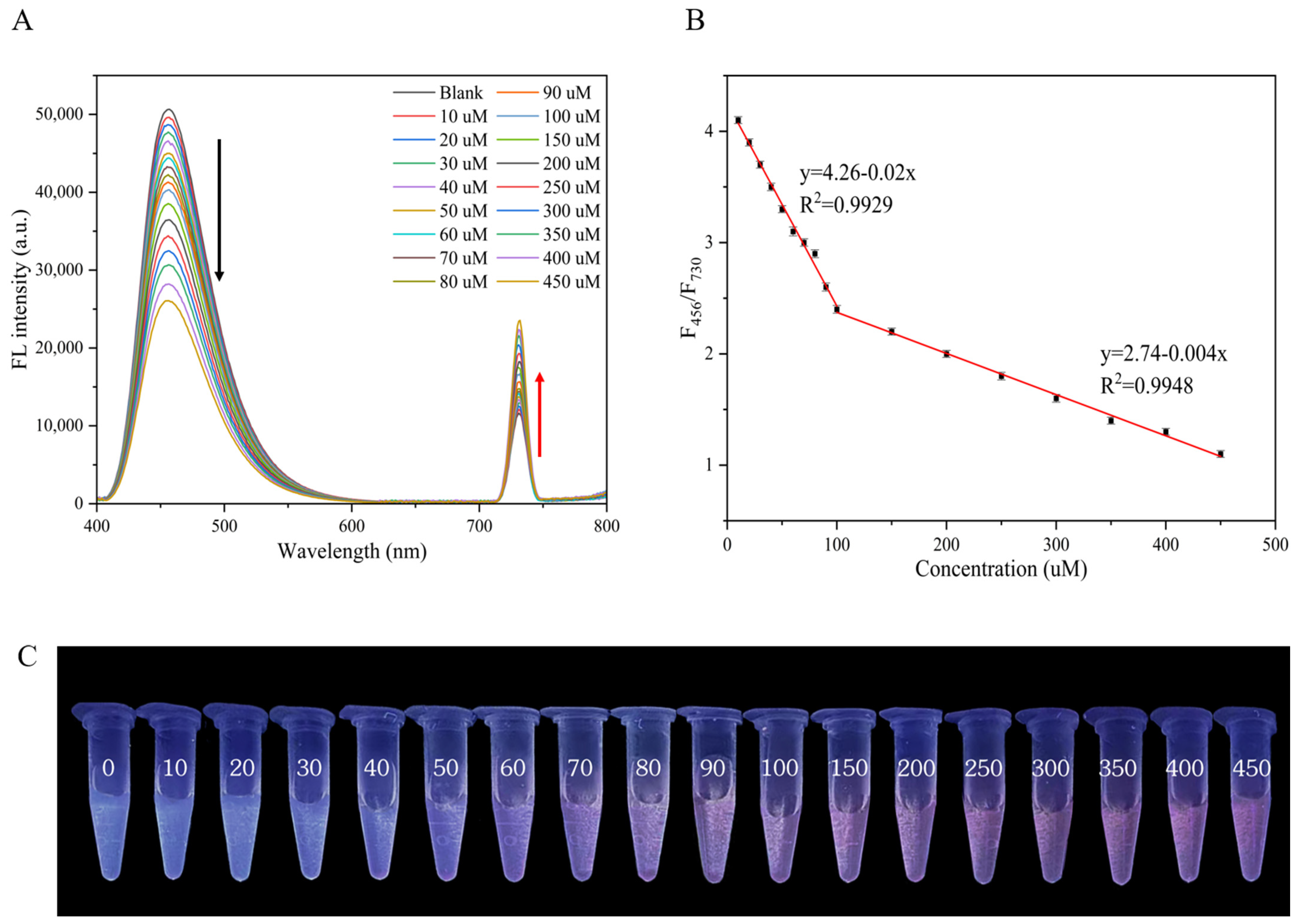
Employing these parameters, the nanosensor was incubated with serial concentrations of NO2 (10–450 μM). After 60 min at 40 °C, the fluorescence spectra were recorded (Figure 5A). The two emission intensities exhibited strong, opposing linear correlations with NO2 concentration across the range of 10–100 μM (R2 = 0.9929) and 100–450 μM (R2 = 0.9948), enabling a robust ratiometric calibration (Figure 5B). Notably, two distinct linear regions were observed in the calibration curve. This behavior is likely related to the porous nature of the N-CDs@UiO-66-NH2 composite and the concentration-dependent accessibility of reactive amino sites. At low nitrite concentrations, diazotization preferentially occurs at amino groups located on the external surface of the material, resulting in a rapid fluorescence response and higher sensitivity. As the nitrite concentration increases, the reaction gradually extends into the internal pores of the MOF, where diffusion limitations and the progressive occupation of reactive sites reduce the response efficiency, leading to a lower slope in the higher concentration range. The limits of detection (LOD) and quantification (LOQ) were calculated according to the equations LOD = 3δ/S and LOQ = 10δ/S, respectively, where δ is the standard deviation of the blank signal and S is the slope of the first linear calibration range. The calculated LOD and LOQ were 1.76 μM and 5.86 μM, respectively. As shown in Figure 5C, the fluorescence color of the N-CDs@UiO-66-NH2 sensing solution gradually changed from blue to red with increasing NO2 concentration under 365 nm UV illumination. This distinct color evolution was consistent with the corresponding ratiometric fluorescence response and could be readily distinguished by the naked eye. The result demonstrated the potential of the proposed sensing platform for rapid visual screening of nitrite.
To further minimize the possible contribution of the inner filter effect (IFE), the concentration of the N-CDs@UiO-66-NH2 suspension was reduced so that the absorbance at both the excitation wavelength (365 nm) and the emission wavelength (456 nm) remained below approximately 0.1 (Figure S3). Under these conditions, the diluted (1:5) sensing system retained essentially the same two-stage calibration behavior toward nitrite as the original system (Figure S4). Although the calculated LOD increased slightly because of the reduced fluorescence intensity after dilution, the calibration slopes, concentration-dependent ratiometric response, and overall analytical conclusions remained unchanged. These results further demonstrated that the observed fluorescence response cannot be explained solely by the inner filter effect, although the IFE may contribute to the overall signal attenuation to a limited extent.
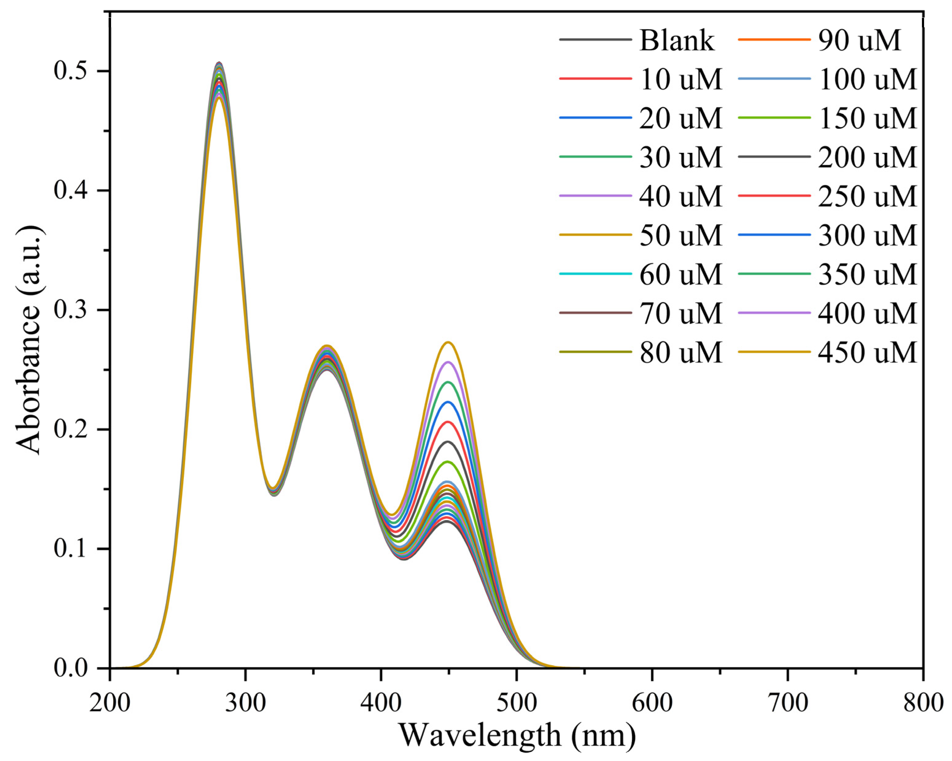
To deeply explore the optical response behavior of the N-CDs@UiO-66-NH2 sensing platform toward NO2, UV–Vis absorption spectra were recorded under the same experimental conditions used for fluorescence measurements. As shown in Figure 6, the characteristic absorption bands located at approximately 280 and 360 nm remained essentially unchanged in position with increasing NO2 concentration, while only slight variations in absorbance were observed. The absence of significant peak shifts indicates that the framework structure of N-CDs@UiO-66-NH2 remains largely intact during the sensing process and that no substantial alteration of the ground-state electronic structure occurs upon nitrite addition.
Compared with previously reported methods, this strategy demonstrated enhanced sensitivity and a wider linear dynamic range (Table 1). In addition, among the reported MOF/carbon-dot hybrid sensors, He et al. developed a UiO-66-NH2-capped carbon-dot nanosensor for ratiometric nitrite detection [31]. Although both studies employ UiO-66-NH2/carbon-dot hybrid materials, notable differences exist in their sensing behavior and signal output characteristics. In the present work, the N-CDs@UiO-66-NH2 composite exhibits an opposite dual-signal response upon exposure to nitrite, where fluorescence quenching of the MOF component is accompanied by simultaneous enhancement of N-CD emission. This signal-coupled response produces a concentration-dependent ratiometric signal and a distinct fluorescence color transition from blue to red under UV illumination, enabling both quantitative analysis and visual detection. Furthermore, the dual-emission behavior provides a useful model for understanding the photophysical interaction between the MOF and CDs, which may facilitate the rational design of future MOF/CDs-based ratiometric sensing platforms.

3.3. Responsive Mechanism of N-CDs@MOF-NH2 to NO2

Traditional single-signal fluorescence detection methods are typically susceptible to external conditions (e.g., probe concentration, excitation intensity) and exhibit low efficiency for nitrite detection in complex samples or aqueous matrices [38]. To overcome this limitation, a self-calibrated fluorescence sensing platform was constructed based on the dual independent fluorescence emission peaks of N-CDs@MOF-NH2. This ratiometric strategy effectively reduces interference from the external environment and enhances signal readability, thereby enabling more sensitive and reliable detection of NO2 [39].
Under optimized experimental conditions, the N-CDs@UiO-66-NH2 sensing system exhibited a distinct dual-fluorescence response toward NO2 over the concentration range of 10–450 μM (Figure 5A). With increasing NO2 concentration, the fluorescence emission of UiO-66-NH2 at 456 nm gradually decreased, whereas the emission centered at 730 nm increased simultaneously. Considering that the UV–Vis absorption spectra showed only minor spectral variations and that probe-dilution experiments yielded nearly identical calibration behavior (Figures S3 and S4), the fluorescence response cannot be explained solely by the inner filter effect (IFE). Instead, the fluorescence response is mainly governed by nitrite-induced modulation of the excited-state photophysical properties of the sensing system. Under acidic conditions, nitrite selectively reacts with the amino groups of the BDC-NH2 linker through a diazotization reaction, converting the electron-donating amino groups into diazonium species. This transformation is expected to reduce the electron-donating capability of the linker and suppress the excited-state intramolecular charge transfer (ICT) process, thereby favoring nonradiative relaxation pathways and resulting in fluorescence quenching at 456 nm. This proposed mechanism is consistent with previous reports in which UiO-66-NH2 was successfully employed as a fluorescent probe for nitrite detection through diazotization-based reactions [40].
Meanwhile, the emission band centered at 730 nm exhibited a concentration-dependent enhancement upon nitrite addition, giving rise to the characteristic ratiometric fluorescence response. Supplementary spectroscopic experiments, including excitation spectra monitored at different emission wavelengths and emission spectra recorded under multiple excitation wavelengths (Figures S1 and S2), provided strong evidence that the 730 nm band originates from the intrinsic fluorescence of the embedded N-CDs rather than from second-order Rayleigh scattering. Although the detailed photophysical origin of the fluorescence enhancement requires further investigation, the reproducible increase in the 730 nm emission, together with the simultaneous quenching of the 456 nm band, establishes a robust self-calibrating ratiometric fluorescence signal. Consequently, the fluorescence intensity ratio (F456/F730) effectively compensates for fluctuations arising from excitation intensity, probe concentration, instrumental drift, and matrix effects, thereby improving the analytical reliability and anti-interference capability of the sensing platform compared with conventional single-emission fluorescent probes.

3.4. Selectivity and Anti-Interference Performance

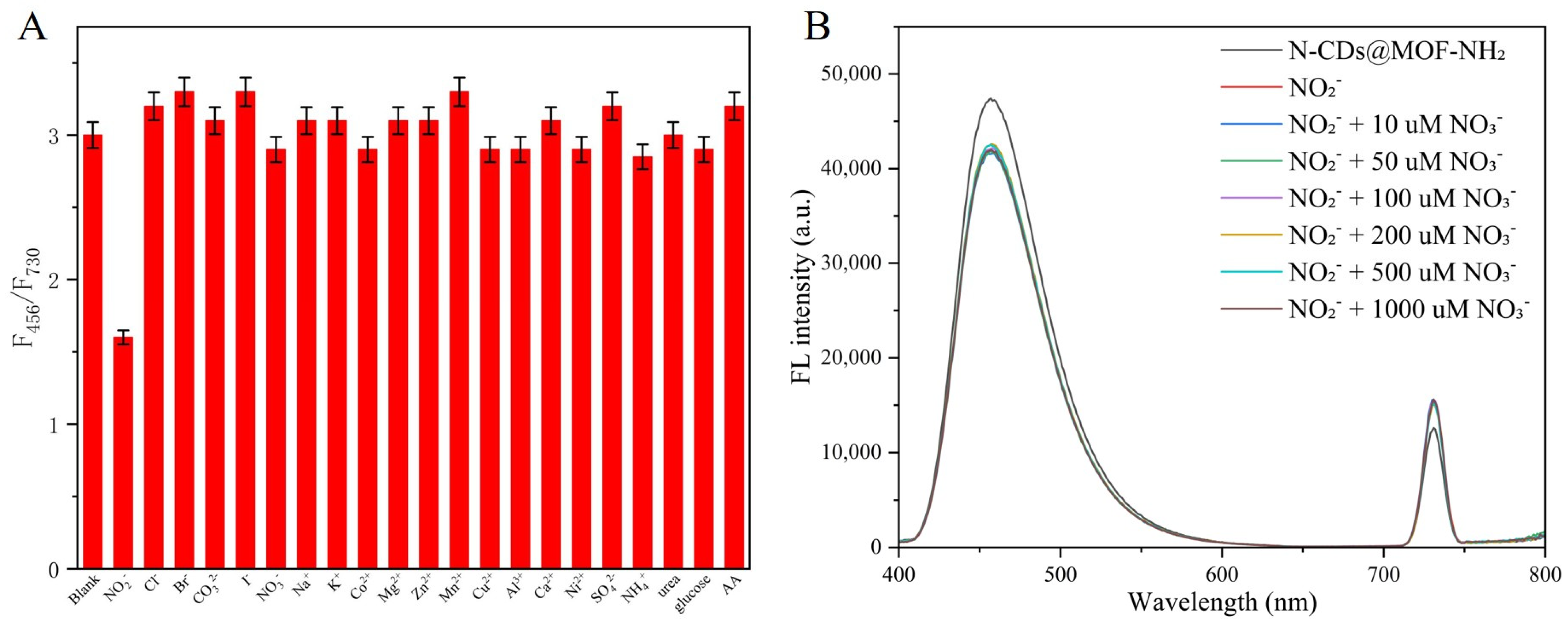
The selectivity and anti-interference ability of the present N-CDs@UiO-66-NH2-based ratiometric nanosensor were systematically investigated by introducing a series of potential coexisting interfering species, including Cl, Br, CO32−, I, NO3, Na+, K+, Ca2+, Mg2+, Zn2+, Mn2+, Cu2+, Al3+, Co2+, Ni2+, SO42−, NH4+, urea, glucose, and ascorbic acid (AA). Notably, NH4+ and urea were included because they are common nitrogen-containing species frequently encountered in environmental and food matrices and could potentially interfere with nitrite determination. As shown in Figure 7A, the fluorescence intensity ratio F456/F730 exhibited a remarkable response toward 100 μM NO2, whereas negligible changes were observed in the presence of 1.0 mM interfering substances (10-fold higher concentration). Neither NH4+ nor urea produced a significant fluorescence response, indicating that the proposed sensing platform is highly selective toward nitrite. These results demonstrated that only NO2 induced a substantial variation in the ratiometric signal, while other coexisting ions and molecules exerted minimal interference even at elevated concentrations. Considering that nitrite and nitrate frequently coexist in environmental and food samples, additional experiments were conducted to evaluate the influence of nitrate on nitrite determination. A fixed concentration of NO2 (100 μM) was measured in the presence of different concentrations of NO3 (10–1000 μM). As shown in Figure 7B, the fluorescence response toward nitrite remained essentially unchanged with increasing nitrate concentration, indicating that NO3 does not significantly interfere with nitrite quantification. This result can be attributed to the fact that the sensing mechanism relies on the diazotization reaction of nitrite with amino groups under acidic conditions, whereas nitrate does not participate in this reaction. Therefore, the proposed sensing platform exhibits excellent discrimination between nitrite and nitrate. The excellent selectivity, together with the satisfactory recoveries obtained in real water samples, confirmed the reliability and strong anti-interference capability of the N-CDs@UiO-66-NH2 sensor and highlighted its potential for nitrite determination in complex environmental matrices.

3.5. Detection of Nitrite in Real Water Samples

To evaluate the practical application potential of the developed method, NO2 in natural lake water samples was analyzed using the standard addition method. As shown in Table 2, the recoveries was calculated according to the equation: recovery (%) = (measured value − endogenous value)/added value × 100%. The recoveries for the spiked samples ranged from 97% to 101%, with the coefficient of variation (CV) below 1.8%. The satisfactory recoveries obtained from lake water samples demonstrated the feasibility of the proposed sensing platform for nitrite determination in environmental water matrices. However, food samples generally contain more complex components, such as proteins, lipids, pigments, and various additives, which may affect the sensing performance. Therefore, although the present results indicate the potential applicability of the N-CDs@UiO-66-NH2 probe for nitrite analysis, further validation using representative food samples is required before practical implementation in food safety monitoring.

4. Conclusions

In summary, a ratiometric fluorescent nanosensor based on N-CDs@UiO-66-NH2 was successfully constructed for the sensitive and selective detection of nitrite. The composite integrated the stable intrinsic fluorescence of UiO-66-NH2 and the strong emission of N-CDs, forming a dual-emission system that is well-suited for self-calibrated fluorescence sensing. The nanosensing mechanism is based on the specific diazotization reaction between NO2 and amino groups in UiO-66-NH2 under acidic conditions, which results in the fluorescence quenching of the UiO-66-NH2. Meanwhile, the fluorescence emission of N-CDs is relatively enhanced due to the weakened inner filter effect, thereby achieving a reliable ratiometric signal output. The proposed nanosensing platform exhibited a good linear response to NO2 in the concentration range of 10–450 μM with a low detection limit of 1.76 μM. It also demonstrated high selectivity for NO2 over a variety of interfering ions and molecules. The proposed N-CDs@UiO-66-NH2 sensing platform enables sensitive and selective determination of nitrite and was successfully applied to lake water samples with satisfactory recoveries. These results demonstrate its potential for environmental monitoring. Further investigations involving complex food matrices will be conducted to evaluate its applicability in food safety analysis.

Supplementary Materials

The following supporting information can be downloaded at: https://www.mdpi.com/article/10.3390/chemosensors14070156/s1, Figure S1: Excitation spectra of N-CDs@UiO-66-NH2 recorded by monitoring the emission at 456 and 730 nm, respectively; Figure S2: Emission spectra of N-CDs@UiO-66-NH2 recorded under different excitation wavelengths (345, 355, 365, 375, 385 and 395 nm); Figure S3: UV–Vis absorption spectra of the N-CDs@UiO-66-NH2 suspension at different dilution ratios (undiluted, 1:2, 1:5, 1:10 and 1:20 dilution); Figure S4: (A) Ratiometric fluorescence response of the 1:5 diluted sensing system toward vary-ing concentrations of nitrite. (B) Corresponding calibration plots of the fluorescence intensity ra-tio F456/F730 versus nitrite concentration.

Author Contributions

T.X.: Methodology, Investigation, Writing—original draft, Writing—review & editing. C.Z.: Validation, Writing—review & editing. Q.R.: Methodology, Validation, Writing—review & editing, Funding acquisition. J.C.: Validation, Writing—review & editing. L.X.: Methodology, Writing—review & editing, Funding acquisition. X.S.: Conceptualization, Writing—review & editing, Supervision. All authors have read and agreed to the published version of the manuscript.

Funding

This research was funded by the Natural Science Foundation of Henan Province, China [grand number 252300423836, 262300420556], the Key Scientific Research Projects of Colleges and Universities of Henan Provincial Department of Education [grand number 26B416003], and the Outstanding Young Teacher of Henan Province [grant number 2025GGJS087].

Institutional Review Board Statement

Not applicable.

Informed Consent Statement

Not applicable.

Data Availability Statement

The original contributions presented in this study are included in the article/Supplementary Materials. Further inquiries can be directed to the corresponding author.

Conflicts of Interest

The authors declare that the research was conducted in the absence of any commercial or financial relationships that could be construed as a potential conflict of interest.

References

  1. Gassara, F.; Kouassi, A.P.; Brar, S.K.; Belkacemi, K. Green Alternatives to Nitrates and Nitrites in Meat-Based Products—A Review. Crit. Rev. Food Sci. Nutr. 2016, 56, 2133–2148. [Google Scholar] [PubMed]
  2. Hathazi, D.; Scurtu, F.; Bischin, C.; Mot, A.; Attia, A.; Kongsted, J.; Silaghi-Dumitrescu, R. The Reaction of Oxy Hemoglobin with Nitrite: Mechanism, Antioxidant-Modulated Effect, and Implications for Blood Substitute Evaluation. Molecules 2018, 23, 350. [Google Scholar] [CrossRef] [PubMed]
  3. Almeida, L.E.F.; Kamimura, S.; De Souza Batista, C.M.; Spornick, N.; Nettleton, M.Y.; Walek, E.; Smith, M.L.; Finkel, J.C.; Darbari, D.S.; Wakim, P.; et al. Sickle Cell Disease Subjects and Mouse Models Have Elevated Nitrite and cGMP Levels in Blood Compartments. Nitric Oxide 2020, 94, 79–91. [Google Scholar] [CrossRef] [PubMed]
  4. Bhatt, H.K.; Molla, F.A.; Das, P. Electrochemical Sensor Based on Polydopamine-Coated Silver Nanowire/Defect-Ridden UiO-66-NH2 Nanocomposites for Trace Detection of Cd(II) and Pb(II) Ions in Water. ACS Appl. Nano Mater. 2025, 8, 23857–23876. [Google Scholar] [CrossRef]
  5. Mirzababaei, A.; Mahmoodi, M.; Keshtkar, A.; Ashraf, H.; Pashayee-Khamene, F.; Khalili, P.; Mirzaei, K. The Association between Dietary Intakes of Nitrate and Nitrite and Odds of Metabolic Syndrome and Its Component in Adults. Sci. Rep. 2026, 16, 10052. [Google Scholar] [CrossRef] [PubMed]
  6. Antczak-Chrobot, A.; Bąk, P.; Wojtczak, M. The Use of Ionic Chromatography in Determining the Contamination of Sugar By-Products by Nitrite and Nitrate. Food Chem. 2018, 240, 648–654. [Google Scholar] [CrossRef] [PubMed]
  7. Murray, E.; Roche, P.; Briet, M.; Moore, B.; Morrin, A.; Diamond, D.; Paull, B. Fully Automated, Low-Cost Ion Chromatography System for in-Situ Analysis of Nitrite and Nitrate in Natural Waters. Talanta 2020, 216, 120955. [Google Scholar] [PubMed]
  8. Maan, D.; Jain, K.; Kumar, A.; Yadav, P.K.; Goutam, U.K.; Tripathi, B.; Jain, S.K. Comprehensive Characterizations and Enhanced Photocatalytic Performance of Zirconium Based Metal-Organic Framework Integrated with Multiwall Carbon Nanotubes under UV Light Irradiation. Sci. Rep. 2026, 16, 19392. [Google Scholar] [CrossRef] [PubMed]
  9. Shanmugam, M.; Agamendran, N.; Sekar, K. Efficient Charge Transfer of P-n Heterojunction UiO-66-NH2/CuFe2O4 Composite for Photocatalytic Hydrogen Production. Catalysts 2024, 14, 341. [Google Scholar]
  10. Alkaltham, M.S.; Althawab, S.A.; Alsulami, T.; Alzahrani, A. N-Doped Antioxidant Carbon Dots as a Bimodal Probe for Nitrite Detection in Commercial Meat Products. Diam. Relat. Mater. 2024, 149, 111624. [Google Scholar] [CrossRef]
  11. Pérez-Chávez, N.A.; Rafti, M.; Albesa, A.G. Computational Study on the Separation of Pentane Isomers in Functionalized UiO-66 Metal-Organic Frameworks. Separations 2025, 12, 152. [Google Scholar]
  12. Kataphiniharn, C.; Unsuree, N.; Janchaysang, S.; Lumjeak, S.; Tulyananda, T.; Wangkham, T.; Srichola, P.; Nithiwutratthasakul, T.; Chattham, N.; Phanphak, S. Portable Multispectral Imaging System for Sodium Nitrite Detection via Griess Reaction on Cellulose Fiber Sample Pads. Sensors 2025, 25, 7323. [Google Scholar] [CrossRef] [PubMed]
  13. Yin, X.; Wang, C.; Wei, S.; Liu, M.; Hu, K.; Song, X.; Sun, G.; Lu, L. Carbon Dots-Based Dual-Mode Sensor for Highly Selective Detection of Nitrite in Food Substrates through Diazo Coupling Reaction. Food Chem. 2025, 463, 141213. [Google Scholar] [CrossRef] [PubMed]
  14. Liang, M.; Gao, Y.; Sun, X.; Kong, R.-M.; Xia, L.; Qu, F. Metal-Organic Framework-Based Ratiometric Point-of-Care Testing for Quantitative Visual Detection of Nitrite. J. Hazard. Mater. 2024, 469, 134021. [Google Scholar] [PubMed]
  15. Vahdatkhoram, F.; Ghoreyshi, A.A.; Chaichi, M.J. Enhancing Heavy Metals Removal from Water by Thiol-Incorporation of UiO-66 Based Metal Organic Framework Nanosheets with l-Cysteine. Inorg. Chem. Commun. 2024, 170, 113351. [Google Scholar] [CrossRef]
  16. Yu, F.; Du, T.; Wang, Y.; Li, C.; Qin, Z.; Jiang, H.; Wang, X. Ratiometric Fluorescence Sensing of UiO-66-NH2 toward Hypochlorite with Novel Dual Emission in Vitro and in Vivo. Sens. Actuators B Chem. 2022, 353, 131032. [Google Scholar]
  17. Song, S.; Zhao, Y.; Li, Y.; Yang, X.; Wang, D.; Wen, Z.; Yang, M.; Lin, Q. pH-Responsive Copper-Cluster-Based Dual-Emission Ratiometric Fluorescent Probe for Imaging of Bacterial Metabolism. Talanta 2021, 221, 121621. [Google Scholar] [PubMed]
  18. Biju, J.R.; Baskaralingam, P.; Thanarasu, A.; Subramanian, S. Fabrication of Z-Scheme UiO-66-NH2/g-C3N4/BiVO4 Heterojunction for Superior Visible-Light Photocatalytic Degradation of Dyes. Langmuir 2025, 41, 32969–32985. [Google Scholar] [CrossRef] [PubMed]
  19. Mohammadi Ziarani, G.; Moradi-Chaleshtori, F.; Mirhosseyni, M.; Feizi-Dehnayebi, M.; Badiei, A. Functionalizing UiO-66-NH2 by 4-chloro-3-Formylcoumarin, as an Optical Chemosensor of Al3+ and F and Its Computational Studies. J. Mol. Struct. 2025, 1325, 140947. [Google Scholar]
  20. Scimone, V.; Albergo, S.; D’Arrigo, G.; Di Bari, I.; Longo, C.; Longo, D.; Sciuto, A.; Tricomi, A. Design and Feasibility Evaluation of a Prototype Setup for Contemporary Easy Nitrates and Nitrites UV Detection in Water for Agriculture. Sensors 2026, 26, 1668. [Google Scholar] [CrossRef] [PubMed]
  21. Heng, C.; He, B.; Wang, L. A Dual-Mode Ratiometric Fluorometric and Colorimetric Platform Based on Nitrogen-Doped Carbon Dots and o-Phenylenediamine for the Detection of Nitrite. J. Fluoresc. 2024, 34, 2157–2167. [Google Scholar] [PubMed]
  22. Zhou, J.; Zhou, X.; Lin, X.; Xia, Y.; Zhao, L.; Huang, K.; Xiong, X.; Yu, H. A Dual-Mode Fluorometric and Colorimetric Sensor Based on N-Doped Carbon Dots for Selective and Sensitive Detection of Nitrite in Food Samples. Sens. Actuators B Chem. 2024, 416, 136045. [Google Scholar]
  23. Timofeev, K.L.; Kulinich, S.A.; Kharlamova, T.S. NH2-Modified UiO-66: Structural Characteristics and Functional Properties. Molecules 2023, 28, 3916. [Google Scholar] [PubMed]
  24. Rajab, N.; Ibrahim, H.; Zhang, D.; Youssef, A.F.A.; Hassan, R.Y.A. Advances in the Chemical Analysis of Nitrite in Environmental and Biological Samples. Nanoscale Adv. 2025, 7, 6321–6372. [Google Scholar] [CrossRef] [PubMed]
  25. Lin, K.-Y.A.; Liu, Y.-T.; Chen, S.-Y. Adsorption of Fluoride to UiO-66-NH2 in Water: Stability, Kinetic, Isotherm and Thermodynamic Studies. J. Colloid Interface Sci. 2016, 461, 79–87. [Google Scholar] [PubMed]
  26. Khan, S.T.; Moosavi, S.M. Connecting Metal-Organic Framework Synthesis to Applications Using Multimodal Machine Learning. Nat. Commun. 2025, 16, 5642. [Google Scholar] [CrossRef] [PubMed]
  27. Jia, J.; Lu, W.-J.; Li, L.; Jiao, Y.; Gao, Y.-F.; Shuang, S.-M. Orange Luminescent Carbon Dots as Fluorescent Probe for Detection of Nitrite. Chin. J. Anal. Chem. 2019, 47, 560–566. [Google Scholar] [CrossRef]
  28. Moore, J.M.; Miller, T.J.; Mu, M.; Peñas-Defrutos, M.N.; Gullett, K.L.; Elford, L.S.; Quintero, S.; García-Melchor, M.; Fout, A.R. Selective Stepwise Reduction of Nitrate and Nitrite to Dinitrogen or Ammonia. J. Am. Chem. Soc. 2025, 147, 8444–8454. [Google Scholar] [CrossRef] [PubMed]
  29. Sultana, N.; Priyadarshini, P.; Parida, K. UiO-66-NH2 and Its Functional Nanohybrids: Unlocking Photocatalytic Potential for Clean Energy and Environmental Remediation. Sustain. Energy Fuels 2025, 9, 3458–3494. [Google Scholar]
  30. Deng, S.; Liu, J.; Han, D.; Yang, X.; Liu, H.; Zhang, C.; Blecker, C. Synchronous Fluorescence Detection of Nitrite in Meat Products Based on Dual-Emitting dye@MOF and Its Portable Hydrogel Test Kit. J. Hazard. Mater. 2024, 463, 132898. [Google Scholar] [PubMed]
  31. He, M.-Q.; Li, H.-W.; Wu, Y. A Novel Ratiometric Fluorescent Nanosensor Based-on UiO-66-NH2 Capped Carbon Dots for Nitrite Determination. Spectrochim. Acta A Mol. Biomol. Spectrosc. 2025, 327, 125422. [Google Scholar] [PubMed]
  32. Ameen, S.S.M.; Algethami, F.; Omer, K.M. Magnetic Rod-Shaped Mn-Based MOF as a Multi-Functional and Recyclable Platform for Dual-Mode Ratiometric-Based Nitrite Detection. Microchim. Acta 2025, 192, 194. [Google Scholar]
  33. Wang, M.; Zhu, H.; Liu, B.; Hu, P.; Pan, J.; Niu, X. Bifunctional Mn-Doped N-Rich Carbon Dots with Tunable Photoluminescence and Oxidase-Mimetic Activity Enabling Bimodal Ratiometric Colorimetric/Fluorometric Detection of Nitrite. ACS Appl. Mater. Interfaces 2022, 14, 44762–44771. [Google Scholar] [CrossRef] [PubMed]
  34. Ponlakhet, K.; Phooplub, K.; Phongsanam, N.; Phongsraphang, T.; Phetduang, S.; Surawanitkun, C.; Buranachai, C.; Loilome, W.; Ngeontae, W. Smartphone-Based Portable Fluorescence Sensor with Gold Nanoparticle Mediation for Selective Detection of Nitrite Ions. Food Chem. 2022, 384, 132478. [Google Scholar] [CrossRef] [PubMed]
  35. Yang, Y.; Wei, S.; Wang, J.; Li, J.; Tang, J.; Aaron, A.A.; Cai, Q.; Wang, N.; Li, Z. Highly Sensitive and Ratiometric Detection of Nitrite in Food Based on Upconversion-Carbon Dots Nanosensor. Anal. Chim. Acta 2023, 1263, 341245. [Google Scholar] [CrossRef] [PubMed]
  36. Adegoke, O.; Zolotovskaya, S.; Abdolvand, A.; Daeid, N.N. Rapid and Highly Selective Colorimetric Detection of Nitrite Based on the Catalytic-Enhanced Reaction of Mimetic Au Nanoparticle-CeO2 Nanoparticle-Graphene Oxide Hybrid Nanozyme. Talanta 2021, 224, 121875. [Google Scholar] [PubMed]
  37. Hao, X.; Shen, A.; Li, M.; Duan, R.; Hou, L.; Zhao, X.; Li, Z.; Zhao, Y.; Zhang, P.; Wang, X.; et al. Simple Method for Visual Detection of Nitrite Using Fluorescence and Colorimetry by Poly (Tannic Acid) Nanoparticles. Anal. Chim. Acta 2023, 1263, 341280. [Google Scholar] [CrossRef] [PubMed]
  38. Yoo, J.; Ryu, U.; Kwon, W.; Choi, K.M. A Multi-Dye Containing MOF for the Ratiometric Detection and Simultaneous Removal of Cr2O72− in the Presence of Interfering Ions. Sens. Actuators B Chem. 2019, 283, 426–433. [Google Scholar]
  39. Baladastian, A.H.; Imanipoor, J.; Mohammadi, M. Simultaneous Adsorption of Ampicillin and Lead by a Highly Stable UiO-66-NH2/TiO2 Nanocomposite from Water: Mechanism, Kinetics, Equilibrium, Thermodynamics, and Regeneration Studies. Ind. Eng. Chem. Res. 2024, 63, 20343–20355. [Google Scholar] [CrossRef]
  40. Hao, X.; Liang, Y.; Zhen, H.; Sun, X.; Liu, X.; Li, M.; Shen, A.; Yang, Y. Fast and Sensitive Fluorescent Detection of Nitrite Based on an Amino-Functionalized MOFs of UiO-66-NH2. J. Solid State Chem. 2020, 287, 121323. [Google Scholar] [CrossRef]
Figure 1. Schematic illustration for the synthesis of N-CDs@UiO-66-NH2 and its application in ratiometric fluorescence detection of NO2.
Chemosensors 14 00156 g001
Figure 2. TEM images of the (A) pristine UiO-66-NH2 and (B) N-CDs@MOF-NH2. (C) TEM image of N-CDs, with the corresponding HRTEM image shown in the inset. (D) XRD spectra of UiO-66-NH2 and N-CDs@UiO-66-NH2. (EH) EDS elemental mappings of Zr, O, C and N for N-CDs@UiO-66-NH2, respectively. Size distribution of (I) UiO-66-NH2, (J) N-CDs and (K) N-CDs@UiO-66-NH2.
Chemosensors 14 00156 g002
Figure 3. Fluorescence spectra of UiO-66-NH2, N-CDs and N-CDs@MOF-NH2, respectively.
Chemosensors 14 00156 g003
Figure 4. Optimization of pH (A), temperature (B), reaction time (C), and concentration (D) factors affecting the fluorescence ratio of N-CDs@MOF-NH2 nanocomposites in nitrite fluorescence sensing.
Chemosensors 14 00156 g004
Figure 5. (A) Fluorescence response of 15 mg/mL N-CDs@UiO-66-NH2 toward nitrite with varying concentrations. (B) Linear fitting plot of the fluorescence intensity ratio F456/F730 versus nitrite concentration. (C) The fluorescent images of N-CDs@MOF-NH2 after reaction with different amount of NO2 under UV radiation of 365 nm.
Chemosensors 14 00156 g005
Figure 6. UV-Vis Absorption Spectra of the N-CDs@UiO-66-NH2 with increasing concentrations of nitrite added.
Chemosensors 14 00156 g006
Figure 7. (A) Selectivity of the N-CDs@MOF-NH2-based detection system toward NO2 against other potential interfering substances. The fluorescence intensity ratio (F456/F730) was recorded upon addition of 100 μM NO2 and 1.0 mM of each interferent, respectively. Data are presented as mean ± SD (n = 3). (B) Effect of nitrate on nitrite determination. Fluorescence intensity ratio (F456/F730) of the N-CDs@UiO-66-NH2 sensing system toward 100 μM NO2 in the presence of different concentrations of NO3.
Chemosensors 14 00156 g007
Table 1. Comparison of the analytical performance (linear range and limit of detection, LOD) between the designed nanomaterial-based sensor and various previously reported material-based nitrite sensors.
MaterialLinear Range (mM)LOD (µM)Ref.
CDs0–0.060.091[13]
Ru@MOF-NH20.002–0.040.6[14]
Mn-MOF0.005–0.0550.18[32]
Mn-CDs0.002–0.151.07[33]
CTAB-AuNPs0.0005–0.10.17[34]
UCNPs0.001–0.020.25[35]
AuNP–CeO2 NP@GO hybrid0.1–54.6[36]
FPTA NPs0–0.360.03[37]
CDs@UiO-66-NH20.0005–0.020.157[31]
N-CDs@MOF-NH20.01–0.1 and 0.1–0.451.76This work
Table 2. Analytical performance of the developed sensing platform for the detection of NO2 in lake water (n = 3).
SampleNO2 Spiked (μM)Ratiometric Fluorescence Detection
Detected (μM)Recovery (%)RSD (%)
Lake water 10///
Lake water 2109.7497.461.84
Lake water 31515.17101.131.68
Disclaimer/Publisher’s Note: The statements, opinions and data contained in all publications are solely those of the individual author(s) and contributor(s) and not of MDPI and/or the editor(s). MDPI and/or the editor(s) disclaim responsibility for any injury to people or property resulting from any ideas, methods, instructions or products referred to in the content.