Review

Microfluidic Paper-Based Sensors and Their Applications for Glucose Sensing

1
Deparment of Electronic Engineering, Gachon University, Seongnam-si 13120, Republic of Korea
2
Deparment of Semiconductor Engineering, Gachon University, Seongnam-si 13120, Republic of Korea
3
Department of Health Sciences and Technology, Gachon University, Incheon 21999, Republic of Korea
*
Authors to whom correspondence should be addressed.
Chemosensors 2025, 13(8), 293; https://doi.org/10.3390/chemosensors13080293
Submission received: 9 July 2025 / Revised: 29 July 2025 / Accepted: 5 August 2025 / Published: 7 August 2025

Abstract

Recently, the incidence of diabetes has increased across all socioeconomic groups, with a notable increase in developing countries. Although advances in medical devices have enhanced healthcare accessibility, these benefits remain largely out of reach for individuals residing in remote areas. Concurrently, a variety of devices have been created to detect glucose biomarkers. Among these, microfluidic paper-based sensors have received substantial attention due to their affordability, disposability, and ease of production. Research on microfluidic paper-based glucose sensors has become particularly prominent owing to their considerable potential and wide applicability, especially in the integration of artificial intelligence and machine learning in glucose sensor processing. This review aims to examine recent advancements and progress in the development of microfluidic paper-based glucose sensors over the past five years, highlighting their advantages, limitations, and prospects. The sensors combined with artificial intelligence and machine learning have potential for future applications.

1. Introduction

The increasing global prevalence of diabetes has received considerable scientific attention [1,2,3]. According to data from the International Diabetes Federation, the incidence of diabetes is steadily increasing each year, with particularly sharp increases observed in developing nations [2]. Diabetes is a major health concern not only due to its direct impact but also because it significantly increases the risk of comorbidities, such as kidney failure and cardiovascular mortality [2]. In light of these concerns, the ability to accurately quantify and monitor glucose levels is essential and offers critical insights for clinicians regarding the management and treatment of patients with diabetes.
Glucose sensors are broadly categorized into enzymatic, non-enzymatic, and hybrid types depending on their core functional elements [4,5,6,7,8]. Enzymatic sensors typically use natural enzymes, such as horseradish peroxidase (HRP) and glucose oxidase (GOx), which have high specificity and catalytic efficiency. In colorimetric sensors, the GOx catalyzes the oxidation of glucose to gluconolactone/gluconic acid and oxygen is reduced to hydrogen peroxide. Subsequently, HRP catalyzes to reduce hydrogen peroxide to water. The presence of hydroxyl radicals in the solution can change the chromogenic substrate from colorless to a typical color [9,10]. In electrochemical glucose sensors, GOx oxidizes glucose to gluconolactone/gluconic acid and oxygen is reduced to hydrogen peroxide. Subsequently, the generated hydrogen peroxide is oxidized at the electrode to generate electricity. In electrochemical glucose sensors, the mediator is reduced at the electrode to produce electrochemical signals [11,12]. However, these systems are hindered by high production costs, difficulties in enzyme purification, and susceptibility to denaturation under harsh environmental conditions [5,13]. To address these limitations, second-generation sensors that reduce the use of natural enzymes have emerged, thereby lowering costs while maintaining considerable specificity. However, their dependence on GOx imposes certain constraints. The third-generation includes nanozyme-based sensors that outperform earlier models in catalytic activity and cost efficiency. Despite these advantages, they generally have reduced specificity, posing a challenge that requires further research and innovation [5,13]. Across all generations, glucose sensors function by detecting glucose biomarkers using a range of signal transduction techniques. These include optical methods, such as colorimetry [14,15,16], fluorescence [17,18], and surface-enhanced Raman spectroscopy [19,20], and electrochemical techniques, such as cyclic voltammetry [21,22,23], chronoamperometry [24,25], differential pulse voltammetry [21,26], mass spectrometry [27,28], and microwave techniques [29,30]. However, these sophisticated techniques often require expensive instrumentation and trained personnel, which limits their use in rural or under-resourced settings. Consequently, there is a pressing need for affordable, portable, and user-friendly glucose sensing platforms to broaden accessibility, particularly in remote areas.
Technological advancements have propelled the integration of biosensors into healthcare devices, including wearable electronics, which have been widely adopted. Despite this progress, designing healthcare devices that align with market demand and personal use, while being accessible in underserved regions, remains a formidable challenge. Recently, paper-based diagnostic devices have emerged as potential solutions, owing to their low cost, portability, disposability, and minimal sample requirements [31,32]. These devices have been successfully applied to a range of biomarkers, including glucose (for diabetes) [32,33], amyloid-beta and tau proteins (for Alzheimer’s disease) [34,35], and various cancer markers [36,37]. Paper-based sensors are compatible with numerous biological fluids, such as plasma [38,39], blood [40,41], sweat [42,43], and urine [44,45]. They can be fabricated using methods, such as wax [46,47] and screen [48,49] printing, and operate through diverse detection modalities, including colorimetric, fluorescent, and electrochemical techniques. With technological developments, the integration of artificial intelligence (AI) and machine learning (ML) has further enhanced the performance and applicability of these devices.
These microfluidic paper-based platforms are particularly well suited for point-of-care testing (POCT), a critical diagnostic approach that enables rapid, accurate, and cost-effective testing without requiring specialized training. POCT is particularly advantageous in resource-limited environments, offering time-efficient diagnostics [50,51]. This approach has been applied across various domains ranging from monitoring human health indicators [52] to detecting environmental contaminants [53] and identifying disease markers [51]. Owing to its practicality and responsiveness to urgent diagnostic needs, POCT has recently undergone rapid advancements.
Therefore, the development of microfluidic paper-based glucose sensors is of strategic importance to expand their real-world applications and improve their accessibility, especially by integrating AI and ML. This review provides a comprehensive overview of the recent advancements in microfluidic paper-based glucose-sensing technologies. It also explores the recently emerging applications of glucose sensors, and discusses future directions and ongoing challenges in this rapidly evolving field.

2. Recent Development of the Paper-Based Glucose Sensor and Their Application in Glucose Sensing

2.1. Overview of Microfluidic Paper-Based Sensors

Paper-based devices, paper chips, and microfluidic paper analytical devices (µPADs) are analytical devices fabricated on paper substrates; however, they have distinct structures and complexities. A paper-based device refers to all devices fabricated on paper, such as pH or pregnancy test trips [54,55]. However, this method does not require microfluidic components. Paper chips are small and compact devices made on paper with one or several reaction zones and do not require a microfluidic channel, as with paper-based devices [56,57]. The µPAD contains the microfluidic channel to route the liquid flow through capillary driving force [58,59]. This enabled multiple biochemical reactions to occur in the zones designed on the paper. Compared with the paper-based device and paper chip, the µPAD are more sophisticated, allowing more multi-step analysis in various applications.
The µPAD has garnered substantial interest due to their expanding utility across various fields, including biotechnology, biomedicine, food safety, and the chemical industry [58,59]. In these systems, cellulose-based paper serves as the primary substrate, providing essential functions, such as reagent filtration and solution transport via capillary action without the need for external pumping [32]. Compared with traditional substrates, such as silicon, glass, and plastic, cellulose stands out for its light weight, flexibility, inherent porosity, chemical and biological inertness, and tunable physicochemical characteristics [31]. The structural properties of the microfluidic paper are integral to facilitating fluid permeability, enabling sample filtration, and anchoring reagents onto its surface. Moreover, membranes fabricated on cellulose substrates provide mechanical robustness and elasticity, thereby enhancing the device functionality [31]. Given these advantages, the µPAD is a promising platform for POCT, and their further development is both timely and essential.
A critical component of the design of microfluidic paper-based sensors is the creation of hydrophobic channels. These barriers are vital for accurately directing fluid flow while preventing unintended leakage. Fabrication techniques for these channels generally fall into two categories: non-printing and printing methods. Nonprinting approaches include photolithography [60], chemical vapor deposition (CVD) [61], and laser-based patterning [62]. Among these, photolithography is a precise and cost-prohibitive technique because of the need for expensive, high-end instrumentation. Similarly, CVD requires sophisticated and expensive equipment. Although laser treatment allows for a one-step fabrication, it is limited by the high operational cost and rigidity of the resulting devices, which restrict their foldability. Examples of µPADs produced via photolithography, laser processing, and wax dipping are illustrated in Figure 1.
Additional fabrication techniques for µPAD include screen printing [64], embossing [65], and stamping [54]. Among printing-based methods, wax and screen printing are particularly prominent. In wax printing, a wax layer is deposited onto a microfluidic paper substrate and subsequently subjected to heat treatment to define the hydrophobic barriers. This technique typically yields higher resolution than that with screen printing. However, each method has distinct advantages and limitations, and the choice of fabrication method often depends on the specific application requirements and accessibility of the equipment. An example of a sensor fabricated using wax printing is shown in Figure 2.

2.2. Overview of Microfluidic Paper-Based Glucose Sensors

As discussed previously, microfluidic paper-based sensors have demonstrated versatility across a wide range of applications, including the detection of diabetes, cancer, and COVID-19. Diabetes-related diagnostics are particularly prominent due to their close relevance to daily health monitoring. Similarly to µPADs, paper−based glucose sensors are typically fabricated by incorporating enzymes or nanozymes into cellulose substrates. The core functional components of these sensors are GOx and HRP. Chromogenic substrates are often integrated to facilitate visual detection, allowing colorimetric responses to be easily observed by the naked eye and quantitatively analyzed using smartphone cameras or smart imaging systems coupled with analytical software. Research on the design, fabrication, and real-world applications of microfluidic paper-based glucose sensors is of substantial importance, particularly in the realm of biomedical diagnostics.

2.3. Signal Analysis and Technological Relevance

In paper-based sensors, the chemical reactions occurring on a paper substrate can be observed either visually or through advanced instrumentation. Signal processing techniques include colorimetry, electrochemistry, and mass spectrometry. Each method was applied to a specific type of sensor. This section addresses the types of signals generated and methods for processing them to extract meaningful information.

2.3.1. Paper-Based Colorimetric Sensor

Paper-based colorimetric sensors detect chemical or biological substances based on color changes that correlate with the concentration of the analyte. In sensor design, a chromogenic agent is typically used to produce a visible color following a chemical reaction. Various colors, such as blue, yellow for 3,3′,5,5′-tetramethylenebenzidine, green for 2,2′-azino-bis(3-ethylbenzothiazoline-6-sulfonic acid), and yellow for cerium oxide [67,68,69], have been observed. These color changes can be detected using the naked eye, portable spectrophotometers, scanners, digital cameras, and smartphones [32,70,71], with smartphone cameras being common. Quantitative analysis is typically performed using a spectrophotometer at a specific wavelength corresponding to a chromogenic substrate. However, these spectrophotometers are not designed to quantify colorimetric signals from paper substrates; therefore, electronic devices with integrated cameras, including smartphones, digital cameras, and scanners, offer a more practical solution for capturing these signals, supported by analytical software for quantitative analysis. Given the widespread use of smartphones, scanners, and digital cameras, this method is both cost-effective and convenient.

2.3.2. Paper-Based Electrochemical Sensor

The electrochemical sensor was constructed with a configuration of two, three, or four electrodes that converted chemical redox reactions into measurable electrical signals. These electrical signals can be detected using advanced electronic systems that filter out noise and amplify the signals. Compared with colorimetric systems, electrochemical sensors are simpler and more sensitive, and provide faster response times. In paper-based sensors, electrodes, such as working, counter, and reference electrodes are fabricated on paper substrates using techniques, such as screen or inkjet printing [41,43,72], and are made from conductive materials, including carbon, conductive polymer, metals, and metal oxides. Signal processing in paper-based sensors follows the same principles as in conventional sensors, using an electronic circuit board for data acquisition.

2.3.3. Paper-Based Mass Spectrometry

In this approach, the sample is applied to a paper substrate, where it is dried to enhance its stability. A microliter of the solvent was introduced into the substrate [73,74]. A high voltage in the kilovolt range was applied to the paper, initiating the ionization process, which converts all molecules into their ionic or charged forms. These ions were subsequently directed to a mass spectrometer, which measured the molecule-to-ion ratio, thereby providing both qualitative and quantitative information about the analyte. This method is rapid and suitable for POCT applications.

3. Study of the Paper-Based Glucose Sensor Advancement Through Typical Examples

In this section, we discuss microfluidic paper-based glucose sensors, including enzymatic, non-enzymatic, and hybrid glucose sensors.

3.1. Recent Development of Enzymatic Microfluidic Paper-Based Glucose Sensor

The earliest microfluidic paper-based biosensors for glucose detection were developed using natural enzymes, such as GOx and HRP. For example, Amor-Gutiérrez et al. reported a microfluidic paper-based enzymatic sensor designed for glucose monitoring [75]. In their approach, glucose oxidase, HRP, and ferrocyanide were deposited onto a carbon-based working electrode, which was then aligned with the reverse side of the screen-printed electrode. Through optimization, the ideal enzyme concentrations were determined to be 1.6 U/µL for GOx and 2.5 U/µL for HRP. This sensor exhibited a linear detection range from 0.3 to 15 mM with a detection limit of 0.12 mM and demonstrated good reproducibility, with a relative standard deviation of 5.4% in real food samples.
Jun et al. developed a microfluidic paper-based enzymatic glucose sensor using a wax printing technique on a cellulose substrate [76]. The sensor consisted of two functional layers: the first served as a fluidic channel directing the sample from the application site to the detection zone, whereas the second housed the detection zone itself, containing GOx, HRP, and chromogenic reagents for visual signal generation. This sensor was capable of detecting glucose concentrations ranging from 0 to 20 mM (or 0 to 17 mM, specifically in human serum) within a matter of minutes.
Despite their high sensitivity and specificity, enzymatic paper-based glucose sensors are often limited by high production costs and limited long-term stability.

3.2. Recent Development of Hybrid Microfluidic Paper-Based Glucose Sensor

In this section, the development of a microfluidic paper-based glucose sensor is examined with a particular focus on replacing either GOx or HRP with a synthetic enzyme.
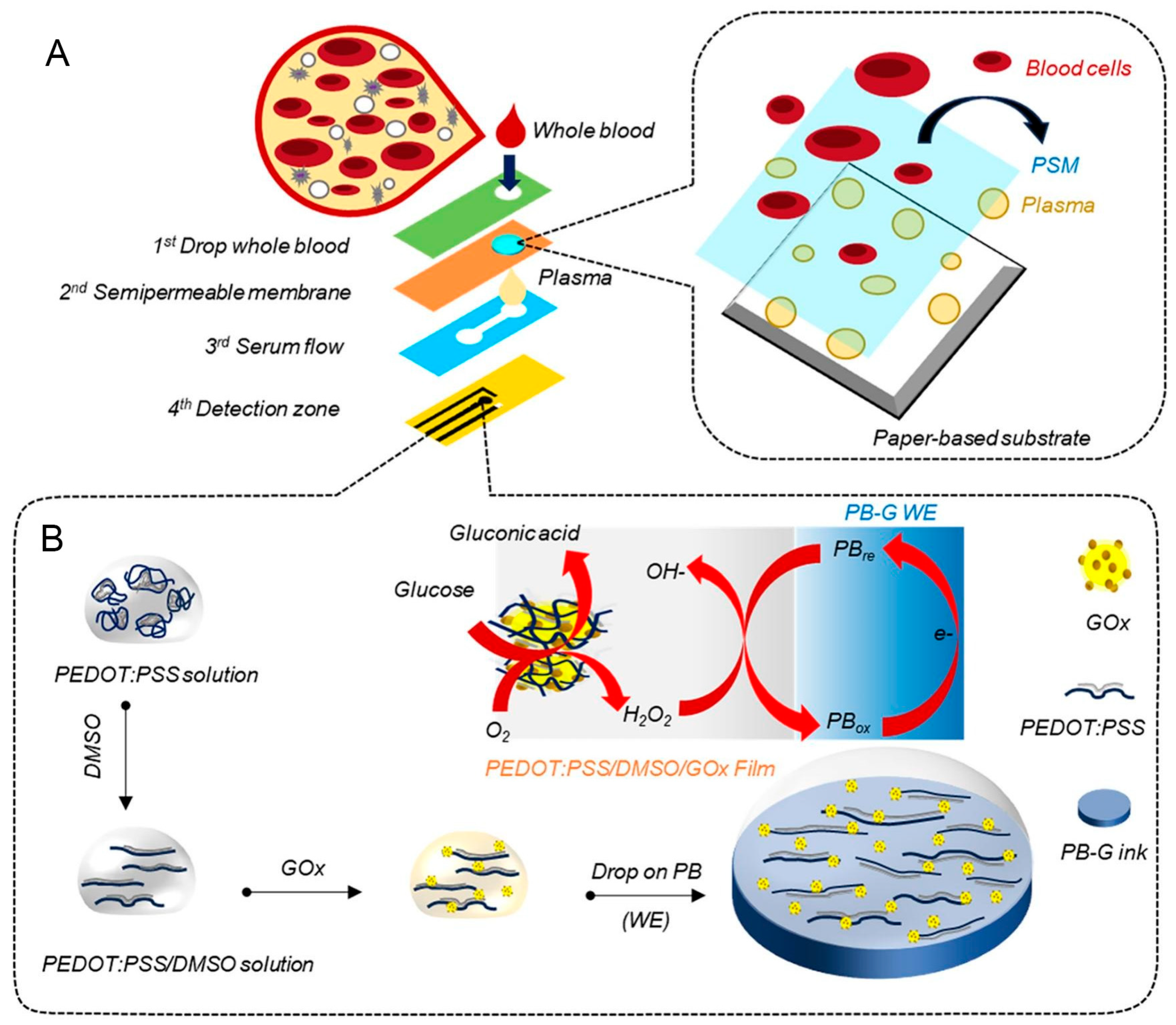
Tong et al. [8] constructed a multilayer microfluidic paper glucose sensor by incorporating Prussian blue nanoparticles, glucose peroxidase, and a chromogenic reagent into PES paper. This assembly was subsequently overlaid with the filtration and spreading layers. The resulting sensor demonstrated the capability to detect glucose concentrations ranging from 2.5 to 25 mM in blood, with a detection limit of 0.13 mM and coefficient of variation below 4%. In a separate study, Somsiri et al. [7] developed a three-dimensional paper-based microfluidic electrochemical sensor integrated with a smartphone and NFC-enabled potentiostat for glucose quantification in human blood plasma. Hydrophobic microfluidic channels were patterned on chromatographic paper using wax printing, whereas the electrode was fabricated via screen printing using Prussian blue-graphite ink. This ink was further modified by incorporating PEDOT:poly(sodium 4-styrenesulfonate) (PSS) blended with DMSO, followed by the immobilization of GOx onto the dried electrode surface to facilitate glucose catalysis. The device comprises four distinct layers: the top layer serves as the sample inlet zone; second layer, a semipermeable membrane, filters plasma from whole blood through gravitational separation; third layer contains microfluidic channels that direct the filtered plasma; and fourth layer hosts the screen-printed, enzyme-modified electrode. The sensor demonstrated a linear glucose detection range from 0.2 to 12 mM in phosphate-buffered electrolyte, with a markedly low detection limit of 0.034 mM. The performance of the sensor was comparable to that of standard clinical methods and commercially available glucometers. An illustration of the sensor fabrication process and glucose detection workflow is provided in Figure 3.
In a separate study, Le et al. [32] developed a microfluidic paper-based sensor featuring six detection zones for the quantification of glucose biomarkers in human serum [32]. The sensor utilized Zn, N-co-doped reduced graphene oxide as a functional material. Fabrication was performed via wax printing on a microfluidic paper substrate to define the hydrophobic microchannels. A graphene-based nanozyme was immobilized on a cellulose surface to which GOx was covalently attached. To prevent reagent leakage, a Nafion membrane was applied over the detection zones. The sensor exhibited a linear detection range of 1–30 mM with detection limit of 0.78 mM, making it suitable for monitoring both hypoglycemic and hyperglycemic conditions.
Although hybrid glucose sensors partially mitigate the challenges posed by natural enzyme use, leading to reduced production costs, the continued reliance on enzymatic components imposes limitations on long-term storage stability and restricts the feasibility of deployment in remote or resource-limited settings.

3.3. Recent Development of Non-Enzymatic Microfluidic Paper-Based Glucose Sensor

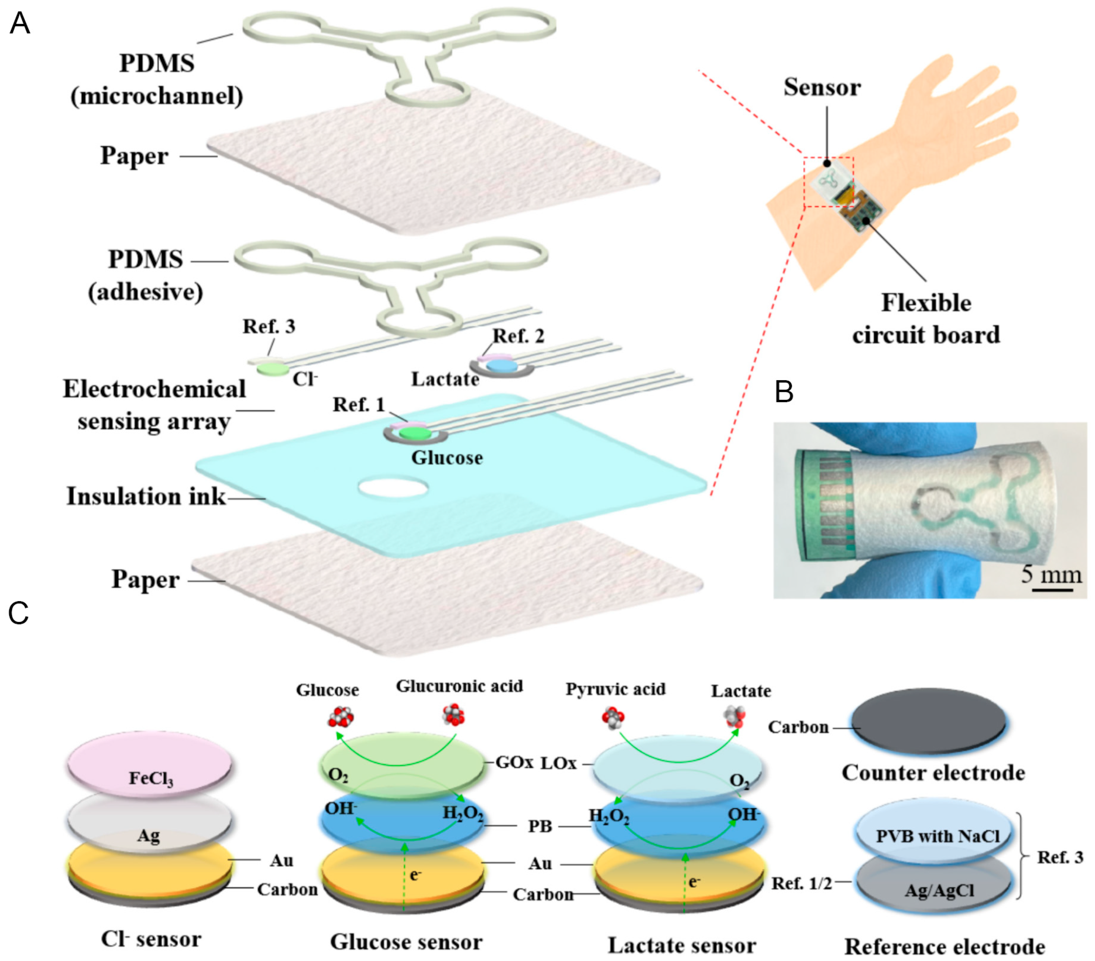
This section focuses on microfluidic paper-based non-enzymatic glucose sensors, wherein traditional enzymes, such as GOx and HRP are substituted by nanozymes. Mei et al. [66] developed a non-enzymatic microfluidic paper sensor using gold (Au) and Prussian blue to electrochemically detect glucose and lactate in sweat. The sensor architecture used a layer-by-layer design, beginning with the screen printing of an insulating layer with an inlet onto a paper substrate. Subsequently, an electrode array, modified with Au and Prussian blue, was screen printed onto the insulation layer for analyte detection. A polydimethylsiloxane (PDMS) adhesive layer was then applied, followed by the attachment of a second paper layer over the electrode. The final PDMS layer, which was printed and cured onto the second paper layer, ensured robust bonding between the layers. The patterned PDMS contains an inlet, three microfluidic channels, and three assay chambers. Au and Prussian blue were deposited on the working electrodes, followed by the addition of GOx and lactate oxidase, which were encapsulated in a Nafion membrane. The sensor exhibited linear detection ranges of 0–20 mM for lactate and 0–200 µM for glucose, with respective detection limits of 0.47 mM and 4.8 µM. Sensitivity was notably high, at 63.3 nA·mM−1 for lactate and 3.1 nA·µM−1 for glucose. In addition, a device integrated with an electronic circuit enables Bluetooth connectivity. It effectively collected sufficient sweat and generated measurable signals after 15 min of exercise.
Yeon et al. [33] designed a microfluidic paper-based glucose sensor that combined electrochemical and colorimetric detection modes. Fabricated on Whatman paper via screen-printing and electrochemical processes, the sensor featured GOx/Prussian blue/carbon as the working electrode, PANI/ITO/carbon as the counter electrode, and Ag/AgCl as the reference electrode. It detected glucose in phosphate-buffered saline (PBS, pH 5) with a linear range of 0–0.2 mM and detection limit of 126 µM, demonstrating rapid response within 30 s. The sensor exhibited excellent selectivity, even in the presence of potential interferents such as ascorbic and uric acid. Glucose concentrations were successfully quantified in commercial beverages (Coca-Cola and orange juice) diluted 500-fold and commercial human serum diluted 20-fold in PBS, achieving a confidence level of 95%.
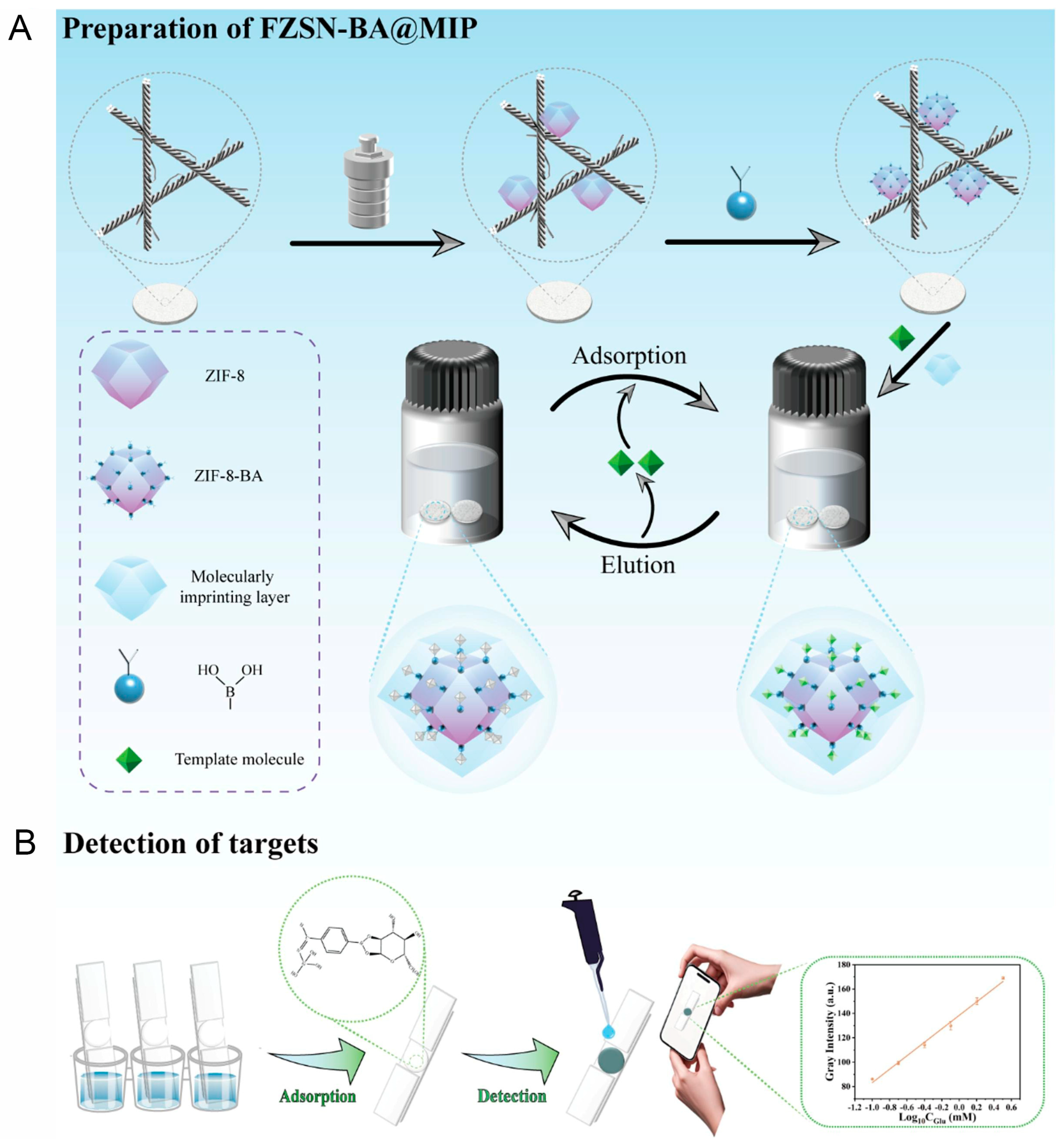
Li et al. [77] fabricated a microfluidic paper chip using metal–organic framework (MOF)/molecularly imprinted polymer (MIP) biomimicry to detect glucose in salivary microfluidics. The device was constructed on a Whatman No. 1 filter paper, which was first soaked in hydrochloric acid for 30 min and subsequently sonicated in PSS for another 30 min to obtain FP@PSS. Successive layers of ZIF-8 (FZ), FZ@SiO2 (FZS), FZS@NH2 (FZSN), and FZSN@FPBA (FZSN-BA) were synthesized and used to prepare FZSN-BA@MIP/NIP with and without a glucose template. The resulting paper chip exhibited a high absorption capacity of 213.4 mg·g−1, excellent selectivity with a factor of 3.7, and a rapid response time of 30 min. Moreover, the sensor demonstrated strong anti-interference properties and detected glucose concentrations ranging from 0.1 to 3.2 mM, with a detection limit of 0.02 mM. This device shows potential for applications in clinical sample analysis, self-testing, and point-of-care diagnostics. The synthesis pathway of FZSN-BA@MIP and target detection process are illustrated in Figure 4.
Therefore, non-enzymatic glucose sensors demonstrate clear advantages over their enzymatic counterparts owing to their straightforward fabrication, elevated catalytic efficiency, and reduced manufacturing costs. These attributes make them highly promising candidates with significant potential for widespread applications in POCT.

3.4. Assistance of AI and Machine Learning for Microfluidic Paper-Based Glucose Sensor Processing

The operation of paper-based glucose sensors generates signals that are analyzed and processed using readout devices. However, these signals can vary across devices owing to factors, such as environmental conditions and human influence [12]. To address these issues, AI and ML techniques have been used for signal processing.
AI and ML have become indispensable tools for enhancing the signal processing ability of microfluidic paper-based glucose sensors. These sensors, which typically rely on colorimetric, electrochemical, or mass signals, encounter challenges, such as environmental inconsistencies, nonlinearity, and variability in user handling. For example, in colorimetric sensors, ML algorithms can process images captured by smartphones, digital cameras, or scanners to quantify subtle color-intensity changes [78,79]. Image processing techniques powered by ML can correct variations in the lighting, camera angles, and background of a paper. Convolutional neural networks are commonly used to extract advanced image features [80], whereas models, such as support vector machines and k-nearest neighbors classify glucose levels into clinically relevant categories [81,82]. Regression models, including support vector and Random Forest regression [83], have been used to predict precise glucose concentrations [84]. ML algorithms can be trained on extensive datasets linking sensor outputs to known glucose levels [85], allowing them to learn complex nonlinear relationships. ML also facilitates real-time error detection, alerting users when a measurement is inaccurate or outside the expected range, similar to an advanced driver assistance system. This enhances the accuracy and enables the system to adapt to different sample types, such as blood, saliva, or urine. For instance, Agrawal et al. applied a ML mode to noninvasive glucose measurement for diabetes management in smart healthcare [86]. Two datasets and many algorithms were applied, using various algorithms for comparison. The Random Forest output had 84% accuracy, 68% recall, 76% precision, and 72% f1-score for PIMA Indian diabetic data. The decision tree resulted in 70% accuracy, 8% mean absolute error, and 8.5% root mean square error for intelligent glucometer data. The results obtained in this study were better than those from other studies. In another study, Abreu et al. used a ML model to predict the glucose concentration [87]. Using the algorithm, the decision tree accurately predicted calibration parameters and obtained a coefficient of determination of approximate 0.9 for most metrics. Multilayer perceptron models effectively predicted the glucose concentrations with a coefficient of determination of 0.828. These typical examples confirm the usefulness of ML-mode applications in glucose sensor processing.
AI offers solutions by enabling an accurate and reliable interpretation of these signals, even under non-ideal circumstances. It also assists in baseline correction and drift compensation, particularly for electrochemical signals. Furthermore, data fusion techniques enable the integration of various data types, including electrochemical signals, colorimetric values, and time-based information, to improve the predictive performance. AI models can be adapted to the physiological characteristics and usage patterns of individual users. Adaptive learning allows a sensor to continuously improve its performance over time [88]. The incorporation of AI enables automatic data logging, cloud synchronization, and remote health monitoring. Consequently, microfluidic paper-based glucose sensors have become increasingly intelligent and user-friendly. These smart systems reduce dependence on laboratory infrastructure or trained personnel and improve accessibility, especially for individuals in low-resource or remote settings. The integration of microfluidic paper-based sensing with AI-driven signal processing is a substantial advancement in affordable and personalized healthcare diagnostics. For example, Medanki et al. used AI for diabetes management by stabilizing glucose levels with insulin pump control [89]. In this study, the glucose levels were monitored before and after meals. The study indicated that a basal insulin management rate and single high-concentration injection before a meal may not be sufficient to maintain a healthy blood glucose level, whereas basal insulin rate treatment can stabilize blood glucose over the long term.
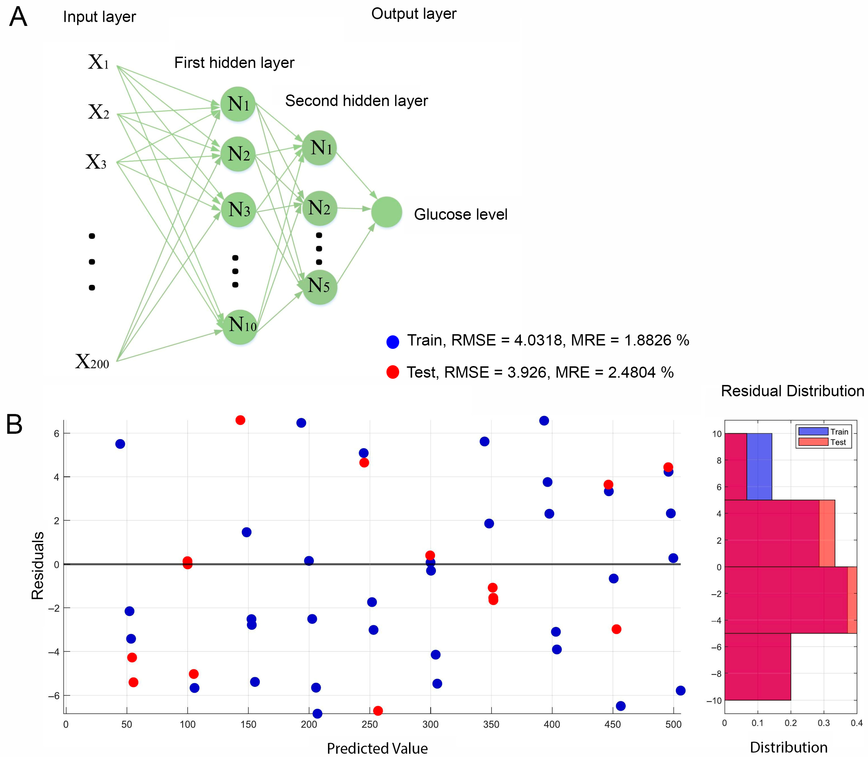
The presence and application of AI and ML in microfluidic paper-based glucose sensors and sensor research have substantially enhanced the accuracy of signal processing, and reduced the need for extensive monitoring and oversight of patient health. The structure and performance of the trained ML neural network is presented in Figure 5.

4. Conclusions and Future Perspectives

Paper-based glucose sensors have become integral to biosensor fabrication owing to their inherent biocompatibility, low cost, flexibility, and porous structure, making them particularly suitable for POCT applications. To date, a wide variety of paper-based biosensors have been developed and validated using both model systems and clinical samples, thereby establishing a solid foundation for future sensor designs and innovations. The recent surge in the COVID-19 pandemic has accelerated the fabrication and deployment of paper-based biosensors for the rapid identification of infected individuals. In addition to infectious diseases, these sensors have been increasingly applied to the detection of various disease biomarkers.
The commercialization of paper-based biosensors is underway, although much of the research remains focused on model matrices, such as human serum and plasma, rather than on extensive clinical trials. Several key challenges must be addressed in order to develop these biosensors for widespread commercial use:
  • The development of novel nanomaterials integrated within paper substrates enhances the sensor response speed and sensitivity.
  • Surface engineering of paper-based biosensors for improving target specificity.
  • The transformation of conventional paper-based biosensors into wearable formats to broaden their applicability and user convenience.
  • Commercial products incorporating AI and ML are required

Author Contributions

Conceptualization, writing, original draft preparation, methodology, review, editing, and supervision: P.G.L.; review, editing, supervision, and funding acquisition: S.C. All authors have read and agreed to the published version of the manuscript.

Funding

This research was supported by the National Research Foundation of Korea (NRF-2023R1A2C1003669) and the Korea Environmental Industry and Technology Institute (KEITI) through the “Technology Development Project for Biological Hazards Management in Indoor Air” Project funded by the Korea Ministry of Environment (MOE) (G232021010381).

Institutional Review Board Statement

Not applicable.

Informed Consent Statement

Not applicable.

Data Availability Statement

Not applicable.

Conflicts of Interest

The authors declare no conflicts of interest.

Abbreviations

The following abbreviations are used in this manuscript:
µPADMicrofluidic paper analytic devices
AIArtificial intelligence
HRPHorseradish peroxidase
GOxGlucose oxidase
MIPMolecularly imprinted polymer
MLMachine learning
PDMSPolydimethylsiloxane
POCTPoint-of-care testing

References

  1. Lu, X.; Xie, Q.; Pan, X.; Zhang, R.; Zhang, X.; Peng, G.; Zhang, Y.; Shen, S.; Tong, N. Type 2 Diabetes Mellitus in Adults: Pathogenesis, Prevention and Therapy. Signal Transduct. Target Ther. 2024, 9, 262. [Google Scholar] [CrossRef] [PubMed]
  2. Antar, S.A.; Ashour, N.A.; Sharaky, M.; Khattab, M.; Ashour, N.A.; Zaid, R.T.; Roh, E.J.; Elkamhawy, A.; Al-Karmalawy, A.A. Diabetes Mellitus: Classification, Mediators, and Complications; A Gate to Identify Potential Targets for the Development of New Effective Treatments. Biomed. Pharmacother. 2023, 168, 115734. [Google Scholar] [CrossRef] [PubMed]
  3. Ojo, O.A.; Ibrahim, H.S.; Rotimi, D.E.; Ogunlakin, A.D.; Ojo, A.B. Diabetes Mellitus: From Molecular Mechanism to Pathophysiology and Pharmacology. Med. Nov. Technol. Devices 2023, 19, 100247. [Google Scholar] [CrossRef]
  4. Lee, H.; Hong, Y.J.; Baik, S.; Hyeon, T.; Kim, D.-H. Enzyme-Based Glucose Sensor: From Invasive to Wearable Device. Adv. Healthc. Mater. 2018, 7, 1701150. [Google Scholar] [CrossRef]
  5. Govindaraj, M.; Srivastava, A.; Muthukumaran, M.K.; Tsai, P.-C.; Lin, Y.-C.; Raja, B.K.; Rajendran, J.; Ponnusamy, V.K.; Arockia Selvi, J. Current Advancements and Prospects of Enzymatic and Non-enzymatic Electrochemical Glucose Sensors. Int. J. Biol. Macromol. 2023, 253, 126680. [Google Scholar] [CrossRef]
  6. Ghosh, R.; Li, X.; Yates, M.Z. Non-enzymatic Glucose Sensor Using Bimetallic Catalysts. ACS Appl. Mater. Interfaces 2024, 16, 17–29. [Google Scholar] [CrossRef]
  7. Somsiri, S.; Kaewjangwad, C.; Malarat, N.; Wangchuk, S.; Saichanapan, J.; Samoson, K.; Promsuwan, K.; Saisahas, K.; Soleh, A.; Phairatana, T.; et al. Portable NFC Potentiostat Integrated with a 3D Paper-based Microfluidic Electrochemical Device for Glucose Detection in Whole Blood using PEDOT:PSS/DMSO/GOx Sensitive Film. Microchem. J. 2025, 213, 113623. [Google Scholar] [CrossRef]
  8. Tong, L.; Wu, L.; Zai, Y.; Zhang, Y.; Su, E.; Gu, N. Paper-based Colorimetric Glucose Sensor using Prussian Blue Nanoparticles as Mimic Peroxidase. Biosens. Bioelectron. 2023, 219, 114787. [Google Scholar] [CrossRef]
  9. Nguyen, Q.H.; Lee, D.H.; Nguyen, P.T.; Le, P.G.; Kim, M.I. Foldable Paper Microfluidic Device Based on Single Iron Site-containing Hydrogel Nanozyme for Efficient Glucose Biosensing. Chem. Eng. J. 2023, 454, 140541. [Google Scholar] [CrossRef]
  10. Le, P.G.; Le, X.A.; Duong, H.S.; Jung, S.H.; Kim, T.; Kim, M.I. Ultrahigh Peroxidase-like Catalytic Performance of Cu–N4 and Cu–N4S Active Sites-containing Reduced Graphene Oxide for Sensitive Electrochemical Biosensing. Biosens. Bioelectron. 2024, 255, 116259. [Google Scholar] [CrossRef]
  11. Le, P.G.; Wu, Q.; Kong, D.Y.; Ge, J.; Kim, M.I. Tailoring Nanostructured Supports to Achieve High Performance in Enzymatic Biofuel Cells. ACS Appl. Energy Mater. 2022, 5, 13113–13127. [Google Scholar] [CrossRef]
  12. Le, P.G.; Cho, S. Recent Developments in MXene-Based Enzyme-Free Electrochemical Glucose Sensing. BioChip J. 2024, 18, 521–534. [Google Scholar] [CrossRef]
  13. Huang, Y.; Ren, J.; Qu, X. Nanozymes: Classification, Catalytic Mechanisms, Activity Regulation, and Applications. Chem. Rev. 2019, 119, 4357–4412. [Google Scholar] [CrossRef]
  14. Kanchan, M.; Tambe, P.K.; Bharati, S.; Powar, O.S. Convolutional Neural Network for Colorimetric Glucose Detection using a Smartphone and Novel multilayer polyvinyl film microfluidic device. Sci. Rep. 2024, 14, 28377. [Google Scholar] [CrossRef] [PubMed]
  15. Xu, B.; Xiang, X.; Ding, Z.; Luo, Z.; Huang, J. A Colorimetric Assay for Detection of Glucose by Enzymatic Etching of Triangular Gold Nanosheets. Mater. Chem. Phys. 2023, 301, 127619. [Google Scholar] [CrossRef]
  16. Lan, N.T.; Phan, L.G.; Hoang, L.H.; Huan, B.D.; Van Hong, L.; Anh, T.X.; Chinh, H.D. Hydrothermal Synthesis, Structure and Photocatalytic Properties of La/Bi Co-Doped NaTaO3. Mater. Trans. 2016, 57, 1–4. [Google Scholar] [CrossRef]
  17. Colvin, L.; Tu, D.; Dunlap, D.; Rios, A.; Coté, G. A Polarity-Sensitive Far-Red Fluorescent Probe for Glucose Sensing through Skin. Biosensors 2023, 13, 788. [Google Scholar] [CrossRef]
  18. Sawayama, J.; Okitsu, T.; Nakamata, A.; Kawahara, Y.; Takeuchi, S. Hydrogel Glucose Sensor with In Vivo Stable Fluorescence Intensity Relying on Antioxidant Enzymes for Continuous Glucose Monitoring. iScience 2020, 23, 101243. [Google Scholar] [CrossRef]
  19. Li, D.; Zhang, Y.; Sun, F.; Felidj, N.; Gagey-Eilstein, N.; Lamouri, A.; Hémadi, M.; Nizard, P.; Luo, Y.; Mangeney, C. Dual-Probe SERS Nanosensor: A Promising Approach for Sensitive and Ratiometric Detection of Glucose in Clinical Settings. ACS Appl. Bio Mater. 2024, 7, 2254–2263. [Google Scholar] [CrossRef]
  20. Sun, X. Glucose Detection through Surface-enhanced Raman Spectroscopy: A Review. Anal. Chim. Acta 2022, 1206, 339226. [Google Scholar] [CrossRef]
  21. Naikoo, G.A.; Bano, M.; Arshad, F.; Hassan, I.U.; BaOmar, F.; Alfagih, I.M.; Tambuwala, M.M. Non-enzymatic glucose sensors composed of trimetallic CuO/Ag/NiO based composite materials. Sci. Rep. 2023, 13, 6210. [Google Scholar] [CrossRef]
  22. Guesmi, S.; Moulaee, K.; Bressi, V.; Kahri, H.; Khaskhoussi, A.; Espro, C.; Barhoumi, H.; Neri, G. Non-Enzymatic Amperometric Glucose Sensing by Novel Cu-MOF Synthesized at Room Temperature. Mater. Adv. 2024, 5, 1160–1170. [Google Scholar] [CrossRef]
  23. Dung, N.Q.; Duong, T.T.T.; Lam, T.D.; Dung, D.D.; Huy, N.N.; Van Thanh, D. A Simple Route for Electrochemical Glucose Sensing using Background Current Subtraction of Cyclic Voltammetry Technique. J. Electroanal. Chem. 2019, 848, 113323. [Google Scholar] [CrossRef]
  24. Phasuksom, K.; Sirivat, A. Chronoampermetric Detection of Enzymatic Glucose Sensor Based on Doped Polyindole/MWCNT Composites Modified onto Screen-printed Carbon Electrode as Portable Sensing Device for Diabetes. RSC Adv. 2022, 12, 28505–28518. [Google Scholar] [CrossRef] [PubMed]
  25. Scandurra, A.; Iacono, V.; Boscarino, S.; Scalese, S.; Grimaldi, M.G.; Ruffino, F. Model of Chronoamperometric Response towards Glucose Sensing by Arrays of Gold Nanostructures Obtained by Laser, Thermal and Wet Processes. Nanomaterials 2023, 13, 1163. [Google Scholar] [CrossRef] [PubMed]
  26. Patil, V.B.; Sawkar, R.R.; Ilager, D.; Shetti, N.P.; Tuwar, S.M.; Aminabhavi, T.M. Glucose-based Carbon Electrode for Trace-level Detection of Acetaminophen. Electrochem. Sci. Adv. 2022, 2, e202100117. [Google Scholar] [CrossRef]
  27. Patras, M.A.; Davalos, J.Z.; Kuhnert, N. Understanding the Fragmentation of Glucose in Mass Spectrometry. J. Mass Spectrom. 2023, 58, e4972. [Google Scholar] [CrossRef]
  28. Taylor, V.F.; March, R.E.; Longerich, H.P.; Stadey, C.J. A Mass Spectrometric Study of Glucose, Sucrose, and Fructose using an Inductively Coupled Plasma and Electrospray Ionization. Int. J. Mass Spectrom. 2005, 243, 71–84. [Google Scholar] [CrossRef]
  29. Omer, A.E.; Shaker, G.; Safavi-Naeini, S.; Kokabi, H.; Alquié, G.; Deshours, F.; Shubair, R.M. Low-cost Portable Microwave Sensor for Non-invasive Monitoring of Blood Glucose Level: Novel Design Utilizing a Four-cell CSRR Hexagonal Configuration. Sci. Rep. 2020, 10, 15200. [Google Scholar] [CrossRef]
  30. Rattan, P.; Houngkamhang, N.; Orankitanun, T.; Phasukkit, P. Metamaterial Microwave Sensor for Glucose Level Measurement Based on Strip Line with Complementary Split Ring Resonator. Phys. Status Solidi (A) 2025, 222, 2400180. [Google Scholar] [CrossRef]
  31. Brazaca, L.C.; Imamura, A.H.; Blasques, R.V.; Camargo, J.R.; Janegitz, B.C.; Carrilho, E. The Use of Biological Fluids in Microfluidic Paper-based Analytical Devices (μPADs): Recent Advances, Challenges and Future Perspectives. Biosens. Bioelectron. 2024, 246, 115846. [Google Scholar] [CrossRef]
  32. Le, P.G.; Jung, S.-C.; Han, J.-H.; Cho, S. Nanozyme-integrated Paper Chip Based on High Peroxidase-like Activity of Zn-doped Reduced Graphene Oxide for Glucose Sensing. Microchem. J. 2025, 213, 113895. [Google Scholar] [CrossRef]
  33. Yeon, S.Y.; Seo, M.; Kim, Y.; Hong, H.; Chung, T.D. Paper-based Electrochromic Glucose Sensor with Polyaniline on Indium Tin Oxide Nanoparticle Layer as the Optical Readout. Biosens. Bioelectron. 2022, 203, 114002. [Google Scholar] [CrossRef]
  34. Sung, W.-H.; Hung, J.-T.; Lu, Y.-J.; Cheng, C.-M. Paper-Based Detection Device for Alzheimer’s Disease—Detecting β-amyloid Peptides (1–42) in Human Plasma. Diagnostics 2020, 10, 272. [Google Scholar] [CrossRef] [PubMed]
  35. Duan, S.; Cai, T.; Liu, F.; Li, Y.; Yuan, H.; Yuan, W.; Huang, K.; Hoettges, K.; Chen, M.; Lim, E.G.; et al. Automatic Offline-Capable Smartphone Paper-based Microfluidic Device for Efficient Biomarker Detection of Alzheimer's Disease. Anal. Chim. Acta 2024, 1308, 342575. [Google Scholar] [CrossRef] [PubMed]
  36. Singh, S.; Raucci, A.; Cimmino, W.; Cinti, S. Paper-Based Analytical Devices for Cancer Liquid Biopsy. Anal. Chem. 2024, 96, 3698–3706. [Google Scholar] [CrossRef] [PubMed]
  37. Shalaby, A.A.; Tsao, C.-W.; Ishida, A.; Maeki, M.; Tokeshi, M. Microfluidic Paper-based Analytical Devices for Cancer Diagnosis. Sens. Actuators B Chem. 2023, 379, 133243. [Google Scholar] [CrossRef]
  38. Fan, H.; Sasaki, Y.; Zhou, Q.; Tang, W.; Nishina, Y.; Minami, T. Non-enzymatic Detection of Glucose Levels in Human Blood Plasma by a Graphene Oxide-modified Organic Transistor Sensor. Chem. Commun. 2023, 59, 2425–2428. [Google Scholar] [CrossRef]
  39. Psotta, C.; Cirovic, S.; Gudmundsson, P.; Falk, M.; Mandal, T.; Reichhart, T.; Leech, D.; Ludwig, R.; Kittel, R.; Schuhmann, W.; et al. Continuous Ex Vivo Glucose Sensing in Human Physiological Fluids using an Enzymatic Sensor in a Vein Replica. Bioelectrochemistry 2023, 152, 108441. [Google Scholar] [CrossRef]
  40. Lee, H.-A.; Lin, P.-Y.; Solomatina, A.I.; Koshevoy, I.O.; Tunik, S.P.; Lin, H.-W.; Pan, S.-W.; Ho, M.-L. Glucose Sensing in Human Whole Blood Based on Near-Infrared Phosphors and Outlier Treatment with the Programming Language “R”. ACS Omega 2022, 7, 198–206. [Google Scholar] [CrossRef]
  41. Math, C.; Income, K.; Khachornsakkul, K.; Duenchay, P.; Dungchai, W. A Sensitive and Facile Electrochemical Paper-based Sensor for Glucose Detection in Whole Blood using the Pd/CB-Ni@rGO Modified Electrode. Analyst 2023, 148, 4753–4761. [Google Scholar] [CrossRef] [PubMed]
  42. Akiiga, N.S.; El-Bab, A.M.R.F.; Yoshihisa, M.; El-Moneim, A.A. Enzyme-Free Glucose Detection in Sweat using 2D inkjet-printed Cobalt Sulfide Anchored on Graphene in a Paper-based Microfluidic Device. J. Colloid Interface Sci. 2025, 688, 490–504. [Google Scholar] [CrossRef]
  43. Moradi, S.; Hosseini, M.; Firoozbakhtian, A.; Barshan Tashnizi, M.; Tashnizi, M.B. Electrochemical Detection of Glucose in Sweat using a Paper-based Origami Biosensor Based on PEDOT:PSS/Ti3C2/MWCNT Composite. J. Electroanal. Chem. 2024, 973, 118675. [Google Scholar] [CrossRef]
  44. Li, Y.; Kong, Y.; Hu, Y.; Li, Y.; Asrosa, R.; Zhang, W.; Boruah, B.D.; Yetisen, A.K.; Davenport, A.; Lee, T.-C.; et al. A Paper-based Dual Functional Biosensor for Safe and User-friendly Point-of-care Urine Analysis. Lab Chip 2024, 24, 2454–2467. [Google Scholar] [CrossRef]
  45. Lee, T.; Kim, I.; Cheong, D.Y.; Roh, S.; Jung, H.G.; Lee, S.W.; Kim, H.S.; Yoon, D.S.; Hong, Y.; Lee, G. Selective Colorimetric Urine Glucose Detection by Paper Sensor Functionalized with Polyaniline Nanoparticles and Cell Membrane. Anal. Chim. Acta 2021, 1158, 338387. [Google Scholar] [CrossRef]
  46. Ruiz, R.A.; Gonzalez, J.L.; Vazquez-Alvarado, M.; Martinez, N.W.; Martinez, A.W. Beyond Wax Printing: Fabrication of Paper-Based Microfluidic Devices Using a Thermal Transfer Printer. Anal. Chem. 2022, 94, 8833–8837. [Google Scholar] [CrossRef]
  47. Malik, S.; Singh, J.; Saini, K.; Chaudhary, V.; Umar, A.; Ibrahim, A.A.; Akbar, S.; Baskoutas, S. Paper-based Sensors: Affordable, Versatile, and Emerging Analyte Detection Platforms. Anal. Methods 2024, 16, 2777–2809. [Google Scholar] [CrossRef]
  48. Costa-Rama, E.; Fernández-Abedul, M.T. Paper-Based Screen-Printed Electrodes: A New Generation of Low-Cost Electroanalytical Platforms. Biosensors 2021, 11, 51. [Google Scholar] [CrossRef]
  49. Cao, Q.; Liang, B.; Tu, T.; Wei, J.; Fang, L.; Ye, X. Three-dimensional Paper-based Microfluidic Electrochemical Integrated Devices (3D-PMED) for Wearable Electrochemical Glucose Detection. RSC Adv. 2019, 9, 5674–5681. [Google Scholar] [CrossRef]
  50. Lee, S.; Park, J.S.; Woo, H.; Yoo, Y.K.; Lee, D.; Chung, S.; Yoon, D.S.; Lee, K.-B.; Lee, J.H. Rapid Deep Learning-assisted Predictive Diagnostics for Point-of-care Testing. Nat. Commun. 2024, 15, 1695. [Google Scholar] [CrossRef] [PubMed]
  51. Han, G.-R.; Goncharov, A.; Eryilmaz, M.; Ye, S.; Palanisamy, B.; Ghosh, R.; Lisi, F.; Rogers, E.; Guzman, D.; Yigci, D.; et al. Machine Learning in Point-of-care Testing: Innovations, Challenges, and Opportunities. Nat. Commun. 2025, 16, 3165. [Google Scholar] [CrossRef] [PubMed]
  52. Gao, Q.; Li, S. Intelligent Point of Care Testing for Medicine Diagnosis. Interdiscip. Med. 2024, 2, e20230031. [Google Scholar] [CrossRef]
  53. Wang, K.; Meng, X.; Yan, X.; Fan, K. Nanozyme-based Point-of-Care testing: Revolutionizing Environmental Pollutant Detection with High Efficiency and Low Cost. Nano Today 2024, 54, 102145. [Google Scholar] [CrossRef]
  54. Kuswandi, B.; Hidayat, M.A.; Noviana, E. Paper-based Sensors for Rapid Important Biomarkers Detection. Biosens. Bioelectron. X 2022, 12, 100246. [Google Scholar] [CrossRef]
  55. Bilgen, E.; Suvacı, Z.; Çetinkol, Ö.P.; Forough, M. 26—Disposable Paper-based Sensors. In Fundamentals of Sensor Technology; Barhoum, A., Altintas, Z., Eds.; Woodhead Publishing: Cambridge, UK, 2023; pp. 803–860. [Google Scholar]
  56. Tong, X.; Ga, L.; Zhao, R.; Ai, J. Research Progress on the Applications of Paper Chips. RSC Adv. 2021, 11, 8793–8820. [Google Scholar] [CrossRef]
  57. Wang, L.; Chen, Y.; Ji, Y.; Zheng, S.; Wang, F.; Li, C. Cheap and Portable Paper Chip with Terrific Oxidase-like Activity and SERS Enhancement Performance for SERS-colorimetric Bimodal Detection of Intracellular Glutathione. Biosens. Bioelectron. 2024, 244, 115817. [Google Scholar] [CrossRef]
  58. Noviana, E.; Ozer, T.; Carrell, C.S.; Link, J.S.; McMahon, C.; Jang, I.; Henry, C.S. Microfluidic Paper-Based Analytical Devices: From Design to Applications. Chem. Rev. 2021, 121, 11835–11885. [Google Scholar] [CrossRef]
  59. Chen, J.L.; Njoku, D.I.; Tang, C.; Gao, Y.; Chen, J.; Peng, Y.-K.; Sun, H.; Mao, G.; Pan, M.; Tam, N.F.-Y. Advances in Microfluidic Paper-Based Analytical Devices (µPADs): Design, Fabrication, and Applications. Small Methods 2024, 8, 2400155. [Google Scholar] [CrossRef]
  60. Yu, L.; Shi, Z.Z. Microfluidic Paper-based Analytical Devices Fabricated by Low-cost Photolithography and Embossing of Parafilm®. Lab Chip 2015, 15, 1642–1645. [Google Scholar] [CrossRef]
  61. García-Casas, X.; Aparicio, F.J.; Budagosky, J.; Ghaffarinejad, A.; Orozco-Corrales, N.; Ostrikov, K.; Sánchez-Valencia, J.R.; Barranco, Á.; Borrás, A. Paper-based ZnO Self-powered Sensors and Nanogenerators by Plasma Technology. Nano Energy 2023, 114, 108686. [Google Scholar] [CrossRef]
  62. Mukhopadhyay, M.; Subramanian, S.G.; Durga, K.V.; Sarkar, D.; DasGupta, S. Laser Printing Based Colorimetric Paper Sensors for Glucose and Ketone Detection: Design, Fabrication, and Theoretical Analysis. Sens. Actuators B Chem. 2022, 371, 132599. [Google Scholar] [CrossRef]
  63. Aghababaie, M.; Foroushani, E.S.; Changani, Z.; Gounani, Z.; Mobarakeh, M.S.; Hadady, H.; Khedri, M.; Maleki, R.; Asadnia, M.; Razmjou, A. Recent Advances In the Development of Enzymatic Paper-based Microfluidic Biosensors. Biosens. Bioelectron. 2023, 226, 115131. [Google Scholar] [CrossRef] [PubMed]
  64. Verma, D.; Dubey, N.; Yadav, A.K.; Saraya, A.; Sharma, R.; Solanki, P.R. Disposable Paper-based Screen-printed Electrochemical Immunoplatform for Dual Detection of Esophageal Cancer Biomarkers in Patients’ Serum Samples. Mater. Adv. 2024, 5, 2153–2168. [Google Scholar] [CrossRef]
  65. Juang, Y.-J.; Chen, P.-S.; Wang, Y. Rapid Fabrication of Microfluidic Paper-based Analytical Devices by Microembossing. Sens. Actuators B Chem. 2019, 283, 87–92. [Google Scholar] [CrossRef]
  66. Mei, X.; Chen, Z.; Wen, A.; Zhang, J.; Wei, X.; Wang, F.; Zhou, L.; Wang, B.; Wu, Y. Wearable Three-dimensional Paper-based Microfluidic Electrochemical Sensors for Real-time Sweat Monitoring. Chem. Eng. J. 2025, 515, 163786. [Google Scholar] [CrossRef]
  67. Ilyasov, I.R.; Beloborodov, V.L.; Selivanova, I.A.; Terekhov, R.P. ABTS/PP Decolorization Assay of Antioxidant Capacity Reaction Pathways. Int. J. Mol. Sci. 2020, 21, 1131. [Google Scholar] [CrossRef]
  68. Liu, D.-M.; Dong, C.; Ma, R.-T. A Colorimetric Method for Screening α-glucosidase Inhibitors from Flavonoids using 3,3′,5,5′-tetramethylbenzidine as a Chromogenic Probe. Colloids Surf. B Biointerfaces 2021, 197, 111400. [Google Scholar] [CrossRef]
  69. Singh, K.R.B.; Nayak, V.; Sarkar, T.; Singh, R.P. Cerium Oxide Nanoparticles: Properties, Biosynthesis and Biomedical Application. RSC Adv. 2020, 10, 27194–27214. [Google Scholar] [CrossRef]
  70. Monisha; Shrivas, K.; Kant, T.; Patel, S.; Devi, R.; Dahariya, N.S.; Pervez, S.; Deb, M.K.; Rai, M.K.; Rai, J. Inkjet-printed Paper-based Colorimetric Sensor Coupled with Smartphone for Determination of Mercury (Hg2+). J. Hazard. Mater. 2021, 414, 125440. [Google Scholar] [CrossRef]
  71. Uhlikova, N.; Almeida, M.I.G.S.; McKelvie, I.; Morrison, R.; Kolev, S.D. Use of Scanners for Colorimetric Analysis of Microfluidic Paper-based Analytical Devices (µPADs): A Practical Guide. Microchem. J. 2023, 191, 108879. [Google Scholar] [CrossRef]
  72. Nota, G.; Cimmino, W.; Singh, S.; Darwish, I.A.; La Rocca, C.; Carbone, F.; Matarese, G.; Cinti, S. A Portable and Ecological Paper-based Device for Glucose Monitoring in Peripheral Blood Mononuclear Cell Lysates. Anal. Methods 2025, 17, 2529–2535. [Google Scholar] [CrossRef]
  73. Zheng, Y.; Huang, Y.; Zuo, Q.; Zhang, Y.; Wu, Y.; Zhang, Z. On-Demand Portable Paper-Based Electrospray Ionization Mass Spectrometry for High-Sensitivity Analysis of Complex Samples. Anal. Chem. 2023, 95, 6163–6171. [Google Scholar] [CrossRef]
  74. Pumbua, R.; Sricharoen, N.; Wongravee, K.; Praneenararat, T. Paper Spray Mass Spectrometry as an Effective Tool for Differentiating Coffees Based on their Geographical Origins. Food Chem. X 2023, 18, 100624. [Google Scholar] [CrossRef]
  75. Amor-Gutiérrez, O.; Costa Rama, E.; Costa-García, A.; Fernández-Abedul, M.T. Paper-based Maskless Enzymatic Sensor for Glucose Determination Combining Ink and Wire Electrodes. Biosens. Bioelectron. 2017, 93, 40–45. [Google Scholar] [CrossRef] [PubMed]
  76. Chun, H.J.; Park, Y.M.; Han, Y.D.; Jang, Y.H.; Yoon, H.C. Paper-based Glucose Biosensing System Utilizing a Smartphone as a Signal Reader. BioChip J. 2014, 8, 218–226. [Google Scholar] [CrossRef]
  77. Li, N.; Zhou, Y.; Sun, H.; Wang, N.; Yang, N.; Ren, P.; Fu, L.; Zhang, Y.; Liu, W.; Li, Y.; et al. Rapid Analysis of Salivary Glucose Content using MOF/MIPs Biomimetic Microfluidic Paper Chips. Chem. Eng. J. 2025, 504, 159023. [Google Scholar] [CrossRef]
  78. Lian, M.; Shi, F.; Cao, Q.; Wang, C.; Li, N.; Li, X.; Zhang, X.; Chen, D. Paper-based Colorimetric Sensor using Bimetallic Nickel-Cobalt Selenides Nanozyme with Artificial Neural Network-assisted for Detection of H2O2 on Smartphone. Spectrophim. Acta A Mol. Biomol. Spectrosc. 2024, 311, 124038. [Google Scholar] [CrossRef]
  79. Yan, L.; Zhang, B.; Zhou, W.; Hao, J.; Shi, H.; Wang, S.; Shuang, S.; Shi, L. An Artificial Intelligence Handheld Sensor for Direct Reading of Nickel Ion and Ethylenediaminetetraacetic Acid in Food Samples using Ratiometric Fluorescence Cellulose Paper Microfluidic Chip. Int. J. Biol. Macromol. 2024, 279, 135083. [Google Scholar] [CrossRef]
  80. Coskun, M. Intrinsic Graph Topological Correlation for Graph Convolutional Network Propagation. Comput. Stand. Interfaces 2023, 83, 103655. [Google Scholar] [CrossRef]
  81. Kumbure, M.M.; Luukka, P. Generalizing Fuzzy K-Nearest Neighbor Classifier using an OWA Operator with a RIM Quantifier. Expert Syst. Appl. 2025, 282, 127795. [Google Scholar] [CrossRef]
  82. Vásquez, J.C.G.; Kumral, M. Artificial Neural Networks and Support Vector Machines for More Accurate Cost Estimation in Underground Mining: A Contractor’s Viewpoint. Mach. Learn. Appl. 2025, 21, 100689. [Google Scholar] [CrossRef]
  83. Hamdi, T.; Ben Ali, J.; Di Costanzo, V.; Fnaiech, F.; Moreau, E.; Ginoux, J.-M. Accurate Prediction of Continuous Blood Glucose Based on Support Vector Regression and Differential Evolution Algorithm. Biocybern. Biomed. Eng. 2018, 38, 362–372. [Google Scholar] [CrossRef]
  84. Pantic, I.V.; Paunovic Pantic, J.; Valjarevic, S.; Corridon, P.R.; Topalovic, N. Artificial Intelligence—Based Approaches Based on Random Forest Algorithm for Signal Analysis: Potential Applications in Detection of Chemico—Biological Interactions. Chem. Biol. Interact. 2025, 418, 111624. [Google Scholar] [CrossRef] [PubMed]
  85. Karim, R.A.H.; Vassányi, I.; Kósa, I. After-meal Blood Glucose Level Prediction using an Absorption Model for Neural Network Training. Comput. Biol. Med. 2020, 125, 103956. [Google Scholar] [CrossRef]
  86. Agrawal, H.; Jain, P.; Joshi, A.M. Machine Learning Models for Non-invasive Glucose Measurement: Towards Diabetes Management in Smart Healthcare. Health Technol. 2022, 12, 955–970. [Google Scholar] [CrossRef]
  87. Abreu, A.; dos Santos Oliveira, D.; Vinagre, I.; Cavouras, D.; Alves, J.A.; Pereira, A.I.; Lima, J.; Moreira, F.T.C. A Machine Learning Approach for Enhanced Glucose Prediction in Biosensors. Chemosensors 2025, 13, 52. [Google Scholar] [CrossRef]
  88. Jeee, S.; Kumar, L.A. An Explainable Artificial Intelligence Driven Fall System for Sensor Data Analysis Enhanced by Butterworth Filtering. Eng. Appl. Artif. Intell. 2025, 158, 111364. [Google Scholar] [CrossRef]
  89. Medanki, S.; Dommati, N.; Bodapati, H.H.; Katru, V.; Moses, G.; Komaraju, A.; Donepudi, N.S.; Yalamanchili, D.; Sateesh, J.; Turimerla, P. Artificial Intelligence Powered Glucose Monitoring and Controlling System: Pumping Module. World J. Exp. Med. 2024, 14, 87916. [Google Scholar] [CrossRef]
  90. Zhang, G.; Zhang, Q.; Wang, C.; Ji, X.; Weng, Z.; Mayet, A.M.; Miao, X. A Microstrip-based Sensor for Glucose Monitoring: Towards Non-invasive Blood Glucose Detection. Measurement 2025, 256, 118116. [Google Scholar] [CrossRef]
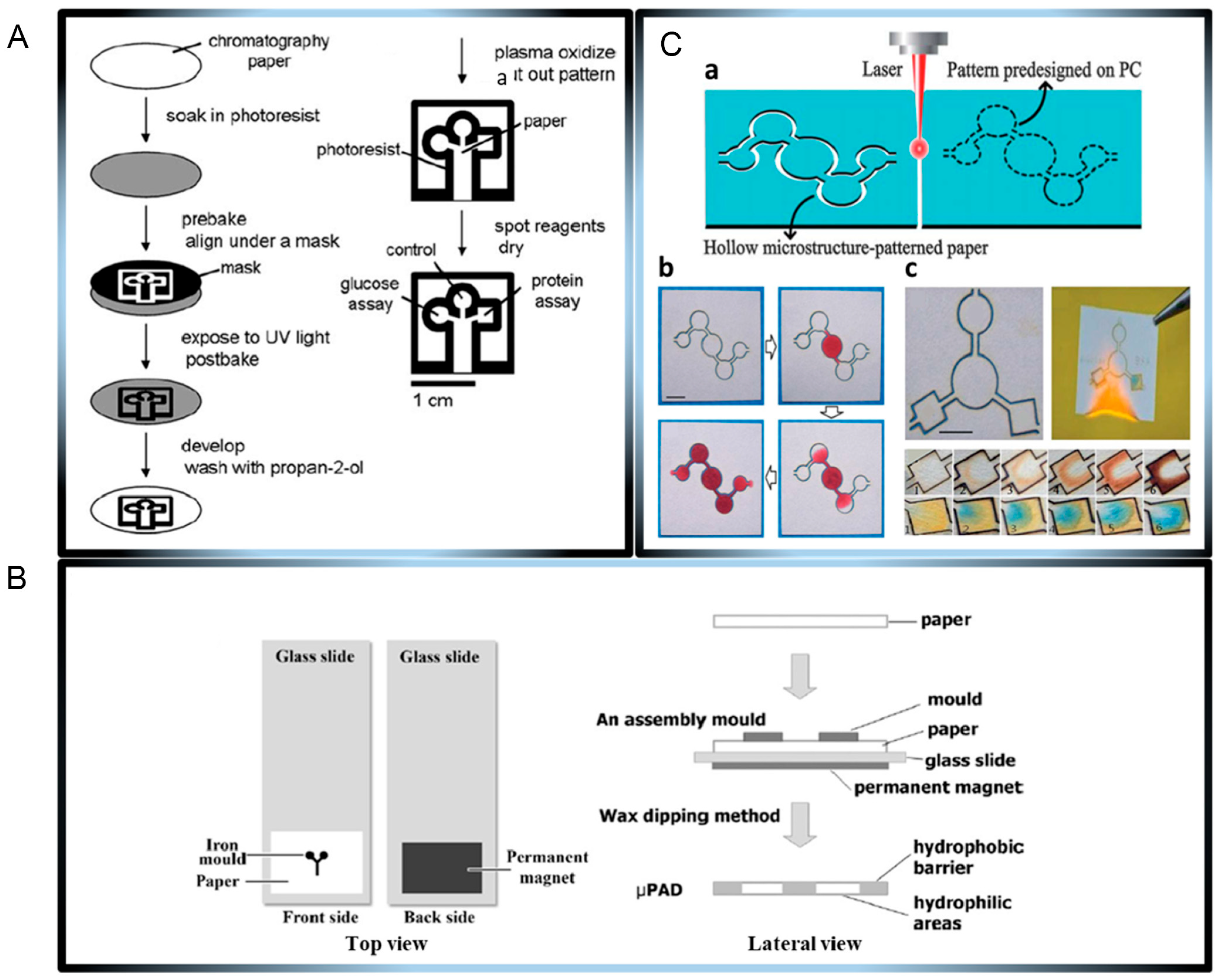
Figure 1. (A) The microfluidic paper-based sensor was fabricated using the lithography method using photoresist into the paper. (B) The microfluidic paper-based sensors were prepared by the wax dipping fabrication process, with procedures for both top and lateral views. (C) The microfluidic paper-based sensor was produced using a one-step CO2 laser cutting or engraving machine. (a) The hollow microstructures were made using a predesigned pattern. (b) The microstructures before and after ink addition. (c) The three-path µPAD for simultaneous detection of Bovine Serum Albumin (from yellow to blue colour) and glucose (from clear to brown colour). Reprinted with permission from Elsevier [63]. µPAD, microfluidic paper analytical device.
Chemosensors 13 00293 g001
Figure 2. The microfluidic paper-based chloride, glucose, and lactate electrochemical sensors were fabricated using PDMS-patterned paper. (A) The photograph. (B) The 3D microfluidic paper-based electrochemical sensor for sweat analysis. (C) Configuration of the sensor electrodes. Reprinted with permission from Elsevier [66]. GOx, glucose oxidase; LOx, lactate oxidase; PDMS, polydimethylsiloxane.
Chemosensors 13 00293 g002
Figure 3. The 3D microfluidic paper-based electrochemical device. (A) The operational procedure and basic functions of the device. (B) Fabrication of the PEDOT:PSS/DMSO/GOx sensing film for glucose detection. Reprinted with permission from Elsevier [7]. GOx, glucose oxidase: PB, Prussian blue; PSS, poly(sodium 4−styrenesulfonate).
Chemosensors 13 00293 g003
Figure 4. (A) The synthesis procedure of FZSN−BA@MIP; (B) The flow diagram of the fabricated microfluidic paper-based sensor for the glucose detection. Reprinted with permission from Elsevier [77].
Chemosensors 13 00293 g004
Figure 5. (A) The structure of the designed neural network and (B) performance of trained machine learning neural network. Reprinted with permission from Elsevier [90].
Chemosensors 13 00293 g005
Disclaimer/Publisher’s Note: The statements, opinions and data contained in all publications are solely those of the individual author(s) and contributor(s) and not of MDPI and/or the editor(s). MDPI and/or the editor(s) disclaim responsibility for any injury to people or property resulting from any ideas, methods, instructions or products referred to in the content.