


Real-Time Fluorescence Imaging of His-Tag-Driven Conjugation of mCherry Proteins to Silver Nanowires
 , ,
, ,  and
and 
Abstract
1. Introduction
2. Materials and Methods
3. Results and Discussion
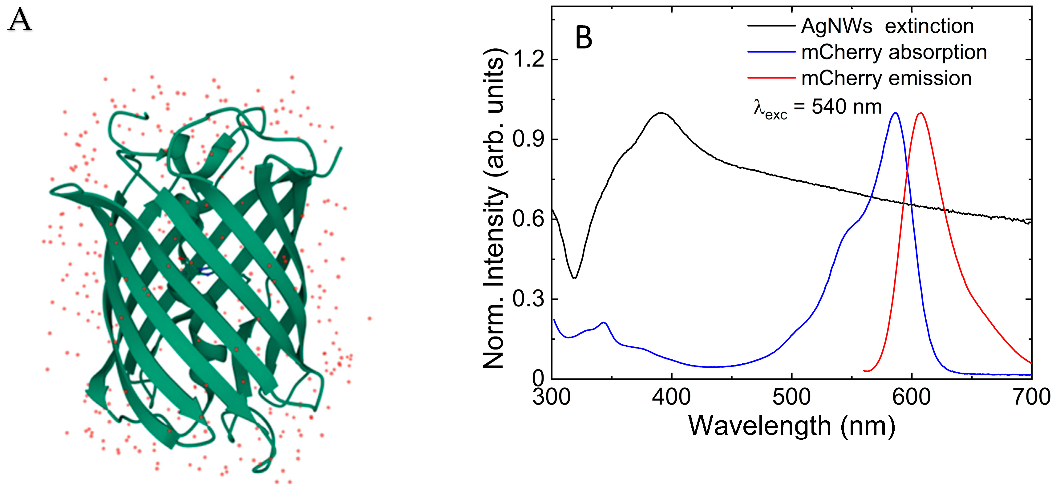
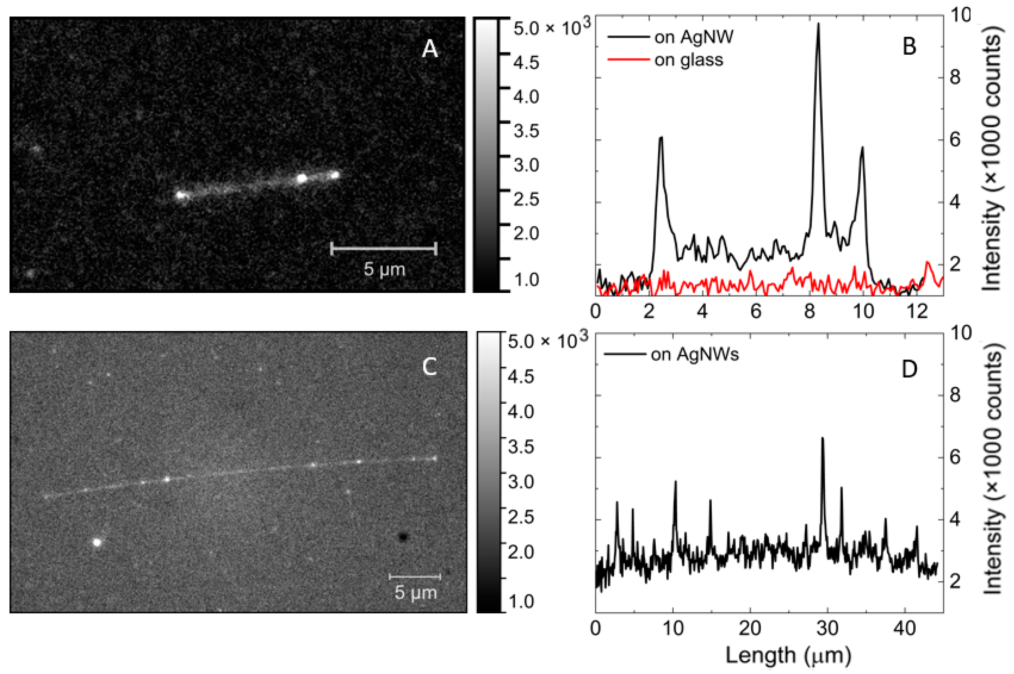
3.1. Fluorescence of mCherry Proteins Mixed with AgNWs
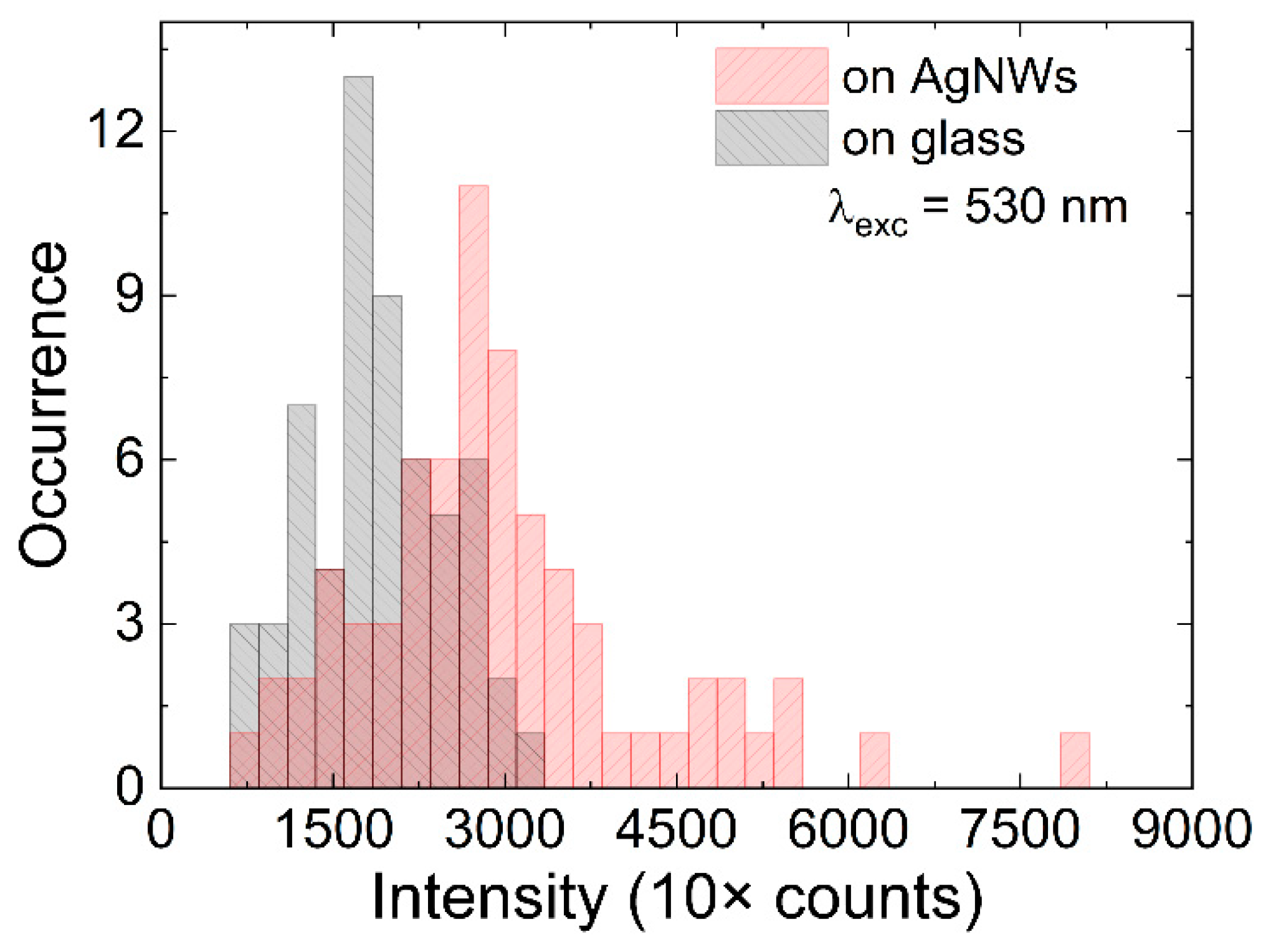
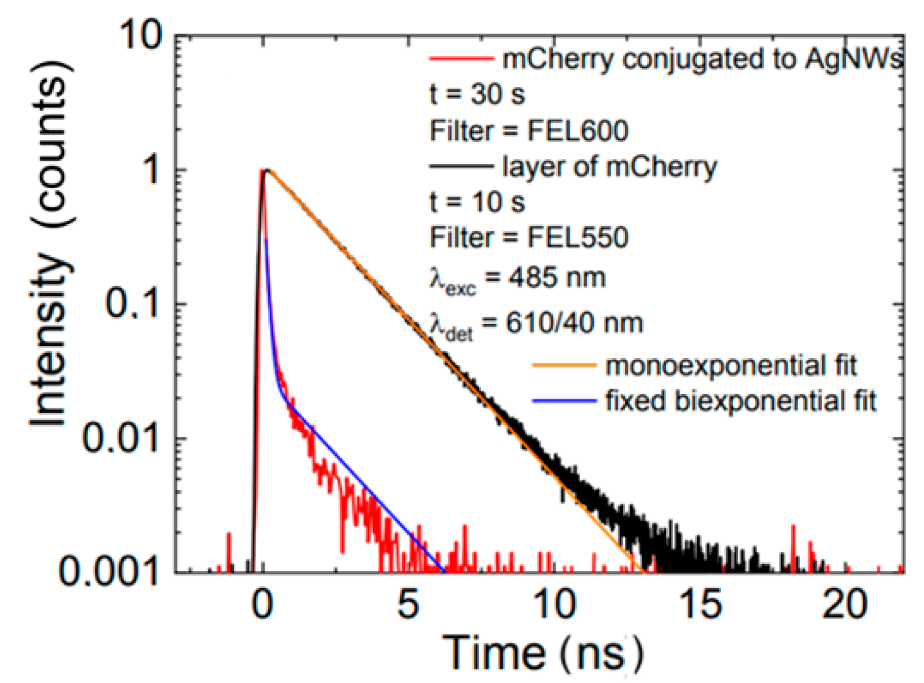
3.2. Fluorescence of mCherry/AgNWs Conjugates
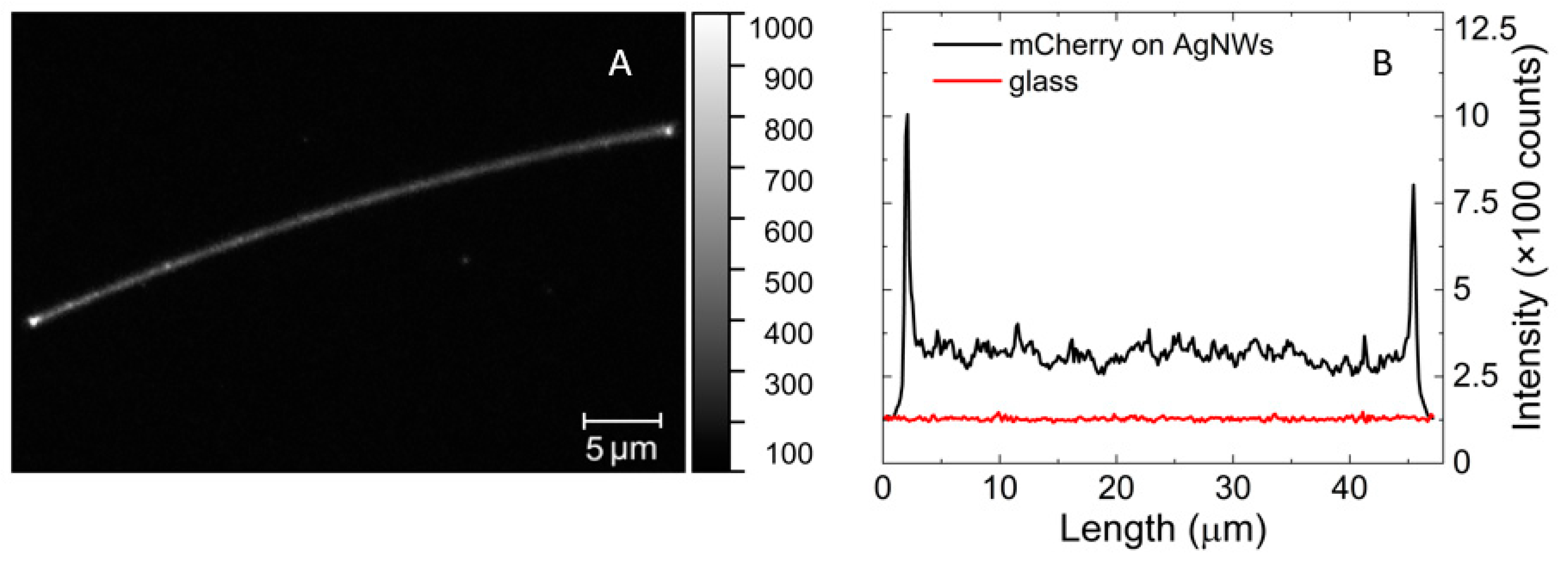
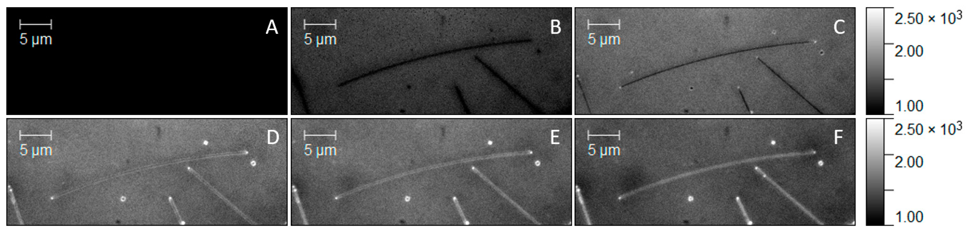
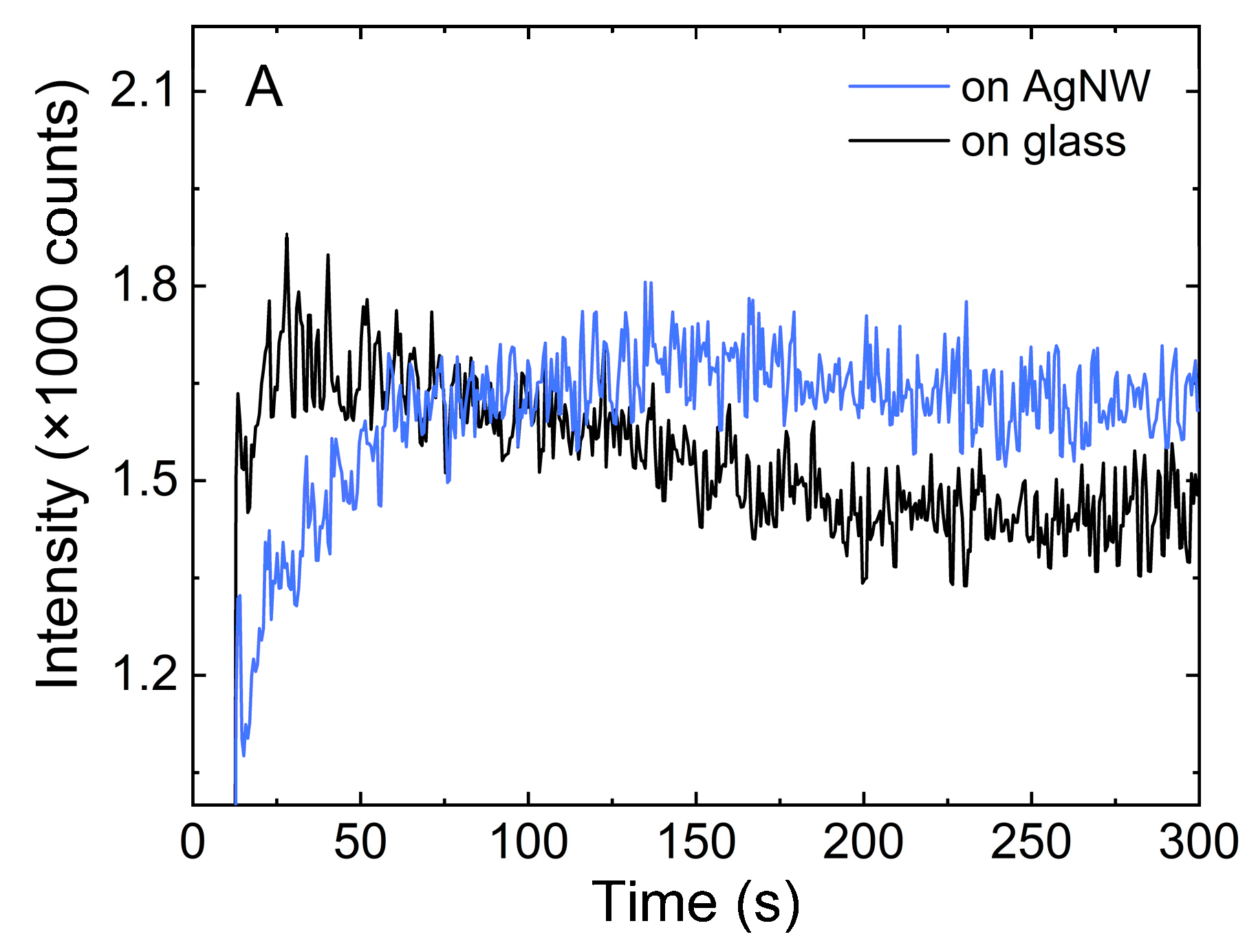
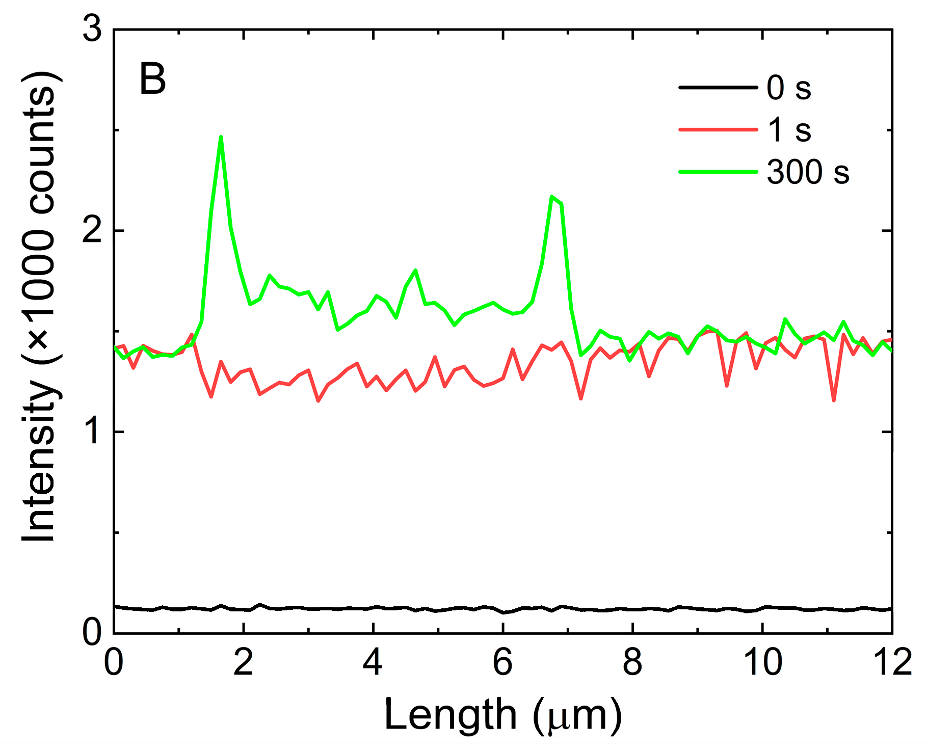
3.3. Real-Time Fluorescence Imaging of His-Tag-Driven Conjugation
4. Conclusions
Supplementary Materials
Author Contributions
Funding
Institutional Review Board Statement
Informed Consent Statement
Data Availability Statement
Conflicts of Interest
References
- Arregui, F.; Andreu, T. Sensors Based on Nanostructured Materials; Springer: New York, NY, USA, 2009; Chapter 15; pp. 79–129. [Google Scholar]
- Choi, I.; Choi, Y. Plasmonic Nanosensors: Review and Prospect. IEE J. Sel. Top. Quantum Electron. 2012, 18, 1110–1121. [Google Scholar] [CrossRef] [Scilit]
- Bauch, M.; Toma, K.; Toma, M.; Zhang, Q.; Dostalek, J. Plasmon-enhanced fluorescence biosensors: A review. Plasmonics 2014, 9, 781–799. [Google Scholar] [CrossRef] [Scilit] [PubMed]
- Amiri, M.; Bezaatpour, A.; Jafari, H.; Boukherroub, R.; Szunerits, S. Electrochemical methodologies for the detection of pathogens. ASC Sens. 2018, 3, 1069–1086. [Google Scholar] [CrossRef] [Scilit] [PubMed]
- Anker, J.N.; Hall, W.P.; Lyandres, O.; Shah, N.C.; Zhao, J.; van Duyne, R.P. Biosensing with plasmonic nanosensors. Nat. Mater. 2008, 7, 442–453. [Google Scholar] [CrossRef] [Scilit]
- Nasu, Y.; Shen, Y.; Kramer, L.; Campbell, R.E. Structure and mechanism guided design of single fluorescent protein-based biosensors. Nat. Chem. Biol. 2021, 17, 509–518. [Google Scholar] [CrossRef] [Scilit]
- Tan, P.; Hesheng, L.; Wang, J.; Gopinath, S.C.B. Sliver nanoparticle in biosensor and bioimaging: Clinical perspectives. Biotechnol. Appl. Biochem. 2020, 68, 1236–1242. [Google Scholar]
- McNamara, K.; Tofail, S.A.M. Nanoparticles in biomedical applications. Adv. Phys. X 2017, 2, 54–88. [Google Scholar] [CrossRef] [Scilit]
- Joo, Y.; Byun, J.; Seong, N.; Ha, J.; Kim, H.; Kim, S.; Kim, T.; Im, H.; Kim, D.; Hong, Y. Silver nanowire-embedded PDMS with a multiscale structure for highly sensitive and robust flexible pressure sensor. Nanoscale 2015, 7, 6208. [Google Scholar] [CrossRef] [Scilit]
- Sun, Y.G.; Yin, Y.D.; Mayers, B.T.; Herricks, T.; Xia, Y.N. Uniform silver nanowires synthesis by reducing AgNO3 with ethylene glycol in the presence of seeds and Poly (Vinyl Pyrrolidone). Chem. Mater. 2002, 14, 4736–4745. [Google Scholar] [CrossRef] [Scilit]
- Coronado, E.A.; Encina, E.R.; Stefani, F.D. Optical properties of metallic nanoparticles: Manipulating light, heat and forces at the nanoscale. Nanoscale 2011, 3, 4042. [Google Scholar] [CrossRef] [Scilit]
- Reineck, P.; Gómez, D.; Ng, S.H.; Karg, M.; Bell, T.; Mulvaney, P.; Bach, U. Distance and wavelength dependent quenching of molecular fluorescence by Au@SiO2 core-shell nanoparticles. ACS Nano 2013, 7, 6636–6648. [Google Scholar] [CrossRef] [Scilit] [PubMed]
- Li, J.; Krasavin, A.V.; Webster, L.; Segovia, P.; Zayats, A.V.; Richards, D. Spectral variation of fluorescence lifetime near single metal nanoparticles. Sci. Rep. 2016, 6, 21349. [Google Scholar] [CrossRef] [Scilit] [PubMed]
- Fu, Y.; Zhang, J.; Lakowicz, J.R. Plasmonic enhancement of single-molecule fluorescence near a silver nanoparticle. J. Fluoresc. 2007, 17, 811–816. [Google Scholar] [CrossRef] [Scilit] [PubMed]
- Anger, P.; Bharadwaj, P.; Novotny, L. Enhancement and quenching of single-molecule fluorescence. PRL 2006, 96, 113002. [Google Scholar] [CrossRef] [Scilit]
- Viste, P.; Plain, J.; Jaffiol, R.; Vial, A.; Adam, P.-M.; Royer, P. Enhancement and quenching regimes in metal-semiconductor hybrid optical nanostructures. ACS Nano 2010, 4, 759–764. [Google Scholar] [CrossRef] [Scilit] [PubMed]
- Olejnik, M.; Bujak, Ł.; Maćkowski, S. Plasmonic molecular nanohybrids-spectral dependence of fluorescence quenching. Int. J. Mol. Sci. 2012, 13, 1018–1028. [Google Scholar] [CrossRef] [Scilit]
- Focsan, M.; Campu, A.; Craciun, A.-M.; Potara, M.; Leordean, C.; Maniu, D.; Astilean, S. A simple and efficient design to improve the detection of biotin-streptavidin interaction with plasmonic nanobiosensors. Biosens. Bioelectron. 2016, 86, 728–735. [Google Scholar] [CrossRef] [Scilit]
- Lv, Q.; Wang, Y.; Su, C.; Lakshmipriya, T.; Gopinath, S.; Pandian, K.; Perumal, V.; Liu, Y. Human papilloma virus DNA-biomarker analysis for cervical cancer: Signal enhancement by gold nanoparticle-coupled tetravalent streptavidin-biotin strategy. Int. J. Biol. Macromol. 2019, 134, 354–360. [Google Scholar] [CrossRef] [Scilit]
- Bolduc, O.R.; Lambert-Lanteigne, P.; Colin, D.Y.; Zhao, S.S.; Proulx, C.; Boeglin, D.; Lubell, W.D.; Pelletier, J.N.; Féthière, J.; Ong, H.; et al. Modified peptide monolayer binding His-tagged biomolecules for small ligand screening with SPR biosensors. Analyst 2011, 136, 3142. [Google Scholar] [CrossRef] [Scilit]
- Evers, T.H.; Appelhof, M.A.; Meijer, E.; Merkx, M. His-tag as Zn(II) binding motifs in a protein-based fluorescent sensor. PEDS 2008, 21, 529–536. [Google Scholar] [CrossRef] [Scilit]
- Zhang, C.; Chen, Y.; Liang, X.; Zhang, G.; Ma, H.; Nie, L.; Wang, Y. Detection of hepatitis B virus M204I mutation by quantum dot-labeled DNA probe. Sensors 2017, 17, 961. [Google Scholar] [CrossRef] [Scilit] [PubMed]
- Holford, T.R.; Davis, F.; Higson, S.P. Recent trends in antibody based sensors. Biosens. Bioelectron. 2012, 34, 12–24. [Google Scholar] [CrossRef] [Scilit] [PubMed]
- Chivers, C.E.; Koner, A.L.; Lowe, E.D.; Howarth, M. How the biotin-steptavidin interaction was made even stronger: Investigation via crystallography and chimaeric tetramer. Biochem. J. 2011, 435, 55–63. [Google Scholar] [CrossRef] [Scilit] [PubMed]
- Szalkowski, M.; Sulowska, K.; Grzelak, J.; Niedziółka-Jönsson, J.; Roźniecka, E.; Kowalska, D.; Mackowski, S. Wide-Field fluorescence microscopy of real-time bioconjugation sensing. Sensors 2018, 18, 290. [Google Scholar] [CrossRef] [Scilit]
- Sulowska, K.; Wiwatowski, K.; Ćwierzona, M.; Niedziółka-Jönsson, J.; Maćkowski, S. Real-time fluorescence sensing of single photoactive proteins using silver nanowires. Methods Appl. Fluoresc. 2020, 8, 045004. [Google Scholar] [CrossRef] [Scilit]
- Fischer, N.O.; Blanchette, C.D.; Chromy, B.A.; Kuhn, E.A.; Segelke, B.W.; Corzett, M.; Bench, G.; Mason, P.W.; Hoeprich, P.D. Immobilization of his-tagged proteins on nickel-chelating nanolipoprotein particles. Bjoconjug. Chem. 2009, 20, 460–465. [Google Scholar] [CrossRef] [Scilit]
- Malhotra, A. Tagging for protein expression. Methods Enzimol. 2009, 463, 239–258. [Google Scholar]
- Shu, X.; Shaner, N.C.; Yarbrough, C.A.; Tsien, A.R.Y.; Remington, S.J. Novel chromophores and buried charges control color in mFruits. Biochemistry 2006, 45, 9639–9647. [Google Scholar] [CrossRef] [Scilit]
- Borrell, K.L.; Cancglin, C.; Stinger, B.L.; DeFrates, K.G.; Caputo, G.A.; Wu, C.; Vaden, T.D. An experimental and molecular dynamics study of red fluorescent protein mCherry in novel aqueous amino acid ionic liquids. Phys. Chem. B 2017, 12, 4823–4832. [Google Scholar] [CrossRef] [Scilit]
- Grzelak, J.; Sulowska, K.; Leśniewski, A.; Roźniecka, E.; Janczuk-Richter, M.; Richter, Ł.; Łoś, M.; Jönsson-Niedziółka, M.; Maćkowski, S.; Niedziółka-Jönsson, J. Capturing fluorescing viruses with silver nanowires. Sens. Actuators B Chem. 2018, 273, 689–695. [Google Scholar] [CrossRef] [Scilit]
- Szalkowski, M.; Olmos, J.D.J.; Buczyńska, D.; Maćkowski, S.; Kowalska, D.; Kargul, J. Plasmon-induced absorption of binding chlorophylls in photosynthetic proteins assembled on silver nanowires. Nanoscale 2017, 9, 10475–10486. [Google Scholar] [CrossRef] [Scilit] [PubMed]
- Tantama, M.; Hung, Y.P.; Yellen, G. Imaging intracellular pH in live cells a genetically encoded red fluorescent protein sensor. J. Am. Chem. Soc. 2011, 133, 10034–10037. [Google Scholar] [CrossRef] [Scilit] [PubMed]
- Olejnik, M.; Krajnik, B.; Kowalska, D.; Twardowska, M.; Czechowski, N.; Hofmann, E.; Mackowski, S. Imaging of fluorescence enhancement in photosynthetic complex coupled to silver nanowires. Appl. Phys. Lett. 2013, 102, 08703. [Google Scholar] [CrossRef] [Scilit]
Publisher’s Note: MDPI stays neutral with regard to jurisdictional claims in published maps and institutional affiliations. |
© 2022 by the authors. Licensee MDPI, Basel, Switzerland. This article is an open access article distributed under the terms and conditions of the Creative Commons Attribution (CC BY) license (https://creativecommons.org/licenses/by/4.0/).
Share and Cite
Jankowska, M.; Sulowska, K.; Wiwatowski, K.; Niedziółka-Jönsson, J.; Mackowski, S. Real-Time Fluorescence Imaging of His-Tag-Driven Conjugation of mCherry Proteins to Silver Nanowires. Chemosensors 2022, 10, 149. https://doi.org/10.3390/chemosensors10040149
Jankowska M, Sulowska K, Wiwatowski K, Niedziółka-Jönsson J, Mackowski S. Real-Time Fluorescence Imaging of His-Tag-Driven Conjugation of mCherry Proteins to Silver Nanowires. Chemosensors. 2022; 10(4):149. https://doi.org/10.3390/chemosensors10040149
Chicago/Turabian StyleJankowska, Martyna, Karolina Sulowska, Kamil Wiwatowski, Joanna Niedziółka-Jönsson, and Sebastian Mackowski. 2022. "Real-Time Fluorescence Imaging of His-Tag-Driven Conjugation of mCherry Proteins to Silver Nanowires" Chemosensors 10, no. 4: 149. https://doi.org/10.3390/chemosensors10040149
APA StyleJankowska, M., Sulowska, K., Wiwatowski, K., Niedziółka-Jönsson, J., & Mackowski, S. (2022). Real-Time Fluorescence Imaging of His-Tag-Driven Conjugation of mCherry Proteins to Silver Nanowires. Chemosensors, 10(4), 149. https://doi.org/10.3390/chemosensors10040149

