1. Introduction
Traumatic brain injury (TBI) is a major public-health problem and a well-established contributor to long-term neurological dysfunction [
1]. Across contact sports, military exposure, falls, and road traffic accidents, both single moderate-to-severe injuries and repetitive mild impacts have been associated with persistent cognitive, behavioral, and emotional deficits [
2,
3]. Among the molecular alterations that have attracted greatest attention in this context is the abnormal accumulation of phosphorylated tau, which progressively disrupts cytoskeletal integrity, axonal transport, neuronal communication, and network-level brain function [
4,
5]. In CTE, tau pathology is considered the defining neuropathological hallmark, and post mortem studies have shown that it follows a characteristic anatomical distribution, typically involving focal cortical lesions at early stages and progressively broader cortical and subcortical involvement as the disease advances [
6,
7].
A growing body of evidence supports the view that mechanical injury and tau pathology are linked through a mechanobiological cascade. At the tissue scale, fast mechanical deformation may compromise axonal integrity by affecting microtubule stability, cytoskeletal organization, and transport mechanisms in white matter [
8,
9,
10]. At the experimental level, TBI has been shown to induce tau aggregation and facilitate the subsequent spreading of pathology over time [
11]. In parallel, neuronal activity has been shown to enhance tau propagation in vivo, further reinforcing the idea that post-traumatic tau accumulation is not merely a passive consequence of damage, but part of an evolving biological process [
12]. Multiscale computational biomechanics has also demonstrated that head trauma can generate regionally heterogeneous strain fields capable of producing axonal injury, thereby providing a plausible bridge between organ-scale loading and microscopic damage mechanisms [
13,
14].
The post-traumatic evolution of tau should not be interpreted solely as a local lesion response. Some investigations suggest that tau pathology propagates through anatomically and functionally connected neural systems, both in animal models and in human disease [
15,
16,
17,
18,
19,
20]. This suggests that mechanical insult may act as the initiating condition for a broader degenerative cascade, in which local injury creates vulnerable microenvironments, seeds misfolded tau, and subsequently enables the regional expansion and network-level dissemination of pathology. This concept is particularly relevant in the context of repetitive head trauma, where cumulative mechanical exposure may amplify biological vulnerability over time [
6,
7,
21,
22].
However, the connection between a controlled mechanical insult and the subsequent temporal development of tau burden is still not fully quantified. Several computational approaches have been proposed to describe neurodegenerative progression, including network diffusion models and vulnerability–connectivity formulations [
17,
18,
19], as well as broader mathematical descriptions of neurodegenerative pathogenesis [
23]. These models have been valuable in explaining spatial propagation patterns, but they are generally not designed to represent the onset of post-traumatic tau aggregation from a mechanically localized initiating insult. Likewise, mechanical frameworks relating head trauma to tau burden have been proposed, including cumulative damage formulations applied to athlete cohorts [
21], but such approaches usually remain phenomenological and do not explicitly incorporate nucleation-growth kinetics governing tau transformation.
The Kolmogorov–Johnson–Mehl–Avrami (KJMA) framework provides a useful mathematical basis for describing processes governed by the initiation and growth of transformed domains [
24,
25,
26]. In these models, the overall transformed amount increases through the formation of new domains and the expansion of existing ones, producing delayed, nonlinear, and saturating kinetics. Although developed in materials science, Avrami-type approaches have proved sufficiently general to be applied beyond classical phase transformations [
27], including protein aggregation phenomena such as amyloid fibrillization and polyglutamine aggregation [
28,
29]. For tauopathy modelling, this analogy allows pathological tau seeds to be represented as initiation sites, while the subsequent increase in local tau burden is treated as the growth of tau-positive domains. This provides a compact and interpretable way to translate sparse temporal experimental data into a continuous kinetic law.
At the same time, the mechanical environment must also be represented realistically if such kinetics are to be embedded in a biomechanical framework. Human brain tissue exhibits marked regional heterogeneity and rate-dependent behavior, and indentation and FE studies have shown that local geometry and tissue composition strongly affect strain concentrations [
30,
31,
32,
33]. Previous finite-element studies have shown that brain deformation is spatially nonuniform and that specific anatomical interfaces or deep structures may experience locally amplified mechanical responses [
13,
14,
32]. However, those mechanical fields are rarely incorporated directly into explicit mathematical models of tau transformation.
To address this limitation, the present study develops a region-calibrated framework that connects experimental post-TBI tau measurements with a human-scale cortical indentation simulation. Instead of assuming a single uniform post-traumatic tau trajectory, the study focuses on cortex, hippocampus, and brainstem, three regions that show distinct temporal burdens in the experimental CCI reference dataset [
11]. This enables the model to retain biologically relevant regional heterogeneity while connecting the experimental injury paradigm to a human-scale biomechanical representation.
A key aspect of the framework is that the reference insult is treated as a localized cortical event rather than as a generic whole-head loading condition. This distinction is important because controlled cortical impact is mechanically dominated by local contact and deformation near the cortical surface. The present study therefore aims to preserve the local mechanical meaning of the experimental insult while translating it into a human-scale setting.
More broadly, the proposed framework combines two complementary ideas: region-specific temporal evolution of tau burden and mechanically informed spatial organization of that burden. In this way, it seeks to provide a mechanobiological bridge between a controlled experimental model of post-traumatic tauopathy and a human-scale spatiotemporal representation of tau accumulation.
Accordingly, this study aims to formulate a region-specific biomechanics-informed mathematical model for post-traumatic tau aggregation; to calibrate the temporal kinetics independently in cortex, hippocampus, and brainstem using experimental mouse data; and to integrate those calibrated laws with a finite-element-derived strain field obtained from a human-controlled cortical indentation simulation, thereby generating spatiotemporal predictions that identify mechanically favored territories for post-traumatic tau accumulation.
2. Materials and Methods
The methodological framework of the present study was organized into four sequential steps. First, a phenomenological Avrami-type law was defined to represent the time-dependent increase in regional tau burden after injury. Second, the murine CCI configuration was translated into a human-scale localized cortical indentation simulation by scaling the indenter geometry using cortical thickness as the only scaling quantity. The finite-element simulation then supplied the nodal MPS field used to characterize the local mechanical response. Third, the temporal component of the model was calibrated with experimental tau data obtained in mice after TBI for the three specific regions analyzed in this work: hippocampus, brainstem, and cortex. This yielded the regional kinetic laws used in the subsequent simulations. Finally, the calibrated regional kinetics were coupled with the normalized strain field extracted from the finite-element model to generate spatiotemporal predictions of regional tau aggregation, allowing the framework to represent both the temporal evolution and the mechanically driven spatial distribution of post-traumatic tau burden. All mathematical modelling, regional calibration, and tau-field projection were implemented in Python version 3.11.9, whereas the finite-element cortical indentation simulation was performed in Abaqus/Explicit 2017.
2.1. Experimental Dataset
The biological calibration of the present model was based on the CCI study reported by Edwards et al. [
11], in which 3-month-old P301S transgenic mice were subjected to TBI and then evaluated for the subsequent development of tau pathology over time. In that experimental work, multiple specimens were analyzed after injury, and the evolution of pathological tau burden was assessed at several post-traumatic time periods, allowing the progression of tau accumulation to be examined longitudinally. Animals were studied at 1 day, 1 week, 1–2 months, and 6 months after injury, which provided a temporally structured dataset spanning the acute, subacute, and longer-term phases after CCI.
Tau pathology was quantified by AT8 immunostaining, and the burden was reported separately in three relevant anatomical regions: cortex, hippocampal area, and brainstem. This regional structure of the experimental dataset is fundamental for the present computational framework. Since the biological study did not report a single global burden value only, but region-dependent temporal results, the computational calibration was also performed independently for the three corresponding regions.
These experimental regional datasets therefore provide the biological basis for the independent kinetic calibration of the three regions analyzed in the present study, after which the calibrated temporal laws can be spatially redistributed using the finite-element mechanical field.
2.2. Mathematical Modeling: Tau Equation
The post-traumatic evolution of tau burden was represented through an Avrami-type transformation law inspired by nucleation-and-growth kinetics. The purpose of this formulation was to provide a compact phenomenological description of the delayed, nonlinear, and saturating temporal behavior observed in the regional post-TBI tau-burden data.
The analogy with phase transformation in materials arises because both processes can be described, at a mesoscale level, by initiation, local expansion, impingement, and saturation. In the present biological interpretation, pathological tau seeds are considered conceptually analogous to localized transformed domains that emerge within tissue, expand as tau-positive microdomains, and progressively occupy a larger fraction of the region until overlap and finite tissue availability limit further increase. The full derivation of the extended Avrami-type expression used in this work is provided in
Appendix A.
For each anatomical region, the homogeneous tau burden was defined as:
where
is the extended transformed quantity and
is the corresponding true transformed burden after impingement and saturation. In the present study, the saturation parameter was fixed as α = 0.4, in the same tau-percentage scale as the experimental AT8 data and consistent with previous studies using digitally quantified regional tau-burden measurements [
11,
34].
In the mouse dataset used for calibration, the maximum regional tau burden at the longest available follow-up time, approximately 6 months after injury, was close to 0.35%. Therefore, (α = 0.40) was selected as a slightly higher phenomenological saturation level for the short-to-intermediate post-injury time window represented by the available data. In this way, α limits the maximum tau-burden value that the model can reach in the experimental tau-percentage scale, keeping the fitted curve close to the range observed in the available mouse data. Since CTE and trauma-associated tauopathy may evolve over much longer time scales, α should be interpreted as a dataset-dependent parameter that could be recalibrated when longer-term data become available.
The temporal law in Equation (1) defines the region-averaged tau burden. To introduce spatial heterogeneity, the regional kinetics were coupled to the finite-element mechanical field. Each mesh node
i carries a scalar mechanical value
, taken in this work as the nodal maximum principal strain extracted from the finite-element simulation. This field was normalized within each anatomical region as:
so that
ranges between 0 and 1. In this normalized form,
represents the relative mechanical intensity of node i within the region considered.
To enhance the distinction between mildly and highly loaded zones, a nonlinear mechanical weighting was introduced:
where
p is a spatial contrast exponent. In the present study,
p = 4 was adopted to emphasize highly strained regions and to generate a more focal redistribution of tau burden. Lower-order exponents, such as quadratic or cubic mappings, would preserve a larger contribution from intermediate mechanical values and would therefore lead to smoother and less localized tau distributions. By contrast, the fourth-power weighting concentrates the redistribution toward the most mechanically affected areas. This exponent is phenomenological and should be reassessed in future studies once spatially resolved experimental tau data become available.
A direct heterogeneous transformation would not generally preserve the calibrated regional mean tau burden, because the transformation
is nonlinear. Therefore, a time-dependent correction factor
S(t) was introduced. The final local nodal tau burden was defined as:
The factor
S(t) is not an additional biological state variable and does not introduce a new physical mechanism. Its role is purely mathematical: it rescales the heterogeneous nodal driving term so that the spatial average of the nodal tau burden remains equal to the experimentally calibrated regional burden. Here,
i denotes the finite-element node within the analyzed region, and
N is the total number of nodes in that region, so that the summation represents the average nodal tau burden. Specifically,
S(t) is determined at each time by enforcing:
In this way, the model preserves the experimental kinetics at the regional level while allowing the mechanical field to determine the relative spatial distribution of tau within each region. Therefore, X(t) controls how much tau is present on average, controls where tau preferentially accumulates, and S(t) ensures mathematical consistency between the calibrated regional kinetics and the heterogeneous nodal field.
2.3. Material Modelling
The mechanical behaviour of brain tissue was represented with a visco-hyperelastic formulation combining an instantaneous nonlinear elastic response and time-dependent relaxation. The instantaneous mechanical response was described by means of a compressible Neo-Hookean hyperelastic formulation. Let
be the deformation gradient,
the right Cauchy–Green tensor,
the first invariant, and
the elastic volume ratio. Under these definitions, the strain-energy density function was written as
where
is the isochoric form of the first invariant. The material constants
and
are linked to the instantaneous shear modulus
and bulk modulus
through
Because brain tissue behaves as an almost incompressible material, the bulk modulus was assigned to a value several orders of magnitude larger than the shear modulus, in accordance with common practice in soft-tissue biomechanics.
To account for rate dependence and stress relaxation, the hyperelastic formulation was complemented with a quasi-linear viscoelastic description. In this framework, the stress at time
is obtained by convolving the instantaneous elastic response with a reduced relaxation function
. This relaxation function was represented by a finite Prony-series expansion,
where
denotes the long-term modulus fraction,
is the relative contribution of the
-th viscoelastic branch, and
is its characteristic relaxation time. With this representation, the response at very short times is governed by the instantaneous Neo-Hookean law, whereas the Prony terms control the progressive decay of stress with time. This material description allows the model to account for both nonlinear deformation and transient stress relaxation during the indentation loading. This constitutive choice was adopted because brain tissue exhibits nonlinear deformation, near-incompressibility, and rate-dependent relaxation under dynamic loading. The Neo-Hookean component provides a simple representation of the instantaneous nonlinear response, while the Prony-series terms account for transient stress relaxation. Therefore, this visco-hyperelastic formulation offers a practical compromise between biomechanical interpretability, numerical stability, and compatibility with explicit dynamic simulation.
2.4. Indentation Test: CCI, Experiment Versus Simulation
The present study is based on a translational comparison between a CCI experiment performed in mice and a localized cortical indentation simulation defined at human scale. This comparison is central to the proposed framework because the biological calibration and the mechanical spatial field do not come from the same species. The murine experiment provides the temporal evolution of tau burden after injury, whereas the human computational model provides the mechanical field used for regional spatial redistribution. The key requirement of this translation is therefore to preserve the local mechanical meaning of the original insult.
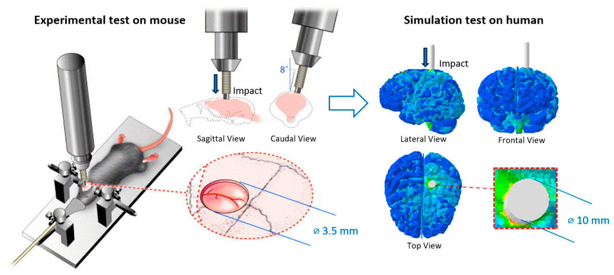
Figure 1 summarizes this translational scheme. On the left, the biological reference corresponds to a murine CCI configuration involving a focal unilateral cortical insult delivered after craniotomy. On the right, the corresponding human-scale representation is formulated as a localized cortical indentation problem intended to reproduce the same contact-driven mechanical logic. The figure therefore links the experimental murine configuration used for biological calibration with the human computational framework used for spatial tau mapping.
The biological reference used in this study was based on the CCI protocol of Edwards et al. [
11], in which 3-month-old P301S transgenic mice underwent a focal cortical injury after craniotomy. In that protocol, a 5 mm craniotomy was created over the right parietal cortex, and the insult was delivered by a pneumatic impactor with a diameter of 3.5 mm, an impact velocity of approximately 3.0 m/s, and a cortical deformation depth of 1.0 mm [
11]. The same study quantified post-traumatic tau burden in cortex, hippocampal area, and brainstem at 1 day, 1 week, 1–2 months, and 6 months after injury, thereby providing the temporal regional dataset used here for calibration.
The purpose of the computational model was to construct a mechanically interpretable human-scale analogue of the focal cortical insult in mice. The main translational question was how to transfer the murine indentation geometry to a human configuration without losing the local meaning of the contact problem.
To support this transfer,
Table 1 summarizes representative anatomical descriptors reported in the literature for adult mouse and adult human brain [
35,
36,
37,
38,
39,
40,
41]. These descriptors were compiled to compare local and global geometric scales relevant to the present problem.
In this research, cortical thickness was the only quantity used to scale the indentation geometry from mouse to human. This choice follows directly from the local nature of the contact problem, since the mechanically relevant dimensions are the indenter diameter, the penetration depth, and the thickness of the cortical layer directly engaged beneath the contact zone. MRI-based cortical thickness analysis in adult mice has reported a mean cortical thickness of 0.89 ± 0.016 mm [
35], whereas human neuroimaging studies report an overall average cortical thickness of approximately 2.5 mm [
36]. This yields an interspecies ratio of about 2.81, which provides a mechanically consistent basis for scaling the indenter dimensions. By contrast, whole-brain mass, whole-brain volume, cortical surface area, and neocortical volume produce much larger human-to-mouse ratios, typically in the order of
to
[
37,
38,
39,
40,
41], which are informative for anatomical context but not appropriate for scaling a localized cortical indentation problem.
The values in
Table 1 show that global anatomical descriptors produce interspecies ratios in the order of
to
[
37,
38,
39,
40,
41]. If such descriptors were used directly to transfer the murine impactor geometry, the corresponding human indenter would be unrealistically large for a localized cortical contact problem. Cortical thickness gives a much smaller ratio, close to 3, and directly characterizes the local tissue layer involved in the contact event [
35,
36]. For this reason, cortical thickness was used as the only scaling quantity in the present study. Using the murine indenter diameter of 3.5 mm reported by Edwards et al. [
11] and a thickness ratio of approximately 2.8 yields a human-equivalent diameter of about 9.8 mm, which supports the use of a 10 mm flat-ended indenter in the present simulation. Applying the same logic to indentation depth, a murine cortical deformation of 1.0 mm corresponds to a human cortical indentation of approximately 2.8 mm. To preserve the short-duration nature of the murine CCI event, the imposed displacement in the human simulation was applied over a short time interval selected to reproduce the order of magnitude of the experimental impact velocity, approximately 3 m/s. Under this interpretation, the computational model should be understood as a translational human-scale analogue of the murine focal cortical insult, while the full finite-element implementation is described in the following section.
2.5. Finite Element Simulation: Brain Biomechanics
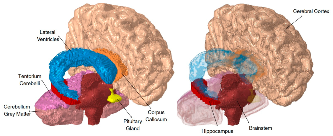
A three-dimensional human brain model was reconstructed from T1-weighted magnetic resonance imaging using 3D Slicer. The segmented anatomical components included cerebral gray matter, cerebral white matter, cerebellum gray matter, cerebellum white matter, hippocampus, brainstem, corpus callosum, lateral ventricles, pituitary gland, falx cerebri, and tentorium cerebelli. Surface defects and local mesh irregularities were corrected in Autodesk Meshmixer, and the volumetric finite-element mesh was generated in Altair HyperMesh. The human brain model and material properties adopted in the present study were based on a computational framework previously validated in earlier studies [
42,
43]. The anatomical components included in the model are shown in
Figure 2.
The deformable intracranial tissues were discretized predominantly with 8-node reduced-integration hexahedral elements (C3D8R). Soft brain tissues were modeled as nearly incompressible visco-hyperelastic materials using an instantaneous Neo-Hookean response combined with Prony-series viscoelasticity.
The material constants listed in
Table 2 were compiled from experimental and computational sources, following the material-characterization strategy used in the female finite element head model (FeFEHM) developed by Carmo et al. [
31]. Specifically, the viscoelastic parameters assigned to cerebral gray matter, cerebral white matter, hippocampus, cerebellar gray matter, cerebellar white matter, brainstem, and corpus callosum were based on the magnetic-resonance-elastography-informed characterizations reported by Alshareef et al. [
33,
44]. The instantaneous Neo-Hookean hyperelastic response of the soft brain tissues was supported by the regional dynamic microindentation measurements reported by Menichetti et al. [
30]. The hyperelastic parameters used for the lateral ventricles were adopted from the head-impact modelling work of Gilchrist [
45], using a Mooney–Rivlin-type formulation. The pituitary gland was modeled using the elastic properties reported by Bouchonville et al. [
46], while the falx cerebri and tentorium cerebelli were assigned linear elastic properties according to the experimental characterization of cranial soft tissues reported by Galford and McElhaney [
47].
Accordingly,
Table 2 includes visco-hyperelastic parameters for the soft brain tissues, hyperelastic parameters for the lateral ventricles, and linear elastic parameters for the pituitary gland and meningeal structures.
To reproduce the mechanically relevant features of the murine CCI paradigm in a human-scale setting, the finite-element analysis was formulated as a localized dynamic cortical indentation problem. A rigid flat-ended cylindrical indenter was defined as an analytical rigid surface and coupled to a reference node through a rigid-body constraint. The indenter acted directly on the cortical surface through a hard-contact formulation with frictionless tangential behavior. This setup was intended to preserve the local contact-driven nature of the experimental insult.
The simulation was performed in Abaqus/Explicit using a displacement-based Lagrangian finite-element formulation with explicit time integration and geometric nonlinearity activated, thereby retaining inertial effects, transient wave propagation, and contact nonlinearities. The deformable brain tissues were discretized mainly with 8-node reduced-integration hexahedral elements, while the rigid indenter was defined as an analytical rigid surface coupled to a reference node. Contact between the indenter and the cortical surface was modeled using hard normal contact and frictionless tangential behaviour. A Dirichlet-type fixed displacement boundary condition was imposed at the inferior base of the brainstem by constraining all translational degrees of freedom of the nodes, in order to mechanically stabilize the model while allowing the localized cortical indentation to generate a distributed deformation field across the brain.
The loading protocol was divided into two sequential explicit dynamic steps. In Step 1, with a total duration of 0.00133 s, the indenter was prescribed a displacement of 4 mm along the loading direction. Since the indenter was initially positioned approximately 1 mm above the cortical surface, this motion corresponds to a short approach phase followed by about 3 mm of effective cortical indentation. The duration of this step was selected so that the imposed indenter motion reproduced the order of magnitude of the experimental murine CCI velocity, approximately 3 m/s, thereby preserving the impulsive character of the reference insult.
In Step 2, the indenter was no longer advanced and the configuration reached at the end of Step 1 was maintained while the model evolved dynamically over a longer time interval of 0.32 s. This second step allowed the stress and strain waves generated during the rapid indentation phase to propagate through the brain and progressively attenuate.
The mechanical field used to drive the tau-aggregation model was the MPS. After completion of the dynamic indentation analysis, nodal strain values were extracted from the finite-element solution and assigned to the corresponding anatomical regions. This mechanically derived field constitutes the raw spatial driver of the subsequent tau model. For coupling with the regional Avrami kinetics, the nodal MPS field was normalized separately within cortex, hippocampus, and brainstem. This region-wise normalization allowed the model to use the relative spatial contrast of the mechanical field within each region while the calibrated temporal laws controlled the regional evolution of tau burden over time.
3. Results
3.1. Calibration
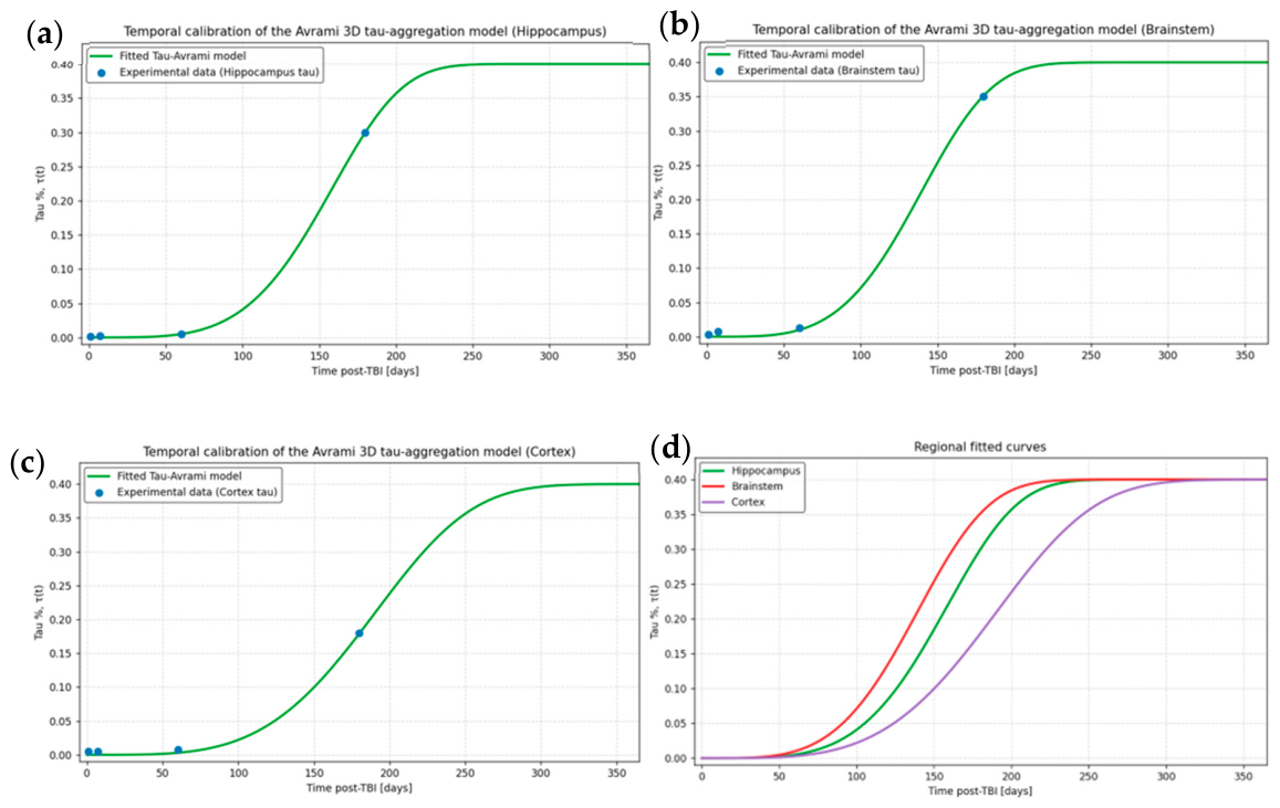
The independent calibration of the Avrami model in cortex, hippocampus, and brainstem produced three distinct regional trajectories, each consistent with the post-TBI experimental progression extracted from the biological dataset. For each region, the kinetic parameters were obtained by minimizing a weighted least-squares calibration error between the model-predicted tau burden and the experimental tau-burden values at the available time points. The resulting curves are shown in
Figure 3a–c, while the direct comparison among the three fitted regional trajectories is presented in
Figure 3d.
In the hippocampus, the fitted curve reproduced an initially negligible tau burden followed by a strong delayed increase, reaching the highest long-term burden among the three regions together with the brainstem. The transition from the quiescent regime to the nonlinear rise occurred later than in the brainstem but earlier than in the cortex, indicating an intermediate kinetic onset.
The brainstem calibration displayed the earliest marked acceleration among the three regions. Although the early burden remained very low, the fitted trajectory rose sooner and more steeply than the hippocampal and cortical curves, indicating a comparatively earlier regional response within the mathematical framework. This behavior is visible in
Figure 3b, where the fitted curve intersects the experimental trend with a clear early nonlinear transition.
The cortex presented the slowest kinetic progression of the three calibrated compartments. Its fitted curve remained lower for longer and then increased more gradually before approaching the imposed saturation level. As shown in
Figure 3c, the cortical trajectory therefore occupies the right-shifted position in time among the three regional laws. When all fitted curves are examined together in
Figure 3d, the relative ordering becomes evident: brainstem rises first, hippocampus follows, and cortex exhibits the latest transition.
This region-dependent temporal separation constitutes one of the main strengths of the present formulation. It shows that the experimental post-TBI burden cannot be adequately represented by a single homogeneous law if the goal is to preserve anatomical specificity. Instead, each region requires its own calibrated trajectory, which later acts as the temporal backbone for spatial redistribution within the finite-element domains.
Table 3 summarizes the experimental tau-burden values and the corresponding model estimates at the four calibration time points for each anatomical region.
The apparently large relative errors at 1 and 7 days in the hippocampus are a consequence of the extremely small experimental tau values at these early time points. Although the percentage error reaches 100%, the corresponding absolute errors remain very small, indicating that the model reproduces a near-zero early burden consistent with a quiescent initial phase. This reflects the sensitivity of percentage errors for near-zero values and should be interpreted together with the low RMSE and excellent overall fit.
The fitted kinetic parameters
,
,
, and
obtained from the independent regional calibration are in
Table 4.
Parameter-Sensitivity Analysis
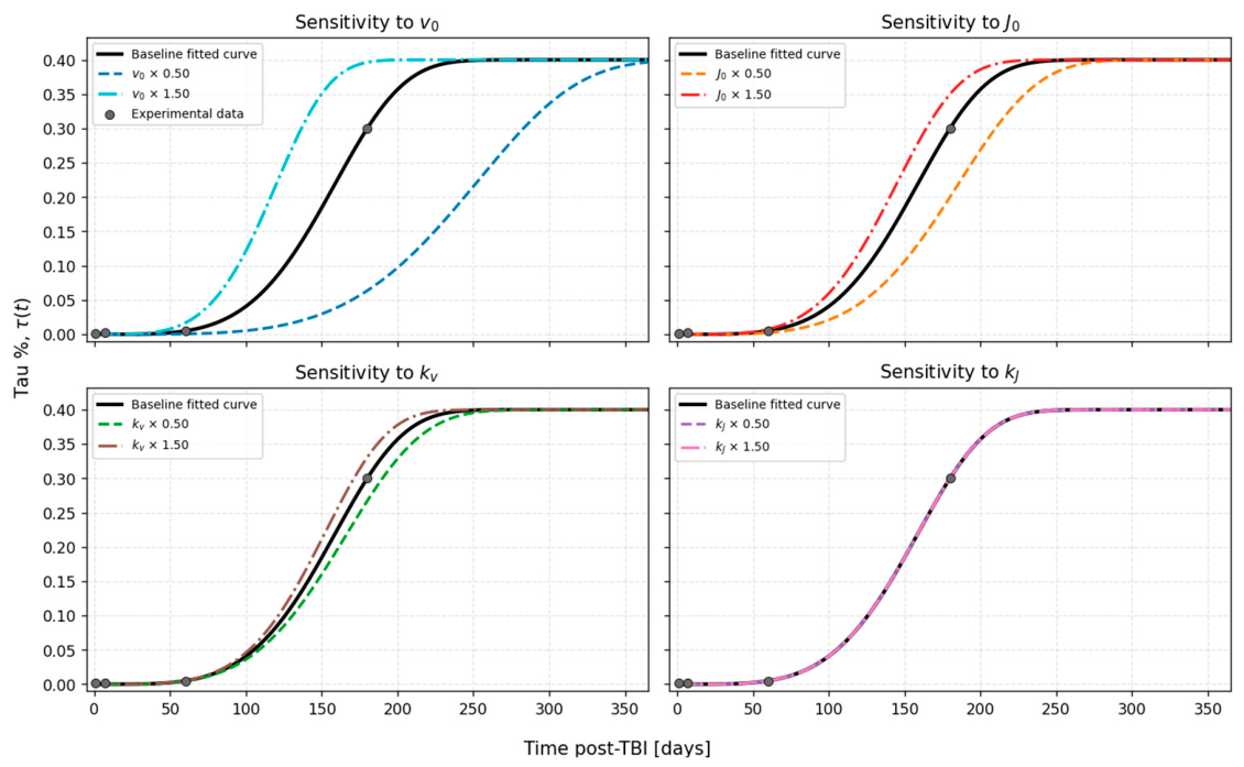
A parameter-sensitivity analysis was performed to evaluate how the calibrated Avrami trajectories respond to variations in the four kinetic parameters
,
,
, and
. For the hippocampus anatomical region, each parameter was independently multiplied by 0.5 and 1.5 with respect to its optimized value, while the remaining three parameters were kept fixed. The resulting curves were then compared with the baseline fitted trajectory over the post-injury time interval. The corresponding sensitivity curves are shown in
Figure 4.
The sensitivity results show that the characteristic sigmoidal shape of the tau-burden trajectory was preserved for all tested perturbations. Among the four parameters, produced the most pronounced changes, mainly shifting the timing of the nonlinear increase. Variations in also affected the onset and progression of the curve, although less strongly than . By contrast, and especially produced comparatively smaller changes within the tested 0.5–1.5 scaling range, with the perturbed curves remaining close to the baseline trajectory. These results suggest that the fitted temporal response is mainly controlled by the baseline growth and nucleation scales, whereas the exponential modulation parameters have a weaker local influence within the explored perturbation interval. However, larger variations of or could still produce relevant changes because these parameters enter the model through exponential time-dependent terms.
3.2. Finite Element Response Under Localized Cortical Indentation
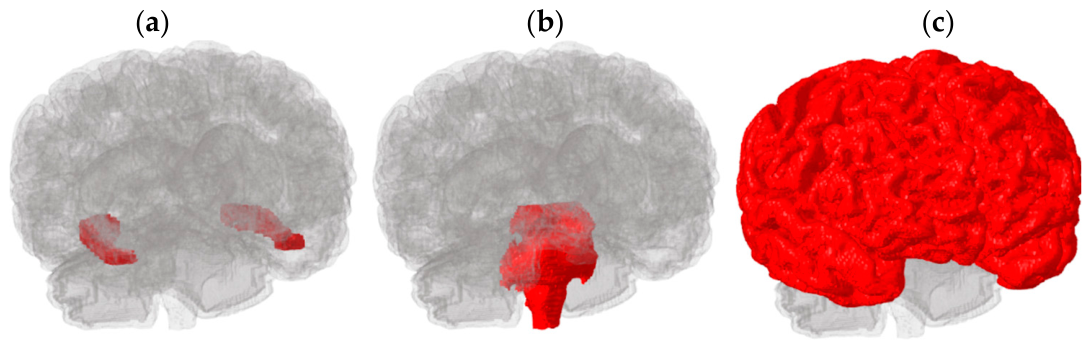
The three anatomical regions independently calibrated in the present framework are shown in
Figure 5. These regions were selected because they correspond to the territories quantified in the experimental reference study and constitute the anatomical domains in which the mechanical response and subsequent tau redistribution were analyzed.
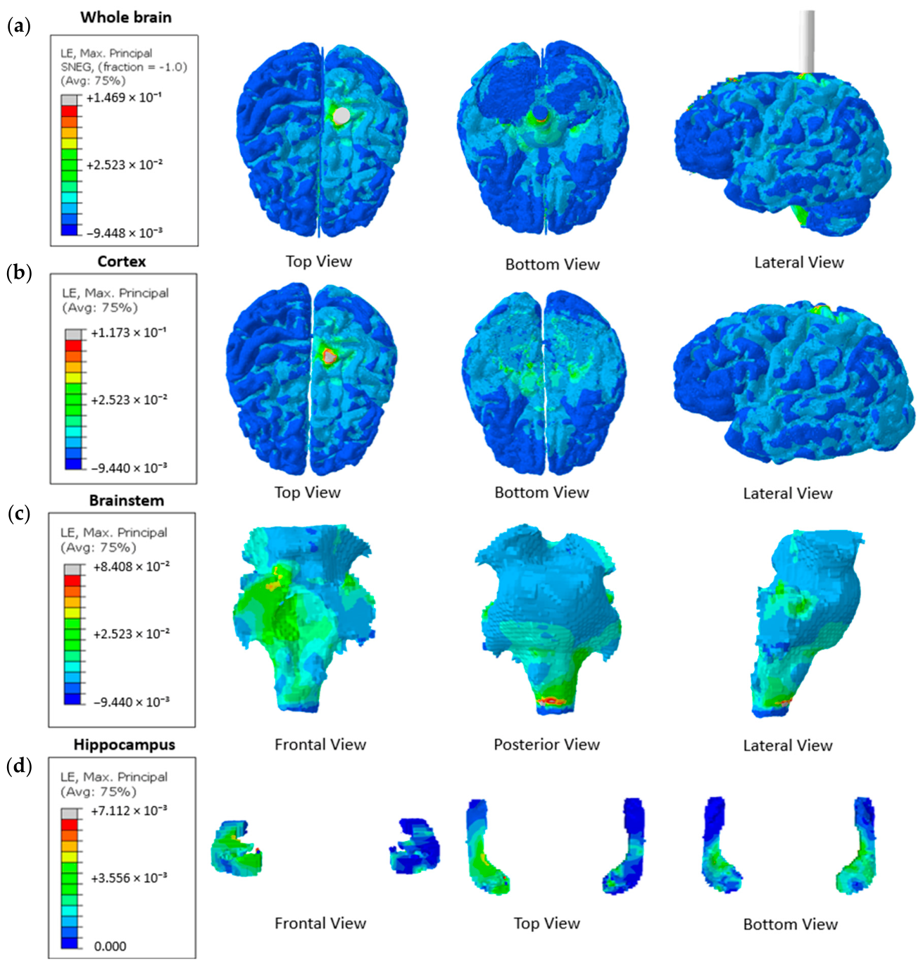
The explicit dynamic indentation generated a heterogeneous field of maximum principal strain (MPS) across the brain model, with the highest values concentrated near the cortical contact region beneath the indenter. This behavior is consistent with the local nature of the applied insult, since the imposed displacement acts directly on the cortical surface and produces a steep deformation gradient away from the contact zone.
Figure 6 shows the finite-element response of the human brain under controlled cortical indentation, including whole-brain and region-focused views of MPS.
In the cortical region, the strain field is strongly localized around the indentation site and decays rapidly toward the surrounding tissue. This produces a clear ipsilateral pattern, with the hemisphere directly exposed to the indenter showing substantially higher strain levels than the contralateral side. The cortical response therefore reflects the direct mechanical footprint of the localized contact event.
In the hippocampal region, the strain field is more distributed and follows the internal curvature of the structure. Higher strain levels are predominantly observed in the ipsilateral hemisphere, that is, on the same side as the applied indentation, whereas the contralateral hippocampal territory exhibits lower magnitudes. This asymmetry is mechanically plausible, since the impact is introduced locally and the transmitted deformation remains stronger on the side of contact, even though the response is no longer confined to the superficial cortex.
In the brainstem, significant strain values are observed in an inferior region close to the constrained base. This pattern must be interpreted carefully, because it is influenced by both the transmitted response from the cortical indentation and the boundary condition imposed at the bottom brainstem support. Thus, the high-strain zone in this region reflects, at least in part, the local mechanical effect of the constraint.
3.3. Spatiotemporal Tau Prediction
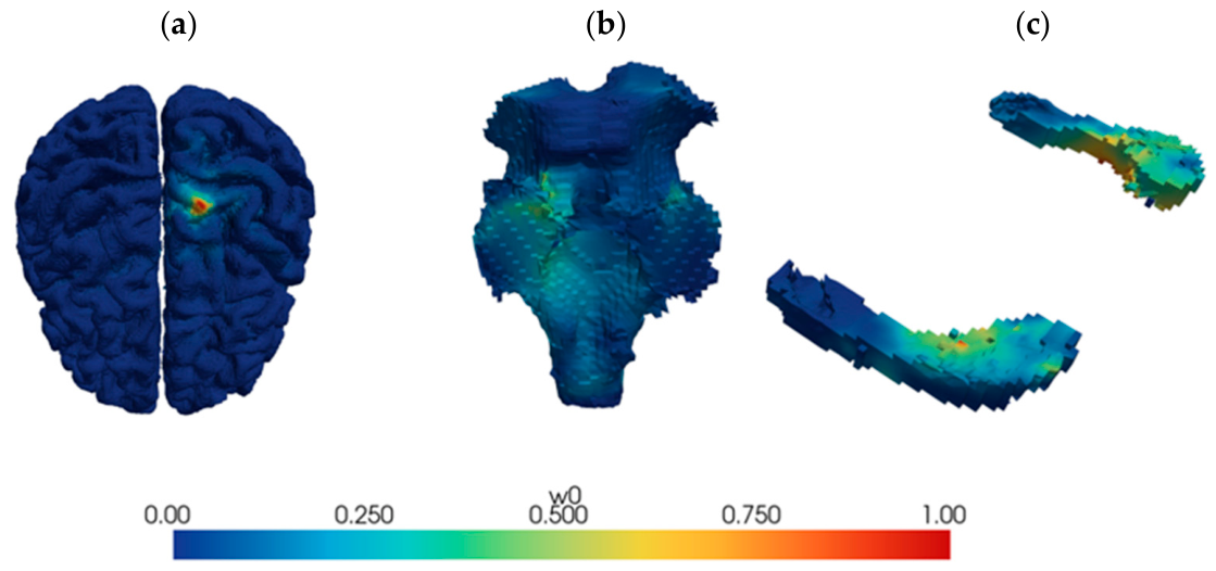
The coupling between the calibrated regional Avrami kinetics and the finite-element strain field yielded spatiotemporal tau predictions for cortex, hippocampus, and brainstem. Before examining the resulting tau maps, the normalized mechanical field
used in each region must be considered, since it defines the relative spatial weighting of tau accumulation. These normalized strain fields are shown in
Figure 7.
The normalized fields in
Figure 7 preserve the main regional structure of the underlying strain patterns and define the mechanical weighting used for tau redistribution. Once coupled to the calibrated regional kinetics, these fields generate tau maps that evolve consistently over time. At early stages, tau burden remains very low in all regions, although local maxima already appear at nodes with the highest mechanical weights. At later stages, regional differences become more evident, with earlier intensification in the brainstem, marked hotspots in the hippocampus, and a comparatively delayed cortical response.
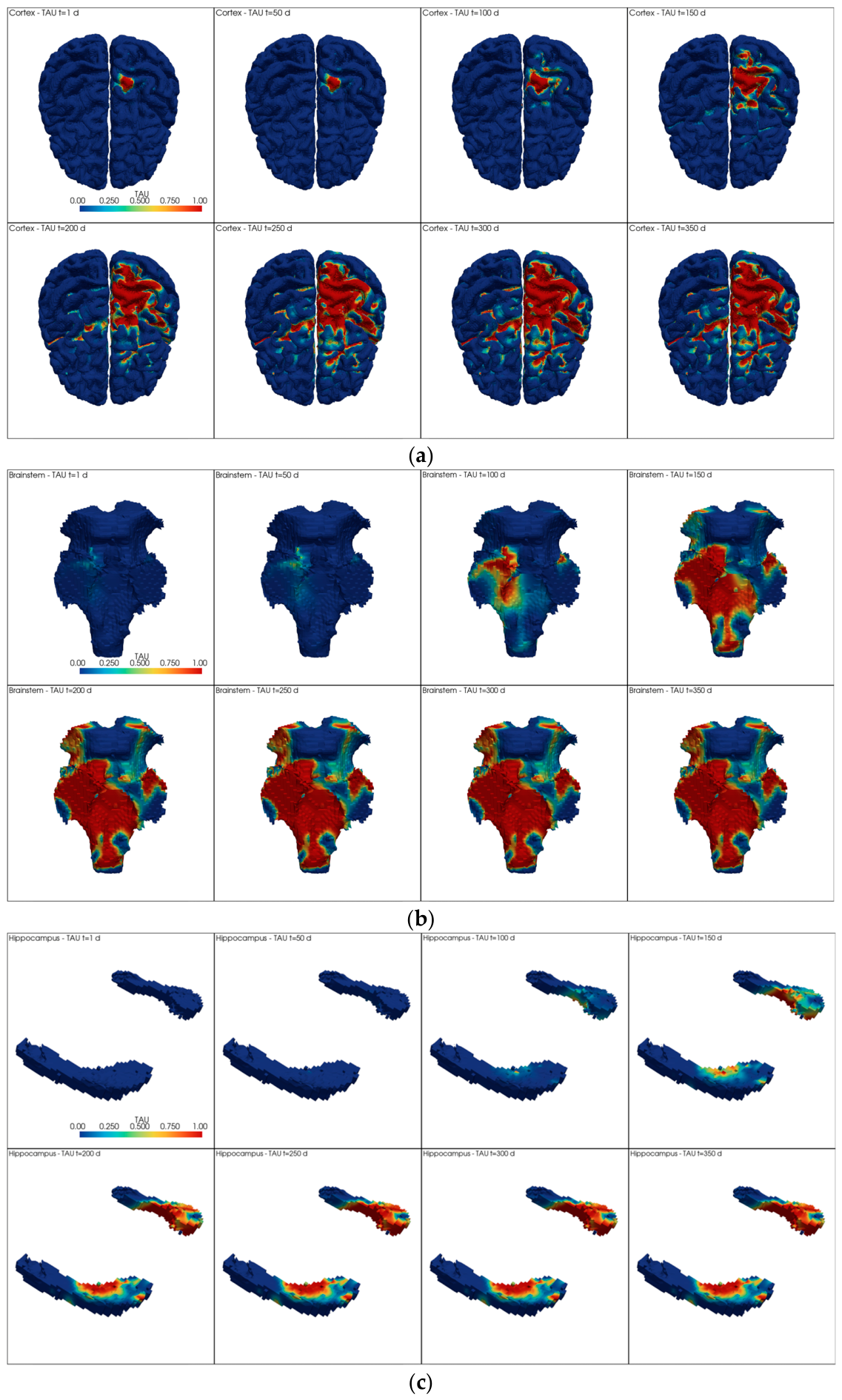
The mean burden in each region remains equal to the corresponding calibrated Avrami trajectory. Therefore, the experimental calibration determines how much tau is present on average, whereas the mechanical field determines where it is preferentially concentrated. For visualization purposes, the spatial maps were plotted with an upper color scale of 1% nodal tau burden. This value reflects localized hotspots rather than regional mean burden, which remains below the imposed saturation limit of 0.4%. The resulting spatiotemporal predictions are shown in
Figure 8.
The maps in
Figure 8 show that tau accumulation is strongly shaped by the spatial distribution of mechanical loading. In the cortex, tau first appears as a focal hotspot in the ipsilateral hemisphere and later expands over a broader but still asymmetric territory. The highest values remain concentrated beneath and around the loaded cortical zone, while the contralateral side is less affected. Part of this predicted cortical burden also follows sulcal regions, which are mechanically vulnerable because of local geometric concentration effects. This is qualitatively consistent with histopathological observations of trauma-associated tauopathy and CTE, where abnormal tau deposition is frequently reported at the depths of cortical sulci [
6,
7].
A similar ipsilateral predominance is observed in the hippocampus, where the side of the insult develops higher tau levels than the contralateral hemisphere. This suggests that a focal cortical insult induces not only superficial cortical vulnerability but also mechanically modulated burden in deeper subcortical structures.
In the brainstem, the burden is more spatially extended and less focal, consistent with its earlier fitted kinetics and its distinct mechanical template. This indicates that remote regions may also develop substantial tau accumulation when both strain exposure and temporal susceptibility are sufficient.
Taken together, these results show that the model combines two levels of heterogeneity: a regional temporal heterogeneity, since cortex, hippocampus, and brainstem follow different calibrated trajectories, and an intra-regional spatial heterogeneity, since tau preferentially accumulates in mechanically weighted subregions.
4. Discussion
The present study introduces a translational framework that combines region-specific tau kinetics with a mechanically localized finite-element loading scenario. In doing so, it addresses two limitations that are common in current computational studies of post-traumatic neurodegeneration. First, many mathematical models of tau progression are spatially informative but not directly anchored to a controlled mechanical insult [
17,
18,
19,
23]. Second, many biomechanical models of brain trauma provide detailed strain fields but do not explicitly couple those fields to a biochemical transformation law capable of reproducing delayed pathological accumulation [
13,
14,
21,
32]. By integrating both components through a region-specific Avrami formulation, the present work attempts to bridge this gap in a conceptually transparent way.
One of the main strengths of the framework is the independent calibration of cortex, hippocampus, and brainstem. This preserves regional specificity and avoids the use of a single global law. This choice is strongly supported by the experimental post-TBI dataset [
11], in which the three territories exhibit distinct burdens and temporal trends. From a biological perspective, this regional separation is reasonable because different brain structures are expected to respond differently to trauma depending on local cytoarchitecture, connectivity, vulnerability, and strain transmission. From a mathematical perspective, the region-specific calibration improves interpretability and prevents the loss of biologically relevant heterogeneity that would result from averaging all regions into a single trajectory.
The use of an Avrami-type formulation is also advantageous. The KJMA framework is particularly well suited to processes with delayed onset, nonlinear increase, and saturation [
24,
25,
26,
27]. These features are consistent with the experimental tau trajectories observed after TBI, where the early burden is very low and the more pronounced increase occurs later [
11]. Moreover, the nucleation-growth interpretation provides a mechanistically appealing analogy for tau aggregation. In this context, pathological tau seeds are represented as effective nuclei, while the subsequent increase in tau burden is represented as the deterministic growth and overlap of transformed domains. The formulation is therefore more informative than a purely empirical curve fit and remains computationally manageable for region-wise application.
Another important contribution of the present work is the use of a localized indentation framework as the translational biomechanical counterpart of the experimental CCI. This point is especially relevant. The experimental mouse injury is not a generic head impact, but a controlled local cortical insult delivered under known geometric and kinematic conditions [
11]. Representing it computationally through a localized human-scale indentation therefore provides a more faithful translational analogue than a broad head-loading or inertial scenario. For this reason, the indenter was scaled using cortical thickness and not global brain size. In a contact-driven cortical indentation problem, the mechanically relevant length scale is local cortical thickness, since it governs the ratio between indenter geometry and the deformed cortical layer. This makes the scaling choice physically interpretable and more appropriate for the present application than whole-organ size scaling.
The finite-element results support the idea that a localized cortical indentation can generate mechanically heterogeneous strain patterns not only in the cortex but also in deeper and more distant regions. In the cortical territory, this heterogeneity is not purely focal but also follows anatomically structured zones of enhanced vulnerability, including sulcal regions, which is of particular interest given the known association between sulcal depths and trauma-related tau deposition in histopathological studies [
6,
7]. This is directly reflected in the predicted tau maps, where the hemisphere ipsilateral to the insult consistently develops the highest burden in cortex and hippocampus, while the brainstem shows a broader but still structured pattern of accumulation. These findings indicate that the three structures are not mechanically isolated from the cortical insult. Instead, the local impact generates a distributed deformation field that creates region-dependent spatial templates for subsequent tau accumulation. This observation is consistent with the broader biomechanical literature showing that localized or global loading can produce anatomically structured strain concentrations across distributed brain territories [
13,
14,
32].
The coupling strategy adopted here also deserves discussion. In the present framework, the biological calibration is temporal and regional, whereas the mechanical field determines the internal spatial weighting within each region. This means that the finite-element field does not calibrate the mean tau burden; it redistributes a previously calibrated regional burden over space. The introduction of the corrective factor is essential in this context. Without it, the nonlinear exponential transformation applied to a heterogeneous strain field would not preserve the calibrated regional mean kinetics. Therefore, should not be interpreted as an arbitrary fitting artifact, but as a deterministic consistency factor that reconciles the heterogeneous nodal field with the biological regional target. So, the present model does not claim direct spatial calibration from histological maps, because such maps are not available here in a directly mesh-compatible form. Instead, it enforces exact agreement with regional kinetics while letting the mechanical field control the relative spatial pattern.
From a mechanobiological standpoint, the model supports a picture in which tau accumulation is not solely determined by the existence of injury, but by the combination of regional temporal susceptibility and local mechanical amplification. Regions with earlier calibrated kinetics, such as brainstem in the present fitted set, begin to accumulate more rapidly, while regions with later onset, such as cortex, remain comparatively quiescent for longer. Within each region, however, tau does not accumulate uniformly. Instead, the highest local burden appears preferentially in the mechanically most weighted subdomains, which in the present simulations results in a clear ipsilateral predominance in cortex and hippocampus and a broader, more distributed pattern in brainstem. The model therefore combines two different levels of heterogeneity: one temporal and biological, the other spatial and mechanical. This dual structure is one of the main conceptual contributions of the work.
The framework also aligns with the broader literature on tau propagation and trauma-associated degeneration. Previous studies have emphasized network-mediated spreading [
15,
16,
17,
18,
19,
20], prion-like self-propagation [
20], and mechanically induced abnormal tau accumulation in exposed populations [
21]. The present work complements these perspectives by focusing on the earlier stage of post-traumatic tau evolution, namely the conversion of a localized cortical insult into region-specific temporally evolving burden maps. It does not replace connectivity-based propagation models but provides a local initiation and redistribution mechanism that could, in future work, be combined with connectome-mediated transport or repeated-impact scenarios. The present framework is also complementary to recent computational neuropathology approaches that use deep learning to classify temporal stages of AT8-labeled tau pathology after experimental TBI [
48]. Together, such approaches may help bridge image-level phenotyping, regional burden quantification, and predictive mechanobiological modeling.
The present framework can also be positioned with respect to data-driven, network-based, and PDE-based computational biology approaches. Data-driven methods are especially useful when large imaging or histopathological datasets are available, because they can identify spatial or temporal patterns without prescribing a detailed mechanistic structure [
48]. Network-diffusion and connectivity-based models are well suited to represent connectivity-mediated propagation in neurodegenerative disease [
17,
18,
19], whereas PDE-based reaction-diffusion or transport models can describe continuous spatiotemporal spreading of pathological proteins and related biological processes. The present approach is complementary to these methodologies. It uses sparse experimental tau-burden measurements [
11] to define region-specific temporal kinetic laws and then couples these laws to a finite-element-derived mechanical field [
31,
32] to obtain a mechanically weighted spatial redistribution of tau burden.
From a numerical perspective, the model is based on a Lagrangian explicit dynamic formulation, which is suitable for extracting a mechanically interpretable strain field after localized cortical indentation. More advanced Eulerian, operator-splitting, and phase-field/contact formulations may be useful in future extensions involving larger deformations, moving interfaces, tissue remodeling, or fluid–structure interaction [
49,
50,
51].
Limitations
Although the proposed framework provides a structured and mechanically interpretable basis for translational post-traumatic tau modeling, several limitations should be acknowledged. The temporal calibration of tau burden was performed using only four post-injury points per region, with a maximum follow-up of 6 months. This is sufficient for a proof-of-concept Avrami-type fit, but it does not capture the full temporal complexity of tauopathy as a long-term neurodegenerative process. In this sense, the experimental points used here represent only an early pathological phase of disease progression. Future studies should seek richer temporal datasets with longer follow-up periods in order to better characterize the phases of pathological accumulation.
Although the nonlinear Avrami-type calibration provided stable regional fits, the limited number of experimental points should be considered when interpreting the fitted kinetic parameters. In multi-parameter mathematical models, different combinations of parameters may provide similar curve-fitting results, and this effect becomes more relevant when the calibration dataset is small. Therefore, the fitted parameters should be interpreted as effective descriptors of the regional tau-burden curves within the available experimental window. Other sigmoidal formulations could also reproduce the same sparse temporal trends. In the present study, the Avrami-type formulation was retained because it provides an interpretable way to describe delayed growth, nonlinear increase, and saturation of tau burden over time, in a similar way to nucleation-and-growth processes during phase transformations in materials, where transformed domains or grains progressively appear and grow over time.
Another important limitation is that trauma-associated tauopathy is likely a multiscale and multiphysics problem, whereas the present framework only incorporates one physical component explicitly, namely mechanics. In the current model, the finite-element strain field is used to define the spatial weighting of tau redistribution, so that regions or subregions exposed to higher strain develop a greater predicted burden. While this assumption is mechanobiologically plausible, it is only a partial representation of the real disease process. In reality, post-traumatic tau pathology is also likely modulated by neuroinflammation, glial activation, axonal dysfunction, impaired axonal transport, and other biochemical and cellular cascades. These processes may interact across scales, from tissue-level deformation to cellular signaling and protein aggregation kinetics. The present model should therefore be interpreted as a mechanically informed approximation of spatial vulnerability, not as a complete representation of post-traumatic tauopathy.
Moreover, the simulated tau maps were not directly calibrated against mesh-compatible histological distributions at regional or voxel level. As a result, the model should not be interpreted as providing a validated ground-truth map of in vivo spatial pathology, but as a plausible mechanically structured redistribution framework constrained by regional temporal calibration.
A further numerical limitation is that a dedicated mesh-convergence study was not performed. The finite-element model used small element sizes and a large number of elements, providing a detailed discretization of the brain geometry. However, because the nodal MPS field is used to guide tau redistribution, local features such as peak strain values may still be influenced by mesh resolution. Future work should assess this effect through a specific mesh-sensitivity analysis.
Additional limitations arise from some of the simplifying assumptions adopted in the coupling procedure. The saturation parameter α = 0.40 was selected according to the available mouse dataset, in which the maximum regional tau burden at 6 months was close to 0.35%. Thus, α represents a practical saturation level in the experimental tau-percentage scale, slightly above the maximum value observed in the calibration data. Since CTE and trauma-associated tauopathy may evolve over much longer time scales, this parameter should not be interpreted as a universal upper limit and could be recalibrated when longer-term experimental or human data become available.
The nonlinear weighting , with p = 4 in the present study, is phenomenological and should be interpreted as a contrast-enhancing spatial assumption, not as an experimentally established law linking strain to tau accumulation. This fourth-power weighting was selected to increase the contrast between highly strained and mildly strained regions. Lower exponents, such as p = 2 or p = 3, would preserve a greater contribution from intermediate mechanical values and would therefore produce smoother and less focal tau redistributions. By contrast, p = 4 reduces the influence of mildly strained areas and concentrates the predicted burden in the most mechanically loaded subregions. This exponent could be modified or recalibrated in future applications if more complete experimental datasets, especially spatially resolved tau pathology data from human brains, become available. A formal sensitivity analysis of p was not performed in this exploratory study; future work should assess how different exponents affect hotspot size and spatial tau dispersion.
Likewise, the transfer from mouse to human was based primarily on cortical thickness, which is a rational local scaling quantity for a contact-driven indentation problem, but does not exhaust all aspects of interspecies similarity. Although the human-scale model preserved the main local features of the murine CCI experiment, including unilateral cortical indentation, a flat-ended impactor, and scaled impact area and indentation depth, some simplifications were introduced. In particular, the approximately 8° indentation angle reported in the murine experiment was neglected, and a normal indentation direction relative to the local cortical surface was used instead.
In addition, because mouse and human brain geometries differ substantially, the exact equivalent point of application cannot be directly established. Therefore, the indentation simulation should be interpreted as a translational human-scale analogue of the murine focal cortical insult and as a way to obtain a localized mechanical field for the proposed tau-aggregation framework. Differences in anatomy, gyrification, tissue composition, constitutive behavior, structural connectivity, and biological susceptibility are not explicitly represented in this simplified scaling strategy. Although cortical thickness is a rational scaling criterion for a localized indentation problem, it does not ensure full mouse-to-human mechanical similarity; therefore, the simulation represents a human-scale analogue of the murine CCI insult, not a complete interspecies equivalence model.
Finally, greater translational relevance would require more extensive human datasets, but these are intrinsically difficult to obtain. Longitudinal monitoring of post-traumatic tauopathy in humans, particularly in exposed populations such as boxers or other contact-sport athletes, is limited by ethical, clinical, and practical constraints. The disease may evolve over years, whereas well-resolved spatiotemporal pathological measurements in living subjects remain scarce. If richer long-term datasets become available in the future, especially in humans, they could be used to refine the temporal laws, improve regional specificity, and evaluate the extent to which the present framework captures clinically meaningful patterns of post-traumatic tau evolution.
Even with these limitations, the proposed framework provides a useful and coherent starting point for translational post-traumatic tau modeling. It preserves experimental regional specificity, uses a mechanically interpretable scaling rule, and couples localized biomechanics to an explicit temporal transformation law. Future extensions may include repeated loading, anisotropic structural dependence, diffusion or connectivity-based tau spread, explicit inflammatory or neurobiological coupling, and more detailed calibration using richer regional or voxel-resolved pathological datasets.




 ,
,