Article

Identification of Protein Biomarker Signatures for Acute Myeloid Leukemia (AML) Using Both Nontargeted and Targeted Approaches

1
Department of Biology, Maynooth University, W23 F2H6 Maynooth, Ireland
2
Institute for Molecular Medicine Finland—FIMM, HiLIFE—Helsinki Institute of Life Science, iCAN Digital Cancer Medicine Flagship, University of Helsinki, FI-00014 Helsinki, Finland
3
Department of Haematology, Mater Misericordiae University Hospital, D07 KH4C Dublin, Ireland
*
Author to whom correspondence should be addressed.
These authors have contributed equally to the work and share first authorship.
Proteomes 2021, 9(4), 42; https://doi.org/10.3390/proteomes9040042
Submission received: 18 October 2021 / Accepted: 20 October 2021 / Published: 30 October 2021

Abstract

:
Acute myeloid leukemia (AML) is characterized by an increasing number of clonal myeloid blast cells which are incapable of differentiating into mature leukocytes. AML risk stratification is based on genetic background, which also serves as a means to identify the optimal treatment of individual patients. However, constant refinements are needed, and the inclusion of significant measurements, based on the various omics approaches that are currently available to researchers/clinicians, have the potential to increase overall accuracy with respect to patient management. Using both nontargeted (label-free mass spectrometry) and targeted (multiplex immunoassays) proteomics, a range of proteins were found to be significantly changed in AML patients with different genetic backgrounds. The inclusion of validated proteomic biomarker panels could be an important factor in the prognostic classification of AML patients. The ability to measure both cellular and secreted analytes, at diagnosis and during the course of treatment, has advantages in identifying transforming biological mechanisms in patients, assisting important clinical management decisions.

1. Introduction

Acute myeloid leukemia (AML) is a highly heterogenous disease of the blood and bone marrow, characterized by the uncontrolled proliferation of cells from the myeloid lineage [1]. AML is the second most common form of leukemia in adults, and accounts for approximately 1% of new cancer diagnoses [2]. The majority of patients, following intensive chemotherapy, achieve complete remission; however, AML remains a highly fatal disease, with a disheartening five-year survival rate of ~24% [3]. The high fatality of this disease is attributed to the presence of primary resistance in a subset of patients and a high relapse rate with therapy-resistant disease following complete remission [4].
According to the French-American-British (FAB) system, there are eight types of AML (M0–M7), each with precise morphological characteristics and differentiation stages [5]. The more recent World Health Organization (WHO) classification considers clinical features, morphology, immunophenotyping, cytogenetics, and molecular genetics [6].
Cytogenetic abnormalities, including chromosomal translocations, deletions, and duplications, in addition to molecular mutations, such as mutations in the nucleophosmin-1 (NPM-1) or FMS-like tyrosine kinase 3 (FLT3) genes, are powerful prognostic markers in AML. In 2017, European LeukemiaNet (ELN) published revised recommendations to aid the interpretation of genetic abnormalities for risk stratification in AML [7,8]. The ELN recommendations stratify patients into three prognostic categories; “favorable”, “intermediate” or “adverse”, based on the genetic profile of patients. These recommendations are used to determine the risk of chemoresistance, a key factor in determining the best course of treatment for patients, as the benefit of highly intensive chemotherapy may outweigh the potential therapy-induced toxicities in patients with “intermediate” or “adverse” risk [8,9].
Despite many clinicians adopting this method of risk stratification, the crucial factors involved in relapse and chemoresistance have yet to be identified, with several studies proposing additional prognostic markers which may improve the prognostic value of the ELN recommendations [10,11,12]. Furthermore, the risk of relapse with therapy-resistant disease in patients with “favorable” risk remains relatively high, i.e., occurring in 30–35% of cases [8]. The high recurrence rate and aggressiveness of this disease heightens the need for an expansion of current biomarkers to improve prognostic classification, risk assessment and therapeutic decision-making in AML.
Advances in high-throughput proteomic techniques, especially in the area of mass spectrometry, has pushed proteomics to the forefront of current efforts in the discovery of novel clinically relevant biomarkers [13]. Analyzing the proteomic profile of AML patients has led to the identification of various potential therapeutic targets and candidate protein biomarkers to predict AML relapse and therapeutic efficiency [14,15,16,17].
In this study, we compared the proteomic profiles of clinical AML cells from favorable (group 1), intermediate (group 2) and adverse (group 3) risk patients based on the ELN recommendations to identify specific proteins associated with each risk group. Further proteomic analyses of the matched AML serum samples were conducted to identify and quantify cytokine levels between the three risk groups. The proteomic profiling of the ELN based risk groups facilitated the identification of differential protein levels between risk groups, thus, improving our current understanding of AML prognosis and identifying potential biomarkers to aid prognostic classifications in AML.

2. Materials and Methods

2.1. Clinical Samples

Matching Peripheral Blood (PB) and bone marrow (BM) patient samples were collected at the same time after receipt of written informed consent from the Helsinki University Hospital Comprehensive Cancer Center (Helsinki University Hospital Ethics Committee, decision number 3613/06.01.05.01.00/2014) and in compliance with the Declaration of Helsinki. In total, 41 samples from AML patients were collected. The patient characteristics are presented in Table 1. Cytogenetic and molecular genetic data were available for all patients, and genetic group (favorable, intermediate, and adverse) was defined according to the European LeukemiaNet. Mononuclear cells (MNCs) were isolated from the BM samples by Ficoll density gradient centrifugation (GE Healthcare, Little Chalfont, UK). MNCs were viably cryopreserved in 10% DMSO and 90% fetal calf serum until further analysis.

2.2. Sample Preparation for Mass Spectrometry

BM aspirate cells were lysed in an SDT-lysis buffer (4% SDS, 100 mM Tris/HCl, 100 mM DTT, pH 7.6) using 1:10 sample to buffer ratio and heated at 95 °C for 3–5 min. DNA was sheared by sonication to reduce the viscosity of the sample. Before starting sample processing, the lysate was clarified by centrifugation at 16,000× g for 5 min. A Pierce 660 nm protein assay system was used to determine protein concentration. Suspensions were then buffer exchanged using the filter-aided sample preparation (FASP) method in a buffer containing 8 M urea/50 mM NH4HCO3/0.1% ProteaseMax. After reduction with dithiothreitol and iodoacetic acid-mediated alkylation, a double digestion was performed using Lys-C (for 4 h at 37 °C) and trypsin (overnight at 37 °C) on 5 μg of BM aspirate protein. Digested samples were desalted prior to analysis using C18 spin columns (ThermoFisher Scientific, Hemel Hempstead, UK), dried through vacuum centrifugation and resuspended in mass spectrometry loading buffer (2% acetonitrile (ACN), 0.05% trifluoroacetic acid (TFA) in LC-MS grade water). Peptides were vortexed, sonicated and briefly centrifuged at 14,000× g and the supernatant transferred to mass spectrometry vials for label-free liquid chromatography mass spectrometry (LC-MS/MS).

2.3. Label-Free Liquid Chromatography Mass Spectrometry

First, 500 ng of each digested sample was loaded onto a Q-Exactive high-resolution accurate mass spectrometer connected to a Dionex Ultimate 3000 (RSLCnano) chromatography system (ThermoFisher Scientific, Hemel Hempstead, UK). Sample loading was carried out by an auto-sampler onto a C18 trap column (C18 PepMap, 300 μm id × 5 mm, 5 μm particle size, 100 Å pore size; Thermo Fisher Scientific). The trap column was switched on-line with an analytical Biobasic C18 Picofrit column (C18 PepMap, 75 μm id × 50 cm, 2 μm particle size, 100 Å pore size: Dionex). Peptides were eluted over a 65 min binary gradient [solvent A: 2% (v/v) ACN and 0.1% (v/v) formic acid in LC-MS grade water and solvent B: 80% (v/v) ACN and 0.1% (v/v) formic acid in LC-MS grade water]: 3% solvent B for 5 min, 3–10% solvent B for 5 min, 10–40% solvent B for 30 min, 40–90% solvent B for 5 min, 90% solvent B for 5 min and 3% solvent B for 10 min. The column flow rate was set to 0.3 μL/min. Data were acquired with Xcalibur software (Thermo Fisher Scientific). The mass spectrometer was externally calibrated and operated in positive, data-dependent mode. A full survey MS scan was performed in the 300–1700 m/z range with a resolution of 140,000 (m/z 200) and a lock mass of 445.12003. Collision-induced dissociation (CID) fragmentation was carried out with the fifteen most intense ions per scan and at 17,500 resolution. Within 30 s a dynamic exclusion window was applied. An isolation window of 2 m/z and one microscan were used to collect suitable tandem mass spectra.

2.4. Protein Identification and Quantification

Data analysis, processing and visualization for urine protein identification and label-free quantification (LFQ) normalization of MS/MS data was performed using MaxQuant v1.5.2.8 (http://www.maxquant.org) (accessed on 4 March 2019) and Perseus v.1.5.6.0 (www.maxquant.org/) (accessed on 8 March 2019) software. Differential protein expression patterns in the AML (favorable, intermediate, and adverse prognostic groups) proteomes were initially identified using MaxQuant software and the Andromeda search engine to explore the detected features against the UniProtKB/SwissProt database for Homo sapien. The following search parameters were used: (i) first search peptide tolerance of 20 ppm, (ii) main search peptide tolerance of 4.5 ppm, (iii) cysteine carbamidomethylation set as a fixed modification, (iv) methionine oxidation set as a variable modification, (v) a maximum of two missed cleavage sites and (vi) a minimum peptide length of seven amino acids. The false discovery rate (FDR) was set to 1% for both peptides and proteins using a target-decoy approach. Relative quantification was performed using the MaxLFQ algorithm. The “proteinGroups.txt” file produced by MaxQuant was further analyzed in Perseus. Proteins that matched to the reverse database or a contaminants database or that were only identified by site were removed. The LFQ intensities were log2 transformed, and only proteins found in all seven replicates in at least one group were used for further analysis. Data imputation was performed to replace missing values with values that simulate signals from peptides with low abundance chosen from a normal distribution specified by a downshift of 1.8 times the mean standard deviation of all measured values and a width of 0.3 times this standard deviation. A two-sample t-test was performed using p < 0.05 on data post imputation, to identify statistically significant differentially abundant proteins. The freely available software package PANTHER (http://pantherdb.org/) (accessed on 4 April 2019), was used to identify protein classes and characterize potential protein interactions, respectively. Statistical overrepresentation tests (PANTHER GO-Pathway) of protein sets were performed using the PANTHER database (http://PANTHERdb.org/) (accessed on 6 April 2019). Protein lists were uploaded in gene symbol format, and default whole genome lists from the appropriate species were used as reference [18]. To analyze statistical significance, Fisher’s exact test with Benjamini–Hochberg False Discovery Rate correction (FDR) was applied [19].

2.5. Luminex Assay

Blood samples were evaluated using panels of analytes on the Luminex xMAP technology bead-based multiplexed immunoassay system. Panel 1 (L1CAM, CA9, Mesothelin, Midkine, Hepsin, Kallikrein 6, TGM2, ALDH1A1, EpCAM and CD44). Panel 2 (EGF, Eotaxin, G-CSF, GM-CSF, IFNα2, IFNγ, IL-10, IL-12p40, IL-12p70, IL-13, IL-15, IL-17A, IL-1RA, IL-1α, IL-1β, IL-2, IL-3, IL-4, IL-5, IL-6, IL-7, IL-8, IP-10, MCP-1, MIP-1α, MIP-1β, TNFα, TNFβ and VEGF). Panel 3 (eotaxin-2, MCP-2, BCA-1, MCP-4, I-309, IL-16, TARC, 6CKine, eotaxin-3, LIF, TPO, SCF, TSLP, IL-33, IL-20, IL-21, IL-23, TRAIL, CTACK, SDF-1α+β, ENA-78, MIP-1d and IL-28A). Samples were run in duplicate, along with blanks, standards, and high and low concentration controls. Fluorescent values beyond the range of the standards were extrapolated (unless the fluorescence intensity was below that of the blanks). Average value of duplicates was used for data analysis.

3. Results

3.1. Proteomics Profiling of Human Bone Marrow Cells

Proteomic profiles on bone marrow aspirates from “favorable”, “intermediate” and “adverse” risk AML patients were generated using label-free mass spectrometry. Label-free mass spectrometry was selected as the quantitative approach because of the minimal sample processing necessary to achieve relative quantification (Supplementary Tables S1–S3). Bone marrow samples of high quality from 41 human subjects, 15 males and 26 females, were available for proteomics analysis. Their age ranged from 16.5 to 78.1 years with a median of 57 years (Table 1). Group 1 had male (3), female (8) with an average diagnostic age of 48.3; Group 2, male (6), female (10) with an average diagnostic age of 57.5 and Group 3, male (6), female (8) with an average diagnostic age of 54.3.
Comparing group 1 (favorable) to group 2 (intermediate), 18 proteins were found to be significantly changed, with CAH1 (Carbonic anhydrase 1), 5.2-fold elevated in group 2 (p = 0.035) representing the largest abundance difference between these groups (Table 2; Supplementary Tables S5–S7). Comparing group 2 to group 3 (adverse), 41 proteins were found to be significantly changed, with PRKDC (protein kinase, DNA-activated, catalytic subunit), 12-fold elevated in group 2 (p = 0.010), representing the largest abundance difference between these groups. The highest number of statistically significant proteins was found when comparing group 1 to group 3. In total, 64 proteins were determined to be significantly changed between these groups, with TIF1B 7.0-fold elevated in group 3 (p = 0.027).

3.2. Pathway Analysis

Focusing on the group 1 vs. group 3 comparison, more proteins associated with metabolic pathways, carbon metabolism, glycolysis/gluconeogenesis and biosynthesis of amino acids were discovered to be elevated in group 3 (Figure 1). Of the proteins identified as significantly changed between group 1 and group 3, those involved in propanoate metabolism, oxytocin signaling pathway, pyruvate metabolism, regulation of the actin cytoskeleton, nucleotide-binding oligomerization domain (NOD)-like receptor signaling pathway, tight junctions, hypoxia-inducible factor 1 (HIF-1) signaling pathway, apoptosis and the glucagon signaling pathway were uniquely associated with group 3. Overall, the majority of proteins were found to be significantly elevated in group 3 compared to group 1 (56 proteins vs. 8 proteins) (Figure 1, Figure 2 and Figure 3).
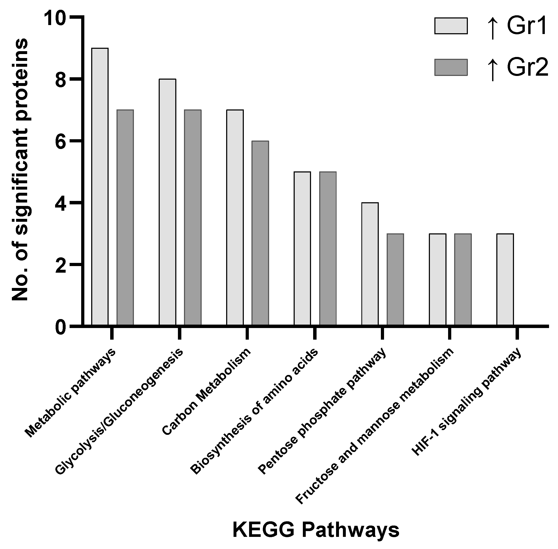
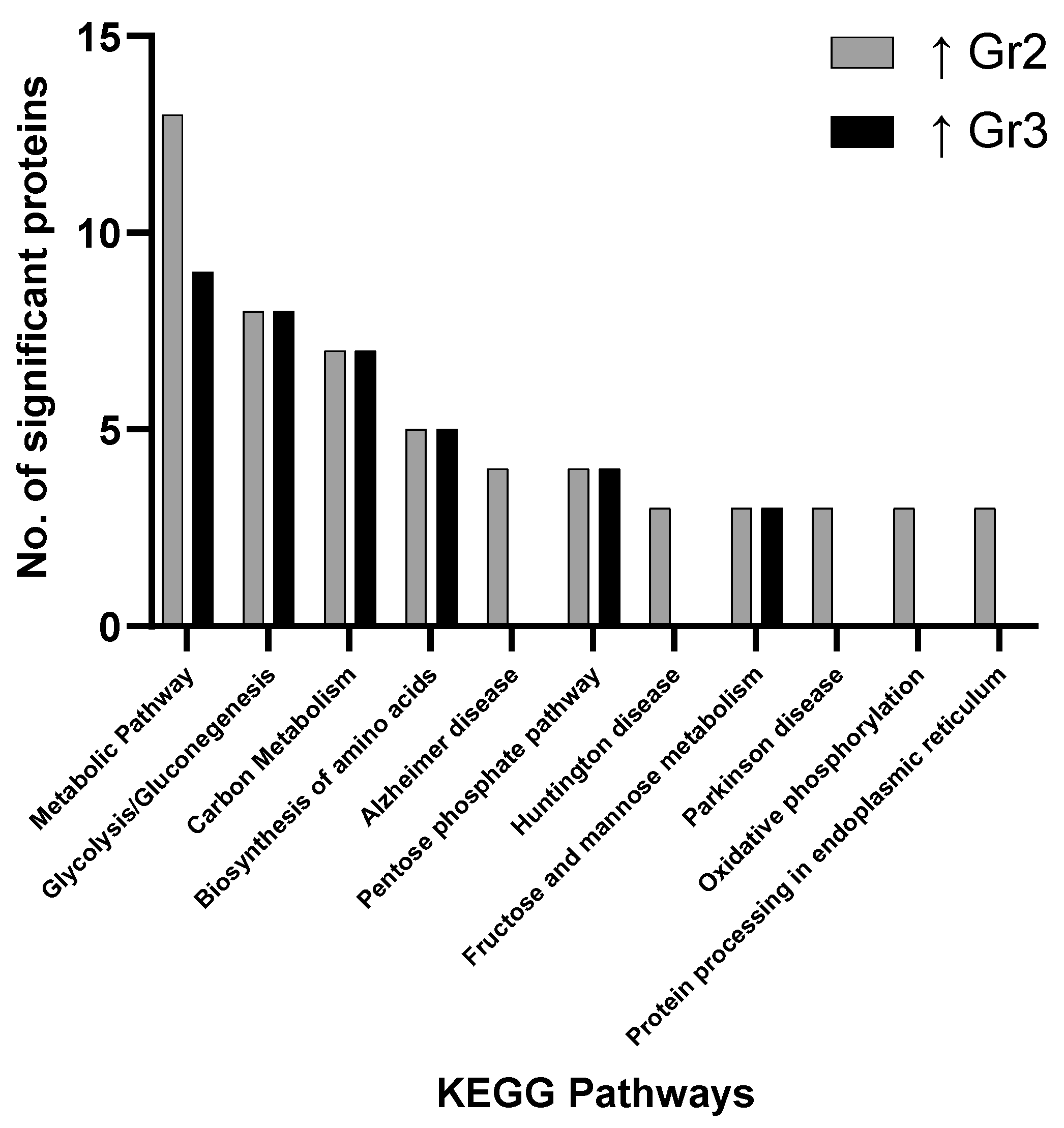
The main pathways involving significantly changed proteins when evaluating all groups focus on metabolic pathways, carbon metabolism, glycolysis/gluconeogenesis, biosynthesis of amino acids, the pentose phosphate pathway, pyruvate metabolism and fructose/mannose metabolism. These results indicate how primary AML samples show variations in distinct metabolic pathway and the associated prognostic impact this may have on clinical outcome (Figure 1, Figure 2 and Figure 3). No pathways were found to be significant based on pathway enrichment analysis filtered by FDR-adjusted p-value (<0.05) using the PANTHER Classification System because of the small protein lists submitted.

3.3. Targeted Proteomics Analysis

The simultaneous measurement of different analytes from a single sample is an emerging area for achieving efficient and high-throughput detection in several applications, including biomarker discovery. In this analysis, the Luminex platform was employed which includes a robust workflow and overcomes sample limitation problems, thus eliminating the need to perform parallel individual measurements using single-plex approaches. Using antibody coated magnetic beads, panels evaluating human circulating cancer biomarkers and human cytokine/chemokine/growth factors were run and subsequently using xPONENT, standard curve fitting models for quantitative analysis were generated (Supplementary Table S4).
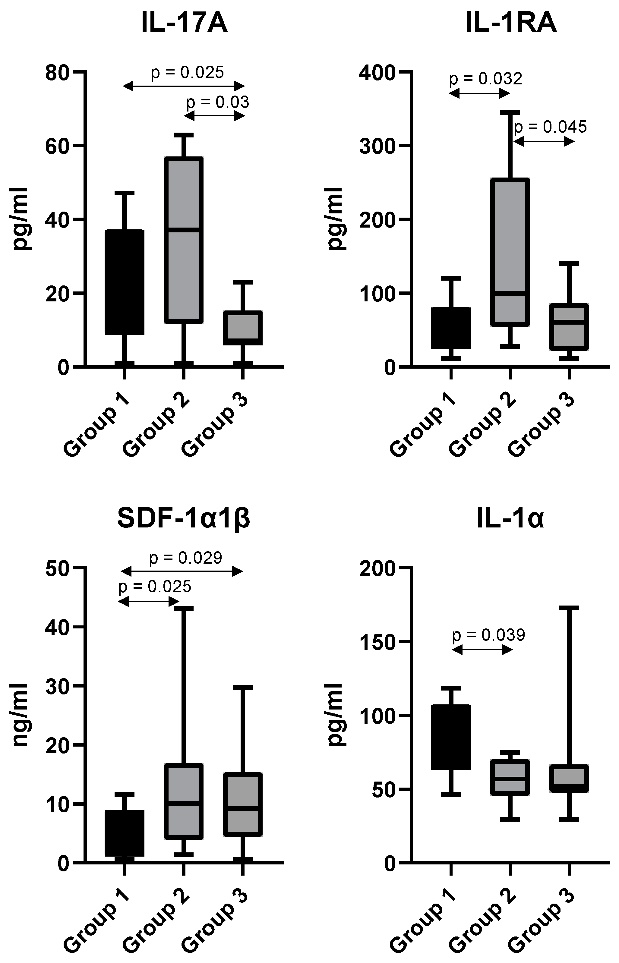
Serum samples from 41 human subjects were analyzed, with IL-17A, IL-1RA, IL-1α and SDF-1α1β found to be significantly changed in abundance across the different risk groups (Figure 4). IL-17A was found to have median values of 30 pg/mL, 37 pg/mL, and 6 pg/mL; IL-1RA 27 pg/mL, 99 pg/mL, and 60 pg/mL; IL-1α 85 pg/mL, 56 pg/mL, and 52 pg/mL; SDF-1α1β 2 ng/mL, 10 ng/mL, and 9 ng/mL in groups 1–3 respectively. In terms of fold-changes for these analytes between the 3 groups, the most significant changes related to IL-17A, with a 5-fold and 6.2-fold decrease in abundance observed when comparing group 3 with groups 1 and 2 respectively. SDF-1α1β was determined to have a 5-fold and 4.5-fold increase in abundance in group 2 and group 3 respectively, when compared to group 1.

4. Discussion

In this study, we used quantitative proteomic techniques to identify specific proteins associated with a “favorable”, “intermediate” or “adverse” prognosis. We identified elevated levels of metabolic-related proteins in AML cells from patients with an “adverse” prognosis when compared with AML cells from patients with a “favorable” prognosis. A change in the levels of several cytokines (IL-17A, IL-1RA, IL-1α and SDF-1α1β) between the risk groups was also identified in matched AML serum samples.
The complexity and heterogeneity of AML is illustrated by its classification into different disease subtypes based on distinct differences in genetic make-up, morphology and clinical presentation of AML [1]. Furthermore, AML patients often display intra-tumoral heterogeneity with molecularly distinct subclones, often present at a low frequency, enhancing the difficulties associated with efficient biomarker discovery and limits the efficacy of target-specific drugs [20,21,22,23]. To combat the heterogeneity of this disease, extensive research is required to boost current efforts in biomarker discovery to facilitate more accurate prognostic classifications and better therapeutic decisions.
Altered metabolism is a well-known hallmark of cancer associated with the reprogramming of metabolic activities to support a number of pro-anabolic pathways promoting tumorigenesis and disease progression [24]. Metabolomics has emerged as an important -omics technology in identifying novel biomarkers and therapeutic targets. Several studies have reported dysregulation in metabolic pathways in AML [25,26,27,28,29,30]. Mutations in the gene encoding the isocitrate dehydrogenase (IDH) enzyme involved in the tricarboxylic acid (TCA) cycle results in the production of the oncometabolite 2-hydroxyglutarate (2HG) and has been identified in ~6% of AML patients [31]. Analysis of AML patient serum revealed 2HG levels correlate with IDH mutational status and suggest a potential role as a prognostic, predictive and therapeutic-monitoring biomarker in AML [32,33]. A mass-spectrometry-based metabolomics study by Chen et al. identified a glucose metabolism prognostic biomarker signature consisting of 6 metabolites [34]. The broadening of metabolomics studies has resulted in various preclinical and/or clinical trials analyzing the effect of chemotherapies targeting metabolism in AML [15,35,36].
Previous studies describing dysregulated metabolism in AML correlate with our findings. Our studies revealed elevated metabolic-related protein levels in “adverse” risk versus “favorable” risk clinical AML cell lysates. A total of 10 metabolic-related proteins were elevated including peroxiredoxin-6, neutral alpha-glucosidase AB, 3-ketoacyl-CoA thiolase (3-KAT), hydroxyacyl-CoA dehydrogenase alpha (HADHA), transaldolase, cytosolic nonspecific dipeptidase, lactate dehydrogenase A and B, dihydrolipoyl dehydrogenase and nucleoside diphosphate kinase B. Several of these metabolic proteins have previously been investigated in AML.
3-KAT and HADHA enzymes make up part of the trifunctional protein complex involved in catalyzing acetyl-CoA production by β-oxidation during fatty acid oxidation (FAO) [37]. FAO was previously reported to be increased in chemo-resistant AML cells [38]. The overexpression of the FAO-related enzyme carnitine palmitoyl transferase 1A (CPT1A) was also shown to be a predictor of poor outcome in AML [39]. Furthermore, bone marrow adipocytes were found to promote acute monocytic leukemia (AMoL) survival via increased FAO, determined by detecting increasing levels of HADHA following AMoL co-culturing [40]. The FAO inhibitor, Avocatin B, was also found to possess potent anti-AML activity [41]. When combined, these studies implicate increased FAO and elevated FAO-related protein levels in the pathogenesis of AML.
Lactate dehydrogenase (LDH) is a well-established biomarker capable of aiding prognosis in a variety of cancers including AML [42,43,44,45]. Recent studies have indicated the potential of LDH as a prognostic marker in AML patients undergoing allogeneic hematopoietic stem cell transplantation (HSCT), as well as a predictive marker of AML patient outcome following sibling bone marrow transplant (BMT) [46,47]. Dihydrolipoyl dehydrogenase is a catalytic subunit of the pyruvate dehydrogenase complex, a target of the lipoate mimetic compound CPI-613 currently being evaluated in a Phase 3 clinical trial in combination with other approved chemotherapeutics for the treatment of relapsed or refractory AML [48]. Low expression levels of nm23-H2, also known as nucleoside diphosphate kinase B, was previously reported to be a good prognostic biomarker in AML [49]. Similarly, the overexpression of nm23-H2 was found to be associated with the “adverse” risk group in our study.
Research in the field of oncometabolism has revealed certain metabolic alterations as key contributors to tumorigenesis and tumor progression [50]. In AML, alterations in several metabolic pathways such as the production of reactive oxygen species (ROS) and increased levels of oxidative phosphorylation, have been linked to clinicopathological features of AML including aggressive disease and chemoresistance [38,51]. A recent study by Lo Presti et al. highlighted the strong influence of metabolic reprogramming on AML prognosis by identifying distinct changes in the metabolic profile of leukemic cells according to their mutational profile and stage of differentiation [52].
Future validation studies, a limitation of this study, on these elevated metabolic-related proteins may lead to the development of a metabolism-based prognostic biomarker signature to boost the prognostic value of current ELN recommendations. Our results show a clear increase in the number of significantly elevated metabolic-related proteins in the “intermediate” risk group. This risk group is the largest subset with many patients displaying heterogenous outcomes, suggesting a need for further stratification [53]. Several studies have identified specific genetic mutations in “intermediate” risk patients that veer towards a more favorable outcome whereas others indicate a poor outcome [54,55]. Thus, further analysis of the dysregulated metabolic-related proteins identified in the “intermediate” risk group and the survival outcome of the patients analyzed may reveal interesting patterns corresponding patient prognosis.
The assessment of bone marrow remains the “gold standard” for the diagnosis and monitoring of AML following treatment. Bone marrow biopsies are invasive and painful procedures; therefore, considerable efforts are being made to develop less invasive means of disease diagnosis and monitoring [56,57]. Analysis of AML patient serum provides an easier and less intrusive method of identifying biomarkers. Our multiplex assays focused on identifying altered chemokine/cytokine levels in AML serum samples matched to the previously analyzed AML cells. Four interesting cytokines (IL-1RA, IL-1α, IL-17A, SDF-1α1β) were found to be significantly dysregulated between single or multiple risk groups.
Interleukin-1 receptor antagonist (IL-1RA) is an anti-inflammatory cytokine that competitively binds to the IL-1 receptor (IL-1R), inhibiting binding of IL-1α and IL-1β and thus preventing downstream signaling cascade initiation [58]. Heterogeneous results from previous studies imply fluctuating levels of IL-1RA in AML patient sera [59,60,61]. One study reported increased, unchanged, and decreased levels of AML proliferation following exposure to IL-1RA, which causes further confusion on the role of IL-1RA in AML [62]. Despite a significant increase in serum IL-1RA levels in the “intermediate” risk group, additional studies are required to validate these findings.
The IL-1 cytokines, IL-1α and IL-1β, exert their pro-inflammatory effects via binding to IL-1R. IL-1α is constitutively secreted and has been reported to possess tumor-promoting or tumor-suppressing properties depending on the type of malignancy [63]. Despite, many articles focusing on IL-1β in AML, studies focusing on IL-1α are lacking [64]. We report increased serum IL-1α levels in patients with a favorable prognosis. Further studies are required to elucidate whether IL-1α plays an anti-tumorigenic or pro-tumorigenic role in AML to determine the prognostic relevance of high levels of this cytokine in “favorable” risk AML.
IL-17A is a hematopoietic stimulatory cytokine mainly secreted by T helper 17 cells. IL-17A has previously been reported to promote the proliferation of IL-17 receptor (IL-17R)-positive AML cells through the activation of proliferative signaling pathways such as JAK2/STAT3 [65]. IL-17A was found to be significantly decreased in the “adverse” risk group compared to “favorable” or “intermediate” risk groups. Despite reports that increased serum IL-17A represents a poor prognostic marker in AML, our findings may suggest an immuno-protective role of IL-17A in some cases, as described in other cancer types [66,67,68,69]. Heterogeneous and contradicting results regarding serum cytokines levels complicates the ability to accurately define these cytokines as useful biomarkers in AML. Further studies to elucidate the lone actions and crosstalk between cytokines is required to confidently identify a panel of serum cytokine biomarkers to aid disease monitoring and prognostication in AML.
Stromal cell-derived factor-1 α and β (SDF-1/CXCL12) are commonly expressed cytokines in various cells and tissues. CXCL12 binds to the CXCR4 receptor leading to the activation of intracellular events such as chemotaxis, proliferation, and transcription [70,71]. CXCR4 is expressed on almost all hematological cell types including lymphocytes and hematopoietic stem cells (HSCs). The CXCL12/CXCR4 ligand/receptor complex is associated with tumor progression, angiogenesis, metastasis, and survival in various malignancies [72,73,74]. CXCL12 levels has been shown to have prognostic significance in cancer, with high levels associated with adverse outcome in esophagogastric, pancreatic, and lung cancer; and, conversely, associated with enhanced survival in breast cancer [75].
In AML, the CXCL12/CXCR4 complex is exploited to initiate pro-survival signaling and homing of AML blasts to the protective bone marrow niche [76,77]. The frequency of bone marrow stromal cells secreting CXCL12 (CXCL12+) in the BM and CXCR4 expression is increased in AML, potentially due to hypoxic conditions within the microenvironment [78,79]. High CXCL12 causes a migration of CXCR4+ leukemic blasts towards the high CXCL12 levels within the protective bone marrow niche [77,80]. The CXCR4+ stromal cells and CXCR4+ leukemic cells create a bi-directional interaction network within the BM resulting in the constitutive activation of proliferative and survival signaling pathways [79,81]. High levels of CXCL12 is suggested to promote the retention of AML-blasts with the bone marrow microenvironment, thus, reducing the susceptibility of these blasts to chemotherapeutics [77]. Our AML serum analysis found that CXCL12 was elevated in “intermediate” and “adverse” risk AML patients. This finding corresponds with previous studies suggesting that high CXCR4 expression is indicative of poor prognosis in AML [82]. Recently, researchers revealed a new CXCR4 receptor antagonist IgG1 antibody (PF-06747143) capable of binding strongly to AML cell lines and to AML primary cells inhibiting their chemotaxis in response to CXCL12 [83]. Monitoring of serum CXCL12 levels between the “favorable” risk group and the “intermediate” and “adverse” risk groups represents a potential marker of disease progression in AML with higher levels corresponding to more adverse outcomes. Blockade of the CXCL12 pathway, using a commercially available CXCR4 antagonists such as plerixafor, may be an efficient method of modulating AML cell proliferation and chemotherapy resistance [84]. As interpatient variabilities are often seen in serum cytokine levels, further context-dependent studies in relation to AML subtypes or age will improve our understanding of changing cytokine levels in AML sera [85,86,87].
As our understanding of the pathophysiology mechanisms associated with AML increases, this has directly contributed to be generation of new therapeutic approaches to treat this malignancy. Much success has been achieved recently using inhibitors of FMS-like tyrosine kinase 3 (FLT3), isocitrate dehydrogenase (IDH) and Bcl-2 (B-cell lymphoma 2). Within the AML cohort, kinase 3 (FLT3) mutations are detected in approximately one-third of patients [88]. Sorafenib is the most common FLT3 inhibitor used, with high activity against internal tandem duplication ITD mutations instead of wild-type FLT3 and tyrosine kinase domain (TKD) mutations [89]. Mutations in IDH1 or IDH2 are detected in approximately 20% of AML patients inducing amino acid changes in conserved residues [90]. Specific IDH1 and IDH2 inhibitors include ivosidenib and enasidenib. Strategies to increase the efficacy of these inhibitors continues, for example in combination with venetoclax [91]. Indeed, venetoclax-based combinations have improved outcomes, including both remission rates and overall survival for older patients. Combinations of venetoclax, with either hypomethylating agents (HMA) or low dose cytarabine (LDAC), have shown promising results in clinical trials [92]. Glasdegib, a hedgehog pathway inhibitor and immune checkpoint inhibitors (nivolumab—PD-1 and ipilimumab—CTLA-4) are currently other therapeutic strategies that are continually being refined with respect to the most efficacious combination to include these therapeutic as part of [93,94]. In the near future, epigenetic modifiers (RMDs), microRNAs and suppressor of cytokine signaling are likely to be utilized clinically with respect to their anti-leukemia activity [95].
Many benefits exist for using a liquid biopsy for the analysis of biomarkers, with plasma, serum and saliva routinely employed. A liquid biopsy is relatively noninvasive to acquire and is generally a less time-consuming procedure than other approaches. Together with soluble proteins found in biofluids, circulating tumor cells (CTCs), circulating tumor DNA (ctDNA), and exosomes are regularly investigated because of their wide range of clinical applications. The presence of leukemic myeloblasts in peripheral blood from AML patients makes this biofluids especially attractive for the analysis of biomarkers, soluble or cellular-based. An advantage of measuring soluble proteins present in peripheral blood is that less processing of the samples needs to be completed (for examples measuring cytokine levels by enzyme-linked immunosorbent assay (ELISA)), speeding up the diagnostic/prognostic test result and ultimately providing a more cost-effective methodology.
An important consideration for this study is that identifications are based on peptides derived from canonical sequences of public databases, and subsequent validation studies will need to take into account the complexity of associated proteoforms that may be present (for example, splice variants and post-translational modifications (PTMs)), when designing these studies [96].

5. Conclusions

Current clinically-used biomarkers in AML are based almost solely on genomic abnormalities, as outlined in the ELN recommendations [8]. The inclusion of validated proteomic biomarkers would establish a more powerful array of factors to improve the prognostic classification of AML patients and ensure more calculated therapeutic decisions are made based on a patient’s prognosis and various other factors. Our study identified elevated metabolic-related proteins associated with adverse risk in AML, supporting evidence of the involvement of increased metabolism in AML. Several differentially expressed cytokines were also identified in the sera of patients from the three risk groups. IL-17A and IL-1RA levels were found to fluctuate across the different prognostic groups; however, both SDF-1α1β (CXCL12) and IL-1α were consistently found to increase and decrease in abundance, respectively, across the different groups. The consistent decrease in IL-1α in the different prognostic groups may point to its role as an antitumorigenic protein, as reported by others. High CXCL12 levels in intermediate and adverse risk groups were of particular interest due to the role of this chemokine in AML pathogenesis. Therefore, further efforts to validate CXCL12 as a prognostic biomarker are warranted. This study supports a role for increased CXCL12 (serum levels) and increased metabolism-related proteins in the risk profile associated with different cohorts of AML patients.

Supplementary Materials

The following are available online at https://www.mdpi.com/article/10.3390/proteomes9040042/s1. Table S1: Raw mass spec data; Table S2: Raw mass spec data; Table S3: Raw mass spec data; Table S4: Raw Luminex Data; Table S5: Protein identification data Gr1 v Gr2; Table S6: Protein identification data Gr1 v Gr3; Table S7: Protein identification data Gr2 v Gr3.

Author Contributions

P.D., C.T. and P.O. designed the study. All authors contribute to data acquisition, analysis, or interpretation. P.D., K.D. and D.B. wrote the manuscript. C.A.H. and J.J.M. participated in the writing and editing of the manuscript. P.D. supervised the study. All authors have read and agreed to the published version of the manuscript.

Funding

Research was supported by the Kathleen Lonsdale Institute for Human Health Research at Maynooth University.

Institutional Review Board Statement

The samples and data used were provided by the Finnish Hematology Registry and Clinical Biobank (FHRB) with appropriate ethics approval. Decision of the FHRB Biobank entry in the National Biobank Register maintained by the Finnish Medicines Agency was received on 15 July 2014 (decision number 3613/06.01.05.01.00/2014). All samples were collected according to the criteria set by the declaration of Helsinki.

Informed Consent Statement

Informed consent was obtained from all subjects involved in the study.

Data Availability Statement

The data will be posted on the Open Science Framework (OSF website, https://osf.io, accessed on 18 October 2021) so that the data may be analyzed by other researchers.

Acknowledgments

Research was supported by funding from the Kathleen Lonsdale Institute for Human Health Research at Maynooth University. The Q-Exactive quantitative mass spectrometer was funded under the Research Infrastructure Call 2012 by Science Foundation Ireland (SFI-12/RI/2346/3). We thank all the patients for their generous participation. The FHRB Biobank is supported by the Finnish Association of Hematology, the Finnish Red Cross Blood Service, Institute for Molecular Medicine Finland, and the participating hospitals in Finland.

Conflicts of Interest

C.A.H. has received research funding from Celgene/BMS, Kronos Bio, Novartis, Oncopeptides, Orion Pharma and the IMI2 projects HARMONY and HARMONY PLUS unrelated to this work.

References

  1. De Kouchkovsky, I.; Abdul-Hay, M. Acute myeloid leukemia: A comprehensive review and 2016 update. Blood Cancer J. 2016, 6, e441. [Google Scholar] [CrossRef]
  2. Hao, T.; Li-Talley, M.; Buck, A.; Chen, W. An emerging trend of rapid increase of leukemia but not all cancers in the aging population in the United States. Sci. Rep. 2019, 9, 12070. [Google Scholar] [CrossRef] [PubMed] [Green Version]
  3. Shallis, R.M.; Wang, R.; Davidoff, A.; Ma, X.; Zeidan, A.M. Epidemiology of acute myeloid leukemia: Recent progress and enduring challenges. Blood Rev. 2019, 36, 70–87. [Google Scholar] [CrossRef] [PubMed]
  4. Zhang, J.; Gu, Y.; Chen, B. Mechanisms of drug resistance in acute myeloid leukemia. OncoTargets Ther. 2019, 12, 1937–1945. [Google Scholar] [CrossRef] [PubMed] [Green Version]
  5. Bennett, J.M.; Catovsky, D.; Daniel, M.T.; Flandrin, G.; Galton, D.A.; Gralnick, H.R.; Sultan, C. Proposed revised criteria for the classification of acute myeloid leukemia. A report of the French-American-British Cooperative Group. Ann. Intern. Med. 1985, 103, 620–625. [Google Scholar] [CrossRef] [Green Version]
  6. Vardiman, J.W.; Thiele, J.; Arber, D.A.; Brunning, R.D.; Borowitz, M.J.; Porwit, A.; Harris, N.L.; Le Beau, M.M.; Hellström-Lindberg, E.; Tefferi, A.; et al. The 2008 revision of the World Health Organization (WHO) classification of myeloid neoplasms and acute leukemia: Rationale and important changes. Blood 2009, 114, 937–951. [Google Scholar] [CrossRef] [PubMed] [Green Version]
  7. Patel, J.P.; Gönen, M.; Figueroa, M.E.; Fernandez, H.; Sun, Z.; Racevskis, J.; Van Vlierberghe, P.; Dolgalev, I.; Thomas, S.; Aminova, O.; et al. Prognostic relevance of integrated genetic profiling in acute myeloid leukemia. N. Engl. J. Med. 2012, 366, 1079–1089. [Google Scholar] [CrossRef] [Green Version]
  8. Döhner, H.; Estey, E.; Grimwade, D.; Amadori, S.; Appelbaum, F.R.; Büchner, T.; Dombret, H.; Ebert, B.L.; Fenaux, P.; Larson, R.A.; et al. Diagnosis and management of AML in adults: 2017 ELN recommendations from an international expert panel. Blood 2017, 129, 424–447. [Google Scholar] [CrossRef] [Green Version]
  9. Hernandez-Valladares, M.; Bruserud, Ø.; Selheim, F. The Implementation of Mass Spectrometry-Based Proteomics Workflows in Clinical Routines of Acute Myeloid Leukemia: Applicability and Perspectives. Int. J. Mol. Sci. 2020, 21, 6830. [Google Scholar] [CrossRef]
  10. Eisfeld, A.K.; Kohlschmidt, J.; Mrózek, K.; Blachly, J.S.; Walker, C.J.; Nicolet, D.; Orwick, S.; Maharry, S.E.; Carroll, A.J.; Stone, R.M.; et al. Mutation patterns identify adult patients with de novo acute myeloid leukemia aged 60 years or older who respond favorably to standard chemotherapy: An analysis of Alliance studies. Leukemia 2018, 32, 1338–1348. [Google Scholar] [CrossRef]
  11. Patel, S.S.; Kuo, F.C.; Gibson, C.J.; Steensma, D.P.; Soiffer, R.J.; Alyea, E.P., 3rd; Chen, Y.A.; Fathi, A.T.; Graubert, T.A.; Brunner, A.M.; et al. High NPM1-mutant allele burden at diagnosis predicts unfavorable outcomes in de novo AML. Blood 2018, 131, 2816–2825. [Google Scholar] [CrossRef] [PubMed] [Green Version]
  12. Papaemmanuil, E.; Gerstung, M.; Bullinger, L.; Gaidzik, V.I.; Paschka, P.; Roberts, N.D.; Potter, N.E.; Heuser, M.; Thol, F.; Bolli, N.; et al. Genomic Classification and Prognosis in Acute Myeloid Leukemia. N. Engl. J. Med. 2016, 374, 2209–2221. [Google Scholar] [CrossRef] [PubMed]
  13. Srivastava, A.; Creek, D.J. Discovery and Validation of Clinical Biomarkers of Cancer: A Review Combining Metabolomics and Proteomics. Proteomics 2019, 19, e1700448. [Google Scholar] [CrossRef]
  14. Aasebø, E.; Berven, F.S.; Bartaula-Brevik, S.; Stokowy, T.; Hovland, R.; Vaudel, M.; Døskeland, S.O.; McCormack, E.; Batth, T.S.; Olsen, J.V.; et al. Proteome and Phosphoproteome Changes Associated with Prognosis in Acute Myeloid Leukemia. Cancers 2020, 12, 709. [Google Scholar] [CrossRef] [PubMed] [Green Version]
  15. Grønningsæter, I.S.; Reikvam, H.; Aasebø, E.; Bartaula-Brevik, S.; Tvedt, T.H.; Bruserud, Ø.; Hatfield, K.J. Targeting Cellular Metabolism in Acute Myeloid Leukemia and The Role of Patient Heterogeneity. Cells 2020, 9, 1155. [Google Scholar] [CrossRef]
  16. Stockard, B.; Garrett, T.; Guingab-Cagmat, J.; Meshinchi, S.; Lamba, J. Distinct Metabolic features differentiating FLT3-ITD AML from FLT3-WT childhood Acute Myeloid Leukemia. Sci. Rep. 2018, 8, 5534. [Google Scholar] [CrossRef]
  17. Riether, C.; Schürch, C.M.; Bührer, E.D.; Hinterbrandner, M.; Huguenin, A.L.; Hoepner, S.; Zlobec, I.; Pabst, T.; Radpour, R.; Ochsenbein, A.F. CD70/CD27 signaling promotes blast stemness and is a viable therapeutic target in acute myeloid leukemia. J. Exp. Med. 2017, 214, 359–380. [Google Scholar] [CrossRef] [PubMed]
  18. Mi, H.; Muruganujan, A.; Ebert, D.; Huang, X.; Thomas, P.D. PANTHER version 14: More genomes, a new PANTHER GO-slim and improvements in enrichment analysis tools. Nucleic Acids Res. 2019, 47, D419–D426. [Google Scholar] [CrossRef]
  19. Benjamini, Y.; Hochberg, Y. Controlling the False Discovery Rate: A Practical and Powerful Approach to Multiple Testing. J. R. Stat. Soc. Ser. B Methodol. 1995, 57, 289–300. [Google Scholar] [CrossRef]
  20. Stanta, G.; Bonin, S. Overview on Clinical Relevance of Intra-Tumor Heterogeneity. Front. Med. 2018, 5, 85. [Google Scholar] [CrossRef] [Green Version]
  21. Fisher, R.; Pusztai, L.; Swanton, C. Cancer heterogeneity: Implications for targeted therapeutics. Br. J. Cancer 2013, 108, 479–485. [Google Scholar] [CrossRef] [Green Version]
  22. Pogosova-Agadjanyan, E.L.; Moseley, A.; Othus, M.; Appelbaum, F.R.; Chauncey, T.R.; Chen, I.L.; Erba, H.P.; Godwin, J.E.; Fang, M.; Kopecky, K.J.; et al. Impact of Specimen Heterogeneity on Biomarkers in Repository Samples from Patients with Acute Myeloid Leukemia: A SWOG Report. Biopreserv. Biobank. 2018, 16, 42–52. [Google Scholar] [CrossRef]
  23. Klco, J.M.; Spencer, D.H.; Miller, C.A.; Griffith, M.; Lamprecht, T.L.; O’Laughlin, M.; Fronick, C.; Magrini, V.; Demeter, R.T.; Fulton, R.S.; et al. Functional heterogeneity of genetically defined subclones in acute myeloid leukemia. Cancer Cell 2014, 25, 379–392. [Google Scholar] [CrossRef] [PubMed] [Green Version]
  24. Kaushik, A.K.; DeBerardinis, R.J. Applications of metabolomics to study cancer metabolism. Biochim. Biophys. Acta Rev. Cancer 2018, 1870, 2–14. [Google Scholar] [CrossRef] [PubMed]
  25. Henkenius, K.; Greene, B.H.; Barckhausen, C.; Hartmann, R.; Märken, M.; Kaiser, T.; Rehberger, M.; Metzelder, S.K.; Parak, W.J.; Neubauer, A.; et al. Maintenance of cellular respiration indicates drug resistance in acute myeloid leukemia. Leuk Res. 2017, 62, 56–63. [Google Scholar] [CrossRef] [PubMed]
  26. Ryu, M.J.; Han, J.; Kim, S.J.; Lee, M.J.; Ju, X.; Lee, Y.L.; Son, J.H.; Cui, J.; Jang, Y.; Chung, W.; et al. PTEN/AKT signaling mediates chemoresistance in refractory acute myeloid leukemia through enhanced glycolysis. Oncol. Rep. 2019, 42, 2149–2158. [Google Scholar] [CrossRef]
  27. Stuani, L.; Riols, F.; Millard, P.; Sabatier, M.; Batut, A.; Saland, E.; Viars, F.; Tonini, L.; Zaghdoudi, S.; Linares, L.K.; et al. Stable Isotope Labeling Highlights Enhanced Fatty Acid and Lipid Metabolism in Human Acute Myeloid Leukemia. Int. J. Mol. Sci. 2018, 19, 3325. [Google Scholar] [CrossRef] [Green Version]
  28. Chen, Y.; Xu, Q.; Ji, D.; Wei, Y.; Chen, H.; Li, T.; Wan, B.; Yuan, L.; Huang, R.; Chen, G. Inhibition of pentose phosphate pathway suppresses acute myelogenous leukemia. Tumour Biol. 2016, 37, 6027–6034. [Google Scholar] [CrossRef]
  29. Forte, D.; García-Fernández, M.; Sánchez-Aguilera, A.; Stavropoulou, V.; Fielding, C.; Martín-Pérez, D.; López, J.A.; Costa, A.S.H.; Tronci, L.; Nikitopoulou, E.; et al. Bone Marrow Mesenchymal Stem Cells Support Acute Myeloid Leukemia Bioenergetics and Enhance Antioxidant Defense and Escape from Chemotherapy. Cell Metab. 2020, 32, 829–843. [Google Scholar] [CrossRef]
  30. Pikman, Y.; Puissant, A.; Alexe, G.; Furman, A.; Chen, L.M.; Frumm, S.M.; Ross, L.; Fenouille, N.; Bassil, C.F.; Lewis, C.A.; et al. Targeting MTHFD2 in acute myeloid leukemia. J. Exp. Med. 2016, 213, 1285–1306. [Google Scholar] [CrossRef]
  31. Schnittger, S.; Haferlach, C.; Ulke, M.; Alpermann, T.; Kern, W.; Haferlach, T. IDH1 mutations are detected in 6.6% of 1414 AML patients and are associated with intermediate risk karyotype and unfavorable prognosis in adults younger than 60 years and unmutated NPM1 status. Blood 2010, 116, 5486–5496. [Google Scholar] [CrossRef] [PubMed]
  32. DiNardo, C.D.; Propert, K.J.; Loren, A.W.; Paietta, E.; Sun, Z.; Levine, R.L.; Straley, K.S.; Yen, K.; Patel, J.P.; Agresta, S.; et al. Serum 2-hydroxyglutarate levels predict isocitrate dehydrogenase mutations and clinical outcome in acute myeloid leukemia. Blood 2013, 121, 4917–4924. [Google Scholar] [CrossRef] [PubMed]
  33. Miller, A.; Tong, A.W.; Sweetman, L.; Theiss, A.; Murtaza, M.; Daoud, Y.; Wong, L. Characterization of Acute Myeloid Leukaemia (AML) Patients with Elevated Peripheral Blood Plasma D-2-Hydroxyglutarate (D-2HG) and/or Isocitrate Dehydrogenase (IDH) Mutational Status. Blood 2017, 130, 3923. [Google Scholar] [CrossRef]
  34. Chen, W.L.; Wang, J.H.; Zhao, A.H.; Xu, X.; Wang, Y.H.; Chen, T.L.; Li, J.M.; Mi, J.Q.; Zhu, Y.M.; Liu, Y.F.; et al. A distinct glucose metabolism signature of acute myeloid leukemia with prognostic value. Blood 2014, 124, 1645–1654. [Google Scholar] [CrossRef] [PubMed]
  35. Vitkevičienė, A.; Janulis, V.; Žučenka, A.; Borutinskaitė, V.; Kaupinis, A.; Valius, M.; Griškevičius, L.; Navakauskienė, R. Oxidative phosphorylation inhibition induces anticancerous changes in therapy-resistant-acute myeloid leukemia patient cells. Mol. Carcinog. 2019, 58, 2008–2016. [Google Scholar] [CrossRef] [PubMed]
  36. Advani, A.S.; Li, H.; Michaelis, L.C.; Medeiros, B.C.; Liedtke, M.; List, A.F.; O’Dwyer, K.; Othus, M.; Erba, H.P.; Appelbaum, F.R. Report of the relapsed/refractory cohort of SWOG S0919: A phase 2 study of idarubicin and cytarabine in combination with pravastatin for acute myelogenous leukemia (AML). Leuk Res. 2018, 67, 17–20. [Google Scholar] [CrossRef]
  37. Rector, R.S.; Payne, R.M.; Ibdah, J.A. Mitochondrial trifunctional protein defects: Clinical implications and therapeutic approaches. Adv. Drug Deliv. Rev. 2008, 60, 1488–1496. [Google Scholar] [CrossRef] [Green Version]
  38. Farge, T.; Saland, E.; de Toni, F.; Aroua, N.; Hosseini, M.; Perry, R.; Bosc, C.; Sugita, M.; Stuani, L.; Fraisse, M.; et al. Chemotherapy-Resistant Human Acute Myeloid Leukemia Cells Are Not Enriched for Leukemic Stem Cells but Require Oxidative Metabolism. Cancer Discov. 2017, 7, 716–735. [Google Scholar] [CrossRef] [Green Version]
  39. Shi, J.; Fu, H.; Jia, Z.; He, K.; Fu, L.; Wang, W. High Expression of CPT1A Predicts Adverse Outcomes: A Potential Therapeutic Target for Acute Myeloid Leukemia. EBioMedicine 2016, 14, 55–64. [Google Scholar] [CrossRef] [PubMed] [Green Version]
  40. Tabe, Y.; Yamamoto, S.; Saitoh, K.; Sekihara, K.; Monma, N.; Ikeo, K.; Mogushi, K.; Shikami, M.; Ruvolo, V.; Ishizawa, J.; et al. Bone Marrow Adipocytes Facilitate Fatty Acid Oxidation Activating AMPK and a Transcriptional Network Supporting Survival of Acute Monocytic Leukemia Cells. Cancer Res. 2017, 77, 1453–1464. [Google Scholar] [CrossRef] [Green Version]
  41. Lee, E.A.; Angka, L.; Rota, S.G.; Hanlon, T.; Mitchell, A.; Hurren, R.; Wang, X.M.; Gronda, M.; Boyaci, E.; Bojko, B.; et al. Targeting Mitochondria with Avocatin B Induces Selective Leukemia Cell Death. Cancer Res. 2015, 75, 2478–2488. [Google Scholar] [CrossRef] [Green Version]
  42. Kornberg, A.; Polliack, A. Serum lactic dehydrogenase (LDH) levels in acute leukemia: Marked elevations in lymphoblastic leukemia. Blood 1980, 56, 351–355. [Google Scholar] [CrossRef] [Green Version]
  43. Liu, D.; Wang, D.; Wu, C.; Zhang, L.; Mei, Q.; Hu, G.; Long, G.; Sun, W. Prognostic significance of serum lactate dehydrogenase in patients with breast cancer: A meta-analysis. Cancer Manag. Res. 2019, 11, 3611–3619. [Google Scholar] [CrossRef] [PubMed] [Green Version]
  44. Lippert, M.C.; Javadpour, N. Lactic dehydrogenase in the monitoring and prognosis of testicular cancer. Cancer 1981, 48, 2274–2278. [Google Scholar] [CrossRef]
  45. Krykowski, E.; Polkowska-Kulesza, E.; Robak, T.; Matusewicz, W.; Urbańska-Rys, H.; Hołub, A. Analysis of Prognostic Factors in Acute Leukemias in Adults. In Acute Leukemias; Springer: Berlin/Heidelberg, Germany, 1987; pp. 369–372. [Google Scholar]
  46. Geva, M.; Shouval, R.; Fein, J.A.; Danylesko, I.; Shem-Tov, N.; Yerushalmi, R.; Shimoni, A.; Nagler, A. Lactate Dehydrogenase Is a Key Prognostic Factor in Acute Myeloid Leukemia and Lymphoma Patients Undergoing Allogeneic Hematopoietic Stem Cell Transplantation. Blood 2019, 134, 3304. [Google Scholar] [CrossRef]
  47. Kalaycio, M.; Rybicki, L.; Pohlman, B.; Dean, R.; Sweetenham, J.W.; Andresen, S.; Sobecks, R.; Sekeres, M.A.; Advani, A.; Davis, R.; et al. Lactate Dehydrogenase (LDH) Level Predicts the Outcome of Patients with Acute Myelogenous Leukemia (AML) Following HLA-Matched Sibling Bone Marrow Transplant (BMT). Blood 2006, 108, 3013. [Google Scholar] [CrossRef]
  48. Stuart, S.D.; Schauble, A.; Gupta, S.; Kennedy, A.D.; Keppler, B.R.; Bingham, P.M.; Zachar, Z. A strategically designed small molecule attacks alpha-ketoglutarate dehydrogenase in tumor cells through a redox process. Cancer Metab. 2014, 2, 4. [Google Scholar] [CrossRef] [Green Version]
  49. Wakimoto, N.; Yokoyama, A.; Okabe-Kado, J.; Nagata, N.; Motoyoshi, K.; Honma, Y. Combined analysis of differentiation inhibitory factor nm23-H1 and nm23-H2 as prognostic factors in acute myeloid leukaemia. Br. J. Cancer 1998, 77, 2298–2303. [Google Scholar] [CrossRef] [Green Version]
  50. Oliveira, P.J.; Urbano, A.M. Oncometabolism: The switchboard of cancer—An editorial. Biochim. Biophys. Acta BBA-Mol. Basis Dis. 2021, 1867, 166031. [Google Scholar] [CrossRef]
  51. Sallmyr, A.; Fan, J.; Datta, K.; Kim, K.-T.; Grosu, D.; Shapiro, P.; Small, D.; Rassool, F. Internal tandem duplication of FLT3 (FLT3/ITD) induces increased ROS production, DNA damage, and misrepair: Implications for poor prognosis in AML. Blood 2008, 111, 3173–3182. [Google Scholar] [CrossRef] [Green Version]
  52. Lo Presti, C.; Fauvelle, F.; Jacob, M.-C.; Mondet, J.; Mossuz, P. The metabolic reprogramming in acute myeloid leukemia patients depends on their genotype and is a prognostic marker. Blood Adv. 2021, 5, 156–166. [Google Scholar] [CrossRef] [PubMed]
  53. Döhner, K.; Paschka, P. Intermediate-risk acute myeloid leukemia therapy: Current and future. Hematology 2014, 2014, 34–43. [Google Scholar] [CrossRef] [PubMed]
  54. Lin, P.-H.; Li, H.-Y.; Fan, S.-C.; Yuan, T.-H.; Chen, M.; Hsu, Y.-H.; Yang, Y.-H.; Li, L.-Y.; Yeh, S.-P.; Bai, L.-Y.; et al. A targeted next-generation sequencing in the molecular risk stratification of adult acute myeloid leukemia: Implications for clinical practice. Cancer Med. 2017, 6, 349–360. [Google Scholar] [CrossRef] [PubMed]
  55. Pallarès, V.; Hoyos, M.; Chillón, M.C.; Barragán, E.; Prieto Conde, M.I.; Llop, M.; Falgàs, A.; Céspedes, M.V.; Montesinos, P.; Nomdedeu, J.F.; et al. Focal Adhesion Genes Refine the Intermediate-Risk Cytogenetic Classification of Acute Myeloid Leukemia. Cancers 2018, 10, 436. [Google Scholar] [CrossRef] [PubMed] [Green Version]
  56. Pardini, B.; Sabo, A.A.; Birolo, G.; Calin, G.A. Noncoding RNAs in Extracellular Fluids as Cancer Biomarkers: The New Frontier of Liquid Biopsies. Cancers 2019, 11, 1170. [Google Scholar] [CrossRef] [Green Version]
  57. Percival, M.E.; Lai, C.; Estey, E.; Hourigan, C.S. Bone marrow evaluation for diagnosis and monitoring of acute myeloid leukemia. Blood Rev. 2017, 31, 185–192. [Google Scholar] [CrossRef]
  58. Dinarello, C.A. Immunological and inflammatory functions of the interleukin-1 family. Annu. Rev. Immunol. 2009, 27, 519–550. [Google Scholar] [CrossRef]
  59. Tsimberidou, A.M.; Estey, E.; Wen, S.; Pierce, S.; Kantarjian, H.; Albitar, M.; Kurzrock, R. The prognostic significance of cytokine levels in newly diagnosed acute myeloid leukemia and high-risk myelodysplastic syndromes. Cancer 2008, 113, 1605–1613. [Google Scholar] [CrossRef] [PubMed]
  60. Fredly, H.; Reikvam, H.; Gjertsen, B.T.; Bruserud, Ø. Disease-stabilizing treatment with all-trans retinoic acid and valproic acid in acute myeloid leukemia: Serum hsp70 and hsp90 levels and serum cytokine profiles are determined by the disease, patient age, and anti-leukemic treatment. Am. J. Hematol. 2012, 87, 368–376. [Google Scholar] [CrossRef] [PubMed] [Green Version]
  61. Tao, M.; Li, B.; Nayini, J.; Andrews, C.B.; Huang, R.W.; Devemy, E.; Song, S.; Venugopal, P.; Preisler, H.D. SCF, IL-1beta, IL-1ra and GM-CSF in the bone marrow and serum of normal individuals and of AML and CML patients. Cytokine 2000, 12, 699–707. [Google Scholar] [CrossRef]
  62. Stosić-Grujicić, S.; Basara, N.; Dinarello, C.A. Modulatory in vitro effects of interleukin-1 receptor antagonist (IL-1Ra) or antisense oligonucleotide to interleukin-1 beta converting enzyme (ICE) on acute myeloid leukaemia (AML) cell growth. Clin. Lab. Haematol. 1999, 21, 173–185. [Google Scholar] [CrossRef]
  63. Baker, K.J.; Houston, A.; Brint, E. IL-1 Family Members in Cancer; Two Sides to Every Story. Front. Immunol. 2019, 10, 1197. [Google Scholar] [CrossRef] [Green Version]
  64. Carey, A.; Edwards, D.K.t.; Eide, C.A.; Newell, L.; Traer, E.; Medeiros, B.C.; Pollyea, D.A.; Deininger, M.W.; Collins, R.H.; Tyner, J.W.; et al. Identification of Interleukin-1 by Functional Screening as a Key Mediator of Cellular Expansion and Disease Progression in Acute Myeloid Leukemia. Cell Rep. 2017, 18, 3204–3218. [Google Scholar] [CrossRef] [PubMed]
  65. Han, Y.; Ye, A.; Bi, L.; Wu, J.; Yu, K.; Zhang, S. Th17 cells and interleukin-17 increase with poor prognosis in patients with acute myeloid leukemia. Cancer Sci. 2014, 105, 933–942. [Google Scholar] [CrossRef]
  66. Abousamra, N.K.; Salah El-Din, M.; Helal, R. Prognostic value of Th17 cells in acute leukemia. Med. Oncol. 2013, 30, 732. [Google Scholar] [CrossRef] [Green Version]
  67. Kryczek, I.; Wei, S.; Szeliga, W.; Vatan, L.; Zou, W. Endogenous IL-17 contributes to reduced tumor growth and metastasis. Blood 2009, 114, 357–359. [Google Scholar] [CrossRef] [PubMed] [Green Version]
  68. Bhat, S.; Gardi, N.; Hake, S.; Kotian, N.; Sawant, S.; Kannan, S.; Parmar, V.; Desai, S.; Dutt, A.; Joshi, N.N. Impact of intra-tumoral IL17A and IL32 gene expression on T-cell responses and lymph node status in breast cancer patients. J. Cancer Res. Clin. Oncol. 2017, 143, 1745–1756. [Google Scholar] [CrossRef] [PubMed] [Green Version]
  69. Armstrong, D.; Chang, C.-Y.; Lazarus, D.R.; Corry, D.; Kheradmand, F. Lung Cancer Heterogeneity in Modulation of Th17/IL17A Responses. Front. Oncol. 2019, 9, 1384. [Google Scholar] [CrossRef] [PubMed]
  70. Moore, N.; Moreno Gonzales, M.; Bonner, K.; Smith, B.; Park, W.; Stegall, M. Impact of CXCR4/CXCL12 Blockade on Normal Plasma Cells In Vivo. Am. J. Transplant. 2017, 17, 1663–1669. [Google Scholar] [CrossRef] [PubMed] [Green Version]
  71. Peled, A.; Klein, S.; Beider, K.; Burger, J.A.; Abraham, M. Role of CXCL12 and CXCR4 in the pathogenesis of hematological malignancies. Cytokine 2018, 109, 11–16. [Google Scholar] [CrossRef]
  72. Pitt, L.A.; Tikhonova, A.N.; Hu, H.; Trimarchi, T.; King, B.; Gong, Y.; Sanchez-Martin, M.; Tsirigos, A.; Littman, D.R.; Ferrando, A.A.; et al. CXCL12-Producing Vascular Endothelial Niches Control Acute T Cell Leukemia Maintenance. Cancer Cell 2015, 27, 755–768. [Google Scholar] [CrossRef] [PubMed] [Green Version]
  73. Kaverina, N.; Borovjagin, A.V.; Kadagidze, Z.; Baryshnikov, A.; Baryshnikova, M.; Malin, D.; Ghosh, D.; Shah, N.; Welch, D.R.; Gabikian, P.; et al. Astrocytes promote progression of breast cancer metastases to the brain via a KISS1-mediated autophagy. Autophagy 2017, 13, 1905–1923. [Google Scholar] [CrossRef] [PubMed]
  74. Kryczek, I.; Lange, A.; Mottram, P.; Alvarez, X.; Cheng, P.; Hogan, M.; Moons, L.; Wei, S.; Zou, L.; Machelon, V.; et al. CXCL12 and vascular endothelial growth factor synergistically induce neoangiogenesis in human ovarian cancers. Cancer Res. 2005, 65, 465–472. [Google Scholar] [PubMed]
  75. Samarendra, H.; Jones, K.; Petrinic, T.; Silva, M.A.; Reddy, S.; Soonawalla, Z.; Gordon-Weeks, A. A meta-analysis of CXCL12 expression for cancer prognosis. Br. J. Cancer 2017, 117, 124–135. [Google Scholar] [CrossRef]
  76. Möhle, R.; Bautz, F.; Rafii, S.; Moore, M.A.; Brugger, W.; Kanz, L. The chemokine receptor CXCR-4 is expressed on CD34+ hematopoietic progenitors and leukemic cells and mediates transendothelial migration induced by stromal cell-derived factor-1. Blood 1998, 91, 4523–4530. [Google Scholar] [CrossRef]
  77. Yazdani, Z.; Mousavi, Z.; Moradabadi, A.; Hassanshahi, G. Significance of CXCL12/CXCR4 Ligand/Receptor Axis in Various Aspects of Acute Myeloid Leukemia. Cancer Manag. Res. 2020, 12, 2155–2165. [Google Scholar] [CrossRef] [Green Version]
  78. Abe-Suzuki, S.; Kurata, M.; Abe, S.; Onishi, I.; Kirimura, S.; Nashimoto, M.; Murayama, T.; Hidaka, M.; Kitagawa, M. CXCL12+ stromal cells as bone marrow niche for CD34+ hematopoietic cells and their association with disease progression in myelodysplastic syndromes. Lab. Investig. 2014, 94, 1212–1223. [Google Scholar] [CrossRef] [PubMed] [Green Version]
  79. Fiegl, M.; Samudio, I.; Clise-Dwyer, K.; Burks, J.K.; Mnjoyan, Z.; Andreeff, M. CXCR4 expression and biologic activity in acute myeloid leukemia are dependent on oxygen partial pressure. Blood 2009, 113, 1504–1512. [Google Scholar] [CrossRef] [PubMed] [Green Version]
  80. Burger, J.A.; Peled, A. CXCR4 antagonists: Targeting the microenvironment in leukemia and other cancers. Leukemia 2009, 23, 43–52. [Google Scholar] [CrossRef] [PubMed]
  81. Kittang, A.O.; Hatfield, K.; Sand, K.; Reikvam, H.; Bruserud, Ø. The chemokine network in acute myelogenous leukemia: Molecular mechanisms involved in leukemogenesis and therapeutic implications. Curr. Top. Microbiol. Immunol. 2010, 341, 149–172. [Google Scholar] [CrossRef] [PubMed]
  82. Du, W.; Lu, C.; Zhu, X.; Hu, D.; Chen, X.; Li, J.; Liu, W.; Zhu, J.; He, Y.; Yao, J. Prognostic significance of CXCR4 expression in acute myeloid leukemia. Cancer Med. 2019, 8, 6595–6603. [Google Scholar] [CrossRef]
  83. Zhang, Y.; Saavedra, E.; Tang, R.; Gu, Y.; Lappin, P.; Trajkovic, D.; Liu, S.-H.; Smeal, T.; Fantin, V.; De Botton, S.; et al. Targeting primary acute myeloid leukemia with a new CXCR4 antagonist IgG1 antibody (PF-06747143). Sci. Rep. 2017, 7, 7305. [Google Scholar] [CrossRef] [Green Version]
  84. Shumilov, E.; Novak, U.; Jeker, B.; Mansouri Taleghani, B.; Bacher, U.; Pabst, T. Hematopoietic Stem Cell Mobilization With Plerixafor Is Safe and Effective in Poorly Mobilizing Acute Myeloid Leukemia Patients. Hemasphere 2019, 3, e176. [Google Scholar] [CrossRef] [PubMed]
  85. Walters, H.M.; Pan, N.; Lehman, T.J.; Adams, A.; Kalliolias, G.D.; Zhu, Y.S.; Santiago, F.; Nguyen, J.; Sitaras, L.; Cunningham-Rundles, S.; et al. The impact of disease activity and tumour necrosis factor-α inhibitor therapy on cytokine levels in juvenile idiopathic arthritis. Clin. Exp. Immunol. 2016, 184, 308–317. [Google Scholar] [CrossRef] [Green Version]
  86. Sack, U.; Burkhardt, U.; Borte, M.; Schädlich, H.; Berg, K.; Emmrich, F. Age-dependent levels of select immunological mediators in sera of healthy children. Clin. Diagn Lab. Immunol. 1998, 5, 28–32. [Google Scholar] [CrossRef] [PubMed] [Green Version]
  87. Eide, H.A.; Knudtsen, I.S.; Sandhu, V.; Løndalen, A.M.; Halvorsen, A.R.; Abravan, A.; Kure, E.H.; Bogsrud, T.V.; Brustugun, O.T.; Kyte, J.A.; et al. Serum cytokine profiles and metabolic tumor burden in patients with non-small cell lung cancer undergoing palliative thoracic radiation therapy. Adv. Radiat. Oncol. 2018, 3, 130–138. [Google Scholar] [CrossRef] [PubMed] [Green Version]
  88. Daver, N.; Schlenk, R.F.; Russell, N.H.; Levis, M.J. Targeting FLT3 mutations in AML: Review of current knowledge and evidence. Leukemia 2019, 33, 299–312. [Google Scholar] [CrossRef] [Green Version]
  89. Grunwald, M.R.; Levis, M.J. FLT3 inhibitors for acute myeloid leukemia: A review of their efficacy and mechanisms of resistance. Int. J. Hematol. 2013, 97, 683–694. [Google Scholar] [CrossRef] [PubMed] [Green Version]
  90. Dang, L.; Yen, K.; Attar, E.C. IDH mutations in cancer and progress toward development of targeted therapeutics. Ann. Oncol. 2016, 27, 599–608. [Google Scholar] [CrossRef] [PubMed] [Green Version]
  91. Chan, S.M.; Thomas, D.; Corces-Zimmerman, M.R.; Xavy, S.; Rastogi, S.; Hong, W.-J.; Zhao, F.; Medeiros, B.C.; Tyvoll, D.A.; Majeti, R. Isocitrate dehydrogenase 1 and 2 mutations induce BCL-2 dependence in acute myeloid leukemia. Nat. Med. 2015, 21, 178–184. [Google Scholar] [CrossRef] [Green Version]
  92. Othman, T.A.; Azenkot, T.; Moskoff, B.N.; Tenold, M.E.; Jonas, B.A. Venetoclax-based combinations for the treatment of newly diagnosed acute myeloid leukemia. Future Oncol. 2021, 17, 2989–3005. [Google Scholar] [CrossRef] [PubMed]
  93. Thompson, D.L.; Moore, D.C. Glasdegib: A Novel Hedgehog Pathway Inhibitor for Acute Myeloid Leukemia. J. Adv. Pract. Oncol. 2020, 11, 196–200. [Google Scholar] [CrossRef] [PubMed]
  94. Isidori, A.; Cerchione, C.; Daver, N.; DiNardo, C.; Garcia-Manero, G.; Konopleva, M.; Jabbour, E.; Ravandi, F.; Kadia, T.; Burguera, A.F.; et al. Immunotherapy in Acute Myeloid Leukemia: Where We Stand. Front. Oncol. 2021, 11, 656218. [Google Scholar] [CrossRef]
  95. Kumar, S.; Nagpal, R.; Kumar, A.; Ashraf, M.U.; Bae, Y.-S. Immunotherapeutic Potential of m6A-Modifiers and MicroRNAs in Controlling Acute Myeloid Leukaemia. Biomedicines 2021, 9, 690. [Google Scholar] [CrossRef] [PubMed]
  96. Carbonara, K.; Andonovski, M.; Coorssen, J.R. Proteomes Are of Proteoforms: Embracing the Complexity. Proteomes 2021, 9, 38. [Google Scholar] [CrossRef]
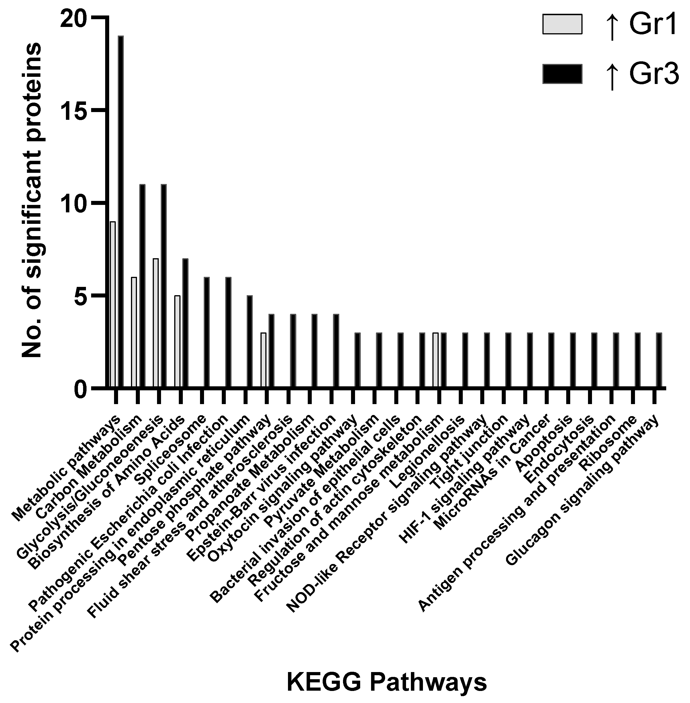
Figure 1. Pathway analysis (cells) comparing significant proteins found to be elevated in Group 1 (Gr1—favourable) and Group 3 (Gr3—adverse) when compared.
Proteomes 09 00042 g001
Figure 2. Pathway analysis (cells) comparing significant proteins found to be elevated in Group 1 (Gr1—favorable) and Group 2 (Gr2—intermediate) when compared.
Proteomes 09 00042 g002
Figure 3. Pathway analysis (cells) comparing significant proteins found to be elevated in Group 2 (Gr2—intermediate) and Group 3 (Gr3—adverse) when compared.
Proteomes 09 00042 g003
Figure 4. Box and Whisker plots for IL-17A, IL-1RA, SDF-1α1β and IL-1α based on results from serum biomarker analysis using targeted multiplexed immunoassays.
Proteomes 09 00042 g004
Table 1. Patient Data including gender, age at diagnosis, risk classification, diagnosis type and percentage (%) blasts in bone marrow (BM). DEK-NUP214: fusion gene created by the translocation occurring between specific introns in the gene DEK on chromosome 6 and the gene NUP214 on chromosome 9. NOS: Not otherwise specified.
Sample IDGenderDiagnosis AgeRisk ClassDiagnosis Type% Blasts in BM
1Female46.419871 Ac. myelomonocytic leuk. w abn. mar. eosinophils50
2Female35.319896 Acute myeloid leukemia, t(8;21)(q22;q22)60
3Male21.619896 Acute myeloid leukemia, t(8;21)(q22;q22)40
4Female67.319861 Acute myeloid leukemia75
5Female68.819861 Acute myeloid leukemian/a
6Male16.819896 Acute myeloid leukemia, t(8;21)(q22;q22)26
7Female55.519873 Acute myeloid leukemia without maturation90
8Female44.819861 Acute myeloid leukemia65
9Female53.519874 Acute myeloid leukemia with maturation20
10Male72.819861 Acute myeloid leukemian/a
11Female48.719891 Acute monocytic leukemia8
12Male76.929861 Acute myeloid leukemian/a
13Female62.929874 Acute myeloid leukemia with maturation30
14Male56.529861 Acute myeloid leukemia23
15Female63.829861 Acute myeloid leukemia70
16Female78.129891 Acute monocytic leukemia60
17Female24.329861 Acute myeloid leukemia63
18Male67.329895 Acute myeloid leuk. with multilineage dysplasia37
19Female48.629873 Acute myeloid leukemia without maturation60
20Male72.629874 Acute myeloid leukemia with maturation33
21Male16.529891 Acute monocytic leukemia80
22Female62.929861 Acute myeloid leukemia22
23Female61.529891 Acute monocytic leukemia40
24Female66.729897 Acute myeloid leukemia, 11q23 abnormalities15
25Male5729874 Acute myeloid leukemia with maturation42
26Female35.429920 Therapy-related acute myeloid leukemia, NOS95
27Female68.22del(9q)w2360
28Female76.639873 Acute myeloid leukemia without maturation91
29Female54.339867 Acute myelomonocytic leukemia12
30Male28.639891 Acute monocytic leukemia45
31Male66.739873 Acute myeloid leukemia without maturation85
32Female5239896 Acute myeloid leukemia, t(8;21)(q22;q22)91
33Female21.839873 Acute myeloid leukemia without maturation79
34Male44.639873 Acute myeloid leukemia without maturation73
35Female71.139873 Acute myeloid leukemia without maturation70
36Female39.739891 Acute monocytic leukemia40
37Male40.639861 Acute myeloid leukemia85
38Female59.439865 Acute myeloid leukemia with t(6;9)(p23;q34) DEK-NUP21485
39Male77.739895 Acute myeloid leuk. with multilineage dysplasia16
40Male62.539727 Precursor cell lymphoblastic lymphoma, NOS91
41Female64.739920 Therapy-related acute myeloid leukemia, NOS65
Table 2. Mass spectrometry data for group 1 vs. group 2; group 2 vs. group 3 and group 2 vs. group 3 including gene name, p-value, and direction of fold-change.
Group 1 vs. Group 2
Gene NameANOVA p-Value↑ in Gr1 (Fold-Change)↑ in Gr2 (Fold-Change)
UBP70.0011.4
HS1050.004 2.0
DPYL20.006 1.2
SRSF20.007 1.1
FUS0.0101.6
RTCB0.0121.4
ANM10.0171.3
PSA10.0201.2
HNRL10.020 1.1
RAB5C0.022 1.3
SYVC0.0301.3
1433Z0.032 1.2
CAH10.035 5.2
SPTN10.0352.3
LDHA0.043 1.3
FLNA0.045 1.5
ANXA60.046 1.3
G6PD0.048 1.8
Group 2 vs. Group 3
Gene NameANOVA p-Value↑ in Gr2 (Fold-Change)↑ in Gr3 (Fold-Change)
DHX90.0003.4
ATPB0.0016.1
GSTK10.0016.7
AHNK0.0046.5
SYNC0.0041.4
TCPA0.0052.2
1433G0.0071.3
CH600.0102.9
VATA0.0102.3
PRKDC0.01012.0
TAGL20.0111.7
RPN10.0121.9
TCPH0.0131.7
UB2V10.013 1.4
PA2G40.016 1.1
ROA20.0161.5
ATPA0.0185.9
UBA10.0201.6
FUBP10.0201.9
TCPG0.0201.6
TBB4B0.0214.4
FUBP20.0222.8
PNPH0.023 2.2
GSTO10.025 1.9
CAN10.0261.5
HBB0.029 4.7
BAX0.0291.9
EF20.0301.4
DDX10.0313.3
URP20.0311.8
HBA0.032 5.4
ESTD0.032 1.4
HBD0.034 8.2
ACTZ0.0381.9
TCPB0.0391.6
CBX30.040 1.2
TIF1B0.0432.8
PGM10.045 1.1
IF4A10.0452.9
CPNS10.0473.5
TCPE0.0481.6
Group 1 vs. Group 3
Gene NameANOVA p-Value↑ in Gr1 (Fold-Change)↑ in Gr3 (Fold-Change)
LA0.001 4.1
OTUB10.001 2.2
CNDP20.001 5.3
RAN0.001 2.5
HNRPC0.002 4.1
HNRPQ0.003 4.2
CH600.003 6.6
PRDX60.004 2.9
TBA1B0.005 3.7
TERA0.006 2.2
SET0.006 2.2
ROA20.006 2.8
CAPZB0.0071.4
RCC20.007 2.0
ECHA0.007 4.2
ARPC40.0071.3
PTPRC0.0072.0
NONO0.008 2.5
THIO0.009 2.9
ILF30.011 2.0
VIME0.011 3.5
TALDO0.012 2.1
LDHA0.013 2.0
TCPH0.013 2.3
NUCL0.014 2.8
NAGK0.0161.7
DHX90.016 4.1
PRDX40.016 1.0
TCP40.017 2.5
HS90A0.018 1.9
ROA10.018 2.5
LDHB0.019 2.6
EF1A30.020 2.4
FEN10.020 1.8
EF20.021 1.9
NPM0.024 2.6
F10A10.025 2.4
1433Z0.026 1.6
TIF1B0.027 7.0
ESTD0.028 2.1
HNRH10.029 2.4
LC7L20.030 2.1
TCPZ0.030 1.7
GANAB0.030 2.3
PGAM10.0311.3
ACTB0.031 1.7
PARP10.032 2.9
RUVB20.032 2.1
NPS3A0.0341.2
NDKB0.034 2.2
RHOA0.035 1.6
SFPQ0.035 1.9
IF4A30.035 2.3
HNRPU0.037 2.4
DLDH0.039 2.6
RSSA0.041 3.6
ROA30.042 2.4
G3P0.042 2.8
RS30.042 4.5
FSCN10.044 1.0
RL400.0461.2
PDIA30.049 1.7
HSP7C0.049 1.7
TSN0.0501.2
Publisher’s Note: MDPI stays neutral with regard to jurisdictional claims in published maps and institutional affiliations.