Article

Proteome Mapping of South African Cassava Mosaic Virus-Infected Susceptible and Tolerant Landraces of Cassava

by
Elelwani Ramulifho
1,2 and
Marie Emma Christine Rey
1,*
1
Plant Biotechnology Laboratory, School of Molecular and Cell Biology, University of the Witwatersrand, Johannesburg 2001, South Africa
2
Germplasm Development, Agricultural Research Council-Small Grain Institute, Bethlehem 9700, South Africa
*
Author to whom correspondence should be addressed.
Proteomes 2021, 9(4), 41; https://doi.org/10.3390/proteomes9040041
Submission received: 7 July 2021 / Revised: 19 October 2021 / Accepted: 20 October 2021 / Published: 23 October 2021

Abstract

:
The production of cassava is threatened by the geminivirus South African cassava mosaic virus (SACMV), which causes cassava mosaic disease. Cassava landrace TME3 shows tolerance to SACMV, while T200 is highly susceptible. This study aimed to identify the leaf proteome involved in anti-viral defence. Liquid chromatography mass spectrometry (LC-MS) identified 2682 (54 differentially expressed) and 2817 (206 differentially expressed) proteins in both landraces at systemic infection (32 days post infection) and symptom recovery (67 days post infection), respectively. Differences in the number of differentially expressed proteins (DEPs) between the two landraces were observed. Gene ontology analysis showed that defence-associated pathways such as the chloroplast, proteasome, and ribosome were overrepresented at 67 days post infection (dpi) in SACMV-tolerant TME3. At 67 dpi, a high percentage (56%) of over-expressed proteins were localized in the chloroplast in TME3 compared to T200 (31% under-expressed), proposing that chloroplast proteins play a role in tolerance in TME3. Ribosomal_L7Ae domain-containing protein (Manes.12G139100) was over-expressed uniquely in TME3 at 67 dpi and interacts with the ribosomal protein Sac52 (RPL10). RPL10 is a known key player in the NIK1-mediated effector triggered immunity (ETI) response to geminivirus infection, indicating a possible role for Sac52 in SACMV recovery in TME3. In conclusion, differential protein expression responses in TME3 and T200 may be key to unravel tolerance to CMD.

1. Introduction

Cassava (Manihot esculenta Crantz), a perennial shrub and root crop belonging to the Euphorbiaceae family, is the third most important food crop in tropical regions after rice and maize. It is grown in over 90 countries as it is a good source of carbohydrates and produces higher yields in adverse conditions compared to most other crops. According to Faostat (http://www.fao.org/faostat/, accessed on 11 August 2021), over 303 million tonnes of cassava were produced in 2019, with Africa being the major contributor (63.3% production share). Cassava does not only provide food but also brings employment to the impoverished regions of Africa. Cassava has many other uses such as in industries producing food, ethanol, paper and cardboard, textiles, pharmaceuticals, and glues and adhesives. Cassava roots are high in carbohydrates, while its leaves are an excellent source of both proteins and vitamins [1]. There is no doubt that the cassava crop is of importance; however, its production is threatened by pests and pathogens, resulting in yield losses.
South African cassava mosaic virus (SACMV) [2] is amongst nine cassava mosaic geminivirus species [3] which cause the devastating cassava mosaic disease (CMD) [4,5]. SACMV has a bipartite single-stranded DNA genome consisting of DNA-A and DNA-B components, both crucial for systemic infection [6]. DNA-A is necessary for transcription and replication of the virus, while DNA-B is crucial for cell-to-cell and long-distance movement [7]. DNA-A encodes the replication-associated protein (Rep) (AC1), transcriptional activator protein (TrAP) (AC2), replication enhancer protein (REn) (AC3), RNA-silencing suppressor (AC4), coat protein (CP) (AV1), and the pre-coat protein (AV2). DNA-B encodes the nuclear-shuttle protein (NSP) (BV1) and the movement protein (MP) (BC1) [8]. In addition to their function in infection, these encoded proteins also influence defence responses, hormonal responses and cell cycle regulation, ubiquitin-proteasomal pathways, and protein-signalling cascades [9]. Since its first report in the late 19th century, CMD has resulted in major cassava production and economic losses to the value of USD 1300–2300 million in Africa [10]. In susceptible cultivars, CMD has caused yield losses up to 82% [11] by significantly reducing the number and size of tubers. However, the severity of the damage is determined by the strain of the virus, cassava variety, age and sensitivity of the host, and environmental factors such as soil fertility [12]. CMD presents itself with symptoms of yellow and/or pale green chlorotic mosaic of leaves accompanied by distortion and an overall reduction in leaf size [13].
Cassava landraces TME3 and T200 have shown different responses to SACMV at different infection stages. TME3 is tolerant to SACMV, as shown by a recovery phenotype approximately 67 days post infection (dpi), whereas T200 is susceptible to the virus throughout the infection period. Findings from a transcriptome profiling study showed many down-regulated and up-regulated genes in the two landraces, including several resistance (R) gene transcripts over-expressed in TME3 and under-expressed in T200 [14]. Although this study revealed some interesting findings which could potentially give some clues to the molecular mechanisms that underly the tolerance phenotype of TME3, levels of mRNA in the cell are not directly proportional to the expression level of their cognate proteins. Proteomics studies the complete set of proteins expressed in a specific cell, tissue, or organism and thus bridges the gap between the other omics [15]. As such, it is important to look at the proteome as this offers a more robust approach to discovering the protein networks and pathways that are crucial for stress responsiveness and disease tolerance, and it will enable further dissecting of the biochemical and molecular responses of cassava against viruses.
The number of proteome mapping studies of crops has increased, and so has the pool of proteomic databases that are now available in the research community. In addition, proteomics has other potentials in the biotechnology field, including the identification of molecular markers to assist in plant breeding, keeping track of changes in protein profiles under different abiotic and biotic treatments at different timelines, understanding pathogen resistance or tolerance mechanisms, and the possibility of developing proteomic-based pesticides [16,17]. Together with the availability of fully sequenced genomes, researchers are now able to integrate different omics data into systems biology in order to seek an understanding of the physiological systems of a cell or tissue in response to different stimuli. Gel-based proteomics techniques such as two-dimensional electrophoresis (2-DE), and gel-free techniques such as mass spectrometry (MS)-based and Isobaric tag for relative and absolute quantitation (iTRAQ), have been used to identify proteomes in different crops [18]. Recently, a review examined tomato proteomes performed on different tissues using different proteomic techniques under different stress/physiological conditions [19]. Two of these studies investigated the differences in protein profiles in tomato fruit infected with tobacco mosaic virus (TMV) [20]. Pathogenesis-related (PR) proteins such as CHI and GLU and antioxidant enzymes such as SOD, DHAR, GST, PHGPX, APX, and TPX were found to protect the plant against TMV infection [20]. PR proteins were further confirmed to be involved in defence against TMV infection in model plant Nicotiana tabacum [21]. Global plant proteome changes during viral infection have been reported in several other studies, showing a range of functions impacted such as translation, protein processing and degradation, intracellular trafficking, primary metabolism, amino acid metabolism, stress responsive proteins, and cell wall biogenesis [22]. In tomato plants infected with cucumber mosaic virus (CMV), the responsive leaf proteome revealed proteins involved in photosynthesis, primary metabolism, and defence activity to be under-expressed following infection [23]. Several proteome studies have been performed on geminivirus-tomato interactions, comparing resistant and susceptible germplasm and revealing hypothetical models for host cell responses to infection [24,25]. A cassava proteome has been investigated in storage roots and leaves under abiotic stress and physiological conditions [26,27,28,29,30,31]; however, there has only been one study that investigated the leaf proteome in response to viral infection. In this study, cassava was infected with both Indian cassava mosaic virus (ICMV) and Sri Lankan cassava mosaic virus (SLCMV), which causes CMD [32]. Proteins such as the DnaJ-like proteins involved in molecular chaperone functions, signalling, and disease resistance (ascorbate peroxidase and protein kinase-coding resistance proteins) were over-expressed, implying their involvement in viral infection resistance. Proteomes have also been established from other crops such as sorghum, rice, soybean, wheat, maize, and mungbean [33,34,35,36,37,38] in response to different stimuli.
This study is the first to profile a cassava proteome in response to infection by a geminivirus, SACMV, on the sub-Saharan African continent. Furthermore, the study compares changes in cellular proteins between a susceptible T200 and tolerant TME3 landrace at the systemic infection stage 32 days post inoculation (dpi) and symptom recovery (67 dpi). Overall, the results showed differences in protein expression profiles between the two landraces and also revealed proteins that are proposed key players in reduced symptoms and virus tolerance in TME3. The results will go towards understanding the biochemical mechanisms involved in both susceptibility and symptom/virus recovery phenotypes in cassava infected with CMD.

2. Materials and Methods

2.1. Cassava Infection with SACMV

Nodal cutting of cassava T200 and TME3 landraces were micro-propagated on full Murashige and Skoog (MS) medium [39] supplemented with 2% sucrose, 0.78% plant tissue culture agar, and 0.002 mM CuSO4, with a pH of 5.8. To induce root growth, the nodal cultures were incubated in a temperature-controlled growth room at 28 °C under a 12 h photoperiod at a light intensity of 150 μEm−2/s. Once the roots appeared (approximately 3 weeks), the plantlets were transplanted into 40 mm × 45 mm Jiffy® pellets (Jiffy International AS, Kristiansand, Norway), placed in a plastic container, and covered with a plastic film for acclimatization. Two slits were made to the plastic film to gradually acclimatize plantlets until they could be infected with SACMV at a 4–6 leaf stage. Agro-inoculation of the plantlets with SACMV infectious dimer clones, DNA A, and DNA B, in Agrobacterium Agl1 was performed as outlined by [14], with minor modifications. Agrobacterium harbouring the infectious clones were grown in yeast extract peptone medium (1% tryptone, 1% Bacto peptone, 0.5% sodium chloride, pH 7) supplemented with 50 mg/mL carbenicillin and 100 mg/mL kanamycin, while Agrobacterium Agl1 medium was only supplemented with 50 mg/mL carbenicillin. The cultures were grown at 30 °C until an OD600 between 1.8–2.0 was reached. SACMV DNA A and DNA B cultures were mixed in equal volumes before plantlets were agro-inoculated along the stem with 100 μL of the culture using a hypodermic needle. Control plants were mock-inoculated with Agrobacterium Agl1 only, incubated in a temperature-controlled growth room, and monitored over a period of 67 days.

2.2. Protein Extraction and Isolation by TCA/Acetone Precipitation

Leaves below the apex from SACMV-infected and mock-inoculated plants were sampled at time points 32- and 67-days post inoculation (dpi). Three infection trials (biological replicates) were carried out independently for both T200 and TME3 landraces, with two leaves from a single plant and nine plants sampled per treatment per trial. Leaves from 3 plants were pooled into one tube/sample, and each treatment had 3 samples. Sampled leaves were immediately flash frozen in liquid nitrogen to halt any metabolic activity. A total of 2 g of crushed leaf samples was used for protein extraction using a HEPES-based buffer (50 mM HEPES-KOH, pH 7.4; 100 mM KOAc; 2 mM MgCl2; 0.5 mM PMSF; and 1% Proteinase inhibitor (ThermoFisher Scientific, Waltham, USA, 78438), 0.1% Triton x-100). Samples containing the extraction buffer were gently mixed in a low-speed vortex and placed on ice for 30 min before centrifuging at 4 °C for 10 min at 3000 rpm. A total of 400 μL lysate was transferred to a 2 mL tube. Proteins were precipitated by the addition of 1.6 mL of ice-cold TCA extraction buffer (10% v/v TCA (trichloroacetic acid; Sigma-Aldrich, Darmstadt, Germany, T9159) in acetone (Sigma-Aldrich, 34850)) with 2% 2-mercaptoethanol (Honeywell, Charlotte, USA, 63700) and incubated at −20 °C for a minimum of 1 h. Protein was pelleted by centrifugation at 21,000× g for 15 min at 4 °C. The supernatant was then removed and the pellet washed with 500 μL of ice-cold acetone. This was repeated a further two times for a sum of three washes in total. The pellets were then dried in a fume hood. Protein pellets were solubilised in 20 μL of 4% sodium dodecyl sulphate (SDS; Sigma-Aldrich, 71736) and 100 mM triethylammonium bicarbonate (TEAB, Sigma-Aldrich, T7408), briefly vortexed, and then heated at 95 °C for 5 min. Insoluble debris was removed by centrifugation at 10,000× g for 5 min. The supernatant was then transferred to a new tube and quantified using the QuantiPro BCA assay kit (Sigma-Aldrich, QBCA) according to the manufacturer’s instructions.

2.3. On-Bead Hydrophilic Interaction Liquid Chromatography (HILIC) Digest

In preparation for the HILIC magnetic bead workflow, the HILIC beads (ReSyn Biosciences, Pretoria, South Africa, HLC010) were aliquoted into a new tube and the shipping solution removed. Beads were then washed with 250 μL wash buffer (15% acetonitrile (ACN), 100 mM ammonium acetate (Sigma-Aldrich, 14267) pH 4.5) for 60 s. This was repeated for a total of two washes. The beads were then resuspended in loading buffer (30% ACN, 200mM ammonium acetate pH 4.5) to a concentration of 5 mg/mL. A total of 50 μg of protein from each sample was transferred to a 96-well protein LoBind plate (Merck, Darmstadt, Germany, 0030504.100). Protein was reduced with tris (2-carboxyethyl) phosphine (TCEP; Sigma-Aldrich, 646547), which was added to a final concentration of 10 mM TCEP and incubated at 60 °C for 1 h. Samples were cooled to room temperature and then alkylated with methyl-methane-thiosulphonate (MMTS; Sigma-Aldrich, 208795), which was added to a final concentration of 10 mM MMTS and incubated at room temperature for 15 min. HILIC magnetic beads were added at an equal volume to that of the sample and a ratio of 5:1 total protein. The plate was then incubated at room temperature on the shaker at 900 RPM for 30 min for binding of protein to beads. After binding, the beads were washed four times with 500 μL of 95% ACN for 60 s. For digestion, trypsin (Promega, Madison, USA, PRV5111), made up in 50 mM TEAB, was added at a ratio of 1:12.5 total protein, and the plate was incubated at 37 °C on the shaker for 4 h. After digestion, the supernatant containing peptides was removed and dried down. Samples were then resuspended in LC loading buffer (0.1% FA, 2% ACN) prior to clean up by Zip-Tip (Sigma-Aldrich, Z720070). Samples were then dried down once more and resuspended in a final volume of 12 μL LC loading buffer.

2.4. Liquid Chromatography-Mass Spectrometry (LC-MS)

LC-MS analysis was conducted with a Q-Exactive quadrupole-Orbitrap mass spectrometer (ThermoFisher Scientific, Waltham, USA) coupled with a Dionex Ultimate 3000 nano-UPLC system. Data was acquired using Xcalibur v4.1.31.9, Chromelean v6.8 (SR13), Orbitrap MS v2.9 (build 2926), and Thermo Foundations 3.1 (SP4). Peptides were dissolved in 0.1% Formic Acid (FA, Sigma-Aldrich, 56302) and 2% acetonitrile (ACN, Burdick & Jackson BJLC015CS) and loaded on a C18 trap column (PepMap100, 9027905000, 300 μm × 5 mm × 5 μm). Approximately 400 ng of peptide was injected. Samples were trapped onto the column and washed for 3 min before the valve was switched and peptides eluted onto the analytical column, as described hereafter. Chromatographic separation was performed with a Waters nanoEase (Zenfit) M/Z Peptide CSH C18 column (186008810, 75 μm × 25 cm × 1.7 μm), as described below. The solvent system employed was solvent A: LC water (Burdick and Jackson BJLC365), 0.1% FA, and solvent B: ACN, 0.1% FA. The multi-step gradient for peptide separation was generated at 300 nL/min as follows: time change, 100 min; gradient change, 5–30% Solvent B; time change, 10 min; gradient change, 30–80% Solvent B. The gradient was then held at 80% Solvent B for 10 min before returning it to 5% Solvent B and equilibrating the column for 15 min. All data acquisition was obtained using Proxeon stainless steel emitters (ThermoFisher Scientific, TFES523). The mass spectrometer was operated in positive ion mode with a capillary temperature of 320 °C. The applied electrospray voltage was 1.95 kV.

2.5. Viral Load

Genomic DNA was extracted from SACMV-infected leaf tissue using a CTAB-based method [40] with modifications. CTAB extraction buffer was supplemented with 40 mg/mL polyvinylpolypyrrolidone (PVPP) and 0.2% 2-mercaptoethanol before the buffer was preheated at 65 °C for 10 min. A total of 500 μL of CTAB buffer was added to 0.5 g ground leaf tissue and incubated at 65 °C for 45 min. Chloroform-isoamyl alcohol (24:1 ratio) was added to the samples, followed by gentle mixing and centrifuging at full speed for 10 min. The top aqueous phase was transferred into a new tube and the latter step repeated. Ice cold isopropanol (1:1 ratio) was added and the samples were thoroughly mixed and incubated in the freezer for 45 min. Samples were then centrifuged at full speed for 10 min and the DNA pellet was washed with ice cold 70% and 95% ethanol, respectively, and air-dried. DNA was resuspended in nuclease free water with 1% RNase and incubated at 37 °C for 1 h to allow digestion. Thereafter, DNA was stored at 4 °C overnight, then quantified and stored at −20 °C. DNA (25 ng) was used as a template for SACMV relative viral load quantification using qPCR. Quantification was carried out in triplicates using Maxima SYBR Green/ROX qPCR Master Mix (2X) (ThermoFisher Scientific, Waltham, USA) with SACMV CP primers (Fwd: 5′ ACGTCCGTCGCAAGTAC 3′, Rev: 5′ ATTGTCATGTCGAATAGTACG 3′). SACMV viral loads were calculated using the log 2(ΔΔCT) method [41]. Student’s t-test was used to access statistical differences between samples.

2.6. Changes in Gene Expression Levels

RNA was extracted from SACMV-infected and Agl1-mock inoculated leaf tissue using QIAzol Lysis Reagent (QIAGEN, Hilden, Germany) according to the manufacturer’s protocol. A RevertAid First Strand cDNA Synthesis Kit (ThermoFisher Scientific) was used to synthesise first strand cDNA using Oligo(dT)18 primer according to the manufacturer’s protocol. Gene expression of selected proteins of interest was measured using relative expression. RT-qPCR was carried out in triplicates with cDNA as a template using Maxima SYBR Green/ROX qPCR Master Mix (2X) with the primers listed in Table S1. UBQ10 was used as a reference gene for qPCR quantifications. Gene expression fold change was calculated using the log 2(ΔΔCT) method [41]. Student’s t-test was used to access statistical differences genes.

2.7. Bioinformatics Analyses

Relative quantification of identified proteins was conducted using Progenesis QI for Proteomics v2.0.5556.29015 (Nonlinear Dynamics, Durham, UK). Data processing included peak picking, run alignment, and normalisation (singly charged spectra were removed from the processing pipeline). Only proteins that contained two or more unique peptides were considered for further analysis. Database interrogation was performed with Byonic Software v3.8.13 (Protein Metrics, Cupertino, USA) using a cassava reference proteome (Cassava_RefProt_UP000091857_25032020.fasta) sourced from UniProt (www.uniprot.org, accessed on 1 August 2021) dated 25/03/2020. Phytozome v13 (https://phytozome-next.jgi.doe.gov/, accessed on 25 July 2021) was used to assess M. esculenta v8.1, v7.1, and v6.1 genomes and retrieve protein family data. UniProt and TAIR were used to retrieve theoretical molecular weight and Arabidopsis homologs of identified proteins, respectively. STRING version 11.0 (https://string-db.org/, accessed on 26 September 2021) was used to study protein–protein interaction networks. STRING network parameters were selected as follows: (i) Full network; (ii) Confidence edge; (iii) Interaction sources selected were Textmining, Experiments, Databases, and Co-expression; (iv) Confidence levels were between 0.8 and 0.9; (v) More than 50 interactors 1st and 2nd shell; and (vi) Hide disconnected nodes in the network. KEGG parameters were as follows: (i) Search background was selected at the whole genome level; (ii) Statistical test method was Fisher; (iii) Multi-test adjustment method was performed by the method of Benjamini and Hochberg [42]; and Significance level was 0.05. Gene ontology (GO) annotations were carried out on Gene Ontology at TAIR (https://www.arabidopsis.org/tools/bulk/go, accessed on 10 September 2021) using default settings.

2.8. CMD Symptom Severity Scoring

The symptom severity score index was used as a guideline for the assessment of CMD symptom development in TME3 and T200 cassava landrace plants following infection with SACMV. The mean severity score was calculated from a total of six leaves from three plants per time point and treatment using a 0–5 score index from Fauquet and Fargette [43] as a guideline for the symptoms assessment, where 0 represent no disease symptoms; 1 represent mild/faint mosaic with mild distortions at bases of most leaves; 2 represent pronounced mosaic pattern on most leaves, leaf malformation, 5% size reduction; 3 represent severe mosaic, distortion, reduced size; 4 represent severe mosaic, severe distortion, up to 50% size reduction; and 5 represent very severe mosaic, distortion, leaf twisting, 50–80% size reduction.

3. Results

3.1. SACMV Infection Development and Viral Load

CMD characteristic symptoms were observed in both susceptible T200 and tolerant TME3 landraces at 32 and 67 dpi with SACMV (Figure 1a). These include leaf deformation, yellow/pale-green mosaic, and reduced leaf size (red arrows). Agl1 mocked plants did not show any disease symptoms (white arrows). Minor symptoms were observed on newly formed leaves of TME3 at recovery (67 dpi), whereas severe symptoms were still observed in T200 plants. T200 infected plants generally showed severe symptoms, with a significantly high CMD symptoms score of 5 at 67 dpi compared to TME3 infected, which had a lower symptom score of 2.7 (Figure 1b). There was significant difference in the accumulation of SACMV DNA A in T200 and TME3 at 32 and 67 dpi (Figure 1c). T200 had viral loads of 7.7 and 10.4-fold higher relative to UBQ10 at 32 and 67 dpi, respectively. TME3 had a fold change of 6.8 and 8.0 relative to UBQ10 at 32 and 67 dpi, respectively, which was lower compared to the fold change in T200.

3.2. Protein Identification and Quantification

Leaf tissue proteins were extracted and isolated from T200 and TME3 landraces inoculated with SACMV infectious dimers, DNA-A, and DNA-B. LC-MS analysis and relative quantification resulted in the overall identification of 2682 proteins in both T200 and TME3 at 32 dpi, 49 of which were differentially expressed (DE) with fold change >1.0 (q < 0.05) (Table 1, Sheets S1 and S2). In T200, 30 and 19 were under and over-expressed, respectively, while in TME3, 27 and 22 were under and over-expressed, respectively. At 67 dpi, a total of 2817 proteins were identified in both T200 and TME3, with 206 of the proteins differentially expressed with fold change >1.0 (q < 0.05) (Table 1, In T200, 63 and 143 were under and over-expressed, respectively, while in TME3, 81 and 125 were under and over-expressed, respectively (Sheets S1 and S2). Ten proteins were expressed in both TME3 and T200 at 32 and 67 dpi. A large number of proteins were not characterised based on the status of the current cassava genome and protein annotations (Manihot esculenta v6.1; phytozome v12.1; https://phytozome.jgi.doe.gov, accessed on 25 July 2021).
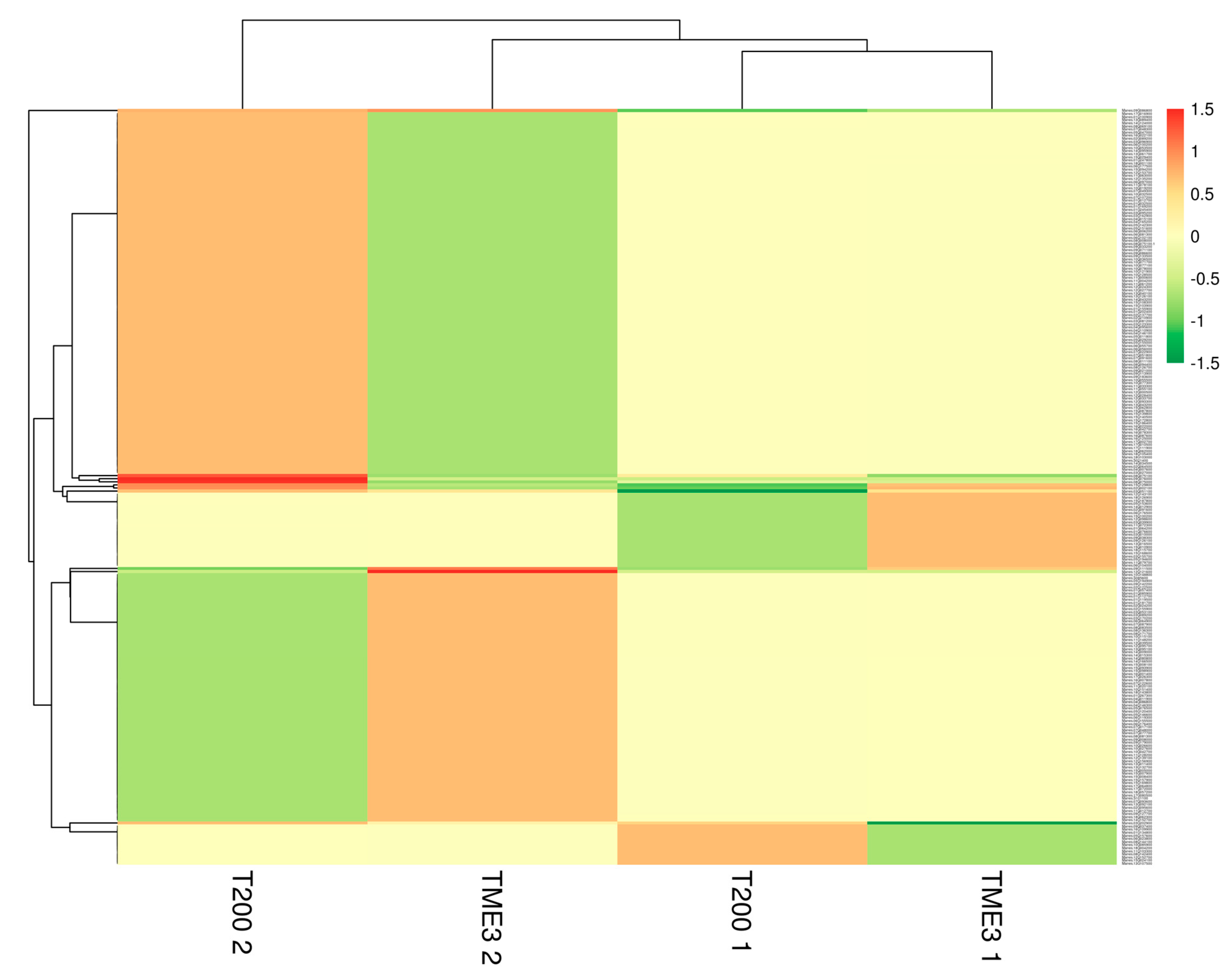
Figure 2 shows a heatmap of all DEPs in T200 and TME3 at 32 and 67 dpi. Dendrogram showed differences between leaf proteome at 32 dpi and 67 dpi, with the proteome of T200 at 67 dpi clustering separately from T200 at 32 dpi and TME3 at 32 and 67 dpi. At 67 dpi, proteins that were relatively over-expressed grouped together (red band), while under-expressed proteins grouped together (dark green band). Most over-expressed proteins branched out from the same node at 67 dpi, and the same was observed for under-expressed proteins. Interestingly, at both 32 and 67 dpi, an opposite pattern between susceptible T200 and tolerant TME3 was observed, where over-expression of a group of proteins in one landrace was under-expressed in the other landrace (Figure 2).

3.3. Gene Ontology Analysis

Gene ontology analyses were performed on differentially expressed (DE) proteins of T200 and TME3 landraces at 32 and 67 dpi to obtain an overview representation of how SACMV infection affects different processes in the plant. In T200 at 32 dpi, more than 25% of the 19 over-expressed proteins were localised in cellular components such as cytoplasm, mitochondrion, and plastid. A total of 33% of over-expressed proteins involved in stress response were positively induced by SACMV infection, while induced molecular functions included catalytic activity, hydrolase activity, and nucleotide binding, all with more than 30% of over-expressed proteins involved (Table S2). Of the 30 under-expressed proteins, more than 30% were localized in the chloroplast, cytoplasm, nucleus, and plastid. Stress response-related proteins (30%) and proteins with catalytic activity (32%) were negatively affected by SACMV infection in T200. In TME3, at 32 dpi, of the 22 and 27 proteins that were over-expressed and under-expressed, respectively (Table 1), more than 30% of the 22 over-expressed proteins were found in the cytoplasm and cytosol and were involved in stress response and catalytic and hydrolase activity. The 27 proteins that were under-expressed in TME3 were distributed across several cellular components, including chloroplast, cytoplasm, nucleus, and plastid, and they were involved in biosynthetic processes and catalytic activity (Table S2).
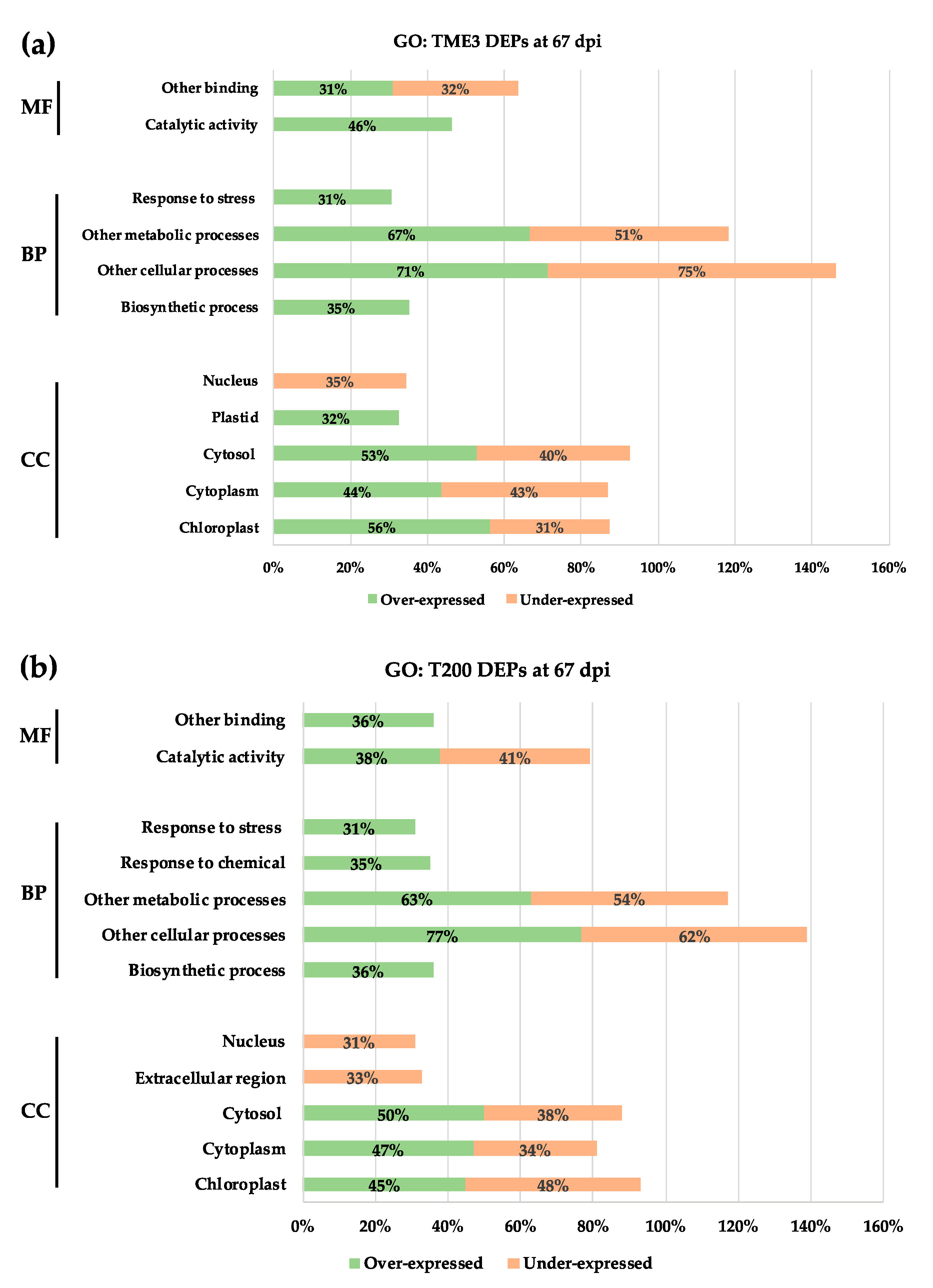
At 67 dpi, the over-expressed (125) and under-expressed (81) proteins in TME3 in response to SACMV infection (Table 1) were distributed across different GO groups (Table S3); however, only over-represented GO annotations with at least 30% of DEPs are presented in Figure 3. SACMV infection induced an increase in the expression of proteins with functions in catalytic activity and binding, which are involved in biological processes such as stress response and biosynthetic processes. These over-expressed proteins were localised in the plastid, cytosol, cytoplasm, and chloroplast (Figure 3a). Under-expressed proteins in TME3 at 67 dpi were localised in the nucleus, cytosol, cytoplasm, and chloroplast. These were involved in metabolic and cellular processes and had binding functions (Figure 3a). In T200, 143 proteins were over-expressed and 63 under-expressed at 67 dpi (Table 1). These proteins are also annotated across a range of GO groups (Table S3). Over-expressed proteins in T200 had catalytic and binding functions and were involved in biosynthetic, cellular, and metabolic processes and response to stress and chemical response. These proteins were found in the cytosol, cytoplasm, and chloroplast (Figure 3b). More than 30% of proteins were repressed by SACMV in T200, and these proteins had catalytic functions; were involved in metabolic and cellular processes; and localised in the nucleus, extracellular region, cytosol, cytoplasm, and chloroplast (Figure 3b).

3.4. KEGG, Protein–Protein Interaction Network and Reactome Pathway Analysis

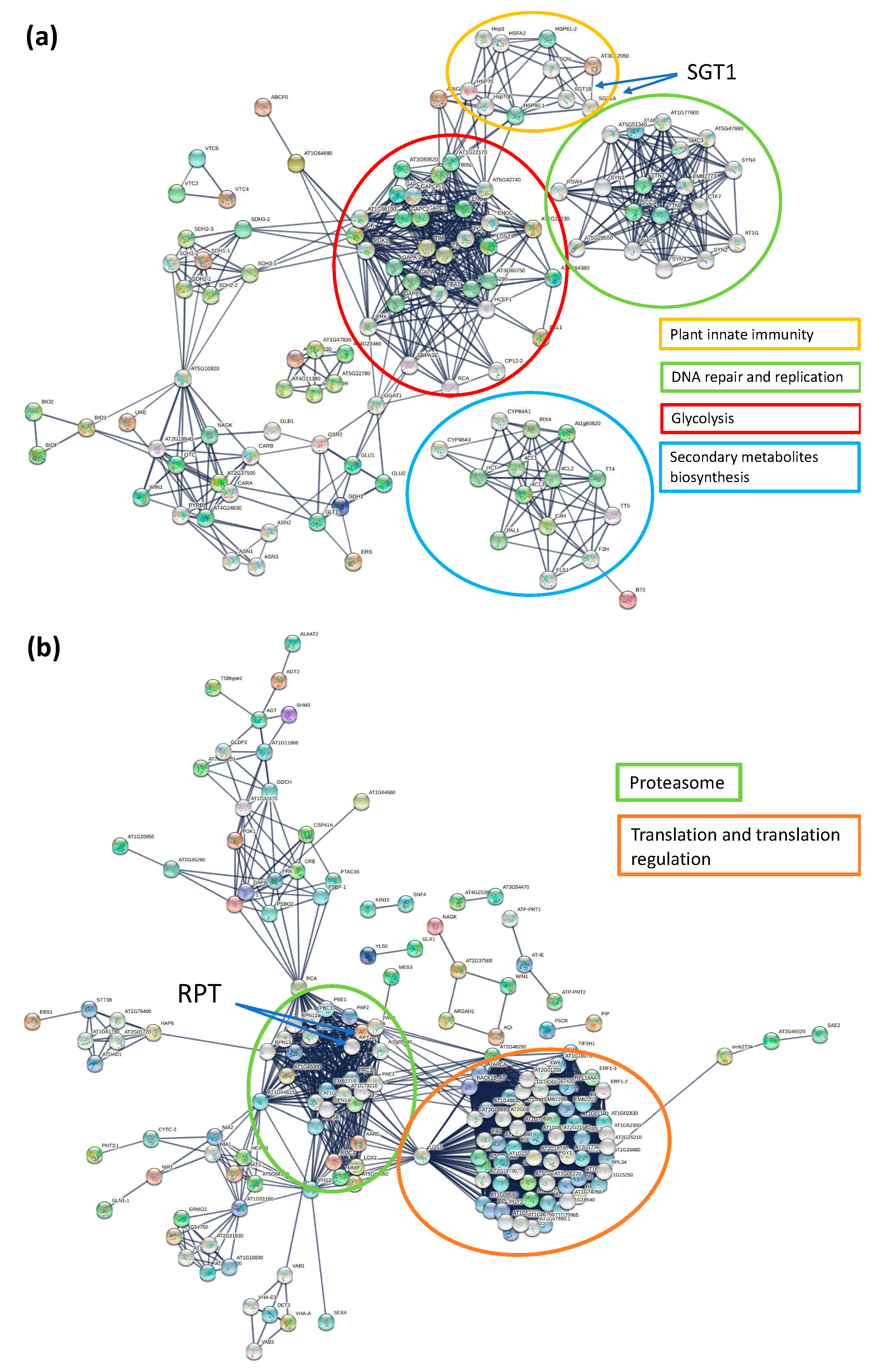
Proteins do not function alone, and therefore the metabolic pathways and network protein partners associated with the differentially expressed proteins in response to SACMV were explored. KEGG analysis of DEPs in T200 and TME3 at 32 dpi demonstrated 6 significantly enriched pathways, whereas 22 pathways were significantly enriched at 67 dpi in response to SACMV infection compared to mock-inoculated (Table 2, Figure S1b). KEGG pathways that were unique to 67 dpi included nitrogen metabolism, pyruvate metabolism, glycolysis/gluconeogenesis, ribosome, and proteasome. STRING applies known and predicted protein–protein interactions extracted from genomic context predictions, high-throughput lab experiments, co-expression, automated text mining, and previous knowledge in databases to create interaction networks. At 32 dpi, the differentially expressed protein–protein network (p-value < 1.0 × 10⁻¹⁶) revealed four major functional groups: DNA repair and replication, secondary metabolites biosynthesis, glycolysis, and plant innate immunity (Figure 4a). Proteins such as suppressors of the G2 allele of skp1 (SGT1) subunit A and SGT1B were some of the interactors with functions in plant innate immunity. SGT1 directly interacts with heat shock protein 90 (HSP90) and Arabidopsis homolog of Manes.09G037400 (AT3G12050; activator of Hsp90 ATPase family), which was under-expressed in both TME3 and T200 with ratios of 1.61 and 1.48, respectively (Figure 4a; Table 3). SGT1 is also an essential S-phase kinase-associated protein (SKP1)-interacting eukaryotic protein which is part of the Cullin1-based RING ligases SCF complexes that function in plant-virus interactions. At 67 dpi, proteins mainly formed two groups: proteasome and translation and translation regulation (Figure 4b). Regulatory particle triple-ATPase (RPT) subunit 2a and RPT2b directly interacted with Manes.01G032500 (AT1G29150, Proteasome component family) and Manes.05G155000 (AT1G45000, Holliday junction DNA helicase RuvB P-loop family), which were both over-expressed in T200 and under-expressed in TME3 (Figure 4b, Table 3). Protein–protein interaction network functional enrichment analysis of over-expressed proteins in TME3 at 67 dpi revealed an interaction with Sac52 (Figure S1a), an important plant defence response protein. At 32 dpi, metabolism (six proteins) was the only pathway that was significantly enriched (FDR < 0.05). Three pathways were significantly enriched at 67 dpi: metabolism (28 proteins), L13a-mediated translational silencing of Ceruloplasmin expression (eight proteins), and antigen processing-Cross presentation (three proteins).

3.5. Differentially Expressed Protein Groups in Response to SACMV Infection at 67 dpi

Because the molecular mechanisms of tolerance to CMD in TME3 are of key interest, we chose to focus on and compare responses to SACMV in T200 and TME3 at 67 dpi, which is when recovery to infection in TME3 has already begun. Differentially expressed proteins between mock-inoculated and SACMV-infected leaves in T200 and TME3 at 67 dpi are presented in Table 3.

3.5.1. Metabolic Pathways

A total of 56 proteins were involved in primary metabolic pathways in TME3 infected with SACMV at 67 dpi (Table 2). Functions of up- and under-expressed DEPs in this group included amino acid metabolism, secondary metabolism, photorespiration, and carbohydrate metabolism. Primary metabolites are crucial for the normal development of the plant. In T200, glycine cleavage system H proteins (Manes.07G087900, Manes.15G169800) and aminotran_5 domain-containing protein (Manes.01G181700), also functioning in the biosynthesis of secondary metabolites, were under-expressed. In addition, phosphoglycerate kinase (PGK, Manes.14G009000), a major role player in energy generation, was also under-expressed (Table 3). In Nicotiana benthamiana, chloroplast PGK was found to be one of the proteins that binds to bamboo mosaic virus (BaMV) RNA [45]. Trehalose-6-phosphate synthase 7 (Manes.16G042700) protein plays a role in plant growth, development, and regulation of defence response against pathogens, and this protein was over-expressed in T200 and under-expressed in TME3. Other proteins involved in the biosynthesis of metabolites include Inositol-1-monophosphatase, Phosphoacetylglucosamine mutase, AA-kinase domain-containing protein, and Arginine biosynthesis bifunctional protein, to mention a few (Table 3). One uncharacterised protein, Manes.09G142200, belonging to the family Enoyl-(Acyl carrier protein) reductase, was highly over-expressed by 67-fold in TME3 and under-expressed (1.63-fold) in T200.

3.5.2. Biosynthesis of Secondary Metabolites

A total of 36 proteins were involved in the biosynthesis of secondary metabolites (Table 2). Plant hosts require secondary metabolites for several roles, including pigmentation, growth, reproduction, and resistance to pathogens, amongst many others. Peroxidase protein, Manes.15G103900, was over-expressed in T200 and under-expressed in TME3 (Table 3). Peroxidases are known to play important roles in oxidative stress, auxin catabolism, and cross-linking of structural cell wall proteins, amongst other things. In this study, a Methyltransferase protein (Manes.05G142300) was under-expressed almost seven-fold in TME3 and over-expressed three-fold in T200 (Table 3). 3-ketoacyl-CoA synthase (Manes.16G087600) and AB hydrolase-1 domain containing proteins (Manes.12G095700, Manes.04G110900, Manes.05G029200, and Manes.13G092100) are some of the structural proteins that play a role in lipid homeostasis (Table 3) [46,47]. Other characterised proteins in this group include PALP domain-containing protein, Phospholipase D, Serine hydroxymethyltransferase, FAD-binding PCMH-type domain-containing protein, GPAT_N domain-containing protein, SAICAR_synt domain-containing protein, Inositol-1-monophosphatase, and F420_oxidored domain-containing protein (Table 3). Some of the proteins, however, were not characterised, but these belonged to protein families including KR domain (Manes.10G026600), p450 (Manes.06G006200 and Manes.09G183600), and aminotransferase_3 (Manes.13G089400), to mention a few.

3.5.3. Glycolysis/Gluconeogenesis

This pathway is associated with the generation of energy. When a plant is attacked by an external stimulus, it direct most of its energy to defence response mechanisms and less to cellular metabolic pathways. In this functional group, five proteins were involved. Three proteins were over-expressed in both T200 and TME3, and these are also involved in metabolic pathways: FBPase domain-containing protein (Manes.08G083500), pyrophosphate-fructose 6-phosphate 1-phosphotransferase subunit alpha (Manes.18G001100), and Manes.04G146300 (uncharacterised). FBPase (fructose 1,6-bisphosphatase) catalyses the hydrolysis of fructose 1,6-phosphate to fructose 6-phosphate in gluconeogenesis and the Calvin cycle. The reverse is true for glycolysis with phosphofructokinase (PFK) and pyrophosphate-fructose 6-phosphate 1-phosphotransferase subunit alpha enzymes catalysing the reaction. With FBPase over-expressed, it means that the plant had enough cellular ATP concentration and there was no need to produce more in both T200 and TME3 at 67 dpi. Uncharacterised Manes.04G146300 protein originates from the family glycoside hydrolase family 19, which consists of chitinase class I. Phosphoglycerate kinase (Manes.14G009000) was over-expressed in TME3, with a ratio of 1.51, and under-expressed in T200, with a ratio of 1.12 (Table 3). This enzyme catalyses the high-energy phosphoryl transfer of the acyl phosphate of 1,3-bisphosphoglycerate to ADP to produce ATP [48]. The last uncharacterised protein in this group, Manes.08G094400, belongs to the thiamine pyrophosphate enzyme (TTP) family, and it was only over-expressed in T200 (ratio 1.91; Table 3).

3.5.4. Ribosome

Ribosomal proteins are known to be components of the protein synthesis machinery. However, with recent studies, it would seem that these proteins have extra functions such as stress signalling [49]. Seven DEPs were grouped to the ribosome pathway (Table 3), and three of these are uncharacterised. Three proteins were under-expressed in T200 and over-expressed in TME3, three over-expressed in T200 and under-expressed in TME3, and one was under-expressed in both landraces. For example, ribosomal_L14e domain-containing protein (Manes.03G162900) was over-expressed with ratio of 4.36 in T200 and under-expressed with ratio of −1.86 in TME3 (Table 3). This protein binds to the 60S ribosomal subunit and plays a role in translation [50]. The 40S ribosomal protein S4 (Manes.03G095200) had ratios of 1.59 in T200 and −1.06 in TME3. An increase in transcript levels of this protein was observed in Vanilla planifolia Jacks infected with fungi [51], whereas there was a decrease in transcript in Arabidopsis 48 h after inoculation with A. tumefaciens [52]. The 60S ribosomal protein L13 (Manes.07G017100) was under-expressed in T200 (−3.86) and over-expressed in TME3 (2.11). This protein has been shown to interact with CMV P6 (translation re-initiator) in A. thaliana [53]. Uncharacterised proteins, Manes.10G151400 (−3.90 ratio in T200 and −1.14 in TME3), Manes.17G064800 (−4.61 in T200 and 1.83 in TME3), and Manes.10G019200 (3.30 in T200 and −1.16 in TME3) are members of the ribosomal protein family L3, L32, and L35Ae. The last protein of interest in this group was ribosomal_L7Ae domain-containing protein (Manes.12G139100), which was under-expressed in T200, with a ratio of 5.24, and over-expressed in TME3, with the ratio of 2.18 (Table 3). Much like the other proteins in this group, very little has been reported on this protein; however, its molecular function includes RNA binding.

3.5.5. Proteasome

Proteasomes form a large portion of the ubiquitin proteasome system, responsible for degrading the intracellular proteins involved in most cellular metabolic processes. Three proteins, also involved in metabolic pathways, with functions in proteasomes, were only over-expressed in T200. PCI domain-containing protein (Manes.01G032500) was over-expressed by up to seven-fold compared to mock sample in T200, and under-expressed in TME3 at 67 dpi. PCI (Proteasome, COP9 signalosome, and initiation factor 3) domain mediates and stabilizes protein–protein interactions within multi subunit protein complexes [54]. The other two proteins were both AAA domain-containing proteins with ratios of 4.75 in T200 and −1.32 in TME3 (Manes.13G043200) and 2.82 in T200 and −1.07 in TME3 (Manes.05G15500) (Table 3). ATPase associated with various cellular activities (AAA) plays a role in the cell, including cell-cycle regulation, protein proteolysis, and disaggregation, organelle biogenesis, and intracellular transport.

3.5.6. Transcription of Selected Differentially Expressed Proteins

Based on proteomics analysis, ten proteins that were differentially expressed in response to SACMV were selected for transcription analyses using RT-qPCR (Figure 5). These selected proteins have functions in disease response and cell morphogenesis and were differentially expressed in both landraces, two at 32 dpi (C3HC4-type RING finger and LRR) and eight at 67 dpi (kinase, SnRK1.1, two receptor-like kinases, LRX2, receptor-like protein, two pathogenesis-related proteins). At 32 dpi, RT-qPCR results demonstrated that C3HC4-type RING finger (Manes.08G075100) was expressed with fold change of 0.5, whereas LRR had a fold change of −1.7 compared to the mock-inoculated control (Figure 5). Transcription levels of the latter genes did not reflect protein translation expression regulation (Table 3). At 67 dpi, all eight genes were demonstrated to be over-expressed with fold changes ranging from 1.7 to 10.8 higher than the mock-inoculated control, with pathogenesis-related protein_2 (Manes.15G007900) showing the highest expression levels (Figure 5). Transcripts of receptor-like kinases (Manes.05G194900, Manes.14G124000), Kinase (Manes.08G126700), and receptor-like protein (Manes.06G055700) were over-expressed with fold change of 7.9, 3.8, 1.7, and 2.5, respectively (Figure 5), whereas the expression levels of their proteins were under-expressed based on the proteomics results (Table 3). The results herein suggest differences between transcript and protein levels may be due to post-transcriptional regulatory processes.

4. Discussion

Cassava is an economically important crop and a major source of nutrition in the tropical and sub-tropical regions of Africa and Asia, and it additionally shows tolerance to drought. This makes it a targeted crop in future climate change where temperatures are expected to rise. However, cassava production is threatened by viral diseases such as CMD, which is caused by a number of viruses, including geminiviruses. As such, it is important to identify proteins that are differentially expressed in response to SACMV as these will help bridge the gap that exist in understanding plant-virus interactions in this and other orphan crops, and furthermore inform engineering strategies for crop varieties that will better tolerate and/or resist virus diseases. This study reports the first SACMV-cassava proteome map comparing susceptible and tolerant landraces and further contributes new information to the proteomic data pool. Finding more clues to the molecular mechanisms of tolerance to SACMV in TME3 at 67 dpi is also of paramount interest. Several important GO processes in response to SACMV in TME3 were identified herein that may play a role in CMD tolerance. In particular, we also focussed on individual proteins shown to be involved in geminivirus responses in other plants that may also play a role in cassava. While it is well documented that protein modifications are a consequence of pathogen infection of plants, fewer studies have been performed on virus infections, in particular on geminiviruses. It is noteworthy that 8 out of the 18 genes involved in processes related to protein modification or protein translation and metabolism (such as ubiquitination, rubylation, phosphorylation, acetylation, or protein folding) were differentially expressed in N. benthamiana infected with the geminivirus tomato yellow leaf curl Sardinia virus (TYLCSV) [55]. It is therefore not surprising that, in SACMV-infected T200 and TME3, seven of the DEPs were related to the ribosome pathway. Ribosomal proteins are not only known to be components of the protein synthesis machinery, but are also involved in stress signalling [49]. In cassava infected with Indian cassava mosaic virus (ICMV) and Sri Lankan cassava mosaic virus (SLCMV), components of the 80S ribosome and 40S small subunit were upregulated in susceptible cassava cultivar H226 [32]. In a proteome study of the begomovirus, tomato chlorotic mottle virus (ToCMoV), in resistant tomato, two ribosomal proteins (40S ribosomal protein S7 and ribosomal protein L11/12) were also differentially expressed [25]. These results suggest that translational control via ribosome activity may be a general response to geminivirus-induced stress. Because T200 and TME3 showed contrary expression (up or down) in six ribosomal proteins, we conclude that these play a role in susceptibility and tolerance in T200 and TME3, respectively. Perturbations in protein translation also reflect dual expression of virus and plant host proteins induced during their interaction.
Notably, a large number of the SACMV-responsive cassava DEPs annotated by GO were located in different cellular components, but largely in the cytoplasm and plastid, including the chloroplast and thylakoid (Figure 3, Tables S2 and S3). From this observation, we conclude that chloroplasts may be involved in response to SACMV. Plastids have versatile roles providing essential metabolic and signalling functions, and affect photosynthesis, plant growth, and development. They also play role in plant-microbe interactions [56]. Chloroplasts have more recently been shown to have other functions, including involvement in plant defence responses through the production of reactive oxygen species, defence-related hormones, and plastid-to-nucleus retrograde signalling pathways [57]. Together with the fact that chloroplasts are central hubs for facilitating communication with different plant cellular compartments, chloroplasts functions have become targets of plant pathogens. Pathogens deliver effector proteins that disrupt the normal functioning of the chloroplast and result in suppressed defence responses and subsequent infection [58]. We propose that the identified differentially expressed chloroplast proteins in our study play a role in virus tolerance and susceptibility in cassava. The majority of over-expressed proteins (56%) in TME3 at 67 dpi were localized in the chloroplast (Figure 3a), whereas 31% of these chloroplast proteins were under-expressed in T200 at 67 dpi (Figure 3b). Manes.15G029400 Arabidopsis homolog AT4G14880, a chloroplast protein whose gene expression is mediated by abscisic acid (ABA), was over-expressed in TME3 (1.25 ratio) at a symptom recovery stage (67 dpi). ABA is an essential signal for plant resistance to pathogens, and it is synthesized in the chloroplast. The over-expression of Manes.15G029400 indicates that ABA production in the chloroplast was not affected by SACMV infection. We conclude that ABA-mediated defence mechanisms are active in TME3 and play a role in the reduction of SACMV replication and symptom reduction at 67 dpi. Results from other studies have shown the importance of ABA in biotic stress and host defence against viral pathogens. For example, in N. benthamiana, it was proved that Chinese wheat mosaic virus suppressed the ABA pathway, which was important in inducing plant host defence against this virus [59]. Although the contribution of these chloroplast-associated proteins to tolerance is not currently known, we suggest that over-expressed chloroplast proteins in tolerant TME3 at 67 dpi play a role in virus and symptom recovery phenotype (Figure 1).
Coat protein (CP) from a number of viruses, including TMV, Potato virus X, and CMV, has been shown to accumulate in the chloroplast of their host and cause chloroplast ultrastructure disruption as well as the development and severity of mosaic or chlorosis symptoms caused by viral infections [60,61,62]. TME3 landrace shows a recovery phenotype and significant reduction of SACMV DNA accumulation at 67 dpi (Figure 1), compared with susceptible T200 landrace. A decrease in DNA also implies that virus coat protein synthesis is decreased as these are co-regulated, which suggests that a decrease in CP in chloroplasts in TME3 at 67 dpi could contribute to symptom attenuation at the recovery stage. It was also reported that some of these viral CPs are associated with the thylakoid [63,64]. In this study, a relatively high number of over-expressed proteins (15; 13%) were localised in the thylakoid at 67 dpi in TME3 (Figure 3a). Proteins such as Manes.10G027600 (Rubisco activase; 6.89 ratio) and Manes.14G009000 (Phosphoglycerate kinase 1; 1.15 ratio) were amongst the over-expressed proteins localised in the thylakoid (Table 2) and are known to interact with virus nucleic acids or proteins [58]. A difference that was seen between T200 and TME3 at 67 dpi was the presence of the Manes.08G136300 (linker histone; 1.08 ratio) protein involved in epigenetic regulation of gene expression in TME3 (Table 1, Figure 3a). Linker histones are major negative regulators that limit the accessibility of DNA to various trans-acting factors, which in turn enables epigenetic suppression of genes [65]. It is not clear how the mechanism of this protein would contribute to SACMV tolerance phenotype in TME3; however, further studies on this protein are warranted.
Additionally, of particular interest with regard to SACMV DNA accumulation was the upregulation and downregulation of the SUMO-activating enzyme subunit in T200 and TME3 at 67 dpi, respectively (Table 1). SUMOylation is an essential post-translational modification in plants and is also affected by oxidative stresses. Geminiviruses replicate in the nuclei of infected plant cells using the plant DNA replication machinery, including proliferating cellular nuclear antigen (PCNA), a cofactor that orchestrates genome duplication and maintenance by recruiting crucial players to replication forks. Posttranslational modification of PCNA by SUMO plays an essential role in the switching of PCNA between interacting partners during host replication, recombination, and repair mechanisms. The geminivirus Rep induces the accumulation of the host replication machinery by interfering with the cell cycle and impairs SUMO conjugation of proliferating cellular nuclear antigen (PCNA) and the SUMO E2 conjugation enzyme (SCE1) [66]. The significant down-regulation (−5.5 ratio) of the SUMO-activating enzyme subunit in T200 would lead to impairment of SUMO conjugation, thereby switching on the host cell machinery for its own replication. This would contribute to the persistent high SACMV replication levels in T200 at 67 dpi (Figure 1). In contrast, over-expression (4.65 ratio) of SUMO-activating enzyme in TME3 would contribute to a reduction in SACMV replication, leading to recovery at 67 dpi.
At 32 dpi (systemic infection stage), virus replication and symptoms are high in both landraces. At this stage, STG1A and the molecular chaperone HSP90.1 were over-expressed. These proteins directly interact with AT3G12050, a homolog of Manes.09G037400 from the Activator of the Hsp90 ATPase family (Figure 4a). SGT1 is an essential S-phase kinase-associated protein (SKP1)-interacting eukaryotic protein which is part of the Cullin1-based RING ligases SCF complexes that function in plant-virus interactions. SGT1 also binds to HSP90, resulting in the positive regulation of disease resistance conferred by many resistance (NB-LRR) proteins in plants [67]. In N. benthamiana, a study found that SGT1a controls the abundance of an R protein Rx after the silencing of SGT1a resulted in the reduction in steady-state levels of Rx. It is understood that any compromise in the activity of HSP90 and levels of SGT1a leads to reduced accumulation of R proteins [67,68]. This is in agreement with what was observed in this study, as Hsp90 activator was under-expressed in both T200 (−1.48 ratio) and TME3 (−1.61 ratio), which may have compromised HSP90. The implications of this would be the observed severe CMD symptoms (Figure 1a,b) as well the accumulation of SACMV DNA (Figure 1c) at 32 dpi. Moshe et al. [69] also proved the roles that SGT1a and HSP90 play in susceptibility to TYLCV in infected tomatoes. Interestingly, in ICMV and SLCMV infected susceptible cassava, a member of the HSP70 chaperones, namely chaperone DNAj, was upregulated. It has been suggested that chaperones may be involved in cell-to-cell movement via modified plasmadesmata (PD), and molecular chaperones are known to be involved in modification of the size exclusion limit of PD [28]. This makes a case for HSP90.1 involvement in virus movement in T200 and TME3 at 32 dpi, corresponding to the systemic infection and high virus loads observed in both landraces.
At 67 dpi in TME3, two overexpressed unique protein interactors, RPT2 and Sac52 (Figure 4b), that we propose are involved in SACMV recovery, are discussed. Firstly, RPT2 is a member of the proteasome regulatory subunits, known to be of importance in defence response [70]. Arabidopsis homologs of Manes.01G032500 (AT1G29150; 7.14 ratio in T200 and −1.40 ratio in TME3) and Manes.05G155000 (AT1G45000; 2.82 ratio in T200 and −1.07 ratio in TME3) (Table 3) interact directly with RPT2a and RPT2b proteins (Figure 4b). RPT2a has been linked with PAMP triggered immunity (PTI) and the establishment of broad-spectrum pathogen resistance and systemic acquired response in Arabidopsis in response to bacterial infections. With the up-regulation of proteins directly interacting with RPT2a, one would propose that there would be the reduction in systemic infection due to RPT2a-induced responses. However, this is not the case for susceptible landrace T200, which shows no recovery phenotype at 67 dpi as disease symptoms are still severe and SACMV accumulation is significantly high (Figure 1). Interestingly, a study showed that Cauliflower mosaic virus relies on the proteasome subunits RPT2a and RPT2b for robust infection [71], indicating that these proteasome subunits could have different functions in different host-virus interactions. This observation supports the results observed in our study, where higher expression of the proteasome interactors was linked to T200 with a greater virus load and down-regulation linked to TME3 (Table S2) with low virus accumulation and a recovery phenotype (Figure 1, Table 3).
The second significant protein of interest is Sac52, also known as the ribosomal protein L10 (RPL10), which interacts with Ribosomal_L7Ae domain-containing protein homolog (Manes.12G139100, AT1G77940), which was over-expressed uniquely in TME3 only at symptom recovery (67 dpi) (Table 3, Figure S1, Sheet S2). RPL10 is a known key player in the NIK1-mediated antiviral immunity defence response to geminivirus infection as it plays the role of viral effector detection, triggering effector triggered immunity (ETI) mechanisms in plants [72]. This defence mechanism is established with interaction between membrane-bound LRR receptor-like serine/threonine kinase and its virulence target nuclear shuttle protein (NSP), found on DNA B component of the bipartite geminiviruses. During geminivirus infection, NIK1 autophosphorylates the kinase domain on threonine-469 and threonine-474, resulting in NIK1 kinase activation [9,73. Once activated, NIK1 indirectly phosphorylates the cytoplasmic RPL10, which is then subsequently translocated to the nucleus where it interacts with the L10-interacting MYB domain-containing protein (LIMYB). This interaction mounts a defence response in the form of translational inhibition of several host genes and decreased geminiviral transcript association with polysomes, consequently negatively impacting virus infection. With regard to the role that RPL10 may play in TME3 tolerance to SACMV, we propose that this protein may be expressed in abundance and then phosphorylated by membrane-bound NIK1, which would then lead to downstream translational suppression of viral proteins and thus virus movement, which would contribute to the symptom and virus recovery phenotype observed in TME3 (Figure 1a). This is in agreement with observations in other different plant hosts [73,74,75,76,77,78]. It is also important to note that the RPL10 interactor (Manes.12G139100) is located on chromosome 12, known to be the location of the QTL for dominant CMD2 resistance [79,80,81], thus supporting the likelihood of this protein being a key player in TME3 tolerance to SACMV.
Ubiquitination of geminiviral proteins can contribute to plant defence, and geminiviruses often subvert or hijack ubiquitin-proteasomal pathways [9]. At 67 dpi, one of the highly represented functional group proteins was the proteasome (Figure 4b). The PCI domain-containing protein (Manes.01G032500), involved in the COP9 signalosome, was over-expressed (7.14-fold ratio) compared to the mock in T200, suggesting a major role in susceptibility via interference of this complex. Plant geminiviruses redirect ubiquitination by interfering with the activity of the CSN (COP9 signalosome) complex. The geminivirus C2 protein has been demonstrated to compromise CSN activity on CUL1. Among several responses regulated by the CUL1-based SCF ubiquitin E3 ligases, is response to jasmonic acid (JA), ethylene (ET), and abscisic acid (ABA), known to function in defence responses. A C3HC4-type RING finger protein, Manes.08G075100, was under-expressed in TME3 at 67 dpi and over-expressed in TME3 at 32 dpi (Table 3), supporting a strong role for ubiquitination in TME3 tolerance. This RING finger protein is an E3 ubiquitin ligase (E3L) that is a component of the ubiquitin proteasome system (UPS), with many of their functions involved in plant stress resistance [82,83,84,85]. Recently, it was shown that CRISPR knockdown of a E3L in cassava TME3 protoplasts resulted in an increase in SACMV replication, suggesting that E3L target proteins may play a role in SACMV tolerance [82]. Ubiquitin activating enzyme UBA1 and RING-type E3 ubiquitin ligase are also reported by [55] to be trans-activated by the geminivirus C2 protein. Further evidence for the role of the ubiquitin-proteasome in response to viral infection was recorded in a proteome study on ToCMoV resistant tomato, which showed decreased levels of ubiquitination pathway-related proteins [25].

5. Conclusions

SACMV is a highly pathogenic virus in susceptible hosts, leading to major yield losses. In perennial hosts such as cassava, tolerance may represent accommodation of persistent virus infection throughout its life span [86]. Our proteome results identified the proteins involved in different metabolic pathways, some with functions in defence responses to SACMV. We identified proteins in the ribosome, chloroplast, ubiquitination, and cytoplasm GOs, and we propose that these proteins and their connected networks are contributors to SACMV and symptom recovery in TME3. One of these identified proteins of importance is Sac52 (RLP10), which interacts with NIK1 and may contribute to an anti-virus response in cassava. This is highly significant because this finding provides further evidence that PAMP triggered immunity (PTI) may be involved in resistance to plant geminiviruses. While ETI has long been recognized as an efficient specific defence against viruses, PTI characterized in non-viral pathogen-plant interactions has not been found to operate against plant viruses until recently [87]. Geminivirus nucleic acids were recently shown to act as viral pattern associated molecular patterns (PAMPs) and activate transmembrane receptor NIK1, which shares PTI co-receptors regulatory mechanisms for activation [87]. However, geminiviral PAMPs and their cognate PRRs have yet to be identified, and investigation of antiviral PTI mechanisms in cassava is highly warranted. In the cassava TME3 landrace, we conclude that tolerance allows the virus to accumulate to some degree without causing significant loss of fitness, implying that the virus and host co-exist. The results herein support the proposal that symptom recovery in TME3 may be associated with stress tolerance and maintenance of cellular homeostasis [85]. In perennial crops, this would be advantageous as long-term defence responses are energy-intensive. SACMV-tolerance in TME3 would mitigate energy costs, leading to growth recovery and a reduction in symptoms and virus load. The results from this study form the foundation for further in planta functional studies to understand cassava tolerance to geminiviruses.

Supplementary Materials

The following are available online at https://www.mdpi.com/article/10.3390/proteomes9040041/s1, Table S1: Primers used for RT-qPCR, Table S2: Gene ontology annotation of differentially expressed proteins of T200 and TME3 at 32 dpi, Table S3: Gene ontology annotation of differentially expressed proteins of T200 and TME3 at 67 dpi, Figure S1: TME3 differential expressed protein interactions at 67 dpi. (a) STRING protein–protein interaction network functional enrichment analysis of over-expressed proteins with p-value < 1.0 × 10⁻¹⁶. (b) KEGG pathways analysis of over-expressed and under-expressed proteins with false discovery rate (FDR) <0.05. Sheet S1: Excel spreadsheet with details of proteins identified in the leaf proteome of cassava T200 and TME3 at 32 and 67 dpi. Sheet S2: Excel spreadsheet with the full list of differentially expressed proteins in the leaf proteome of cassava T200 and TME3 at 32 and 67 dpi. Sheet S3: Excel spreadsheet of STRING protein–protein interactions of differentially expressed proteins in T200 and TME3 at 32 and 67 dpi, and over-expressed proteins in TME3 at 67 dpi.

Author Contributions

Conceptualization, M.E.C.R.; methodology, E.R.; software, E.R.; formal analysis, E.R.; investigation, E.R.; resources, M.E.C.R.; data curation, E.R.; writing—original draft preparation, E.R.; writing—review and editing, M.E.C.R.; supervision, M.E.C.R.; project administration, M.E.C.R.; funding acquisition, M.E.C.R. All authors have read and agreed to the published version of the manuscript.

Funding

This research was funded by the National Research Foundation, South Africa grant number 104829. E.R. was supported by bursaries from the National Research Foundation and Agricultural Research Council, South Africa.

Institutional Review Board Statement

Not applicable.

Informed Consent Statement

Not applicable for studies not involving humans.

Data Availability Statement

The datasets generated and analysed during the current study are available from the corresponding author on request.

Conflicts of Interest

The authors declare no conflict of interest. The funders had no role in the design of the study; in collection, analysis, or interpretation of data; in the writing of the manuscript, or in the decision to publish the results.

References

  1. Eze, A.V.; Nwibo, S.U. Economic and technical efficiency of cassava production in Ika North East Local Government Area of Delta State, Nigeria. J. Dev. Agric. Econ. 2014, 6, 429–436. [Google Scholar] [CrossRef] [Green Version]
  2. Berrie, L.C.; Palmer, K.; Rybicki, E.P.; Hiyadat, S.H.; Maxwell, D.P.; Rey, M.E.C. A new isolate of African cassava mosaic virus in South Africa. Afr. J. Root Tuber Crop. 1997, 2, 49–52. [Google Scholar]
  3. Rey, C.; Vanderschuren, H. Cassava Mosaic and Brown Streak Diseases: Current Perspectives and Beyond. Annu. Rev. Virol. 2017, 4, 429–452. [Google Scholar] [CrossRef] [PubMed]
  4. Brown, J.K.; Zerbini, F.M.; Navas-Castillo, J.; Moriones, E.; Ramos-Sobrinho, R.; Silva, J.C.F.; Fiallo-Olivé, E.; Briddon, R.W.; Hernández-Zepeda, C.; Idris, A.; et al. Revision of Begomovirus taxonomy based on pairwise sequence comparisons. Arch. Virol. 2015, 160, 1593–1619. [Google Scholar] [CrossRef] [PubMed]
  5. Jacobson, A.L.; Duffy, S.; Sseruwagi, P. Whitefly-transmitted viruses threatening cassava production in Africa. Curr. Opin. Virol. 2018, 33, 167–176. [Google Scholar] [CrossRef]
  6. Pradhan, B.; van Tien, V.; Dey, N.; Mukherjee, S.K. Molecular Biology of Geminivirus DNA Replication. Avid Sci. 2017, 2–35. [Google Scholar]
  7. Gutierrez, C. Geminivirus DNA replication. Cell. Mol. Life Sci. 1999, 56, 313–329. [Google Scholar] [CrossRef] [PubMed]
  8. Alabi, O.; Kumar, P.; Naidu, R. Cassava mosaic disease: A curse to food security in sub-Saharan Africa. APSnet Features 2011, 1–16. [Google Scholar] [CrossRef]
  9. Kumar, R.V. Plant antiviral immunity against geminiviruses and viral counter-defense for survival. Front. Microbiol. 2019, 10, 1460. [Google Scholar] [CrossRef] [Green Version]
  10. Saeed, S.T.; Samad, A. Emerging threats of begomoviruses to the cultivation of medicinal and aromatic crops and their management strategies. Virus Dis. 2017, 28, 1–17. [Google Scholar] [CrossRef] [Green Version]
  11. Owor, B.; Legg, J.P.; Okao-Okuja, G.; Obonyo, R.; Ogenga-Latigo, M.W. The effect of cassava mosaic geminiviruses on symptom severity, growth and root yield of a cassava mosaic virus disease-susceptible cultivar in Uganda. Ann. Appl. Biol. 2004, 145, 331–337. [Google Scholar] [CrossRef]
  12. Raji, S.N.; Subhash, N.; Ravi, V.; Saravanan, R.; Mohanan, C.N.; MakeshKumar, T.; Nita, S. Detection and Classification of Mosaic Virus Disease in Cassava Plants by Proximal Sensing of Photochemical Reflectance Index. J. Indian Soc. Remote Sens. 2016, 44, 875–883. [Google Scholar] [CrossRef]
  13. Houngue, J.A.; Zandjanakou-Tachin, M.; Ngalle, H.B.; Pita, J.S.; Cacai, H.T.; Ngatat, S.E.; Bell, J.M.; Ahanhanzo, C. Evaluation of resistance to cassava mosaic disease in selected African cassava cultivars using combined molecular and greenhouse grafting tools. Physiol. Mol. Plant Pathol. 2019, 105, 47–53. [Google Scholar] [CrossRef] [PubMed]
  14. Allie, F.; Pierce, E.J.; Okoniewski, M.J.; Rey, C. Transcriptional analysis of South African cassava mosaic virus-infected susceptible and tolerant landraces of cassava highlights differences in resistance, basal defense and cell wall associated genes during infection. BMC Genom. 2014, 15, 1006. [Google Scholar] [CrossRef] [Green Version]
  15. Wilkins, M.R.; Sanchez, J.-C.; Gooley, A.A.; Appel, R.D.; Humphery-Smith, I.; Hochstrasser, D.F.; Williams, K.L. Progress with proteome projects: Why all proteins expressed by a genome should be identified and how to do it. Biotechnol. Genet. Eng. Rev. 1996, 13, 19–50. [Google Scholar] [CrossRef] [Green Version]
  16. Eldakak, M.; Milad, S.I.M.; Nawar, A.I.; Rohila, J.S. Proteomics: A biotechnology tool for crop improvement. Front. Plant Sci. 2013, 4, 35. [Google Scholar] [CrossRef] [PubMed] [Green Version]
  17. Acero, F.J.F.; Carbú, M.; El-Akhal, M.R.; Garrido, C.; González-Rodríguez, V.E.; Cantoral, J.M. Development of proteomics-based fungicides:New strategies for environmentally friendly control of fungal plant diseases. Int. J. Mol. Sci. 2011, 12, 795–816. [Google Scholar] [CrossRef] [Green Version]
  18. Vanderschuren, H.; Lentz, E.; Zainuddin, I.; Gruissem, W. Proteomics of model and crop plant species: Status, current limitations and strategic advances for crop improvement. J. Proteom. 2013, 93, 5–19. [Google Scholar] [CrossRef]
  19. Sant’Ana, D.V.P.; Lefsrud, M. Tomato proteomics: Tomato as a model for crop proteomics. Sci. Hortic. 2018, 239, 224–233. [Google Scholar] [CrossRef]
  20. Casado-Vela, J.; Sellés, S.; Martínez, R.B. Proteomic analysis of tobacco mosaic virus-infected tomato (Lycopersicon esculentum M.) fruits and detection of viral coat protein. Proteomics 2006, 6, S196–S206. [Google Scholar] [CrossRef]
  21. Saboki Ebrahim, K.U.; Singh, B. Pathogenesis related (PR) proteins in plant defense mechanism. Sci. Against Microb. Pathog. 2011, 2, 1043–1054. [Google Scholar]
  22. Alexander, M.M.; Cilia, M. A molecular tug-of-war: Global plant proteome changes during viral infection. Curr. Plant Biol. 2016, 5, 13–24. [Google Scholar] [CrossRef] [Green Version]
  23. Di Carli, M.; Villani, M.E.; Bianco, L.; Lombardi, R.; Perrotta, G.; Benvenuto, E.; Donini, M. Proteomic analysis of the plant-virus interaction in cucumber mosaic virus (CMV) resistant transgenic tomato. J. Proteome Res. 2010, 9, 5684–5697. [Google Scholar] [CrossRef] [PubMed]
  24. Huang, Y.; Ma, H.Y.; Huang, W.; Wang, F.; Xu, Z.S.; Xiong, A.S. Comparative proteomic analysis provides novel insight into the interaction between resistant vs susceptible tomato cultivars and TYLCV infection. BMC Plant Biol. 2016, 16, 162. [Google Scholar] [CrossRef] [PubMed] [Green Version]
  25. Carmo, L.S.T.; Murad, A.M.; Resende, R.O.; Boiteux, L.S.; Ribeiro, S.G.; Jorrin-Novo, J.V.; Mehta, A. Plant responses to tomato chlorotic mottle virus: Proteomic view of the resistance mechanisms to a bipartite begomovirus in tomato. J. Proteom. 2017, 151, 284–292. [Google Scholar] [CrossRef]
  26. Ding, Z.; Fu, L.; Tie, W.; Yan, Y.; Wu, C.; Hu, W.; Zhang, J. Extensive Post-Transcriptional Regulation Revealed by Transcriptomic and Proteomic Integrative Analysis in Cassava under Drought. J. Agric. Food Chem. 2019, 67, 3521–3534. [Google Scholar] [CrossRef]
  27. Chang, L.; Wang, L.; Peng, C.; Tong, Z.; Wang, D.; Ding, G.; Xiao, J.; Guo, A.; Wang, X. The chloroplast proteome response to drought stress in cassava leaves. Plant Physiol. Biochem. 2019, 142, 351–362. [Google Scholar] [CrossRef]
  28. Shan, Z.; Luo, X.; Wei, M.; Huang, T.; Khan, A.; Zhu, Y. Physiological and proteomic analysis on long-term drought resistance of cassava (Manihot esculenta Crantz). Sci. Rep. 2018, 8, 17982. [Google Scholar] [CrossRef] [Green Version]
  29. Vanderschuren, H.; Nyaboga, E.; Poon, J.S.; Baerenfaller, K.; Grossmann, J.; Hirsch-Hoffmann, M.; Kirchgessner, N.; Nanni, P.; Gruissen, W. Large-scale proteomics of the cassava storage root and identification of a target gene to reduce postharvest deterioration. Plant Cell 2014, 26, 1913–1924. [Google Scholar] [CrossRef] [Green Version]
  30. An, F.; Fan, J.; Li, J.; Li, Q.X.; Zhu, W.; Wen, F.; Carvalho, L.J.C.B.; Chen, S. Comparison of Leaf Proteomes of Cassava (Manihot esculenta Crantz) Cultivar NZ199 Diploid and Autotetraploid Genotypes. PLoS ONE 2014, 9, e85991. [Google Scholar]
  31. Mitprasat, M.; Roytrakul, S.; Jiemsup, S.; Boonseng, O.; Yokthongwattana, K. Leaf proteomic analysis in cassava (Manihot esculenta, Crantz) during plant development, from planting of stem cutting to storage root formation. Planta 2011, 233, 1209–1221. [Google Scholar] [CrossRef] [PubMed]
  32. Duraisamy, R.; Natesan, S.; Muthrajan, R.; Gandhi, K.; Lakshmanan, P.; Janaky, J.G.; Karuppusamy, N.; Chokkappan, M. Proteomic Analysis of Cassava Mosaic Virus (CMV) Responsive Proteins in Cassava Leaf. Int. J. Curr. Microbiol. Appl. Sci. 2019, 8, 2988–3005. [Google Scholar] [CrossRef]
  33. Schulze, W.X.; Altenbuchinger, M.; He, M.; Kränzlein, M.; Zörb, C. Proteome profiling of repeated drought stress reveals genotype-specific responses and memory effects in maize. Plant Physiol. Biochem. 2021, 159, 67–79. [Google Scholar] [CrossRef] [PubMed]
  34. Chauhan, R.; Awasthi, S.; Indoliya, Y.; Chauhan, A.S.; Mishra, S.; Agrawal, L.; Srivastava, S.; Dwivedi, S.; Singh, P.C.; Mallick, S.; et al. Transcriptome and proteome analyses reveal selenium mediated amelioration of arsenic toxicity in rice (Oryza sativa L.). J. Hazard. Mater. 2020, 390, 122122. [Google Scholar] [CrossRef] [PubMed]
  35. Ma, Q.; Shi, C.; Su, C.; Liu, Y. Complementary analyses of the transcriptome and iTRAQ proteome revealed mechanism of ethylene dependent salt response in bread wheat (Triticum aestivum L.). Food Chem. 2020, 325, 126866. [Google Scholar] [CrossRef] [PubMed]
  36. Ngara, R.; Ndimba, R.; Borch-Jensen, J.; Jensen, O.N.; Ndimba, B. Identification and profiling of salinity stress-responsive proteins in Sorghum bicolor seedlings. J. Proteom. 2012, 75, 4139–4150. [Google Scholar] [CrossRef] [PubMed] [Green Version]
  37. Vengavasi, K.; Pandey, R.; Abraham, G.; Yadav, R.K. Comparative analysis of soybean root proteome reveals molecular basis of differential carboxylate efflux under low phosphorus stress. Genes 2017, 8, 341. [Google Scholar] [CrossRef] [PubMed] [Green Version]
  38. Kundu, S.; Chakraborty, D.; Pal, A. Proteomic analysis of salicylic acid induced resistance to Mungbean Yellow Mosaic India Virus in Vigna mungo. J. Proteom. 2011, 74, 337–349. [Google Scholar] [CrossRef] [PubMed]
  39. Murashige, T.; Skoog, F.A. Revised Medium for Rapid Growth and Bio Assays with Tobacco Tissue Cultures. Physiol. Plant. 1962, 15, 473–497. [Google Scholar] [CrossRef]
  40. Doyle, J.J.; Doyle, J.L. A rapid DNA isolation procedure for small quantities of fresh leaf tissue. Phytochem. Bull. 1987, 19, 11–15. [Google Scholar]
  41. Livak, K.J.; Schmittgen, T.D. Analysis of relative gene expression data using real-time quantitative PCR and the 2−ΔΔCT method. Methods 2001, 25, 402–408. [Google Scholar] [CrossRef] [PubMed]
  42. Benjamini, Y.; Hochberg, Y. Controlling the false discovery rate: A practical and powerful approach to multiple testing. J. R. Stat. Soc. Ser. B Methodol. 1995, 57, 289–300. [Google Scholar] [CrossRef]
  43. Fauquet, C.; Fargette, D. African cassava mosaic virus: Etiology, epidemiology and control. Plant Dis 1990, 74, 404–411. [Google Scholar] [CrossRef]
  44. Metsalu, T.; Vilo, J. ClustVis: A web tool for visualizing clustering of multivariate data using Principal Component Analysis and heatmap. Nucleic Acids Res. 2015, 43, W566–W570. [Google Scholar] [CrossRef] [PubMed]
  45. Cheng, S.F.; Huang, Y.P.; Chen, L.H.; Hsu, Y.H.; Tsai, C.H. Chloroplast phosphoglycerate kinase is involved in the targeting of Bamboo mosaic virus to chloroplasts in Nicotiana benthamiana plants. Plant Physiol. 2013, 163, 1598–1608. [Google Scholar] [CrossRef] [PubMed] [Green Version]
  46. Anitha, V.; Ram, R. Distinct Roles of Alpha/Beta Hydrolase Domain Containing Proteins. Biochem. Mol. Biol. J. 2016, 2, 1–3. [Google Scholar]
  47. Kim, J.; Jung, J.H.; Lee, S.B.; Go, Y.S.; Kim, H.J.; Cahoon, R.; Markham, J.E.; Cahoon, E.B.; Suh, M.C. Arabidopsis 3-ketoacyl-coenzyme a synthase9 is involved in the synthesis of tetracosanoic acids as precursors of cuticular waxes, suberins, sphingolipids, and phospholipids. Plant Physiol. 2013, 162, 567–580. [Google Scholar] [CrossRef] [Green Version]
  48. Blake, C.C.; Rice, D.W. Phosphoglycerate kinase. Biol. Sci. 1981, 293, 93–104. [Google Scholar]
  49. Warner, J.R.; McIntosh, K.B. How Common Are Extraribosomal Functions of Ribosomal Proteins? Mol. Cell 2009, 34, 3–11. [Google Scholar] [CrossRef] [Green Version]
  50. Edmondson, S.P.; Turri, J.; Smith, K.; Clark, A.; Shriver, J.W. Structure, stability, and flexibility of ribosomal protein L14e from Sulfolobus solfataricus. Biochemistry 2009, 48, 5553–5562. [Google Scholar] [CrossRef] [Green Version]
  51. Solano de la Cruz, M.T.; Adame-Garcia, J.; Gregorio-Jorge, J.; Jimenez-Jacinto, V.; Vega-Alvarado, L.; Iglesias-Andreu, L.; Escobar-Hernández, E.E.; Luna-Rodríguez, M. Increase in ribosomal proteins activity: Translational reprogramming in Vanilla planifolia Jacks., against Fusarium infection. bioRxiv 2019, 1–42. [Google Scholar] [CrossRef]
  52. Ditt, R.F.; Kerr, K.F.; De Figueiredo, P.; Delrow, J.; Comai, L.; Nester, E.W. The Arabidopsis thaliana transcriptome in response to Agrobacterium tumefaciens. Mol. Plant-Microbe Interact. 2006, 19, 665–681. [Google Scholar] [CrossRef] [PubMed] [Green Version]
  53. Bureau, M.; Leh, V.; Haas, M.; Geldreich, A.; Ryabova, L.; Keller, M. P6 protein of Cauliflower mosaic virus, a translation reinitiator, interacts with ribosomal protein L13 from Arabidopsis thaliana. J. Gen. Virol. 2004, 85, 3765–3775. [Google Scholar] [CrossRef] [PubMed]
  54. Tsuge, T.; Matsui, M.; Wei, N. The subunit 1 of the COP9 signalosome suppresses gene expression through its N-terminal domain and incorporates into the complex through the PCI domain. J. Mol. Biol. 2001, 305, 1–9. [Google Scholar] [CrossRef] [PubMed] [Green Version]
  55. Lozano-Durán, R.; Rosas-Diaz, T.; Gusmaroli, G.; Luna, A.P.; Taconnat, L.; Deng, X.W.; Bejarano, E.R. Geminiviruses subvert ubiquitination by altering CSN-mediated derubylation of SCF E3 ligase complexes and inhibit jasmonate signalling in Arabidopsis thaliana. Plant Cell 2011, 23, 1014–1032. [Google Scholar] [CrossRef] [PubMed] [Green Version]
  56. Inaba, T.; Ito-Inaba, Y. Versatile roles of plastids in plant growth and development. Plant Cell Physiol. 2010, 51, 1847–1853. [Google Scholar] [CrossRef] [Green Version]
  57. Yang, F.; Xiao, K.; Pan, H.; Liu, J. Chloroplast: The Emerging Battlefield in Plant-Microbe Interactions. Front. Plant Sci. 2021, 12, 637853. [Google Scholar] [CrossRef]
  58. Zhao, J.; Zhang, X.; Hong, Y.; Liu, Y. Chloroplast in plant-virus interaction. Front. Microbiol. 2016, 7, 1565. [Google Scholar] [CrossRef] [PubMed] [Green Version]
  59. He, L.; Jin, P.; Chen, X.; Zhang, T.-Y.; Zhong, K.-L.; Liu, P.; Chen, J.-P.; Yang, J. Comparative proteomic analysis of Nicotiana benthamiana plants under Chinese wheat mosaic virus infection. BMC Plant Biol. 2021, 21, 51. [Google Scholar] [CrossRef]
  60. Lehto, K.; Tikkanen, M.; Hiriart, J.B.; Paakkarinen, V.; Aro, E.M. Depletion of the Photosystem II Core Complex in Mature Tobacco Leaves Infected by the Flavum Strain of Tobacco mosaic virus. Mol. Plant-Microbe Interact. 2003, 16, 1135–1144. [Google Scholar] [CrossRef] [PubMed] [Green Version]
  61. Qiao, Y.; Li, H.F.; Wong, S.M.; Fan, Z.F. Plastocyanin transit peptide interacts with Potato virus X coat protein, while silencing of plastocyanin reduces coat protein accumulation in chloroplasts and symptom severity in host plants. Mol. Plant-Microbe Interact. 2009, 22, 1523–1534. [Google Scholar] [CrossRef] [Green Version]
  62. Mazidah, M.; Lau, W.H.; Yusoff, K.; Habibuddin, H.; Tan, Y.H. Ultrastructural Features of Catharanthus roseus Leaves Infected with Cucumber Mosaic Virus. Pertanika J. Trop. Agric. Sci. 2012, 35, 85–92. [Google Scholar]
  63. Gunasinghe, U.B. Association of Potato Virus Y Gene Products with Chloroplasts in Tobacco. Mol. Plant-Microbe Interact. 1991, 4, 452–457. [Google Scholar] [CrossRef]
  64. Hodgson, R.A.J.; Beachy, R.N.; Pakrasi, H.B. Selective inhibition of photosystem II in spinach by tobacco mosaic virus: An effect of the viral coat protein. Febs Lett. 1989, 245, 267–270. [Google Scholar] [CrossRef] [Green Version]
  65. Yang, S.M.; Kim, B.J.; Toro, L.N.; Skoultchi, A.I. H1 linker histone promotes epigenetic silencing by regulating both DNA methylation and histone H3 methylation. Proc. Natl. Acad. Sci. USA 2013, 110, 1708–1713. [Google Scholar] [CrossRef] [PubMed] [Green Version]
  66. Arroyo-Mateos, M.; Sabarit, B.; Maio, F.; Sanchez-Duran, M.A.; Rosas-Diaz, T.; Prins, M.; Ruiz-Albert, J.; Luna, A.P.; van den Burg, H.A.; Bejarano, E.R. Geminivirus replication protein impairs SUMO conjugation of proliferating cellular nuclear antigen at two acceptor sites. J. Virol. 2018, 92, e00611-18. [Google Scholar] [CrossRef] [Green Version]
  67. Azevedo, C.; Betsuyaku, S.; Peart, J.; Takahashi, A.; Noël, L.; Sadanandom, A.; Casais, C.; Parker, J.; Shirasu, K. Role of SGT1 in resistance protein accumulation in plant immunity. EMBO J. 2006, 25, 2007–2016. [Google Scholar] [CrossRef] [Green Version]
  68. Lu, R.; Malcuit, I.; Moffett, P.; Ruiz, M.T.; Wu, A.-J.; Rathjen, J.P.; Bendahmane, A.; Day, L.; Baulcombe, D.C. High throughput virus-induced gene silencing implicates heat shock protein 90 in plant disease resistance. EMBO J. 2003, 22, 5690–5699. [Google Scholar] [CrossRef]
  69. Moshe, A.; Gorovits, R.; Liu, Y.; Czosnek, H. Tomato plant cell death induced by inhibition of HSP90 is alleviated by Tomato yellow leaf curl virus infection. Mol. Plant Pathol. 2016, 17, 247–260. [Google Scholar] [CrossRef]
  70. Langin, G.; Gouguet, P.; ÜstünMicrobial, S. Effector Proteins—A Journey through the Proteolytic Landscape. Trends Microbiol. 2020, 28, 523–535. [Google Scholar] [CrossRef] [PubMed]
  71. Shukla, A.; Ustun, S.; Hafrén, A. Proteasome homeostasis is essential for a robust cauliflower mosaic virus infection. bioRxiv 2021, 436740, 1–22. [Google Scholar] [CrossRef]
  72. Calil, I.P.; Fontes, E.P.B. Plant immunity against viruses: Antiviral immune receptors in focus. Ann. Bot. 2017, 119, 711–723. [Google Scholar] [CrossRef] [PubMed] [Green Version]
  73. Santos, A.A.; Carvalho, C.M.; Florentino, L.H.; Ramos, H.J.O.; Fontes, E.P.B. Conserved threonine residues within the A-loop of the receptor NIK differentially regulate the kinase function required for antiviral signalling. PLoS ONE 2009, 4, e5781. [Google Scholar] [CrossRef] [Green Version]
  74. Zorzatto, C.; Machado, J.P.B.; Lopes, K.V.G.; Nascimento, K.-J.T.; Pereira, W.A.; Brustolini, O.J.B.; Reis, P.A.B.; Calil, L.P.; Deguchi, D.; Sachetto-Martin, S.M.; et al. NIK1-mediated translation suppression functions as a plant antiviral immunity mechanism. Nature 2015, 520, 679–682. [Google Scholar] [CrossRef] [Green Version]
  75. Brustolini, O.J.B.; Machado, J.P.B.; Condori-Apfata, J.A.; Coco, D.; Deguchi, M.; Loriato, V.A.P.; Pereira, W.A.; Alfenas-Zerbini, P.; Zerbini, F.M.; Inoue-Nagata, A.K.; et al. Sustained NIK-mediated antiviral signalling confers broad-spectrum tolerance to begomoviruses in cultivated plants. Plant Biotechnol. J. 2015, 13, 1300–1311. [Google Scholar] [CrossRef] [PubMed] [Green Version]
  76. Santos, A.A.; Lopes, K.V.G.; Apfata, J.A.C.; Fontes, E.P.B. NSP-interacting kinase, NIK: A transducer of plant defence signalling. J. Exp. Bot. 2010, 61, 3839–3845. [Google Scholar] [CrossRef] [Green Version]
  77. Carvalho, C.M.; Santos, A.A.; Pires, S.R.; Rocha, C.S.; Saraiva, D.I.; Machado, J.P.; Mattos, E.C.; Fietto, L.G.; Fontes, E.P. Regulated nuclear trafficking of rpL10A mediated by NIK1 represents a defense strategy of plant cells against virus. PloS Pathog. 2008, 4, e1000247. [Google Scholar] [CrossRef] [Green Version]
  78. Rocha, C.S.; Santos, A.A.; Machado, J.P.B.; Fontes, E.P.B. The ribosomal protein L10/QM-like protein is a component of the NIK-mediated antiviral signalling. Virology 2008, 380, 165–169. [Google Scholar] [CrossRef] [Green Version]
  79. Akano, A.O.; Dixon, A.G.O.; Mba, C.; Barrera, E.; Fregene, M. Genetic mapping of a dominant gene conferring resistance to cassava mosaic disease. Theor. Appl. Genet. 2002, 105, 521–525. [Google Scholar] [CrossRef]
  80. Lozano, R.; Hamblin, M.T.; Prochnik, S.; Jannink, J.L. Identification and distribution of the NBS-LRR gene family in the Cassava genome. BMC Genom. 2015, 16, 360. [Google Scholar] [CrossRef] [Green Version]
  81. Wolfe, M.D.; Rabbi, I.Y.; Egesi, C.; Hamblin, M.; Kawuki, R.; Kulakow, P.; Lozano, R.; Pino Del Carpio, D.; Ramu, P.; Jannink, J.-L. Genome-Wide Association and Prediction Reveals Genetic Architecture of Cassava Mosaic Disease Resistance and Prospects for Rapid Genetic Improvement. Plant Genome 2016, 9, 1–13. [Google Scholar] [CrossRef] [PubMed] [Green Version]
  82. Chatukuta, P.; Rey, M.E.C. A cassava protoplast system for screening genes associated with the response to South African cassava mosaic virus. Virol. J. 2020, 17, 184. [Google Scholar] [CrossRef]
  83. Shen, Q.; Hu, T.; Bao, M.; Cao, L.; Zhang, H.; Song, F.; Xie, Q.; Zhou, X. Tobacco RING E3 Ligase NtRFP1 Mediates Ubiquitination and Proteasomal Degradation of a Geminivirus-Encoded β C1. Mol. Plant 2016, 9, 911–925. [Google Scholar] [CrossRef] [PubMed] [Green Version]
  84. Wu, W.; Cheng, Z.; Liu, M.; Yang, X.; Qiu, D. C3HC4-type RING finger protein NbZFP1 is involved in growth and fruit development in Nicotiana benthamiana. PLoS ONE 2014, 9, e99352. [Google Scholar] [CrossRef] [Green Version]
  85. Yu, Y.; Xu, W.; Wang, J.; Wang, L.; Yao, W.; Yang, Y.; Xu, Y.; Ma, F.; Du, Y.; Wang, Y. The Chinese wild grapevine (Vitis pseudoreticulata) E3 ubiquitin ligase Erysiphe necator-induced RING finger protein 1 (EIRP1) activates plant defense responses by inducing proteolysis of the VpWRKY11 transcription factor. New Phytol. 2013, 200, 834–846. [Google Scholar] [CrossRef]
  86. Louis, B.; Rey, C. Resistance gene analogs involved in tolerant cassava—Geminivirus interaction that shows a recovery phenotype. Virus Genes 2015, 51, 393–407. [Google Scholar] [CrossRef] [PubMed]
  87. Teixeira, R.M.; Ferreira, M.A.; Raimundo, G.A.S.; Fontes, E.P.B. Geminiviral Triggers and Suppressors of Plant Antiviral Immunity. Microorganisms 2021, 9, 775. [Google Scholar] [CrossRef]
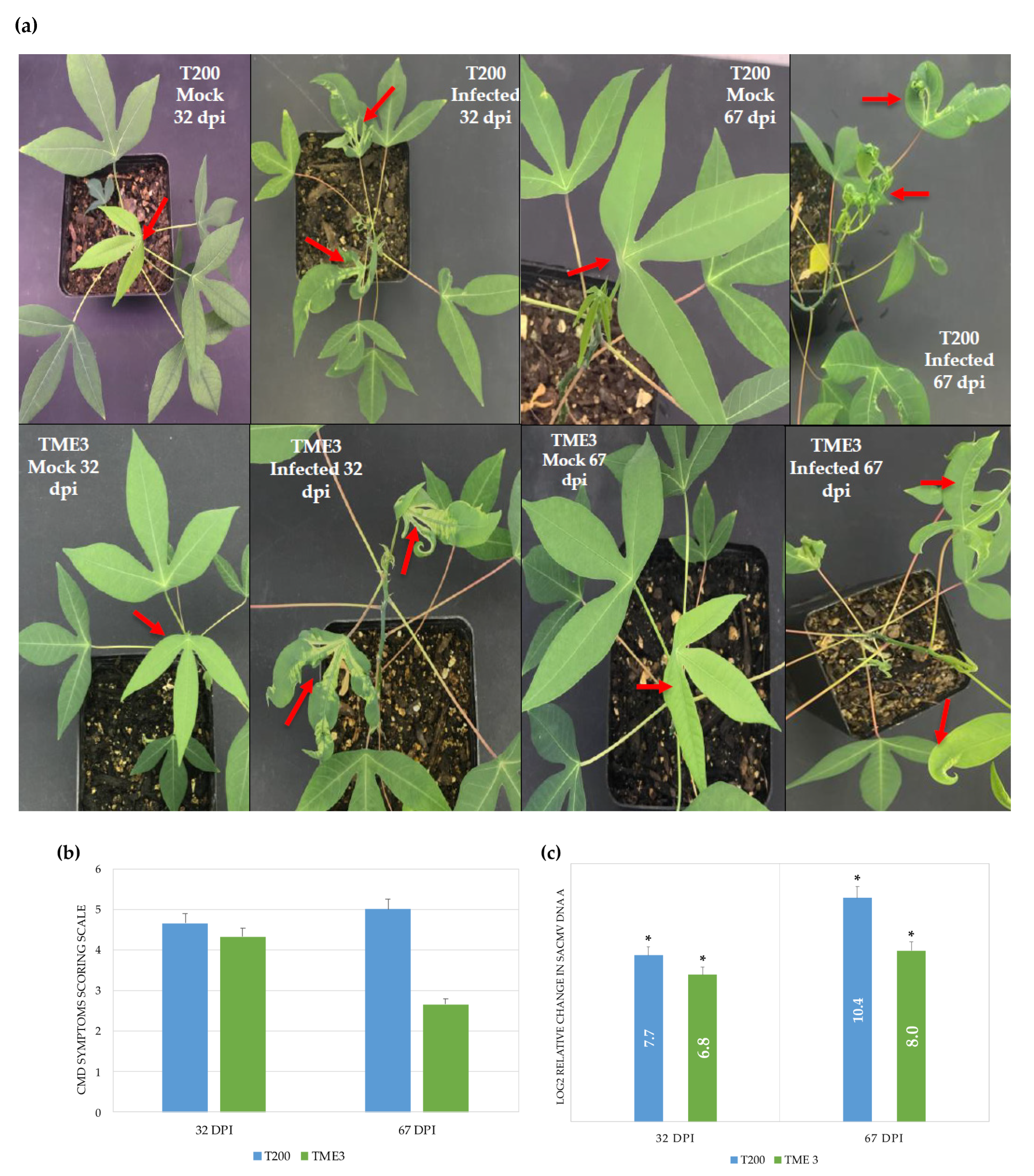
Figure 1. SACMV symptoms scores and viral load quantification in T200 and TME3 landraces at 32 and 67 dpi. (a) Characteristic CMD symptoms on T200 and TME3 compared with mock-inoculated plants. The white arrow points at CMD-free leaves, while the red arrow points at CMD-infected leaves. (b) CMD symptoms scoring on infected plants. (c) Relative change (log 2(ΔΔCT)) of SACMV DNA A to internal reference gene UBQ10 in infected plants. Error bars represent means ± S.D. (n = 3). Asterisks indicate statistically significance at p ≤ 0.05 using t-test. Symptoms Scoring Scale: 0 = No disease symptoms; 1 = Mild/faint mosaic with mild distortions at bases of most leaves; 2 = Pronounced mosaic pattern on most leaves, leaf malformation, 5% size reduction; 3 = Severe mosaic, distortion reduced size; 4 = Severe mosaic, severe distortion, up to 50% size reduction; 5 = Very severe mosaic, distortion, leaf twisting, 50–80% size reduction.
Proteomes 09 00041 g001
Figure 2. Heatmap of TME3 and T200 differentially expressed proteins at 32 and 67 dpi. 1 represents 32 dpi and 2 represents 67 dpi. ClustVis [44] was used to generate the heatmap. Rows are centred; unit variance scaling is applied to rows. Imputation is used for missing value estimation. Both rows and columns are clustered using Euclidean distance and average linkage.
Proteomes 09 00041 g002
Figure 3. Gene ontology analysis of over-represented differentially expressed proteins from TME3 and T200 at 67 dpi. (a) Functional grouping of TME3 proteins. (b) Functional grouping of T200 proteins. MF represents molecular function; BP, biological process; CC, cellular component.
Proteomes 09 00041 g003
Figure 4. STRING protein–protein interaction network of differentially expressed proteins with p-value < 1.0 × 10⁻¹⁶ in T200 and TME3 at 32 dpi (a) and 67 dpi (b). Network Settings: full network; confidence edge; textmining, experiments, databases, co-expression interaction sources; confidence of 0.8 and 0.9, respectively; more than 50 interactors 1st and 2nd shell; hide disconnected nodes in the network.
Proteomes 09 00041 g004
Figure 5. Validation of cassava gene expression levels by real-time RT-qPCR in TME3. Gold represents 32 dpi; Blue represents 67 dpi. Readings are averaged from three biological replicates normalized against endogenous gene UBQ10. Error bars represent means ± S.D. (n = 3). Asterisks indicate statistically significance at p ≤ 0.05 using t-test.
Proteomes 09 00041 g005
Table 1. Summary of differentially expressed proteins in SACMV-infected T200 and TME3 plants compared with mock-inoculated plants at 32 and 67 dpi.
TimepointRegulationProtein CountProtein CharacterisationGO Categories aFunctional Groups
32 dpiUnder-expressed30 T20014 characterised
35 uncharacterised
49 CC19
27 TME342 BP35
Over-expressed19 T20036 MF13
22 TME349 KEGG11
Total=49
67 dpiUnder-expressed63 T200128 characterised
78 uncharacterised
198 CC21
81 TME3178 BP44
Over-expressed143 T200198 MF23
125 TME3199 KEGG22
Total=206
a Categories based on cellular component, CC; biological process, BP; molecular function, MF; and KEGG analysis of the total number of differentially expressed proteins.
Table 2. KEGG analysis of differentially expressed proteins in SACMV-infected T200 and TME3 plants at 32 and 67 dpi.
TimepointPathway Protein CountFDR a
32 dpiMetabolic pathways132.30 × 10−4
Biosynthesis of secondary metabolites82.30 × 10−3
Carbon metabolism57.30 × 10−4
Arginine biosynthesis35.40 × 10−4
Biosynthesis of amino acids33.86 × 10−2
Carbon fixation in photosynthetic organisms23.64 × 10−2
67 dpiMetabolic pathways561.46 × 10−18
Biosynthesis of secondary metabolites362.88 × 10−13
Biosynthesis of amino acids141.04 × 10−7
Glycine, serine and threonine metabolism91.04 × 10−7
Carbon metabolism91.30 × 10−3
Arginine biosynthesis72.83 × 10−7
Ribosome73.26 × 10−2
Amino sugar and nucleotide sugar metabolism62.80 × 10−3
Alanine, aspartate and glutamate metabolism54.80 × 10−4
Carbon fixation in photosynthetic organisms51.60 × 10−3
2-Oxocarboxylic acid metabolism51.80 × 10−3
Glyoxylate and dicarboxylate metabolism51.90 × 10−3
Glycolysis/Gluconeogenesis59.10 × 10−3
Arginine and proline metabolism44.60 × 10−3
Pyruvate metabolism41.92 × 10−2
Nitrogen metabolism32.07 × 10−2
Ascorbate and aldarate metabolism32.32 × 10−2
Valine, leucine and isoleucine degradation32.32 × 10−2
Pentose phosphate pathway33.41 × 10−2
Proteasome33.41 × 10−2
Histidine metabolism23.26 × 10−2
Other glycan degradation23.41 × 10−2
a false discovery rate.
Table 3. Differentially expressed proteins between mock-inoculated and SACMV-infected leaves at 67 dpi. Proteins with at least a ratio of 2.0 in one landrace are presented here.
AccessionManes IDProtein NameUnique Peptidesq ValueT200 Ratio aTME3 Ratio a
tr|A0A2C9U824Manes.16G022000PKS_ER domain-containing protein3.002.83 × 10−42.38−1.15
tr|A0A2C9VEA2Manes.08G075000Uncharacterized protein4.007.56 × 10−42.37−2.04
tr|A0A2C9U4J9Manes.17G026300Lactoylglutathione lyase 2.002.59 × 10−3−1.352.82
tr|A0A2C9VIL5Manes.07G049300Proline iminopeptidase 7.003.91 × 10−32.261.05
tr|A0A0A1E5H6Manes.11G078100Phosphate transporter2.004.63 × 10−35.621.01
tr|A0A251JR98Manes.11G020100Germin-like protein6.005.29 × 10−3−3.26−1.03
tr|A0A2C9VJP2Manes.07G087900Glycine cleavage system H protein2.006.82 × 10−3−3.321.43
tr|A0A2C9WL85Manes.01G155900Uncharacterized protein2.006.82 × 10−32.46−2.07
tr|A0A2C9V1F6Manes.11G148200Uncharacterized protein3.007.50 × 10−3−2.795.63
tr|A0A2C9UCJ8Manes.15G029400Cysteine synthase10.007.85 × 10−32.941.25
tr|A0A2C9VK05Manes.07G093600GLOBIN domain-containing protein 2.001.11 × 10−21.143.05
tr|A0A2C9UGM3Manes.15G103900Peroxidase9.001.16 × 10−22.09−1.03
tr|A0A2C9UYJ6Manes.11G012700Aminotran_1_2 domain-containing protein10.001.16 × 10−21.012.01
tr|A0A2C9VAT8Manes.09G071100Uncharacterized protein2.001.22 × 10−22.231.27
tr|A0A2C9VSW1Manes.05G029200AB hydrolase-1 domain-containing protein 2.001.26 × 10−22.091.25
tr|A0A2C9WBE7Manes.02G064500PALP domain-containing protein3.001.30 × 10−23.271.14
tr|A0A2C9URN3Manes.13G132700Uncharacterized protein4.001.32 × 10−2−1.403.51
tr|A0A2C9VLP8Manes.06G006200Uncharacterized protein3.001.34 × 10−23.25−1.85
tr|A0A2C9UGX0Manes.15G172800Phosphoacetylglucosamine mutase9.001.49 × 10−22.46−1.12
tr|A0A2C9VP29Manes.06G081300Uncharacterized protein3.001.51 × 10−218.46−1.91
tr|A0A2C9UBX9Manes.15G008100Bet_v_1 domain-containing protein9.001.76 × 10−2−1.113.20
tr|A0A2C9V4E1Manes.10G042700Uncharacterized protein2.001.76 × 10−2−1.202.12
tr|A0A2C9WLB8Manes.01G085900Uncharacterized protein2.001.76 × 10−21.172.43
tr|A0A251K3C9Manes.09G021000HP domain-containing protein 18.001.81 × 10−22.18−1.71
tr|A0A2C9UTJ3Manes.12G039500Uncharacterized protein6.001.81 × 10−2−1.182.10
tr|A0A2C9W314Manes.04G086800SUMO-activating enzyme subunit2.001.81 × 10−2−5.154.65
tr|A0A251K980Manes.08G011100Uncharacterized protein3.001.81 × 10−211.82−2.66
tr|A0A251K915Manes.08G008000Uncharacterized protein4.001.81 × 10−21.82−2.13
tr|A0A2C9VGL2Manes.08G075100VWFA domain-containing protein11.002.00 × 10−23.13−1.29
tr|A0A2C9WEH9Manes.02G089200Uncharacterized protein6.002.07 × 10−22.411.99
tr|A0A2C9UID8Manes.15G186400Uncharacterized protein6.002.07 × 10−24.47−5.30
tr|A0A2C9UV72Manes.12G093300Glycosyltransferase2.002.24 × 10−29.06−1.92
tr|A0A2C9VFU9Manes.08G126700Protein kinase domain-containing protein2.002.66 × 10−223.94−1.43
tr|A0A2C9W2J9Manes.04G146100Uncharacterized protein7.002.66 × 10−22.21−1.44
tr|A0A2C9UPJ8Manes.13G061700FAS1 domain-containing protein10.002.66 × 10−2−1.45−2.01
tr|A0A2C9U6Y4Manes.17G080500Uncharacterized protein5.002.67 × 10−2−2.301.59
tr|A0A2C9U1Y7Manes.18G103000Uncharacterized protein3.002.68 × 10−23.18−1.22
tr|A0A2C9UQH1Manes.13G092100AB hydrolase-1 domain-containing protein18.002.70 × 10−21.302.76
tr|A0A2C9UYF6Manes.11G055100Phospholipase D34.002.73 × 10−22.05−1.17
tr|A0A251K1M8Manes.10G151400Uncharacterized protein2.002.73 × 10−2−3.90−1.14
tr|A0A2C9WAR9Manes.03G202900Uncharacterized protein9.002.73 × 10−22.281.45
tr|A0A2C9UXW4Manes.12G139100Ribosomal_L7Ae domain-containing protein3.002.73 × 10−2−5.242.18
tr|A0A2C9UYT0Manes.12G153700Uncharacterized protein9.002.79 × 10−22.401.20
tr|A0A2C9UNI9Manes.14G166500Non-specific lipid-transfer protein6.002.81 × 10−2−5.8810.33
tr|A0A2C9V3Z4Manes.10G027600ATPase_AAA_core domain-containing protein3.002.89 × 10−2−52.296.89
tr|A0A251JSV5Manes.11G063000Uncharacterized protein3.002.94 × 10−22.161.11
tr|A0A2C9WF62Manes.02G137700Alpha-mannosidase13.003.05 × 10−22.321.63
tr|A0A2C9W858Manes.03G096900M20_dimer domain-containing protein3.003.05 × 10−22.471.62
tr|A0A2C9VPI9Manes.06G102100Uncharacterized protein2.003.05 × 10−22.09−2.96
tr|A0A2C9VT96Manes.06G155500Glyco_hydro_18 domain-containing protein4.003.05 × 10−22.672.71
tr|A0A2C9VIN8Manes.07G051800Uncharacterized protein3.003.07 × 10−22.441.35
tr|A0A2C9UXQ1Manes.11G033300TauD domain-containing protein4.003.14 × 10−22.871.67
tr|A0A0C4ZQZ2Manes.12G135200Annexin13.003.14 × 10−22.781.41
tr|A0A2C9UCR5Manes.15G007900Bet_v_1 domain-containing protein2.003.14 × 10−2−1.083.69
tr|A0A2C9U3Q4Manes.17G010500RRM domain-containing protein4.003.14 × 10−22.41−1.45
tr|A0A2C9WMF8Manes.01G202400Uncharacterized protein3.003.21 × 10−26.53−1.13
tr|A0A2C9VRV6Manes.06G177500Uncharacterized protein2.003.26 × 10−23.261.27
tr|A0A2C9UUG2Manes.12G027700Uncharacterized protein14.003.32 × 10−22.281.56
tr|A0A2C9U185Manes.18G062000NAD(P)H-hydrate epimerase7.003.42 × 10−24.79−2.12
tr|A0A2C9V3P1Manes.10G055500WD_REPEATS_REGION domain-containing protein2.003.43 × 10−22.24−1.84
tr|A0A2C9VBZ8Manes.09G142200Uncharacterized protein2.003.59 × 10−2−1.6367.74
tr|A0A2C9UV10Manes.12G095700AB hydrolase-1 domain-containing protein3.003.59 × 10−21.422.21
tr|A0A2C9UJE4Manes.14G034500UMP-CMP kinase2.003.59 × 10−23.101.14
tr|A0A2C9WRD2Manes.01G245400Importin N-terminal domain-containing protein9.003.59 × 10−23.16−1.16
tr|A0A251KS16Manes.05G011800Uncharacterized protein12.003.61 × 10−22.05−1.21
tr|A0A2C9UGV6Manes.15G140500Eukaryotic translation initiation factor 3 subunit I10.003.61 × 10−22.10−1.40
tr|A0A2C9UA86Manes.16G0876003-ketoacyl-CoA synthase2.003.61 × 10−28.18−6.12
tr|A0A2C9UX42Manes.12G156900Hist_deacetyl domain-containing protein5.003.74 × 10−2−4.081.33
tr|A0A2C9W735Manes.03G081200FAD-binding PCMH-type domain-containing protein8.003.77 × 10−25.66−1.54
tr|A0A2C9VMD1Manes.07G122600AAI domain-containing protein4.003.77 × 10−2−10.20−1.40
tr|A0A2C9VAA7Manes.09G127700Bet_v_1 domain-containing protein7.003.77 × 10−21.322.22
tr|A0A2C9URF0Manes.13G126100Uncharacterized protein2.003.77 × 10−22.16−1.04
tr|A0A2C9U7U7Manes.16G007800Uncharacterized protein7.003.77 × 10−2−2.851.88
tr|A0A2C9W3J5Manes.04G095600eRF1_1 domain-containing protein4.003.77 × 10−22.26−1.02
tr|A0A199UC10Manes.s021400ZnMc domain-containing protein 2.003.77 × 10−23.571.09
tr|A0A2C9WH55Manes.01G012700Vac14_Fab1_bd domain-containing protein3.003.80 × 10−22.001.10
tr|A0A2C9U3Q3Manes.18G143800Uncharacterized protein3.003.80 × 10−2−5.261.18
tr|A0A2C9V4F7Manes.10G077300Uncharacterized protein2.003.80 × 10−22.461.12
tr|A0A251LVH3Manes.01G267300Uncharacterized protein3.003.87 × 10−21.542.11
tr|A0A2C9UJT8Manes.14G080800Peptidyl-prolyl cis-trans isomerase2.003.90 × 10−2−3.454.03
tr|A0A2C9V4B1Manes.10G077100Lipase_3 domain-containing protein5.003.92 × 10−22.03−1.24
tr|A0A2C9VG07Manes.08G081300Uncharacterized protein2.003.93 × 10−2−5.026.17
tr|A0A2C9WEB2Manes.02G155900Uncharacterized protein4.003.93 × 10−21.174.80
tr|A0A2C9V5Y8Manes.10G128500Uncharacterized protein5.003.93 × 10−24.19−1.96
tr|A0A2C9WJN9Manes.01G112700AAI domain-containing protein 3.004.06 × 10−2−2.736.45
tr|A0A2C9VN74Manes.06G056000Uncharacterized protein2.004.06 × 10−23.26−1.66
tr|A0A2C9WNU1Manes.01G247800Glycosyltransferase2.004.06 × 10−22.171.39
tr|A0A2C9VYT5Manes.05G155000AAA domain-containing protein9.004.06 × 10−22.82−1.07
tr|A0A2C9W393Manes.04G165200Uncharacterized protein4.004.06 × 10−24.42−1.42
tr|A0A2C9VKB0Manes.07G107200Chalcone-flavonone isomerase family protein10.004.06 × 10−22.891.20
tr|A0A2C9UBT3Manes.15G008400Bet_v_1 domain-containing protein 7.004.06 × 10−22.8513.10
tr|A0A2C9V9W8Manes.09G113900Uncharacterized protein4.004.06 × 10−22.311.48
tr|A0A2C9WD78Manes.02G122500Alpha-1,4 glucan phosphorylase 5.004.06 × 10−22.324.83
tr|A0A2C9WCC9Manes.02G095600CTP_transf_like domain-containing protein3.004.18 × 10−21.162.35
tr|A0A2C9UNS6Manes.13G043200AAA domain-containing protein9.004.22 × 10−24.75−1.32
tr|A0A2C9U6K5Manes.17G064800Uncharacterized protein2.004.23 × 10−2−4.611.83
tr|A0A2C9VE34Manes.08G069100Uncharacterized protein15.004.30 × 10−22.061.34
tr|A0A2C9V5M6Manes.10G121900 Uncharacterized protein13.004.30 × 10−22.52−1.27
tr|A0A2C9U8P4Manes.17G111900Uncharacterized protein3.004.30 × 10−22.331.50
tr|A0A2C9USP9Manes.12G024300Uncharacterized protein2.004.30 × 10−22.64−1.54
tr|A0A2C9V2I1Manes.10G019200Uncharacterized protein2.004.34 × 10−23.30−1.16
tr|A0A2C9VWB9Manes.05G146600Peptidyl-prolyl cis-trans isomeras 3.004.34 × 10−2−1.522.06
tr|A0A2C9UGS7Manes.15G108300Uncharacterized protein6.004.45 × 10−23.10−1.11
tr|A0A2C9VJ85Manes.07G01710060S ribosomal protein L132.004.45 × 10−2−3.862.11
tr|A0A2C9UTD1Manes.12G033700Dolichyl-diphosphooligosaccharide--protein glycosyltransferase subunit 2 4.004.49 × 10−25.39−1.86
tr|A0A2C9WHE0Manes.01G032500PCI domain-containing protein 2.004.52 × 10−27.14−1.40
tr|A0A2C9UFY6Manes.15G139800Transmembrane 9 superfamily member 4.004.55 × 10−24.51−2.11
tr|A0A2C9VXS5Manes.05G194900 Protein kinase domain-containing protein2.004.55 × 10−2−8.12−2.18
tr|A0A2C9VQH2Manes.06G055700Uncharacterized protein7.004.57 × 10−22.10−2.39
tr|A0A2C9VYM5Manes.05G142300Methyltransferase 2.004.62 × 10−23.32−6.67
tr|A0A2C9TZI5Manes.18G001100Pyrophosphate--fructose 6-phosphate 1-phosphotransferase subunit alpha3.004.62 × 10−22.131.34
tr|A0A2C9V233Manes.11G128200Uncharacterized protein3.004.65 × 10−2−6.769.16
tr|A0A2C9V6N1Manes.09G008000F420_oxidored domain-containing protein4.004.68 × 10−21.082.91
tr|A0A251JLF7Manes.12G028400Carbonic anhydrase 2.004.68 × 10−22.95−1.11
tr|A0A2C9W4R1Manes.03G053100Uncharacterized protein5.004.68 × 10−2−5.501.68
tr|A0A2C9V313Manes.10G032500NAD(P)-bd_dom domain-containing protein 3.004.68 × 10−23.111.28
tr|A0A2C9VBY7Manes.09G183600Uncharacterized protein3.004.68 × 10−23.66−1.04
tr|A0A2C9UMZ5Manes.13G011400AAI domain-containing protein 5.004.68 × 10−2−9.136.96
tr|A0A2C9VRT4Manes.06G097000Plasma membrane ATPase 6.004.69 × 10−25.75−1.45
tr|A0A2C9WA59Manes.03G162900Ribosomal_L14e domain-containing protein 2.004.69 × 10−24.36−1.86
tr|A0A2C9VYM8Manes.04G011900Carboxypeptidase 3.004.69 × 10−2−1.363.72
tr|A0A251K5T0Manes.09G086600Uncharacterized protein6.004.69 × 10−23.00−1.22
tr|A0A2C9V462Manes.10G078000Glycosyltransferase 5.004.71 × 10−23.12−1.63
tr|A0A2C9VLD9Manes.07G091600Uncharacterized protein4.004.81 × 10−24.30−2.64
tr|A0A2C9W8C0Manes.03G123300Trafficking protein particle complex subunit 3.004.81 × 10−22.54−1.12
tr|A0A2C9W8D3Manes.03G089200Uncharacterized protein2.004.87 × 10−2−14.912.30
tr|A0A199U962Manes.S101100Lipase_GDSL domain-containing protein3.004.87 × 10−2−2.513.28
tr|A0A2C9UL26Manes.14G124000Protein kinase domain-containing protein 3.004.89E × 10−2−1.70−2.19
tr|A0A2C9U8J5Manes.16G001400Abhydrolase_2 domain-containing protein 2.004.89 × 10−2−1.418.09
tr|A0A2C9VH42Manes.08G171700Non-specific lipid-transfer protein 3.004.97 × 10−2−13.046.81
tr|A0A2C9VWH1Manes.05G151600Uncharacterized protein10.004.97 × 10−23.19−3.49
a Negative ratio = under-expression, positive ratio = over-expression.
Publisher’s Note: MDPI stays neutral with regard to jurisdictional claims in published maps and institutional affiliations.