Article

Modification of Polycaprolactone with TiO2 and ZnO Nanoparticles for Biomedical Applications

by
Polina A. Fomina
1,
Grigorii A. Oloviannikov
1,
Dmitriy A. Serov
1,
Lev R. Sizov
1,
Ilya V. Baimler
1,
Pavel P. Chapala
2,3,4,
Sergey V. Gudkov
1 and
Valeriy A. Kozlov
1,5,*
1
Prokhorov General Physics Institute of the Russian Academy of Sciences, 38 Vavilova St., 119991 Moscow, Russia
2
HARZ Labs LLC, Silikatnaya Str. 51A, bld.5, 141013 Mytischi, Russia
3
Department of Chemical Technology of Polymeric Composite Paints and Coatings, D.I. Mendeleev University of Chemical Technology of Russia, 9 Miusskaya Sq., 125047 Moscow, Russia
4
A.V. Topchiev Institute of Petrochemical Synthesis RAS, 29 Leninskiy Pr., 119991 Moscow, Russia
5
Department of Fundamental Sciences, Bauman Moscow State Technical University, 5 2nd Baumanskaya St., 105005 Moscow, Russia
*
Author to whom correspondence should be addressed.
Technologies 2026, 14(9), 587; https://doi.org/10.3390/technologies14090587
Submission received: 5 August 2026 / Revised: 5 September 2026 / Accepted: 15 September 2026 / Published: 17 September 2026
(This article belongs to the Section Innovations in Materials Science and Materials Processing)

Abstract

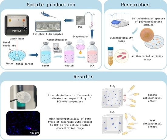
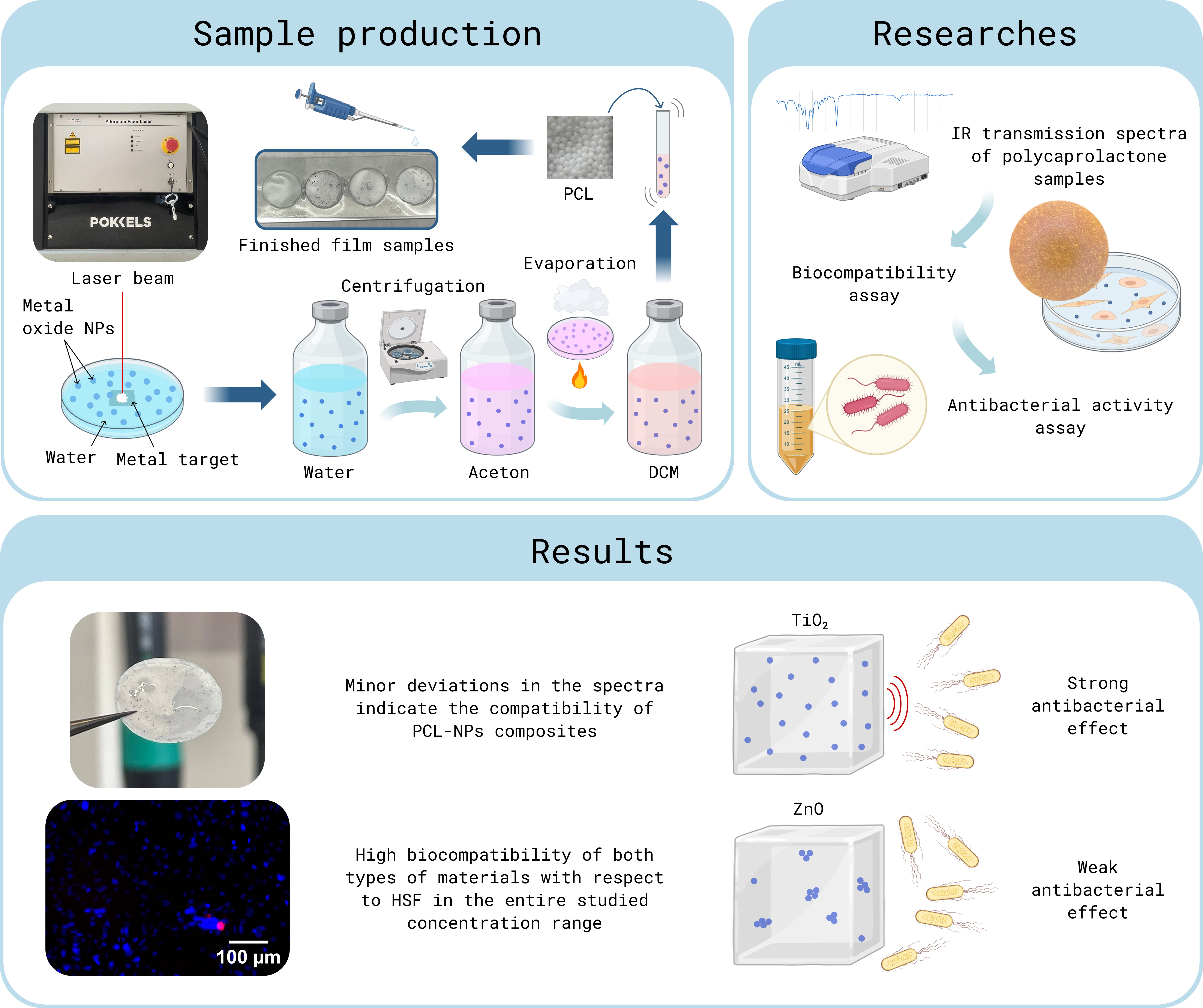
Polycaprolactone (PCL) is a promising polymer in the medical field and is already being used in the creation of suture materials and tissue engineering. Due to the limited properties of PCL, in many medical tasks it is necessary to modify it, for example, the addition of antimicrobial properties to reduce the risk of infection in tissue repair processes. In this work, the effect of the introduction of nanoparticles of titanium oxide (NPs-TiO2) and zinc (NPs-ZnO) into the polycaprolactone matrix on its antibacterial activity and biocompatibility was studied. The studied concentrations of NPs ranged from 0.001 to 0.5 wt. %, and the final composites were obtained by casting from a solution. An investigation into the antibacterial activity against the Gram-negative bacterium Escherichia coli revealed high efficacy of the PCL/NPs-TiO2 composites, which increased with rising nanoparticle concentration to 27.6% at a nanoparticle content of 0.5 wt. %. PCL/NPs-ZnO composites did not show such activity. The results obtained demonstrate that the key factor determining the biological activity of a composite is not only the nature of the nanoparticles but also their ability to maintain a nanoscale state during the manufacture of the final composite. PCL/NPs-TiO2 composites may be a promising candidate for the creation of antibacterial materials for medical purposes, for absorbable scaffolds or sutures.

Graphical Abstract

1. Introduction

Since the beginning of the 21st century, there has been a steady trend in the field of materials science, ecology, and biomedicine aimed at developing composite materials with improved physicochemical and biological properties [1]. In this context, polycaprolactone attracts special attention from researchers. Among the biodegradable polymers used in modern regenerative medicine, polycaprolactone (PCL) stands out for its high biocompatibility and non-toxicity to mammalian cells [2] and has the lowest rate of hydrolytic and enzymatic degradation compared to other popular polymers such as polylactic acid (PLA) and poly(lactic-co-glycolic acid) (PLGA) [3,4]. A longer decomposition time allows the use of PCL in tasks requiring long-term mechanical integrity of the implant, for example, when replacing bone or cartilage tissue. PCL is also convenient for industrial production, as it allows the fabrication of final composites of various shapes and sizes from nanofibers to complex 3D-printed frames using electrospinning, casting from solutions, electrospray and other methods.
However, the expanded clinical use of PCL is constrained by the limited properties of the material: the hydrophobic nature of its surface limits cell adhesion and proliferation, and the complete absence of its own antimicrobial activity poses a serious risk of infection during implantation and healing [5,6]. In recent years, a wide range of strategies have been proposed to give polycaprolactone bactericidal properties. One such approach is to create composite materials with biopolymers such as chitosan. The combination of biocompatible and biodegradable PCL and chitosan, which has antibacterial activity, makes it possible to obtain a polymer framework with new biological properties [7]. Antibacterial properties can also be added by adding plant extracts with a bactericidal effect, such as hop extract [8]. However, one of the most popular approaches is the volumetric modification of PCL by introducing inorganic nanoparticles into its polymer matrix.
Among the inorganic compounds studied so far, nanoparticles of metals and their oxides, which can exhibit bactericidal activity even in the absence of external influences, are of the greatest practical interest. Their mechanism of action, unlike antibiotics, is not tied to a single cellular target and includes a complex set of factors: the generation of reactive oxygen species (ROS), direct physical interaction with the bacterial cell wall and, in the case of some oxides, the release of metal ions [9,10]. This complex mechanism reduces the likelihood of developing resistance in pathogens. For example, the paper [11] demonstrates the high antibacterial efficacy of PCL/NPs-TiO2 composites, which is dose-dependent. Muñoz-Bonilla et al. demonstrated that PCL/TiO2 nanocomposites with a TiO2 content of 0.5–5 wt.% exhibit antimicrobial activity against both E. coli and S. aureus, with the system’s efficacy being linked to the dispersion and physicochemical state of the inorganic phase. A similar approach has been widely developed for PCL/ZnO materials. Subsequently, Seo et al. investigated PCL membranes containing 0.5 and 5 wt. % ZnO for guided periodontal tissue regeneration and demonstrated that both concentrations significantly reduced the adhesion of S. mutans and P. gingivalis without adversely affecting osteoblast proliferation [12]. ZnO-modified electrospun PCL scaffolds have also been investigated for use in wound dressings and tissue engineering. The incorporation of ZnO nanowires into electrospun PCL scaffolds reduces bacterial colonization by half while preserving the metabolic activity of mammalian cells [13]. The body of published data shows that the biological properties of PCL composites with inorganic nanoparticles are determined not only by the chemical nature and nominal concentration of the filler, but also by its morphology, degree of dispersion and other factors. However, the influence of the nanoparticle transfer procedure and the change of solvent prior to their incorporation into PCL has been studied in considerably less detail. This aspect may be of particular significance for nanoparticles synthesized in aqueous media (for example, metal oxide nanoparticles obtained by laser ablation). The transfer of nanoparticles from water into hydrophobic solvents used to dissolve the polymer can alter the colloidal stability of the particles and cause them to aggregate, thereby changing the effective surface area of the nanoparticles in the final composite [14,15]. The use of acetone as an intermediate solvent in the transfer of nanoparticles from water to dichloromethane makes it possible to reduce interfacial effects and make the medium transition smoother [16].
In this work, we synthesized TiO2 and ZnO nanoparticles by laser ablation in water, transferred them to dichloromethane through an intermediate solvent (acetone), and then introduced them into a PCL solution in DCM. Film samples of composites containing 0.001 to 0.5 wt. % of each of the types of NPs were obtained by casting into molds. The key objective of the study was to determine to what extent the change in the content of nanoparticles in the polymer solution correlates with the antibacterial activity and cytotoxicity of the final composites. In addition, the goal was to assess the differences in the impact of various NPs on the biological properties of the polymer composite and to determine what might be driving these differences. To assess cytotoxicity, a culture of normal human fibroblasts of the HSF line was used, and the antibacterial properties were studied on a reference Gram-negative strain of Escherichia coli.

2. Materials and Methods

2.1. Synthesis of Nanoparticles

ZnO and TiO2 nanoparticles were synthesized by laser ablation in deionized water (MQ). An installation based on a pulsed fiber ytterbium laser P-Mark TT 100 (Pokkels, Moscow, Russia) and a scanning system was used. The following laser radiation parameters were set: λ = 1064 nm, τ = 200 ns; v = 30 kHz; average power up to 60 W; E = 1.49 mJ. The beam was scanned over the target surface using an LScanH galvanomechanical scanner (Ateko-TM, Moscow, Russia) and an F-Theta lens with a focal length of 90 mm. The beam waist diameter was approximately 100 μm, corresponding to a laser fluence of about 19 J/cm2 on the target surface. The scanning trajectory consisted of several parallel lines within a 1 × 1 cm2 square area. The scanning speed was 3000 mm/s. The typical ablation time was approximately 30 min. The metal target was filled with MQ so that there was a layer of water 2–3 mm above it. The laser ablation duration was 45 min for a liquid volume of 100 mL.

2.2. Characterization of Nanoparticles

The obtained colloidal solutions of NPs in water were characterized using UV–visible spectroscopy and the dynamic light scattering (DLS) method. The colloidal solutions underwent preliminary treatment in an ultrasonic bath for 5 min (40 kHz, 22 °C) and were mixed during the pipetting process. The zeta potential was measured for all NP dispersions in water, size distributions were constructed, and the NP content in the dispersions was estimated using a Zetasizer ULTRA Red Label device (Malvern Panalytical Ltd., Worcestershire, Malvern, UK). The absorption spectra of radiation in the UV–visible region for NP colloids in water were recorded on a Persee T6U UV–visible region spectrometer (PG Instrument, Beijing, China).
The size of the resulting nanoparticles was also assessed using TEM. TEM images were obtained using a Libra 200 FE HR transmission electron microscope (Carl Zeiss, Jena, Germany), and particle morphology was analyzed from the acquired TEM images. For TEM measurements, samples were deposited on gold and copper TEM grids. TEM-based particle size distributions were obtained by measuring the diameters of spherical NPs and the lengths of NRs (nanorods) from TEM-images using ImageJ software (version 1.54f). At least 50 particles/nanorods were analyzed for each sample.

2.3. Transfer of Nanoparticles from Water to Dichloromethane

Due to the weak miscibility of dichloromethane with water, nanoparticles were transferred from water to dichloromethane (DCM) (Lenreactive, Saint Petersburg, Russia) through an intermediate medium, acetone. The transfer of NPs from water to acetone was carried out by centrifugation on a 3-16KL centrifuge (Sigma, Darmstadt, Germany). First, 80 mL of colloidal NP solution in water was dispersed for 3–5 min in an ultrasonic bath (40 kHz, 22 °C) and stirred for 3–5 min on a Vortex V-1 plus vortex mixer (Biosan, Riga, Latvia). Subsequently, the water dispersion underwent the first centrifugation cycle for 40 min at 22 °C at 7012× g. After the first centrifugation, the solvent was changed from water to acetone, and all the previous steps were repeated in order again.
The transfer of nanoparticles from acetone to DCM was carried out by evaporation of a solvent. The NP dispersion in acetone was poured into a glass Petri dish and heated on a tile at a boiling point of acetone (56 °C). Acetone evaporation was carried out to a residue of 5 mL in a Petri dish, after which 40 mL of DCM was poured into it, and the heating temperature of the tile dropped to the boiling point of DCM (40 °C). The solvent was evaporated again to a residue of 5 mL, after which 35 mL of DCM was added to it, and the entire resulting dispersion of NPs in DCM was poured into a vial with a lid. The final concentration of NPs in DCM is twice as high as the initial concentration of NPs in water.

2.4. Preparation of Solutions of Polycaprolactone and PCL Composites with NPs

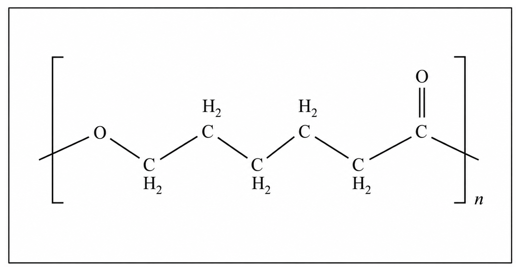
A literature review has shown that researchers most often prepare a solution of polycaprolactone with its content in the range of 10–15% (Table 1). Therefore, we chose the average value of 12.5%. This concentration of PCL in DCM ensures the density of the solution without losing its fluidity, which is most suitable for producing samples by casting. The initial polycaprolactone is a set of granules about 2 mm in size. The PCL polymer’s chemical structure is given in Figure 1. On an analytical balance, 3.75 g PCL was measured in granules and transferred to a glass cylinder, into which dichloromethane was then added until a total solution volume of 30 mL was reached. The solution was thoroughly mixed using a vortex mixer until uniformity was achieved and poured into a glass vial with a lid.
To study the effect of nanoparticles on the biological properties of composites, 4 concentrations of NPs in composite solutions were selected: 0.001, 0.01, 0.1, and 0.5 wt. %. Briefly, 1.25 g PCL in granules was measured on analytical scales and transferred to a glass tube, into which the required volume of NP dispersion in DCM was added. DCM was added to the resulting mixture to 10 mL of the total volume of the solution, after which the entire liquid composite was thoroughly mixed using a vortex mixer until uniformity was achieved. The volume of NP dispersion in DCM required to obtain each specific concentration was calculated based on the concentration of each type of NPs in DCM (ZnO—0.414 mL; TiO2—0.4 mL). The finished liquid composites were also poured into glass vials.

2.5. Preparation of Film Samples

The film samples were obtained by casting into molds. Round silicone molds with a diameter of 15.8 mm and rectangular silicone molds with a size of 35 mm × 5 mm were placed on the slides. All surfaces were carefully treated with isopropyl alcohol (HIMMED, Moscow, Russia). Liquid composites were poured into ready-made molds and placed in a fume hood for 2–3 h until the DCM completely evaporated. At the end of this time, solid translucent white films 1–2 mm thick were removed from the molds, treated with isopropyl alcohol and exposed to a 30 W UV lamp.

2.6. Characterization of the Obtained PCL and PCL/NPs Composites

The degree of crystallinity of the final PCL composites and the effect of the introduced nanoparticles on the polymer matrix were evaluated by taking IR spectra of the attenuation total reflectance (ATR). PCL disks with a diameter of 15.8 mm were used as samples. The IR spectra of the ATR were recorded on an IR-8000 IR Fourier spectrometer (Siberian Analytical Systems LLC, Krasnoyarsk, Russia).

2.7. Biocompatibility Assay

The cytotoxicity of the composites was assessed by fluorescence microscopy. Human spleen fibroblast cells (HSFs) (the cells were kindly provided by the Collection of Human Cell Cultures for Biomedical Purposes at VILAR) were cultured in TC-treated T-25 culture vials in DMEM/F12 medium supplemented with 10% (v/v) fetal veal serum, 250 U/mL penicillin, 250 µg/mL streptomycin and 2 mM L-glutamine (all PanEco, Moscow, Russia) at 37 °C with 5% CO2. Subcultivation of cells was performed according to standard protocols using a mixture of 0.25% trypsin and EDTA 0.2 g/L EDTA (PanEco, Moscow, Russia). Cells of 5–10 passages were used in the experiments. The finished round samples of materials were thoroughly washed with isopropyl alcohol and phosphate-buffered saline solution (PBS) (SigmaAldrich, Burlington, MA, USA), after which they were treated with ultraviolet light for 30 min. HSFs (105 cells in 100 µL of culture medium) were planted on round cover glasses with a diameter of 24 mm (MiniMed, Moscow, Russia) placed on the bottom of the cells of a 6-well culture plate, and sterile composites were placed next to the glasses in the wells, after which the tablet was placed in a CO2 incubator for three days. Cells cultivated on a cover glass without a sample were used as a control. Cells were stained with Hoechst 33342 and propidium iodide (PI) (all Lumiprobe, Hunt Valley, MD, USA) to assess viability. Cells on cover glass were stained with 5 µg/mL Hoechst 33342 for 30 min at 37 °C after incubation with samples. After that, the cells were washed with PBS again and stained with 2 µM of PI for 1 min. Data collection was performed on an NSZ818m fluorescence microscope (Ningbo Yongxin Optics Co., Ltd., Ningbo, China) equipped with a Micrus 20C cooled camera (Helicon Micrus, Moscow, Russia). The analysis of the obtained images was carried out using the ImageJ (Fiji) program (National Institutes of Health University of Wisconsin, Bethesda, WI, USA). The presented survival rate was calculated as the average percentage for 5–6 cell samples. About 500–1000 cells were analyzed in each independent sample. Analysis of the obtained micrographs and subsequent quantitative calculation showed that, for all the composites studied, without exception, the proportion of dead cells did not exceed the values characteristic of the negative control (cell culture without a polymer sample). Cell viabilities were calculated by Formulas (1) and (2):
D e a d   c e l l s = P I   p o s i t i v e   c e l l s H o e c h s t   p o s i t i v e   c e l l s × 100 %
V i a b i l i t y = 100 % D e a d   c e l l s

2.8. Antibacterial Activity Assay

Antibacterial activity was studied against Gram-negative E. coli bacteria at an initial concentration of 104 CFU/mL under conditions of periodic cultivation of microorganisms. Rectangular samples were placed along the cylindrical walls of the culture plate, after which 1 mL of LB broth (Dia-m, Moscow, Russia) containing cells was added to them. Pure broth was used as a form, and cells with bacteria without samples were used as a control. To assess the antibacterial activity, the optical density (OD600) of the samples was used during 36 h of incubation (the end point of the growth curve) at a temperature of 37 °C and constant stirring at 200 rpm. The optical density was measured every 30 min during the entire incubation time at a wavelength of 600 nm using a tablet reader, Fancy-A400 (Allsheng, Hangzhou, China). After 36 h of incubation, the optical density (OD600) values for each experimental series were statistically processed: at least ten independent measurements were performed for each type of sample, after which the average value was calculated, which was used to plot the growth curve characterizing this sample. All the processed growth curves were summarized on one common graph, and boxplots containing information about the endpoints of the growth curves for all measurements performed for each of the samples were constructed separately. A bacterial culture incubated without a composite sample was used as a negative control.

2.9. Statistical Processing

The experimental results were analyzed using nonparametric statistics. Data are presented as median values, minima and maxima, 25th and 75th percentiles. The statistical significance of differences was assessed using the Mann–Whitney test. Sample sized are indicated in the corresponding figures’ captions.

3. Results

3.1. Characterization of Nanoparticles

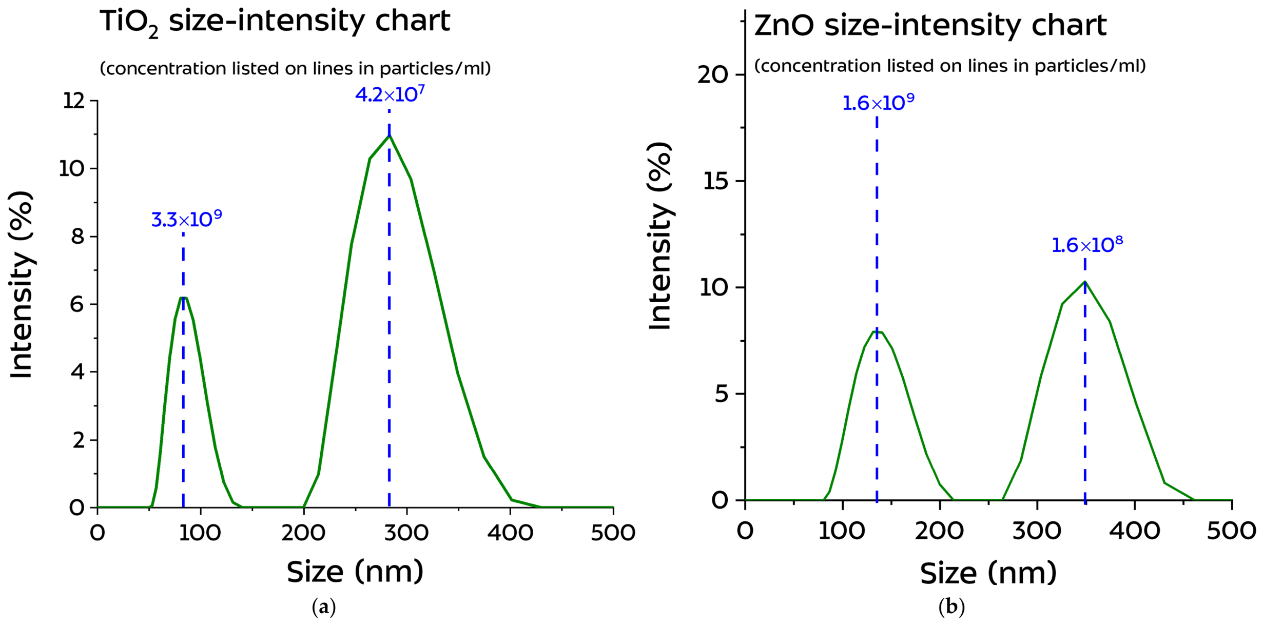
The size distribution graphs of NPs are presented in Figure 2. The obtained results show that NPs in water undergo significant aggregation and have a large spread in their diameters.
Table 2 shows the results of measuring the ζ-potential of aqueous dispersions of nanoparticles, as well as their size range (mean value and dispersion) and concentration in pcs/mL in water. The modulus of the ζ-potential allows us to estimate the stability of a colloidal solution: |ζ| > 25–30 mV is considered characteristic of stable systems [30,31].
Based on the data in Figure 2 and Table 2, both colloidal solutions are metastable. This is also reflected in the size of NPs obtained by the DLS method: nanoparticles of both oxides strongly aggregate in water, while NPs-ZnO agglomerate into larger particles. In view of this, the measurement of the size of an individual nanoparticle using DLS may be inaccurate and may need to be verified by another method, such as TEM.
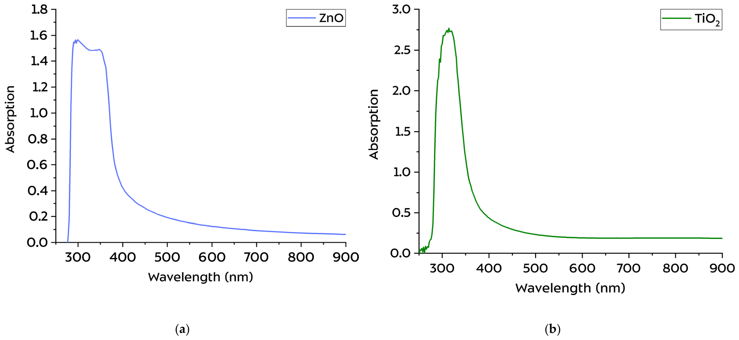
The absorption spectra of radiation in the UV–visible region for dispersions in water are shown in Figure 3. The spectra of nanoparticles based on zinc and titanium make it possible to observe the presence of semiconductor compounds (corresponding oxides) by the characteristic absorption spectra in the UV–visible violet region [32,33].
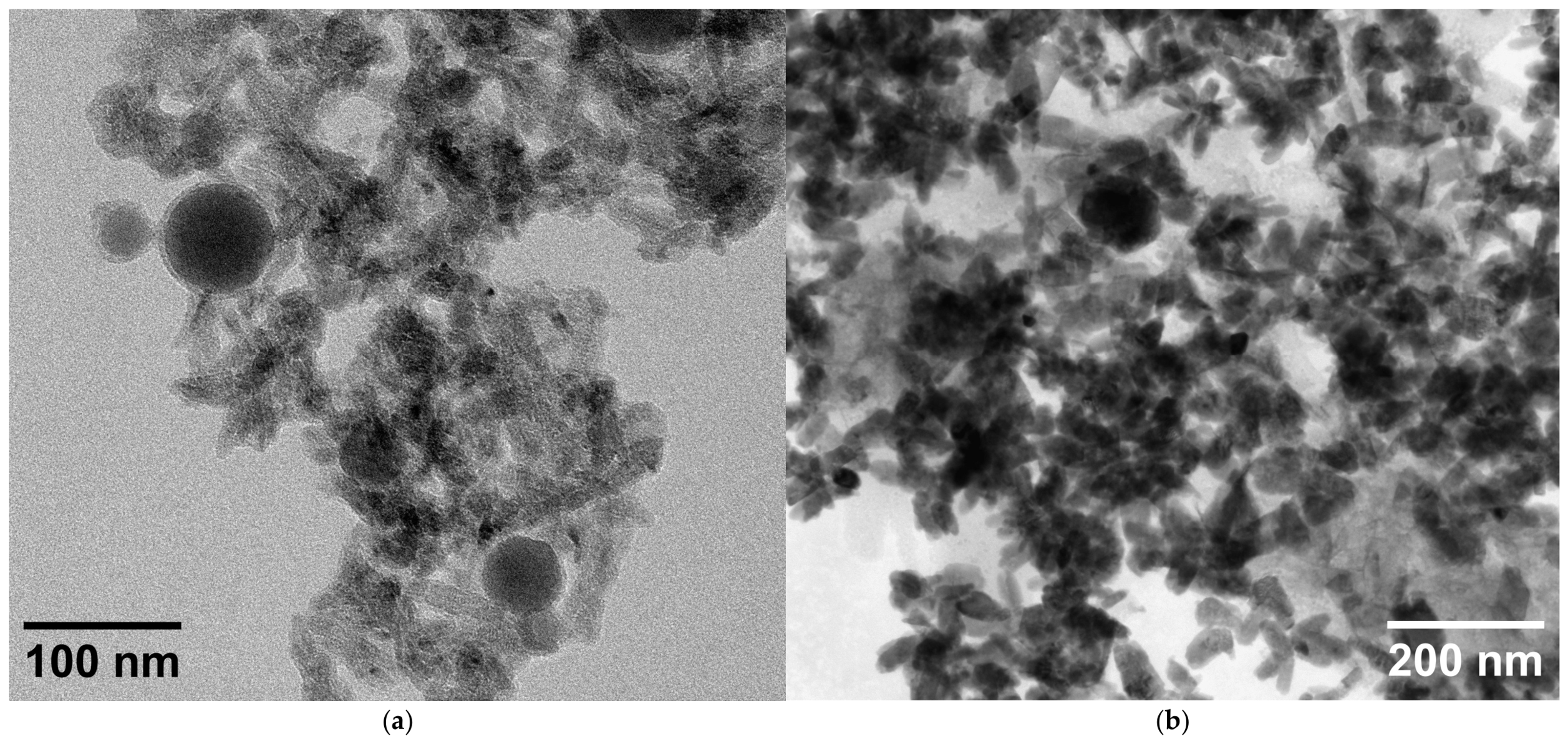
The images of NPs-ZnO and NPs-TiO2 obtained using TEM are shown in Figure 4. Visually, in both cases, NPs were observed predominantly in an aggregated form. Generally, NPs have an elongated rod-like shape, but particularly large NPs are spherical-shaped.
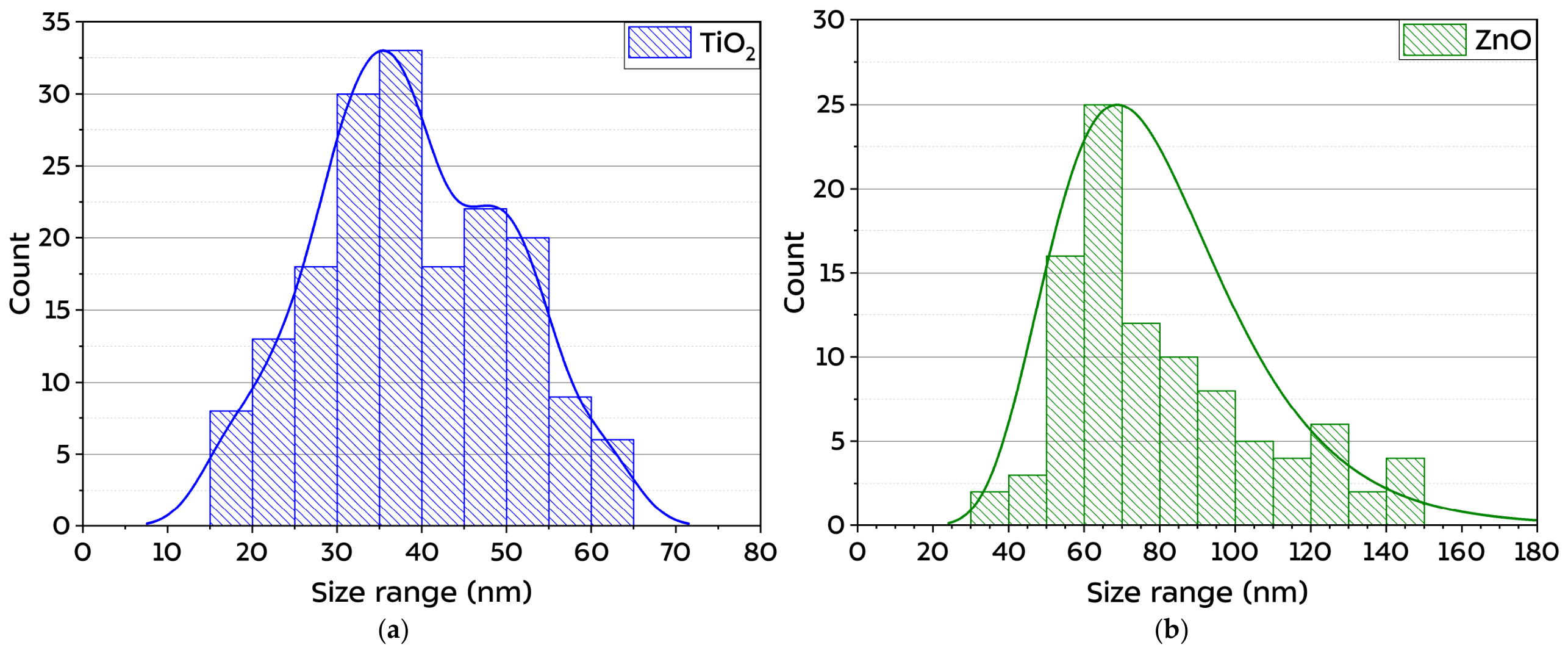
An analysis of the NPs’ size was carried out using the ImageJ program. As a result of image processing, the NPs’ size distributions were obtained, as shown in Figure 5. It is worth noting that the average nanoparticle sizes obtained using this method are half of those obtained by DLS: the average size of NPs-TiO2 is around 35–40 nm, while that of NPs-ZnO is approximately 70 nm. Other researchers have also encountered a similar variation in their work; however, this can be explained by various factors [34]. The paper [35] examines in detail the reasons for the differences in nanoparticle sizes obtained by TEM and DLS methods, such as non-spherical shape and high NP concentration. Thus, differences in the sizes of the same NPs measured by different methods can be explained by the rod-like shape of the NPs, which is not typical for the DLS method, as well as by their high concentration, leading to the formation of larger agglomerates. The difference is also explained by the operating principles of the methods: TEM measures the physical/projected dimensions of particles in a dried sample, while DLS determines the hydrodynamic size of particles and/or their aggregates in a suspension.

3.2. Characterization of the Obtained PCL and PCL/NP Composites

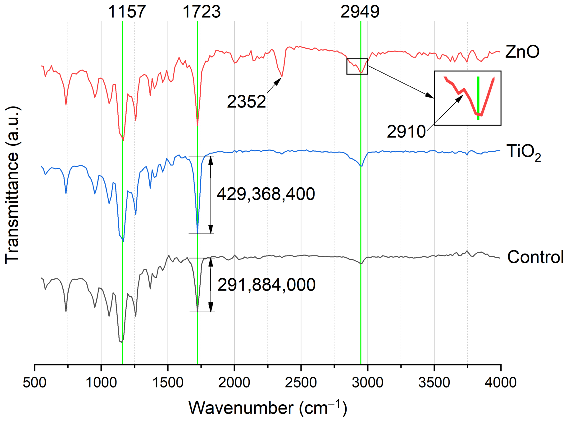
The IR transmission spectra for PCL samples with metal oxide NPs in the range of 400–4000 cm−1 are shown in Figure 6. The characteristic peaks of PCL are shown in Table 3. The characteristic peaks and their changes (the area under the peaks or intensity) allow us to judge the effect of the introduced NPs on the polymer matrix itself. Changes in peak values in the range of 1700–1750 cm−1 (C=O) are the most informative for the analysis of PCL crystallinity. Here we consider the ratio of the areas under the peaks in the region of 1725 cm−1 and 1735 cm−1, representing the crystalline and amorphous phases, respectively. This makes it possible to estimate the degree of crystallinity of the obtained PCL-based material.
Dips in the range of 2800–3000 cm−1 (CH2 wavenumber) reflect structural conformational changes in molecules (trans and gauche isomers). A decrease in the transmission intensity in 2945–2949 cm−1 indicates an increase in the crystallinity of the substance, which is also confirmed by the formation of an inflection in the graph near the value of 2900 cm−1 (Table 3, Figure 6). The absence of a shift in the characteristic dip of the PCL carbonyl group (1723 cm−1) towards the amorphous phase or its expansion confirms the absence of chemical destruction of the polymer base after the introduction of NP metal oxides. The graphs of the spectra also show a decrease in the transmission intensity in the region of this dip for samples impregnated with NPs, which makes it possible to suggest a positive effect of NPs on the formation of the crystalline structure of the composite during polymerization (formation of new crystallization centers) [36,37]. The presence of an amorphous phase in all composites is confirmed by dips in the 1157 cm−1 band, which allows us to conclude that the amorphous–crystalline structure of the composite is mixed.
The absence of characteristic dips/peaks in transmission intensity for TiO2 (600 cm−1 and 1125 cm−1) and ZnO (3400 cm−1) due to the formation of bonds in the PCL matrix indicates a too low concentration of NPs in the sample, which does not allow the detection of NPs in composites using FTIR. The occurrence of a dip in the 2352 cm−1 region for the PCL/NPs-ZnO composite is characteristic of the antisymmetric valence vibration of a linear CO2 molecule (O=C=O) and can be associated with both the adsorption of NPs-ZnO carbon dioxide from the atmosphere and its retention in the pores of the material during polymerization [38].
In general, the analysis of the obtained spectra revealed minor changes in the modified PCL samples, which are due to the peculiarities of the polymerization process of composites. Minor deviations in the spectra of the obtained composites indicate the compatibility of the components and the absence of visible obstacles to the production and use of PCL-NP composites of titanium and zinc oxides.
Table 3. Characteristic PCL peaks in the IR absorption spectra [39,40].
No.Wavenumber (cm−1)Oscillation (Type)Effect of Changes in Peak Area/Intensity on Physicochemical Properties of PCL
12945–2949νas(CH2)
(methylene chain groups)
The decrease in intensity corresponds to an increase in the crystallinity of the material and the ordering of polymer chains
22865–2866νs(CH2)A decrease in intensity in the amorphous phase, the appearance of an inflection at about 2900 cm−1 in the crystalline phase
31727–1735ν(C=O)
(carbonyl group in the amorphous phase)
A decrease in intensity in the amorphous phase, the appearance of an inflection at 2900 cm−1 in the crystalline phase
41722–1726ν(C=O)
(carbonyl group in the crystalline phase)
The main peak of the polyester structure, an increase in the area under the peak during crystallization, a narrow intense peak
51293–1294ν(C–O)
ν(C–C)
(the crystalline phase)
The peak is associated with ordered segments of the macromolecule; an increase in intensity indicates an increase in the proportion of the crystalline phase
61240νas(C–O–C) (the ether group)Reflects the orientation of the main PCL circuit, an increase in intensity with a perpendicular orientation
71190ν(OC–O)
(hydrolytic cleavage of ester and ester bonds)
A decrease in the intensity or a shift in this peak indicates a transition from surface to bulk degradation (especially in alkaline or acidic conditions)
81170νs(C–O–C)
(the amorphous phase)
An indicator of the regularity of the chain (assessment of orientation and orderliness), a decrease in intensity indicates an increase in orderliness (parallel orientation)
91157ν(C–O–C)
(the amorphous phase)
Indicates disordered sections of the chain (assessment of the degree of amorphousness), an increase in intensity indicates an increase in the proportion of the amorphous phase
10940–960ν(C–O–C)
(trans configuration)
The appearance of a double peak during crystallization

3.3. Biocompatibility Assay

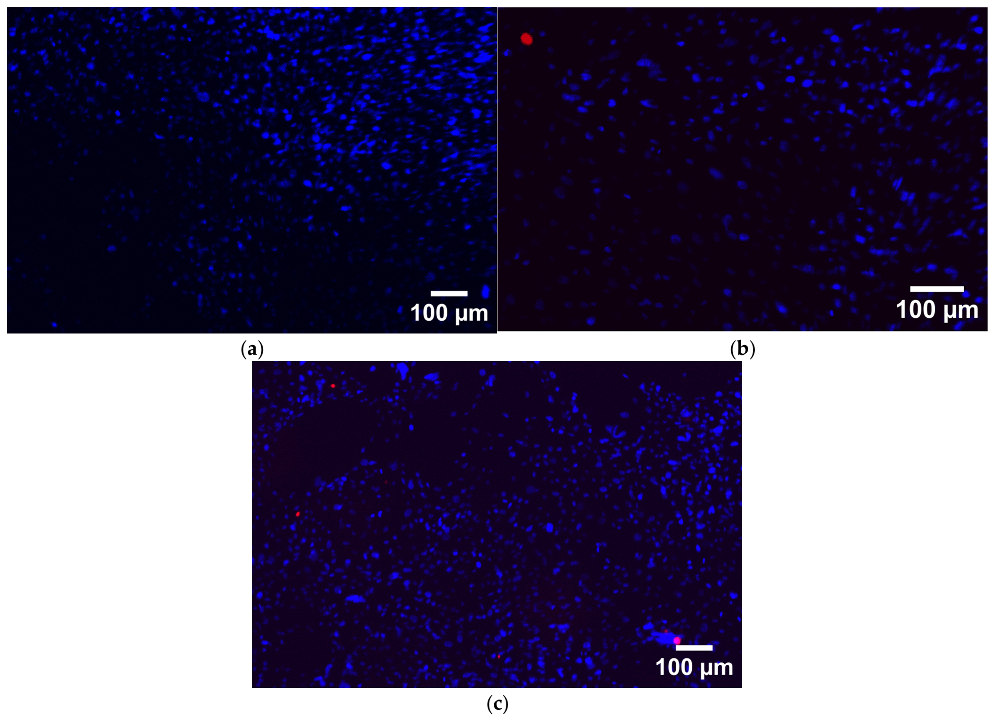
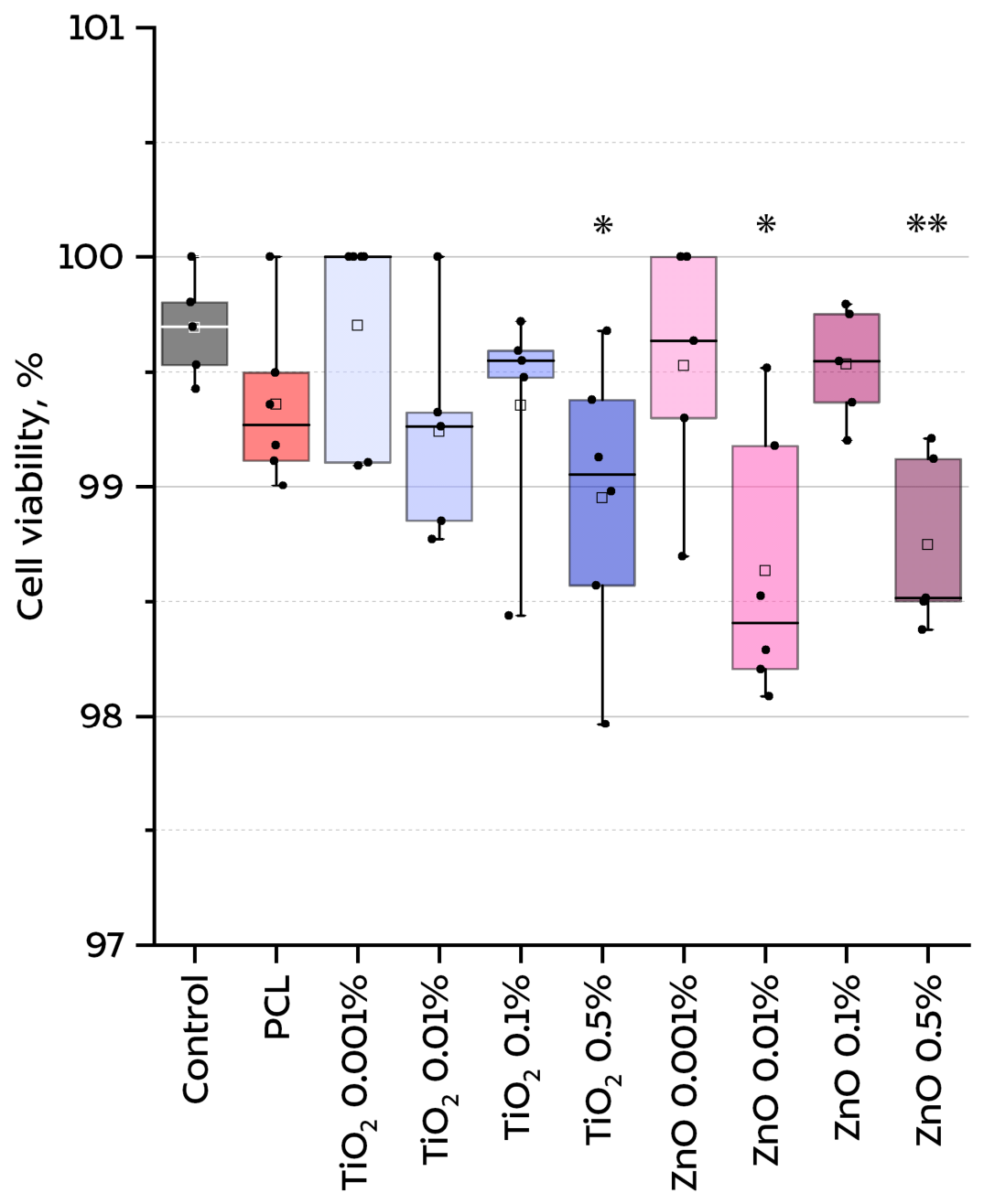
Examples of images of cells obtained using a fluorescence microscope are shown in Figure 7. As an illustrative example, images for samples of PCL/NPs-TiO2 (0.5%) and PCL/NPs-ZnO (0.5%) were taken. The results of photo processing using ImageJ are shown in Figure 8. It is clearly seen that all samples have a very high survival rate. Compared with the control, pure polycaprolactone gave a result of 99.56%. Thus, all PCL/NP composites synthesized in this work are fully biocompatible with respect to HSFs in vitro. Similar results were obtained by other authors for PCL and its composites with starch or polyvinylpyrrolidone on human fibroblasts of the L929 and MRC5 lines, and the KG1 macrophage line [41,42]. The absence of cytotoxicity in vitro allows us to conclude that all current modifications can be used in more-complex biological tests.

3.4. Antibacterial Activity Assay

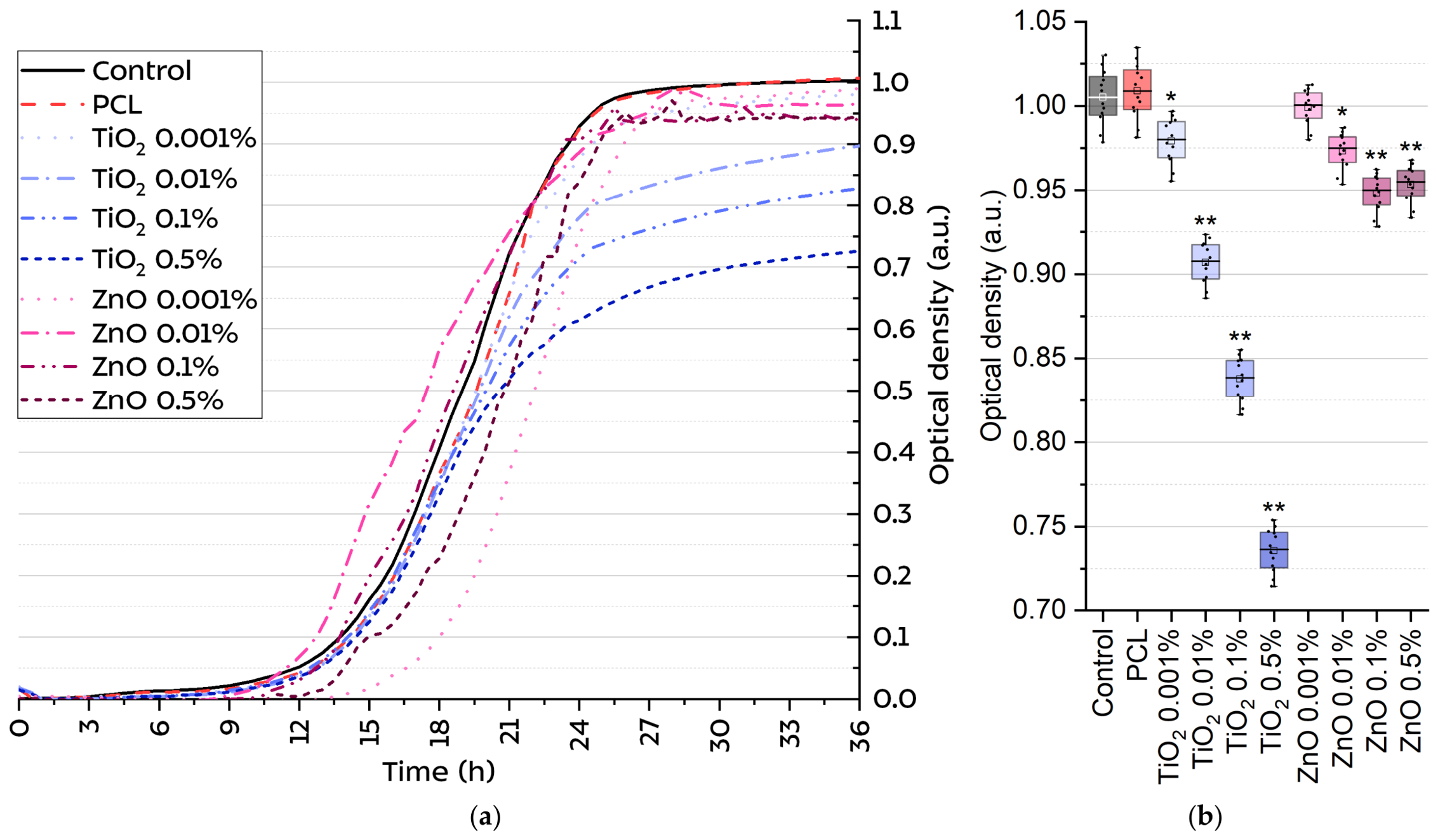
Unmodified PCL did not show its own antibacterial activity: its growth curve practically did not differ from the curve of the negative control, and the endpoints almost coincided (1.003 and 1.007 for the control and pure PCL, respectively) (Figure 9).
Composites containing ZnO nanoparticles demonstrated a weak antibacterial effect: with an increase in ZnO concentration from 0.001 to 0.5 wt. %, a slight decrease in optical density was observed; however, the degree of suppression of E. coli growth remained low compared with the negative control (approximately 5.9%). On the contrary, the introduction of titanium oxide nanoparticles led to a more pronounced antibacterial effect correlating with the concentration of NPs in the composite. For a sample with the highest concentration of TiO2 (0.5 wt. %), bacterial growth suppression was 27.6% compared with the negative control. Similar results for the PCL/NPs-TiO2 composite are given in the work of Munoz-Bonilla [11], where the minimum studied concentration of NPs in the composite corresponds to the maximum in our work and is equal to 0.5 wt. %. Also, the introduction of 3 wt. % TiO2 nanoparticles into the PLA/PCL matrix led to an increase in the growth inhibition of E. coli from 16.88 ± 0.29 mm to 21.71 ± 1.74 mm, which corresponds to a relative increase of 28.6% [43]. A number of studies provide data on the high antibacterial effect of ZnO nanoparticles in composites with PCL [44,45]. Such strong differences in the results obtained may be due to several factors, including the strong aggregation of NPs-ZnO during solvent replacement [46].

4. Discussion

The results obtained demonstrate that the biological properties of PCL/NP composites are determined not only by the chemical nature of the nanoparticles used but also by their colloidal stability at the stages of synthesis and transfer between solvents with different polarities. The ability of nanoparticles to maintain a nanoscale state during the transition from an aqueous medium to an organic phase and subsequent introduction into a polymer matrix has a significant effect on the final antibacterial activity of composites [46].
The results of the ζ-potential measurement showed that both types of nanoparticles in the aquatic environment are in a metastable state: the ζ-potential of NPs-TiO2 was −26.2 mV and that of NPs-ZnO was +27.3 mV. As mentioned earlier, colloidal solutions are considered stable at <25–30 mV, whereas at lower values the probability of particle aggregation increases greatly due to a decrease in electrostatic repulsion [30,31].
It should also be noted that the sign of the ζ-potential does not determine the stability of the dispersion by itself but reflects the different state of the surface of the oxides in an aqueous medium and the features of the acid–base equilibrium of the surface hydroxyl groups. The negative surface charge for TiO2 nanoparticles is typical at pH values above the isoelectric point, which is in the pH range 5–7 [47,48]. The negative ζ-potential of TiO2 in the system under study is a consequence of deprotonation of surface hydroxyl groups [47,49,50]. The isoelectric point of zinc oxide lies higher than that of titanium oxide; therefore, in water (pH ≈ 7), in which NP synthesis occurs, they have a different surface charge [49,50]. ZnO nanoparticles retain a protonated state and have a positive surface charge at pH values below the 8–10 range corresponding to its isoelectric point [47,48]. Under these conditions, the predominance of Zn–OH2+ groups makes the NPs’ surface more sensitive to environmental changes.
The increased chemical sensitivity of the ZnO surface compared with TiO2 is also confirmed by studies of the behavior of oxide nanoparticles in organic and mixed solvents. It has been shown that ZnO reacts significantly more strongly to changes in the polarity of the medium, ionic strength, and solvent composition, demonstrating a pronounced tendency toward aggregation and rearrangement of the surface hydroxyl layer [51,52,53]. For TiO2, such effects are significantly weaker due to the higher chemical stability of the surface and a lower tendency toward interparticle coordination [54,55,56]. Thus, NPs of zinc oxide and titanium oxide synthesized by the same method and transferred in the same way without additional stabilizers to another solvent, DCM, have fundamentally different properties, which may explain such a difference in the biological properties of PCL/NPs-TiO2 and PCL/NPs-ZnO composites.
The results of the viability assay of HSF culture in the presence of PCL/NPs-ZnO and PCL/NPs-TiO2 composites confirm the high biocompatibility of both types of materials in the entire concentration range studied (0.001–0.5 wt. %). The proportion of dead cells slightly exceeded the values of the negative control, and the minimum percentage of viability obtained was 98.75%, which is consistent with the literature data on the low cytotoxicity of PCL and PCL composites modified with metal oxide nanoparticles [46,57,58,59,60]. As shown in Babaei’s work, the introduction of up to 6 wt. % NPs-ZnO into the PCL/PLA matrix led to fibroblast viability above 90% in the MTT test, which indicates the biocompatibility of the materials [61]. Johari’s research using porous functionally graded PCL/ZnO frameworks with a ZnO content of up to 15 wt. % also demonstrated high cell adhesion in the complete absence of cytotoxicity [62]. Seo et al. report that polycaprolactone membranes containing 0.5 and 5 wt. % ZnO significantly reduce bacterial adhesion, while osteoblast proliferation in them occurs at a level comparable to that in membranes made of unmodified polycaprolactone [12]. The authors report no effect of NPs-ZnO at the studied concentrations on osteoblast viability. Taking into account the identical level of NPs-ZnO content in the composites in the work [12] and the current study, it is possible to compare the biological properties of closely related, very similar composites. Even though in our study cytotoxicity tests were carried out using a different culture, namely, HSF, the results were similar. Similar conclusions were obtained for TiO2 nanoparticles: surface modification of titanium implants with PCL/TiO2 coating increased osteoblast viability by 40% already on the third day of cultivation [63], and studies of PCL-ZnO/TiO2/HAp multicomponent fibers confirmed the absence of a cytotoxic effect and an increase in the number of metabolically active cells with normal morphology [64]. Thus, the high viability observed in this study should be considered specifically as high cytocompatibility under in vitro conditions, and not as evidence of complete biological safety. To confirm the biomedical safety of the developed composite materials, further studies will be required, involving various types of cells, longer exposure periods, the study of inflammatory reactions, and the use of in vivo models.
The antibacterial activity of metal oxide nanoparticles, including ZnO and TiO2, is due to a complex set of interrelated mechanisms [65,66,67]. The generation of reactive oxygen species (ROS), such as hydroxyl radicals, superoxidanion (O2) and hydrogen peroxide (H2O2), is the central link in the antibacterial action of most metal oxide nanoparticles [68,69]. This mechanism is confirmed in the work on the introduction of NP Ag2O into the matrix of borosiloxane and PLGA [70,71]. Upon contact with a bacterial cell, ROS initiate lipid peroxidation, leading to damage to membrane proteins and lipids, as well as disrupting the redox balance of the cell [72]. In addition to oxidative stress, nanoparticles can have a direct physical effect on the bacterial cell wall, disrupting their integrity through electrostatic and hydrophobic interactions, as well as directly damaging the cell’s DNA and RNA [72]. In addition, for some oxides, especially for ZnO, a significant contribution to the antibacterial effect is made by the release of zinc ions, which penetrate the cell, disrupt ATP synthesis and inhibit the activity of key enzymes. The nanoparticles introduced into the polymer matrix retain their antibacterial effect, continuing to generate ROS, which is confirmed in the works [73,74,75]. Evaluation of the mechanisms of antibacterial action of the composites obtained in this work is a task for future research. As a result of the experiments, unmodified PCL did not show its own antibacterial activity, which is fully consistent with the literature data [76,77]. The antibacterial properties identified in this study require more-detailed investigation, as the relatively weak activity of PCL/NPs-ZnO composites seems somewhat unexpected, given the well-known antibacterial potential of zinc oxide nanoparticles. In the work by [12], where the minimum studied concentration of zinc oxide nanoparticles is 0.5 wt. %, which coincides with the maximum concentration in our study, it is stated that bacterial adhesion is inhibited when nanoparticles are added to the composite. Raghupathi et al., for example, demonstrated an inverse relationship between the size of zinc oxide nanoparticles and their antibacterial activity, and linked the antibacterial effect to the production of reactive oxygen species and the interaction of the nanoparticles with cells [78]. Similarly, other studies have shown that surface modification and particle size can significantly affect the antibacterial efficacy of zinc oxide nanoparticles [79]. PCL/NPs-ZnO composites demonstrated a weak effect: the maximum suppression of E. coli growth was only about 5.9%, whereas PCL/NPs-TiO2 composites at an NP concentration of 0.5 wt. % provided suppression of 27.6% (reduction in optical density from 1.007 in the control to 0.726). Thus, the weak activity of the ZnO-containing composites based on polycaprolactone obtained in this study should not be interpreted as evidence of the initially low antibacterial activity of ZnO. This difference is primarily explained by the different colloidal stability of the nanoparticles during transfer from water to DCM and further introduction into the polymer matrix. As mentioned earlier, zinc oxide NPs are more sensitive to solvent change than titanium oxide NPs and are more prone to aggregation in the absence of additional stabilizers. According to the numerous literature data, the aggregation of nanoparticles leads to a critical decrease in their antibacterial effectiveness for several reasons: the specific surface area in contact with bacterial cells decreases; the rate of metal ion release decreases (which is especially important for ZnO); the efficiency of ROS generation decreases due to a decrease in the number of active centers per unit mass [80,81,82,83]. Even a slight increase in the size of NPs affects their antibacterial activity: a 3-fold increase in the size of nanoparticles may require an increase in their concentration by 1.5–2 times for the same effect, which is clearly shown in the work of El-Habib [84]. In the case of TiO2, the higher chemical stability of the surface and a lower tendency toward aggregation make it possible to maintain the active surface, which fully realizes the antibacterial potential of nanoparticles. Our results are consistent with studies in which PCL/NPs-TiO2 composites demonstrated a pronounced antibacterial effect against E. coli [11,43,58,85]. For example, in the article by Muñoz-Bonilla et al., PCL/NPs-TiO2 composites with a nanoparticle concentration of 0.5 wt. % exhibited noticeable bacteriostatic activity, which is consistent with our results [11]. In this study, the fact that TiO2 demonstrated more-pronounced antibacterial activity than ZnO, despite the fact that both types of nanoparticles were introduced into the composites at the same nominal concentrations and using the same transfer procedure, confirms the hypothesis that the physicochemical history of nanoparticles during the composite preparation process may influence their final biological properties. At the same time, the moderate intensity of the observed antibacterial effect indicates that, under these conditions, the materials obtained should be classified as weakly or moderately antibacterial rather than highly bactericidal. It should also be noted that the antibacterial test used in this study was conducted under simplified in vitro conditions using a single reference strain—E. coli. Therefore, further research should include the use of additional bacterial strains, in particular Gram-positive bacteria and clinically significant isolates, biofilm models, longer antibacterial tests, and, ultimately, in vivo assessment.

5. Conclusions

In this work, TiO2 and ZnO nanoparticles were synthesized by laser ablation in water and transferred to dichloromethane through an intermediate solvent (acetone), and PCL/NP composite films with filler concentrations from 0.001 to 0.5 wt. % were obtained by casting from a solution.
All synthesized PCL/NP composites, regardless of the type and concentration of nanoparticles, demonstrated high biocompatibility with respect to HSF culture: cell viability in all samples exceeded 98.75%, which is comparable with negative control and pure PCL.
When assessing the antibacterial activity against E. coli, PCL/NPs-TiO2 composites showed a pronounced correlation between the content of nanoparticles and the antimicrobial effect: the maximum growth suppression was 27.6% at a concentration of 0.5 wt. %. However, PCL/NPs-ZnO composites showed only weak activity (≈5.9% growth suppression). The key factor determining this difference was the different colloidal stability of nanoparticles, determined by the magnitude of the zeta potential, which affects their aggregation when replacing a solvent from a more polar to a less polar one.
Thus, PCL/NPs-TiO2 composites are fully biocompatible and have high antibacterial activity, which makes them candidates for further study as medical materials such as absorbable scaffolds or suture filaments. It is shown that the success of PCL modification with metal oxide nanoparticles is determined not only by the nature of the filler but also by the preservation of its nanoscale state at all stages of the technological process.

Author Contributions

Conceptualization, V.A.K. and P.A.F.; methodology, P.A.F., G.A.O. and I.V.B.; software, D.A.S.; validation, L.R.S. and V.A.K.; formal analysis, D.A.S. and I.V.B.; investigation, P.A.F., G.A.O. and D.A.S.; resources, P.P.C., V.A.K. and S.V.G.; data curation, D.A.S. and P.A.F.; writing—original draft preparation, P.A.F. and D.A.S.; writing—review and editing, L.R.S., I.V.B., V.A.K. and S.V.G.; visualization, D.A.S. and P.A.F.; supervision, V.A.K. and S.V.G.; project administration, V.A.K.; funding acquisition, S.V.G. All authors have read and agreed to the published version of the manuscript.

Funding

This research received no external funding.

Institutional Review Board Statement

Not applicable.

Data Availability Statement

The raw data supporting the conclusions of this article will be made available by the authors on request.

Acknowledgments

The authors thank their colleagues Dmitriy E. Burmistrov, Ann V. Gritsaeva, Ivan A. Popov, and Alexander V. Simakin for their assistance in carrying out the experiments, their valuable tips on raw data obtaining, and discussions on the obtained results.

Conflicts of Interest

Author Pavel P. Chapala was employed by the company HARZ Labs LLC. The remaining authors declare that the research was conducted in the absence of any commercial or financial relationships that could be construed as potential conflicts of interest.

Abbreviations

The following abbreviations are used in this manuscript:
PCLPolycaprolactone
NPsNanoparticles
PLAPolylactic acid
PLGAPoly(lactic-co-glycolic acid)
DCMDichloromethane
DLSDynamic light scattering
ATRAttenuation total reflectance
HSFsHuman spleen fibroblast cells
PIPropidium iodide
ODOptical density
HApHydroxyapatite
ROSReactive oxygen species

References

  1. Gudkov, S.V.; Antipov, A.V.; Astashev, M.E.; Baybursky, V.L.; Baimler, I.V.; Beldova, D.A.; Bunkin, A.F.; Burmistrov, D.E.; Validov, S.Z.; Vasilyev, V.A.; et al. Ecology of Industrial Cities: Non-Standard Scientific and Technological Solutions for Environmental Monitoring, Neutralization, and Subsequent Advanced Processing of Industrial and Municipal Waste (A Review). Russ. J. Gen. Chem. 2025, 95, S449–S480. [Google Scholar] [CrossRef] [Scilit]
  2. Ntrivala, M.A.; Pitsavas, A.C.; Lazaridou, K.; Baziakou, Z.; Karavasili, D.; Papadimitriou, M.; Ntagkopoulou, C.; Balla, E.; Bikiaris, D.N. Polycaprolactone (PCL): The biodegradable polyester shaping the future of materials—A review on synthesis, properties, biodegradation, applications and future perspectives. Eur. Polym. J. 2025, 234, 114033. [Google Scholar] [CrossRef] [Scilit]
  3. Kim, Y.; Kwak, J.; Lim, M.; Lim, S.Y.; Chae, S.; Ha, S.-J.; Won, Y.-W.; Kim, H.-O.; Lim, K.S. Advances in PCL, PLA, and PLGA-Based Technologies for Anticancer Drug Delivery. Pharmaceutics 2025, 17, 1354. [Google Scholar] [CrossRef] [Scilit] [PubMed]
  4. Ramírez-Ruiz, F.; Núñez-Tapia, I.; Piña-Barba, M.C.; Alvarez-Pérez, M.A.; Guarino, V.; Serrano-Bello, J. Polycaprolactone for hard tissue regeneration: Scaffold design and in vivo implications. Bioengineering 2025, 12, 46. [Google Scholar] [CrossRef] [Scilit] [PubMed]
  5. Nashchekina, Y.; Chabina, A.; Moskalyuk, O.; Voronkina, I.; Evstigneeva, P.; Vaganov, G.; Nashchekin, A.; Yudin, V.; Mikhailova, N. Effect of functionalization of the polycaprolactone film surface on the mechanical and biological properties of the film itself. Polymers 2022, 14, 4654. [Google Scholar] [CrossRef] [Scilit] [PubMed]
  6. Zhang, C.; Ru, Y.; You, J.; Lin, R.; Chen, S.; Qi, Y.; Li, D.; Zhang, C.; Qiu, Z. Antibacterial properties of PCL@45s5 composite biomaterial scaffolds based on additive manufacturing. Polymers 2024, 16, 3379. [Google Scholar] [CrossRef] [Scilit] [PubMed]
  7. Ozkan, O.; Sasmazel, H.T. Antibacterial performance of PCL-chitosan core–shell scaffolds. J. Nanosci. Nanotechnol. 2018, 18, 2415–2421. [Google Scholar] [CrossRef] [Scilit] [PubMed]
  8. Villanueva-Lumbreras, J.; Rodriguez, C.; Aguilar, M.R.; Aviles-Arnaut, H.; Cordell, G.A.; Rodriguez-Garcia, A. Nanofibrous ε-polycaprolactone matrices containing nano-hydroxyapatite and Humulus lupulus L. extract: Physicochemical and biological characterization for oral applications. Polymers 2024, 16, 1258. [Google Scholar] [CrossRef] [Scilit] [PubMed]
  9. Generalova, A.N.; Dushina, A.O. Metal/metal oxide nanoparticles with antibacterial activity and their potential to disrupt bacterial biofilms: Recent advances with emphasis on the underlying mechanisms. Adv. Colloid Interface Sci. 2025, 345, 103626. [Google Scholar] [CrossRef] [Scilit] [PubMed]
  10. Gudkov, S.V.; Burmistrov, D.E.; Fomina, P.A.; Validov, S.Z.; Kozlov, V.A. Antibacterial properties of copper oxide nanoparticles. Int. J. Mol. Sci. 2024, 25, 11563. [Google Scholar] [CrossRef] [Scilit] [PubMed]
  11. Muñoz-Bonilla, A.; Cerrada, M.L.; Fernández-García, M.; Kubacka, A.; Ferrer, M.; Fernández-García, M. Biodegradable polycaprolactone-titania nanocomposites: Preparation, characterization and antimicrobial properties. Int. J. Mol. Sci. 2013, 14, 9249–9266. [Google Scholar] [CrossRef] [Scilit] [PubMed]
  12. Seo, N.; Park, C.; Stahl, A.M.; Cho, H.; Park, S.-W.; Yim, S.-H.; Yun, K.-D.; Ji, M.-K.; Kim, H.; Yang, Y.P.; et al. Effect of Zinc Oxide Nanoparticle Addition to Polycaprolactone Periodontal Membranes on Antibacterial Activity and Cell Viability. J. Nanosci. Nanotechnol. 2021, 21, 3683–3688. [Google Scholar] [CrossRef] [Scilit] [PubMed]
  13. Tian, J.; Paterson, T.E.; Zhang, J.; Li, Y.; Ouyang, H.; Asencio, I.O.; Hatton, P.V.; Zhao, Y.; Li, Z. Enhanced Antibacterial Ability of Electrospun PCL Scaffolds Incorporating ZnO Nanowires. Int. J. Mol. Sci. 2023, 24, 14420. [Google Scholar] [CrossRef] [Scilit] [PubMed]
  14. Coplan, C.D.; Watkins, N.E.; Lin, X.-M.; Schaller, R.D. Scalable and adaptable two-ligand co-solvent transfer methodology for gold bipyramids to organic solvents. Nanoscale Adv. 2024, 6, 4877–4884. [Google Scholar] [CrossRef] [Scilit] [PubMed]
  15. Alkilany, A.M.; Bani Yaseen, A.I.; Park, J.; Eller, J.R.; Murphy, C.J. Facile phase transfer of gold nanoparticles from aqueous solution to organic solvents with thiolated poly(ethylene glycol). RSC Adv. 2014, 4, 52676–52679. [Google Scholar] [CrossRef] [Scilit]
  16. Goulet, P.J.; Bourret, G.R.; Lennox, R.B. Facile phase transfer of large, water-soluble metal nanoparticles to nonpolar solvents. Langmuir 2012, 28, 2909–2913. [Google Scholar] [CrossRef] [Scilit] [PubMed]
  17. Altun, E.; Ahmed, J.; Aydogdu, M.O.; Harker, A.; Edirisinghe, M. The effect of solvent and pressure on polycaprolactone solutions for particle and fibre formation. Eur. Polym. J. 2022, 173, 111300. [Google Scholar] [CrossRef] [Scilit]
  18. Polonio-Alcala, E.; Casanova-Batlle, E.; Puig, T.; Ciurana, J. The solvent chosen for the manufacturing of electrospun polycaprolactone scaffolds influences cell behavior of lung cancer cells. Sci. Rep. 2022, 12, 19440. [Google Scholar] [CrossRef] [Scilit] [PubMed]
  19. Syed Jaafar, S.N.; Zambari, I.F.; Amini, N.A.I.; Norizan, B.A.-Z.; Daud, M.F. Electrosprayed low toxicity polycaprolactone microspheres from low concentration solutions. J. Polym. Eng. 2023, 43, 516–525. [Google Scholar] [CrossRef] [Scilit]
  20. Toopghara, S.A.H.; Hwang, Y.-O.; Kim, Y.S.; Kim, Y.R.; Zhang, P.; Park, C.; Park, S.-W.; Kang, J.-H. Optimization of a porous polycaprolactone coating to enhance the mechanical properties and bioactivity of an additively manufactured hydroxyapatite scaffold. Ceram. Int. 2025, 51, 53646–53656. [Google Scholar] [CrossRef] [Scilit]
  21. Ramírez-Cedillo, E.; Ortega-Lara, W.; Rocha-Pizaña, M.R.; Gutierrez-Uribe, J.A.; Elías-Zúñiga, A.; Rodríguez, C.A. Electrospun polycaprolactone fibrous membranes containing Ag, TiO2 and Na2Ti6O13 particles for potential use in bone regeneration. Membranes 2019, 9, 12. [Google Scholar] [CrossRef] [Scilit] [PubMed]
  22. Piskunova, A.; Ashikhmin, A.; Chobotova, V.; Piskunov, M. Hydrodynamics and phase transition during polycaprolactone solution droplet impact on glass surface. Int. J. Heat Fluid Flow 2026, 119, 110271. [Google Scholar] [CrossRef] [Scilit]
  23. Feng, D.; Li, Y.; Zheng, Y.; Chen, J.; Zhang, X.; Li, K.; Shen, J.; Guo, X. Study on the Electrospinning Fabrication of PCL/CNTs Fiber Membranes and Their Oil–Water Separation Performance. Polymers 2025, 17, 1705. [Google Scholar] [CrossRef] [Scilit] [PubMed]
  24. Ibrahim, N.A.; Affandi, N.D.N.; Samsulrizal, N. The effects of intrinsic parameters on the formation of electrospun polycaprolactone fibre. Int. J. Eng. Adv. Technol. 2019, 9, 5699–5704. [Google Scholar] [CrossRef] [Scilit]
  25. Elangovan, J.; Paulraj, G.E.; Nellaiappan, S.N.T.; Kulanthaivelu, R. Fabrication of Electrospun Polycaprolactone Nanofibers for Drug Delivery Application and Development of an Electrospinnability Map. J. Appl. Polym. Sci. 2025, 142, e57334. [Google Scholar] [CrossRef] [Scilit]
  26. Gil-Castell, O.; Badia, J.; Ribes-Greus, A. Tailored electrospun nanofibrous polycaprolactone/gelatin scaffolds into an acid hydrolytic solvent system. Eur. Polym. J. 2018, 101, 273–281. [Google Scholar] [CrossRef] [Scilit]
  27. Prado-Prone, G.; Bazzar, M.; Letizia Focarete, M.; García-Macedo, J.A.; Perez-Orive, J.; Ibarra, C.; Velasquillo, C.; Silva-Bermudez, P. Single-step, acid-based fabrication of homogeneous gelatin-polycaprolactone fibrillar scaffolds intended for skin tissue engineering. Biomed. Mater. 2020, 15, 035001, Correction in Biomed. Mater. 2023, 18, 049504. [Google Scholar] [CrossRef] [Scilit] [PubMed]
  28. Tampau, A.; González-Martinez, C.; Chiralt, A. Carvacrol encapsulation in starch or PCL based matrices by electrospinning. J. Food Eng. 2017, 214, 245–256. [Google Scholar] [CrossRef] [Scilit]
  29. Alberto, L.; Kalluri, L.; Qu, J.; Zhao, Y.; Duan, Y. Influence of polycaprolactone concentration and solvent type on the dimensions and morphology of electrosprayed particles. Materials 2023, 16, 2122. [Google Scholar] [CrossRef] [Scilit] [PubMed]
  30. Sizochenko, N.; Mikolajczyk, A.; Syzochenko, M.; Puzyn, T.; Leszczynski, J. Zeta potentials (ζ) of metal oxide nanoparticles: A meta-analysis of experimental data and a predictive neural networks modeling. NanoImpact 2021, 22, 100317. [Google Scholar] [CrossRef] [Scilit] [PubMed]
  31. Toropov, A.A.; Sizochenko, N.; Toropova, A.P.; Leszczynski, J. Towards the development of global nano-quantitative structure–property relationship models: Zeta potentials of metal oxide nanoparticles. Nanomaterials 2018, 8, 243. [Google Scholar] [CrossRef] [Scilit] [PubMed]
  32. Awad, M.A.; Ortashi, K.M.; Alenazi, W.; Alfaifi, F.S.; Al-Huqail, A.A. Green Synthesis of Titanium Dioxide Nanoparticles: Characterization and Evaluation of Their Potential for Photocatalytic and Dielectric Applications. Molecules 2025, 30, 4701. [Google Scholar] [CrossRef] [Scilit] [PubMed]
  33. Kulkarni, S.S.; Shirsat, M.D. Optical and structural properties of zinc oxide nanoparticles. Int. J. Adv. Res. Phys. Sci. 2015, 2, 14–18. [Google Scholar]
  34. Valentini, X.; Rugira, P.; Frau, A.; Tagliatti, V.; Conotte, R.; Laurent, S.; Colet, J.-M.; Nonclercq, D. Hepatic and Renal Toxicity Induced by TiO2 Nanoparticles in Rats: A Morphological and Metabonomic Study. J. Toxicol. 2019, 2019, 5767012. [Google Scholar] [CrossRef] [Scilit] [PubMed]
  35. Filippov, S.K.; Khusnutdinov, R.; Murmiliuk, A.; Inam, W.; Zakharova, L.Y.; Zhang, H.; Khutoryanskiy, V.V. Dynamic light scattering and transmission electron microscopy in drug delivery: A roadmap for correct characterization of nanoparticles and interpretation of results. Mater. Horiz. 2023, 10, 5354–5370. [Google Scholar] [CrossRef] [Scilit] [PubMed]
  36. Coşkun, M.; Öner, E.Ö.; Tatar, C.; Kök, M. MgO NPs reinforced PCL/PVC nanocomposite films with enhanced UV shielding and thermal stability for packaging applications. Open Phys. 2025, 23, 20250240. [Google Scholar] [CrossRef] [Scilit]
  37. Luyt, A.S.; Gasmi, S. Influence of TiO2 Nanoparticles on the Crystallization Behaviour and Tensile Properties of Biodegradable PLA and PCL Nanocomposites. J. Polym. Environ. 2018, 26, 2410–2423. [Google Scholar] [CrossRef] [Scilit]
  38. Hosakun, Y.; Halász, K.; Horváth, M.; Csóka, L.; Djoković, V. ATR-FTIR study of the interaction of CO2 with bacterial cellulose-based membranes. Chem. Eng. J. 2017, 324, 83–92. [Google Scholar] [CrossRef] [Scilit]
  39. Elzein, T.; Nasser-Eddine, M.; Delaite, C.; Bistac, S.; Dumas, P. FTIR study of polycaprolactone chain organization at interfaces. J. Colloid Interface Sci. 2004, 273, 381–387. [Google Scholar] [CrossRef] [Scilit] [PubMed]
  40. Phillipson, K.; Hay, J.N.; Jenkins, M.J. Thermal analysis FTIR spectroscopy of poly(ε-caprolactone). Thermochim. Acta 2014, 595, 74–82. [Google Scholar] [CrossRef] [Scilit]
  41. Viscusi, G.; Paolella, G.; Lamberti, E.; Caputo, I.; Gorrasi, G. Quercetin-loaded polycaprolactone-polyvinylpyrrolidone electrospun membranes for health application: Design, characterization, modeling and cytotoxicity studies. Membranes 2023, 13, 242. [Google Scholar] [CrossRef] [Scilit] [PubMed]
  42. Zhao, Y.-Q.; Yang, J.-H.; Ding, X.; Ding, X.; Duan, S.; Xu, F.-J. Polycaprolactone/polysaccharide functional composites for low-temperature fused deposition modelling. Bioact. Mater. 2020, 5, 185–191. [Google Scholar] [CrossRef] [Scilit] [PubMed]
  43. Evcimen Duygulu, N. Functionalization of PLA nanofibers with PCL blending and TiO2 nanoparticle addition. Mater. Res. Express 2024, 11, 075403. [Google Scholar] [CrossRef] [Scilit]
  44. Augustine, R.; Malik, H.N.; Singhal, D.K.; Mukherjee, A.; Malakar, D.; Kalarikkal, N.; Thomas, S. Electrospun polycaprolactone/ZnO nanocomposite membranes as biomaterials with antibacterial and cell adhesion properties. J. Polym. Res. 2014, 21, 347. [Google Scholar] [CrossRef] [Scilit]
  45. Tian, G.; Huang, Z.; Wang, H.; Cui, C.; Zhang, Y. Polycaprolactone nanofiber membrane modified with halloysite and ZnO for anti-bacterial and air filtration. Appl. Clay Sci. 2022, 223, 106512. [Google Scholar] [CrossRef] [Scilit]
  46. Liu, L.; Zhang, Y.; Li, C.; Cao, J.; He, E.; Wu, X.; Wang, F.; Wang, L. Facile preparation PCL/modified nano ZnO organic-inorganic composite and its application in antibacterial materials. J. Polym. Res. 2020, 27, 78. [Google Scholar] [CrossRef] [Scilit]
  47. Kosmulski, M. Isoelectric points and points of zero charge of metal (hydr)oxides: 50 years after Parks’ review. Adv. Colloid Interface Sci. 2016, 238, 1–61. [Google Scholar] [CrossRef] [Scilit] [PubMed]
  48. Kosmulski, M. The pH dependent surface charging and points of zero charge. VIII. Update. Adv. Colloid Interface Sci. 2020, 275, 102064. [Google Scholar] [CrossRef] [Scilit] [PubMed]
  49. Degen, A.; Kosec, M. Effect of pH and impurities on the surface charge of zinc oxide in aqueous solution. J. Eur. Ceram. Soc. 2000, 20, 667–673. [Google Scholar] [CrossRef] [Scilit]
  50. Parks, G.A. The Isoelectric Points of Solid Oxides, Solid Hydroxides, and Aqueous Hydroxo Complex Systems. Chem. Rev. 1965, 65, 177–198. [Google Scholar] [CrossRef] [Scilit]
  51. Bian, S.-W.; Mudunkotuwa, I.A.; Rupasinghe, T.; Grassian, V.H. Aggregation and Dissolution of 4 nm ZnO Nanoparticles in Aqueous Environments: Influence of pH, Ionic Strength, Size, and Adsorption of Humic Acid. Langmuir 2011, 27, 6059–6068. [Google Scholar] [CrossRef] [Scilit] [PubMed]
  52. Liu, L.; Yadav Schmid, S.; Feng, Z.; Li, D.; Droubay, T.C.; Pauzauskie, P.J.; Schenter, G.K.; De Yoreo, J.J.; Chun, J.; Nakouzi, E. Effect of Solvent Composition on Non-DLVO Forces and Oriented Attachment of Zinc Oxide Nanoparticles. ACS Nano 2024, 18, 16743–16751. [Google Scholar] [CrossRef] [Scilit] [PubMed]
  53. Šarić, A.; Despotović, I. Hydrogen Bonds as Stability-Controlling Elements of Spherical Aggregates of ZnO Nanoparticles: A Joint Experimental and Theoretical Approach. Materials 2023, 16, 4843. [Google Scholar] [CrossRef] [Scilit] [PubMed]
  54. İkizler, B.; Peker, S.M. Synthesis of TiO2 coated ZnO nanorod arrays and their stability in photocatalytic flow reactors. Thin Solid Film. 2016, 605, 232–242. [Google Scholar] [CrossRef] [Scilit]
  55. Kim, M.; Kim, J. A Study on the Stability of TiO2 Nanoparticles as an Electron Transport Layer in Quantum Dot Light-Emitting Diodes. Korean J. Met. Mater. 2021, 59, 476–480. [Google Scholar] [CrossRef] [Scilit]
  56. Tso, C.-p.; Zhung, C.-m.; Shih, Y.-h.; Tseng, Y.-M.; Wu, S.-c.; Doong, R.-a. Stability of metal oxide nanoparticles in aqueous solutions. Water Sci. Technol. 2010, 61, 127–133. [Google Scholar] [CrossRef] [Scilit] [PubMed]
  57. Banerjee, A.; Roy, P.; Chakraborty, J.; Majumder, M. Environmentally degradable curcumin/zinc oxide nanoparticles-incorporated polycaprolactone films for use as top-sheets in feminine sanitary hygiene napkins. Mater. Today Commun. 2024, 40, 109452. [Google Scholar] [CrossRef] [Scilit]
  58. Chen, L.; Zhang, J.; Cheng, J.; Mao, Y.; Xu, J.; Yin, M.; He, Y.; He, M. Multilayered PCL/MAO@TiO2 nanoparticle coatings: Optimizing degradation and mechanical stability of biodegradable magnesium alloy bone implants. J. Magnes. Alloys 2025, 13, 5059–5076. [Google Scholar] [CrossRef] [Scilit]
  59. Ghiyasi, Y.; Salahi, E.; Esfahani, H. Synergy effect of Urtica dioica and ZnO NPs on microstructure, antibacterial activity and cytotoxicity of electrospun PCL scaffold for wound dressing application. Mater. Today Commun. 2021, 26, 102163. [Google Scholar] [CrossRef] [Scilit]
  60. Sharma, H.; Pathak, M. Development of PCL/TiO2 composite as an efficient antibacterial, anticancer drug and biocompatible properties. Results Chem. 2024, 7, 101534. [Google Scholar] [CrossRef] [Scilit]
  61. Babaei, A.; Haji Abdolrasouli, M.; Rostami, A. Polylactic acid/polycaprolactone bionanocomposites containing zinc oxide nanoparticles: Structure, characterization and cytotoxicity assay. J. Thermoplast. Compos. Mater. 2023, 36, 2998–3020. [Google Scholar] [CrossRef] [Scilit]
  62. Johari, N.; Rafati, F.; Zohari, F.; Tabari, P.G.; Samadikuchaksaraei, A. Porous functionally graded scaffolds of poly (ε-caprolactone)/ZnO nanocomposite for skin tissue engineering: Morphological, mechanical and biological evaluation. Mater. Chem. Phys. 2022, 280, 125786. [Google Scholar] [CrossRef] [Scilit]
  63. Kiran, A.S.K.; Kumar, T.S.S.; Sanghavi, R.; Doble, M.; Ramakrishna, S. Antibacterial and Bioactive Surface Modifications of Titanium Implants by PCL/TiO2 Nanocomposite Coatings. Nanomaterials 2018, 8, 860. [Google Scholar] [CrossRef] [Scilit] [PubMed]
  64. Jinga, S.-I.; Zamfirescu, A.-I.; Voicu, G.; Enculescu, M.; Evanghelidis, A.; Busuioc, C. PCL-ZnO/TiO2/HAp Electrospun Composite Fibers with Applications in Tissue Engineering. Polymers 2019, 11, 1793. [Google Scholar] [CrossRef] [Scilit] [PubMed]
  65. Chaturvedi, A.; Ranjan, R. Antimicrobial nanoparticles: A new horizon to combat multidrug-resistant bacteria. Front. Nanotechnol. 2025, 7, 1611126. [Google Scholar] [CrossRef] [Scilit]
  66. Kaur, R.; Kaur, K.; Alyami, M.H.; Lang, D.K.; Saini, B.; Bayan, M.F.; Chandrasekaran, B. Combating Microbial Infections Using Metal-Based Nanoparticles as Potential Therapeutic Alternatives. Antibiotics 2023, 12, 909. [Google Scholar] [CrossRef] [Scilit] [PubMed]
  67. Samal, P.; Satpathy, S.; Rath, G.; Sabut, T.; Sahoo, A.P.; Arakha, M. Lignin-Encapsulated ZnO Nano-Hybrids Exhibit Enhanced Antibacterial and Photocatalytic Activity via ROS-Mediated Oxidative Stress and Bacterial Membrane Disruption. ACS Omega 2026, 11, 6133–6146. [Google Scholar] [CrossRef] [Scilit] [PubMed]
  68. Jamil, Y.; Ali, M.; Ali, S.; Law, D.; Alrefaei, A.F.; Ahmad, A. Insights into the antibacterial mode of action of cress polysaccharide-mediated NiO nanoparticles. Sci. Rep. 2026, 16, 14839. [Google Scholar] [CrossRef] [Scilit] [PubMed]
  69. Zuo, J.; Qin, Y.; Zhao, Z.; Xing, L.; Liu, T.; Wang, S.; Liu, W. Preparation, properties and antibacterial applications of medical nano-metals and their oxides: A review. Sheng Wu Gong Cheng Xue Bao 2023, 39, 1462–1476. [Google Scholar] [CrossRef] [Scilit] [PubMed]
  70. Chausov, D.N.; Smirnova, V.V.; Burmistrov, D.E.; Sarimov, R.M.; Kurilov, A.D.; Astashev, M.E.; Uvarov, O.V.; Dubinin, M.V.; Kozlov, V.A.; Vedunova, M.V. Synthesis of a novel, biocompatible and bacteriostatic borosiloxane composition with silver oxide nanoparticles. Materials 2022, 15, 527. [Google Scholar] [CrossRef] [Scilit] [PubMed]
  71. Smirnova, V.V.; Chausov, D.N.; Serov, D.A.; Kozlov, V.A.; Ivashkin, P.I.; Pishchalnikov, R.Y.; Uvarov, O.V.; Vedunova, M.V.; Semenova, A.A.; Lisitsyn, A.B. A novel biodegradable composite polymer material based on plga and silver oxide nanoparticles with unique physicochemical properties and biocompatibility with mammalian cells. Materials 2021, 14, 6915. [Google Scholar] [CrossRef] [Scilit] [PubMed]
  72. Ajayi, A.S.; Adanigbo, P.; Olaifa, J.T.; Obaleye, M.O.; Ofoka, E.A.; Ukanwa, S.O.; Duru, P.; Moshood, F.; Aigbodion, A.I.; Ifijen, I.H. Tellurium nanoparticles as antimicrobial agents for multi-drug-resistant infections. RSC Adv. 2025, 15, 36272–36299. [Google Scholar] [CrossRef] [Scilit] [PubMed]
  73. Burmistrov, D.E.; Ivliev, P.A.; Serov, D.A.; Baimler, I.V.; Simakin, A.V.; Liubimovskii, S.O.; Astashev, M.E.; Kozlov, V.A.; Nastulyavichus, A.A.; Nizameeva, G.R.; et al. Composite Materials Based on L-Polylactide with Titanium or Titanium Dioxide Nanoparticles: Dark Antibacterial Activity Through ROS Generation. J. Compos. Sci. 2026, 10, 214. [Google Scholar] [CrossRef] [Scilit]
  74. Serov, D.A.; Yanbaev, F.M.; Burmistrov, D.E.; Baimler, I.V.; Liubimovskii, S.O.; Kozlova, L.Y.; Popov, I.A.; Gritsaeva, A.V.; Fomina, P.A.; Sizov, L.R.; et al. A Composite Based on L-Polylactide with Cu or CuO Nanoparticles: Physical Properties and Biological Activity. Polymers 2026, 18, 976. [Google Scholar] [CrossRef] [Scilit] [PubMed]
  75. Yanbaev, F.M.; Ignatenko, D.N.; Shabalina, A.V.; Baimler, I.V.; Burmistrov, D.E.; Astashev, M.E.; Lednev, V.N.; Nastulyavichus, A.A.; Pishchalnikov, R.Y.; Sarimov, R.M.; et al. Antibacterial and Non-Toxic to Mammalian Cell Composite Material Based on Polymethyl-Methacrylate-like Resin Containing Grain-Shaped Copper Oxide Nanoparticles. J. Compos. Sci. 2025, 9, 706. [Google Scholar] [CrossRef] [Scilit]
  76. Ganbaatar, S.E.; Kim, H.-K.; Kang, N.-U.; Kim, E.C.; U, H.J.; Cho, Y.-S.; Park, H.-H. Calcium Phosphate (CaP) Composite Nanostructures on Polycaprolactone (PCL): Synergistic Effects on Antibacterial Activity and Osteoblast Behavior. Polymers 2025, 17, 200. [Google Scholar] [CrossRef] [Scilit] [PubMed]
  77. Vilamová, Z.; Šimonová, Z.; Bednář, J.; Mikeš, P.; Cieslar, M.; Svoboda, L.; Dvorský, R.; Rosenbergová, K.; Kratošová, G. Silver-loaded poly(vinyl alcohol)/polycaprolactone polymer scaffold as a biocompatible antibacterial system. Sci. Rep. 2024, 14, 11093. [Google Scholar] [CrossRef] [Scilit] [PubMed]
  78. Raghupathi, K.R.; Koodali, R.T.; Manna, A.C. Size-Dependent Bacterial Growth Inhibition and Mechanism of Antibacterial Activity of Zinc Oxide Nanoparticles. Langmuir 2011, 27, 4020–4028. [Google Scholar] [CrossRef] [Scilit] [PubMed]
  79. Lallo da Silva, B.; Caetano, B.L.; Chiari-Andréo, B.G.; Pietro, R.C.L.R.; Chiavacci, L.A. Increased antibacterial activity of ZnO nanoparticles: Influence of size and surface modification. Colloids Surf. B Biointerfaces 2019, 177, 440–447. [Google Scholar] [CrossRef] [Scilit] [PubMed]
  80. Wu, L.; Wei, S.; Cheng, X.; He, N.; Kang, X.; Zhou, H.; Cai, Y.; Ye, Y.; Li, P.; Liang, C. Release of ions enhanced the antibacterial performance of laser-generated, uncoated Ag nanoparticles. Colloids Surf. B Biointerfaces 2024, 243, 114131. [Google Scholar] [CrossRef] [Scilit] [PubMed]
  81. Viswanathan, K.; Kim, I.; Kasi, G.; Sadeghi, K.; Thanakkasaranee, S.; Seo, J. Facile approach to enhance the antibacterial activity of ZnO nanoparticles. Adv. Appl. Ceram. 2020, 119, 414–422. [Google Scholar] [CrossRef] [Scilit]
  82. Bélteky, P.; Rónavári, A.; Zakupszky, D.; Boka, E.; Igaz, N.; Szerencsés, B.; Pfeiffer, I.; Vágvölgyi, C.; Kiricsi, M.; Kónya, Z. Are Smaller Nanoparticles Always Better? Understanding the Biological Effect of Size-Dependent Silver Nanoparticle Aggregation Under Biorelevant Conditions. Int. J. Nanomed. 2021, 16, 3021–3040. [Google Scholar] [CrossRef] [Scilit] [PubMed]
  83. Phan, T.L.; Le, T.T.; Doan, V.D.; Vu Truong, H.A.; Le, V.T. Size-dependent catalytic and antibacterial effects of phytogenically synthesized silver nanoparticles. Kuwait J. Sci. 2025, 52, 100366. [Google Scholar] [CrossRef] [Scilit]
  84. El-Habib, I.; Maatouk, H.; Lemarchand, A.; Dine, S.; Roynette, A.; Mielcarek, C.; Traoré, M.; Azouani, R. Antibacterial Size Effect of ZnO Nanoparticles and Their Role as Additives in Emulsion Waterborne Paint. J. Funct. Biomater. 2024, 15, 195. [Google Scholar] [CrossRef] [Scilit] [PubMed]
  85. Ando, H.; Kawasaki, N.; Yamano, N.; Uegaki, K.; Nakayama, A. Biodegradation of a poly(ε-caprolactone-co-l-lactide)–visible-light-sensitive TiO2 composite with an on/off biodegradation function. Polym. Degrad. Stab. 2015, 114, 65–71. [Google Scholar] [CrossRef] [Scilit]
Figure 1. Chemical structure of PCL.
Technologies 14 00587 g001
Figure 2. Graphs showing the size distribution of TiO2 (a) and ZnO (b) NPs based on DLS results. The corresponding concentrations are given for the peak maxima.
Technologies 14 00587 g002
Figure 3. Absorption spectra of ZnO (a) and TiO2 (b) nanoparticles in water in the UV–visible region of the spectrum.
Technologies 14 00587 g003
Figure 4. TEM images of NPs-TiO2 (a) and NPs-ZnO (b) in water.
Technologies 14 00587 g004
Figure 5. Graphs showing the size distribution of TiO2 (a) and ZnO (b) NPs based on TEM processing results.
Technologies 14 00587 g005
Figure 6. IR transmission spectra of polycaprolactone samples without and with nanoparticles in the range of 550–4000 cm−1.
Technologies 14 00587 g006
Figure 7. Fluorescent microphotographs of HSFs cultured for 72 h: (a) in the absence of polymer samples (negative control); (b) in the presence of a sample of PCL/NPs-TiO2 (0.5%); (c) in the presence of a sample of PCL/NPs-ZnO (0.5%). The image of the cells was obtained by combining micrographs of cells stained with Hoechst 33342 (blue) and cells stained with PI (red) and allows us to estimate the number of dead cells among their total number.
Technologies 14 00587 g007
Figure 8. Viability indicators of the samples obtained by fluorescence microscopy. The data is presented in the form of median values (horizontal line inside the box), percentiles of 25 and 75% (lower and upper borders of the boxes), minima and maxima (bars). Each point corresponds to an individual measurement. * p < 0.01, ** p < 0.001 (regarding control), according to the Mann–Whitney test (n = 5–6).
Technologies 14 00587 g008
Figure 9. Antibacterial activity of the samples: (a) E. coli growth curves obtained by measuring OD600 for 36 h and 30 min between measurements; (b) the OD600 values at the end points of the growth curves for each sample (in the stationary growth phase). The data is presented in the form of median values (horizontal line inside the box), percentiles of 25 and 75% (lower and upper borders of the boxes), minima and maxima (bars). Each point corresponds to an individual measurement. * p < 0.01, ** p < 0.001 (regarding control), according to the Mann–Whitney test (n = 12).
Technologies 14 00587 g009
Table 1. Information on the solvents used to prepare PCL solutions, as well as the final mass content of PCL in them.
No.Solventwt. % of PCLReference
1Chloroform + ethanol (3:1/1:1/1:3)15[17]
2Chloroform + DCM (3:1/1:1/1:3)
3Chloroform
4DCM
5Chloroform + DCM (7:3)15[18]
6Acetic acid + formic acid (3:1)
7Chloroform1–5[19]
8DCM
9DCM3–10[20]
10Chloroform10[21]
11DCM1–10[22]
12DCM + dimethylformamide (7:3)17[23]
13DCM10[24]
14Chloroform + ethyl alcohol + formic acid (9:1:0.1)10[25]
15Acetic acid + formic acid (1:1)15[26]
16Acetic acid19[27]
17Adipic acid5–15[28]
18Chloroform + dimethylformamide (1:1/3:1)2–6[29]
19Chloroform
20Acetic acid + chloroform (1:1/3:1)
21Acetic acid
The symbol “―” means that the value is the same as the previous one.
Table 2. Values of electrokinetic potentials, sizes and concentrations of metal oxide NPs obtained by laser ablation in water.
No.NPsTiO2ZnO
1ζ-potential, mV−26.227.3
2Mean size, nm (concentration [particles/mL])80 ± 15 (109)
283 ± 48 (107)
131 ± 25 (109)
349 ± 46 (108)
Disclaimer/Publisher’s Note: The statements, opinions and data contained in all publications are solely those of the individual author(s) and contributor(s) and not of MDPI and/or the editor(s). MDPI and/or the editor(s) disclaim responsibility for any injury to people or property resulting from any ideas, methods, instructions or products referred to in the content.