1. Introduction
Breast cancer screening relies on the “gold”-standard mammography technique as well as on advanced imaging, such as breast tomosynthesis, cone-beam CT, and breast CT. Phantoms play a critical role in procedures, concerning safety, quality assurance, and protocol optimization in these imaging systems. However, conventional phantoms are often oversimplified and do not adequately replicate the complex anatomical and radiological characteristics of the breast tissue [
1,
2].
Anthropomorphic phantoms, both physical and virtual, are composed of tissue-equivalent materials to provide realistic and accurate representations of human anatomy and tissue properties for both research and routine clinical applications [
2,
3]. A realistic representation involves mimicking both the external morphology and internal anatomical structures of the modeled tissue or organ. In X-ray imaging, tissue-equivalent materials need to accurately reproduce the attenuation and scattering properties of human tissues within the relevant diagnostic energy range.
Recent advancements in 3D printing have made the fabrication of highly customizable physical phantoms increasingly accessible. The growing affordability of additive manufacturing technologies, combined with the widespread availability of high-resolution medical imaging data, has positioned 3D printing as an essential tool in modern radiology [
4]. Such phantoms can be produced for whole-body applications or specific organs and are widely used for evaluating novel imaging techniques, establishing clinical protocols, and performing routine quality control procedures [
5,
6].
The ELPIDA research team at the Medical University of Varna (
https://muve-team.mu-varna.bg/en/early-diagnosis-and-prevention-of-oncological-breast-diseases-by-using-new-technologies-elpida (accessed on 15 April 2026)) has been extensively involved in the development and testing of anthropomorphic breast phantoms. Through international collaborations, the team participated in the assessment of 3D printing methodologies based on the direct conversion of DICOM imaging data into G-code for the case of breast phantoms, developed by Morphe, Thessaloniki (
https://morphe.cc/ (accessed on 18 March 2026)). This workflow enables anatomically accurate phantom fabrication by translating imaging voxel information directly into printer instructions. Within this framework, developments are focused on correlating filament extrusion rates with Hounsfield Unit (HU) values derived from MRI datasets [
7]. The methodology was further advanced through a voxel-by-voxel material mixing strategy, in which a single contoured region is printed sequentially using two materials—polylactic acid (PLA) and polypropylene (PP)—to precisely control X-ray attenuation properties [
8]. Additionally, the integration of direct voxel-based printing with advanced extrusion techniques allows for the variation in both rate and speed, enabling a single material to replicate the full spectrum of breast soft tissues [
9]. This DICOM-to-G-code approach has also been successfully applied to the fabrication of other anatomical models, including lung tissue, cervical vertebrae, knee, heart, and head structures [
10,
11,
12,
13,
14,
15].
An alternative printing strategy involves segmenting anatomical structures from DICOM datasets to generate STL files, which are subsequently printed using tailored infill patterns to simulate variations in tissue density. This methodology has been successfully implemented into a range of 3D-printed anthropomorphic models, including neck-thyroid [
16], head [
17], chest [
18,
19], artery [
20], and cardiac phantoms [
21]. By combining segmentation-based modeling with controlled infill design and G-code parameter adjustment, complex and customizable internal architectures can be achieved.
Building on this concept, the ELPIDA team adapted the segmentation-based workflow to breast imaging applications using MRI and breast tomosynthesis (BT) datasets. The objective was to establish an efficient and reproducible manufacturing protocol, initially employing a 100% infill baseline, to produce high-fidelity anthropomorphic breast phantoms. To achieve improved material control, we developed a dual-head 3D printing approach utilizing two carefully selected filaments, which are suitable to reproduce the radiological attenuation properties of breast tissue [
22].
For clinical and research applications, particularly in dose measurement, quality assurance, and the optimization of emerging imaging techniques, the manufacturing process must demonstrate high reproducibility and stable radiological performance. Ensuring that printed phantoms consistently exhibit the intended X-ray attenuation characteristics represents a critical technical requirement [
22]. Therefore, the aim of this study is to evaluate the reproducibility of the proposed dual-head manufacturing workflow in terms of mammographic performance by comparing the imaging characteristics of multiple nominally identical breast phantoms.
2. Materials and Methods
2.1. Data Acquisition and Modeling
The breast phantom design was derived from patient-specific imaging data obtained from T1-weighted, contrast-enhanced MRI scans of a 26-year-old female patient (Siemens Verio (Siemens Healthineers, Erlangen, Germany); 1.0 mm isotropic voxel resolution), following approval by the Ethics Committee of the Medical University of Varna (Approval No. 102/22.04.2021). For the purposes of this study, we focus on a section of the breast where tissue variability is prominent. Adipose, glandular and skin tissues were initially segmented automatically using a MATLAB (R2022a)-based script employing thresholding techniques. The segmentation was subsequently refined through manual inspection and correction, with additional editing performed using Microsoft Paint to ensure accurate delineation of tissue boundaries, including the skin. Since the breast sections are printed with a dual-head 3D printer, the skin is also assigned the properties of the glandular tissue.
2.2. STL Model
The next step is to convert the segmented breast to an STL file for 3D printing. However, generating high-quality STL representations from voxel-based datasets presents significant challenges [
23], particularly in multi-material printing applications [
22]. The conversion of voxel-based models to STL format is frequently complicated by triangulation artifacts and excessive mesh complexity, which increase with anatomical heterogeneity and spatial resolution [
24].
In previous developments, the object surface was extracted in MATLAB (R2022a) using the
isosurface function, which segments a 3D volume based on a selected threshold (isovalue) and generates a triangulated surface representation. The popular function
stlwrite by Holcombe [
25] relies on the Marching Cubes algorithm [
26] to compute surface vertices and faces from volumetric data. While this approach produces a smoothed, chamfered surface relative to the original prismatic voxels, it results in slight deviations in the enclosed volume.
To overcome the limitations of isosurface-based STL generation, an alternative approach was developed based on the explicit representation of voxel geometry. Instead of interpolating surfaces, the method identifies shared voxel faces between adjacent segmented regions and constructs the surface directly from these interfaces. Each voxel face is treated as a rectangular element, which is subdivided into two triangular facets with outward-facing normal vectors. This procedure preserves the original prismatic voxel structure and ensures a watertight, manifold surface without internal voids, which is critical for reliable multi-material 3D printing. In contrast to conventional isosurface methods, which may introduce smoothing artifacts and geometric inconsistencies, the proposed approach maintains geometric fidelity and improves the structural integrity of the printed object. A detailed description and validation of the algorithm are provided in [
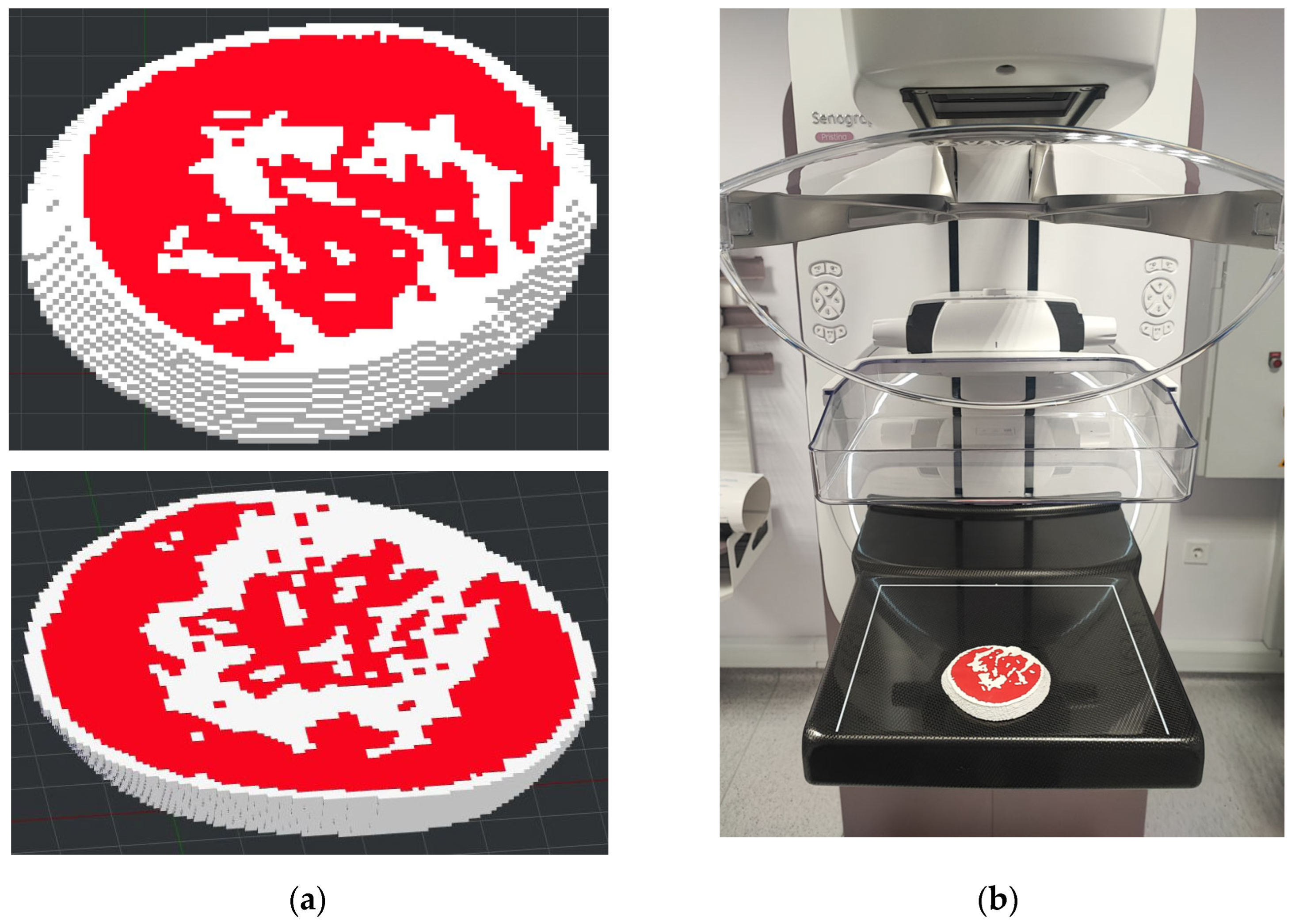
27]. The STL file of the breast section is shown in
Figure 1a.
Due to the computational and manufacturing demands associated with high-resolution voxel-based dual-material printing, full-breast fabrication presents increased susceptibility to cumulative extrusion variability and prolonged print instability. Therefore, representative sectional volumes were selected to optimize process stability and enable rigorous assessment of manufacturing reproducibility under controlled conditions.
2.3. Physical Model
Two filaments were used to print the breast sections, Acrylonitrile Styrene Acrylate (ASA, ρ = 1.11 g·cm
−3, Formfutura, Nijmegen, The Netherlands) and High-Impact Polystyrene (HIPS, ρ = 1.00 g·cm
−3, Formfutura), to represent the glandular and adipose tissues of the breast. The exact molecular formula of the materials was not experimentally determined in this study. As ASA and HIPS are polymeric systems with formulation-dependent composition rather than single chemical compounds, they are described in terms of their constituent monomer units. ASA is a terpolymer composed of styrene (C
8H
8), acrylonitrile (C
3H
3N), and acrylic ester units (general form C
4H
6O
2), while HIPS is a two-phase polymer consisting of a polystyrene matrix (C
8H
8)
n with dispersed polybutadiene rubber [
28]. This combination was chosen based on an extensive experimental study of commercial filaments for 3D printing [
29].
The breast phantom slices were manufactured using a dual-extrusion fused filament fabrication (FFF) 3D printer (Raise3D Pro3 Plus (Raise3D, Shanghai, China)). A nozzle diameter of 0.6 mm was used, with a layer height of 0.25 mm, 100% line infill, and a 25% infill overlap to ensure structural uniformity. To minimize warping and ensure material stability, cooling fans were disabled, and the heated bed was maintained at 74 °C. Further, ASA and HIPS materials were printed using separate extruders, with extrusion temperatures of 243 °C (ASA) and 255 °C (HIPS) and flow rates of 91% and 93%, respectively. Printing was performed at a speed of 50 mm/s using a rectilinear infill pattern and a single perimeter.
Slicing was carried out using ideaMaker (5.3.2.8640), where the spatial distribution of ASA and HIPS was defined for dual-extrusion printing. The glandular and adipose regions were segmented separately and exported as individual STL files, each assigned to a dedicated extruder, ASA or HIPS, during slicing. In this way, the material distribution within the phantom was defined at the model level, and no voxel-level mixing was implemented. Printing was carried out in a layer-by-layer manner, with the 3D printer sequentially depositing the two materials according to the predefined geometry for each layer before proceeding to the next.
All samples were printed using identical process parameters, including extrusion temperatures, layer height, printing speed, and infill strategy, and in the same build orientation: axial plane, along the Z-axis. No support material or post-processing steps, such as sanding or cleaning, were applied. The samples were not printed in a single batch but were fabricated on different days using the same printer, materials, and slicing configuration, with standard preparation procedures, including bed leveling and nozzle checks, in order to ensure consistent operating conditions and minimize inter-batch variability.
2.4. Mammography and BT Images
The four 3D-printed breast slabs were imaged in a clinical mammography unit, shown in
Figure 1b, using the standard craniocaudal (CC) projection and BT. All exposures were performed at a tube voltage of 26 kV to assess the radiological consistency and attenuation reproducibility of the printed phantom sections.
2.5. Evaluation
Evaluation was accomplished with an in-house developed software application used to extract 23 different features from mammography and tomosynthesis images [
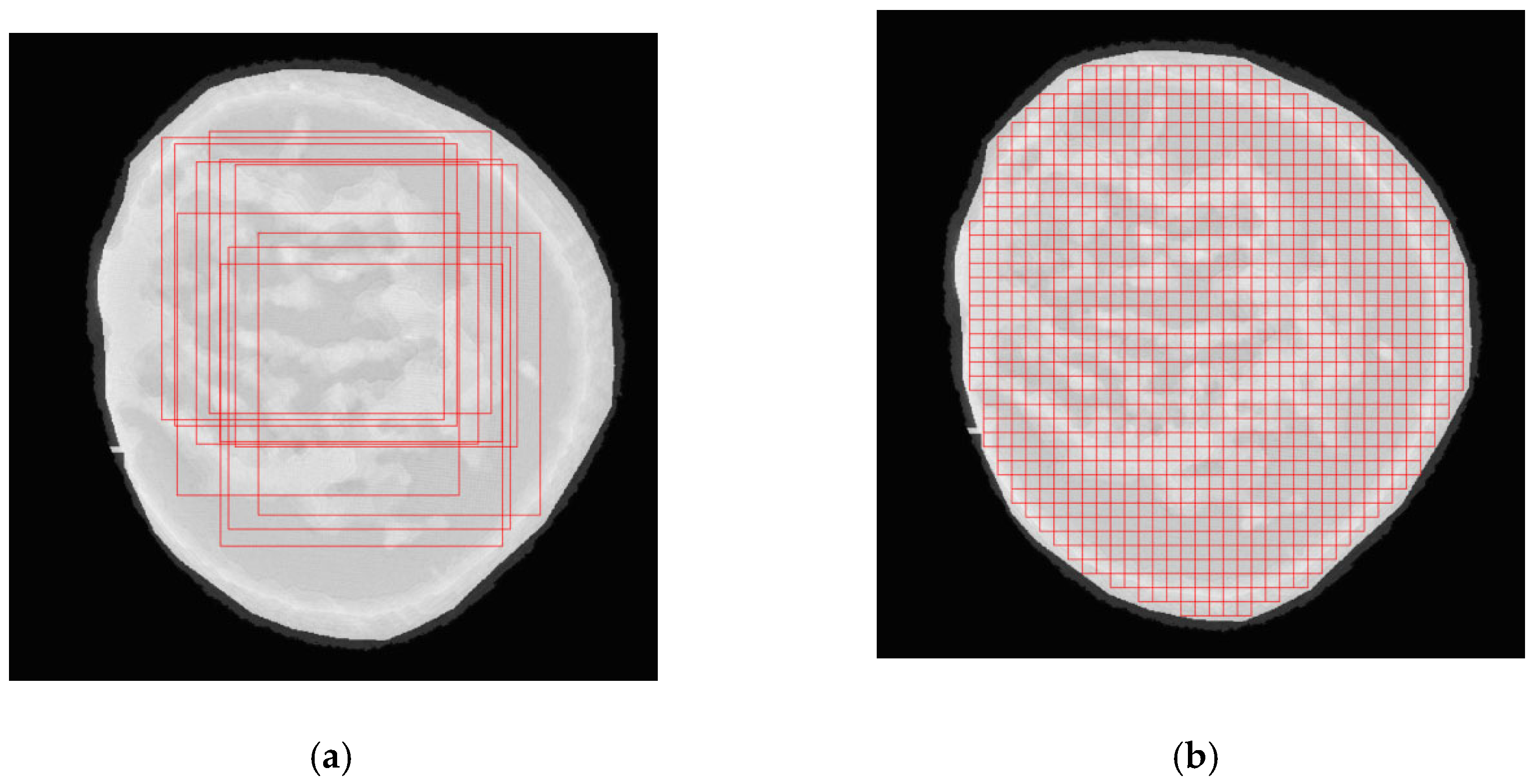
30]. The fractal dimension and power analysis beta parameter were evaluated using ten randomly positioned 500 pixels × 500 pixels overlapping Regions of Interest (ROIs). The rest of the features, including mean, medium, contrast, and energy, were calculated using 50 pixels × 50 pixels ROIs, sampled with a systematic 50% overlap, as shown in
Figure 2.
Based on these ROIs, both intra-phantom and inter-phantom variability were assessed. Intra-phantom variability was quantified for each feature as the coefficient of variation (
CV), calculated from the distribution of ROI values within each phantom as:
where
μ and
σ represent the mean and standard deviation of the feature values across ROIs within the given phantom. Inter-phantom variability was evaluated by first computing the mean value of each feature for each phantom (
μ) and subsequently calculating the
CV across the four phantoms (
μall). In addition, the relative deviation of each phantom from the global mean was computed as:
where
fi is the feature value for phantom
i. The ratio between inter-phantom and intra-phantom
CVs was used to assess the relative contribution of fabrication-related variability compared with intrinsic structural heterogeneity.
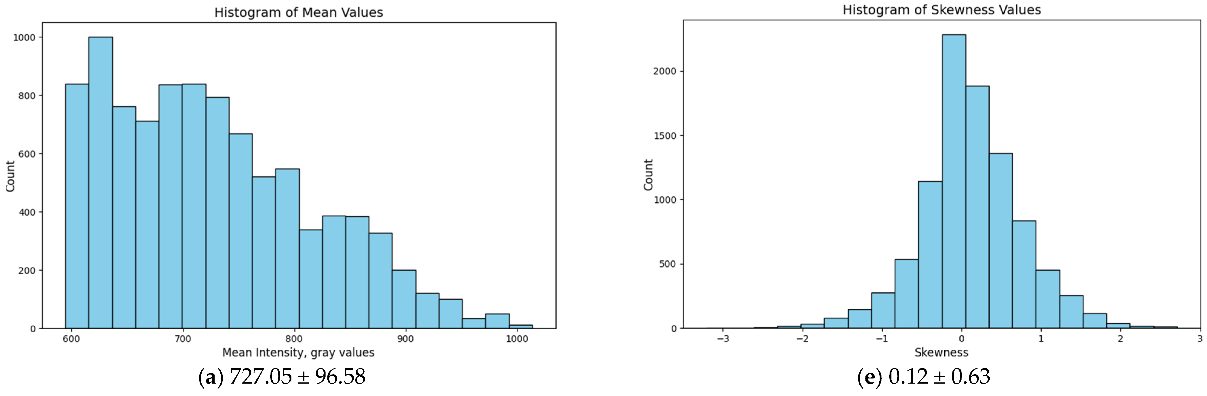
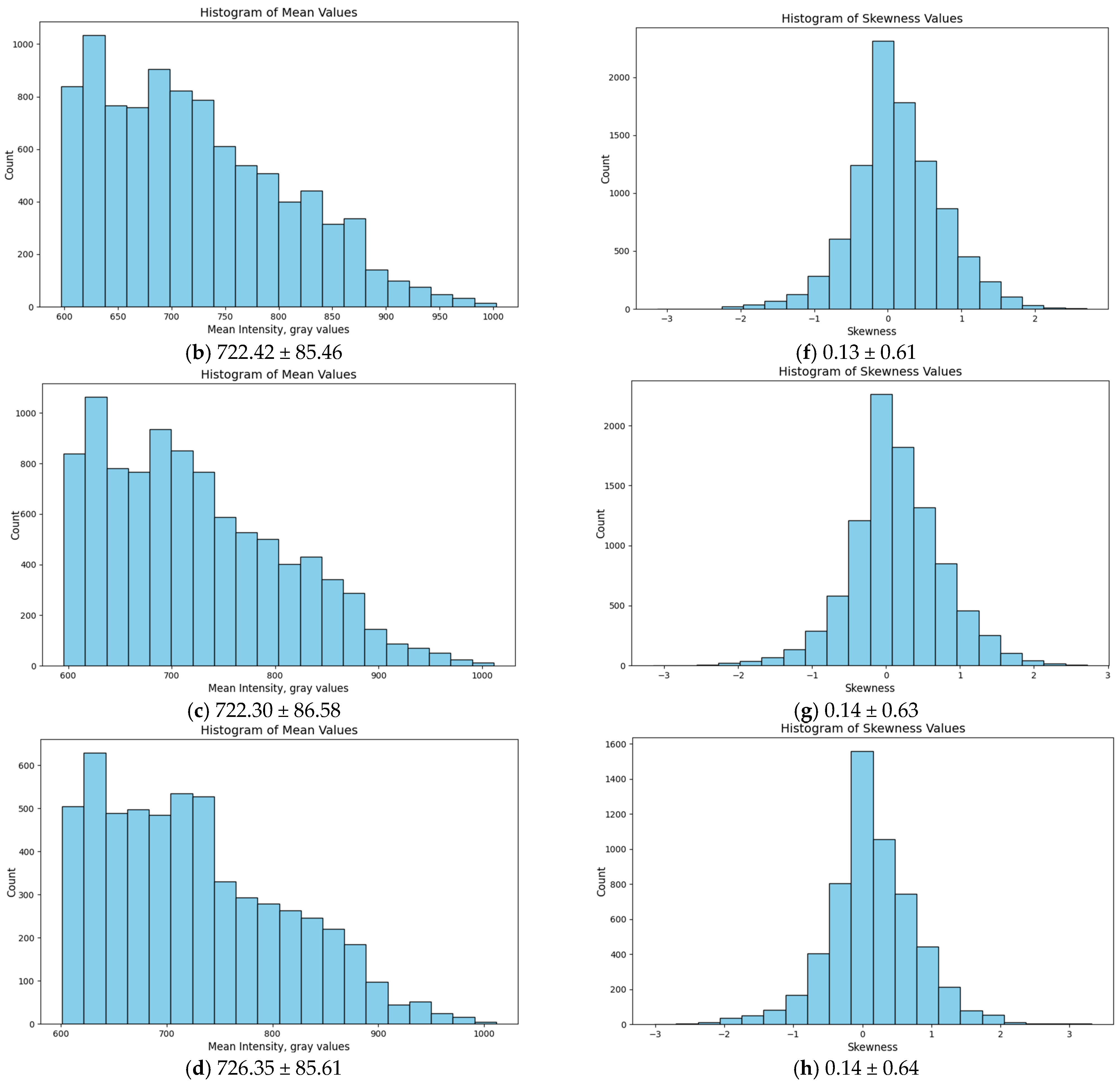
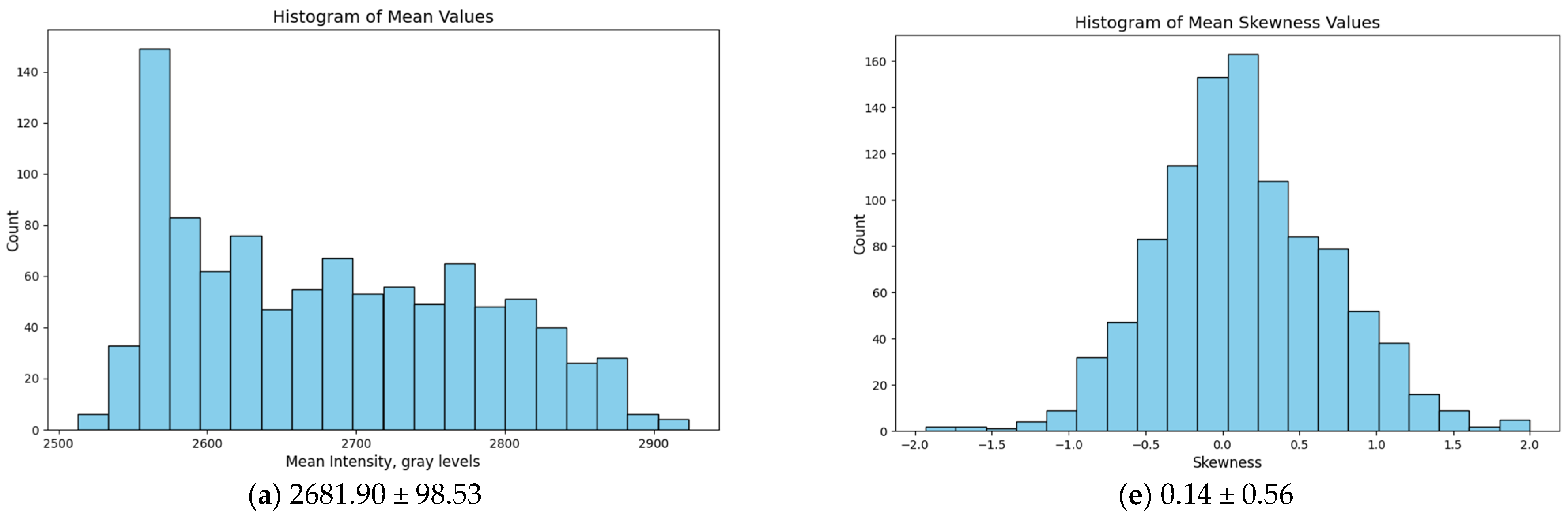
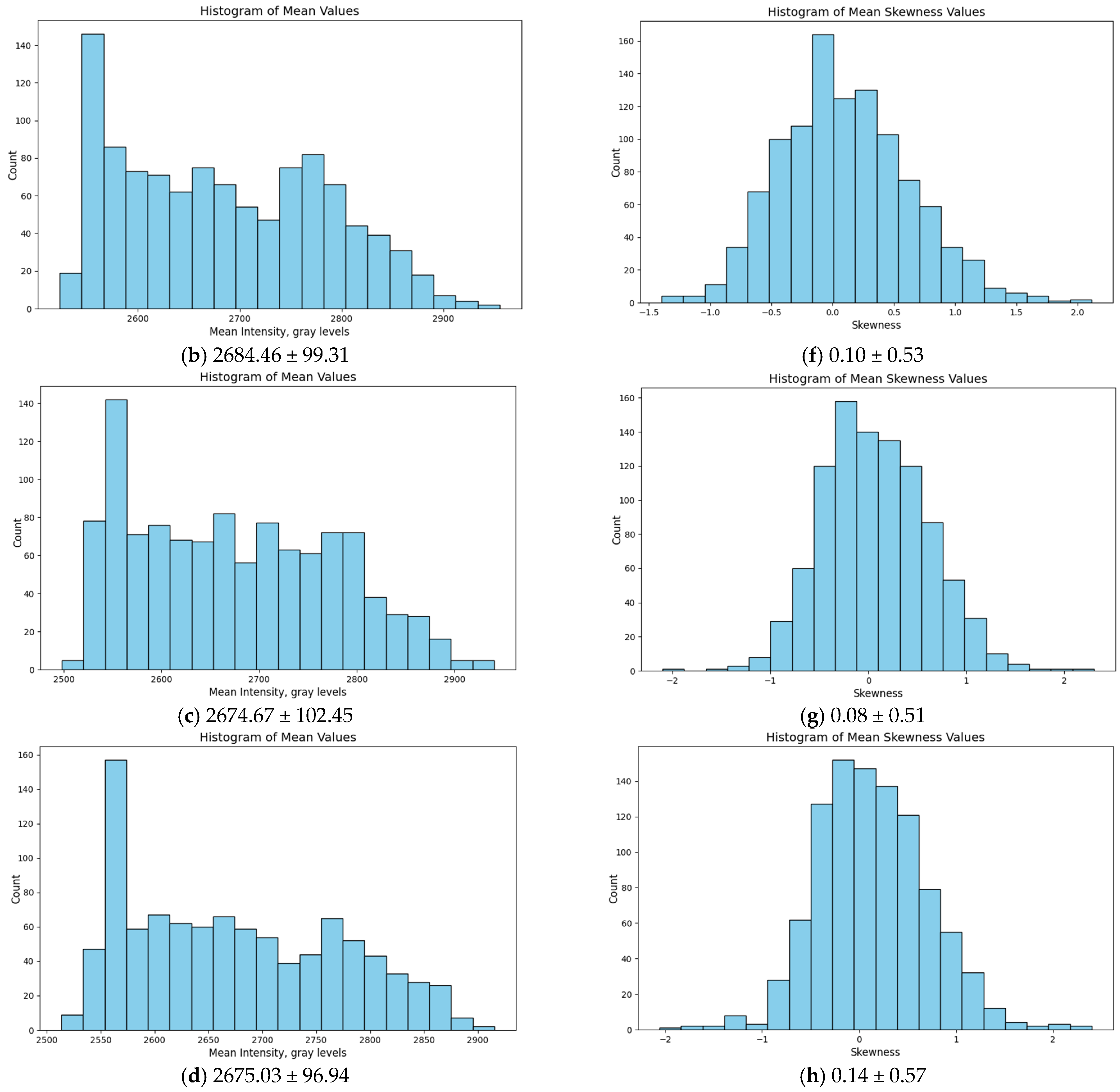
Histograms based on the mean intensity and mean skewness were also plotted. These were chosen as representative of the features where the models exhibited the minimum and maximum differences.
We used the CV as our primary measure of similarity. Because of the high resolution and large sample size, the Kruskal–Wallis test (used because the features were not normally distributed) became overly sensitive to minor shifts that do not actually matter in practice. A CV below 5% was considered indicative of high inter-phantom reproducibility.
4. Discussion
Anthropomorphic phantoms play an important role in dose optimization, quality assurance, and the evaluation of emerging imaging techniques. In our previous work, we reported a simple approach for the development of an anthropomorphic breast phantom using FFF 3D printing with two commercially available filaments, ASA and HIPS, selected based on their radiological characteristics [
22]. However, for such phantoms to be reliably used in research and clinical settings, particularly for multi-center studies and technology comparison, the reproducibility of the manufacturing process becomes critical. If phantoms are to be reproduced and distributed across institutions, it is essential to ensure that they exhibit consistent imaging performance. This is a prerequisite for fair and meaningful comparison of results between different systems and sites. To our knowledge, such systematic evaluation is unexplored.
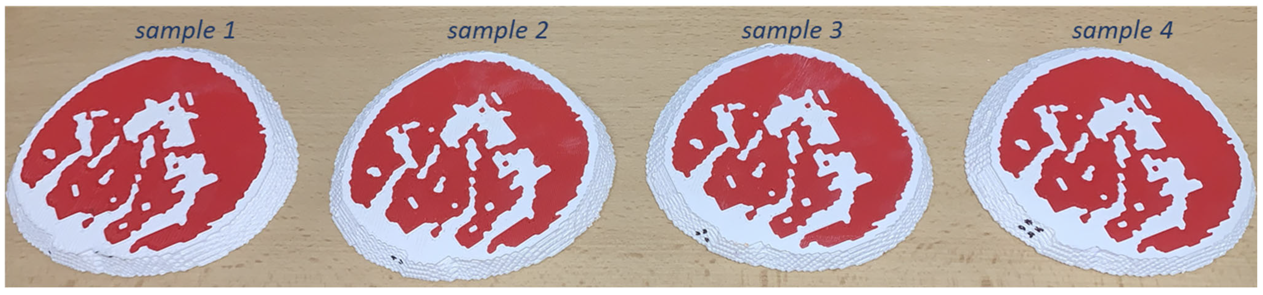
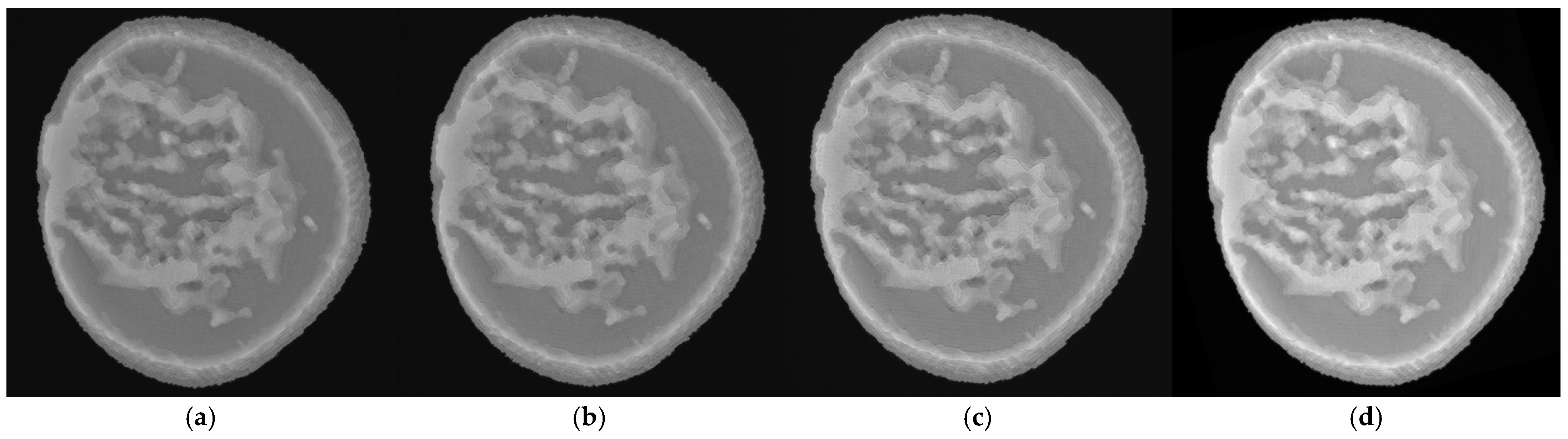
The visual comparison of the physical breast sections shown in
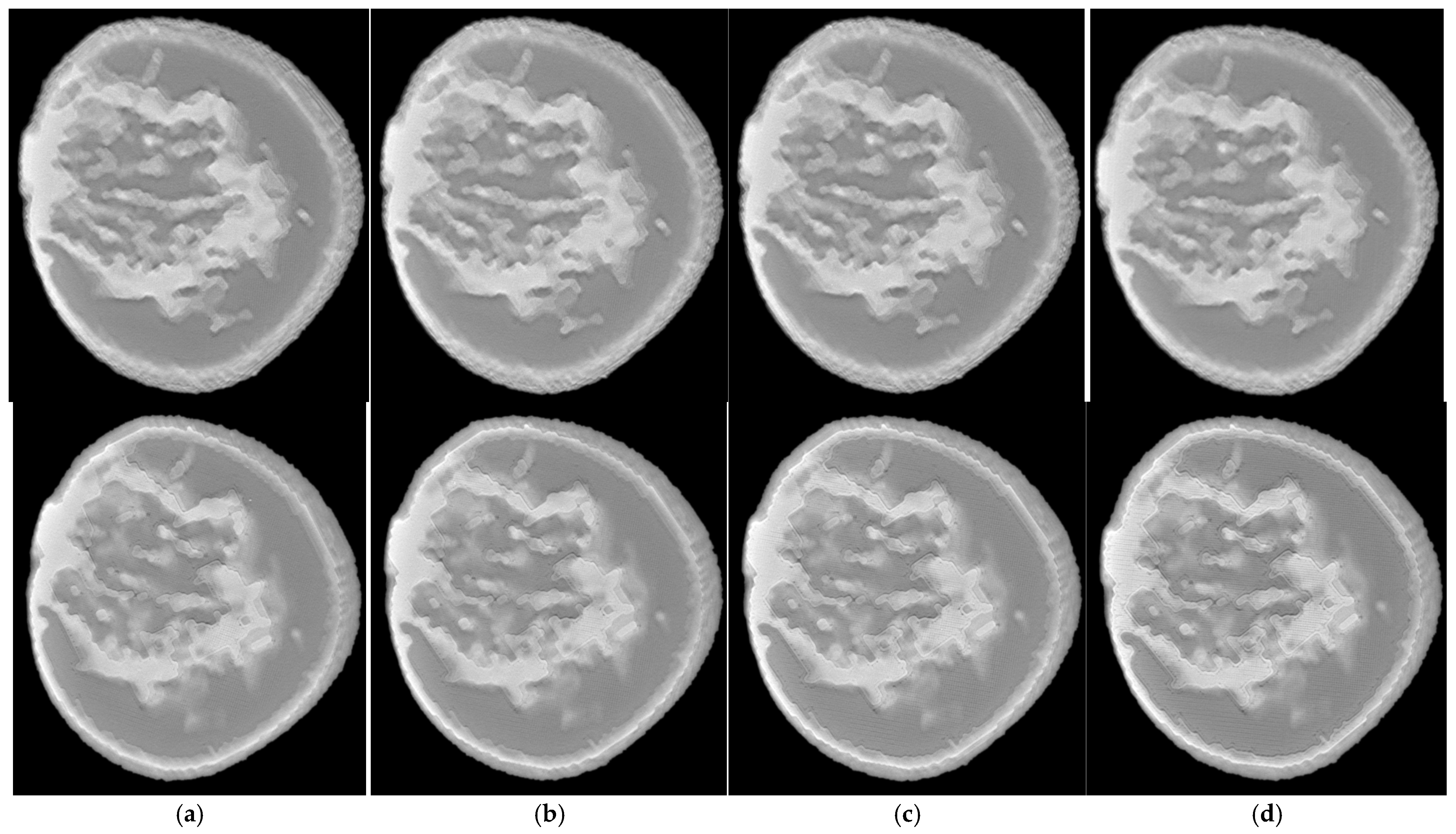
Figure 3 revealed no observable differences in the distribution of glandular and adipose tissues. The sections appeared structurally similar. Similarly, a comparison between the mammography images of the phantoms and the BT slices of imaged breast sections (
Figure 4 and
Figure 5) demonstrates highly consistent visual characteristics; any observed variations are minor and limited to stochastic material deposition.
Because all phantoms were derived from the same digital template, the expectation is that their statistical texture properties will be highly consistent. The results for the fractal dimension and β parameter obtained from the power spectra analysis, presented in
Table 1, confirm that the fabrication process achieved a high degree of reproducibility. Specifically, for mammography images, the calculated fractal dimension values ranged from 2.95 to 2.99, with a variability of approximately 0.32% across the four phantoms. The fractal dimension characterizes the spatial complexity and self-similarity of the underlying texture [
30]. The very small variation observed indicates that the large-scale texture characteristics of the printed phantoms were reproduced with high fidelity. The power spectrum exponent β, describing the frequency-dependent distribution of image texture, also showed very small variations across all phantoms. In mammography images, β values ranged from 2.10 to 2.20, corresponding to a variability of approximately 2.23%. This level of variation remains low and indicates that the spatial frequency characteristics of the texture were preserved across all manufactured models. Since the power spectrum reflects the relative contribution of coarse versus fine structural patterns, these results suggest that the printing process maintained consistent spatial texture characteristics.
Comparable consistency was also observed in the BT images. The fractal dimension values were nearly identical for all four phantoms (approximately 2.88–2.89), with very small associated standard deviations. Further, the β values for tomosynthesis ranged between 2.06 and 2.08, supporting the conclusion that the phantoms exhibit similar spatial frequency characteristics when imaged under different acquisition modalities.
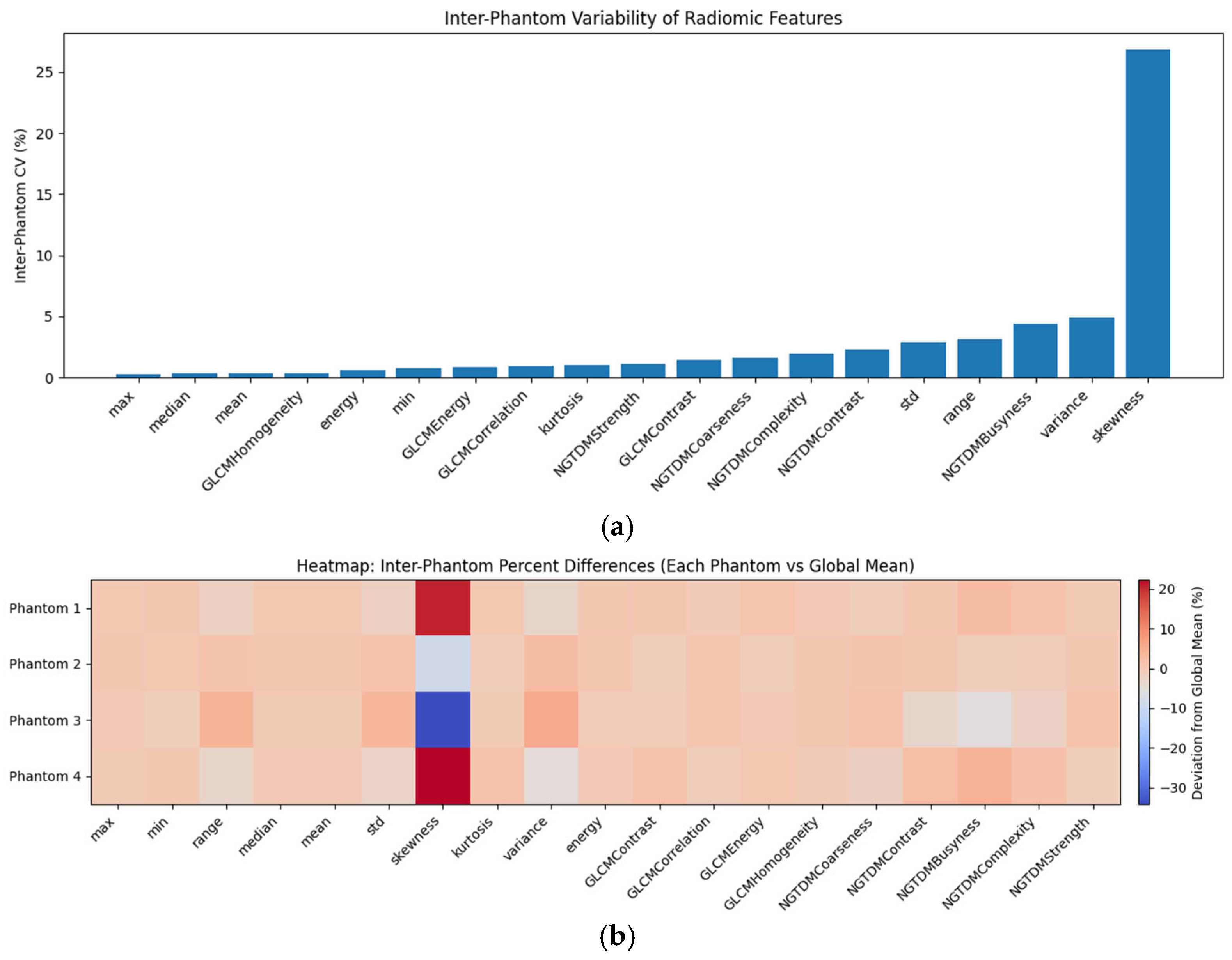
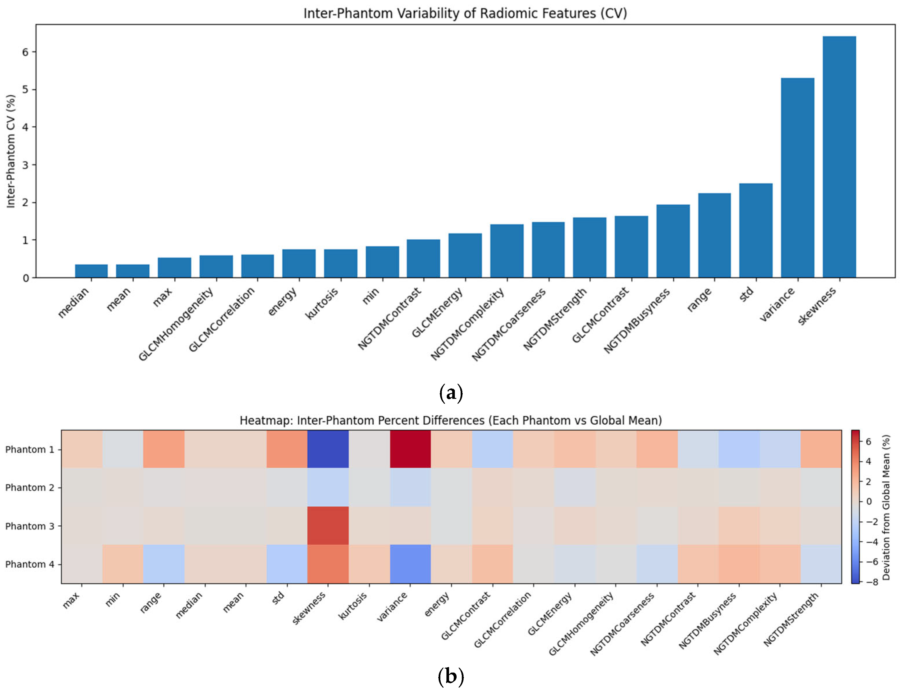
Most of the features stayed consistent across the phantoms, confirming a reliable manufacturing process. In particular, in the case of mammography, first-order and GLCM (Gray-Level Co-occurrence Matrix) features remained highly stable, with less than 1%. Neighborhood-based texture features showed slightly higher variability (1–5%), reflecting their increased sensitivity to micro-structural heterogeneity. However, the skewness values demonstrated higher variability (26.8%). Because skewness relies on a cubic calculation, it acted as a sensitive indicator for minor localized asymmetries, specifically outliers caused by incidental material deposition, which is largely because extra material deposition created outliers. The histograms in
Figure 8 further validate this high reproducibility. Mean intensity distributions (
Figure 8a–d) are very similar with overlapping gray value ranges (2500–2900), reinforcing the stability of the fabrication process. Skewness distributions (
Figure 8e–h) feature broader bases relative to their means, confirming that while bulk material properties are stable, skewness captures subtle distribution asymmetries. Finally, the heatmap, shown in
Figure 6b, confirms minimal directional deviation for the majority of first-order and GLCM features, with most differences remaining small relative to the global mean. As expected, skewness shows larger variation.
In the case of tomosynthesis, the inter-phantom analysis also demonstrated excellent stability (
Figure 7a). Most first-order and GLCM features exhibited coefficients of variation below 1–2%, while features like mean and median remained below 1%, confirming that the bulk attenuation properties are consistently reproduced. The larger deviation was in skewness (up to 6%); however, it was significantly reduced in comparison to that observed in mammography. This was expected, as the tomosynthesis reconstruction process effectively mitigates the effects of incidental printing artifacts by distributing their influence volumetrically rather than exposing them in a single 2D projection [
31]. These results are further validated by the heatmap (
Figure 7b) and the histograms in
Figure 9. Mean intensity distributions (
Figure 9a–d) show overlapping gray value ranges and consistent averages (722–727), reinforcing the stability of the fabrication process. The skewness distributions (
Figure 9e–h) feature broader bases relative to their means (0.12–0.14), confirming that while bulk material properties are stable, skewness remains a sensitive indicator for suitable distribution asymmetries. The results in
Figure 8 and
Figure 9 are further supported by Kolmogorov–Smirnov analysis, which shows that the observed distributional differences are minor in magnitude and consistent with the low inter-phantom variability. The smaller Kolmogorov–Smirnov statistics observed in breast tomosynthesis indicate a high degree of consistency between phantoms, despite the increased sensitivity of this modality to structural heterogeneity.
These findings align well with recent research highlighting the impact of micro-structural details on inter-phantom variability. In a study analyzing 64 image quality metrics across 22 “identical” TORMAS phantoms, researchers discovered that inter-phantom variability consistently exceeded intra-phantom variability [
32]. On average, these inter-phantom differences accounted for 84.2% of the total variance, which was concluded to be primarily attributed to manufacturing inconsistencies, material property differences, and structural variations between phantoms. Similarly, Zhao et al. [
33], who developed silicone and epoxy-resin phantoms, reported that “critical steps” in the preparation process could lead to inconsistent scattering properties and create “batch outliers” in the field of the diffused optics. Sarno et al. [
34] also conclude that using FFF printing for mammography and BT can result in phantoms characterized by high anatomical realism; however, they also acknowledged that micro-structural details are the hardest to replicate. They also observed that printing artifacts result in non-realistic textures. In our analysis, these artifacts manifest as “outliers”. It is important to distinguish these incidental material depositions from intentional microcalcifications, as the former are unintended consequences of the printing process.
The present study shows that the variability of radiomic features is predominantly driven by intrinsic structural heterogeneity rather than by differences arising from the fabrication process (
Table 2). The observed variability patterns provide insight into the origin of radiomic feature instability in anthropomorphic phantoms. The consistently lower inter-phantom variability indicates that the fabrication process introduces only minimal differences between independently produced phantoms. Instead, the dominant source of variability arises from intrinsic structural heterogeneity and the sensitivity of radiomic features to ROI sampling. This is particularly evident for higher-order texture features, which are designed to capture local intensity fluctuations and therefore inherently exhibit larger variability.
The increased variability observed in breast tomosynthesis compared to mammography can be attributed to the nature of the imaging modality. Tomosynthesis reconstructs pseudo-three-dimensional information from limited-angle projections, making it more sensitive to variations in tissue structure, noise propagation, and reconstruction artifacts. As a result, radiomic features derived from tomosynthesis are more influenced by local structural complexity than those derived from projection imaging. Importantly, this increased sensitivity does not reflect instability of the phantoms themselves, but rather the expected behavior of the imaging modality.
From a practical perspective, these findings highlight that reproducibility studies of radiomic features should distinguish between variability introduced by the object and variability introduced by the imaging process. The present results demonstrate that, when using the proposed fabrication approach, the contribution of manufacturing-related variability is minimal, even under imaging conditions that amplify structural heterogeneity. This supports the use of such phantoms for comparative imaging studies, protocol optimization, and multi-center evaluations, where consistent and reproducible reference objects are required.
While this study focuses on about 1 cm thick breast slices rather than full-volume phantoms, this approach was selected to facilitate a high-resolution assessment of structural reproducibility. Using slices allowed for a controlled environment to validate the manufacturing process without the excessive production timelines currently associated with large-scale FFF printing. This approach offers a scalable proof-of-concept that can be extended to full-volume models as fabrication speeds improve.
The evaluation in this study demonstrated that this technology is suitable for the production of imaging phantoms. However, one limitation of the manufacturing process is the occasional uncontrolled extrusion of material during printing. In some cases, small excess filaments are deposited on the phantom surface and may appear in the images as bright spots resembling calcifications. Such artifacts may influence some radiomic features.
The use of ASA and HIPS reflects the current limitations of available 3D-printing materials, as no single material is presently able to satisfactorily reproduce both the radiological and mechanical properties of breast tissue. This is a recognized challenge in phantom development, where materials are typically selected based on specific application requirements rather than full multimodal realism [
35]. For this reason, the present work was intentionally focused on mammographic performance and reproducibility of the manufacturing workflow. The phantoms prepared by using this approach are radiological anthropomorphic models, designed to reproduce X-ray imaging characteristics rather than mechanical tissue behavior.
In this study, no additional spectroscopic characterizations of the printed slices were performed, such as Fourier-transform infrared spectroscopy or Raman spectroscopy. While such techniques provide valuable insights into the chemical structure of materials, they are not directly relevant to X-ray imaging performance, which is primarily governed by density and effective atomic number. In the present work, the focus was on assessing the reproducibility of mammographic characteristics.
Spectroscopic analysis may be beneficial in future studies aimed at material development or in the design of phantoms for optical or infrared imaging modalities, where molecular composition directly influences the imaging signal. In such applications, tissue-mimicking phantoms are typically based on polymeric or hydrogel matrices, including silicone, polyvinyl alcohol cryogels, fibrin, and resins, whose optical properties are tuned through the incorporation of absorbing and scattering agents rather than their intrinsic composition [
36,
37,
38]. This approach will be explored in future work using our in-house developed optical 2D/3D imaging system [
39].
In contrast, common thermoplastic filaments such as ASA and HIPS are not considered primary materials for optical or infrared imaging, as they lack the required optical tunability and transparency. Instead, they are predominantly used as structural materials. Within the context of the present study, the emphasis remains on ensuring consistent and reliable X-ray imaging performance, demonstrating that the selected materials are suitable for mammographic applications.
Future work will focus on improving the control of the extrusion process in order to deposit the material more precisely. This may be achieved through better printer calibration, optimization of printing parameters, or the use of alternative printing technologies with higher deposition accuracy. Better control of the material deposition would reduce imaging artifacts and further improve the reproducibility of the printed phantoms.
These findings demonstrate that the phantom production method provides highly reproducible structural properties, supporting its suitability for quantitative imaging studies where consistent texture characteristics are essential.




 ,
,